the original - caimed.com.arcaimed.com.ar/docs/aesculap/3_craniofix .pdf · the removal forceps ......
TRANSCRIPT

Seit 1997 weltweit in über
200.000 Patienten erfolgreich implantiert!
Successful implanted in over
200.000 patients worldwide since 1997!
The Original
Inklusive NeuheitenNew Products included

CRANIOFIX® wurde entwickelt in Zusammenarbeit mit:
CRANIOFIX® has been developed in cooperation with:
Dr. med. K.-D. Lerch
Neurochirurgische KlinikKlinikum Dortmund gGmbH
Münsterstr. 24044145 Dortmund
CRANIOFIX® - Das Implantate-System zur Refixierung von kraniotomierten Schädelknochendeckeln
CRANIOFIX® - Implant system for refixation of cranial bone flaps after craniotomy

Bone flaps after craniotomy are typically fixed with wire, suture material or mini platesand screws.
In some cases, fixation with wire or suture material is not secure. Shifting of the bone flap may result in dislocation, causing depressionor protrusion of the flap. This phenomenon occurs more frequently with the progressive shift to smaller craniotomies for minimally invasive surgical procedures.
Fixation of the bone flap using mini plates and screws has improved the attachment of the bone flap. This technique, however, demandsa considerable amount of time and added cost.
This has led to the development ofCRANIOFIX® - a totally new, unique systemfor fixation of the bone flap to the cranium.
A distinguishing feature of CRANIOFIX®
is its quick and easy application. It provides optimal stability and reliable fixation of the bone flap to the cranium.
3
DBP, German Patent DE 196 03 887US-Patent No. 5 800 436
Die Refixierung von kraniotomierten Knochendeckeln erfolgt in der Regel durch eine Adaptation mittels Draht, Nahtmaterial oder verschraubbarer Mini-Platten.
Bei der Verwendung von Draht oder Naht-material kann die Forderung nach einer sicheren, unverschiebbaren Fixierung des Knochendeckels in vielen Fällen nicht erreichtwerden, da naturgemäß mit Draht oder Fadennur schwer die notwendige Haltefestigkeit und damit eine lagestabile Fixierung an den Verbindungsstellen erzielt werden kann.Daraus können Dislokationen des Knochen-deckels (Einsinken, Stufenbildung) resultieren. Dieses Phänomen tritt verstärkt bei der fort-laufenden Miniaturisierung der operativen Zugänge auf, da sich der Anteil von Fräs- bzw.Sägefurche in Relation zur gesamten Kranio-tomiefläche ungünstig erhöht.
Die Fixierung des Knochendeckels mittels Mini-Platten-Systemen, die mit Mini-Schraubenbefestigt werden, hat die Knochendeckeladap-tation zwar verbessert, erfordert jedoch einennicht unerheblichen Aufwand an Zeit und Kosten.
All dies führte zur Entwicklung von CRANIO-FIX® - einem völlig neuen und einzigartigen System zur Refixierung von kraniotomiertenKnochendeckeln an der Schädelkalotte.
CRANIOFIX® zeichnet sich durch eine einfache und schnelle Applikation aus und garantiert eine optimale, lagestabile und sichere Fixierung des Knochendeckels zur Schädelkalotte.
4
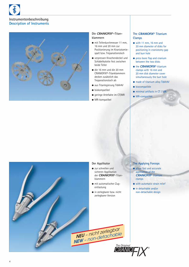
InstrumentenbeschreibungDescription of Instruments
Die CRANIOFIX®-Titan-klammern● mit Tellerdurchmesser 11 mm,
16 mm und 20 mm zur Positionierung im Kraniotomie-spalt bzw. Trepanationsloch
● verpressen Knochendeckel undSchädelkalotte fest zwischen beide Teller
● die 16 mm und die 20 mm CRANIOFIX®-Titanklammern decken zusätzlich das Trepanationsloch ab
● aus Titanlegierung Ti6Al4V
● biokompatibel
● geringe Artefakte im CT/MR
● MR-kompatibel
The CRANIOFIX® TitaniumClamps● with 11 mm, 16 mm and
20 mm diameter of disks for positioning in craniotomy gapand burr hole
● press bone flap and cranium between the two disks
● the CRANIOFIX® titaniumclamps with 16 mm and 20 mm disk diameter cover simultaneously the burr hole
● made of titanium alloy Ti6Al4V
● biocompatible
● minimal artifacts in CT / MR
● MR-compatible
Der Applikator● zur schnellen und
sicheren Applikation der CRANIOFIX®-Titan-klammern
● mit automatischer Zug-entlastung
● in zerlegbarer bzw. nicht zerlegbarer Version
The Applying Forceps● allow fast and accurate
application of the CRANIOFIX® titanium clamps
● with automatic strain relief
● in detachable and/or non-detachable design
The Original
NEU – nicht zerlegbar
NEW – non-detachable

5
Die Halteklemme● zur temporären Sicherung der
CRANIOFIX®-Titanklammern
● verhindert Druck der CRANIOFIX®-Titanklammer auf Dura mater während der Applikation
The Holding Forceps● for temporarily securing
of the CRANIOFIX® titaniumclamps
● prevent pressure of the CRANIOFIX® titanium clamp on the dura during application
Die Abziehzange● zum problemlosen Entfernen
der CRANIOFIX®-Titan-klammern
● zwei ausgeprägte Zähne greifen exakt in Aussparungen am oberen Teller der CRANIOFIX®-Titanklammern
The Removal Forceps● for simple removal of the
CRANIOFIX® titanium clamps
● two teeth fit precisely into notches on the upper disk of the CRANIOFIX® titanium clamps
Die Schneidezange● zur einfachen Entfernung des
überstehenden Stiftes an denCRANIOFIX®-Titanklammern
● kuppenförmig abgesetzte Schneide trennt den Stift direkt über dem oberen Teller der CRANIOFIX®-Titan-klammern ab
The Pin Cutter● for simple removal of the
protruding portion of the pin on the CRANIOFIX® titanium clamps
● blades are recessed so the protruding pin is cut off directlyabove the upper disk
The Original

6
Applikationsbeschreibung am künstlichen SchädelmodellApplication Procedure with an Artificial Skull Model
Die Applikation von CRANIOFIX® wird aneiner kreisförmigen Kraniotomie mit einem12 mm Trepanationsloch beschrieben.
The application of CRANIOFIX® is described with a circular craniotomy with a 12 mm burr hole.
Der Applikator wird über den Stift der CRANIOFIX®-Titanklammer gebracht.
The applier is brought into position above the pin of the CRANIOFIX® titanium clamp.
Die Halteklemme wird an die zu applizierende CRANIOFIX®-Titan-klammer angelegt, um während der Applikation ein unbeabsichtigtes Herunterdrücken der CRANIOFIX®-Titanklammer auf die Dura mater zu verhindern.
The holding forceps is placed on the first CRANIOFIX® titanium clamp and holds the clamp in place to prevent unintentional pressure on the dura during application.
Die CRANIOFIX®-Titanklammer mit 16 mm Durchmesser wird im Trepanations-loch positioniert; die CRANIOFIX®-Titan-klammern mit 11 mm Durchmesser im Kraniotomiespalt.
Die CRANIOFIX®-Titanklammern sollten in gleichmäßigem Abstand positioniert werden, in dem jeweils der untere Tellerhalbseitig zwischen Dura Mater und LaminaInterna der Schädelkalotte geschoben wird.
The CRANIOFIX®-titanium clamp with 16 mm diameter is positioned in the burrhole; the CRANIOFIX®-titanium clampwith 11 mm diameter in the craniotomy gap.
The CRANIOFIX®-titanium clamps shouldbe positioned equidistant to one another.The lower disks are inserted between theDura and the cranium.
Der zuvor entnommene Knochendeckel wird anschließend in seine ursprünglicheLage in die Kraniotomieöffnung zurück-gelegt.
The bone flap is then placed in its original position.
The Original

7
Durch Zusammendrücken des Applikator-griffes wird der obere Teller über die Zahnung des Stiftes dem unteren Tellergenähert und gegen ihn verpreßt. Eine automatische Zugentlastung stellt beim Erreichen der maximal aufnehmbaren Kraft durch die CRANIOFIX®-Titan-klammer sicher, daß beide Teller in einemoptimalen Verhältnis zueinander appliziertwerden. Der untere Teller faßt mit seiner gezahnten Konkavseite die Lamina internader Schädelkalotte und des Knochendeckelsvon innen, der obere Teller entsprechend die Lamina externa von außen.
The handles of the applying forceps are pressed together to move the upper disk downward along the circumferential grooves toward the Iower disk. Automaticstrain relief upon reaching the maximumabsorbable force through the CRANIOFIX®titanium clamp ensures that both disks areapplied in an optimum ratio to one another.The lower disk holds the lamina interna of the vault of the cranium and bone flapfrom the inside with its toothed concaveside while the upper disk holds the laminaexterna from the outside.
Der Knochendeckel geht mit dem knöchernen Kraniotomierand der Schädelkalotte einen perfekten Form- und Kraftschluß ein. Eventuelle Durahochnähte können zuvor unter dem oberen Teller durchgeführt und fixiert werden.
CRANIOFIX® forms a perfect closureconnecting the bone fIap to the craniotomy edge. Any supra-sutures in the dura can be performed and fixed in place below the upper disk.
Nachdem mit allen CRANIOFIX®-Titan-klammern entsprechend verfahren wurde,zeigt sich als Resultat eine absolut feste, lagestabile und sichere Fixierung des Knochendeckels zur Schädelkalotte mitgleichzeitiger Abdeckung des Trepanations-lochs.
The same procedure is performed with all remaining CRANIOFIX® titaniumclamps. The result is a firm, positionally stable, reliable closure of the bone flap to the cranium with a simultaneously covering of the burr hole.
Der restliche, außen überstehende Stift wird festgehalten und mit der Schneide-zange entfernt.
The pin cutter is used to remove the remaining external protruding portion of the pin, which must be held.
The Original

8
Intraoperative ApplikationsbeschreibungIntraoperative Application Procedure
Der vorliegende Fall zeigt die Refixierung eines Knochendeckelsin einer links pterionealen Kranio-tomieöffnung mit basalem osteoklastischem Anteil.
Drei CRANIOFIX®-Titan-klammern werden in gleich-mäßigem Abstand zueinander im Kraniotomiespalt positioniert, in dem jeweils der untere Tellerhalbseitig zwischen Dura materund Lamina interna der Schädel-kalotte geschoben wird.
Three CRANIOFIX® titaniumclamps are positioned equidistantto one another along the cranio-tomy opening. The lower disks are inserted between the dura and the cranium.
Der zuvor entnommene Knochen-deckel wird anschließend in seine ursprüngliche Lage in die Kranio-tomieöffnung zurückgelegt.
The bone flap is then placed in its originaI position.
Die Halteklemme wird an die zuapplizierende CRANIOFIX®-Titanklammer angelegt, umwährend der Applikation ein un-beabsichtigtes Herunterdrücken der CRANIOFIX®-Titanklammer auf die Dura mater zu verhindern.
The holding forceps is placed onthe first CRANIOFIX® titaniumclamp and holds the clamp in place to prevent unintentionalpressure on the dura during application.
The case shown demonstrates the fixation of the bone flap in a Ieft pteronial craniotomy.
The Original

9
Der Applikator wird über den Stift der CRANIOFIX®-Titan-klammer gebracht.
The applier is brought into position above the pin of the CRANIOFIX® titanium clamp.
Eine automatische Zugentlastung stellt beim Erreichen der maximal aufnehmbarenKraft durch die CRANIOFIX®-Titan-klammer sicher, daß beide Teller in einemoptimalen Verhältnis zueinander appliziertwerden. Der untere Teller faßt mit seiner gezahnten Konkavseite die Lamina internader Schädelkalotte und des Knochendeckelsvon innen, der obere Teller entsprechend die Lamina externa von außen.
Automatic strain relief upon reaching themaximum absorbable force through theCRANIOFIX® titanium clamp ensures that both disks are applied in an optimumratio to one another. The lower disk holdsthe lamina interna of the vault of the cranium and bone flap from the inside withits toothed concave side while the upper disk holds the lamina externa from the outside.
Durch Zusammendrücken des Applikatorgriffes wird der obere Teller über die Zahnung des Stiftes dem unteren Teller genähert und gegen ihn verpreßt.
The handles of the applying forceps are pressed together tomove the upper disk downwardalong the circumferential grooves toward the Iower disk.

10
Der Knochendeckel geht mit demknöchernen Kraniotomierand derSchädelkalotte einen perfektenForm- und Kraftschluß ein. Eventuelle Durahochnähte könnenzuvor unter dem oberen Tellerdurchgeführt und fixiert werden.
CRANIOFIX® forms a perfectclosure connecting the bone fIap to the craniotomy edge. Anysupra-sutures in the dura can be performed and fixed in place below the upper disk.
Nachdem mit allen CRANIOFIX®-Titanklammern entsprechend ver-fahren wurde, zeigt sich als Resultat eine absolut feste, lage-stabile und sichere Fixierung desKnochendeckels zur Schädelkalotte.
The same procedure is performedwith all remaining CRANIOFIX® titanium clamps. The result is a firm, positionally stable, reliable closure of the bone flap to the cranium.
Der restliche, außen überstehendeStift wird festgehalten und mit der Schneidezange entfernt.
The pin cutter is used to removethe remaining external protrudingportion of the pin, which must beheld.
The Original

Bei der postoperativen Kontrollemittels Röntgenaufnahmen zeigtsich die lagestabile Fixierung des Knochendeckels in der Kranio-tomieöffnung und dessen formge-naue Anpassung an die Rundungder Schädelkalotte (im vorliegen-den Fall links pterioneal sowierechts temporal).
Post operative follow up x-raysshow the stable fixation of thebone flap in the craniotomy opening and the precise formmatch to the curve of the cranialvault. (In this case, left pteronialand right temporal.)
Eine Aussagebeeinträchtigung der CT- bzw. MR-Aufnahmen findet aufgrund der geringen Artefakte der Titanlegierung nicht statt.
Interpretation of CT and/or MR results are not adversely affectedsince titanium alloy produces minimal artifact.
11
Postoperative KontrollePostoperative Follow-up Checks
Fixierung einer multiplen Schädelimpressions-fraktur mit CRANIOFIX®.
Fixation of a multiple skull impression fracture with CRANIOFIX®.

12
Implantate und InstrumentariumImplants und Instruments
CRANIOFIX® TitanklammernCRANIOFIX® Titanium clamps
FF 106 RCRANIOFIX®
Applikator, zerlegbarApplying Forceps, detachable
FF 107 RCRANIOFIX®
Applikator, nicht zerlegbarApplying Forceps, non-detachable
The Original
Art.-nr. Ø PackungArt.-no. diam. Packaging
FF 100 T 11 mm 12/Box
FF 101 T 16 mm 6/Box
FF 099 T 20 mm 6/Box
NEUNEW

13
FF 104 RCRANIOFIX®AbziehzangeRemoval Forceps
FF 105 RCRANIOFIX®HalteklemmeHolding Forceps
FF 094 PCRANIOFIX®
Lagerung (ohne Instrumente)Tray (instruments not included)
JK 300 PContainer für Lagerung FF 094 P(ohne Abb.)
Container for Tray FF 094 P(not illustrated.)
FF 103 RCRANIOFIX®
SchneidezangePin Cutter
C73311 Silikon HirnspatelSilicone Brain Spatula
C21711 KERRISONZerlegbare StanzenDetachable Punches
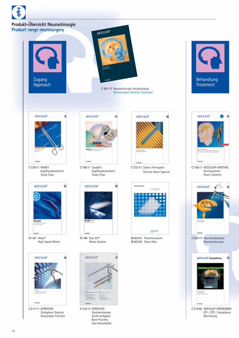
Produkt-Übersicht NeurochirurgieProduct range neurosurgeryProdukt-Übersicht NeurochirurgieProduct range neurosurgery
C79011 ScalpFix KopfhautklammernScalp Clips
C75611 AESCULAP-MIETHKEVentilsystemeShunt Systems
C21811 KERRISONKnochenstanzen (nicht zerlegbar)Bone Punches (not detachable)
C35511 NeuroendoskopieNeuroendoscopy
C72911 RANEY KopfhautklammernScalp Clips
O134 Hilan®High Speed Motor
ZugangApproach
BehandlungTreatment
C77302 AESCULAP-SPIEGELBERGICP-, CPP-, ComplianceMonitoring
O148 Elan EC®Motor System
B02201 KnochenwachsB02202 Bone Wax
C20111 Neurochirurgie HauptkatalogNeurosurgery General Catalogue
14

C30211 Bipolares HF-GerätBipolar HF Unit
C46511 Jet SpülgerätJet Irrigation Unit
C79111 PaediScopeKinderventrikuloskopPediatric Ventriculoscope
B055 Neuropatch®DuraersatzDura Substitute
B076 Lyoplant®DuraersatzDura Substitute
B037 Premilene®Nahtmaterial
Safil® Synthetic Absorbable Suture
C47111 CranioFix®KnochendeckelfixierungBone Flap Fixation
C79211 NeuroPilotSteuerungseinheitSteering Device
C74511 SENSATIONMikroinstrumenteMicro Instruments
C45611 YASARGILTitan AneurysmenclipsTitanium Aneurysmclips
C20211 YASARGIL Phynox AneurysmenclipsPhynox Aneurysmclips
VerschlußClosure
C77011 XS MikroinstrumenteMicro Instruments
15

Surgeon name
Hospital (Adress and Phone no.)
Patient medical record number (MR #)
Date of surgery
Pre-printed label:
Aesculap Reference numberAesculap Lot number
Pre-printed label:
Aesculap Reference numberAesculap Lot number
Pre-printed label:
Aesculap Reference numberAesculap Lot number
Pre-printed label:
Aesculap Reference numberAesculap Lot number
The Aesculap CranioFix® Clamp for bone flap fixation is made of a Titanium Alloy. It is safe to
perform with Aesculap CranioFix® Clamps MR Imaging up to 1.5 Tesla.
The bearer of this card has undergone a surgical procedure where the following Aesculap
CranioFix® Clamps have been implanted:
LiteraturLiterature
● Ebel H, Schillinger G, Walter C, Brockhagen HG, Klug N"Titanium Clamps for Refixation of Bone Fragments inthe Repair of Depressed Skull Fractures: Technical Note"Minim Invas Neurosurg 2000; 43: 212-214
● Estin D, Troffkin N, Heilman CB"Bone Flap Fixation with Titanium Clamps: A New Technique"Surg Neurol 2000; 53: 391-395
● Frenzel D, Lerch KD"The craniofix“ Titanium clamp for refixation of cranial bone flaps and the treatment of skullfractures. Experimental load-bearing tests and clinical long-term results in comparison with suture material"Zentralbl Neurochir (1998) Suppl 24, Abstrakt O84
● Lerch KD"Reliability of Cranial Flap Fixation Techniques: Comparative Experimental Evaluation of Suturing, Titanium Miniplates, and a New Rivet-like TitaniumClamp (CranioFix): Technical Note"Neurosurgery, Vol. 44, No. 4, April 1999: 902-905
● Lerch KD, Morgenstern F, Lau KT, Hoffmann G"Nietenartige Titanklammern zur Refixierung von Knochendeckeln nach Kraniotomie – Radiologische Identifizierung, Anwendungssicherheit und Bildqualitätbei CT- und MR-Untersuchungen"Fortschr. Röntgenstr. 169,6 (1998): 601-604
● Shellock FG, Shellock VJ"Cranial Bone Flap Fixation Clamps: Compatibility at MR Imaging"Radiology 1998; 207: 822-825
● Wörner B, Lange M, Herzog A, Fink U, Oeckler R"A new method for surgical repair of impression fractures of the cranial vault and frontal sinus with rivet-like titanium clamps"Neurosurg Rev (2001) 24: 83-87
Patient
Identification Card
Germany and international:
AESCULAP AG & CO. KG
Am Aesculap-Platz
78532 Tuttlingen/Germany
Phone +49 (74 61) 95-0
Fax +49 (74 61) 95-26 00
E-mail [email protected]
C 79
8 02
Date of birth
m/f
Patient
United Kingdom
B. Braun Medical Ltd.
Thorncliffe Park
GB-Sheffield S35 2PW
Phone +44 (114) 2 25 90 00
Fax +44 (114) 2 25 9111
http://www.bbraun.com
USAAESCULAP INC.
3773 Corporate Parkway
Center Valley PA 18034
Phone +1 (800) 282-90 00
Fax +1 (610) 791-68 86
http://www.aesculap-usa.com
CRANIOFIX® Patientenausweis zur Aushändigung an den Patienten für spätere radiologische Untersuchungen – bitte kontaktieren SieIhren zuständigen Außendienstmitarbeiter.
CRANIOFIX® Patient Identification Card to handing out to the patient for post operativeradiological imaging – please contact your local sales representative (available in English, French, Spanish, Italian or Japanese).
Brochure No. C 47111
All rights reserved. Technical alterations are possible. This leaflet may be used for no other purposes than offering, buying and selling of our products. No part may be copied or reproduced in any form. In the case of misuse we retain the rights to recall our catalogues and pricelists and to take legal actions.
0103/3.0/7
Technische Änderungen vorbehalten. Dieser Prospekt darf ausschließlich zur Anbietung und dem An- und Verkauf unserer Erzeugnisse dienen. Nachdruck, auch auszugsweise, verboten. Bei Missbrauch behalten wir uns Rücknahme der Kataloge und Preislisten sowie Regressmaßnahmen vor.
AESCULAP AG & CO. KG
Am Aesculap-Platz78532 Tuttlingen/Germany
Phone +49 (74 61) 95-0Fax +49 (74 61) 95-26 00
www.aesculap.de
NEUNEW