the pattern of genetic transmission of the leukocyte defect in fatal
TRANSCRIPT

The Pattern of Genetic Transmission
of the Leukocyte Defect in Fatal
Granulomatous Disease of Childhood
D. B. WINDHORST, A. R. PAGE, B. HOLMES, P. G. QUIE, and R. A. GOOD
From the Pediatric Research Laboratories of the Variety Club Heart Hospitaland the Department of Pediatrics, University of Minnesota,Minneapolis, Minnesota 55455
A B S T R A C r The leukocyte-phagocytic functiontest which was found to be abnormal in boys withfatal granulomatous disease of childhood has beenfound to be abnormal to an intermediate extent intheir mothers. Nine of nine mothers were shownto be abnormal, whereas none of eight fathers andnone of five healthy brothers exhibited a defect.10 of 16 female siblings were abnormal to the samedegree as their mothers, as were all three maternalgrandmothers available for study. Assuming thatthis intermediate functional defect represents theheterozygous state, the nine family pedigrees areentirely compatible with the concept that the traitis transmitted on the X-chromosome.A tetrazolium dye-phagocytosis histochemical
test was also abnormal in the carrier females andprovided independent confirmation of the selectionof the female siblings suspected of being carriersfor the trait. In addition, this procedure gives in-direct evidence that the gene in question is subjectto the random inactivation that appears to affectmany X-linked genes in mammalian females.The family members were also studied with two
of the metabolic assays that have been shown tobe abnormal in the cells of affected boys. Oneassay, the oxidation of the first carbon of glu-cose-1-14C by the isolated leukocytes, was signifi-cantly abnormal in the cells of carrier females.The other assay, the oxidation of formate-'4C by
Dr. R. A. Good is American Legion Memorial HeartResearch Professor of pediatrics and microbiology.
Received for publication 16 August 1967 and in revisedform 30 October 1967.
leukocytes of heterozygotes was not significantlydifferent from control values.The practical problem of diagnosing patients
would appear to be best solved with a tetrazoliumdye procedure, whereas the more subtle abnormal-ity in carrier females is best detected with theleukocyte function test. Improved methods for thefunction test are being developed.
INTRODUCTION
Peripheral blood leukocytes of children with thegranulomatous disease of childhood have recentlybeen shown to be defective in their capacity tokill certain bacteria in vitro. Furthermore, theseleukocytes have been found to have abnormalitiesin the known metabolic concomitants of activelyphagocytizing cells.The disease in which the abnormal leukocytes
are found has been reported from several areas inthe United States (for review, see reference 1)and recently from Europe (2, 3). The initial mani-festations are an eczematous dermatitis and fluc-tuant cervical or inguinal adenopathy, developingin male children during the first 18 months of life.Chronic infiltrative pulmonary disease with super-imposed acute episodes, hepatosplenomegaly, andosteomyelitis are the more serious continuingproblems, and pericarditis, stomatitis, and ab-scesses in a variety of locations are frequent.Various bacteria usually considered to be of lowvirulence can often, but not always, be culturedfrom suppurative lesions and granulomas. Studies
1026 The Journal of Clinical Investigation Volume 47 1968

of immune capacity yield normal findings and thepatients react appropriately to known virus agents,although MacFarlane, Speirs, and Sommerville(2) found abnormalities in the capacity of theseleukocytes to reduce the number of Vaccinia orHerpes simplex organisms in vitro.
Berendes, Bridges, and Good (4) and Bridges,et al. (5) suggested that this syndrome was fa-milial. Subsequent recognition of affected brothersof patients studied in our clinic has strengthenedthis possibility as has the study of Carson, Chad-wick, Brubaker, Cleland, and Landing (6).
Early in our studies of polymorphonuclear leu-kocyte function, it became evident that the leuko-cytes of the mothers of affected boys were alsoabnormal in the in vitro test. Further, three malematernal cousins of patients were found to havedeveloped early signs of granulomatous disease.Thus it became probable that this defect wasgenetically determined and X-linked.
It is the purpose of this paper to report thatstudies of the polymorphonuclear leukocyte defectsindicate that they are transmitted in an X-linkedmanner, and that the carrier state in female sib-lings can be predicted. Our original term "fatal"seems justified, since the gene exhibits a highdegree of lethality for the affected boys.
METHODS53 members of nine families were studied. One familyincludes affected half brothers, each from a differentfather. All first degree family members except 3 of 11fathers and 4 of 15 diseased boys were examined. Threematernal grandmothers representing four families and onematernal grandfather were also studied. This populationwas analyzed using three different approaches; (a) aleukocyte function test, (b) two metabolic analyses, and(c) a combined histochemical-phagocytosis procedure.
I. Leukocyte function test
The leukocyte function test developed by Holmes, Quie,Windhorst, and Good (7) was used. The major differencebetween this procedure and that of Quie, White, Holmes,and Good (8) is the addition of antibiotics to the cultureat 20-30 min to eliminate extracellular bacteria. Thatmost of the bacteria which can be cultured at the 2-hrtime interval are intracellular is inferred from the factthat cultures set up without cells, but which are otherwiseidentical to the test system, will not have culturable bac-teria present at that time (9 and footnote 1).
1Alexander, J. W., and R. A. Good. Effect of anti-biotics on the bactericidal activity of human leukocytes.Submitted for publication.
That the carrier females' leukocytes are intermediatein capacity to kill staphylococci is demonstrable withoutadding antibiotics; however, this addition accentuates theabnormality and provides increased reliability in recog-nition of the carrier state." 2 Antibiotics make it possibleto obtain reliable results with a shaking water bath asthe culture method, if end-over-end tube mixers are notavailable. We found that a compact, inexpensive tilterLab-Tech aliquot mixer, (Lab-Tech Instrument Co.,Westmont, Ill.) gives excellent results in the procedureeither with or without antibiotics. Substitution of sterile,disposable, plastic equipment (such as culture tubes) forsiliconized glassware has proved feasible.
Because of the variability in the leukocyte functiontest, each day's study was made complete in itself. Aculture using normal cells was always included as anegative control and one using cells from one of theaffected boys was usually included as a positive control.In eight families, this control was their own familymember.
II. Metabolic studies
Holmes, Page, and Good (1) have found the peripheralleukocytes of affected boys to be abnormal in 4 of 12major metabolic parameters which they examined. Twoof these, (a) the capacity of the phagocytizing cells toincrease the oxidation of the first carbon of glucose and(b) the oxidation of formate were selected for familyscreening studies. The methods used were those ofHolmes et al.
III. Histochemical procedureFor these studies, an adaptation of the original method
of Baehner and Nathan (10) was developed. This testis based on their observation that phagocytizing leuko-cytes of the affected boys do not reduce tetrazolium dyesas do normal leukocytes. Our modification was developedto examine the carrier female's cells in an effort to dem-onstrate that two populations of leukocytes, normal anddefective, may be present. The cells were isolated, incu-bated, and smears were prepared as previously described(11). The nitro-blue tetrazolium appears to vary con-siderably from batch to batch, and a new lot should firstbe tested with normal cells to determine its capacity togive positive results.
Duplicate cultures were prepared from each collectionof cells, and all cultures were coded so that the smearscould be examined without knowledge of the sourceof the cells.
Cells from normal individuals show reduction of thetetrazolium dye, most noticeably in cells that have phago-cytized the latex particles.
Differential counts were made on smears by counting100 cells containing 10 or more phagocytized latex par-ticles. Cells were positive if any deep-blue granular dyeprecipitate could be seen. A faint homogenous blue tingeof the cell cytoplasm was not considered a positive finding.2Alexander, W., D. B. Windhorst, and R. A. Good.
In preparation.
Family Studies in Fatal Granulomatous Disease 1027

RESULTS
I. Results of the leukocyte function test
Bacterial counts were plotted as a function oftime on five-cycle semilogarithmic paper. A rep-resentative family study is illustrated in Fig. 1.It is evident that bactericidal capacity of cells fromthe father, a normal brother, and one sister wassimilar to that of the control. However, the moth-er's cells and those of another sister functioned atan intermediate level. A partial abnormality hasbeen present without exception in multiple exami-
nations of each of the nine mothers. Three mater-nal grandmothers have shown the same pattern.An example is presented in Fig. 2. None of thefathers or the one maternal grandfather has beenabnormal. Five healthy brothers were all entirelynormal in this test.
Since interpretation of the plots of the leukocytefunction tests has been so consistent for recogni-tion of carrier mothers, we used this method toselect the suspected carriers among the femalesiblings. Thus, 10 of the 16 sisters have been cate-gorized as carriers.
8
106
G.)- 104
a)0
86
co 0
a)
Ez
0 2 3 4 5 6 7 0 2 3 4Hours Hours
5 6 7 0 2 3 4 5 6 7Hours
FIGURE 1 Surviving intracellular bacteria in leukocytes of the members of the S family (broken lines) examined onthe same day and plotted in relation to that day's control cells (solid lines). The father, the normal brother, and onesister have cells which function well, whereas the mother's cells and those of the other sister kill bacteria at a rate in-termediate between that of the cells from the patient and that of the control.
1028 D. B. Windhorst, A. R. Page, B. Holmes, P. G. Quie, and R. A. Good

L Family
2 4Hours
FIGURE 2 Surviving intracellular bacteria in leukocytesof members of the L family. These are plotted to illustratethat the maternal grandmother's cells exhibit an abnor-mality comparable to that of the mother's.
The pedigrees constructed on the basis of thisevaluation are recorded in Fig. 3. It should benoted that 9 of the 15 diseased boys can beregarded as probands, and that the oldest of the9 living affected boys is only 11 yr. The J familyand the L family were represented by case 3 andcase 4 in the report by Bridges et al. (5).One carrier sister has had a diagnosis of ulcera-
tive colitis since the age of 11, and one mother hashad chronic furunculosis. Several of the mothersand carrier sisters have recurrent apthous stoma-titis, and one mother is said to have had rheumaticfever.
Despite findings suggestive of disease in thecarrier state, the three maternal grandmothers arein their 70s and two have outlived their husbands.
Reliability of the interpretation of the leukocytefunction test. For the purpose of statistical analy-sis, the bacterial counts at the 2-3 hr samplingpoint were utilized. The ratios of the remainingviable bacteria in the culture of the experimentalcells to those remaining in the culture containing
cells from a known normal, unrelated individualwere determined. Thus, the ratios were based onthe control cells used for the given day's study,and a figure of one or less would be indicative ofbactericidal function which was as good as thatof the control cells.
Table I shows these data plotted as means with95% confidence intervals for the means. It is seenthat, (a) the ratios for normal family membersapproached unity, (b) the ratios for carrier sisters(mean of ratios = 95) were very similar to theratios of their mothers (mean = 100), and (c) theratios of these carrier groups were different notonly from those ratios of the normal family mem-bers but also from those of the diseased boys(mean of ratios = 674). The probability that themean of the grandmother-mother group is in thesame population as the mean of the abnormal boysis 0.042 and a similar figure for this known carriergroup compared with the normal family membersis less than 0.0001. There is no overlapping ofthe 95%o confidence intervals for normals, affectedboys, and carrier females.
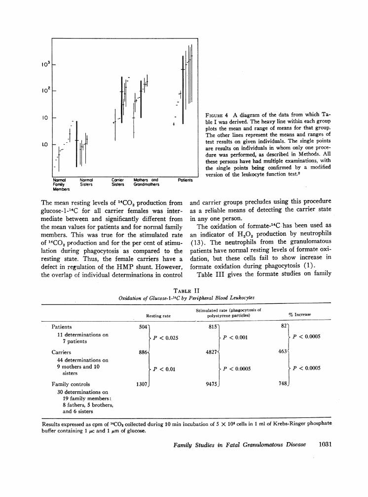
Because statistics are of little value in the actualprediction of the status of a given individual, therange of individual determinations and the rangeof means for individuals from which Table I wasderived are shown in Fig. 4. It will be noted, thatonly one determination is given for a number ofindividuals. All persons in the study were givenmultiple tests and these showed consistent find-ings; however, as this work progressed, modifica-tions were made in the procedure and only thosestudies actually performed, as described in Meth-ods, were included in the statistical analysis.
In the range of the means, as well as the rangesof individual determinations, a considerableamount of overlap can be seen. The wide varia-bility in the patients' values is unexplained, butmay be related to the extremes of conditions underwhich these boys were studied. Of more immediateinterest is the critical problem of predicting thecarrier status of females. The mother (Mrs. B.)whose mean of ratios was 10 is also an aunt ofthe affected boys; hence, she is surely a carrier.For this reason, we feel that a technically adequateexamination in which a girl's ratio is found tobe greater than 10 can be taken as strong evidencefor the carrier state. Ratios less than 10 in poten-tial carriers must be confirmed by repeat examina-
Family Studies in Fatal Granulomatous Disease 1029

dldYffS °
"Boilsl eart Trouble
AN2(S. TNM)* a Ad)Ki*(bomn-1949. born-950 on-95 /n-1956 born-158 bon-%l born-1964 bern 1947 torn-1949 bovr- baorn-%99 bow-S9 bonb - or-93 born-1965died-O Famld-950OrnlSfni
N Family W Family S Family B Family
TN d<I yreInfection Infection
born-1960 born 1961/born-1962 born-1964 boen-1966 bon-1949 born-1951 borr1950or-,955 "Albino"d/d--9G ded- 197
D Family L Family
borm- 1962 /bsmn-l"35
S-P FamilyJ Family
KEY,Clinical gronulomatous disease,died before test was available
* Granulomatous disease, testabnormal
No Granulomotous diseaseIntermediate abnormality inphogocytosis test
4 Early AbortionTN Tested and found NORMAL/ Proband
I.3' Case 3 rne,~oCase 4 8ridgesBerendes,&GoodAmer. J Dis. Child. (1959)
97:387
born-1963 bomn- 91-64b r 6
L-B Family
FIGURE 3 Pedigrees of the nine families diagrammed on the basis of the leukocyte function test. Note the affectedmaternal half brothers in the L-B family. Note also that maternal uncles in the L, N, and W families died in in-fancy with probable sepsis. There are no living affected maternal uncles.
TABLE IRatios of Viable Intracellular Bacteria at 2-3 Hr:
Family Members/Same Day Controls
Patients25 determinations on
11 patientsMothers and grandmothers
21 determinations on 8 mothersand 1 grandmother
Carrier sisters15 determinations on 9 sisters
Fathers, brothers, and normal sisters29 determination on 8 fathers,
6 sisters, and 5 brothers
tions. Consistent findings of ratios of 5-10 requirea guarded prognosis.
II. Results of the metabolic studiesMeans of ratios
Normal neutrophils increase their oxidation ofthe first carbon of glucose almost 10-fold when
674 K464 they are presented with particles to phagocytize.This oxidation is a function of glucose-6-phosphatedehydrogenase (G-6-PD) and is in consequence
100 i 52 of an increased over-all activity of the hexosemonophosphate (HMP) shunt (12). Cells of pa-tients with fatal granulomatosis have reduced
95 79 HMP shunt activity during the resting state andfail to show stimulation of the shunt during
1.5 0.8 phagocytosis.The results of the studies on the families of the
granulomatous boys are summarized in Table II.
1030 D. B. Windhorst, A. R. Page, B. Holmes, P. G. Quie, and R. A. Good
103k
02 S
10
1.0
I :t
NormalFanilyMembers
Normal Carrier Mothers andSisters Sisters Grandmothers
-I
+
Patients
The mean resting levels of 14CO2 production fromglucose-1-14C for all carrier females was inter-mediate between and significantly different fromthe mean values for patients and for normal familymembers. This was true for the stimulated rateof 14CO2 production and for the per cent of stimu-lation during phagocytosis as compared to theresting state. Thus, the female carriers have adefect in regulation of the HMP shunt. However,the overlap of individual determinations in control
FIGURE 4 A diagram of the data from which Ta-ble I was derived. The heavy line within each groupplots the mean and range of means for that group.The other lines represent the means and ranges oftest results on given individuals. The single pointsare results on individuals in whom only one proce-dure was performed, as described in Methods. Allthese persons have had multiple examinations, withthe single points being confirmed by a modifiedversion of the leukocyte function test.2
and carrier groups precludes using this procedureas a reliable means of detecting the carrier state
in any one person.The oxidation of formate-14C has been used as
an indicator of H202 production by neutrophils(13). The neutrophils from the granulomatouspatients have normal resting levels of formate oxi-dation, but these cells fail to show increase informate oxidation during phagocytosis (1).
Table III gives the formate studies on family
TABLE I IOxidation of Glucose-1-14C by Peripheral Blood Leukocytes
Stimulated rate (phagocytosis ofResting rate polystyrene particles) % Increase
Patients 5047 815' 82'
11 determinations on P < 0.025 P < 0.001 P < 0.00057 patients
Carriers 886 4827 463'44 determinations on 19 mothers and 10 P < 0.01 P < 0.0005 P <0.0005
sisters J J
Family controls 1307 9475 748,30 determinations on
19 family members:8 fathers, 5 brothers,and 6 sisters
Results expressed as cpm of 14C02 collected during 10 min incubation of 5 X 10⁶ cells in 1 ml of Krebs-Ringer phosphatebuffer containing 1 Asc and 1 ,um of glucose.
Family Studies in Fatal Granulomatous Disease
7I
1031
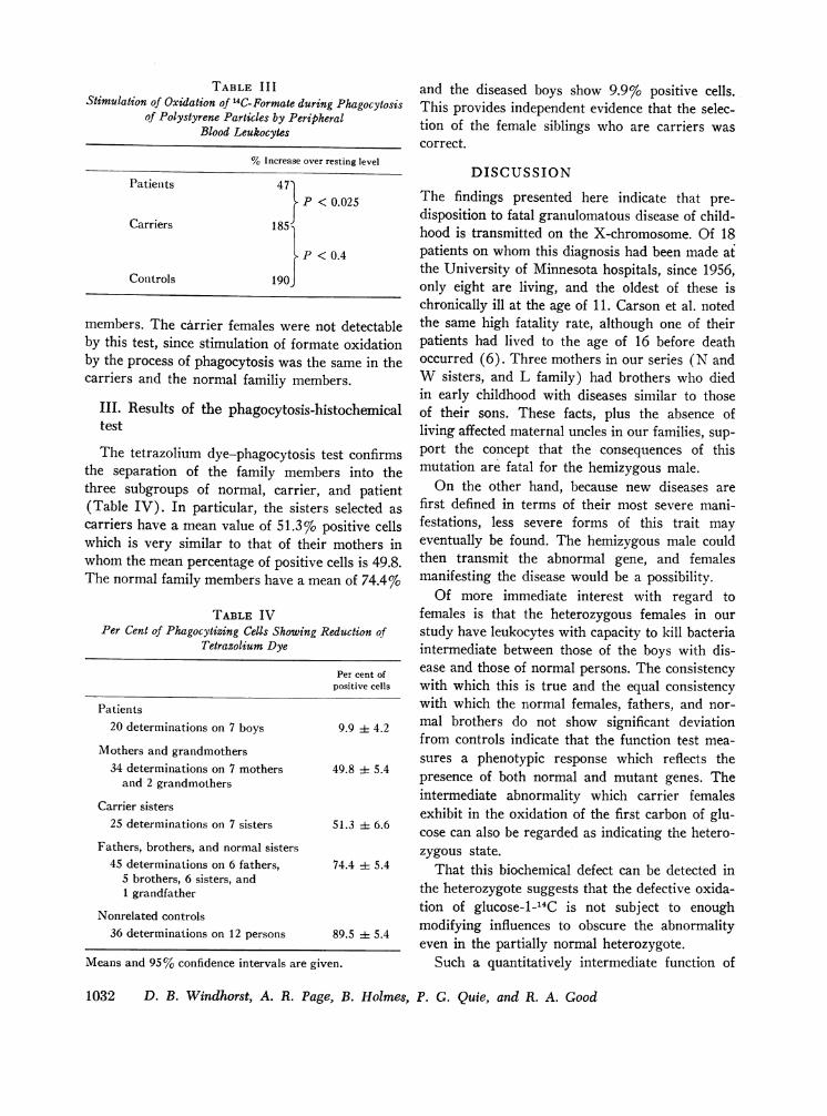
TABLE IIIStimulation of Oxidation of 14C-Formate during Phagocytosis
of Polystyrene Particles by PeripheralBlood Leukocytes
% Increase over r
Patients 47}
Carriers 185
Controls 1901
members. The carrier females were noby this test, since stimulation of formaby the process of phagocytosis was thecarriers and the normal familiy memb
III. Results of the phagocytosis-hitest
The tetrazolium dye-phagocytosis tIthe separation of the family membethree subgroups of normal, carrier,(Table IV). In particular, the sisterscarriers have a mean value of 51.3% pwhich is very similar to that of theirwhom the mean percentage of positiveThe normal family members have a me-
TABLE IVPer Cent of Phagocytizing Cells Showing R
Tetrazolium Dye
Patients20 determinations on 7 boys
Mothers and grandmothers34 determinations on 7 mothersand 2 grandmothers
Carrier sisters25 determinations on 7 sisters
Fathers, brothers, and normal sisters45 determinations on 6 fathers,
5 brothers, 6 sisters, and1 grandfather
Nonrelated controls36 determinations on 12 persons
resting level
and the diseased boys show 9.9% positive cells.This provides independent evidence that the selec-tion of the female siblings who are carriers wascorrect.
DISCUSSION
0.025 The findings presented here indicate that pre-disposition to fatal granulomatous disease of child-hood is transmitted on the X-chromosome. Of 18
0.4 patients on whom this diagnosis had been made atthe University of Minnesota hospitals, since 1956,only eight are living, and the oldest of these ischronically ill at the age of 11. Carson et al. noted
)t detectable the same high fatality rate, although one of theirte oxidation patients had lived to the age of 16 before deathsame in the occurred (6). Three mothers in our series (N anders. W sisters, and L family) had brothers who died
in early childhood with diseases similar to thosestochemical of their sons. These facts, plus the absence of
living affected maternal uncles in our families, sup-est confirms port the concept that the consequences of thisrs into the mutation are fatal for the hemizygous male.and patient On the other hand, because new diseases areselected as first defined in terms of their most severe mani-ositive cells festations, less severe forms of this trait maymothers in eventually be found. The hemizygous male could
cells is 49.8. then transmit the abnormal gene, and femalesan of 74.4%o manifesting the disease would be a possibility.
Of more immediate interest with regard tofemales is that the heterozygous females in our
'eduction of study have leukocytes with capacity to kill bacteriaintermediate between those of the boys with dis-
Per cent of ease and those of normal persons. The consistencypositive cells with which this is true and the equal consistency
with which the normal females, fathers, and nor-
9.9 ± 4.2 mal brothers do not show significant deviationfrom controls indicate that the function test mea-
49.8 + 5.4 sures a phenotypic response which reflects thepresence of both normal and mutant genes. Theintermediate abnormality which carrier femalesexhibit in the oxidation of the first carbon of glu-cose can also be regarded as indicating the hetero-zygous state.
74.4 ± 5.4 That this biochemical defect can be detected inthe heterozygote suggests that the defective oxida-tion of glucose-1-14C is not subject to enough
89.5 A 5.4 modifying influences to obscure the abnormalityeven in the partially normal heterozygote.
en. Such a quantitatively intermediate function of
1032 D. B. Windhorst, A. R. Page, B. Holmes, P. G. Quie, and R. A. Good
Means and 95% confidence intervals are giv(

the female carriers' cells could be due to a partialdefect in all cells or to a mixed population of cellsin which some are completely normal and othersfully abnormal. The latter situation would obtainif random inactivation of one X-chromosome nor-mally occurs in female cells as hypothesized byLyon (14, 15) and Beutler, Yeh, and Fairbanks(16).We have shown that carrier females for the
granulomatous disease do appear to have a mixedpopulation of neutrophils, some abnormal andsome normal by their ability to reduce tetrazoliumdye. This is compatible with the assumption thatX-chromosome inactivation is operating to affectthe gene under consideration (11).The practical clinical consequences of this as-
sumption are of importance, since X inactivationis presumably random and may be capable of oper-ating to produce a wide range of phenotypic find-ings in heterozygous females. Thus in G-6-PDdeficiency, females whose enzyme levels are withinthe normal range, as well as women with com-pletely abnormal levels of enzyme, have beendemonstrated to be heterozygous on the basis oftheir normal and abonormal sons (17). Therefore,it seems reasonable to think that in the granulo-matous trait, heterozygous females may be foundwho have the same clinical disease on this basisas the boys.The analogy of fatal granulomatous disease to
G-6-PD deficiency can be extended. Besides beingX-linked, the two disorders affect closely relatedcell lines, neutrophils, and erythrocytes, respec-tively, which are circulating, highly specialized,nondividing cells. In such cells, major areas ofmetabolism might be expected to have evolved inthe direction of serving the specialized functionof the cell. For instance, the HMP shunt providesmechanisms for stabilization of the red cell mem-brane whereas in nonterminal dividing cells, theHMP shunt is involved in fatty acid turnover andnucleic acid synthesis, among other things. Therole of the increased HMP shunt activity in phago-cytizing neutrophils is unclear.G-6-PD is the entering enzyme for the HMP
shunt, and the over-all functioning of this shuntin the granulomatous carriers' and patients' leuko-cytes is abnormal. The first two steps of the HMPshunt generate reduced nicotinamide adenine di-nucleotide phosphate (NADPH). The reduction
of tetrazolium dyes is apparently a function of theoxidation of nicotinamide adenine dinucleotide(NADH) or NADPH by specific oxidases (18).NADH and NADPH apparently do not diffuseinto intact cells8 and it is not possible to knowwhich, if either, is involved in the dye reductionseen in normal leukocytes but not in the leukocytesof granulomatous patients. It is neverthelesstempting to speculate that the enzyme defect in thepatients with granulomatous disease is in the elec-tron transport system for the hydrogen ions, whichare activated by G-6-PD and 6-phosphogluconatedehydrogenase.Such speculation must not obscure the imme-
diate practical problems of detection of new pa-tients with fatal granulomatous disease and theestimation of carrier status in female relatives.These require evaluation of the diagnostic pro-cedures now available. For simplicity and speeda tetrazolium dye test, employed with a simul-taneous control of normal cells, is the procedure ofchoice for patient diagnosis. However, the diffi-culties in interpretation of the test for the inter-mediate results found in the carrier state are suchthat it is unsuitable for carrier diagnosis.
Simultaneously, the abnormalities observed inglucose-1-14C metabolism in the carrier females asa group are widely variable in individual deter-minations; hence, this procedure is not applicableto the diagnosis of individual females. In fact,neither the dye test nor the metabolic test offeradditional help to the leukocyte function test in theselection of a given individual as a carrier.The technically more complicated leukocyte bac-
tericidal test provides a solid indication as the thecarrier status of a female within 24-48 hr of theinitial venipuncture, particularly when abnormalcontrol cells, as well as normal cells, are includedin the day's run. Improved and simplified proce-dures for this test are being developed.
ACKNOWLEDGMENTS
The untiring and enthusiastic technical assistance of MissJudy Sater was basic to this project.
Dr. D. B. Windhorst is an assistant professor and arecipient of special fellowship AM-22,103 from the Na-tional Institutes of Health. Dr. A. R. Page is an associ-ate professor and a U. S. Public Health Service CareerDevelopment awardee. Dr. B. Holmes is a research as-
3 Windhorst, D., and A. Page. Unpublished observations.
Family Studies in Fatal Granulomatous Disease 1033

sistant and recipient of a U. S. Public Health ServiceTraining grant (AI 90-06) in medical microbiology. Dr.P. G. Quie is a John and Mary R. Markle Scholar in aca-demic medicine and a recipient of U. S. Public HealthService Career Development Award 5-K3-AI-10,109-05.
This work was supported by the Minnesotal Heart As-sociation, the Graduate School of the University of Min-nesota, U. S. Public Health Service grants AI-00798 andAM-05830, and The National Foundation.
REFERENCES1. Holmes, B., A. R. Page, and R. A. Good. 1967. Stud-
ies of the metabolic activity of leucocytes from pa-tients with a genetic abnormality of phagocyticfunction. J. Clin. Invest. 46: 1422.
2. Macfarlane, P. S., A. L. Speirs, and R. G. Sommer-ville. 1967. Fatal granulomatous disease of childhoodand benign lymphocytic infiltration of the skin (con-genital dysphagocytosis). Lancet. 1: 408.
3. Jansz, A., and J. Huber. 1967. Fatal granulomatousdisease of childhood. Lancet. 1: 844.
4. Berendes, H., R. A. Bridges, and R. A. Good. 1957.A fatal granulomatosis of childhood. Minn. Med. 40:309.
5. Bridges, R. A., H. Berendes, and R. A. Good. 1959.A fatal granulomatous disease of childhood. Am. J.Diseases Children. 97: 387.
6. Carson, M. J., D. L. Chadwick, C. A. Brubaker, R. S.Cleland, and B. H. Landing. 1965. Thirteen boys withprogressive septic granulomatosis. Pediatrics. 35:405.
7. Holmes, B., P. G. Quie, D. B. Windhorst, and R. A.Good. 1966. Fatal granulomatous disease of childhood-an inborn abnormality of phagocytic function.Lancet. 1: 1225.
8. Quie, P. G., J. G. White, B. Holmes, and R. A. Good.
1967. In vitro bactericidal capacity of human poly-morphonuclear leukocytes: Diminished activity inchronic granulomatous disease of childhood. J. Clin.Invest. 46: 668.
9. Holmes, B., P. G. Quie, D. B. Windhorst, B. Pollara,and R. A. Good. 1966. Protection of phagocytizedbacteria from the killing action of antibiotics. Nature.210: 1131.
10. Baehner, R. L., and D. G. Nathan. 1966. Deficientglucose oxidation in the intact leukocytes of chronicgranulomatous disease. Blood. 28: 1010. (Abstr.)
11. Windhorst, D. B., B. Holmes, and R. A. Good. 1967.A newly defined X-linked trait in man with demon-stration of the Lyon effect in carrier females. Lancet.1: 737.
12. Karnovsky, M. L. 1962. Metabolic basis of phagocyticactivity. Physiol. Revs. 42: 143.
13. Iyer, G. Y. N., M. F. Islam, and J. H. Quastel. 1961.Biochemical aspects of phagocytosis. Nature. 192:535.
14. Lyon, M. F. 1961. Gene action in the X-chromosomeof the mouse (Mus. Musculus. L.). Nature. 190: 372.
15. Lyon, M. F. 1962. Sex chromatin and gene action inthe mammalian X-chromosome. Am. J. Human Genet.14: 135.
16. Beutler, E., M. Yeh, and V. F. Fairbanks. 1962. Thenormal human female as a mosaic of X-chromosomeactivity: Studies using the genes for G-6-PD-defi-ciency as a marker. Proc. Natl. Acad. Sci. U. S. 48: 9.
17. Nance, W. E. 1964. -Genetic Tests with a Sex-linkedMarker: Glucose-6-phosphate Dehydrogenase. Symp.Quant. Biol. 29: 415.
18. Novikoff, A. B. 1963. Electron transport enzymes:Biochemical and tetrazolium staining studies. In His-tochemistry and cytochemistry. R. Wegmann, editor.The Macmillan Co., New York. 465.
1034 D. B. Windhorst, A. R. Page, B. Holmes, P. G. Quie, and R. A. Good