the phosphatidylinositol-transfer protein nir2 binds...
TRANSCRIPT

EMBO reports - Peer Review Process File - EMBOR-2013-37285
© European Molecular Biology Organization 1
Manuscript EMBOR-2013-37285 The phosphatidylinositol-transfer protein Nir2 binds phosphatidic acid and positively regulates phosphoinositide signaling SoHui Kim, Amir Kedan, Merav Marom, Nancy Gavert, Omer Keinan, Michael Selitrennik, Orly Laufman and Sima Lev Corresponding author: Sima Lev, Weizmann Institute of Science Review timeline: Submission date: 18 March 2013 Editorial Decision: 08 April 2013 Revision received: 01 July 2013 Editorial Decision: 09 July 2013 Revision received: 11 July 2013 Accepted: 11 July 2013 Editor: Barbara Pauly Transaction Report: (Note: With the exception of the correction of typographical or spelling errors that could be a source of ambiguity, letters and reports are not edited. The original formatting of letters and referee reports may not be reflected in this compilation.)
1st Editorial Decision 08 April 2013
Thank you very much for the submission of your research manuscript to our editorial office. Please accept my apologies for the slight delay in the review process. We have now received the full set of reviews on your manuscript. As the detailed reports are pasted below I will only repeat the main points here. You will see that the reviewers appreciate the interest of your findings and are, in principle, supportive of publication of your study in our journal. However, they also point out aspects of your study that would need to be further strengthened before publication. For instance, both referee 1 and 2 state that the quantities of PIs in Nir2-depleted cells would need to be determined by an additional method. Referee 1 also feels that stronger evidence is needed to support the idea that PLD is upstream of Nir2-mediated PI production. Referee 2 states that further experimental evidence is needed to support a causal link between Nir2's interaction with PA and its recruitment to the plasma membrane and for the link between PA-binding/PM-translocation and regulation of PI production and s/he suggests potential ways of how to achieve this. Finally, referee 3 suggests testing whether Nir2 also binds to PI4P and PI(4,5)P2 and checking whether knockdown of Nir2 also leads to decreased levels of PI4P in the

EMBO reports - Peer Review Process File - EMBOR-2013-37285
© European Molecular Biology Organization 2
plasma membrane. Given these evaluations, the reviewers constructive comments and the potential interest of the study, I would like to give you the opportunity to revise your manuscript, with the understanding that the main concerns of the reviewers should be addressed, especially with regard to the concerns of referee 2 about the causal link between Nir2's PA binding-ability and the regulation of PI production. Acceptance of the manuscript will depend on a positive outcome of a second round of review and I should also remind you that it is EMBO reports policy to allow a single round of revision only and that therefore, acceptance or rejection of the manuscript will depend on the completeness of your responses included in the next, final version of the manuscript. Revised manuscripts should be submitted within three months of a request for revision; they will otherwise be treated as new submissions. If you feel that this period is insufficient for a successful submission of your revised manuscript I can potentially extend this period slightly. Also, the length of the revised manuscript should not exceed roughly 29,000 characters (including spaces and references). Currently your manuscript contains about 36,000 characters, so some shortening will be required. If you feel that the additional data requested by the reviewers would make the manuscript too long you may consider including some peripheral data in the form of Supplementary information. However, I have noticed that at the moment, there are no materials and methods in the main body of the text and I would kindly ask you to expand this section before submitting your revised version. Materials and Methods essential for the repetition of the main experiments should not be displayed as supplementary information only. When submitting your revised manuscript, please include: • a word-formatted version of the manuscript text • editable, high-resolution TIFF or EPS-formatted or figure files • a separate single PDF file of all the Supplementary information (in its final format) • a letter detailing your responses to the referee comments (as word file if possible) • a short, two-sentence summary of the manuscript As part of the EMBO publication's Transparent Editorial Process, EMBO reports publishes online a Review Process File to accompany accepted manuscripts. This File will be published in conjunction with your paper and will include the referee reports, your point-by-point response and all pertinent correspondence relating to the manuscript. You are able to opt out of this by letting the editorial office know ([email protected]). If you do opt out, the Review Process File link will point to the following statement: "No Review Process File is available with this article, as the authors have chosen not to make the review process public in this case." We also welcome the submission of cover suggestions or motifs that might be used by our Graphics Illustrator in designing a cover. I look forward to seeing a revised form of your manuscript when it is ready. Should you in the meantime have any questions, please do not hesitate to contact me. REFEREE REPORTS: Referee #1: The manuscript by Kedan et al. describes a novel PA-binding region present in the PI transfer protein Nir2. The authors demonstrate that the interaction between PA and Nir2 is important for its translocation to the plasma membrane upon EGF stimulation or by PA treatment. It is also demonstrated that Nir2 plays an important role in the activation of AKT as well as MAPK pathways by maintaining the proper level of phosphoinositide metabolism which involves PI3K and PLC. This study may provide important insights into how PA acts as precursor and product lipids in PI

EMBO reports - Peer Review Process File - EMBOR-2013-37285
© European Molecular Biology Organization 3
metabolism through a direct interaction with Nir2. However, there are several points that need to be improved before considered as a strong candidate for publication in EMRO Reports. Major points: 1. A major concern for this study is lack of direct measurement of the quantity of phosphoinositides in Nir2-knockdown cells. The authors only demonstrate changes in PI(4,5)P2 and PI(3,4,5)P3 by using exogenously expressed PH domains or anti-PIP2 antibody staining. Are these phosphoinositides really decreased or increased by the Nir2 knockdown or drug treatments? Because the possible function of Nir2 in the control of PI-signaling is one of the key findings in this manuscript, it is quite important to discuss these phenotypes based on quantitative data of each phosphoinositide species. 2. The second problem would be that the PA-binding region of Nir2 is not fully characterized in this study. As the authors have presented very nice alignment (Fig.2A), it should be examined which of these conserved residues are really important. By doing so, more detailed information would be provided concerning the molecular mechanism of this novel interaction between PA and Nir2. Such data are also very useful in localization assays shown in Figs 1C, F and 2B, as well as rescue experiments in Fig. 4E. 3. There is no evidence supporting the role of PLD upstream of Nir2-mediated PIs production. In Fig. 2D, it was shown that 1-BuOH inhibited the plasma membrane translocation of Nir2 upon EGF stimulation, suggesting that the process is mediated by a PLD activity. Does the same 1-BuOH treatment also inhibit PIP2 production as shown in Fig. 4C-E? Minor points: 1. In Fig.2F, nearly 100% of proteins bound to vesicles containing 5% PA. In such a case, lower concentrations of PA need to be tested, in order to know how strong the binding is. 2. In Fig. 3A-D, there are no western blots for "total" Akt, therefore it is impossible to judge whether the Akt phosphorylations are really affected by Nir2 overexpression/knockdown, or the total amounts of Akt have been changed. Referee #2: This study reveals the a role for the PI transfer protein NIR1 in regulating the levels of phosphoinositides. The authors also demonstrate that NIR1 binds to PA via it's C-terminal domain. This report is interesting, however the research does not make the confections that would make this study strong enough for publication in EMBO reports. If the authors are able to provide information on the points below then it would deserve consideration for publication. Major points. 1. There is no clear relationship between PA interaction and plasma membrane interaction. The authors demonstrate tha deletion of the liPIn domain reduces membrane interaction but this does not clarify the issue. Do they have point mutations that diminish PA binding that eliminate membrane interaction. The addition of extracellular Pa is interesting, however it would be expected that this would saturate the whole of the plasma membrane leading to a general recruitment of NIR1 to the membrane, however the authors show quite localised membrane interaction suggestion that more than Pa interaction is required (figure 2c ) . the demonstration that butanol suppresses the translocation needs to be properly controlled with measurements of PA. Cells could be labeled with 32-P ortho-phosphate and stimulated in the presence and absence of butanol and PA should be measured. 2. There is no clear relationship between PA/membrane interaction and phosphoinositide regulation. If the authors could generate a PA binding mutant this would help to assess the role of membrane interaction and PI regulation. Furthermore can the authors then target the mutant back to the membrane and regenerate PI signaling, akt and map kinase activation. This might be done using either a caax or- a myristoylation motif. Alternatively, the fkbp targeting system system could be utilised. 3. The measurement of changes in PI need to be assessed using conventional methods of measurement such as radiolabelling. Although the authors have utilised two different methods to

EMBO reports - Peer Review Process File - EMBOR-2013-37285
© European Molecular Biology Organization 4
visualise PIp2 they both suffer from similar limitations. For example it is clear that knock down of NIR1 changes the morphology of the cells ( figure 4a,b,c) which would certainly compromise these visualisation methods. Measurement by radiolabelling would also allow the authors to measure the levels of PA in cells to determine if PLC signaling is enhanced in the NIR1 knockdown. The authors should also measure DAG levels. Minor points. 1 the regulation of maps kinase by either knock down or over-expression are not convincing and should be quantified over a number of experiments. 2 the authors need to measure binding of the C- terminal domain to PA containing liposomes at lower concentrations of PA to determine its approximate affinity as they have saturated binding at this level. 3. if NIR1 knock down stimulates PI hydrolysis does the C- terminal domain translocate to the membrane as a consequence of increased PA formation ? Referee #3: This is a very interesting set of observations that show, really for the first time, that a PI exchange protein can limit inositol lipid synthesis in an intact cell. This is by no means taken as being what these proteins do, so it is actually a very novel set of data that will be widely cited. There are a couple of simple (for the authors because they already have the methods in hand) experiments that they could do which would not change the overall message, but I think would help to clarify it. (1) The liposome data nicely show that Nir2 binds to PA and not PS, and that this is unlikely to be a non-specific charge effect. But it would be really interesting to put PI4P and (separately) PI45P2 into the same assay. They are present in the plasma membrane at levels similar to PA, and if NIr2 binds PIP2 thenthe PLD-dependence of the Nir2 translocation could be at least in part by a (PA)-stimulated PIP2 production. PI4P would be equally interesting because it makes a similar contribution to general p.m. charge (GRV Hammond et al Science 2012). (2) The interpretation is that it is PI that is ultimately limiting, because it is PI that is transferred by Nir2. If that is correct then PI4P should be similarly depleted in the p.m. after Nir2 knock-down, and using an anti-PI4P antibody instead of anti-PI45P2 (as in Fig 4C) would be very informative in this context (a suitable antibody is used in the above Hammond et al publication). 1st Revision - authors' response 01 July 2013
Response to referees We would like to thank the three reviewers for their productive and thoughtful comments. Referee #1 Major Points 1. The major concern of the referee is that our study lacks a direct measurement of the phosphoinositides levels in Nir2-knockdown cells. We accept this comment and measured the level of PI(4,5)P2 in control and Nir2 knockdown (KD) HeLa cells using metabolic labeling with [32P]Orthophosphate. We also measured the level of PI(3,4,5)P3 in serum-starved and EGF-stimulated control or Nir2-depleted HeLa cells using the PIP3 Mass Elisa Kit of Echelon Biosc. Inc. (Catalog No. K-2500s). In brief, for metabolic labeling with 32P-PO4 (orthophosphate), control or Nir2-depleted HeLa cells (4 x 10⁶) were labeled for 1 hr with 100 µCi 32P-orthophosphate/ml in PO4-free media. Acidic lipids were extracted with chloroform/methanol as previously described (Serunian et al., 1991), dried under N2 stream, and separated by TLC in a solvent containing: chloroform:acetone:methanol:acetic-acid:DDW; 40:15:13:12:7 (Traynor-Kaplan et al., 1989). PI, PIP and PIP2 standards (Sigma, Avanti) were identified by iodine vapor and used as marks for the

EMBO reports - Peer Review Process File - EMBOR-2013-37285
© European Molecular Biology Organization 5
radiolabeled phosphoinositides. PI(4,5)P2 synthesis was quantified by phosphorimager and densitomery analysis. The experiments were performed in duplicate, and were repeated at least 6 times. The intensity of the radiolabeled lipids was normalized according total radiolabeled lipids. A representative TLC is shown in Fig. 4F of the revised manuscript. The PIP2 levels obtained from several additional experiments are also shown along with a graph that summarizes the results of all the experiments. As shown, PI(4,5)P2 synthesis was significantly reduced (by ~28%) in Nir2-KD cells. This is a very important observation that complements and supports our staining results using anti-PI(4,5)P2 antibody and the subcellular distribution of the GFP-PH-PLCδ reporter (Fig. 4C,D). For PI(3,4,5)P3 measurements, we used the PIP3 Mass Elisa Kit, as metabolic labeling with 32P-PO4 following by TLC separation is not sensitive enough. We used the Echelon’s PIP3 mass elisa kit that was previously used in many studies (Jo et al., 2011). The kit measures PI(3,4,5)P3 levels utilizing a competitive ELISA. It contains a PI(3,4,5)P3-coated Elisa plate and a PI(3,4,5)P3 detector protein. Binding of the detector protein to PI(3,4,5)P3 produced in cells competes with its binding to the PI(3,4,5)P3-coated plate. We used the kit for two experiments that were performed in triplicate. Ten million HeLa cells (control and Nir2-depleted) were used for each measurement. In brief, the cells were serum-starved for 18 hr and then stimulated with EGF for 3 or 6 min. The level of PI(3,4,5)P3 in the serum-starved and EGF-stimulated cells was measured by the kit according the manufacturer’s instructions. The amount of PI(3,4,5)P3 was calculated according to a standard curve. The results of two experiments (the maximum capacity of the kit) that were performed in triplicates are shown in Fig. 4B of the revised manuscript. As seen, depletion of Nir2 substantially reduced the production of PI(3,4,5)P3 as compared to control cells: at 3 min following EGF treatment ~32 pmol PI(3,4,5)P3 was produced in the control cells but only ~6.5 in Nir2-KD cells. The level of PI(3,4,5)P3 was reduced to ~24 pmol at 6 min in the control cells and could hardly be detected in Nir2-KD cells. These results are consistent with the subcellular distribution of the GFP-PH-AKT reporter in control and Nir2-depleted HeLa cells (Fig. 4A). 2. The second concern of the referee was related to the PA-binding region of Nir2, which was not fully characterized. We agree that we did not fully characterize the PA-binding site within Nir2 protein. In light of the referee’s comment, we used site directed mutagenesis along with in vitro PA-binding assays and localization studies in intact cells, and demonstrated that the LNS2 HAD-like domain of Nir2 mediates the PA-binding. In brief, according to structural data and conserved motifs in HAD superfamily, we decided to mutate the aspartic acid residue in loop 4 (D1128) which participates in Mg2+ binding residue (see attached figure from Karen Allen’s review) (Allen and Dunaway-Mariano, 2004). This site is highly conserved in HAD superfamily, Nir proteins (Nir1-3) and lipins (labeled in red in motif 3, Fig. 2A). We assumed that mutating this site would greatly reduce PA binding as Mg2+ both electrostatically and through coordinate bonds allows phosphoryl-group binding. Furthermore, folding studies show that most HAD members are not dependent on Mg2+ for folding and stability and therefore mutating this site was not expected to affect the folding of the protein. To be on the safe side, we mutated two additional sites that participate in Mg2+ binding: Asp (1028) and Ser (1026) residues from loop1 (motif 1, Fig. 2A) (see figure below). We established two C-terminal mutants for expression in bacteria as recombinant proteins: D1128A, and the triple mutant S1026/D1028/D1128A. In addition, we established two full-length Nir2 mutants: D1128A and S1026/D1028/D1128A for expression in mammalian cells as Myc-tagged proteins. The recombinant C-terminal mutants were expressed in bacteria in the correct size, but were not as soluble as the wild-type (WT) protein. The triple mutant was completely insoluble and mainly found in inclusion bodies, whereas the D1128A mutant was partially insoluble. Nevertheless, after optimizing the induction conditions, we could purify soluble D1128A mutant and perform the in vitro PA binding assays. The results were very convincing; this point mutation substantially reduced the binding of PA in vitro. As shown in Fig. 5B of the revised manuscript, we could not detect any binding to liposomes containing 20% PA and could hardly detect binding to liposomes containing 30% PA. This result strongly suggests that the LNS2 domain of Nir2 mediates the PA-binding. We went one step further and could demonstrate that the single D1128A mutation substantially reduced the translocation to the plasma membrane in response to either PA or EGF. The effect was more profound with the triple mutant (see Fig. 5C revised manuscript). It is worth mentioning that the mutations had no effect on the Golgi localization, consistent with the results shown in Fig. 1. Most importantly, we found that neither the single nor the triple mutant could rescue the PIP2 levels in Nir2-depleted cells (Fig. 5D). This extensive analysis, at the resolution of a single amino-acid,

EMBO reports - Peer Review Process File - EMBOR-2013-37285
© European Molecular Biology Organization 6
demonstrates (i) that Nir2 binds PA via its LNS2 domain, (ii) that PA can mediate the translocation of Nir2 to the PM in response to certain external stimuli, and (iii) that its PM translocation is essential for PIP2 production. We believe that these are important observations that support our findings and markedly improve our manuscript.
3. The referee asked whether 1-BuOH treatment inhibits PIP2 production. The connection between PLD activity and PIP2 production was demonstrated by many previous studies as PIP2 activates PLD and PA activates PIP5K, the rate-limiting enzyme of PIP2 biosynthesis (Divecha et al., 2000; Jones et al., 2000; Liscovitch et al., 1994). Specifically, previous studies have shown that treatment with 1-butanol diminishes PIP2 in the PM and eliminates EGF-induced calpain activation in NR6 fibroblasts (Shao et al., 2006). We performed a similar experiment in HeLa cells and found that 1-butanol markedly reduces the level of the GFP-PH-PLCδ reporter at the PM (see below). However, this observation is not directly related to our studies, and therefore we did not include it in the revised manuscript.
Localization of GFP-PH-PLCδ in control and 1-butanol-treated HeLa cells. HeLa cells were transfected with GFP-PH-PLCδ reporter and either treated with 0.3% 1-butanol or left untreated. The localization of the GFP-PH-PLCδ reporter was determined either in live cells or following fixation with 4% PFA/0.1% GA. Minor points 1.In Fig.2F, nearly 100% of proteins bound to vesicles containing 5% PA. In such a case, lower concentrations of PA need to be tested, in order to know how strong the binding is. We agree with this comment and repeated the binding experiments utilizing liposomes containing different concentrations of PA. It is worth mentioning that these binding experiments were performed with different batch of PA and recombinant protein prep, as all the original reagents that were used in our previous experiments have been finished or were too old (we performed the binding experiments almost two years ago). We essentially repeated the lipid specificity experiment (Fig. 2E, revised manuscript) utilizing liposomes of different lipid compositions and obtained similar results. As for the PA concentrations experiment, we could detect weak binding at 5% PA, and not as described in our previous version. We don’t know the reason for these differences, but could be related to the PA batch (we found that some batches are not good or contain different concentrations). We therefore replaced the original figure with the new analysis using the new PA (Avanti) and a new purification of the recombinant C-terminal region. As shown in Fig. 2F, the binding of the C-terminal is increased with the increased concentration of PA and reach saturation at 20-25% PA. We repeated this experiments few times and similar results were obtained. 2. In Fig. 3A-D, there are no western blots for "total" Akt, therefore it is impossible to judge whether the Akt phosphorylations are really affected by Nir2 overexpression/knockdown, or the total amounts of Akt have been changed. We completely agree with this comment. The total amount of AKT did not change in either Nir2

EMBO reports - Peer Review Process File - EMBOR-2013-37285
© European Molecular Biology Organization 7
overexpressing HeLa or MCF7 cells or in Nir2-depleted cells. We should have include it in our previous submission, and corrected this mistake in our revised manuscript (Fig. 3A, 3B, 3C, S2, S4). Referee #2 Major points 1.The referee asked to better characterize the PA-binding site within Nir2, and possibly to establish point mutations that abolish this binding. As seen in our reply to referee 1 (Major point 2), we could establish point mutants that abolished PA-binding. As this comment is similar to comment 2 of referee 1, our response is identical in both cases. The referee also mentioned that the distribution of Nir2 at the PM in response to PA is ‘quite localized’ which could suggest that more than PA interaction is required. The distribution pattern at the plasma membrane (PM) in response to PA treatment could be different from one cell to another. We could detect different distribution patterns of Nir2 at the PM as well as of GFP-Spo20 (a PA-reporter) (Young et al., 2010). As seen in the attached figure, in some cases Nir2 or Spo20 are equally distributed at the PM, whereas in others they are localized at certain regions.
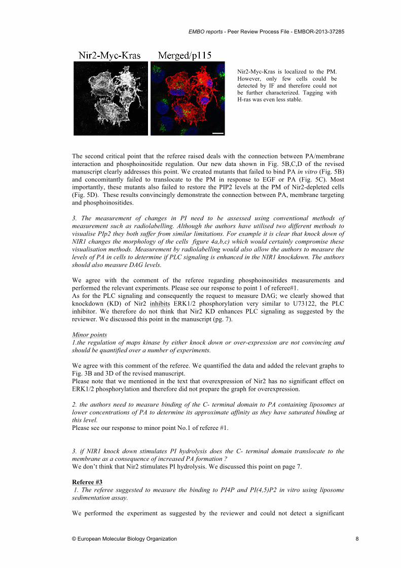
Localization of GFP-Spo20 in response to PA treatment. As shown, in some cells (picture 3 from left) Spo20-GFP is not equally distributed at the PM in response to PA treatment while in others it does (picture 4 from left). Very similar distribution was obtained for Nir2; pictures 5 and 6, respectively. The referee suggested measuring the suppressive effect of butanol on PA production using orthophosphate labeling. Many previous studies demonstrated the effect of 1-butanol on PA production. The inhibitory effect of 1-butanol on PA production in response to EGF treatment was previously measured using metabolic radiolabeling (see Figure 1 in (Shen et al., 2001)). 2. There is no clear relationship between PA/membrane interaction and phosphoinositide regulation. If the authors could generate a PA binding mutant this would help to assess the role of membrane interaction and PI regulation. Furthermore can the authors then target the mutant back to the membrane and regenerate PI signaling, akt and map kinase activation. This might be done using either a caax or- a myristoylation motif. Alternatively, the fkbp targeting system could be utilised. The idea of targeting Nir2 to the PM by specific targeting motif as suggested by the referee is very relevant, and we actually tried it before. We fused the H-ras (GCMSCKCVLS) or K-ras (SKDGKKKKKKSKTKCVIM) PM targeting motifs (Huang et al., 1993) to the C-terminal end of Nir2. We could detect constitutive targeting of Nir2 to the PM (see figure below), but the protein was possibly unstable. We could hardly observed transfected cells by IF and could not detect the protein by WB. We therefore did not continue with this approach or applied different targeting strategy.
EMBO reports - Peer Review Process File - EMBOR-2013-37285
© European Molecular Biology Organization 8
The second critical point that the referee raised deals with the connection between PA/membrane interaction and phosphoinositide regulation. Our new data shown in Fig. 5B,C,D of the revised manuscript clearly addresses this point. We created mutants that failed to bind PA in vitro (Fig. 5B) and concomitantly failed to translocate to the PM in response to EGF or PA (Fig. 5C). Most importantly, these mutants also failed to restore the PIP2 levels at the PM of Nir2-depleted cells (Fig. 5D). These results convincingly demonstrate the connection between PA, membrane targeting and phosphoinositides. 3. The measurement of changes in PI need to be assessed using conventional methods of measurement such as radiolabelling. Although the authors have utilised two different methods to visualise PIp2 they both suffer from similar limitations. For example it is clear that knock down of NIR1 changes the morphology of the cells figure 4a,b,c) which would certainly compromise these visualisation methods. Measurement by radiolabelling would also allow the authors to measure the levels of PA in cells to determine if PLC signaling is enhanced in the NIR1 knockdown. The authors should also measure DAG levels. We agree with the comment of the referee regarding phosphoinositides measurements and performed the relevant experiments. Please see our response to point 1 of referee#1. As for the PLC signaling and consequently the request to measure DAG; we clearly showed that knockdown (KD) of Nir2 inhibits ERK1/2 phosphorylation very similar to U73122, the PLC inhibitor. We therefore do not think that Nir2 KD enhances PLC signaling as suggested by the reviewer. We discussed this point in the manuscript (pg. 7). Minor points 1.the regulation of maps kinase by either knock down or over-expression are not convincing and should be quantified over a number of experiments. We agree with this comment of the referee. We quantified the data and added the relevant graphs to Fig. 3B and 3D of the revised manuscript. Please note that we mentioned in the text that overexpression of Nir2 has no significant effect on ERK1/2 phosphorylation and therefore did not prepare the graph for overexpression. 2. the authors need to measure binding of the C- terminal domain to PA containing liposomes at lower concentrations of PA to determine its approximate affinity as they have saturated binding at this level. Please see our response to minor point No.1 of referee #1. 3. if NIR1 knock down stimulates PI hydrolysis does the C- terminal domain translocate to the membrane as a consequence of increased PA formation ? We don’t think that Nir2 stimulates PI hydrolysis. We discussed this point on page 7. Referee #3 1. The referee suggested to measure the binding to PI4P and PI(4,5)P2 in vitro using liposome sedimentation assay. We performed the experiment as suggested by the reviewer and could not detect a significant
Nir2-Myc-Kras is localized to the PM. However, only few cells could be detected by IF and therefore could not be further characterized. Tagging with H-ras was even less stable.
EMBO reports - Peer Review Process File - EMBOR-2013-37285
© European Molecular Biology Organization 9
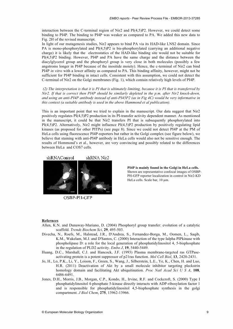
interaction between the C-terminal region of Nir2 and PI(4,5)P2. However, we could detect some binding to PI4P. The binding to PI4P was weaker as compared to PA. We added this new data to Fig. 2H of the revised manuscript. In light of our mutagenesis studies, Nir2 appears to bind PA via its HAD-like LNS2 domain. Since PA is mono-phosphorylated and PI(4,5)P2 is bis-phosphorylated (carrying an additional negative charge) it is likely that the electrostatics of the HAD-like binding site would not be suitable for PI(4,5)P2 binding. However, PI4P and PA have the same charge and the distance between the diacylglycerol group and the phosphoryl group is very close in both molecules (possibly a few angstroms longer in PI4P because of the inositide moiety). Hence, the c-terminal of Nir2 can bind PI4P in vitro with a lower affinity as compared to PA. This binding affinity, however, might not be sufficient for PI4P binding in intact cells. Consistent with this assumption, we could not detect the C-terminal of Nir2 on the Golgi membranes (Fig. 1), which contain relatively high levels of PI4P. (2) The interpretation is that it is PI that is ultimately limiting, because it is PI that is transferred by Nir2. If that is correct then PI4P should be similarly depleted in the p.m. after Nir2 knock-down, and using an anti-PI4P antibody instead of anti-PI45P2 (as in Fig 4C) would be very informative in this context (a suitable antibody is used in the above Hammond et al publication). This is an important point that we tried to explain in the manuscript. Our data suggest that Nir2 positively regulates PI(4,5)P2 production in its PI-transfer activity dependent manner. As mentioned in the manuscript, it could be that Nir2 transfers PI that is subsequently phosphorylated into PI(4,5)P2. Alternatively, Nir2 might influence PI(4,5)P2 production by positively regulating lipid kinases (as proposed for other PITPs) (see page 8). Since we could not detect PI4P at the PM of HeLa cells using fluorescence PI4P-reporters but rather in the Golgi complex (see figure below), we believe that staining with anti-PI4P antibody in HeLa cells would also not be sensitive enough. The results of Hommond’s et al., however, are very convincing and possibly related to the differences between HeLa and COS7 cells.
Refernces Allen, K.N. and Dunaway-Mariano, D. (2004) Phosphoryl group transfer: evolution of a catalytic
scaffold. Trends Biochem Sci, 29, 495-503. Divecha, N., Roefs, M., Halstead, J.R., D'Andrea, S., Fernandez-Borga, M., Oomen, L., Saqib,
K.M., Wakelam, M.J. and D'Santos, C. (2000) Interaction of the type Ialpha PIPkinase with phospholipase D: a role for the local generation of phosphatidylinositol 4, 5-bisphosphate in the regulation of PLD2 activity. Embo J, 19, 5440-5449.
Huang, D.C., Marshall, C.J. and Hancock, J.F. (1993) Plasma membrane-targeted ras GTPase-activating protein is a potent suppressor of p21ras function. Mol Cell Biol, 13, 2420-2431.
Jo, H., Lo, P.K., Li, Y., Loison, F., Green, S., Wang, J., Silberstein, L.E., Ye, K., Chen, H. and Luo, H.R. (2011) Deactivation of Akt by a small molecule inhibitor targeting pleckstrin homology domain and facilitating Akt ubiquitination. Proc Natl Acad Sci U S A, 108, 6486-6491.
Jones, D.H., Morris, J.B., Morgan, C.P., Kondo, H., Irvine, R.F. and Cockcroft, S. (2000) Type I phosphatidylinositol 4-phosphate 5-kinase directly interacts with ADP-ribosylation factor 1 and is responsible for phosphatidylinositol 4,5-bisphosphate synthesis in the golgi compartment. J Biol Chem, 275, 13962-13966.
PI4P is mainly found in the Golgi in HeLa cells. Shown are representative confocal images of OSBP-PH-GFP reporter localization in control in Nir2-KD HeLa cells. Scale bar, 10 µm.
EMBO reports - Peer Review Process File - EMBOR-2013-37285
© European Molecular Biology Organization 10
Liscovitch, M., Chalifa, V., Pertile, P., Chen, C.S. and Cantley, L.C. (1994) Novel function of phosphatidylinositol 4,5-bisphosphate as a cofactor for brain membrane phospholipase D. J Biol Chem, 269, 21403-21406.
Serunian, L.A., Auger, K.R. and Cantley, L.C. (1991) Identification and quantification of polyphosphoinositides produced in response to platelet-derived growth factor stimulation. Methods Enzymol, 198, 78-87.
Shao, H., Chou, J., Baty, C.J., Burke, N.A., Watkins, S.C., Stolz, D.B. and Wells, A. (2006) Spatial localization of m-calpain to the plasma membrane by phosphoinositide biphosphate binding during epidermal growth factor receptor-mediated activation. Mol Cell Biol, 26, 5481-5496.
Shen, Y., Xu, L. and Foster, D.A. (2001) Role for phospholipase D in receptor-mediated endocytosis. Mol Cell Biol, 21, 595-602.
Traynor-Kaplan, A.E., Thompson, B.L., Harris, A.L., Taylor, P., Omann, G.M. and Sklar, L.A. (1989) Transient increase in phosphatidylinositol 3,4-bisphosphate and phosphatidylinositol trisphosphate during activation of human neutrophils. J Biol Chem, 264, 15668-15673.
Young, B.P., Shin, J.J., Orij, R., Chao, J.T., Li, S.C., Guan, X.L., Khong, A., Jan, E., Wenk, M.R., Prinz, W.A., Smits, G.J. and Loewen, C.J. (2010) Phosphatidic acid is a pH biosensor that links membrane biogenesis to metabolism. Science, 329, 1085-1088.
2nd Editorial Decision 09 July 2013
Thank you for the submission of your revised manuscript to our offices. We have now received the enclosed reports from the referees that were asked to assess it. I am very pleased to tell you that all referees now support publication of your manuscript in our journal. Referee 1 has a minor comment on including the statistical significance for the results shown in figure 4F and I would kindly ask you to include this in the final version. Referee 3 still feels that it would be worthwhile to analyze the level of PI4P in the absence of Nir2 by immunostaining. While his/her suggestion of how to do this seems to be rather straightforward, I also understand that you have tried to address this concern using a PI4P reporter, which did not work. If you are willing to test the levels using an antibody as referee 3 suggested I would encourage you to do so, but I would not make this a prerequisite for publication at this point. There are also a few more formal points that I would like to draw your attention to: 1. Unfortunately, the text is still too long for our format (it currently contains almost 35,000 characters, while our limit in fact would be about 27,000). I think it would be okay if you could cut it down to 30,000, but this really is the limit. 2. Fig 4D is referenced twice in the figure legend and I think the second 4D really is Fig 4E and what is now 4E in the legend is Fig4F. Please correct this in the final version. 3. I have taken the liberty of slightly editing the abstract, as one sentence did not read so well. Can you please let me know that you approve of my edited version, which is copied here: "Phosphatidic acid (PA) and phosphoinositides are metabolically interconverted lipid second messengers that play central roles in many growth factor (GF)-stimulated signaling pathways. Yet, little is known about the mechanisms that coordinate their production and downstream signaling. Here we show that the phosphatidylinositol (PI)-transfer protein Nir2 translocates from the Golgi complex to the plasma membrane in response to GF stimulation. This translocation is triggered by PA formation and is mediated by its C-terminal region that binds PA in vitro. We further show that depletion of Nir2 substantially reduces the PI(4,5)P2 levels at the plasma membrane and concomitantly GF-stimulated PI(3,4,5)P3 production. Finally, we show that Nir2 positively regulates the MAPK and PI3K/AKT pathways. We propose that Nir2 through its PA binding capability and PI-transfer activity can couple PA to phosphoinositide-signaling, and possibly coordinates their local lipid metabolism and downstream signaling."

EMBO reports - Peer Review Process File - EMBOR-2013-37285
© European Molecular Biology Organization 11
4. Lastly, we have also started to encourage authors to submit the raw data of biochemical and/or microscopical images to our editorial office. These data will be published online as part of the supplementary information. This is voluntary at the moment, but if you agree that this would be useful for readers I would like to invite you to supply these files when submitting the revised version of your study. I look forward to seeing the final version of your manuscript as soon as possible. REFEREE REPORTS: Referee #1: I am satisfied with all answers and revisions except for the point as follows; As the difference between PIP2 levels in control and KD cells are relatively small, the data shown in new Figure 4F should be statistically examined. Referee #2: The authors have answered many of my questions and the remaining ones are trivial and would not significantly enhance the paper. i would therefore recomend publication of this manuscript. Referee #3: The authors' response to my comments are mixed. Point 1. This is nicely done and the result adds to the paper. Point 2. The authors have done a significant number of further experiments to meet all the reviewers' suggestions, which makes it all the more puzzling why they appear willfully to have avoided the simple extra experiment I suggested. The underlying hypothesis is that Nir2 is limiting PIP2 (and thus also PIP3) availability by limiting the PI in the plasma membrane. I suggested that quantitating PI4P in the p.m. under conditions of Nir2 depletion would be very informative, for two reasons. If PI really is so low that it is becoming limiting, then p.m. PI4P levels should also fall. If they do not, then that could in turn be an interesting result because it may suggest different pools of PI and/or PI4P with different roles as PIP2 precursors (plus Nir2 having differential effects on each) - a result that would build on the recent Hammond et al Science paper that the authors mention. It is a win-win experiment and incredibly simple for these authors to perform. To reiterate what I said before (and I never mentioned anything about transfecting with PI4P binding domains), they simply repeat the PIP2 staining in fixed cells (Fig 4D in the new version - note the numbering in the legend is wrong) but use a PI4P monoclonal instead of a PIP2 antibody. Their fixation conditions are almost identical to those that Hammond et al used for p.m. PI4P quantification, which are described in detail in that group's earlier Biochemical Journal paper (in which, incidentally, they showed that PI4P stains very nicely in the p.m. of HeLa cells!). It is worth adding that the present authors already have the whole-cell PI4P data in their new Fig 4F, which looks like it does not change (but which will of course have a major contribution from Golgi PI4P), and quantifying their tlc plates for PI4P would nicely complement the p.m. data derived from staining with a PI4P antibody.
EMBO reports - Peer Review Process File - EMBOR-2013-37285
© European Molecular Biology Organization 12
2nd Revision - authors' response 11 July 2013
We modified Fig. 4 and included the statistical significance (attached) and I shortened the manuscript by 3,000 characters, it is now 32,074 (including spaces) (attached). If this is not enough, I can further shortened it, but it might affect the manuscript. Nevertheless, I will do what ever you suggest. As for the summary sentences: this is my suggestion: The PI-transfer protein Nir2 is also a PA-binding protein, which translocates from the Golgi complex to the PM in response to growth factor stimulation. Nir2 positively regulates the production of PI(4,5)P2 and PI(3,4,5)P3 and consequently the MAPK and the PI3K/AKT signaling pathways. I look forward to your comments. 3rd Editorial Decision 11 July 2013
I am very pleased to accept your manuscript for publication in the next available issue of EMBO reports. Thank you for your contribution to our journal. As part of the EMBO publication's Transparent Editorial Process, EMBO reports publishes online a Review Process File to accompany accepted manuscripts. As you are aware, this File will be published in conjunction with your paper and will include the referee reports, your point-by-point response and all pertinent correspondence relating to the manuscript. If you do NOT want this File to be published, please inform the editorial office within 2 days, if you have not done so already, otherwise the File will be published by default [contact: [email protected]]. If you do opt out, the Review Process File link will point to the following statement: "No Review Process File is available with this article, as the authors have chosen not to make the review process public in this case." Thank you again for your contribution to EMBO reports and congratulations on a successful publication. Please consider us again in the future for your most exciting work.