the polymorphic transformations of obelia - journal of cell science
TRANSCRIPT
235
The Polymorphic Transformations of Obelia
BY
N. J. BERRILL1
(McGill University, Montreal)
Part I. Stolonic growth and PerisarcPart II. Hydranth developmentPart HI. Gonangium and Medusae
INTRODUCTION
' • "*HE following account is a portrayal of a single hydroid species, ObeliaX commissularis, as a living organism in all of its manifestations.Obelia has long been established as the text-book type of a marine hydroid,
illustrating the colonial condition and so-called alternation of generations.In the present century it has been the subject of several special studies.Huxley and de Beer (1923) have given a detailed account of the processof hydranth resorption. Lund (1921-6), in a series of papers, correlatedhydranth formation and regeneration with electrical polarities. Commencingin 1933 and culminating in a general review started in 1943, Hammett andhis co-workers published numerous papers on the role of amino-acids andother factors upon the several phases of growth as defined by them. Obeliageniculata was employed as the test organism, and while the approach isdynamic and numerous observations on the formation of the living hydranthare reported (Hammett, 1943; Hauscha, 1944), the 'concern here is not withorganism as organism; but with organism as test material suitable for evalua-tion of the interdependencies of growth'. Much of the value of this workdepends upon the validity of the various abstractions made of the develop-mental process as a whole. Since our own emphasis is on Obelia as an organ-ism, it is unnecessary to discuss, the question further at this time. In laterpapers Hammett (1945-6) presents statistical analyses of the condition of thehydranth population of thousands of colonies, undoubtedly a herculean labourbut one that throws into sharp relief the difficulty, if not the impossibility, ofanalysing individual behaviour in terms of statistical averages, for an organismpersists in being primarily organismal in spite of anything an investigatormay have in mind.
Studies of Obelia are included in the older work of Weismann, in hismonograph (1883) on the origin of the sex cells in hydroids. Weismann'sillustrations are unequalled in beauty and accuracy, but they were interpretedin an extremely static manner remote from any concept of a living organism,
1 Aided by a grant from the Penrose Fund.
Quarterly Journal Microscopical Science, Vol. 90, part 3, September 1949.

236 Berrill—The Polymorphic Transformations of Obelia
and with a misleading preconception of the continuity and nature of germplasm, a theory ostensibly but not actually based upon these particular studies(cf. Berrill and Liu, 1948).
The present account was completed before acquaintance was made withthe work of Louis Agassiz of almost a century ago. Yet it was Agassiz (1862),with his profound belief in special creation and his idea of an organism asbeing as final 'as an expressed thought', who saw Obelia and other hydroidssimply as they are. With the aid of A. J. Clark he described and pictured thehydroids in living detail that has not since been equalled. Our own account,even though independent, may well be regarded as a direct extension ofAgassiz's observations, both in kind and in motive, being essentially a deepinterest in and respect for the organism as such.
THE MATURE COLONY
Obelia commissuralis McCrady is a species common along the New Englandcoast, where it forms large branching colonies that attain a length of 20 cm.or more. Mature colonies consist usually of a few long stems bearing secon-dary and tertiary branches, and united proximally to form a narrow base ofattachment. The extent and habit of growth sharply distinguishes it fromO. geniculata and other species, O. longissimus being the only one with whichit might be confused. The form of growth is related to the presence of longdistal processes, with few branches and no hydranths, which are in fact largefree terminal stolons. These are found at all seasons of the year and are notpurely" a response to high summer temperatures as in the case of Bougain-villia superciliaris (cf. Berrill, 1949). They give a distinctive appearance tothe colony. The main lateral branches also end in stolons, though narrowerand not so long.
Below the distal stolonic zone the hydranths are formed profusely. Gonan-gia develop only in the proximal regions of the colony, forming from theangles of hydranths and lateral branches. Annulations of the perisarc, usuallythree or four, separate the internodes and occur also at the base and tip ofeach hydranth stalk.
The questions presented by the general form and detail of a colony arethe nature of stolons, wherever they appear, the development of hydranths,gonangia, and medusae, and the relationship between them; and the originand significance of annulations, together with the formation and influence ofchitin.
GROWTH CYCLE
The preceding description presents a somewhat static picture. In realitythere is a continual change in all parts of a colony, and the organism whichis Obelia can be properly defined only as activity. This is sharply broughtout by the behaviour of isolated pieces of distal stolon, especially whenallowed to attach to glass at a temperature of about 150 C. Under these cir-cumstances a cyclic transformation takes place.

Berrill—The Polymorphic Transformations of Obelia 237
The distal end continues to grow forward, though more slowly. Theproximal end becomes progressively attenuated as cells degenerate and with-draw into the hydroplasm. The effect is a migration of the stolon piece asa whole, much like the motion of an amoeba in spite of multicellularity andslowness. No annulations appear in the attached stolon.
A short distance behind the advancing tip a vertical branch appears.This is annulated from the first, and at its distal end it develops into ahydranth. At the same time the stolen tip continues to advance and anotherand similar upgrowth arises a little behind the tip. The process goes on untilsuch vertical branches with terminal hydranths stand more or less equi-distantly down the whole length of the stolon. In the proximal half, however,there is not only a slow resorption of the basal attached stolon but resorptionof formed hydranths.
The overall picture of the cycle is shown in Text-fig, ic. Behind theadvancing tip of such newly formed stolons, in orderly succession, there isusually a short annulated vertical branch, a longer growth with a developingterminal bud, one or more such branches with functional hydranths, ahydranth in process of resorption, and finally several perisarcal tubes con-taining coenosarc as attenuated as the basal stolon of this region. So it con-tinues, new stolonic growth and new hydranths forming distally, resorptionof the old occurring proximally, the resorbed material passing into the hydro-plasmic stream and serving as nutrient for the building of the new.
PART I. STOLONIC GROWTH AND PERISARC
STOLONIC GROWTH
Stolonic growth occurs at the free end of the main and lateral branchesas part of the normal growth of this species. It also occurs to a more limitedextent as basal attached stolons, and also from either end of cut pieces ofstem. The process appears to be the same wherever it appears, except forvariation in diameter, growth-rate, and the effect of contact.
RATE OF GROWTH
With optimum nutritive conditions, i.e. maintained either by activelyfeeding or by actively resorbing hydranths, the rate of stolonic growth variesdirectly with the temperature. Under such conditions the rate of growth at10-120 C. is a little less than 1 mm. in 24 hours; at 16-17° C. it is about10 mm. in 24 hours; and at 20° C. it may be as much as 15-20 mm.in 24 hours. Rapid growth continues at temperatures as high as 25° C ,possibly 260 C , but at 27° C. growth ceases even though the tissue mayappear to be otherwise healthy.
In most cases the maximum growth-rate possible for a given temperatureis maintained only for limited periods, for production of developing hydranthsbehind the stolon tip introduces competitive demands upon the nutritivesupply. Ideally, however, stolon formation is a process of rapid continuing
238 Berrill—The Polymorphic Transformations of Obelia
growth with no recognizable decrement such as is characteristic of a develop-ing hydranth or other organized structure.
PROCESS OF GROWTH
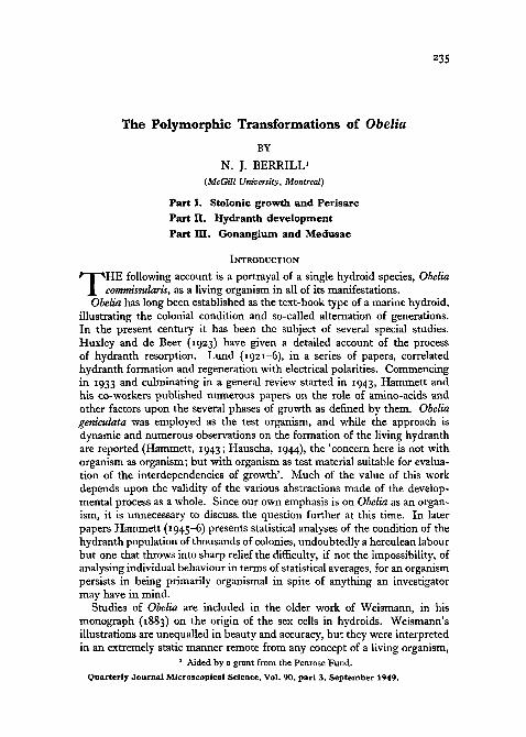
A detailed study of stolon tips reveals significant cell arrangements.Whether a stolon is massive or slender, the cells of both the epidermis and
B
TEXT-FIG, I . A, B. Sections through two stolon tips, respectively growing slowly and rapidly,showing confluence of cell contours of epidermis and endodermis (emphasized by heavybroken lines), and also showing graded vacuolation of distal endodermal cells. Note absenceof vacuolation in the terminal cells of the more rapidly growing stolon (B). C. Diagram ofgrowth sequence of isolated stolon, new stolonic tissue forming distally and old resorbingproximally, direction of growth indicated by arrow. In sequence along stolon are initial andlate stage of developing hydranth, two mature hydranths, and hydrothecae of two resorbedhydranths.
endodermis are streamlined as though flowing in and constituting a singlecurrent moving toward the tip. The intercellular contours of the endodermiscontinue with those of the epidermis. Whatever force is responsible for theconfiguration, the appearance is that the cells of both layers flow in conformitywith the lines of force and are subordinate rather than determinate (Text-fig, IA, B).
Cell proliferation has been found only at the extreme tip of both epidennisand endodermis. In neither layer is there any sign of so-called interstitialcells and in any case they must be so rare that no significant role can be
Berrill—The Polymorphic Transformations of Obelia 239
assigned to them. Since stolon growth is linear and in one direction, pro-liferation must in a sense be polarized, that is, the cells formed toward theproximal side of the proliferation zone cease division, those on the distal sidecontinue to divide.
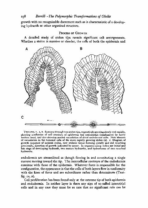
Immediately behind the proliferative tip the endodermal cells presenta very distinctive appearance. Toward the hydrocoel or lumen they retaintheir characteristic granular nature, but on the side toward the epidermis thecells become extended with a large clear vacuole. Passing backwards fromthe tip, vacuolization rises rapidly to a maximum and then slowly decreases,finally disappearing altogether (Text-fig. 2B), when the cells become indis-tinguishable from those of the endodermis in general. When growth is veryrapid the terminal proliferating endodermal cells may be granular throughoutand somewhat smaller than the vacuolate cells. When growth is slower, theterminal cells themselves may be vacuolate.
The cell cycles appear to be as follows. In the terminal division centre ofthe endodermis the cells may be relatively small if division is very rapid, butif not so rapid, and in any case at the margin of the centre, the cells becomegreatly enlarged and vacuolated, the vacuolated cells probably dividing atleast once. The vacuolation is probably a polar imbibition of water, isdefinitely a temporary phase, and is in some way related to the rejuvenescenceor reconstitution of cell organization. The phenomenon is seen elsewhere inthe activated epicardial tissue of the ascidian Archidistoma at the time ofstrobilation (Berrill, 1948).
Whatever the significance of the polar vacuoles in terms of the physiologyof dividing cells, they do afford a useful criterion of growth-rate. In slowlygrowing stolons relatively few new cells are produced in a given period, andcorrespondingly few cells are to be found in the vacuolated or recovery phase.In rapidly growing stolons, many more cells are formed within a given periodand correspondingly more cells will be found in the vacuolated recoveryphase. Each newly formed cell goes through its individual cycle in the sametime. The extent and gradient in degree of vacuolation accordingly form anindicator of the biological or relative growth-rates, in contrast to actualgrowth-rates (Text-fig. 2A, B).
The epidermis exhibits a somewhat similar cycle. Cells in the proliferativezone are large and high columnar, and in this zone and in the recovery phase(assuming it to be comparable and coextensive with that of the endodermis)an extensive but less obvious vacuolated region exists external to the nucleiof the cells.
The flowing aspect of stolon tissue is shown even more strikingly when apiece is cut. When a stolon or branch is cut through there is an immediateclosing over of the end of the endodermal layer to form a closed tube. Theepidermis reacts in a similar manner but a little more slowly, as it flows upand around the tip of the endodermis. Both distal and proximal ends of acut piece react in the same way. The response is so rapid that there is nopossibility of cell proliferation being in any way involved. The condition
240 Berrill—The Polymorphic Transformations of Obelia
1-2 minutes after a cut was made is shown in Text-fig. 2E. The pseudo-fluidnature of the material is further shown by its behaviour under light glasspressure. The coenosarcal tube as a whole remains stationary and unaffected,but the tip slowly extends without significant change in shape.
When a cut is made across a stem or stolon some distance from its distalend, new growth occurs rapidly from the cut proximal end (Text-fig. 2D),
TEXT-FIG. 2. Growth of stolons, A. End of rapidly growing free stolon, showing thickenedepidermis of the growing zone including a few glandular cells, and endodermis with a distalgroup of dense rapidly dividing cells grading into large vacuolated cells which decreaseproximally in size and vacuolation. B. End of slowly growing free stolon, showing reductionin range but not extent of vacuolated zone and absence of non-vacuolated cells distally. c.End of slowly growing attached stolon, showing latero-distal spreading of epidermis. D.Stolonic growth from basal end of a cut stem. E. Distal healing in cut annulated zone of astem, showing distal endodermal fusion and flowing of epidermis (cell division not involved).
that is, the normal polarity of growth may be reversed. When such out-growths become attached to the substratum, growth may be vigorous andmost of the old coenosarc and organized structures may resorb and becomeconverted into nutrient for the new tissues.
Stolon tips readily attach to solid surfaces. Those already attached aredifficult to detach without breaking them. Free stolons attach immediatelyupon contact, the region of attachment corresponding approximately withthe extent of the recovery phase described above. In other words, the stolonsurface is sticky for a limited though variable extent from the tip backwards.

Berrill—The Polymorphic Transformations of Obelia 241
When a free stolon attaches to glass, it assumes the terminal shape charac-teristic of creeping stolons in general. The contrast with the free stolon isshown in Text-fig. 2C. The endodermis remains unaffected, but the epidermisflows forwards and outwards as a pair of shoulders, that is, at the tip it hasa tendency to spread on contact, clearly a surface tension effect.
HYDROPLASMIC STREAMING
Movement of the fluid within the hydrocoel has been generally noted.Huxley and de Beer (1923) recorded changes in contour of the coenosarcwith the ebb and flow of the stream. Hammett(io.43) reports the movement asstreaming distally during hydranth development, proximally during hydranthresorption. Hauschka (1944) found from isolation experiments that the absenceof the stream did not prevent hydranths from completing their development,and that the causal connexion suggested by Hammett did not hold.
While the present account is of O. commtssularis, O. geniculata is essentiallythe same in this respect, and the obvious feature of the hydroplasmic stream-ing in O. comtnissularis is its regular reversal in direction. The rhythmicperiod varies with the temperature and may have from 3- to 7-minute cycle,consisting of a flow in one direction of from 1 to 3 minutes, a short periodof quiescence, and a flow in the opposite direction of similar duration to thefirst. Cells, cell fragments, heavily pigmented cells derived from resorbedor resorbing hydranths all flow along with the stream.
The rhythmical streaming occurs in a stolon or piece of stolon in theabsence of any formed or developing structures. It could be due either to aregular reversal in the direction of beat of the cilia lining the hydrocoel, orto contractions and expansions of one or both of the epidermis and endo-dermis. The activity of the cilia becomes most evident through the dancingmovement imposed upon the travelling particles.
To determine whether cilia are in any way responsible for the current,,let alone its reversal, various sections of stolons and stems were cut andstudied in isolation. Only those that retained or recovered ciliary activitywere employed. In no case could anything but local agitation be observedwith no forward or backward current. There was no evidence that ciliaryactivity is responsible for the hydroplasmic streaming, and this is confirmedby observations on intact stolons in which marked ciliary activity is oftenseen to coincide with the absence of directional movement.
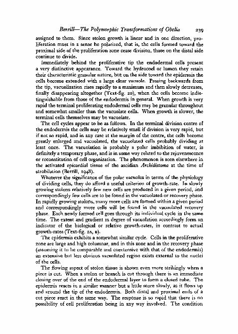
The alternative that coenosarcal contractions are responsible could meaneither a local activity or a general peristaltic wave. Greatly enlarged super-imposed series of camera lucida drawings showed no fluctuations in stolondiameter either in the recovery zone or down the main length of the stem,but in the region immediately proximal to the recovery zone a definiteexpansion and contraction was evident and coincided with the reversals ofthe stream (Text-fig. 3B, c).
The epidermis in this region when expanded comes into fairly close contactwith the chitinous cuticle, when contracted becomes separated from it by
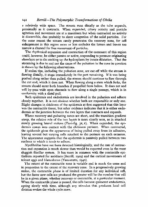
242 Berrill—The Polymorphic Transformations of Obelia
a relatively wide space. The stream runs distally as the tube expands,proximally as it contracts. When expanded, ciliary activity and particleagitation and movement are at a maximum but when contracted no activityis discernible, due probably to sheer congestion of the solid particles. Forthe same reason the stream rarely penetrates the recovery zone, for cellenlargement in this region more or less occludes the lumen and leaves toonarrow a channel for free movement of particles.
The rhythmical expansion and contraction of the coenosarc of this regioncould, however, be either passive or active, responding to pressure originatingelsewhere or to the sucking up the hydroplasm by innate dilatation. That thestreaming is due to and not the cause of the pulsation in the zone in questionis shown by the following observations.
If stolon tips, including the pulsation zone, are cut off while the stream isflowing distally, it stops immediately in the part remaining. If it was beingpushed along rather than pulled, the stream should continue to flow throughthe cut end, which it does not. When flowing along a stem which forks, thestream should enter both branches if propelled from below. It does not andwill by-pass wide open channels to flow along a single passage, which is inconformity with a distal pull.
Both epidermis and endodermis are involved in the pulsation and adhereclosely together. It is not obvious whether both are responsible or only one.Slight changes in thickness of the epidermis at first suggested that this layerwas the contractile tissue, but other evidence indicates that it is either endo-dermis or the junction between the two layers that contracts and expands.
Where recovery and pulsating zones are short, and the transition gradientsteep, the relative role of the two layers is more clearly seen, as in attachedslowly growing lateral stolons (Text-fig. 3E, F). When expanded, the epi-dermis comes into contact with the chitinous perisarc. When contracted,the epidermis gives the appearance of being pulled away from its adhesion,leaving several but varying cells attached to the perisarc on each occasion.The appearance suggests that the epidermis is passively pulled between twosurfaces to which it tends to adhere.
Myofibrillae have not been detected histologically, and the rate of contrac-tion and expansion is much slower than would be expected even in the mostsluggish fibrillar system. It has more in common with the contractile epi-thelium reported for ascidians (Berrill, 1929) and the cortical movements ofteleost eggs and blastoderms (Yamomoto, 1940).
The extent of the contractile zone is variable and is much the same andin proportion to the extent of the recovery zone. In a progressively growingstolon, the contractile phase is of limited duration for any individual cell,but the faster new cells are produced the greater will be the number that willbe in a given phase, whether recovery or contractile, at a particular moment.When the contractile phase is passed, the cells become quiescent endodermis,ageing slowly with time, although any stimulus that produces local celldivision evokes the whole cycle anew.

Berrill—The Polymorphic Transformations of Obelia 243
There is some indication that the vacuolation-contraction cycle is lessa response to a recent cell division than it is to cell growth as such, divisionof the cell being itself a response to the same condition. This is suggestedby the appearance of the large cells initiating the outgrowth of a lateral branch
TEXT-FIG. 3. Stolonic pulsation, A. Camera-lucida drawings of same stolonic tip 2 minutesapart, showing changes in diameter and contour and varying extent of internal ciliaryactivity. B. Rapidly growing stolon, with relatively large terminal flow of epidermis; brokenlines represent contour alternating with that of unbroken lines, showing slight terminalpulsation, and extensive pulsation zone proximal to thick vacuolated zone. C. Similar to B,showing two particles adhering to epidermis and exhibiting merely transverse movementwith the pulsation. D. Lateral outgrowth starting from junction of stolon and base of hydranthstem; both epidermal and endodermal cells greatly enlarged as preliminary and accompani-ment of proliferation. E, F. Contracted and expanded phases of a pulsation of a lateralattached stolon, showing tendency of epidermal cells to adhere to cuticle during contractionphase.
(Text-fig. 3D) and by the short period (2 hours) that elapses between thecutting of a stem and the presence of vacuolate and contractile regions.
BRANCHING
Branching occurs both from creeping stolons and from erect stems andfree stolons.
Creeping stolons. Vertical branches are the first to appear, from the upperside of a stolon a short distance behind the tip. The region from which they
244 Berrill—The Polymorphic Transformations of Obelia
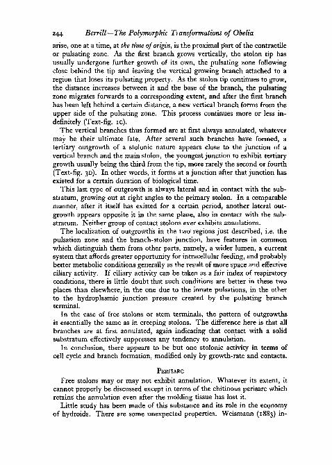
arise, one at a time, at the time of origin, is the proximal part of the contractileor pulsating zone. As the first branch grows vertically, the stolon tip hasusually undergone further growth of its own, the pulsating zone followingclose behind the tip and leaving the vertical growing branch attached to aregion that loses its pulsating property. As the stolon tip continues to grow,the distance increases between it and the base of the branch, the pulsatingzone migrates forwards to a corresponding extent, and after the first branchhas been left behind a certain distance, a new vertical branch forms from theupper side of the pulsating zone. This process continues more or less in-definitely (Text-fig, ic).
The vertical branches thus formed are at first always annulated, whatevermay be their ultimate fate. After several such branches have formed, atertiary outgrowth of a stolonic nature appears close to the junction of avertical branch and the main stolon, the youngest junction to exhibit tertiarygrowth usually being the third from the tip, more rarely the second or fourth(Text-fig. 3D). In other words, it forms at a junction after that junction hasexisted for a Certain duration of biological time.
This last type of outgrowth is always lateral and in contact with the sub-stratum, growing out at right angles to the primary stolon. In a comparablemanner, after it itself has existed for a certain period, another lateral out-growth appears opposite it in the same plane, also in contact with the sub-stratum. Neither group of contact stolons ever exhibits annulations.
The localization of outgrowths in the two regions just described, i.e. thepulsation zone and the branch-stolon junction, have features in commonwhich distinguish them from other parts, namely, a wider lumen, a currentsystem that affords greater opportunity for intracellular feeding, and probablybetter metabolic conditions generally as the result of more space and effectiveciliary activity. If ciliary activity can be taken as a fair index of respiratoryconditions, there is little doubt that such conditions are better in these twoplaces than elsewhere, in the one due to the innate pulsations, in the otherto the hydroplasmic junction pressure created by the pulsating branchterminal.
In the case of free stolons or stem terminals, the pattern of outgrowthsis essentially the same as in creeping stolons. The difference here is that allbranches are at first annulated, again indicating that contact with a solidsubstratum effectively suppresses any tendency to annulation.
In conclusion, there appears to be but one stolonic activity in terms ofcell cycle and branch formation, modified only by growth-rate and contacts.
PERISARC
Free stolons may or may not exhibit annulation. Whatever its extent, itcannot properly be discussed except in terms of the chitinous perisarc whichretains the annulation even after the molding tissue has lost it.
Little study has been made of this substance and its role in the economyof hydroids. There are some unexpected properties. Weismann (1883) in-

Berrill—The Polymorphic Transformations of Obelia 245
eluded an illustration, obviously accurately presented, that Nutting (1915)doubted on a priori grounds, showing an outgrowth rudiment secreting itsown fine perisarc and protruding through a hole in the thick chitin surround-ing an old stem. Louis Agassiz (1862) in fact illustrated almost exactly thesame thing for O. commissularis. Taking Weismann's illustration at facevalue, the chitinous perisarc clearly can both be laid down and dissolvedunder certain more or less obscure conditions.
Hammett calls attention to the elasticity of the perisarc at the distal endof nearly developed hydranths and speaks in terms, no doubt correctly, ofpolymerization of the secreted substance. The brittle rigidity of old perisarcin contrast to the sticky stretchiness of the newly formed material of stolonsand developing hydranths suggests forcibly that polymerization does takeplace.
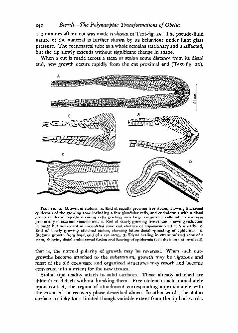
.The increasing thickness of the perisarc with the age of a stem, its laminarquality, and above all, the formation of straight lamellae on the inner sideof annulations, demonstrates that chitin production is continuous and notlimited merely to growing regions (Text-fig. 4B).
Perisarc is secreted at the growing end of a stolon, though hardly visiblein the recovery zone, yet sufficiently for a very thin though non-elastic filmto be present at the distal edge of the pulsation zone, gradually thickeningproximally. In the pulsation zone it is no longer adhesive (unless previouslyattached) and is probably fully polymerized. If polymerization is rapid, asit appears to be, the various shapes permanently assumed by the chitin inconformity with the shape of the secreting tissue is explained. Straightstolons produce straight cylinders, curved surfaces produce curved perisarc.
The evidence is that chitin is secreted continuously and not only bygrowing regions. It is therefore either a general property of epidermal cellsor else specialized cells must be present. If the former, then the propertyis apparently lost in the case of further specialization, as in tentacle epidermis.There is evidence, however, that specialized cells are solely responsible, andthat the greater part of the epidermis is basically unspecialized.
Distinctive ovoid cells are always to be found in the epidermis of a growingstolon tip, and in somewhat smaller numbers throughout the rest of theepidermis. Similar cells occur throughout the stem epidermis, lying parallelto the surface. They are not typical epidermal cells and undoubtedly havesome special function. They may well be the cells Weismann concluded wererudimentary ova, which would then need to migrate to other sites, and ledhim so far astray in his interpretation of hydroid reproduction. Being larger,rounder, and more granular than other cells of the epidermis, they might beinterpreted as rudimentary ova if the desire to do so was excessive.
Their most characteristic feature, however, is that the cytoplasm is packedwith highly refringent and insoluble granules, as clearly visible and refringentin the living cell as in preserved material (Text-fig. 4A). In the cells whenalive and when preserved in formalin, though not stained, the containedgranules have the same appearance and optical properties as the chitinous

246 Berrill—The Polymorphic Transformations of Obelia
cuticle, and it is difficult to avoid the conclusion that these are glandularcells responsible for the secretion of chitin. Chemical tests for chitin arecrude, however, and in any case there is no guarantee that the stainingproperties of secreted chitin and of its mother substance would be the same.While iron haematoxylin has no special affinity for chitin as such, it does stain
TEXT-FIG. 4. A. Section of free stolon, stained with iron haematoxylin, showing numerousglandular cells with deeply staining granules in epidermis, and the three endodermal zonesconsisting of distal non-vacuolated cells, vacuolated zone, and proximal non-vacuolated zone.B. Chitinous perisarc of old stem, showing external loosely laid lamellae laid down duringannular growth, and closely packed straight lamellae laid down subsequently.
the intracellular granules as deeply as chromatin, and darker than the in-clusions of any other cells (Text-fig. 4A).
On other grounds entirely, it is plausible that these cells secrete chitin.They must have a function, they appear to be secretory, are epidermal, andare adjacent to the only epidermal secretion recognizable. Their distributionin Obelia and other hydroids is such as to account well for the presence andnature of perisarcal structures. They also make possible a dual controlmechanism for perisarc that would be difficult to account for otherwise.Special cells lay down the chitin in successive layers, while ordinary epidermalcells when dividing or undergoing rapid growth are able to dissolve it.
Berrill—The Polymorphic Transformations of Obelia 247
ANNULATIONS
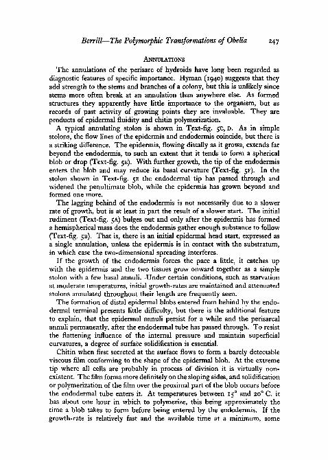
The annulations of the perisarc of hydroids have long been regarded asdiagnostic features of specific importance. Hyman (1940) suggests that theyadd strength to the stems and branches of a colony, but this is unlikely sincestems more often break at an annulation than anywhere else. As formedstructures they apparently have little importance to the organism, but asrecords of past activity of growing points they are invaluable. They areproducts of epidermal fluidity and chitin polymerization.
A typical annulating stolon is shown in Text-fig. 5c, D. As in simplestolons, the flow lines of the epidermis and endodermis coincide, but there isa striking difference. The epidermis, flowing distally as it grows, extends farbeyond the endodermis, to such an extent that it tends to form a sphericalblob or drop (Text-fig. 5E). With further growth, the tip of the endodermisenters the blob and may reduce its basal curvature (Text-fig. 5F). In thestolon shown in Text-fig. 5E the endodermal tip has passed through andwidened the penultimate blob, while the epidermis has grown beyond andformed one more.
The lagging behind of the endodermis is not necessarily due to a slowerrate of growth, but is at least in part the result of a slower start. The initialrudiment (Text-fig. 5A) bulges out and only after the epidermis has formeda hemispherical mass does the endodermis gather enough substance to follow(Text-fig. 5B). That is, there is an initial epidermal head start, expressed asa single annulation, unless the epidermis is in contact with the substratum,in which case the two-dimensional spreading interferes.
If the growth of the endodermis forces the pace a little, it catches upwith the epidermis and the two tissues grow onward together as a simplestolon with a few basal annuli. Under certain conditions, such as starvationat moderate temperatures, initial growth-rates are maintained and attenuatedstolons annulated throughout their length are frequently seen.
The formation of distal epidermal blobs entered from behind by the endo-dermal terminal presents little difficulty, but there is the additional featureto explain, that the epidermal annuli persist for a while and the perisarcalannuli permanently, after the endodermal tube has passed through. To resistthe flattening influence of the internal pressure and maintain superficialcurvatures, a degree of surface solidification is essential.
Chitin when first secreted at the surface flows to form a barely detectableviscous film conforming to the shape of the epidermal blob. At the extremetip where all cells are probably in process of division it is virtually non-existent. The film forms more definitely on the sloping sides, and solidificationor polymerization of the film over the proximal part of the blob occurs beforethe endodermal tube enters it. At temperatures between 150 and 20° C. ithas about one hour in which to polymerize, this being approximately thetime a blob takes to form before being entered by the endodermis. If the.growth-rate is relatively fast and the available time at a minimum, some
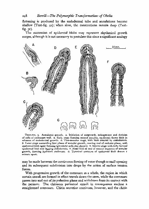
248 Berrill—The Polymorphic Transformations of Obelia
flattening is produced by the endodermal tube and annulations becomeshallow (Text-fig. 5E); when slow, the constrictions remain deep (Text-fig- 5F)-
The succession of epidermal blobs may represent rhythmical growthsurges, although it is not necessary to postulate this since a significant analogy
0-1 mm.
TEXT-FIG, 5. Annulated growth, A. Initiation of outgrowth, enlargement and divisionof cells of coenosarc wall. B. Early stage forming second annulus, epidermis forms blob inadvance of endodermal growth, c. Two-annulus stage, with blob entered by endodermis.D. Later stage succeeding first phase of annular growth, nearing end of stolonic phase, withepidermal blob again forming (glandular cells also shown). E. Similar stage with fully formedepidermal blob and lagging endodermis. F. Final blob at end of second sequence of annulargrowth, forming hydranth rudiment. G. Terminal contours of epidermal blob drawn Iminute apart.
may be made between the continuous flowing of water though a small openingand its subsequent subdivision into drops by the action of surface tensionforces.
With progressive growth of the coenosarc as a whole, the region in whichcertain annuli are formed in effect travels down the stem, while the coenosarcpasses into and out of its pulsation phase and withdraws from its contact withthe perisarc. The chitinous perisarcal annuli in consequence enclose astraightened coenosarc. Chitin secretion continues, however, and the chitin

Berrill—The Polymorphic Transformations of Obelia 249
is subsequently deposited as straight lamellae on the inner side of an annulatedouter layer (Text-fig. 4B).
The mobile fluid-like nature of the epidermal blob is illustrated in Text-fig. 5 c In contrast to the fixed contour of the base of the blob, the distalpart is continually changing every few seconds from a multiple to a singlecurved surface and back again, the single curvature predominating.
An analogy of some significance in the light of the following sections onmorphogenesis, is that of a glass blowers' terminal blob of molten glass(epidermis), with a column of air (endodermis) being blown into it througha tube.
PART II. HYDRANTH DEVELOPMENT
There is a sequence of events leading to the initiation of hydranth develop-ment which in O. commissularis has so far been found to be invariable.
The hydranth always arises from the end of a short stolon that has under-gone the following sequential changes, namely, several initial annulations,a phase of simple (non-annulated) stolonic growth, followed by a second anddistal series of annulations.
The first series of annulations is due mainly to the head start of theepidermis. This overhead, however, is progressively lost as the endoderm ineffect catches up, and as growth-rates and terminals coincide, straight stolonicgrowth becomes established. After a variable period of such growth, thegrowth-rate of the endodermis begins to fall off again relative to that of theepidermis, and terminal epidermal blobs (and annuli) again appear.
There is a difference in the annulated growth of the first and second series.In the first it is a diminuendo, the epidermal blob becoming progressivelyreduced in size at the time the endoderm penetrates it, until none is formed.In the second it is a crescendo, the endodermis lagging more and more sothat the terminal epidermal blob is larger each time it is formed. With thethird, fourth, or fifth epidermal surge, a crisis is reached.
The sequence just described, culminating in the cessation of the secondseries of annulations, is characteristic of a relatively low temperature range,below i8°-20° C. Above this level, in most circumstances, the phase ofsimple stolonic growth succeeding the first series of annulations continuesindefinitely. The appearance is that the endodermis responds to highertemperatures a little more vigorously than the epidermis, becomes the pace-maker and enforces simple growth.
At relatively low temperatures, the absolute reduction in growth-rate ofboth tissues, and the relative lagging of the endodermis result finally in theformation of an epidermal blob larger than its predecessors, and entered bythe endodermis even more slowly than before. Both features are important,the greater quantity of material in the unit, and the longer time in whichsomething can happen.
The terminal unit shown in Text-fig. 5F is typical of the new starting-point, with maximum epidermal blob enveloping the tip of the endodermis.

250 Berrill—The Polymorphic Transformations of Obelia
This is the rudiment of the hydranth. The unit now grows as a whole andquantitatively exhibits a typical growth-curve or decrement until all growthceases.
The slowing of the growth-rate of the endodermis is indicated by thedisappearance of the terminal zone of relatively small rapidly dividing cells,
TEXT-FIG. 6. Hydranth development (chitinous cuticle in heavy black), A-E. Note changingrange of vacuolated zone. A. initial semi-hemispherical' rudiment with terminal epidermalblob fully entered by endodermis. B, C. Enlargement and elongation as cells become addedfrom the anterior disk to the proximal cylinder. D, E. Progressive growth, limitation ofvacuolated cells to anterior disk, proximal formation of pulsation zone causing detachmentof coenosarc from the perisarc. F. Slightly later stage at same scale showing completeseparation of coenosarc and perisarc, and conversion of marginal rim of disk into rudimentsof primary and secondary tentacles.
and the substitution of dividing cells of maximum size and vacuolation. Theeffect is to give expansion a lateral as well as a distal direction, resulting in theslow formation of a flask-shaped structure (Text-fig. 6A, B, C).
At the same time, apart from change in diameter, which increases at thedistal end, and in the absence of a centre of maximum growth-rate, thegrowth sequence of cells is basically similar to that in stolon growth. Celldivision continues in the area extending from the distal centre to the shoulderslope, vacuolation of the endodermis being at a maximum in this region, theepidermis being comparable in its own way. Behind the shoulder, tapering

Berrill—The Polymorphic Transformations of Obelia 251
toward the neck, vacuolation decreases and disappears near the junction withthe annulated stalk (Text-fig. 6c).
As growth proceeds, the distal disk of cells becomes wider. More cellspass from the edge of the disk into the distal part of the slope. Vacuolationdisappears in cells farther and farther from the junction with the stem.Epidermal cells become progressively less columnar in company with thoseof the endodermis.
With the proximal disappearance of vacuolation, a zone appears thatcorresponds in relative location to the pulsation zone of the stolon, and withits appearance the pulsations commence. At first the extent and the amplitudeare such as to be barely detectable. With progressive distal addition to thearea, both the extent and the amplitude increase, the rhythmical movementsbecome obvious, and the epidermis is pulled away from the chitinous perisarc(Text-fig. 6D, E).
The pulsations in the hydranth have been reported already for later stagesin O. geniculata by Hammett (1943), who regards them as the result of hydro-plasmic ebb and flow originating elsewhere in the colony. To test this inO. commissularis, various stages of developing hydranths were isolated bycutting through the stem in the distal annulated zone. The reversing hydro-plasmic stream in the intact stems ceased immediately, while the rhythmicalcontractions and expansions in the hydranth buds continued indefinitely.Accordingly there is no reason to doubt the existence of active pulsation inthe hydranth itself, while the manner of formation of the zone and theidentity of rhythm rates leave little doubt that the phenomenon in hydranthand stolon tip is the same.
The pulsation zone is gradually extended distally until the bud consistsof a long pulsating cylinder of flat and cuboidal cells, surmounted by a capof columnar epidermal and endodermal cells that forms a right angle junctionwith the cylinder. The cylinder becomes the stomach of the hydranth, andthe pulsations persist almost until the hydranth as a whole becomes a func-tional entity. The extent of growth of the whole from the bud rudiment tothe present stage is shown to scale in Text-fig. 6A-F. Text-fig. 7J shows fiveconsecutive positions of the cylinder wall in process of contraction, Text-fig. 7Kfive positions occupied one minute apart.
As the round bottom of the inverted flask (Text-fig. 8A) becomes convertedinto a flat bottom, a new crisis is reached. No further cells are added to theslopes to augment the pulsation zone, while other events take place at themarginal junction. The profile of this stage is shown in Text-fig. 8B. Thecylinder is surmounted by an extremely straight-sided ridge or flange, whichin turn surrounds a slightly convex disk. Both ridge and disk, in contrastto the pulsation zone, consist of high columnar cells in progress of slowdivision. The inner surface of the endodermis of this region bears very activecilia.
The interest in this stage concerns the marginal rim. Seen in lateral profile,nothing appears remarkable, but examined from the more difficult angle of

252 Berrill—The Polymorphic Transformations of Obelia
above or below, the circumference is seen to be no longer circular but many-sided. The number of sides varies from 8 to 12, according to the size of thehydranth bud at this stage. Imperceptibly, the circular outline of a slightlyearlier stage flattens equidistantly to form 8, 9, 10, 11, or 12 sides (Text-fig.7D-E), the length of a side being approximately the same in all, and the
TEXT-FIG.. 7. Hydranth development, A, B. Alternating contours of distal end shortlybefore tentacle initiation, c. Similar stage with endodermis shrunken as consequence ofnarcotization, showing marginal growth of epidermis and endodermis independently. D, E, F.Distal end-views of stage I and Text-fig. 6E, showing polygonal form, and variation in numberof sides with length of circumference. G, H, I. Three stages of early development drawn tosame scale as in Text-figs. 8 and o. j . Pre-tentacle stage showing five consecutive contoursof cylinder wall during contraction. K. Same stage, showing position of cylinder wall duringexpansion and contraction i J minutes apart.
number of sides being directly proportional to the length of the circumfer-ence. This foreshadows tentacle formation, and the number of tentacles thatwill be formed is twice the number of sides, whatever that may be.
At 2o° C. it takes about 4 hours to pass from the stage where polygonalstructure is suspected to the definite configuration, a relatively long periodin view of the rapidity of subsequent changes.
As the cylinder alternately expands and contracts, the contour of the distalregion alternates between those shown in Text-fig, JA and B. At one time themarginal thickening appears to be epidermal, at the other it seems to beendodermal. It is difficult to determine which of the two layers is really

Berrill—The Polymorphic Transformations of Obelia 253
responsible for the variable thickness of the whole, even when vitally stainedwith brilliant cresyl blue or with neutral red. However, a prepared slide wasdiscovered representing stained specimens that had clearly been narcotizedpreparatory to fixation, and while formed structures and the epidermisthroughout appeared to be in excellent condition, the endodermis of rapidlygrowing regions was shrunken or withdrawn. In one instance, at the stagein question, a space had appeared between the two layers (Text-fig. 7 c), andit is evident that in both epidermis and endodermis growth has resulted inthe differentiation of a thick marginal rim and a central dome. Both layersare involved.
After the distal polygon has been definitely established, a relatively, evenremarkably, rapid transformation occurs. Each epidermal corner becomesrounded, again like a bead induced by surface-tension. Only a few minuteslater the unorganized territory remaining between adjacent corners also drawsup into small hemispherical masses (Text-fig. 6F, 8C).
The row of beads thus formed at the margin of the disk represents the fullnumber of tentacles that are to be formed, those arising from the cornersgiving rise to the more or less horizontal tentacles of the expanded hydranth,the intermediate ones becoming more or less erect.
The tentacles of Obelia are small and as they are initiated on a correspond-ingly small scale, they are less suitable than those of many other hydroidsfor an analysis of tentacle histogenesis. At the same time, a tentative analysisis as follows. Local proliferation of marginal endoderm results in the dela-mination of masses of very small cells, a similar process occurring in theadjacent epidermis. The small endodermal cells enlarge individually, andconstitute the endodermal chordal cells of the tentacles without furtherincrease in number. As the epidermal covering becomes thrust out, thesmall cells of epidermal origin are also drawn out, giving rise to the nem-atocysts.
The final phase of hydranth development occupies but a few hours, andconsists mainly of differentiation processes in the three zones already estab-lished (Text-fig. 8D). The central dome grows into a thin-walled manubrium,opening distally to form the mouth. The marginal beads rapidly grow out asthe tentacles, while the stomach.forming from the wall of the cylinder acquirestypically pigmented gastrodermal cells, with inclusions that stain intenselywith neutral red.
Simultaneously, movements commence indicative of the functional dif-ferentiation of muscle-fibres. Twitch contractions appear in the tentacles,occupying merely a fraction of a second. In the stomach, movements occurwhich change the shape rather frequently, each transformation occupyingbut a few seconds (Text-fig. 8E, F). Similar movements develop in the manu-brium though somewhat later. While these enteric contractions are slowcompared with a tentacle twitch, they are very fast compared with therhythmical pulsations described earlier, and there is no possibility of con-fusion between them.

254 Berrill—The Polymorphic Transformations of Obelia
Hammett (1943) has recorded for O. geniculata the necessity of rhythmicsurging of the whole hydranth for the final stretching and rupture of the thinsticky perisarc covering its distal end. In O. commissularis it is not certainthat a coat of quite such consistency is present. In many cases the hydrothecaretains its polygonal character, though it is slight and often disappears.
TEXT-FIG. 8. Later development of Hydranth. A, B. Late pre-tentacle stages, showingorigin of new terminal growth from wall of distal part of non-annulated or internodal stem.C. Distal view of initiation of primary and secondary tentacles. D. Lateral view of stageimmediately following that of Text-fig. 6F. E, F. Later stages with dilated and contractedbody-wall, showing elongating tentacles and formation of manubrium.
With the opening of the mouth, the large pigmented gastric cells derivedfrom the resorption of hydranths elsewhere in the colony and accumulatedin the cul-de-sac of the developing hydranth, escape to the exterior. In younghydranths they may be seen as a ball of yellow-orange material, opaque bytransmitted light, held between the tentacles.
It was mentioned earlier that hydranths usually do not start to form unlessthe temperature is below 200 C. If after development has started the tempera-ture rises even several degrees above this threshold, development continuesto completion. At temperatures in the region of 250 C , however, the hydranthreaches the functional state and then usually resorbs immediately, indicatinga greater susceptibility of fully differentiated specialized cells to high tempera-tures than either cells in the developing system or in the unorganized
Berrill—The Polymorphic Transformations of Obelia 255
coenosarc. The process of hydranth resorption has been adequately describedby Huxley and De Beer (1923).
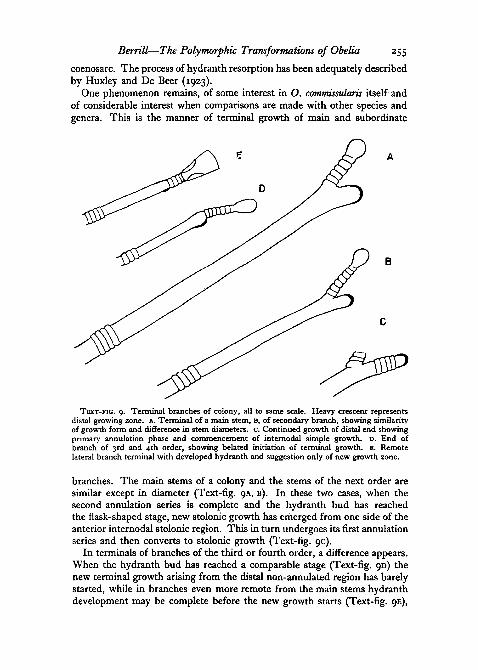
One phenomenon remains, of some interest in O. commissularis itself andof considerable interest when comparisons are made with other species andgenera. This is the manner of terminal growth of main and subordinate
TEXT-FIG. 9. Terminal branches of colony, all to same scale. Heavy crescent representsdistal growing zone. A. Terminal of a main stem, B, of secondary branch, showing similarityof growth form and difference in stem diameters, c. Continued growth of distal end showingprimary annulation phase and commencement of internodal simple growth. D. End ofbranch of 3rd and 4th order, showing belated initiation of terminal growth, E. Remotelateral branch terminal with developed hydranth and suggestion only of new growth zone.
branches. The main stems of a colony and the stems of the next order aresimilar except in diameter (Text-fig, QA, B). In these two cases, when thesecond annulation series is complete and the hydranth bud has reachedthe flask-shaped stage, new stolonic growth has emerged from one side of theanterior internodal stolonic region. This in turn undergoes its first annulationseries and then converts to stolonic growth (Text-fig. 9c).
In terminals of branches of the third or fourth order, a difference appears.When the hydranth bud has reached a comparable stage (Text-fig. 9D) thenew terminal growth arising from the distal non-annulated region has barelystarted, while in branches even more remote from the main stems hydranthdevelopment may be complete before the new growth starts (Text-fig. 9E),

256 Berrill—The Polymorphic Transformations of Obelia
and in fact may arrive at a condition where there is a functional terminalhydranth and no further growth at all.
PART III. GONANGIUM AND MEDUSAE
The production of gonangia raises the following questions. Where andwhen do they arise, and why? What are the formative processes initiatingtheir appearance ? How does the early developmental phase differ from thatof a hydranth, what are the developmental consequences, and what otherfactors intervene to bring about further differences ?
PLACE OF ORIGIN
In this species the gonangia arise only in the basal and central part of acolony, that is, from the older parts. They arise generally from establishedjunctions, regions where a stem junction already exists. From such placesgonangia are second outgrowths, although second outgrowths need not begonangia. In the same general region of a colony, though always toward theproximal end of a branch, gonangia may arise from the unbranched internodalpedicel of a hydranth.
Since junction zones tend to become larger with age, the diameter of sucha zone might relate to the size of outgrowths emerging from it, and this in^turn be a determining factor. On the other hand, those arising from simplehydranth pedicels would remain unaccounted for, and in any case the initialgonangial protrusions do not appear to differ from those leading to hydranthdevelopment. It is therefore more likely that the primary determinant ismetabolic rather than physical or dimensional. If the lower growth-rate oflateral branch terminals is taken as a criterion of metabolic conditions, itindicates a lower growth tendency in the basal parts compared with the apicalparts of a colony. The rates of stolonic outgrowth from pieces of stem isolatedfrom various regions confirm this.
GROWTH SEQUENCE
While the initial protrusions appear to be similar for hydranth andgonangium, the succeeding growth sequences are very different. The hy-dranth sequence consisted of a series of annulations of diminishing diameter,changing to simple stolonic growth culminating in annulations increasing toform the hydranth rudiment.
In the gonangial sequence, annulating growth commences as before, butthe endodermis not only fails to overtake the epidermal blob, but lags so thatthe terminal cap enlarges with each annulation. Three or four annulationsoccur in this manner, corresponding in kind to the second series of thehydranth stalk, but forming here as the primary series (Text-fig, IOA-E).Sometimes the number of annulations may be much greater, but it is stilla crescendo and the final state is the same.

Berrill—The Polymorphic Transformations of Obelia
GONANGIAL RUDIMENT
257
As the final surge occurs, which produces the gonangial rudiment, anotherdifference appears. The final unit is flatter and wider than the hydranthrudiment, and from the first consists of a considerably larger number of cells(cf. Text-fig. 6A and IOD). This difference was noted by Weismann (1883)
TEXT-FIG. 10. Development of Gonangium. A. Initial stage, B, C, D. Primary annulation.growth phase with expanding diameter culminating in gonangial rudiment, E, F. Growthof rudiment, especially of disk. G, H. Initiation of medusa buds from wall of anterior cylinder,and consequent shrinkage of distal endodermis (stippled in G).
and by Hammett (1943), who compares the two primary caps with a cylinder(hydranth) and tam-o'-shanter (gonangium). However, this is a differencethat develops rather than one which at first exists.
If we compare the formative blobs at the time of endodermal entrance, thedifference is less striking. It is none the less significant. The distal flowof the epidermis is reduced and there is greater lateral spreading, so thatmore cells lie in the transverse plane, and the greater total number ofcells present in the initial gonangial rudiment both lead to the develop-ment of a larger final structure, final size being a reflection of initial scaleof organization.
258 Berrill—The Polymorphic Transformations of Obelia
DEVELOPMENT OF THE GONANGIUM
The unit which continues to enlarge as the developing gonangium shortlyconsists of a convex cap and two semi-annulations which remain too shallowto prohibit the contained coenosarc from participating in the formative zone(Text-fig, IOF).
Both hydranth and gonangium rudiments follow a growth-rate curve toa final state of equilibrium or cessation of growth. On the reasonable assump-tion that the growth-curves are typical, then stage for stage the gonangiumis larger than the hydranth, or at similar sizes is developmentally younger.This is borne out by a comparison of the final length and mass of hydranthand gonangium. It is also significant that at the same temperature the relativelylarge gonangium, as well as can be determined considering the vagueness ofthe developmental end-point, develops in approximately the same time asthe hydranth.
In essence, therefore, the gonangium in its early development may beregarded as a developing large hydranth, commencing to form a wide distaldisk, and proximally a wide tapering enteron. If this interpretation is valid,why does not a gonangium exhibit more of hydranth structure, and whydoes not a hydranth produce medusae ?
The answer to the second question is that during the critical developmentphase, the hydranth is not large enough for the initiation of medusa disks,and a second opportunity never arises since no growth occurs in the functionalhydranth.
On the other hand, a developing gonangium forms medusa rudimentsprimarily because there is sufficient territory in its wall. This matter is dis-cussed further in connexion with medusa formation and development. Inthe present connexion, what is important is the effect of medusa initiationupon the continuing development of the gonangium as a potential hydranth.Medusa initiation is precocious, that is, it occurs during the developmentalperiod of the parent individual, and not later as in some other hydroids. Theconsequences are to be seen in the later development of the gonangium.
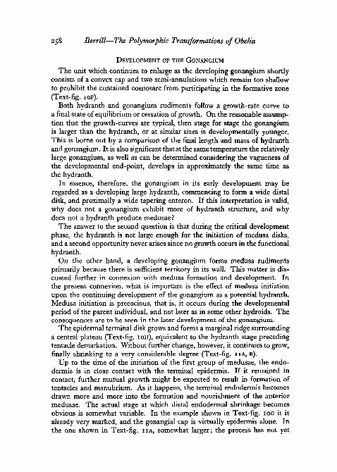
The epidermal terminal disk grows and forms a marginal ridge surroundinga central plateau (Text-fig. IOH), equivalent to the hydranth stage precedingtentacle demarkation. Without further change, however, it continues to grow,finally shrinking to a very considerable degree (Text-fig, IIA, B).
Up to the time of the initiation of the first group of medusae, the endo-dermis is in close contact with the terminal epidermis. If it remained incontact, further mutual growth might be expected to result in formation oftentacles and manubrium. As it happens, the terminal endodermis becomesdrawn more and more into the formation and nourishment of the anteriormedusae. The actual stage at which distal endodermal shrinkage becomesobvious is somewhat variable. In the example shown in Text-fig, IOG it isalready very marked, and the gonangial cap is virtually epidermis alone. Inthe one shown in Text-fig, IIA, somewhat larger; the process has not yet
Berrill—The Polymorphic Transformations of Obelia 259
commenced. Sooner or later, unable to serve two masters, the endodermisprogressively abandons its contact with the epidermal plate, and the latter isclearly unable alone to develop tentacular or manubrial structure. Frequentlythe epidermis shrinks as rapidly as the endodermis, and the two layersremain in contact.
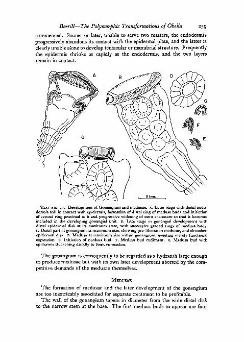
TEXT-FIG, I I . Development of Gonangium and medusae, A. Later stage with distal endo-dermis still in contact with epidermis, formation of distal ring of medusa buds and initiationof second ring proximal to it and progressive widening of stem coenosarc so that it becomesincluded in the developing gonangial unit. B. Late stage in gonangial development withdistal epidermal disk at its maximum state, with successive graded rings of medusa buds,c. Distal part of gonangium at maximum size, showing pre-liberation medusae, and shrunkenepidermal disk. D. Medusa at maximum size within gonangium, awaiting merely functionalexpansion. E. Initiation of medusa bud. F. Medusa bud rudiment. G. Medusa bud withepidermis thickening distally to form entocodon.
The gonangium is consequently to be regarded as a hydranth large enoughto produce medusae but with its own later development aborted by the com-petitive demands of the medusae themselves.
MEDUSAE
The formation of medusae and the later development of the gonangiumare too inextricably associated for separate treatment to be profitable.
The wall of the gonangium tapers in diameter from the wide distal diskto the narrow stem at the base. The first medusa buds to appear are four
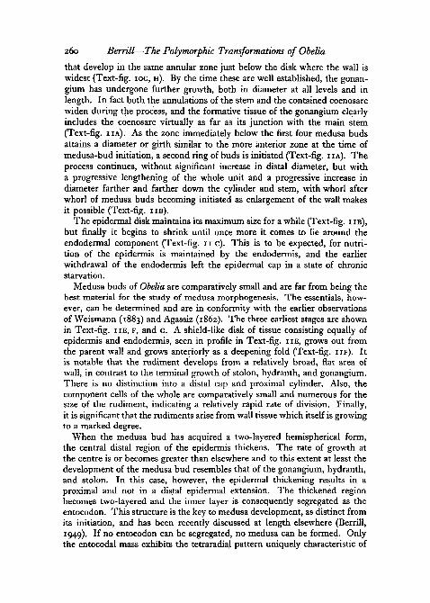
260 Berrill—The Polymorphic Transformations of Obelia
that develop in the same annular zone just below the disk where the wall iswidest (Text-fig, IOG, H). By the time these are well established, the gonan-gium has undergone further growth, both in diameter at all levels and inlength. In fact both the annulations of the stem and the contained coenosarcwiden during the process, and the formative tissue of the gonangium clearlyincludes the coenosarc virtually as far as its junction with the main stem(Text-fig. 11A). AS the zone immediately below the first four medusa budsattains a diameter or girth similar to the more anterior zone at the time ofmedusa-bud initiation, a second ring of buds is initiated (Text-fig, I IA) . Theprocess continues, without significant increase in distal diameter, but witha progressive lengthening of the whole unit and a progressive increase indiameter farther and farther down the cylinder and stem, with whorl afterwhorl of medusa buds becoming initiated as enlargement of the wall makesit possible (Text-fig, I IB) .
The epidermal disk maintains its maximum size for a while (Text-fig, i IB),but finally it begins to shrink until once more it comes to lie around theendodermal component (Text-fig, n c). This is to be expected, for nutri-tion of the epidermis is maintained by the endodermis, and the earlierwithdrawal of the endodermis left the epidermal cap in a state of chronicstarvation.
Medusa buds of Obelia are comparatively small and are far from being thebest material for the study of medusa morphogenesis. The essentials, how-ever, can be determined and are in conformity with the earlier observationsof Weismann (1883) and Agassiz (1862). The three earliest stages are shownin Text-fig, H E , F, and G. A shield-like disk of tissue consisting equally ofepidermis and endodermis, seen in profile in Text-fig, H E , grows out fromthe parent wall and grows anteriorly as a deepening fold (Text-fig, I I F ) . Itis notable that the rudiment develops from a relatively broad, fiat area ofwall, in contrast to the terminal growth of stolon, hydranth, and gonangium.There is no distinction into a distal cap and proximal cylinder. Also, thecomponent cells of the whole are comparatively small and numerous for thesize of the rudiment, indicating a relatively rapid rate of division. Finally,it is significant that the rudiments arise from wall tissue which itself is growingto a marked degree.
When the medusa bud has acquired a two-layered hemispherical form,the central distal region of the epidermis thickens. The rate of growth atthe centre is or becomes greater than elsewhere and to this extent at least thedevelopment of the medusa bud resembles that of the gonangium, hydranth,and stolon. In this case, however, the epidermal thickening results in aproximal and not in a distal epidermal extension. The thickened regionbecomes two-layered and the inner layer is consequently segregated as theentocodon. This structure is the key to medusa development, as distinct fromits initiation, and has been recently discussed at length elsewhere (Berrill,1949). If no entocodon can be segregated, no medusa can be formed. Onlythe entocodal mass exhibits the tetraradial pattern uniquely characteristic of
Berrill—The Polymorphic Transformations of Obelia 261
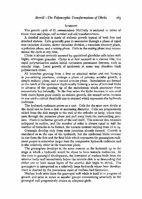
medusa organization. The details of its later development are not followedhere, except to indicate the final size of the medusa attained before thefunctional expansion which occurs at the time of liberation. This is shownin Text-fig. 1 ID at the same scale as the rudiment. All medusae of this speciesare liberated with sixteen tentacles.
Within the limits of observational accuracy, it is notable that at a giventemperature the time taken for a medusa to develop from initiation to libera-tion is approximately the same as the developmental periods of the hydranth,and of the gonangium up to its stage of maximal development as such (at18-200 C. it is about 24 hours). It is also significant that in each of the threecases the final diameter at the end of its developmental period is approximatelyfour times the diameter of the earliest definitive rudiment. If these two state-ments are accurate, and they appear to be reasonably so, we get the implica-tions that the initial scale of organization determines the final size at the endof development, that the material in each follows a curve of growth repre-senting a definite multiplicative value of its mass, and that each unit of massgrows to the same extent in the same time, and therefore that growth-rateand developmental time are independent of the initial total mass.
During the development of the medusae while attached to the blastostyleof the gonangium, regular contractions and expansions may be seen similarto the pulsation phases of stolon and hydranth. The time phases are similar,and there is the same alternating occlusion and dilatation of the hydrocoel(Text-fig. 12E, F). While this is probably due to a basic biological propertyof cells in a certain phase connected with growth, there is no doubt that inthis case above all a useful function is performed, serving to draw nourish-ment from the hydroplasm streaming through the blastostyle, and effectinga regular metabolic exchange.
The hydroplasm streams in and out of the blastostyle hydrocoel in thesame manner as it does in the developing hydranth. The contractions andexpansions appear to be effected by the endodermis near the distal end. Theresult is that the developing medusae are maintained effectively and thestream is active usually until the last medusa has been liberated, although theinnate pulsations of the developing medusae themselves probably are anadequate means of creating a communal feeding current.
When colonies bearing large numbers of large gonangia are kept understarvation conditions, so that any continued development must necessarilybe at the expense of other tissues, the same phenomenon of differentialresorption appears as that described by Huxley and de Beer for otherhydroid species (1923) and for the ascidian Perophora (Huxley, 1921). Fullyformed hydranths regress, developing hydranths complete development andthen regress, while the medusa buds go on developing and finally all becomeliberated. Developing tissues are clearly in a very different physiologicalstate from the same tissues fully developed. In this case the result isclearly both an inevitable one of colonial economics, and one of obviousvalue for the survival of the species. In fact, the medusa buds in completing

262 Berrill—The Polymorphic Transformations of Obelia
their development may bring about the resorption of most of the stemcoenosarc.
Liberation of medusae is effected not so much by the breaking of themedusa stalks down the length of the blastostyle, but to the continued growthin length of the blastostyle itself in a distal direction. As it continues to
TEXT-FIG. 12. A—D. Liberation of medusae, A. Fully developed gonangium with cap atminimal state, with all medusae developed or initiated. B, c. Same gonangium 12 and 36hours later respectively showing progressive elongation of stem or blastostyle distally, andsuccessive liberation of medusae, D. Twelve hours later, with one medusa in process ofdetachment, and last medusa still within gonangium, with two abortive medusa rudimentson elongating residual blastostyle. E, F. Three stages of developing medusa buds showingcontracted and expanded conditions; arrows show direction of hydroplasmic stream ofblastostyle.
grow, the distal medusae are more or less pushed through the opening leftat the gonangial tip by the shrinkage of the epidermal cap. As these breakfree, further extension carries the next group out, and so on until the lastmedusa is borne up to the threshold. The progression is shown in Text-fig. I2A-D.
The liberated medusa immediately expands. There is no sign of gonadsat this stage. It is equipped with 16 tentacles and 8 lithocysts. At 20° C.there are from 6 to 10 swimming contractions in about 3 seconds, followedby a rest pause of similar duration. The medusa rotates clockwise, completinga full circle with every 7 or 8 contractions.
Berrill—The Polymorphic Transformations of Obelia 263
SUMMARY
The growth cycle of O. comtnissularis McCrady is analysed in terms oftissue mass and shape, cell number and cell transformations.
A detailed analysis is made of stolonic growth typical of both free andattached stolons. Cells generally pass in succession through a phase of rapidnon-vacuolate division, slower vacuolate division, a vacuolate recovery phase,a pulsation phase, and a resting phase. Cells in the resting phase may recom-mence the cycle at any time.
Chitin is almost certainly secreted by specialized glandular cells laden withhighly refringent granules. Chitin is at first secreted as a viscous film, butrapid polymerization makes initial curvatures permanent features, such asannular rings. Local growth of epidermis in some way dissolves thickpolymerized chitin.
All branches growing from a free or attached stolon and not formingat pre-existing junctions, undergo a phase of primary annular growth, asimple stolonic phase, and a second annular phase. Annulations are formedas the result of the epidermis rhythmically forming a series of terminal blobsin advance of the growing tip of the endodermis which penetrates themsuccessively but belatedly. In the first series the blobs decrease in size untilboth tissue layers grow evenly as stolonic growth, the second series increasein size until a critical threshold size is attained which represents the hydranthrudiment.
The hydranth rudiment grows as a unit. Cells for the most part divide atthe distal end to form a disk of increasing diameter. Cells are progressivelyadded from the disk margin to the wall of the cylinder or body, where theypass through the pulsation phase and pull away from the surrounding peri-sarc. There is no further growth of the wall itself. The anterior disk becomespolygonal in outline, and the number of sides is always equal to half thenumber of tentacles to be formed, the tentacle number varying from 16 to 24.
Gonangia develop only from stem junctions already formed. Growth isannulated as in the case of the hydranth, but the epidermal blobs increasein size from the first and the final blob which represents the gonangium rudi-ment proper is somewhat larger than the comparable hydranth rudiment andis also relatively wider in the transverse plane.
The gonangium develops in the same manner as the hydranth up to thestage at which a hydranth would be about to form tentacle rudiments. Atthis time in gonangial development, the formation of medusa buds from theanterior body-wall immediately below the tentacle disk is so demanding thateither one or both tissue layers of the anterior disk begin to shrink. Thegonangium is interpreted as a relatively large hydranth whose later develop-ment is aborted by the precocious onset of medusa-bud formation.
Medusa buds arise from the gonangial wall while it itself is in progress ofgrowth and arise in series as annular groups commencing anteriorly as thegonangial wall progressively attains an adequate girth.

264 Berrill—The Polymorphic Transformations of Obelia
The medusa bud arises directly from the wall and not from the terminalof a growing stolon as in hydranth and gonangium. The cells are small,indicating relative rapidity of division, and an entocodon, essential formedusoid organization, is formed by apical thickening of the medusa-budepidermis, followed by segregation.
As medusae develop, the blastostyle to which they are attached grows andelongates towards the anterior end, thereby carrying medusae in effectthrough the distal aperture of the ripe gonangium, in order of their seniority.Upon liberation the medusae all have sixteen tentacles and no sign of gonads.
REFERENCES
AGASSIZ, L., 1862. Contributions to the Natural History of the United States of America. IV.Boston (Little, Brown & Co.).
BERRILL, N. J., 1929. 'Studies in Tunicate Development. Pt. 1. General Physiology ofDevelopment.' Phil. Trans. Roy. Soc. London, B, ax8, 37-78.1948. 'Structure, Tadpole and Bud Formation in the Ascidian Archidistoma.' J. Mar.
biol. Assoc. U.K., 2:7, 380-8.1949. 'Growth and Form in Gymnoblastic Hydroida. I. Polymorphic Development
in Bougainvillia and Aselomaris.' J. Morph., 84, 1-30.and Liu, C. K., 1948. 'Germplasm, Weismann and Hydrozoa.' Quart. Rev. Biol., 23,124-32.
HAMMETT, F. S., 1933. 'The Influence of Sulphydril and its suboxidized Derivatives on theDevelopmental Cycle of Hydranths of the Genus Obelia.' Protoplasma, 19, 510-40.1943. 'The Role of the Amino-Acids and Nucleic Acid Components in Developmental
Growth. Parti. The Growth of an Obelia Hydranth. Chapter I. Description of Obeliaand its Growth.' Growth, 7, 331-400.1946. 'Correlations between the Hydranth Components of Obelia Colony Populations.'
Ibid., io, 89-172.and HAMMETT, D. W., 1945. 'Seasonal Changes in Obelia Colony Composition.'
Ibid., 9, 55-144.HAUSCHKA, T. S., 1944. 'The Role of Hydroplasmic Pressure in Hydroid Growth.' Growth,
8, 321-36.HUXLEY, J. S., 1921. 'Studies in Dedifferentiation. II. Resorption and Differential Inhibition
in Perophora.' Quart. J. micr. Sci., 65, 643-97.and DE BEER, G. R., 1923. 'Studies in Dedifferentiation. IV. Resorption and Differen-
tial Inhibition in Obelia and Campanularia.' Ibid., 67, 473-94.HYMAN, L., 1940. The Invertebrates. New York and London (McGraw Hill).LUND, E. J., 1921. 'Experimental Control of Organic Polarity by the Electric Current. I.
Effects of the Electric Current on Regenerating Internodes of Obelia Commissularis.'J. exp. Zool., 34, 471-93-1925. 'Ibid. V. TheNatureof the Control of Organic Polarity by the Electric Current.'
Ibid., 41, 155-90.1926. 'The Electrical Polarity of Obelia and Frog's Skin and its Reversible Inhibition
by Cyanide, Ether, and Chloroform.' Ibid., 44, 383-96.NUTTING, C. C, 1915. 'The American Hydroids.* Smithsonian lnst. Spec. Bull., 1-126.WEISMANN, A., 1883. Die Entstehung der Sexualzellen bet den Hydromedusen. Jena. Pp. 1-155.YAMAMOTO, T. O., 1940. 'Rhythmical Contractile Movements of Eggs of Trout.' Annot.
Zool. Jap., 19, 69-79.