the potential impact of various physiological …
TRANSCRIPT
Funct Neurol Rehabil Ergon 2013;3(2-3):xx-xx ISSN: 2156-941X © 2013 Nova Science Publishers, Inc.
THE POTENTIAL IMPACT OF VARIOUS PHYSIOLOGICAL MECHANISMS ON OUTCOMES IN TBI, MTBI,
CONCUSSION AND PPCS
Joel Brandon Brock∗1,2,4, Samuel Yanuck3,4, Michael Pierce1,5,
Michael Powell2,6, Steven Geanopulos7, Steven Noseworthy8, Datis Kharrazian9, Chris Turnpaugh3,11, Albert Comey1,12,
and Glen Zielinski2,3,13 1Carrick Institute for Graduate Studies, Cape Canaveral FL US
2F. R. Carrick Institute for Clinical Ergonomics, Rehabilitation and Applied Neurosciences, Garden City, NY US
3The Yanuck Center, Chapel Hill, NC US 4Cogence, LLC, Chapel Hill, NC US
5Integrated Health Systems, Denver CO US 6Powell Chiropractic Clinic, Cedar Rapids IO, US
7New Heights Integrated Health, New York, NY US 8Essential Medicine, Lakewood Ranch Florida US
9Bastyr University, San Diego, CA US 11Brain Balance Center of Mechanicsburg, Mechanicsburg PA, US
12Comey Chiropractic Clinic, Largo FL US 13Northwest Functional Neurology, Lake Oswega OR, US
ABSTRACT
The need for effective clinical interventions in chronic neurological diseases such as TBI and other variants of chronic neurological conditions have been called for in the literature. The cellular and neurochemical mechanisms addressed in recent literature have focused around three common themes that traverse all of these condition classes: immune and autoimmune mechanisms, inflammatory pathways and oxidative phosphorylation or other energy production damage. Limits to the effectiveness of pharmaceutical and surgical approaches are apparent, and complicated by the physiological interconnectedness of such pathways. A growing call for non-drug, non-surgical options has evolved due to the dangers of poly-pharmacy, the lifestyle illnesses, and emerging evidence pointing to functional measures and methods. This paper surveys and links selected studies of specific, measurable effects of brain injury on several body systems, and it indicates an emerging path toward outcome-based multifactorial functional neurological assessment and treatment of some of the sequelae of chronic TBI and mTBI) (mild traumatic brain injury).
∗ Correspondence: Dr. J. Brandon Brock, 105 Decker Court, Suite 120 Irving, TX 75062 E-mail: [email protected].
Joel Brandon Brock, Samuel Yanuck, Michael Pierce et al.
2
Keywords: Concussion, TBI, mTBI, autoimmune, inflammatory pathways, neuroinflammation, neurodegeneration, neuropsychological, neuroendocrine, vestibular
INTRODUCTION
While many people recover from common brain injuries during the first year of recovery, those who are left with chronic problems have been told to accept their fate according to the dogma of permanence. Brain plasticity research has brought this notion under scrutiny. Studies on the neurochemistry and sensorimotor consequences of chronic brain injury have revealed wide ranging and cross-disciplinary mechanisms.
These measurable phenomena have increased the understanding of both pathological and dysfunctional syndromes within this category of illness. An increasing need for strong generalist thinking is therefore demanded in order to comprehend and utilize this bounty, and to avoid the traps of heuristic decision making and specialist-dumping in a clinical setting. Careful examination of many wide-ranging patient measures must be integrated in order to progress, such as reflexive eye movements, hormone panels, sensorimotor changes, immune and inflammatory markers, and mental and emotional states. History taking will need to expand to include lifestyle factors previously thought unrelated.
The compartmentalization of diagnosis and treatment and the wanton medication of these populations of chronic brain injury may not be their only fate.
Part I of this paper will survey mechanisms that span much of the above chronic neurological illness spectrum; first, through intracellular, and then, endocrine and tissue effects. This includes the underlying milieu prior to the injury, neurochemistry and receptors, fuel, immunity and inflammation, barriers, and endocrine effects. The CDC defines concussion as a subset within TBI and many sources consider it a form of mTBI (mild traumatic brain injury).
There are several criteria for mTBI and concussion, so the reader is warned that each reference may define these with slightly different symptoms. Part II includes trauma effects, clinical neurological rehabilitation applications and mental health related to persistent post-concussion syndrome (PPCS).
PART I: MOLECULAR, RECEPTOR, SECRETORY AND CELLULAR DYNAMICS
Adding Insult to Injury: Non-Traumatic Brain Insults in the Total Picture of TBI and mTBI
Accumulated neuroinflammation and neurodegeneration from multiple concussions cause deleterious effects that are well established. The fact that non-traumatic mechanisms can create pro-inflammatory influences on the brain that influence the same cellular signaling mechanisms to those seen with trauma suggests that, in order to understand the total burden of brain pathology in a given patient, both traumatic (injury) and non-traumatic (insult) sources, and their interactions, must be considered. Non-traumatic sources of brain insult may be instigators of microglial priming in advance of TBI or mTBI, therefore worsening TBI or mTBI outcome.
Traumatic Brain Injury Outcomes and Management
3
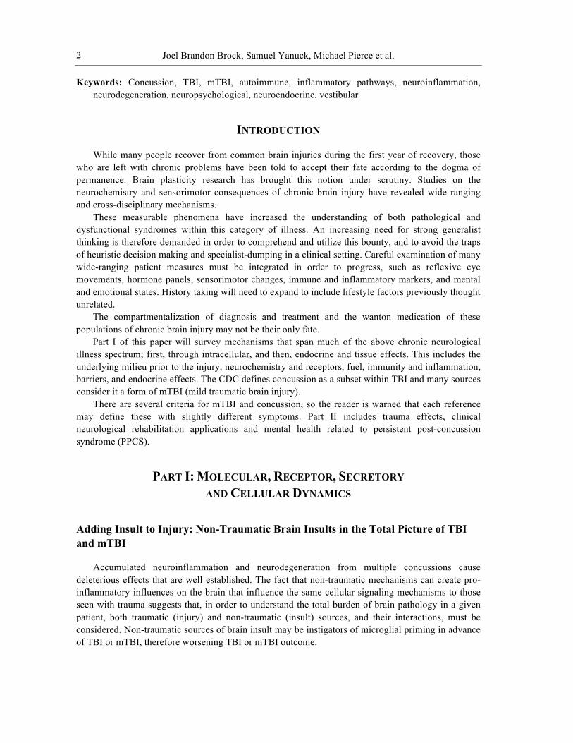
Changes in Microglia / Neuron Relationship and/or Excitotoxic Status
of Brain Environment Favoring
Death of Neurons / Loss of Cortical Integrity
Systemic PathogensRespiratory Infection
UTIGI Dysbiosis
Viral Infections
Fuel: -‐ O2 -‐ GlucoseNeuronal Fuel Exhaustion -‐> Low FOFInsulin Resistance -‐> Inflammation
Glycation -‐> Induction of Inflammatory and AI Potential
Hepatic Biotransformation Issues
• Altered biotransformation of endogenous compounds (cortisol, sex hormones, etc.)
• Haptens• Chemicals
Redox Chemistry Issues Leading to Neuronal Necrosis
Instead of Apoptosis Increased Mesencephalic / Sympathetic Activation
Faster Use of Glucose
Promotion of Pro-‐Inflammatory Cytokines or other Signaling
Molecule Changes / Increased ON Signals Decreased OFF Signals
Central Nervous System-‐Induced
Immunodepression Syndrome (CIDS) & Shift Toward Th2
Dominance in Brain & Body
Decreased Neuronal FOF (a primary OFF signal)
Death of Cortical Neurons & Alteration of
Total Brain Integrity
Damage to BBB – Presentation of Brain Self-‐Antigenic
Markers to Systemic Immune System
Diminished Th1 Surveillance&
Diminished Innate Immune Response
Hormonal IssuesProgesterone in TBI
Neuronal Thyroid RequirementsNFkB inhibition by estrogenAdrenal/Cortisol Inhibition of
Inflammation
Death of Vestibular, Cerebellar, Mesencephalic, PM, Vagal and other neurons & Alteration of Total Brain
Integrity
Increased Peripheral Vascular Resistance – Affects Brain O2?
IL-‐6 Elevation
Inflammatory Promotion of MDSC-‐BasedImmune
Suppression
Has Neuroprotective
Aspects
Multiple Connections
Systemic Inflammation
IL-‐6 causes NTIS…↓T4 & T3; ↑rT3 →
↓ Metabolism including Neuronal Metabolism
TBI / mTBI
Neuropsychiatric Consequences
Figure 1. Chronic TBI sequelae © Samuel Yanuck 2013.
Neuron-Microglial Interactions in the Healthy Brain
In the adult brain, there is a decline in gray matter density over time. For example, Sowell et al found a loss of gray matter density of approximately 32% between ages 7 and 60, and a 5% loss between 40 and 87 [186].. Thus neurons die every day. They need to be cleared, or they will drive damage associated molecular pattern (DAMP) mediated immune system activation, instigating a pro-inflammatory response in the brain [3,187].
Microglia are the predominant immune cells in the healthy brain [1]. In the normal, resting state, it is the job of ramified microglia to phagocytize the dead neurons, much like macrophages phagocytize apoptotic cells and tissue debris in peripheral tissues. In both cases, this housekeeping level of phagocytic activity drives the creation of anti-inflammatory mediators like IL-10 and TGFβ. Phagocytosis of apoptotic cells provides immune regulation through anti-inflammatory cytokines and regulatory T cells [2]
However, since there is no appreciable drainage of lymph from the brain, except minimally at the cribriform plate, the option of clearing phagocytized debris from tissue via the lymph system, as occurs in the periphery, is not available in the brain. Therefore, microglia must fully degrade dead neurons and recycle them as building blocks and fuel. Given billions of neurons and an estimated ten microglia per neuron, and given a motif of continuous surveillance, the number of instantaneous events of microglial cells being activated or inhibited in the decision flow to instigate phagocytosis of a neuron is very large indeed. How do the microglia decide which neurons are dead?
How is this process between microglia and neurons regulated in non-neutral circumstances like trauma, infection, inflammation, or neurodegeneration?
Neurons control microglial activity [3]. ‘Off’ signals from neurons keep microglia in their resting state and reduce pro-inflammatory activity, while ‘On’ signals from neurons are inducible, including purines, chemokines, and glutamate. Thus, neurons should be envisaged as key immune modulators in the brain [3]

Joel Brandon Brock, Samuel Yanuck, Michael Pierce et al.
4
Table 1. Inhibition of Glial Phagocytosis of Neurons (“OFF” Signals) [3]
OFF Signals Signal Effect Released TGFβ
CD22 CX3CL1 Neurotransmitters NGF BDNF NT-3
Inhibition of effector T cells; Promotion of immune tolerance & inflammatory resolution; loss of anti-pathogenic vigilance Inhibition of B cell activity Inhibition of microglial neurotoxicity Multiple functions Nerve growth factor Brain derived neurotrophic factor Neurotrophin-3
Membrane Bound
CD200 CD22 CD47 CX3CL1
Inhibition of glial inflammation Inhibition of B cell activity Inhibition of glial inflammation Inhibition of microglial neurotoxicity
Table 2. Activation of Glial Phagocytosis of Neurons (“ON” Signals) [3]
ON Signals Signal Effect Released CCL21
CXCL10 ATP & UTP Glutamate MMP3
Glial chemoattraction Glial chemoattraction Glial chemoattraction & IL-1β release TNFα release & Neuroexcitotoxicity Induces glial release of TNFα, IL-1β, IL-6 (inflammatory)
Membrane Bound
TREM2 ligand Promotes glial phagocytosis of neurons
In a healthy brain, the interplay of neurons and microglia is balanced. Healthy, viable neurons
produce adequate OFF signals to repel microglial phagocytic interest. In addition, healthy neurons produce abundant electrical activity. The electrical activity of neurons is itself a potent inhibitor of microglial activation [4].
In the healthy, non-inflamed brain, ramified microglia secrete TGFβ, which promotes a tolerogenic and anti-inflammatory tissue environment [5]. In addition, in a healthy brain, microglia and astrocytes express FasL, which induces apoptosis in T cells that migrate from the periphery into the brain [6]. Neurons and glia express cellular death signals, including CD95Fas/CD95L, FasL, tumor necrosis factor-related apoptosis-inducing ligand (TRAIL) and TNF receptor (TNFR), through which they can trigger apoptosis in T cells and other infiltrating cells [2].
Factors Affecting Neuronal-Microglial ‘On’ or ‘Off’ Signaling
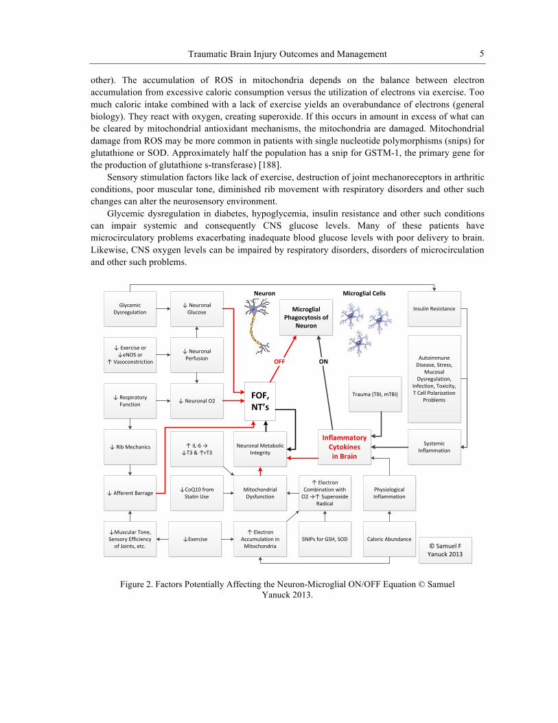
The capacity of neurons to maintain robust frequency of firing (FOF) is essential for sustained electrical activity and neurotransmitter (NT) output, two essential ‘OFF’ signals. The central integrative state (CIS) of the neuron depends on presynaptic stimulation, neuronal oxygen and neuronal glucose. Physiological factors that have the capacity to impair these three factors can contribute to changes in neuronal FOF, thereby changing the neuron-microglial ON/OFF equation. The capacity of neurons to maintain robust FOF also depends on the metabolic integrity of the neurons themselves. A host of factors contribute to neuronal metabolic integrity. Among the variables that can perturb function and lead to diminished FOF are a lack of exercise, excessive accumulation of mitochondrial ROS, diminished thyroid hormone signaling, and CoQ10 deficiency (statin-induced or
Traumatic Brain Injury Outcomes and Management
5
other). The accumulation of ROS in mitochondria depends on the balance between electron accumulation from excessive caloric consumption versus the utilization of electrons via exercise. Too much caloric intake combined with a lack of exercise yields an overabundance of electrons (general biology). They react with oxygen, creating superoxide. If this occurs in amount in excess of what can be cleared by mitochondrial antioxidant mechanisms, the mitochondria are damaged. Mitochondrial damage from ROS may be more common in patients with single nucleotide polymorphisms (snips) for glutathione or SOD. Approximately half the population has a snip for GSTM-1, the primary gene for the production of glutathione s-transferase) [188].
Sensory stimulation factors like lack of exercise, destruction of joint mechanoreceptors in arthritic conditions, poor muscular tone, diminished rib movement with respiratory disorders and other such changes can alter the neurosensory environment.
Glycemic dysregulation in diabetes, hypoglycemia, insulin resistance and other such conditions can impair systemic and consequently CNS glucose levels. Many of these patients have microcirculatory problems exacerbating inadequate blood glucose levels with poor delivery to brain. Likewise, CNS oxygen levels can be impaired by respiratory disorders, disorders of microcirculation and other such problems.
Figure 2. Factors Potentially Affecting the Neuron-Microglial ON/OFF Equation © Samuel Yanuck 2013.
Neuron Microglial Cells
FOF, NT’s
OFF
↓ Neuronal O2
↓ Afferent Barrage
↓Muscular Tone, Sensory Efficiency of Joints, etc.
↓ Neuronal Perfusion
↓ Respiratory Function
↓ Rib Mechanics
↓ Exercise or↓eNOS or
↑ Vasoconstriction
↓ Neuronal Glucose
Glycemic Dysregulation
Neuronal Metabolic Integrity
Inflammatory Cytokines in Brain
ON
Systemic Inflammation
Insulin Resistance
Mitochondrial Dysfunction
Trauma (TBI, mTBI)
↓CoQ10 from Statin Use
↑ IL-‐6 →↓T3 & ↑rT3
Autoimmune Disease, Stress,
Mucosal Dysregulation,
Infection, Toxicity, T Cell Polarization
Problems
↓Exercise↑ Electron
Accumulation in Mitochondria
SNIPs for GSH, SOD Caloric Abundance
↑ Electron Combination with O2 →↑ Superoxide
Radical
Microglial Phagocytosis of
Neuron
© Samuel F Yanuck 2013
Physiological Inflammation
Joel Brandon Brock, Samuel Yanuck, Michael Pierce et al.
6
Clearance and Inflammation in Injury
When the brain is injured, however, the resulting inflammatory chemistry can change the equation. With inflammation, neurons and microglia both change in ways that promote microglial phagocytosis of neurons. As with any cell, inflammation compromises metabolic integrity. In neurons, this yields diminished production of neurotransmitters and reduced electrical activity. Thus, two potent “Off” signals are lost. Meanwhile, inflamed microglia change their morphology. Brain inflammation differs from inflammation in the periphery by the relative absence of leukocytes and antibodies. There is a limited traffic across this barrier and this traffic can be increased by inflammation which can recruit leukocytes into the brain [1].
However, in the inflamed brain, microglia (which are after all a specialized form of macrophage) acquire antigen presenting cell capacity [4]. Instead of inducing apoptosis in invading T cells, inflamed microglia present antigen to invading T cells. Antigenic material can be fragments of processed pathogens. This is a useful antimicrobial effect, favoring clearance of pathogen. However, microglia can also present fragments of neuronal tissue debris as antigen, promoting a self-antigenic response in the T cells to which the antigen is presented. A mild and transient form of this T cell self-antigenic activation appears to be reparative. However, prolonged or overly exuberant self-antigenic T cell activation can cause irreversible damage to brain [4]. It is noteworthy that some of the research in this field occurred before there was a full appreciation of the role of TH17 polarization in T cell/microglial interactions.
Inflammation is known to induce mitochondrial uncoupling, diminishing mitochondrial integrity in all cells. Inflammation is therefore also a driver of diminished mitochondrial integrity and FOF of neurons. In a variety of inflammatory and neurodegenerative diseases, glial cells such as microglia gain antigen-presenting capacity through the expression of MHC molecules. The pro-inflammatory cytokines stimulate microglial MHC expression in the lesioned CNS areas only [4].
Neuronal signaling, a strong “Off” signal, works to suppress the immune system’s inflammatory activation in the brain so that induction of brain immunity is strongly counterregulated in intact CNS areas. The signaling activity of neurons also constitutes an inhibitory signal. The control of MHC expression by neurons is dependent on their electrical activity. Immunity in the CNS is inhibited by the local microenvironment, in particular by physiologically active neurons, to prevent unwanted immune mediated damage of neurons [4] (emphasis not original).
Research on the role of T cells and peripheral innate immune cells in the brain is evolving. Most authors suggest that maintenance of the non-inflamed CNS environment depends on the ability of microglial cells to induce apoptosis in peripheral T cells that gain access to the brain. However, the failure of systemic anti-inflammatory medications to yield improvement in neuroinflammatory disorders has led some authors to a view that CNS-infiltrating T cells provide crucial anti-inflammatory cytokine signals that are essential for the resolution of neuroinflammation [7].
It is noteworthy that non-steroidal, anti-inflammatory medications have been described in the literature as “resolution toxic” because they inhibit signaling mechanisms involved in the resolution of inflammation [8]. This insight may prove useful in understanding the failure of anti-inflammatories as a viable therapy to address chronic neuroinflammation.
In the presence of systemic inflammation, T cells invading the CNS are likely to be influenced by pro-inflammatory cytokines and other factors into a pro-inflammatory morphology, as they are in the periphery. Once so influenced, they are likely to produce pro-inflammatory cytokines, further promoting the pro-inflammatory CNS environment.
Pathogens in tissue create pathogen associated molecular patterns (PAMPs). Damaged tissue creates damage associated molecular patterns (DAMPs). Microglial pattern recognition receptors (PRRs) sense PAMPs and DAMPs, triggering phagocyte NADPH oxidase (PHOX), which turns

Traumatic Brain Injury Outcomes and Management
7
molecular oxygen into reactive oxygen species (ROS). This conversion depletes the tissue of molecular oxygen, yielding hypoxia, driving hypoxia inducible factor 1α (HIF-1α), driving NF-κB, driving IL-1β and TNFα, which increase gene expression of NF-κB. NF-κB drives iNOS (inducible nitric oxide synthase) expression. Though nitric oxide (NO) is cytoprotective, the combination of NO and hypoxia impair cellular respiration, yielding excitotoxicity. Though balanced amounts of ROS are normal to microglial function, ROS in combination with NO yields peroxynitrite, driving neuronal apoptosis. In patients with antioxidant depletion, from glutathione (gsh) snips or other factors, the likelihood of microglia producing neurotoxic amounts of ROS is increased.
Figure 3. Factors Contributing to Perpetuation or Resolution of Neuroinflammation [11] © Samuel Yanuck 2013.
Microglial Priming and the Contribution of Non-Traumatic Influences to Neuroinflammation
Microglial priming is a condition in which microglia move from the ramified state in which they perform housekeeping functions and reduce neuro-inflammation to a state in which they swell and fill with pro-inflammatory cytokines [9,10,11,12] Priming can be induced by aging, trauma, infection, or other stimuli. Microglia can remain in this state for long periods of time, without returning to the ramified state, but without releasing their bolus of cytokines. However, further insult to the brain will cause a flooding release of pro-inflammatory cytokines that can be damaging to the brain [9,10,11,12]. It has been established that increases in pro-inflammatory cytokines in the periphery yield upregulation of brain inflammation and potentiate neuronal death [13,14] It has also been shown that increasing the peripheral LPS (lipopolysaccharide) level can induce the activation of central pro-inflammatory mechanisms, even when the amount of LPS used for stimulation is minimal or when the peripheral inflammatory cytokine levels are suppressed artificially. Both central and peripheral inflammation can exacerbate local brain inflammation and neuronal death [15]. Once neuroinflammation occurs, the key question is whether it will resolve quickly or yield a chronically activated state, in which greater neuronal loss occurs.
Microglia alter their morphology and activate in response to pathophysiological brain insults. Microglial phenotype is also modified by systemic infection or inflammation. Chronic systemic inflammatory components are risk factors for Alzheimer disease. This implies that crosstalk occurs between systemic inflammation and microglia in the CNS [16]. Pathogens, protein aggregates, or damaged neurons may inflammatorily activate glia, which may then kill neurons. IL-1b has been
PAMP or DAMP → ↑ Microglial PRR
↑ PHOX → ↓ Molecular O2
to ↑ ROS
↑ HIF-‐1α → ↑ NFκB
↑ iNOS→ ↑ Nitric Oxide
↑ IL-‐1β, TNFα
↑ O2-‐ & H2O2
↑ ONOO-‐↑ Apoptosis / Phagocytosis
Hypoxia
Cellular Respiratory Inhibition
Excitotoxicity
Microglial Activation
CytoprotectionNeurotoxicity
Antioxidant Depletion
+
Joel Brandon Brock, Samuel Yanuck, Michael Pierce et al.
8
shown to be the main activator of microglia during brain disturbances. Systemic IL-1b can cause CNS inflammation once it enters the brain, thus linking systemic inflammation and immune activation [12].
Microglia can become over-activated through two mechanisms. First, microglia can initiate neuron damage by recognizing pro-inflammatory stimuli, such as lipopolysaccharide (LPS)). Second, microglia can become overactivated in response to neuronal damage [11]. LPS can directly activate the brain endothelium even at relatively low doses, obviating the need for systemic cytokine stimulation [9,10]. Many of the pathological events described in traumatic brain injuries can also be seen with excitotoxicity [12].
Receptor stimulation by pathogens or neuron damage contributes to nuclear factor-kappaB (NF-κB) activation. Simultaneous activation of PHOX (Phagocyte NADPH oxidase) and iNOS (inducible nitric oxide synthase) in microglia resulted in the disappearance of NO, appearance of peroxynitrite and apoptosis. However, the chronic state of activation may progress to “resolution phase” where microglia are amoeboid, highly phagocytic, and produce anti-inflammatory cytokines (including IL-10 and TGFb) in order to resolve the inflammation and clear up the mess [1]
With successful, non-phlogistic microglial phagocytosis of apoptotic neurons, the neuron is engulfed and digested without release of additional damage associated molecular patterns (DAMPs) into the tissue environment. This favors the production of anti-inflammatory cytokines and a movement toward resolution of tissue inflammation in the brain parenchyma. If instead the neuron dies by necrosis through direct or indirect trauma, or is triggered into necrosis by a pathogen or toxin, its death releases cell fragments and cytosolic contents into the tissue environment, triggering a pro-inflammatory response in surrounding microglia. Neuroinflammation favors a more aggressive microglial cell phenotype and a more exuberant phagocytosis of neurons, yielding neuronal loss in excess of that necessary to bring about resolution of the initial condition.
SIDS, SIRS and CARS
Acute neuroimmunological syndromes such as central nervous system injury-induced immune deficiency syndrome (SIDS), systemic inflammatory response syndrome (SIRS) and compensatory anti-inflammatory response syndrome (CARS) have been reviewed elsewhere [17]. It is unclear in the literature, however, whether a gradient of severity exists in these syndromes. For example, it is unclear whether a patient with mTBI (mild traumatic brain injury) might be expected to manifest a modest version of the apoptosis of innate immune cells and TH1 cells seen in SIDS. If this were the case, the patient might be incrementally more susceptible to chronic infection, promoting systemic and therefore neuroinflammatory mechanisms.
From a clinical perspective, whether or not mild forms of these syndromes pertain, the clinician faced with a patient with a TBI or mTBI should be alert for indications of suppressed immune vigilance against pathogens. Such a circumstance would have the potential to yield chronic infection, a known driver of microglial priming and neuroinflammation.
The Injured Neuron
In the realm of traumatic brain injury, the neuron is the center point of the physiological story [18]. Factors including the preexisting central integrated state of various neuronal pools, the integrity of existing neuronal circuitry, peripheral receptor integrity, level of circulating cytokine populations, polarization status of a dynamic immune system, level of glial priming and function, balance and integrity of the endocrine system, various underlying infectious organisms, genetic predisposition, associated comorbidities and the extent of damage sustained as well as related biomechanics of a
Traumatic Brain Injury Outcomes and Management
9
given injury all determine neuronal integrity and probability of recovery post-injury [19]. Other factors that can impact the neuron are the nutritional and digestive status of the patient as well as vascular perfusion and autonomic integrity. The neuron’s ability to survive and maintain optimum functional capacity and appropriate cellular plasticity is vital to recovery and sustaining humanism and vitality post-neurological insult. Understanding and evaluating all converging physiological scenarios that can impact the health of the neuron and how it relates to head injury and damage to the CNS is vital when determining the extent of injury, creating appropriate treatment plans and care of patients suffering from traumatic brain injury or neurodegeneration [20].
The intracellular cascade after head injury is complex and involves organelle function, metabolic and ionic fluctuations and surface receptor interplay. This gross level interplay impacts cellular plasticity, immunoexcitotoxicty, intracellular calcium and binding proteins, caspase cascades, apoptosis, cerebral blood flow, glucose metabolism, phospholipase and free radical production, protease and cytoskeleton breakdown, endonuclease and DNA damage, nitric oxide isomers and superoxide anions [12]. On a smaller intracellular scale, organelle involvement includes changes in mitochondrial function, neurofilaments and microtubules, lysosomes, epigenetic function, protein replication, secretory vesicle production and synaptic capacity. The eventual consequence of intracellular and organelle variations will impact cellular energy, axonal and myelin integrity, cellular swelling, synaptic transmission, lipid membrane stability, synaptic transmission, and cellular summation capabilities. When damage occurs in the CNS, neurons are impacted, glial cells are altered, glutamate receptors can become sensitized, GABA receptors can become internalized, the immunological system is impacted, vasculature is compromised and the blood-brain barrier becomes damaged. This combination of events can lead to cellular damage, microglial priming and sustained inflammation within the CNS, antigen presentation of neural tissues and possible autoimmunity and compromise in the resolution process post-injury [21].
Surface receptor types, densities and their sensitivities also contribute to metabolism [22]. There are many receptor types; however, of particular interest are the ionotropic receptors including NMDA, AMPA, Kainate and voltage gated calcium channels. These receptors types allow a regulated influx of calcium into the cell that impacts multiple intracellular pathways allowing for intracellular cascades, cellular function, cellular plasticity and long term potentiation. The ultimate promotion of NMDAr(define) (N-methyl-D-aspartate, a specific type of ionotropic glutamate receptor) promotes the extracellular influx of calcium and calcium stores within the cell. The increase and appropriate regulation of cytosolic calcium leads to the proper activation of kinase dependent signaling cascades leading to cAMP (cyclic adenosine monophosphate) element binding protein activation. The activation of CREB (cAMP Response Element-Binding protein) ultimately generates the phosphorylation at SER 133 (serine 133) leading the generation of protein synthesis within the nucleus. This generates the ability for the cell to innately generate more surface receptors, intracellular structures, cytokines, neurotrophic factors, cellular efficiency, dendritic development and repair cycles vital for cell survival. The ultimate outcome of this process is synaptic plasticity and long term potentiation. This process is a large part of learning and memory and is a major mechanism of repair after damaged neural circuitry is created after head injury or in a neurodegenerative process [23].
The dysregulation of intracellular calcium, however, can lead to degenerative and excitotoxic mechanisms that are damaging. Under certain circumstance, cell surface receptors become more permeable to calcium while intracellular calcium-buffering proteins become aberrant thus causing the intracellular calcium levels to become dysregulated. Multiple neurodegenerative conditions, inflammatory scenarios and disease processes as well as excitotoxin loads and glutamate levels have the ability to alter NMDA receptors activation and skew AMPA:NMDA receptor ratios allowing a greater influx of extracellular calcium into the cell [18]. The resultant intracellular calcium dysregulation triggers pathology. The triggering of intracellular lipases causes cell membrane damage.
Joel Brandon Brock, Samuel Yanuck, Michael Pierce et al.
10
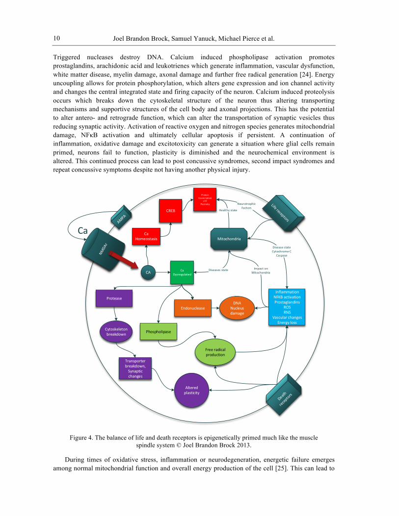
Triggered nucleases destroy DNA. Calcium induced phospholipase activation promotes prostaglandins, arachidonic acid and leukotrienes which generate inflammation, vascular dysfunction, white matter disease, myelin damage, axonal damage and further free radical generation [24]. Energy uncoupling allows for protein phosphorylation, which alters gene expression and ion channel activity and changes the central integrated state and firing capacity of the neuron. Calcium induced proteolysis occurs which breaks down the cytoskeletal structure of the neuron thus altering transporting mechanisms and supportive structures of the cell body and axonal projections. This has the potential to alter antero- and retrograde function, which can alter the transportation of synaptic vesicles thus reducing synaptic activity. Activation of reactive oxygen and nitrogen species generates mitochondrial damage, NFκB activation and ultimately cellular apoptosis if persistent. A continuation of inflammation, oxidative damage and excitotoxicity can generate a situation where glial cells remain primed, neurons fail to function, plasticity is diminished and the neurochemical environment is altered. This continued process can lead to post concussive syndromes, second impact syndromes and repeat concussive symptoms despite not having another physical injury.
Figure 4. The balance of life and death receptors is epigenetically primed much like the muscle spindle system © Joel Brandon Brock 2013.
During times of oxidative stress, inflammation or neurodegeneration, energetic failure emerges among normal mitochondrial function and overall energy production of the cell [25]. This can lead to
Ca
CA
Ca Homeostasis
CREB
Protein trasncription
LTPPlasticity
Ca Dysregulated
Endonuclease
Phospholipase
ProteaseDNA
Nucleus damage
Cytoskeleton breakdown
Free radical production
Mitochondria
Diseases state
Healthy state
Transporter breakdown,Synaptic changes
InflammationNFKB activationProstaglandins
ROSRNS
Vascular changesEnergy loss
Disease stateCytochrome C
Caspase
Altered plasticity
NeurotrophicFactors
Impact onMitochondria

Traumatic Brain Injury Outcomes and Management
11
a breakdown in the electron transport chain and decrease mitochondrial calcium loading capacity through an activated mitochondrial permeability exchanger. The exchanger opens mitochondrial pores and allows the deposition of calcium within the mitochondria into the cellular cytosol disrupting intracellular calcium regulation. Respiratory chain uncoupling and the release of intermembrane space proteins as a result of MPT (membrane permeability transition) activation causes multiple cascades to occur [26]. These include the release of intracellular caspases, caspase independent cell death effectors, NLRP3 inflammasomes, NFκB and interferon regulatory factors. The release of cytochrome C from the mitochondria activates caspase three which programs cell death and apoptosis. These cascades collectively lead to the development of inflammation, loss of cellular energy production and perpetuate further mitochondrial and cellular dysfunction. Thus the milieu of the cell primes the receptor sensitivity even before death or life receptors are triggered. This phenomenon of “setting receptor tone” is conceptually similar to how the tympanum tension is preset to perceive or protect from sound prior to the event, and the muscle spindle tone is preset before a perturbation. The implications of this for clinical prognosis and cellular apoptosis indicate some potential for leverage through clinical cell mediator manipulation. Such manipulation could be through substances administered as well as evoked potentials.
In a normal cell, when there is abnormal stress, organelle damage, accumulation of misfolded proteins or damaged mitochondria, the cell removes the damaged organelles or unwanted proteins [27]. These processes include autophagy and mitophagy. Under periods of aging, inflammation, oxidative stress, mitochondrial damage, intracellular calcium dysregulation or a decrease in cellular activation, appropriate autophagy and mitophagy is impaired. When these cellular processes are impaired after head injury, various diseases can manifest, the health of the neuron can fail and ultimately lead to cellular death and premature neurodegeneration.
Head InjuryCoup
Contra CoupRotationalShearing
Linear forceBleeding
Biomechanical forces
Structural Damage
Chemical Cascade
Neurochemical Damage
ChemicalCascade
Transmitter changesBleeding / stroke / watershed
Vascular changes
Mechanical and ionicDeformation
Axonal damage
Neurofilaments
Damage to Terminal or transporters
Synaptic Damage
Glutamate ↑ GABA↓
CaspaseCalpain
Serotonin ↓Dopamine ↓
Monoamine issue
Reuptake transporter loss
NMDA
Excitotoxicity
LysosomeMitochondrialAutophagy
GolgiPackaging damage
Synaptic vesicle loss
Relates to comorbiditiesAnd number of injuries
Glial primingInflammation
ApoptosisPONiNOSSOA
Energy lossIntracellular
debris
Cellular damage
Decrease in synaptic
transmission
HyperkineticTND
Excitotoxic
DepressionPain
Emotional changes
Second impact /Post concussive
prone
Prone for loss in plasticity
IschemiaTo tissue
Reactive Vasconstriction
Decrease in fuel for delivery
↓Fuel causesOxidative stress
Excitotoxicty
Second impact / Post concussive
prone
Second impact / Post concussive
prone
Second impact / Post
concussiveprone
Emotional changesHead and body pain
Will vary depending On time
Without therapyRecovery ↓
TwistingRotation
Shearing forceBlunt trauma
TractionTo
Peripheral Vestibular System
Contusion or damage to the peripheral vestibular apparatus
Damage to TM / round / oval
windowOr Damage to hair cells
Or Displacement of Otoconia or Fracture to temporal
bone Or InflammationAutoimmunity
Delineate vestibular And Auditory loss or
both
Copyright ©Joel Brandon Brock 2013
Twisting, Shearing, CompressionBleeding, Diffuse and Focal
Damage
Changes to CNSDepends on area of
trauma, extent of force on the head, amount of tissue ischemia, other
comorbidities
PAST MEDICAL HISTORY
Mechanism of InjuryAge / Sex / Other bodily injury
Will vary depending on severity
Knockout
Unconscious
Cellular Failure
Hearing lossCanal / OtolithsFistula / TM BPPV Conditions
Damage to PVNDamage to PV apparatus
Hydrops
Joel Brandon Brock, Samuel Yanuck, Michael Pierce et al.
12
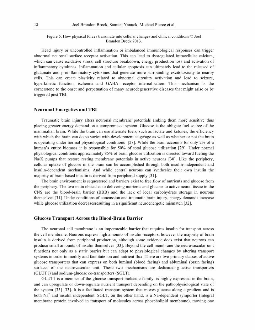
Figure 5. How physical forces transmute into cellular changes and clinical conditions © Joel Brandon Brock 2013.
Head injury or uncontrolled inflammation or imbalanced immunological responses can trigger abnormal neuronal surface receptor activation. This can lead to dysregulated intracellular calcium, which can cause oxidative stress, cell structure breakdown, energy production loss and activation of inflammatory cytokines. Inflammation and cellular apoptosis can ultimately lead to the released of glutamate and proinflammatory cytokines that generate more surrounding excitotoxicity to nearby cells. This can create plasticity related to abnormal circuitry activation and lead to seizure, hyperkinetic function, ischemia and GABA receptor internalization. This mechanism is the cornerstone to the onset and perpetuation of many neurodegenerative diseases that might arise or be triggered post TBI.
Neuronal Energetics and TBI
Traumatic brain injury alters neuronal membrane potentials amking them more sensitive thus placing greater energy demand on a compromised system. Glucose is the obligate fuel source of the mammalian brain. While the brain can use alternate fuels, such as lactate and ketones, the efficiency with which the brain can do so varies with development stage/age as well as whether or not the brain is operating under normal physiological conditions [28]. While the brain accounts for only 2% of a human’s entire biomass it is responsible for 50% of total glucose utilization [29]. Under normal physiological conditions approximately 85% of brain glucose utilization is directed toward fueling the Na/K pumps that restore resting membrane potentials in active neurons [30]. Like the periphery, cellular uptake of glucose in the brain can be accomplished through both insulin-independent and insulin-dependent mechanisms. And while central neurons can synthesize their own insulin the majority of brain-based insulin is derived from peripheral supply [31].
The brain environment is sequestered and barriers exist to free flow of nutrients and glucose from the periphery. The two main obstacles to delivering nutrients and glucose to active neural tissue in the CNS are the blood-brain barrier (BBB) and the lack of local carbohydrate storage in neurons themselves [31]. Under conditions of concussion and traumatic brain injury, energy demands increase while glucose utilization decreasesresulting in a significant neuroenergetic mismatch [32].
Glucose Transport Across the Blood-Brain Barrier
The neuronal cell membrane is an impermeable barrier that requires insulin for transport across the cell membrane. Neurons express high amounts of insulin receptors, however the majority of brain insulin is derived from peripheral production, although some evidence does exist that neurons can produce small amounts of insulin themselves [33]. Beyond the cell membrane the neurovascular unit functions not only as a static barrier but can adapt to physiological changes by altering transport systems in order to modify and facilitate ion and nutrient flux. There are two primary classes of active glucose transporters that can express on both luminal (blood facing) and abluminal (brain facing) surfaces of the neurovascular unit. These two mechanisms are dedicated glucose transporters (GLUT1) and sodium-glucose co-transporters (SGLT).
GLUT1 is a member of the glucose transport molecule family, is highly expressed in the brain, and can upregulate or down-regulate nutrient transport depending on the pathophysiological state of the system [33] [33]. It is a facilitated transport system that moves glucose along a gradient and is both Na+ and insulin independent. SGLT, on the other hand, is a Na-dependent symporter (integral membrane protein involved in transport of molecules across phospholipid membrane), moving one
Traumatic Brain Injury Outcomes and Management
13
glucose molecule along with two Na+ molecules in the same direction across the cell membrane in a co-transport relationship, against the glucose gradient [31]. SGLT is a secondary active transport mechanism that uses ATP generated from ion gradients. SGLT can also function in reverse, moving accumulated Na+ out of the cell into the extracellular space [34]. While GLUT1 is considered the primary glucose transport system studies have shown upregulation of the sodium-dependent glucose transporter SGLT1 in conditions of ischemia-hypoxia [33].
Because of these limitations (cellular and tissue level barriers coupled with a lack of passive diffusion and local neuronal glucose storage) the brain relies heavily on the expression of glucose transporters on the blood-brain barrier as well as the storage capacity of astrocytes. Both of these glucose transporters can be insulin sensitive or insensitive, hormonally regulated or driven by glucose concentrations [31]
Other GLUT and SGLT isoforms (GLUT3, GLUT4, SGLT1, SGLT2) exist in the brain, but in lower concentrations and contribute to a lesser degree to neuroenergetics [5].
Energy Compartmentalization
In the brain, energy metabolism is highly compartmentalized [35]. While neurons have both aerobic and anaerobic pathways they have little capacity to store energy. As such neurons rely heavily on glucose delivery from the periphery as well as the participation of astroglial cells, which function as the brain’s glycogen repository. This presents several problems since there are significant barriers to consistent and stable glucose supply to the brain. Furthermore astrocytic glycogenesis and glycogenolysis as well as neuronal mitochondrial function can be influenced by TBI.
Since neurons do not store glycogen, neuronal glucose utilization is intimately yoked to astroglial compartmentalization of glycogen. In the absence of its own localized glycogen neurons must communicate energetically with astrocytes in order to replenish energy substrates. This allows for resetting of membrane potentials and continued neuronal viability and functionality. Astrocytes support neuronal energetics via multiple pathways, the most notable and well-studied being the Glutamine-Glutamate Cycle and the Astrocyte Neuron Lactate Shuttle (ANLS).
In the former, glutamate produced by neuronal Citric Acid Cycle (TCA) mechanics passes into the extracellular space and is taken up by astroglial cells where it is converted to glutamine [36]. Astroglial glutamine then exits the astrocyte and can be taken up again by a nearby neuron where it can resupply the neurotransmitter pool or reenter the TCA cycle by being converted into either alpha-ketoglutarate or succinate [37].
The Astrocyte Neuron Lactate Shuttle provides substrate for neurons to engage the glycolytic pathway. Extracellular glutamate, from active neuronal signaling, increases astroglial uptake of glucose by upregulating GLUT1 [38]. Astroglial cells then drive the glycolytic process creating lactate as a metabolic byproduct. This lactate is transported out of the astroglia into neurons by a monocarboxylate transporters, MCT1 and MCT 2 respectively. Neurons express several isoforms of lactate dehydrogenase (LDH) causing the conversion of lactate to pyruvate and the initiation of neuronal glycolysis, which eventually yields more glutamate to continue the ANLS. These two systems, Glutamine-Glutamate Cycling and the Lactate Shuttle, bind astroglial cells and central neurons in a symbiotic relationship that ultimately determines the ATP potential of both functional and injured brains.

Joel Brandon Brock, Samuel Yanuck, Michael Pierce et al.
14
TBI Alters Brain Energy Metabolism
In 2001, Giza and Hovda outlined the neurometabolic cascade of concussion. The injured brain shifts into “hypermetabolism”, which increases the demand for ATP. At the same time energy production pathways shift away from the highly efficient oxidative phosphorylation to the much less efficient glycolysis. The net effect is neuronal acidosis, membrane dysfunction and BBB permeability. In addition evidence suggests that in the context of TBI lactate-consuming pathways that would otherwise drive ATP production via the ANLS are compromised [32].
What follows is diminished cerebral blood flow, which may be reduced by as much as 50%. This cellular energy crisis predisposes the injured brain to second injury and prolongs functional deficits associated with the injury [39]. Beyond the direct effects upon ATP producing pathways, post-injury depolarization and K efflux opens NMDA receptors. Calcium influx further impairs neuronal ATP production by impairing both oxidative phosphorylation and glycolysis. This promotes activation of apoptotic pathways, neurofilament compaction, microtubule disassembly, axonotomy and increases the production of inducible nitric oxide synthase [39].
Due to its high lipid content, the brain is highly susceptible to oxidative damage. TBI leads to a significant decrease in glutathione and ascorbic acid, the two primary intracellular antioxidants. Rat brains subjected to TBI showed a three-fold reduction in the reduced-to-oxidized glutathione ratio. Concomitant reductions in cerebral NAD+ promote mitochondrial dysfunction [32]. Reduced glutathione has been shown to impair astroglial glucose metabolism and glycogen utilization [40].
Hypoxia
Epidemiological studies reveal that up to 44% of severe TBI patients experience brain hypoxia that is a direct consequence of hypoperfusion. Proinflammatory cytokine upregulation induces BBB dysfunction via IL-6 and IL-1b and an overall significant hypoglycemia, with brain glucose levels dropping by 50% in injury-induced hypoxia [41].
Peripheral Metabolic Impacts on Injured Brain Fuel Status
Ives et al explored evidence of hypopituitarism following multiple concussions. Growth hormone was the most vulnerable to successive brain injury followed by gonadotropins, TSH and finally ACTH. Furthermore these hormonal imbalances may not be evident until well after the initiating injury [42]. Agha et al explored Glucagon Stimulation and Insulin Tolerance tests in a population of brain injured patients with a median interval of 7 months post-injury. Approximately 28% of patients exhibited at least one anterior pituitary hormone deficiency, 22% showed isolated deficiencies involving either GH, LH/FSH (follicle-stimulating hormone) or ACTH (adrenocorticotropic hormone) and 6% showed evidence of multiple deficiencies [43]. Both GH and ACTH exert significant influence over the action of both glucagon and insulin, and GH deficiency can impair glucose tolerance by decreasing beta cell mass and insulin production [44]. These conditions can have impacts on the injured brain primarily due to the brain’s reliance on peripherally derived insulin for glucose uptake.
Traumatic Brain Injury Outcomes and Management
15
Post-Synaptic Contributions in TBI
TBI and Central Processing of Viscera Traumatic brain injury can lead to various mechanisms of gastrointestinal dysfunction. These
mechanisms include: impairment of digestive enzyme production, impairment of intestinal motility, disruption intestinal autonomics related to circulation, promotion of intestinal permeability, and altered interoceptive processing. Disruption of the brain-gut axis involving the cortico-pontine circuit has been demonstrated with brain imaging studies as a central mechanism of irritable bowel syndrome [45]. Central integration of cortico-pontine circuit is critical for proper vagal temporal summation and autonomic regulation necessary for regulating proper intestinal afferent and efferent communication. Additionally, traumatic brain injury can lead to significant changes of brain-gut peptides in both plasma and small intestine, which may be involved in the pathogenesis of complicated gastrointestinal dysfunction [46]. These cortico-pontine and pontine-cortical central integrations may be altered in TBI. Cortical integration is critical for proper bowel function and bowel disorders associated with lack of cortical level integration of visceral inputs have been demonstrated with percept-related fMRI [47]. Therefore it appears that TBI may potentially lead to altered gastrointestinal function from loss of cortico-pontine central processing.
TBI and Intestinal Permeability One of the major consequences of TBI is lack of cortical activation of the pontine vagal system
leading to altered postsynaptic autonomic changes that promote decreased intestinal autonomics and inflammatory reactions leading to intestinal permeability. Intestinal permeability induced from TBI may be a consequence of lack of post-syanaptic activation of the vagal nuclei. In a mouse model of TBI, vagal stimulation prevented TBI-induced intestinal permeability and also increased enteric glial activity [48]. This study supported the notion that the vagal nuclei disruption from TBI was the central mechanism for intestinal permeability development and that vagal activation has modulating activity on the enteric glia neuroinflammatory responses. Additionally, TBI can induce an increase in intestinal permeability, which may lead to bacterial translocation, sepsis, and system inflammation [49,50]. TBI induced intestinal permeability thus has the potential to promote a vicious inflammatory cascade involving the brain to gut axis and the gut to brain axis. Intestinal permeability has been found to increase proinflammatory cytokines at the intestinal mucosal level and cause lipopolysaccharide translocation that can disrupt brain function [51,52]. Therefore, the alteration of cortico-pontine integration from TBI can lead to gastrointestinal inflammatory consequences from intestinal permeability that then potentially further suppress brain function leading to chronic inflammatory vicious cycles between the brain and the gastrointestinal system. Traumatic brain injury can also lead to pro-inflammatory immune activation in the peripheral blood stream leading to systemic inflammatory response syndrome [53]
TBI and Intestinal Mucosa Compromise At the intestinal level many changes take place in the intestinal mucosa directly after brain injury
including mucosal ischemia, mucosal atrophy, and activation of intestinal inflammatory cascades. These reactions occur rapidly as early as 3 hours following brain injury and last for more than 7 days with marked mucosal atrophy [54]. Additionally, TBI induces profound effects including gastrointestinal mucosa ischemia and motility dysfunction [55]. The inflammatory reactions that occur in the intestinal mucosa following traumatic brain injury appear to increase the expression of intestinal nuclear factor kappa B and intercellular adhesion molecule-1 in the intestine leading to acute gut mucosal injury following TBI [56,57]. There is also rapid and persistent up-regulation of myeloid differentiation primary response protein 88 (Myd88) in combination with systemic inflammatory cytokine activation [58]. These inflammatory changes that occur after TBI are immediate and
Joel Brandon Brock, Samuel Yanuck, Michael Pierce et al.
16
illustrate how cortical injury can lead to inflammatory consequences in the peripheral gastrointestinal mucosa lining.
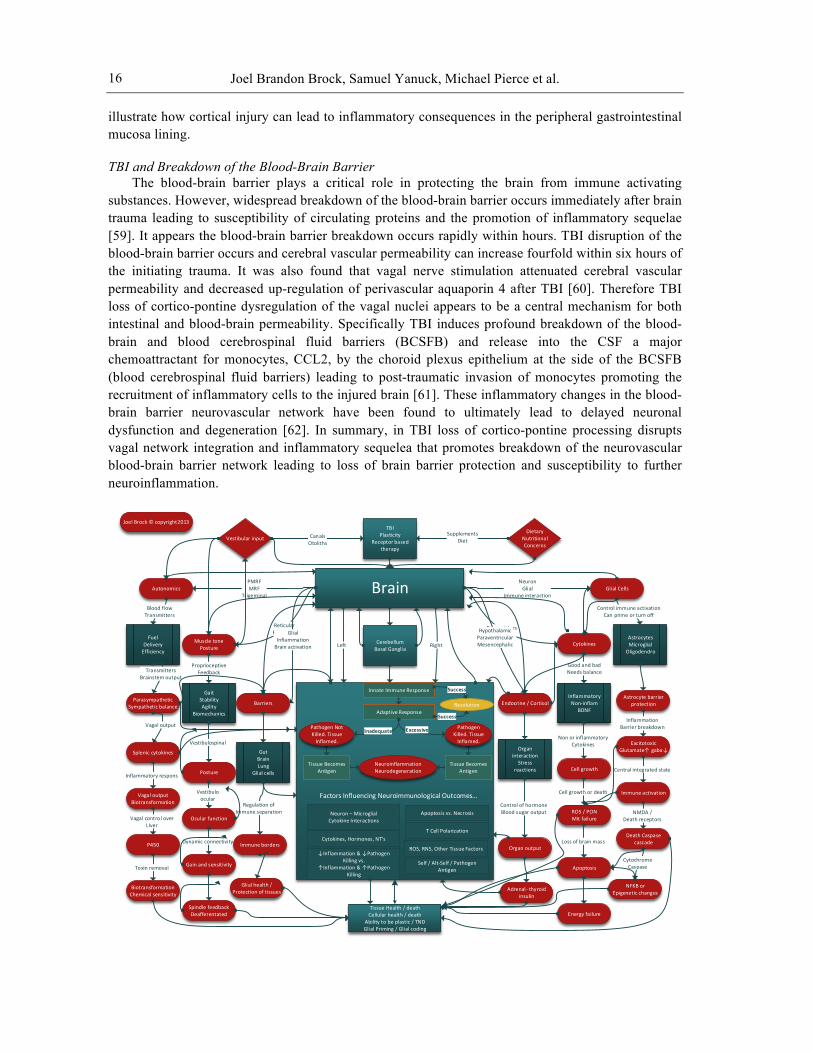
TBI and Breakdown of the Blood-Brain Barrier The blood-brain barrier plays a critical role in protecting the brain from immune activating
substances. However, widespread breakdown of the blood-brain barrier occurs immediately after brain trauma leading to susceptibility of circulating proteins and the promotion of inflammatory sequelae [59]. It appears the blood-brain barrier breakdown occurs rapidly within hours. TBI disruption of the blood-brain barrier occurs and cerebral vascular permeability can increase fourfold within six hours of the initiating trauma. It was also found that vagal nerve stimulation attenuated cerebral vascular permeability and decreased up-regulation of perivascular aquaporin 4 after TBI [60]. Therefore TBI loss of cortico-pontine dysregulation of the vagal nuclei appears to be a central mechanism for both intestinal and blood-brain permeability. Specifically TBI induces profound breakdown of the blood-brain and blood cerebrospinal fluid barriers (BCSFB) and release into the CSF a major chemoattractant for monocytes, CCL2, by the choroid plexus epithelium at the side of the BCSFB (blood cerebrospinal fluid barriers) leading to post-traumatic invasion of monocytes promoting the recruitment of inflammatory cells to the injured brain [61]. These inflammatory changes in the blood-brain barrier neurovascular network have been found to ultimately lead to delayed neuronal dysfunction and degeneration [62]. In summary, in TBI loss of cortico-pontine processing disrupts vagal network integration and inflammatory sequelea that promotes breakdown of the neurovascular blood-brain barrier network leading to loss of brain barrier protection and susceptibility to further neuroinflammation.
BrainPMRFMRF
TrigeminalAutonomics
ReticularPyramidal
Muscle tone Posture
GlialInflammation Brain activation
Barriers
NeuronGlial
Immune interactionGlial Cells
Cytokines
Endocrine / Cortisol
GutBrainLung
Glial cells
Proprioceptive Feedback
GaitStabilityAgility
Biomechanics
Blood flowTransmitters
FuelDeliveryEfficiency
Organ interaction
Stress reactions
Good and badNeeds balance
InflammatoryNon-‐inflam
BDNF
Control immune activationCan prime or turn off
AstrocytesMicroglial
Oligodendro
TBIPlasticity
Receptor based therapy
Astrocyte barrier protection
InflammationBarrier breakdown
ExcitotoxicGlutamate↑ gaba↓
TransmittersBrainstem output
ParasympatheticSympathetic balance
Vagal output
Splenic cytokines
Inflammatory respons
Vagal outputBiotransformation
Vagal control overLIver
P450
Toxin removal
BiotransformationChemical sensitivity
Vestibulospinal
Posture
Vestibuloocular
Ocular function
Dynamic connectivity
Gain and sensitivity
Spindle feedbackDeafferentated
Cerebellum Basal Ganglia
Right
TransmittersImmune
HypothalamicParaventricularMesencephalic
Regulation of Immune separation
Immune borders
Glial health /Protection of tissues
Control of hormoneBlood sugar output
Organ output
Adrenal-‐ thyroid insulin
Non or inflammatoryCytokines
Cell growth
Cell growth or death
ROS / PON Mit failure
Loss of brain mass
Apoptosis
Energy failureTissue Health / deathCellular health / death
Ability to be plastic / TNDGlial Priming / Glial coding
Central integrated state
Immune activation
NMDA /Death receptors
Death Caspase cascade
CytochromeCaspase
NFKB or Epigenetic changes
Vestibular input CanalsOtoliths
DietaryNutritional Concerns
SupplementsDiet
Joel Brock © copyright2013
↓Inflammation & ↓Pathogen Killing vs.
↑Inflammation & ↑Pathogen Killing
T Cell Polarization
Apoptosis vs. NecrosisNeuron – Microglial Cytokine Interactions
Cytokines, Hormones, NT’s
ROS, RNS, Other Tissue Factors
Self / Alt-‐Self / Pathogen Antigen
Innate Immune Response
Resolution
Success
Adaptive ResponseSuccess
Pathogen Killed. Tissue Inflamed.
Excessive
NeuroinflammationNeurodegeneration
Pathogen Not Killed. Tissue Inflamed.
Tissue Becomes Antigen
Inadequate
Tissue Becomes Antigen
Factors Influencing Neuroimmunological Outcomes...
Left
Traumatic Brain Injury Outcomes and Management
17
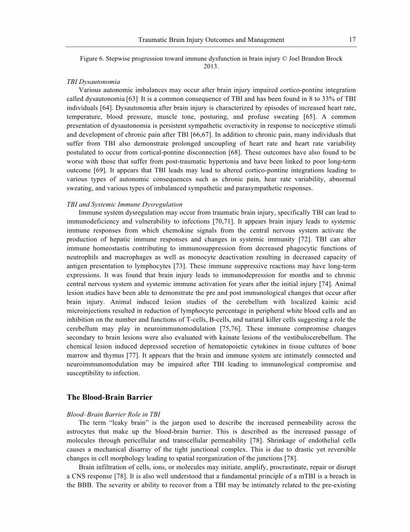
Figure 6. Stepwise progression toward immune dysfunction in brain injury © Joel Brandon Brock 2013.
TBI Dysautonomia Various autonomic imbalances may occur after brain injury impaired cortico-pontine integration
called dysautonomia [63]. It is a common consequence of TBI and has been found in 8 to 33% of TBI individuals [64]. Dysautonomia after brain injury is characterized by episodes of increased heart rate, temperature, blood pressure, muscle tone, posturing, and profuse sweating [65]. A common presentation of dysautonomia is persistent sympathetic overactivity in response to nociceptive stimuli and development of chronic pain after TBI [66,67]. In addition to chronic pain, many individuals that suffer from TBI also demonstrate prolonged uncoupling of heart rate and heart rate variability postulated to occur from cortical-pontine disconnection [68]. These outcomes have also found to be worse with those that suffer from post-traumatic hypertonia and have been linked to poor long-term outcome [69]. It appears that TBI leads may lead to altered cortico-pontine integrations leading to various types of autonomic consequences such as chronic pain, hear rate variability, abnormal sweating, and various types of imbalanced sympathetic and parasympathetic responses.
TBI and Systemic Immune Dysregulation Immune system dysregulation may occur from traumatic brain injury, specifically TBI can lead to
immunodeficiency and vulnerability to infections [70,71]. It appears brain injury leads to systemic immune responses from which chemokine signals from the central nervous system activate the production of hepatic immune responses and changes in systemic immunity [72]. TBI can alter immune homeostastis contributing to immunosuppression from decreased phagocytic functions of neutrophils and macrophages as well as monocyte deactivation resulting in decreased capacity of antigen presentation to lymphocytes [73]. These immune suppressive reactions may have long-term expressions. It was found that brain injury leads to immunodepression for months and to chronic central nervous system and systemic immune activation for years after the initial injury [74]. Animal lesion studies have been able to demonstrate the pre and post immunological changes that occur after brain injury. Animal induced lesion studies of the cerebellum with localized kainic acid microinjections resulted in reduction of lymphocyte percentage in peripheral white blood cells and an inhibition on the number and functions of T-cells, B-cells, and natural killer cells suggesting a role the cerebellum may play in neuroimmunomodulation [75,76]. These immune compromise changes secondary to brain lesions were also evaluated with kainate lesions of the vestibulocerebellum. The chemical lesion induced depressed secretion of hematopoietic cytokines in tissue cultures of bone marrow and thymus [77]. It appears that the brain and immune system are intimately connected and neuroimmunomodulation may be impaired after TBI leading to immunological compromise and susceptibility to infection.
The Blood-Brain Barrier
Blood–Brain Barrier Role in TBI The term “leaky brain” is the jargon used to describe the increased permeability across the
astrocytes that make up the blood-brain barrier. This is described as the increased passage of molecules through pericellular and transcellular permeability [78]. Shrinkage of endothelial cells causes a mechanical disarray of the tight junctional complex. This is due to drastic yet reversible changes in cell morphology leading to spatial reorganization of the junctions [78].
Brain infiltration of cells, ions, or molecules may initiate, amplify, procrastinate, repair or disrupt a CNS response [78]. It is also well understood that a fundamental principle of a mTBI is a breach in the BBB. The severity or ability to recover from a TBI may be intimately related to the pre-existing
Joel Brandon Brock, Samuel Yanuck, Michael Pierce et al.
18
state or the severity of the breach of the BBB. Glial cell types are all capable of producing typical proinflammatory molecules [78]. Inflammatory cytokines have been implicated as modulators of BBB function [79].
TBI induced breaches in the BBB cause an extravasation or movement of albumin from the capillaries to the surrounding tissue in brain, changing the ion channels. This may be an independent activator of astrocytes and result in long-term neocortical abnormalities and functional decline [80].
The brain has a highly specialized glioneuronal system to buffer extracellular potassium [81]. Increased levels of potassium will cause massive reduction in blood flow. This reduction will shift the brain toward hypoxia and loss of metabolic support.
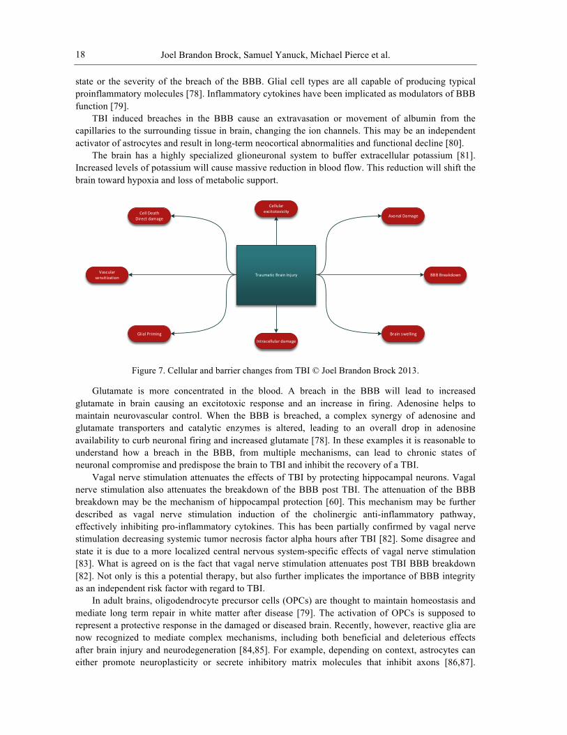
Figure 7. Cellular and barrier changes from TBI © Joel Brandon Brock 2013.
Glutamate is more concentrated in the blood. A breach in the BBB will lead to increased glutamate in brain causing an excitotoxic response and an increase in firing. Adenosine helps to maintain neurovascular control. When the BBB is breached, a complex synergy of adenosine and glutamate transporters and catalytic enzymes is altered, leading to an overall drop in adenosine availability to curb neuronal firing and increased glutamate [78]. In these examples it is reasonable to understand how a breach in the BBB, from multiple mechanisms, can lead to chronic states of neuronal compromise and predispose the brain to TBI and inhibit the recovery of a TBI.
Vagal nerve stimulation attenuates the effects of TBI by protecting hippocampal neurons. Vagal nerve stimulation also attenuates the breakdown of the BBB post TBI. The attenuation of the BBB breakdown may be the mechanism of hippocampal protection [60]. This mechanism may be further described as vagal nerve stimulation induction of the cholinergic anti-inflammatory pathway, effectively inhibiting pro-inflammatory cytokines. This has been partially confirmed by vagal nerve stimulation decreasing systemic tumor necrosis factor alpha hours after TBI [82]. Some disagree and state it is due to a more localized central nervous system-specific effects of vagal nerve stimulation [83]. What is agreed on is the fact that vagal nerve stimulation attenuates post TBI BBB breakdown [82]. Not only is this a potential therapy, but also further implicates the importance of BBB integrity as an independent risk factor with regard to TBI.
In adult brains, oligodendrocyte precursor cells (OPCs) are thought to maintain homeostasis and mediate long term repair in white matter after disease [79]. The activation of OPCs is supposed to represent a protective response in the damaged or diseased brain. Recently, however, reactive glia are now recognized to mediate complex mechanisms, including both beneficial and deleterious effects after brain injury and neurodegeneration [84,85]. For example, depending on context, astrocytes can either promote neuroplasticity or secrete inhibitory matrix molecules that inhibit axons [86,87].
Traumatic Brain Injury
Cell DeathDirect damage Axonal Damage
Vascular sensitization BBB Breakdown
Glial Priming Brain swelling
Intracellular damage
Cellular excitotoxicity
Traumatic Brain Injury Outcomes and Management
19
Similarly, microglia are now known to release both pro-recovery and neurotoxic factors [88,89,90]. OPCs can also release multiple factors to modulate neighboring cells and the microenvironment [91] These intercellular signals may be especially important, since perturbations in the blood-brain barrier (BBB) are known to be a critical part of white matter pathology in a wide range of CNS disorders [92,93]. OPCs in adult brain are precursor cells for white matter remodeling, and repair injury and demyelination. They can play a surprisingly deleterious role in cerebrovascular injury and demyelination in white matter [79].
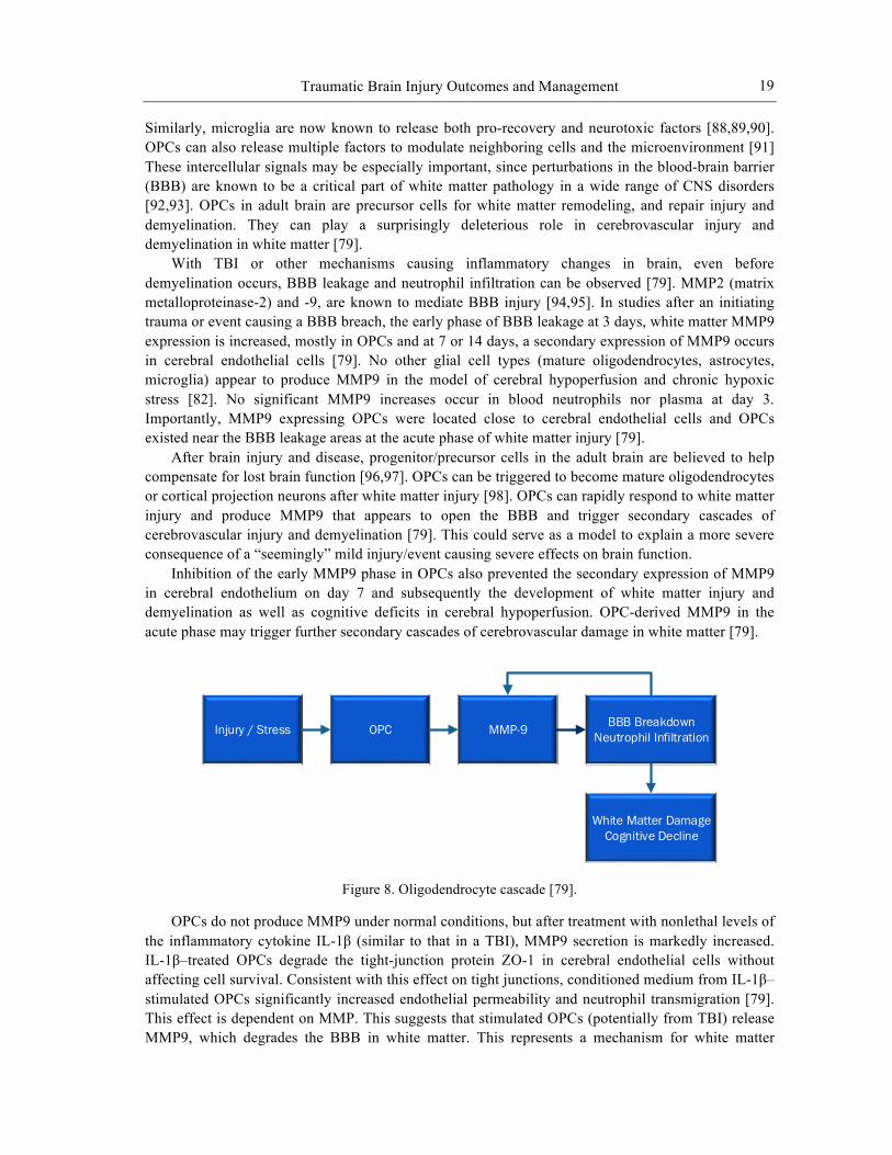
With TBI or other mechanisms causing inflammatory changes in brain, even before demyelination occurs, BBB leakage and neutrophil infiltration can be observed [79]. MMP2 (matrix metalloproteinase-2) and -9, are known to mediate BBB injury [94,95]. In studies after an initiating trauma or event causing a BBB breach, the early phase of BBB leakage at 3 days, white matter MMP9 expression is increased, mostly in OPCs and at 7 or 14 days, a secondary expression of MMP9 occurs in cerebral endothelial cells [79]. No other glial cell types (mature oligodendrocytes, astrocytes, microglia) appear to produce MMP9 in the model of cerebral hypoperfusion and chronic hypoxic stress [82]. No significant MMP9 increases occur in blood neutrophils nor plasma at day 3. Importantly, MMP9 expressing OPCs were located close to cerebral endothelial cells and OPCs existed near the BBB leakage areas at the acute phase of white matter injury [79].
After brain injury and disease, progenitor/precursor cells in the adult brain are believed to help compensate for lost brain function [96,97]. OPCs can be triggered to become mature oligodendrocytes or cortical projection neurons after white matter injury [98]. OPCs can rapidly respond to white matter injury and produce MMP9 that appears to open the BBB and trigger secondary cascades of cerebrovascular injury and demyelination [79]. This could serve as a model to explain a more severe consequence of a “seemingly” mild injury/event causing severe effects on brain function.
Inhibition of the early MMP9 phase in OPCs also prevented the secondary expression of MMP9 in cerebral endothelium on day 7 and subsequently the development of white matter injury and demyelination as well as cognitive deficits in cerebral hypoperfusion. OPC-derived MMP9 in the acute phase may trigger further secondary cascades of cerebrovascular damage in white matter [79].
Injury / Stress OPC MMP-9
White Matter DamageCognitive Decline
BBB BreakdownNeutrophil Infiltration
Figure 8. Oligodendrocyte cascade [79].
OPCs do not produce MMP9 under normal conditions, but after treatment with nonlethal levels of the inflammatory cytokine IL-1β (similar to that in a TBI), MMP9 secretion is markedly increased. IL-1β–treated OPCs degrade the tight-junction protein ZO-1 in cerebral endothelial cells without affecting cell survival. Consistent with this effect on tight junctions, conditioned medium from IL-1β–stimulated OPCs significantly increased endothelial permeability and neutrophil transmigration [79]. This effect is dependent on MMP. This suggests that stimulated OPCs (potentially from TBI) release MMP9, which degrades the BBB in white matter. This represents a mechanism for white matter
Joel Brandon Brock, Samuel Yanuck, Michael Pierce et al.
20
disease at an early stage, and creates further damage and downstream inflammation and demyelination. The state of the BBB both prior to and immediately after injury plays a major role in the probability of, the severity, and the long-term consequences a TBI.
TBI and Its Effect on the Adrenals and Hormones Deficiencies in circulating hormones may not be apparent immediately after injury but can be
demonstrated days to weeks thereafter. Aimaretti et al. reported an incidence of pituitary dysfunction in 33% of TBI patients 3 months after injury, complete panhypopituitarism in 5.7%, multiple defects in another 5.7% and a single hormone deficit in 21%. Secondary hypoadrenalism was found in 8.5% but considering those with panhypopituitarism or multiple hormone abnormalities that might include the adrenal hormones, this incidence may be as high as 20% [99].
In a prospective comparison of 80 TBI patients to 41 trauma patients without TBI, measuring cortisol and ACTH levels twice daily for 9 days after injury, adrenal failure, which was defined as two consecutive cortisols of ≤ 15µg/dL or one cortisol of ≤ 5µg/dL, occurred in 53% of the TBI patients at a mean time of 2.4 days and suggested a secondary cause. Patients with adrenal failure were more severely injured, demonstrated more episodes of hypotension and more often required vasoactive drug support [100]. From such data, it would appear that the risk of secondary hypoadrenalism after TBI is somewhat higher than 25%.
Recently published guidelines recommend acute endocrine evaluation only for patients with documented fractures in the sella turcica or diabetes insipidus. Other authors, however, suggest early endocrine assessment for all patients with moderate-to-severe TBI and routine endocrine testing for all TBI patients at 3 and 12 months. It is also suggested that evaluation be performed for primary and secondary adrenal failure in patients that demonstrate continuing hyponatremia or hypoglycemia or require persistent vasoactive drug treatment during acute care [99].
Endocrine evaluation should include a baseline blood cortisol concentration, a serum ACTH measurement and thyroid studies before the administration of any preparation containing steroids. Although some authors recommend a morning cortisol blood test, stress decreases the normal diurnal variation in cortisol release, making random values acceptable. In both primary and secondary adrenal failure this measurement should be low. The actual concentration that defines a low value is somewhat controversial because of the expected increase in cortisol during ‘stress’ caused by TBI. Therefore, values between 200 and 700 nmol/l or over 15mgm/dl are suggested as minimal basal concentrations expected after trauma [100].
HPA (hypothalamic-pituitary-adrenal) injury may also produce underproduction of thyroid stimulating hormone (TSH) as ‘central’ hypothyroidism, indicated by low TSH and tetraiodothyronine (T4) blood concentrations. The ‘euthyroid sick syndrome’ is expected in such critically injured patients, but in that condition the TSH remains normal. Growth hormone and the several gonadal hormones produced by the pituitary gland may also be low but do not require acute replacement, although treatment during later recovery will be important [100].
Utilization of the traditional ‘high-dose’ (250 mg) versus ‘low-dose’ (1 mg) ACTH stimulation test remains controversial. The higher pharmacological amount may allow a partially dysfunctional gland to release a sufficient amount of hormone to appear falsely normal, while the lower dose is perhaps more difficult to accurately administer. Similarly, criteria regarding the magnitude of response that confirms a responsive adrenal gland have been controversial. Criteria include either a specific post-stimulation (usually 60 min) cortisol concentration (e.g., above 500 nmol/l, or 25 mg/dl) or a specified incremental increase from basal levels (e.g., >250 nmol/l or >9 mg/dl). Bernard et al. found 78% of their TBI patients had a basal cortisol value of under 414 nmol/l, but after a 250 mg ACTH stimulation, only 13% failed to increase the serum cortisol concentration by less than 250 nmol/l [101]. These data again emphasize that the diagnosis of hypoadrenalism often depends upon the criteria selected [100].
Traumatic Brain Injury Outcomes and Management
21
Cortisol is measured in the blood as both an unbound (‘free’) form, which is the biologically active hormone, and as cortisol bound with cortisol-binding globulin (CBG). CBG is commonly low in critical illness/injury, especially when the serum albumin is less than 2.5 gm/dl and may be one cause of an apparent low cortisol serum concentration. Measurement of free-cortisol, therefore, has been suggested as the more accurate assessment of adrenal output. Testing methods for CBG and free cortisol, however, are rarely available [99].
Acute hypoadrenalism after TBI may contribute to hypotension, hyponatremia or hypoglycemia during patient care. Its incidence remains unclear due to variable definitions and testing methods, but appears to be approximately 25%, including both secondary causes related to injury to the central hypothalamic-pituitary axis and primary adrenal failure. We recommend serum testing of basal cortisol, ACTH, and post-ACTH stimulation cortisol at 60 min when hypoadrenalism is suspected. Basal cortisol below 15 mg/dl suggests either primary or secondary adrenal failure and may warrant treatment. Failure of the cortisol concentration to increase by at least 9 mg/dl after stimulation suggests primary adrenal gland hypoadrenalism. Treatment with intravenous hydrocortisone during acute care should be initiated if clinical circumstances warrant. Thyroid function should also be evaluated and, if needed, hormone replacement should be provided if adrenal insufficiency is treated. It is generally recommended that even if acute treatment is not given, all patients should be evaluated for adrenal, thyroid and growth hormone deficiency 3 months after severe TBI [99].
It has been previously reported that experimental mild traumatic brain injury results in increased sensitivity to stressful events during the first post-injury weeks, as determined by analyzing the hypothalamic-pituitary-adrenal (HPA) axis regulation following restraint-induced stress. This is the same time period when rehabilitative exercise has proven to be ineffective after a mild fluid-percussion injury (FPI) [100]. These findings suggest that the increased sensitivity to stressful events during the first post-injury weeks, after a mild FPI, has an impact on hippocampal neuroplasticity [100].
An earlier paper described an increased sensitivity to restraint-induced stress during the first two post-injury weeks as indicated by increases in corticosterone (CORT) and ACTH compared to uninjured rats [100]. The stress response involves the activation of the HPA axis resulting in the release of ACTH from pituitary cells. ACTH stimulates the adrenal gland to release glucocorticoids, such as CORT, which in turn results in the inhibition of ACTH secretion [102].
It has been well characterized that stress decreases neuronal plasticity and favors neurodegeneration [103]. The effects of stress on the central nervous system are most notable within the hippocampus where it substantially influences neuronal excitability and long-term potentiation (LTP). The hippocampus has a high density of glucocorticoid receptors [104]. These receptors exert a variety of effects besides the autoregulation of the stress response, such as influencing mood, learning and memory [105]. Moreover, stress-related increases in glucocorticoids have been associated with cell death and cognitive impairments [106]. Among the effects of stress is the inhibition of hippocampal brain-derived neurotrophic factor (BDNF) [107]. The anatomical and vascular characteristics of the hypothalamic-pituitary complex increase its vulnerability during TBI. Particularly, diffuse TBI, where metabolism and neural connectivity are compromised [108].
Given the above-mentioned effects of glucocorticoids on hippocampal synaptic plasticity and BDNF expression, it is feasible that impaired neuroendocrine function interferes with BDNF-mediated restorative processes after TBI. For example, a hyper-response to stress following TBI may play a role in the inability to increase BDNF during the subacute period, as seen in rats with a mild injury [109].
Affective disorders and cognitive impairments after TBI play a substantial part in decreasing quality of life. A dysregulation of the stress response has been linked to affective disorders in TBI patients. In effect, alterations in the regulation of the HPA axis contribute to negative mood states
Joel Brandon Brock, Samuel Yanuck, Michael Pierce et al.
22
associated with depression [110]. In addition to showing a hyper-responsiveness to stress after TBI, we now provide evidence that post-injury stress has an effect on BDNF regulation within the hippocampus. The effects of stress on cognitive abilities also need to be considered, particularly given BDNF’s effects on plasticity. An increase in glucocorticoids, in human subjects, is associated with impaired memory and hippocampal deterioration [111]. These data add some pieces to the puzzle in understanding the hyper-response and the delay of exercise-induced increases in BDNF during the subacute period. However, they also emphasize the need for more studies regarding HPA dysregulation after a mild TBI. Understanding some of the molecular mechanisms influencing the response to stress will allow us to better address posttraumatic affective and behavioral disorders as well as enhancing rehabilitative therapies.
There were 113 charts that were retrospectively reviewed of traumatic brain injury patients within 10 days of their injury. They all had a high-dose corticotropin stimulation test performed because of haemodynamic instability. Blood cortisol concentrations were measured at baseline, 30 and 60 minutes after the administration of high-dose corticotropin. The incidence of adrenal insufficiency was determined according to various definitions used in the literature [101].
Primary adrenal insufficiency defined by an abnormal baseline cortisol concentration and an abnormal response to the high-dose corticotropin stimulation test was present in 13–28% of patients according to the cut-off values used. The incidence of adrenal insufficiency varies from 25 to 100% in the first 10 days after traumatic brain injury when the charts of 113 traumatic brain injury patients were reviewed [101].
Adrenal insufficiency has emerged in recent years to be a crucial and prevalent problem in intensive care. Hypopituitarism has recently been reported to occur in 35–80% of patients in rehabilitation following head injury. Adrenal insufficiency accounts for 30–50% of these cases [101]. Underestimation of the true incidence of adrenal insufficiency and potentially under-treated patients may be occurring by using only the low-dose stimulation test to define adrenal insufficiency [101].
A concentration of 414 nmol/l is considered to be the normal minimum value in response to severe stress, particularly hypotension. Hence, it is viewed as the minimal appropriate concentration for intensive care patients. A concentration below 690 nmol/l has been suggested instead since patients undergoing surgery or sustaining trauma almost invariably have cortisol concentrations above this value. The intensive care literature tends to support this view. Response to high-dose corticotropin stimulation test, in a study by Annane and colleagues, suggested a rise in plasma cortisol concentration by >250 nmol/l was the appropriate response of the adrenal glands in intensive care patients with sepsis [112]. This definition is probably the most widely used cut-off to interpret a stimulation test in intensive care. Classically, a rise above 500 or 550 nmol/l is considered normal based on the response of non-critically ill patients to insulin-induced hypoglycaemia [101].
Primary adrenal insufficiency defined by an abnormal baseline and abnormal post high-dose corticotropin stimulation test cortisol concentration was present in 13–28% of patients depending on whether a 60 or 30 min sampling time after high-dose corticotropin was used, respectively. Whichever definition was used, the incidence of primary adrenal insufficiency is markedly reduced if the 60 min (rather than the 30 min) cortisol values were used to define adrenal insufficiency [101].This study showed that the incidence of adrenal insufficiency in severe traumatic brain injury varies enormously depending on the definition used. The incidence of adrenal insufficiency defined by baseline cortisol concentrations may be as high as 100%. Primary adrenal insufficiency accounts for 13–25% of the cases. It also demonstrated that sampling for cortisol at 60 min after a high-dose corticotropin stimulation test is more appropriate than doing so at 30 min. 414 mmol/l is probably a more useful cut off in a traumatic brain injury population [103].
Although fatigue is a common experience in the general population it is particularly bothersome to those with TBI [113]. Depending on the scales used to measure fatigue and time since injury, its
Traumatic Brain Injury Outcomes and Management
23
prevalence has been reported in 16–80% of individuals after injury. Associated symptoms include depression, sleep disturbance, pain, cognitive and motor disturbances [113].
Neuroendocrine abnormalities as manifested by pituitary dysfunction can occur after TBI. One-to-several hypothalamic-pituitary axes may be affected; posterior pituitary dysfunction typically resolves during the first several months post-injury, whereas anterior pituitary problems are more likely to persist [113].
Although several lines of evidence indicate that TBI may predispose the pituitary to injury, neuroendocrine dysfunction is rarely considered in current TBI management. Autopsy studies of fatal head-injury victims confirm that up to one third sustain anterior pituitary gland necrosis. Moreover, numerous case reports, retrospective reviews, and recent prospective cohort studies have documented acute and chronic posttraumatic hypopituitarism [100].
Activation of the HPA axis is an important protective response during critical illness. Untreated adrenal insufficiency (AI) may lead to hemodynamic instability and poor outcome. [100] The molecular mechanism of the effects of glucocorticoids on chronic inflammation is not well understood, but there is increasing evidence that they inhibit the action of transcription factors such as AP-1 and NF-kB [114]. Glucocorticoids are potent inhibitors of the activation of NF-kB, which may account for most of their anti-inflammatory actions [115]. Glucocorticoids are effective inhibitors of NF-kB, but they have endocrine and metabolic side effects when given systemically [115].
Activation of NF-kB, for example by cytokines, is blocked by glucocorticoids. Glucocorticoid-receptor complexes bind to the p65 subunit of NF-kB, and this prevents NF-kB activation of the inflammatory genes. Synthesis of nuclear factor of kappa light polypeptide gene enhancer in B-cells inhibitor, alpha (IkBα) is stimulated by the binding of glucocorticoid-glucocorticoid-receptor complexes to a glucocorticoid response element in the promoter region of the IkBα gene [115]. It may be unwise to block the activation of NF-kB for prolonged periods, because the factor plays such a critical part in the immune response and other defensive responses [115].
Part I revealed a wide range and number of cellular and tissue/hormone processes that can be measured and manipulated. Part II describes clinical level measures and interventions.
PART II: CLINICAL CONSEQUENCES AFFECTING SPECIFIC BRAIN SYSTEMS
Neuroanatomical Considerations
Evidence of anatomical brain damage has long been a hallmark in diagnosis of brain injury. For the chronic post-concussion patient this has historically been elusive. Researchers consider concussion to be the result of mechanical trauma to the head or neck that leads to functional impairment without overt structural damage [115]. This results in no gross changes or brain abnormalities seen on imaging [116,117]. A traumatic head injury mechanism combined with a lack of positive findings on imaging is the primary diagnostic scenario. This, the symptom checklists, the opinion of a physician familiar with the patient and ongoing observation by family, coaches or the patient themselves, are standard diagnostic tools. These are commonly used strategies in identifying the presence of concussion and are relied upon for clinical, return to activity and legal purposes in most settings [118,119,120,121].
Current neuroscience has reached a point where evidence of the neuroanatomical consequences of concussion can be identified. The initiating structural damage appears to result from stretching and disruption of neuronal, glial and axonal cell membranes; while cell bodies and myelin sheaths are less affected [122,123]. These injuries are microscopic, involving cellular structures, as well as

Joel Brandon Brock, Samuel Yanuck, Michael Pierce et al.
24
microvasculature and the blood-brain barrier. Although techniques to identify microscopic structural damage are not readily available to clinicians, the pursuit of this information is clinically relevant. Investigators looking at extent of tissue damage using advance imaging techniques have been able to use the data in predicting individuals who will likely have an uncomplicated recovery from concussion versus those who progress to chronic post-concussion syndrome [124]
Postmortem studies of individuals who survived concussion and later died due to apparently unrelated circumstances have provided early evidence of microscopic brain damage in concussion. These types of studies, performed as early as the 1950’s and 1960’s, continue to provide evidence of damage post-concussion. Historically, the overall impression has been that post-concussion brain damage occurs diffusely in the white matter [155,126] More recently diffusion tensor imaging, tractography, perfusion studies and functional neuroimaging have been utilized in identifying areas of damage in individuals with mild traumatic brain injury. These advanced neuroimaging techniques support the notion of axonal and vascular lesions [128,129,130, 131]. Additionally, a mouse model of concussion has provided evidence that degeneration in the gray matter, involving extensive dendrite degeneration and synapse reduction with very minimal cell death, may be a major neuropathological feature in mTBI [132]. Chronic head injury at the concussive or subconcussive level has been associated with morphological change in the brain that is similar to those seen in Alzheimer’s disease and other neurodegenerative disorders. Cortical degeneration with neurofibrillary tangles, neurite heads, neuronal dropout and accumulation of Tau and transactive response DNA-binding protein 43 (TDP-34) proteins are found the human brain chronically after repetitive concussion and subconcussion trauma [133,134,135,136]
Figure 9. Neuroanatomical damage at the cellular level © Joel Brandon Brock 2013.
Work has been done to identify which regions of the brain are most often affected in concussion. In severe and moderate brain injury the location of damage is fairly consistent with proximity to mechanical trauma, contrecoup deformation, areas of the brain in contact with more rigid structures and those displaced by swelling [137,138]. For the chronic post-concussion patient there is less predictability in the specific region of damage. Medial frontal, dorsolateral frontal, occipital, subcortical and corpus callosum have all been reported as damaged in this population [113, 139,140,141] In a subset of chronic post-concussion patients with depression, the frontal and temporal white matter have been identified as injured [142]. Corticocerebellar circuitry disruption has been found as well [115,124,143] Structural involvement of the thalamus was inferred using graph theory analysis and proposed as a useful marker in identifying mTBI [144]. Sheering stresses around the brainstem have been investigated by recreating a series of recorded head to head collisions in football and analyzing the threshold of force that may be necessary to produce a lasting concussion syndrome
Brain DamageStructure damage
Changes in Neurofilament and microtubles
Changes in protein
replication and epigenetic factors
Changes in mitochondrial
function
Changes in secretory vesicles
Changes in lysosomes
Changes in synaptic capacity
Traumatic Brain Injury Outcomes and Management
25
[145]. Additionally, the tissue stressor from concussive trauma has been associated with vascular damage to both the small vessels and integrity of the blood-brain barrier [146,147]
The immediate trauma associated concussion results in a host of other changes at the cellular and molecular levels. They include involvement of the immune and endocrine systems. They affect the metabolism and biochemistry of the brain, as well as distant sites in the body [148,149]. There is very strong and growing evidence that both the immediate and ongoing “non-anatomical” consequences are associated with the vast majority of the difficulties faced by the post-concussion patient [150,151,152].
Visual and Vestibular Dysfunction in the mTBI Patient Beyond neurocognitive testing, many additional methodologies have been evaluated to assess the
consequences and natural history of mTBI. These forms of functional analysis offer new windows for the clinician to assess the severity of mTBI and monitor response to treatment and level of recovery. Further, they imply promising new avenues of treatment for the mTBI patient.
Vestibular dysfunction in mTBI Vestibular impairment is a common consequence of mTBI. As many as 65% of mTBI pateints
will experience some form of vestibular dysfunction during their recovery [153]. Vestibular lesions have been shown to directly produce central inflammatory changes. Unilateral vestibular deafferentation has been demonstrated to result in significant microglial and astroglial activation in the vestibular nuclei [154]. Rat models of vestibular lesion via arsanilate transtympanic injection manifest significant central vestibular inflammatory changes. Elevations in the number of tumor necrosis factor alpha immunoreactive (TNFa-Ir) cells in the bilateral medial and inferior vestibular nuclei can be seen as early as 4 hours after vestibular lesion, with NF-kB upregulation following 8 hours after the lesion, and manganese superoxide dismutase (MnSOD) elevation after 24 hours. Similar changes have been demonstrated in rat models of mechanical unilateral vestibular deafferentation [140]. As a consequence, thorough evaluation and management of vestibular dysfunction in mTBI patients may show promise as a means of limiting central inflammatory processes.
Benign paroxysmal positional vertigo is the most common form of posttraumatic vertigo [139]. As this is a relatively straightforward condition to evaluate via Dix-Hallpike testing, and manage with canalith repositioning maneuvers, Dix-Hallpike testing should be employed in all mTBI patients with vestibular complaints [139]. Sherer et al. evaluated blast-induced mTBI patients reporting persistent dizziness via videonystagmography (VNG), rotational chair, cervical vestibular-evoked myogenic potentials, computerized dynamic posturography, and self-report measures [155].
Evidence of central and peripheral vestibular dysfunction was identified at a higher frequency in symptomatic patients. Abnormal nystagmus or oculomotor findings were present in 50% of symptomatic subjects. Rotational chair testing found evidence of peripheral vestibulopathy in 25% of symptomatic patients, and central vestibular pathology in 17% of symptomatic subjects [141].
Computerized dynamic posturography (CDP) has been proposed as a means to monitor the status of vestibular function. CDP testing assesses the patient’s ability to maintain postural stability against a standardized series of balance and postural challenges. Approximate entropy (ApEn), a measurement of how likely a postural correction is to recur within a time series, is thought to decrease with mTBI [156]. A person swaying in a predictable manner represents a lowered capacity to adapt to subtle changes in environmental stability, and thus lower postural control dynamics. ApEn has been shown to decrease with mTBI, and shifts in stabilization strategies toward greater anterior-posterior and decreased medial-lateral control [142]. This raises the likelihood that concussed patients have a chronic risk of falls without appropriate neurorehabilitation.
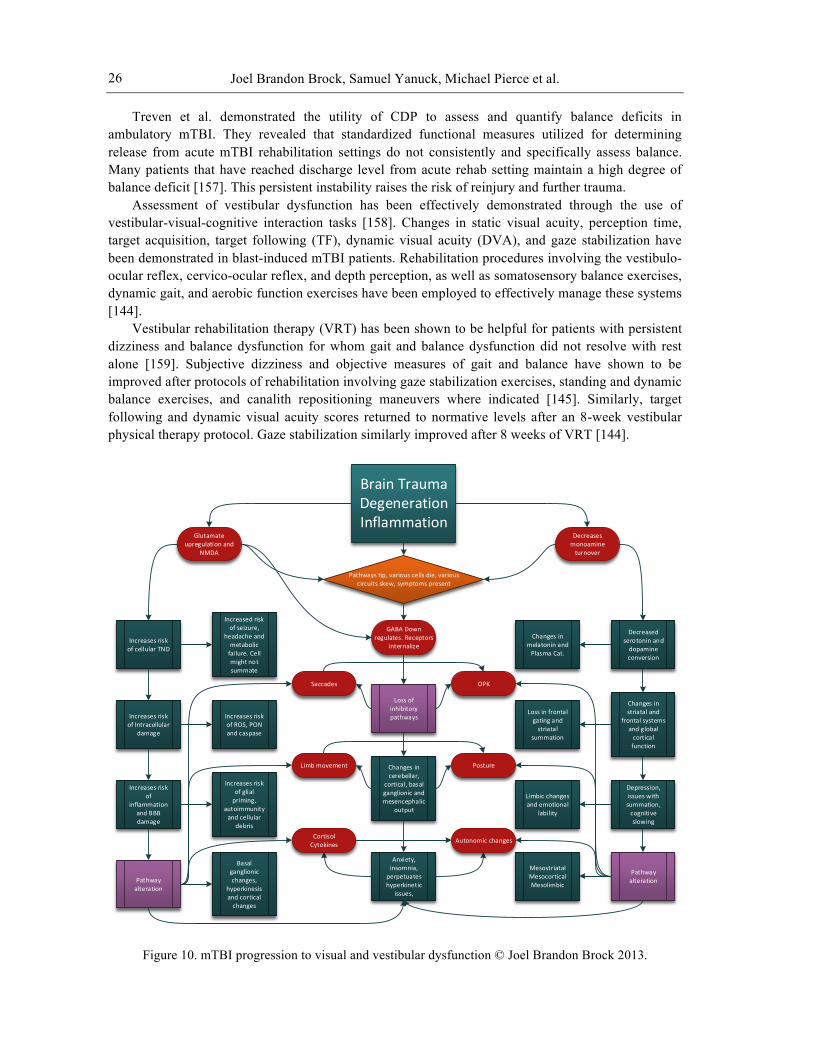
Joel Brandon Brock, Samuel Yanuck, Michael Pierce et al.
26
Treven et al. demonstrated the utility of CDP to assess and quantify balance deficits in ambulatory mTBI. They revealed that standardized functional measures utilized for determining release from acute mTBI rehabilitation settings do not consistently and specifically assess balance. Many patients that have reached discharge level from acute rehab setting maintain a high degree of balance deficit [157]. This persistent instability raises the risk of reinjury and further trauma.
Assessment of vestibular dysfunction has been effectively demonstrated through the use of vestibular-visual-cognitive interaction tasks [158]. Changes in static visual acuity, perception time, target acquisition, target following (TF), dynamic visual acuity (DVA), and gaze stabilization have been demonstrated in blast-induced mTBI patients. Rehabilitation procedures involving the vestibulo-ocular reflex, cervico-ocular reflex, and depth perception, as well as somatosensory balance exercises, dynamic gait, and aerobic function exercises have been employed to effectively manage these systems [144].
Vestibular rehabilitation therapy (VRT) has been shown to be helpful for patients with persistent dizziness and balance dysfunction for whom gait and balance dysfunction did not resolve with rest alone [159]. Subjective dizziness and objective measures of gait and balance have shown to be improved after protocols of rehabilitation involving gaze stabilization exercises, standing and dynamic balance exercises, and canalith repositioning maneuvers where indicated [145]. Similarly, target following and dynamic visual acuity scores returned to normative levels after an 8-week vestibular physical therapy protocol. Gaze stabilization similarly improved after 8 weeks of VRT [144].
Figure 10. mTBI progression to visual and vestibular dysfunction © Joel Brandon Brock 2013.
Brain TraumaDegenerationInflammation
Glutamate upregulation and
NMDA
Decreases monoamine turnover
GABA Down regulates. Receptors
internalizeIncreases risk of cellular TND
Increases risk of Intracellular
damage
Increases risk of
inflammation and BBB damage
Pathway alteration
Increased risk of seizure,
headache and metabolic failure. Cell might not summate
Increases risk of ROS, PON and caspase
Increases risk of glial priming,
autoimmunity and cellular
debris
Basal ganglionic changes,
hyperkinesis and cortical changes
Loss of inhibitory pathways
Changes in cerebellar,
cortical, basal ganglionic and mesencephalic
output
Anxiety, insomnia,
perpetuates hyperkinetic
issues,
Decreased serotonin and dopamine conversion
Changes in striatal and
frontal systems and global cortical function
Depression, issues with summation, cognitive slowing
Pathway alteration
Changes in melatonin and Plasma Cat.
Loss in frontal gating and striatal
summation
Limbic changes and emotional
lability
MesostriatalMesocorticalMesolimbic
OPKSaccades
Limb movement Posture
Autonomic changesCortisol Cytokines
Pathways tip, various cells die, various circuits skew, symptoms present
Traumatic Brain Injury Outcomes and Management
27
Visual Dysfunction in mTBI Visual dysfunction has been shown to be extremely prevalent in the mTBI population. Ciuffreda
et al. assessed for frequency of occurrence of oculomotor dysfunction in TBI, considering accommodation, version, vergence, strabismus, and cranial nerve palsy. They found that oculomotor dysfunction is the norm in TBI [160].
Convergence insufficiency (CI) has been shown to be present in roughly 9% of the visually symptomatic TBI population without simultaneous vestibular dysfunction, saccade or pursuit dysfunction, cranial nerve palsies, visual field deficits, visuospatial neglect, or nystagmus. In many cases it appears that CI may occur in the absence of obvious dysfunction in visual and vestibular systems, rendering a thorough evaluation even more necessary [161].
The neural substrates of eye movements involve diffuse networks including the frontal lobes, parietal lobes, and cerebellar cortices. These systems also serve within networks that facilitate executive functions and higher cognitive experience. Impairments of eye movements have thus been demonstrated to have considerable clinical utility in both diagnosing and rehabilitating the mTBI patient.
TBI patients demonstrate aberrancies in predictive components of smooth pursuit eye movements [162]. These deficits in average target prediction, eye position error, and eye position variability positively correlated with executive function and attentional measures on the California Verbal Learning test (CVLT-II), without apparent reduction in IQ noted on the Weschler Abbreviated Scale of Intelligence (WASI). Impairment of predictive smooth pursuit eye movements thus may be a sensitive indicator of impaired attention processing.
Smooth pursuit movements coupled with target blanking, wherein the target being pursued is intermittently obscured, necessitates the generation of greater predictive eye movements [163]. This process requires greater reliance on brain regions involved in cognitive processing. Deficits in CVLT-II performance have been shown to more positively correlate with oculomotor variability during target blanking than during target tracking [163].
Maruta et al. demonstrated the presence of diffuse axonal injury (DAI) within the frontal lobes in mTBI via diffusion tensor imaging. Fractional Anisotropy indicates frequent involvement of the anterior corona radiata, the uncinate fasciculus, the genu of the corpus callosum, and the cingulum bundle. They compared various measures of eye movement performance to fractional anisotropy (FA) and diffusion tensor imaging (DTI), as well as to neurocognitive tests. Variability of gaze position errors during predictive smooth pursuit proved to be a sensitive measure that correlated with neurocognitive testing data and DTI findings. The degree of variability correlated with the mTBI spectrum severity [164]. They determined that performance variability during predictive visual tracking is a valid and useful indicator of damage to frontal white matter tracts, and is similarly indicative of impaired cognitive function [164].
Similar correlations have been shown between dysfunction in saccade eye movements and measures of neurocognition. Mulhall indicated that bedside tests of saccades utilizing infrared oculography (IRO) produced saccadic impairment data that positively correlated with impairments on neurocognitive tests [165]. Recovering TBI patients were shown to have decreased rates of self-paced saccades, impaired ability to suppress inappropriate saccades in single memory-guided and antisaccade tests, prolonged saccadic latencies, and hypometric saccades in visually guided reflex saccade tests. This implies that IRO testing should be part of any mTBI management protocol [165].
Increased duration of reflexive saccade latencies has also been shown to be present in mTBI [166]. Portable saccadometry has been utilized to evaluate boxers pre- and post-bouts. Latency distributions have been shown to be significantly altered after blows to the head, with these effects reversible over a short period of time. Latency requires cortical decision mechanisms, and is directly affected by processes that affect cerebral function [166].
Joel Brandon Brock, Samuel Yanuck, Michael Pierce et al.
28
Drew et al. investigated the relationship between dysfunctional visuospatial orientation processes and difficulty with orienting attention. Saccadic targets were presented with varying temporal gaps and reaction times were assessed. They found that mTBI patients demonstrated significantly longer saccadic reaction times than controls when the time gap between saccades was short, but not when the temporal gap was long. Shifts in attention require disengagement from the point of fixation, both cognitively and visually. This process appears impaired in mTBI [167].
Heitger found that eye movement function was impaired in post-concussion syndrome, with eye movement deficits found on measures relating to motor functions executed under both conscious and semi-conscious control, as well as on several eye movement functions that are beyond conscious control and indicative of sub-cortical brain function [168]. They demonstrated specific deficits in antisaccade performance, a task that in non-mTBI individuals that requires attentional focus, inhibitory control, working memory, and the ability to generate voluntary goal-directed behavior [169]. They recommended that eye movement testing be utilized with post-concussion syndrome patients to evaluate for incomplete recovery of function [169].
Given the interaction between visual and vestibular function within the maintenance of balance and gait, it is perhaps unsurprising that TBI patients demonstrate abnormal caloric irrigation and optokinetic circularvection testing, as well as lower anterior and posterior and higher medial and lateral center of mass displacements and velocities during gait assessment [170].
Rehabilitation of eye movement dysfunction has been shown to be effective in mTBI patients. Ciuffreda et al. demonstrated a 90% rate of improvement from oculomotor rehabilitation, with reduction in both symptoms and objective findings that persisted at a 2- to 3-month follow-up, demonstrating both the efficacy of optometric rehabilitation and significant neural plasticity in mTBI patients [171].
Novel forms of rehabilitation have been shown to have efficacy in treating the visual and vestibular dysfunction, and may hold promise in the treatment of those same dysfunctions in mTBI patients. Optokinetic stimulation has been shown to activate areas often involved in mTBI, including the prefrontal cortex/frontal eye fields, areas involved in generation of saccades, and modulation of the parieto-insular vestibular cortex, an area involved in visual-vestibular interaction [172]. Repetitive optokinetic stimulation has been shown to be useful for rehabilitation of vestibular deficits and in causing adaptive changes in locomotion in healthy subjects [173,174,175]. Optokinetic stimulation has shown efficacy restoration of mobility in acute stroke patients [176,177]. Vagal nerve stimulation has been shown to be effective in limiting the vasogenic edema that develops secondary to the disruption of the blood-brain barrier in TBI, thus limiting central inflammation and neuronal damage [60,178]. Within a virtual reality environment, combinations of optokinetic stimulation and habituation, visual and physical perturbations, and postural stability exercises have been shown to decrease visual and physical motion intolerance and impairment in static balance [179]. Further research is needed with respect to these interventions.
Given the interaction between the visual and vestibular systems, the frequency of impairment noted in these systems, and their potential influence on central inflammatory processes, thorough and sensitive evaluation of these systems is a prerequisite for effective management of mTBI.
Neuropsychological Effects: Multi-Systems Approach to Persistent Post-Concussion Syndrome (PPCS)
Neurocognitive effects of concussion/TBI are unpredictable. Research to date has given us a wealth of information concerning the mechanism of injury, acute pathophysiology, chronic pathophysiology, imaging, neuro-immune and HPA axis contribution. With all of this information there are still few tools available for clinicians to diagnose the extent of cognitive impact and even fewer tools available for prognosis and treatment. Why is it that some who have suffered more severe TBI have complete resolution of cognitive symptoms but those with a relatively mild history of injury
Traumatic Brain Injury Outcomes and Management
29
have such great difficulty with executive function, attention, anxiety, pain, fatigue, and depression? This leaves clinicians with little choice but to react to symptoms as they occur, thus allowing the potential harm of unrecognized, undiagnosed chronic pathophysiology and dysfunction to fester.
The treatment for persistent post-concussion syndrome (PPCS) due to mTBI does not address the cause, and mostly consists of rest and cognitive behavioral therapy with limited results for those suffering from PPCS [180]. Medications to treat concomitant depression, attention deficit and anxiety are often prescribed.
There are two current sets of research criteria for the post-concussive disorder: the International Classification of Diseases, 10th Edition (ICD-10), and the Diagnostic and Statistical Manual of Mental Disorders, Fourth Edition (DSM-IV). According to the DSM-IV the individual with post-concussion syndrome will have trouble paying attention, concentrating, changing focus from one activity to another, performing more than one mental activity at the same time, remembering information, and learning new information. Three or more of the following symptoms occur after the trauma and last at least 3 months: fatigue; insomnia; headache; vertigo or dizziness; anger that occurs without a good reason; anxiety, depression, or mood swings; and personality changes such as inappropriate behavior, apathy, or lack of spontaneity. The impairment represents a significant decline from pre-trauma functioning and causes significant difficulty with school or workplace performance.
According to the ICD-10, a person must have a history of “head trauma with loss of consciousness” preceding the onset of symptoms by a period of up to 4 weeks and at least three of six symptom categories. These include:
• Headaches, dizziness, general malaise, excessive fatigue, or noise intolerance. • Irritability, emotional lability, depression, or anxiety. • Subjective complaints of concentration or memory difficulty. • Insomnia. • Reduced tolerance to alcohol. • Preoccupation with these symptoms and fear of permanent brain damage. The ICD-10 criteria do not require “objective” evidence of cognitive problems. Both models
attempt to clearly state the symptoms and parameters for diagnosis, however they do not offer much of a direction for understanding pathophysiology once the diagnosis has been made. A clear understanding of the pathophysiology as it relates to multiple systems and circuits will guide research toward better models of prognosis, care and prevention.
Complete or nearly complete recovery from concussion occurs in a timely manner in the majority of individuals, however there is a minority of those who have symptoms that persist for months to years [181]. The research is not clear as to how to predict who will be more likely to experience chronic symptoms. Bigler et al. described PPCS as it is related to structural damage to specific predictable or likely brain regions impacted by brain injury using biomechanical models. Recent advances in imaging using DTI have detected axonal damage previously undetectable. The anatomical regions most likely to suffer structural damage with concussion include the posterior frontal lobe, medial temporal lobe, midbrain structures, fornix, hypothalamus, pituitary, cerebral peduncle, entorhinal cortex and hypothalamus. So, within a few centimeters are critical brain structures that, if affected, could represent the structural basis to many symptoms associated with concussion [182].
Rao and Lyketsos indicate that the PPCS nomenclature is too vague and that the more accurate description is ‘frontotemporal syndrome’ as this better describes the regions most affected by injury. There is considerable evidence linking the vestibular system and objective findings utilizing digital posturography and videonystagmography for ocular involvement and precise evaluation of central circuits involved [183] We recognize that the same symptoms classified as PPCS can be seen in

Joel Brandon Brock, Samuel Yanuck, Michael Pierce et al.
30
people without history of concussion or trauma [184]. This is possibly involving the same pathophysiological characteristics with a different etiology. The methodological quality of neuropsychological research for concussion, although plentiful over the past decade, has lacked scientific rigor [185]. When looking at the confounding factors that must be considered in the design of PPCS research, Bigler notes that the major symptoms of PPCS; fatigue, sleep disorder, headache, dizziness/vertigo, irritability, affective lability, anxiety, apathy and personality changes, share such great overlap that exist with other psychiatric disorders such as depression [182] [182].
Figure 11. Development of mental health problems in the context of mTBI milieu © Joel Brandon Brock 2013.
It appears, as is noted in the research cited above, that PPCS shares a great deal of similarity with depression, anxiety, attention deficit disorder (ADD) and OCD. The common thread that holds PPCS, depression, anxiety, OCD and ADD may be a chronic, immune mediated, inflammatory process also known as immuno-excitotoxicity as well as shared anatomical circuitry [12].
Research conducted which points out the effect peripheral musculoskeletal injury has on cognition is similar to that seen with concussion, indicating that injury itself, regardless of anatomical location can affect objective test scores previously attributed to concussion alone. Thus a narrow assessment of pre- and post-injury neuropsychological scores should be avoided [185]. This finding may indicate that systemic inflammation occurring outside the CNS may play a role in the function of cognitive circuitry.
There appear to be significant shortcomings in conceiving PPCS, mTBI and chronic TBI as a wholly neuropsychological condition requiring a neuropsychological approach to researching treatment, diagnosis and prognosis. A comprehensive model that addresses multi-system physiology including but not limited to sensorimotor processing and neuro-immune function is likely to emerge. The probability of chronic immune inflammatory processes being involved and the likelihood of compromised neural circuitry may be discovered with sensitive analysis of sensorimotor function of neural circuitry shared by cognitive and behavioral centers. A research model can be crafted that incorporates a multisystem approach to function as opposed to simply ameliorating symptoms. With this in mind the direction research can take regarding care must be tangible, quantifiable and very different from rest and cognitive therapy. Research needs to address sensorimotor function of specific
Head Injury
Cognitive
Somatic
Affective
Sleep Disorder
Memory
Consciousness
Attention
AmnesiaConfused
Brain fogVerbal delay
Synaptic and cell capacity
Tau / N degen
Drowsy Disoriented Metabolic capacity
Cortical regions
Head pain
Vestibular
Visual
Peripheral Central
AutonomicsSensitization
Vision/Pur/Sac/OKN
Sleeping more
Sleeping less
Trouble falling asleep
Met / summationFuel
Excitotoxicity
Melatonin / circadian / Th
Anxious
Fatigue / sadness
Lability / Irritable
Dop / Vent mes / Excito GABA
SERT / Monoamine
TND / CIS
CytokinesCellular function
vENGExam
Posurography
Fuel / trans Glut / Gaba
Cell output and fatigue
Inflammationtransmitters
ChemicalCellular function
Chemical Cellular function
Chemical Cellular function
Cellular output Fatigue
Fuel – corticalCellular output
Fuel – corticalCellular output
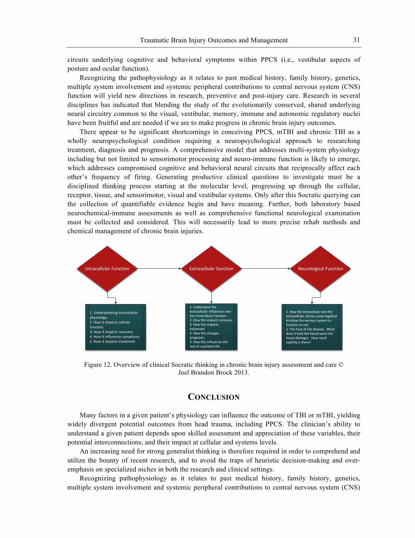
Traumatic Brain Injury Outcomes and Management
31
circuits underlying cognitive and behavioral symptoms within PPCS (i.e., vestibular aspects of posture and ocular function).
Recognizing the pathophysiology as it relates to past medical history, family history, genetics, multiple system involvement and systemic peripheral contributions to central nervous system (CNS) function will yield new directions in research, preventive and post-injury care. Research in several disciplines has indicated that blending the study of the evolutionarily conserved, shared underlying neural circuitry common to the visual, vestibular, memory, immune and autonomic regulatory nuclei have been fruitful and are needed if we are to make progress in chronic brain injury outcomes.
There appear to be significant shortcomings in conceiving PPCS, mTBI and chronic TBI as a wholly neuropsychological condition requiring a neuropsychological approach to researching treatment, diagnosis and prognosis. A comprehensive model that addresses multi-system physiology including but not limited to sensorimotor processing and neuro-immune function is likely to emerge, which addresses compromised cognitive and behavioral neural circuits that reciprocally affect each other’s frequency of firing. Generating productive clinical questions to investigate must be a disciplined thinking process starting at the molecular level, progressing up through the cellular, receptor, tissue, and sensorimotor, visual and vestibular systems. Only after this Socratic querying can the collection of quantifiable evidence begin and have meaning. Further, both laboratory based neurochemical-immune assessments as well as comprehensive functional neurological examination must be collected and considered. This will necessarily lead to more precise rehab methods and chemical management of chronic brain injuries.
Figure 12. Overview of clinical Socratic thinking in chronic brain injury assessment and care © Joel Brandon Brock 2013.
CONCLUSION
Many factors in a given patient’s physiology can influence the outcome of TBI or mTBI, yielding widely divergent potential outcomes from head trauma, including PPCS. The clinician’s ability to understand a given patient depends upon skilled assessment and appreciation of these variables, their potential interconnections, and their impact at cellular and systems levels.
An increasing need for strong generalist thinking is therefore required in order to comprehend and utilize the bounty of recent research, and to avoid the traps of heuristic decision-making and over-emphasis on specialized niches in both the research and clinical settings.
Recognizing pathophysiology as it relates to past medical history, family history, genetics, multiple system involvement and systemic peripheral contributions to central nervous system (CNS)
Intracellular Function Extracellular function Neurological Function
1. Understanding intracellular physiology.2. How it impacts cellular function.3. How it impacts recovery.4. How it influences symptoms.5. How it impacts treatment
1. Understand the extracellular influences over the intracellular function.2. How this impacts recovery.3. How this impacts treatment.4. How this changes prognosis.5. How this influences the rest of a patients life
1. How the intracellular and the extracellular stories come together to allow the nervous system to function or not. 2. The Face of the disease. What does it look like based upon the tissue damages. How much viability is there?
Joel Brandon Brock, Samuel Yanuck, Michael Pierce et al.
32
function will yield new directions in research, preventive and post-injury clinical care. Research in several disciplines has indicated that blending the study of the evolutionarily conserved, shared underlying neural circuitry common to the visual, vestibular, memory, immune and autonomic regulatory nuclei have been fruitful. Their expansion and integration are needed if we are to make progress in improving chronic brain injury outcomes.
In both research and clinical settings, careful examination of many wide-ranging patient measures must be integrated in order to progress, including reflexive eye movements, hormone panels, sensorimotor changes, immune and inflammatory markers, mental and emotional states, and other factors required to appreciate each patient’s uniqueness. History taking will need to expand to include lifestyle factors previously thought unrelated. The health status of individuals pre-concussion will be a significant factor in determining PPCS risk in any individual.
In the research setting, accounting for physiological variables that can impact TBI and mTBI outcomes is essential in appreciating potential non-homogeneity of patient populations enrolled in a given study.
In the clinical setting, accounting for the same physiological variables creates the opportunity to identify key clinical targets that can markedly improve outcomes or, if ignored, stall progress. Once identified, these clinical targets must be addressed with best-matched methodologies, not all of which will be pharmacological. Understanding the post-injury, and ideally the pre-injury status of these patients, utilizing outcome-based multifactorial neurological assessment and treatment are key in the emerging path for effective concussion care.
REFERENCES
[1] Brown GC, Neher JJ. Inflammatory neurodegeneration and mechanisms of microglial killing of neurons. Mol. Neurobiol. 2010;41:242-247.
[2] Griffiths MR, Gasque P, Neal JW. The Multiple Roles of the Innate Immune System in the Regulation of Apoptosis and Inflammation in the Brain. J. Neuropathol Exp Neurol. 2009;68(3):217-226.
[3] Biber K, Neumann H, Inoue K, Boddeke HW. Neuronal ‘On’ and ‘Off’ Signals Control Microglia. Trends Neurosci. 2007;30(11):596-602.
[4] Neumann H. Control of Glial Immune Function by Neurons. Glia. 2001;36:191-199. [5] Neumann H, Kotter MR, Franklin RJ. Debris clearance by microglia: an essential link between
degeneration and regeneration. Brain. 2009;132(Pt 2):288-95. [6] Wraith DC, Nicholson LB. The adaptive immune system in diseases of the central nervous
system. J. Clin. Invest. 2012;122(4):1172-9. [7] Schwartz M, Shechter R.Systemic inflammatory cells fight off neurodegenerative disease. Nat
Rev. Neurol. 2010;6(7):405-10. [8] Yacoubian S, Serhan CN. New endogenous anti-inflammatory and proresolving lipid mediators:
implications for rheumatic diseases. Nat. Clin. Pract Rheumatol. 2007;3(10):570-9. [9] Dilger RN, Johnson RW. Aging, microglial cell priming, and the discordant central
inflammatory response to signals from the peripheral immune system. J. Leukoc Biol. 2008;84(4):932-9.
[10] Mangano EN, Hayley S.Inflammatory priming of the substantia nigra influences the impact of later paraquat exposure: Neuroimmune sensitization of neurodegeneration. Neurobiol Aging. 2009;30(9):1361-78.
[11] Block ML, Zecca L, Hong JS. Microglia-mediated neurotoxicity: uncovering the molecular mechanisms. Nat Rev Neurosci. 2007;8(1):57-69.

Traumatic Brain Injury Outcomes and Management
33
[12] Blaylock RL, Maroon J. Immunoexcitotoxicity as a central mechanism in chronic traumatic encephalopathy – A unifying hypothesis. Surg. Neurol Int. 2011;2:107.
[13] Cunningham C, Campion S, Lunnon K, Murray CL, Woods JF, Deacon RM, Rawlins JN, Perry VH. Systemic Inflammation Induces Acute Behavioral and Cognitive Changes and Accelerates Neurodegenerative Disease. Biol. Psychiatry. 2009;65(4):304-12.
[14] Perry VH, Cunningham C, Holmes C.Systemic infections and inflammation affect chronic neurodegeneration. Nat. Rev. Immunol. 2007 Feb;7(2):161-7.
[15] Cunningham C, Wilcockson DC, Campion S, Lunnon K, Perry VH. Central and Systemic Endotoxin Challenges Exacerbate the Local Inflammatory Response and Increase Neuronal Death during Chronic Neurodegeneration. J. Neurosci. 2005;25(40):9275-84.
[16] Perry VH, Nicoll JA, Holmes C. Microglia in neurodegenerative disease. Nat. Rev. Neurol. 2010 Mar;6:193-201.
[17] Meisel C, Schwab JM, Prass K, Meisel A, Dirnagl U.Central nervous system injury-induced immune deficiency syndrome. Nat. Rev. Neurosci. 2005 Oct;6(10):775-86.
[18] Shaw NA. The neurophysiology of concussion. Prog. Neurobiol. 2002;67(4):281-344. [19] Johnson VE, Stewart W, Smith DH. Traumatic brain injury and amyloid-β pathology: a link to
Alzheimer's disease? Nat. Rev. Neurosci. 2010;11(5):361-70. [20] Henry LC, Tremblay S, Leclerc S, Khiat A, Boulanger Y, Ellemberg D, Lassonde M. Metabolic
changes in concussed American football players during the acute and chronic post-injury phases. BMC Neurol. 2011;11:105.
[21] Das M, Mohapatra S, Mohapatra SS. New perspectives on central and peripheral immune responses to acute traumatic brain injury. J. Neuroinflammation. 2012;9:236.
[22] Yun J, Gaivin RJ, McCune DF, Boongird A, Papay RS, Ying Z, Gonzalez-Cabrera PJ, Najm I, Perez DM. Gene expression profile of neurodegeneration induced by alpha1B-adrenergic receptor overactivity: NMDA/GABAA dysregulation and apoptosis. Brain. 2003;126(Pt 12):2667-81.
[23] Marambaud P, Dreses-Werringloer U, Vingtdeux V. Calcium signaling in neurodegeneration. Mol. Neurodegener. 2009;4:20.
[24] Doble A. The role of excitotoxicity in neurodegenerative disease: implications for therapy. Pharmacol Ther. 1999;81(3):163-221.
[25] Malkus KA, Tsika E, Ischiropoulos H. Oxidative modifications, mitochondrial dysfunction, and impaired protein degradation in Parkinson's disease: how neurons are lost in the Bermuda triangle. Mol. Neurodegener. 2009;4:24.
[26] Green DR, Galluzzi L, Kroemer G. Mitochondria and the autophagy-inflammation-cell death axis in organismal aging. Science. 2011;333(6046):1109-12.
[27] Mizushima N, Levine B, Cuervo AM, Klionsky DJ.Autophagy fights disease through cellular self-digestion. Nature. 2008;451(7182):1069-75.
[28] Babikian T, Prins ML, Cai Y, Barkhoudarian G, Hartonian I, Hovda DA, Giza CC. Molecular and physiological responses to juvenile traumatic brain injury: focus on growth and metabolism. Dev Neurosci. 2010;32:431-441.
[29] Fehm HL, Kern W, Peters A. The selfish brain: competition for energy resources. Prog. Brain Res. 2006;153:129-140.
[30] Hyder F, Rothman DL, Shulman RG. Total neuroenergetics support localized brain activity: Implications for the interpretation of fMRI. PNAS. 202;99:10771-10776.
[31] Shah K, DeSilva S, Abbruscato T. The Role of Glucose Transporters in Brain Disease: Diabetes and Alzheimer’s Disease. Int. J. Mol Sci. 2012;13:12629-12655.
Joel Brandon Brock, Samuel Yanuck, Michael Pierce et al.
34
[32] Prieto R, Tavazzi B, Taya K, Barrios L, Amorini A, Di Pietro V, Marmarou C. Brain energy depletion in a rodent model of diffuse traumatic brain injury is not prevented with administration of sodium lactate. Brain Res. 2011;1404:39-49.
[33] Vemula S, Roder K E. A functional role for sodium-dependent glucose transport across the blood brain barrier during oxygen glucose deprivation. J. Pharmacol Exp. Ther. 2009;328(2)487-495.
[34] Eskandari S, Wright EM, Loo D. (2005, March) Kinetics of the reverse mode of the Na+/glucose cotransporter. J. Membr Biol. 2005;204(204):23-32.
[35] Moreno A, Jego P, Cruz F, Canals S. Neurophysiological, metabolic and cellular compartments that drive neurovascular coupling and neuroimaging signals. Front Neuroenergetics. 2013;5:3.
[36] Shen J. Modeling the glutamate-glutamine neurotransmitter cycle. Front Neurogenetics. 2013;5:1-13.
[37] Caprette DR. Substrate Oxidation: Krebs Reactions. 2005. http://www.ruf.rice.edu /~bioslabs/studies/mitochondria/mitokrebs.html
[38] Stobart JL, Anderson CM. Multifunctional role of astrocytes as gatekeeper of neuronal energy supply. Front Cell Neurosci. 2013;7:38.
[39] Giza CC, Hovda DA. The neurometabolic cascade of concussion. J. Ath Train. 2001;36(3):228-235.
[40] Lavoie S, Allaman I, Petit J, Do KQ, Magistretti PJ. Altered glycogen metabolism in cultured astrocytes from mice with chronic glutathione deficit; relevance for neuroenergetics and schizophrenia. PloS One. 2011;6(7):e22875
[41] Yan EB, Hellewell SC, Bellander B, Agyapomaa DA, Morganti-Kossmann M. Post-traumatic hypoxia exacerbates neurological deficit, neuroinflammation and cerebal metabolism in rats with diffuse traumatic brain injury. J. Neuroinflammation. 2011;8:147.
[42] Ives JC, Alderman M, Stred SE. Hypopituitarism after multiple concussions: a retrospective case study in an adolescent male. J. Ath Train 2007;42(3):431-439.
[43] Agha A, Rogers B, Sherlock M, O’Kelly P, Tormey W, Phillips J, Thompson CJ. Anterior pituitary dysfunction in survivors of traumatic brain injury. J. Clin. Endocrinol. Metab. 2004;89(10):4929-4936.
[44] Oliveira C, Meneguz-Mreno RA, Aguiar-Oliviera MH, Barreto-Filho JA. Emerging role of the GH/IGF-I on cardiometabolic control. Arq Bras Cardiol. 2011;97(5): 434-439.
[45] Chang L. Brain responses to visceral and somatic stimuli in irritable bowel syndrome: a central nervous system disorder? Gastroenterol. Clin. North Am. 2005;34(2):271-9.
[46] Hang CH, Shi JX, Li JS, Wu W, Li WQ, Yin HX. Levels of vasoactive intestinal peptide, cholecystokinin and calcitonin gene-related peptide in plasma and jejunum of rats following traumatic brain injury and underlying significance in gastrointestinal dysfunction. World J. Gastroenterol. 2004;10(6):875-80.
[47] Kwan CL, Diamant NE, Pope G, Mikula K, Mikulis DJ, Davis KD. Abnormal forebrain activity in functional bowel disorder patients with chronic pain. Neurology. 2005;65(8):1268-77.
[48] Bansal V, Costantini T, Ryu SY, Peterson C, Loomis W, Putnam J, Elicieri B, Baird A, Coimbra R. Stimulating the central nervous system to prevent intestinal dysfunction after traumatic brain injury. J. Trauma. 2010;68(5):1059-64.
[49] Bansal V, Costantini T, Kroll L, Peterson C, Loomis W, Eliceiri B, Baird A, Wolf P, Coimbra R. Traumatic brain injury and intestinal dysfunction: uncovering the neuro-enteric axis. J. Neurotrauma. 2009;26(8):1353-9.
[50] Feighery L, Smyth A, Keely S, Baird AW, O'Connor WT, Callanan JJ, Brayden DJ. Increased intestinal permeability in rats subjected to traumatic frontal lobe percussion brain injury. J. Trauma. 2008;64(1):131-7;discussion 137-8.

Traumatic Brain Injury Outcomes and Management
35
[51] Maes M, Kubera M, Leunis JC. The gut-brain barrier in major depression: intestinal mucosal dysfunction with an increased translocation of LPS from gram negative enterobacteria (leaky gut) plays a role in the inflammatory pathophysiology of depression. Neuro Endocrinol Lett. 2008;29(1):117-24.
[52] Leclercq S, Cani PD, Neyrinck AM, Stärkel P, Jamar F, Mikolajczak M, Delzenne NM, de Timary P. Role of intestinal permeability and inflammation in the biological and behavioral control of alcohol-dependent subjects. Brain Behav. Immun. 2012;26(6):911-8.
[53] Lenz A, Franklin GA, Cheadle WG. Systemic inflammation after trauma. Injury. 2007;38(12):1336-45.
[54] Hang CH, Shi JX, Li JS, Wu W, Yin HX. Alterations of intestinal mucosa structure and barrier function following traumatic brain injury in rats. World J. Gastroenterol. 2003;9(12):2776-81.
[55] Santos A, Gonçalves P, Araújo JR, Martel F. Intestinal permeability to glucose after experimental traumatic brain injury: effect of gadopentetate dimeglumine administration. Basic Clin. Pharmacol Toxicol. 2008;103(3):247-54.
[56] Hang CH, Shi JX, Li JS, Li WQ, Yin HX. Up-regulation of intestinal nuclear factor kappa B and intercellular adhesion molecule-1 following traumatic brain injury in rats. World J. Gastroenterol. 2005;11(8):1149-54.
[57] Hang CH, Shi JX, Li JS, Li WQ, Wu W. Expressions of intestinal NF-kappaB, TNF-alpha, and IL-6 following traumatic brain injury in rats. J. Surg. Res. 2005;123(2):188-93.
[58] Ling HP, Li W, Zhou ML, Tang Y, Chen ZR, Hang CH. Expression of intestinal myeloid differentiation primary response protein 88 (Myd88) following experimental traumatic brain injury in a mouse model. J. Surg. Res. 2013;179(1):e227-34.
[59] Fukuda K, Tanno H, Okimura Y, Nakamura M, Yamaura A. The blood-brain barrier disruption to circulating proteins in the early period after fluid percussion brain injury in rats. J. Neurotrauma. 1995;12(3):315-24.
[60] Lopez NE, Krzyzaniak MJ, Costantini TW, Putnam J, Hageny AM, Eliceiri B, Coimbra R, Bansal V. Vagal nerve stimulation decreases blood-brain barrier disruption after traumatic brain injury. J. Trauma Acute Care Surg. 2012;72(6):1562-6.
[61] Szmydynger-Chodobska J, Strazielle N, Gandy JR, Keefe TH, Zink BJ, Ghersi-Egea JF, Chodobski A. Posttraumatic invasion of monocytes across the blood-cerebrospinal fluid barrier. J. Cereb. Blood Flow Metab. 2012;32(1):93-104.
[62] Shlosberg D, Benifla M, Kaufer D, Friedman A. Blood-brain barrier breakdown as a therapeutic target in traumatic brain injury. Nat. Rev. Neurol. 2010;6(7):393-403.
[63] Baguley IJ, Heriseanu RE, Cameron ID, Nott MT, Slewa-Younan S. A critical review of the pathophysiology of dysautonomia following traumatic brain injury. Neurocrit Care. 2008;8(2):293-300.
[64] Kirk KA, Shoykhet M, Jeong JH, Tyler-Kabara EC, Henderson MJ, Bell MJ, Fink EL. Dysautonomia after pediatric brain injury. Dev. Med. Child Neurol. 2012;54(8):759-64.
[65] Hendricks HT, Heeren AH, Vos PE. Dysautonomia after severe traumatic brain injury. Eur. J. Neurol. 2010;17(9):1172-7.
[66] Baguley IJ, Heriseanu RE, Nott MT, Chapman J, Sandanam J. Dysautonomia after severe traumatic brain injury: evidence of persisting overresponsiveness to afferent stimuli. Am. J. Phys. Med. Rehabil. 2009;88(8):615-22.
[67] Nampiaparampil DE. Prevalence of chronic pain after traumatic brain injury: a systematic review. JAMA. 2008;300(6):711-9.
[68] Baguley IJ, Heriseanu RE, Felmingham KL, Cameron ID. Dysautonomia and heart rate variability following severe traumatic brain injury. Brain Inj. 2006;20(4):437-44.

Joel Brandon Brock, Samuel Yanuck, Michael Pierce et al.
36
[69] Hoarau X, Richer E, Dehail P, Cuny E. A 10-year follow-up study of patients with severe traumatic brain injury and dysautonomia treated with intrathecal baclofen therapy. Brain Inj. 2012;26(7-8):927-40.
[70] Frisina PG, Kutlik AM, Barrett AM. Left-Sided Brain Injury Associated With More Hospital-Acquired Infections During Inpatient Rehabilitation. Arch. Phys. Med. Rehabil. 2013;94(3):516-21.
[71] Esmaeili A, Dadkhahfar S, Fadakar K, Rezaei N. Post-stroke immunodeficiency: effects of sensitization and tolerization to brain antigens. Int Rev Immunol. 2012;31(5):396-409.
[72] Anthony DC, Couch Y, Losey P, Evans MC. The systemic response to brain injury and disease. Brain Behav Immun. 2012;26(4):534-40.
[73] Roquilly A, Lejus C, Asehnoune K. [Brain injury, immunity and infections]. Ann Fr Anesth Reanim. 2012;31(6):e97-100.
[74] Kamel H, Iadecola C. Brain-immune interactions and ischemic stroke: clinical implications. Arch. Neurol. 2012;69(5):576-81.
[75] Peng YP, Qiu YH, Qiu J, Wang JJ. Cerebellar interposed nucleus lesions suppress lymphocyte function in rats. Brain Res Bull. 2006;71(1-3):10-7.
[76] Peng YP, Qiu YH, Chao BB, Wang JJ. Effect of lesions of cerebellar fastigial nuclei on lymphocyte functions of rats. Neurosci Res. 2005;51(3):275-84.
[77] Ghoshal D, Sinha S, Sinha A, Bhattacharyya P. Immunosuppressive effect of vestibulo-cerebellar lesion in rats. Neurosci Lett. 1998;257(2):89-92.
[78] Damir J. Are you in or out? Leukocyte, ion, and neurotransmitter permeability across the epileptic blood-brain barrier. Epilepsia. 2012;53(Suppl. 1):26-34.
[79] Pan W, Stone KP, Hsuchou H, Manda VK, Zhang Y, Kastin AJ. Cytokine signaling modulates blood-brain barrier function. Curr. Pharm. 2011;17:336-338.
[80] Tomkins O, Friedman O, Ivens S, Reiffurth C, Major S, Dreier JP, Heinemann U, Friedman A. Blood-brain barrier disruption results in delayed functional and structural alterations in the rat neocortex. Neurobiol Dis. 2007;25(2):367-377.
[81] Kofuji P, Newman EA. Potassium buffering in the central nervous system. Neuroscience. 2004;129(4):1045-56.
[82] Bansal V, Ryu SY, Lopez N, Allexan S, Krzyzaniak M, Eliceiri B, Baird A, Coimbra R. Vagal stimulation modulates inflammation through a ghrelin mediated mechanism in traumatic brain injury. Inflammation. 2012;35:214-220.
[83] Naritoku DK, Terry WJ, Helfert RH. Regional induction of fos immunoreactivity in the brain by anticonvulsant stimulation of the vagus nerve. Epilepsy Res. 1995;22:53-62.
[84] Fitch MT, Silver J. CNS injury, glial scars, and inflammation: Inhibitory extracellular matrices and regeneration failure. Exp. Neurol. 2008;209(2):294–301.
[85] Myer DJ, Gurkoff GG, Lee SM, Hovda DA, Sofroniew MV. Essential protective roles of reactive astrocytes in traumatic brain injury. Brain. 2006;129(pt 10):2761–2772.
[86] Xin H, et al. Increasing tPA activity in astrocytes induced by multipotent mesenchymal stromal cells facilitate neurite outgrowth after stroke in the mouse. PLoS One. 2010;5(2):e9027.
[87] Silver J, Miller JH. Regeneration beyond the glial scar. Nat. Rev. Neurosci. 2004;5(2):146–156. [88] Ransohoff RM, Brown MA. Innate immunity in the central nervous system. J. Clin. Invest.
2012;122(4):1164–1171. [89] Tremblay ME, Stevens B, Sierra A, Wake H, Bessis A, Nimmerjahn A. The role of microglia in
the healthy brain. J. Neurosci. 2011;31(45):16064–16069. [90] Yenari MA, Kauppinen TM, Swanson RA. Microglial activation in stroke: therapeutic targets.
Neurotherapeutics. 2010;7(4):378–391.
Traumatic Brain Injury Outcomes and Management
37
[91] Du Y, Dreyfus CF. Oligodendrocytes as providers of growth factors. J. Neurosci. Res. 2002;68(6):647–654.
[92] Yang Y, Rosenberg GA. Blood-brain barrier breakdown in acute and chronic cerebrovascular disease. Stroke. 2011;42(11):3323–3328.
[93] Saunders NR, Ek CJ, Habgood MD, Dziegielewska KM. Barriers in the brain: a renaissance? Trends Neurosci. 2008;31(6):279–286.
[94] Asahi M, Wang X, Mori T, Sumii T, Jung JC, Moskowitz MA, Fini ME, Lo EH. Effects of matrix metalloproteinase-9 gene knock-out on the proteolysis of blood-brain barrier and white matter components after cerebral ischemia. J. Neurosci. 2001;21(19):7724–7732.
[95] Rosenberg GA, Estrada EY, Dencoff JE. Matrix metalloproteinases and TIMPs are associated with blood-brain barrier opening after reperfusion in rat brain. Stroke. 1998;29(10):2189–2195.
[96] Moskowitz MA, Lo EH, Iadecola C. The science of stroke: mechanisms in search of treatments. Neuron. 2010;67(2):181–198.
[97] Hermann DM, Chopp M. Promoting brain remodeling and plasticity for stroke recovery: therapeutic promise and potential pitfalls of clinical translation. Lancet Neurol. 2012;11(4):369–380.
[98] Rivers LE, Young KM, Rizzi M, Jamen F, Psachoulia K, Wade A, Kessaris N, Richardson WD. PDGFRA/NG2 glia generate myelinating oligodendrocytes and piriform projection neurons in adult mice. Nat. Neurosci. 2008;11(12):1392–1401.
[99] Aimaretti G, Ambrosio MR, Di Somma C, Gasperi M, Cannavò S, Scaroni C, Fusco A, Del Monte P, De Menis E, Faustini-Fustini M, Grimaldi F, Logoluso F, Razzore P, Rovere S, Benvenga S, Degli Uberti EC, De Marinis L, Lombardi G, Mantero F, Martino E, Giordano G, Ghigo E. Residual pituitary function after brain injury-induced hypopituitarism: a prospective 12-month study. J. Clin. Endocrinol Metab. 2005;90(11):6085-92.
[100] Cohan P, Wang C, McArthur DL, Cook SW, Dusick JR, Armin B, Swerdloff R, Vespa P, Muizelaar JP, Cryer HG, Christenson PD, Kelly DF. Acute secondary adrenal insufficiency after traumatic brain injury: a prospective study. Crit. Care Med. 2005;33(10):2358-66.
[101] Bernard F, Outtrim J, Menon DK, Matta BF. Incidence of adrenal insufficiency after severe traumatic brain injury varies according to definition used: clinical implications. Br. J. Anaesth. 2006;96(1):72-6.
[102] Dallman MF, Akana SF, Jacobson L, Levin N, Cascio CS, Shinsako J. Characterization of corticosterone feedback regulation of ACTH secretion. Ann. NY Acad. Sci. 1987;512:402-14.
[103] McEwen BS, Magarinos AM. Stress and hippocampal plasticity: implications for the pathophysiology of affective disorders. Hum Psychopharmacol. 2001 Jan;16(S1):S7-S19.
[104] Joëls M, de Kloet ER. Effect of corticosteroid hormones on electrical activity in rat hippocampus. J. Steroid Biochem. Mol. Biol. 1991;40(1-3):83-6.
[105] de Kloet ER. Stress in the brain. Eur. J. Pharmacol. 2000;405(1-3):187-98. [106] Sapolsky RM. Glucocorticoids, stress, and their adverse neurological effects: relevance to
aging. Exp. Gerontol. 1999;34(6):721-32. [107] Grønli J, Bramham C, Murison R, Kanhema T, Fiske E, Bjorvatn B, Ursin R, Portas
CM.Chronic mild stress inhibits BDNF protein expression and CREB activation in the dentate gyrus but not in the hippocampus proper. Pharmacol. Biochem. Behav. 2006;85(4):842-9.
[108] Barkhoudarian G, Hovda DA, Giza CC. The molecular pathophysiology of concussive brain injury. Clin. Sports Med. 2011;30(1):33-48.
[109] Griesbach GS, Hovda DA, Molteni R, Wu A, Gomez-Pinilla F. Voluntary exercise following traumatic brain injury: brain-derived neurotrophic factor upregulation and recovery of function. Neuroscience. 2004;125(1):129-39.
Joel Brandon Brock, Samuel Yanuck, Michael Pierce et al.
38
[110] Vreeburg SA, Hoogendijk WJ, van Pelt J, Derijk RH, Verhagen JC, van Dyck R, Smit JH, Zitman FG, Penninx BW. Major depressive disorder and hypothalamic-pituitary-adrenal axis activity: results from a large cohort study. Arch. Gen. Psychiatr. 2009;66(6):617-26.
[111] Newcomer JW, Selke G, Melson AK, Hershey T, Craft S, Richards K, Alderson AL. Decreased memory performance in healthy humans induced by stress-level cortisol treatment. Arch. Gen. Psychiatry. 1999;56(6):527-33.
[112] Sprung CL, Annane D, Keh D, Moreno R, Singer M, Freivogel K, Weiss YG, Benbenishty J, Kalenka A, Forst H, Laterre PF, Reinhart K, Cuthbertson BH, Payen D, Briegel J; CORTICUS Study Group. Hydrocortisone therapy for patients with septic shock. N Engl J Med. 2008;358(2):111-24.
[113] Englander J, Bushnik T, Oggins J, Katznelson L. Fatigue after traumatic brain injury: Association with neuroendocrine, sleep, depression and other factors. Brain Inj. 2010;24(12):1379-88.
[114] Barnes PJ, Karin M. Nuclear factor-kappaB: a pivotal transcription factor in chronic inflammatory diseases. N Engl. J. Med. 1997;336(15):1066-71.
[115] Patterson Z, Holahan M.Understanding the neuroinflammatory response following concussion to develop treatment strategies. Front Cell Neurosci. 2012;6(58):1-10.
[116] McCrory P, Meeuwisse W, Aubry M, Cantu B, Dvorak J, Molloy M, Johnston K. Consensus statement on concussion in sport- Third International Conference on Concussion in Sport held in Zurich, November 2008. Phys Sportsmed. 2009;37(2):141-159.
[117] Schrader H, Mickreviciene D, Gleizniene R, Jakstiene S, Surkiene D, Stovner L, Obelieniene D. Magnetic resonance imaging after most common form of concussion. BMC Med Imaging. 2009; 9(11):1-6.
[118] Harmon KG, Drezner JA, Gammons M, Guskiewicz KM, Halstead M, Herring SA, Kutcher JS, Pana A, Putukian M, Roberts WO. American Medical Society for Sports Medicine position statement: concussion in sport.Br. J. Sports Med. 2013;47(1):15-26.
[119] Marshall S, Bayley M, McCullagh S, Velikonja D, Berrigan L. Clinical practice guidelines for mild traumatic brain injury and persistent symptoms. Can Fam Physician. 2012;58(3):257-67
[120] Mott TF, McConnon ML, Rieger BP. Subacute to chronic mild traumatic brain injury. Am. Fam Physician. 2012;86(11):1045-51.
[121] Herbert DL. Medical-legal issues of mild traumatic brain injury. Curr. Sports Med. Rep. 2007;6(1):20-4.
[122] Spain A, Daumas S, Lifshitz J, Rhodes J, Andrews P, Horsburgh K, Fowler J. Mild fluid percussion injury in mice produces evolving selective axonal pathology and cognitive deficits relevant to human brain injury. J. Neurotrauma. 2010;27:1429-38.
[123] Svetlov SI, Prima V, Kirk DR, Gutierrez H, Curley KC, Hayes RL, Wang KK. Morphologic and biochemical characterization of brain injury in a model of controlled blast overpressure exposure. J. Trauma. 2010;69(4):795-804.
[124] Messé A, Caplain S, Pélégrini-Issac M, Blancho S, Montreuil M, Lévy R, Lehéricy S, Benali H. Structural integrity and post-concussion syndrome in mild traumatic brain injury patients. Brain Imaging Behav. 2012;6(2):283-92.
[125] Oppenheimer DR. Microscopic lesions in the brain following head injury. J. Neurol. Neurosurg Psychiatry. 1968;31(4):299-306.
[126] Strich J. Diffuse degeneration of the cerebral white matter in severe dementia following head injury. J. Neurol. Neurosurg Psychiatry. 1956;19(3):163-185.
[127] Bigler E, Maxwell W. Neuropathology of mild traumatic brain injury: relationship to neuroimaging findings. Brain Imaging and Behav. 2012;6(2):108-136.
Traumatic Brain Injury Outcomes and Management
39
[128] Slobounov S, Zhang K, Pennell D, Ray W, Johnson B, Sebastianelli W. Functional abnormalities in normally appearing athletes following mild traumatic brain injury: a functional MRI study. Exp. Brain Res. 2010;202(2):341-354.
[129] Zourdiakis G, Udit Patidar N, Castillo E. Levin H. Papanicolaou A. Functional connectivity changes in mild traumatic brain injury assessed using magnetoencephalography. J. Mech. Med. Biology. 2012;12(2):1-13.
[130] Lewine J, Davis J, Bigler E, Thoma R, Hill D, Funke M, Hall, S. Objective documentation of traumatic brain injury subsequent to mild head trauma: Multimodal brain imaging with MEG, SPECT and MRI. J. Head Trauma Rehabil. 2007;22(3):141-155.
[131] Hattori, N, Swan M, Stobbe G, Uomoto J, Minoshima S, Djang D, Lewis, Differential SPECT activation patterns associated with PASAT performance may indicate frontocerebellar functional dissociation in chronic mild traumatic brain injury. J. Nucl. Med. 2009;50(7):1054-61.
[132] Gao X, Chen J. Mild traumatic brain injury results in extensive neuronal degeneration in the cerebral cortex. J. Neuropathol Exp. Neurol. 2011;70(3):183-91.
[133] Dekosky ST, Blennow K, Ikonomovic MD, Gandy S. Acute and chronic traumatic encephalopathies: pathogenesis and biomarkers. Nat. Rev. Neurol. 2013;9(4):192-200.
[134] Stern RA, Riley DO, Daneshvar DH, Nowinski CJ, Cantu RC, McKee AC. Long-term consequences of repetitive brain trauma: chronic traumatic encephalopathy. PM R. 2011;3(10 Suppl 2):S460-7.
[135] Baugh CM, Stamm JM, Riley DO, Gavett BE, Shenton ME, Lin A, Nowinski CJ, Cantu RC, McKee AC, Stern RA. Chronic traumatic encephalopathy: neurodegeneration following repetitive concussive and subconcussive brain trauma. Brain Imaging Behav. 2012;6(2):244-54.
[136] McKee A, Stein T, Nowinski C, Stern R, Daneshvar D, Alvarez V, Wojtowicz S. The spectrum of disease in chronic traumatic encephalopathy. Brain. 2013;136(Pt 1):43-64.
[137] Maas AI, Stocchetti N, Bullock R. Moderate and severe traumatic brain injury in adults. Lancet Neurol. 2008;7(8):728-41.
[138] Hardman JM, Manoukian A. Pathology of head trauma. Neuroimaging Clin N Am. 2002;12(2):175–87.
[139] Liu W, Wang B, Wolfowitz R, Yeh P, Nathan D, Graner J, Pham D. Perfusion deficits in patients with mild traumatic brain injury characterized by dynamic susceptibility contrast MRI. NMR Biomed. 2013;26(6):651-63.
[140] Bonne O, Gilboa A, Louzoun Y, Kempf-Sherf O, Katz M, Fishman Y, Bocher M. Cerebral blood flow in chronic symptomatic mild traumatic brain injury. Psychiatry Res. 2003;124(3):141-152.
[141] Lipton M, Gellella E, Lo C, Gold T, Ardekani B, Shifteh K, Branch C. Multifocal white matter utrastructural abnormalities in mild traumatic brain injury with cognitive disability: A voxel-wise analysis of diffusion tensor imaging. J. Neurotrauma. 2008;25:1335-42.
[142] Rao V, Mielke M, Xu X, Smith G, McCann U, Bergey A, Mori S. Diffusion tensor imaging atlas-based analyses in major depression after mild traumatic brain injury. J. Neuropsychiatry Clin. Neurosci. 2012;24(3):309-315.
[143] Peskind E, Petrie E, Cross D, Pagulayan K, McCraw K, Hoff D, Satoshi Minoshima S. Cerebrocerebellar hypometabolism associated with repetitive blast exposure mild traumatic brain injury in 12 Iraq War veterans with persistent post-concussive symptoms. Neuroimage. 54(Suppl 1)2011;S76-S82.
[144] Nathan DE, Wang BQ, Wolfowitz RD, Liu W, Yeh PH, Graner JL, Harper J, Pan H, Oakes TR, Riedy G. Examining intrinsic thalamic resting state networks using graph theory analysis: implications for mTBI detection. Conf Proc IEEE Eng Med Biol Soc. 2012;5445-8.
Joel Brandon Brock, Samuel Yanuck, Michael Pierce et al.
40
[145] Zhang L, Yang KH, King AI. 2004A proposed injury threshold for mild traumatic brain injury. J. Biomech. Eng. 2004;126(2):226-36.
[146] Len T, Neary J. Cerebrovascular pathology following mild traumatic brain injury. Clin. Physiol Funct Imaging. 2011;31(2):85-93.
[147] Marchi N, Bazarian J, Puvenna V, Janigro M, Gosh C. Consequences of repeated blood-brain barrier disruption in football players. PLoS ONE. 2013;8(3):1-11.
[148] Griffin GD. The injured brain: TBI, mTBI, the immune system, and infection: connecting the dots. Mil Med. 2011;176(4):364-8.
[149] Caso JR, Leza JC, Menchén L. The effects of physical and psychological stress on the gastro-intestinal tract: lessons from animal models. Curr. Mol. Med. 2008;8(4):299-312.
[150] Barkhoudarian G, Hovda D, Giza C. The molecular pathophysiology of concussive brain injury. Clin. Sports Med. 2011;30:33-48.
[151] Redell JB, Moore AN, Grill RJ Jr, Johnson D, Zhao J, Liu Y, Dash PK. Analysis of Functional Pathways Altered Following Mild Traumatic Brain Injury. J. Neurotrauma. 2013;30(9):752-64
[152] Blennow K, Hardy J, Zetterberg H. The neuropathology and neurobiology of traumatic brain injury. Neuron. 2012;76(5):886-899.
[153] Johnson EG. Clinical management of a patient with chronic recurrent vertigo following a mild traumatic brain injury. Case Rep Med. 2009;2009:910596.
[154] Liberge M, Manrique C, Bernard-Demanze L, Lacour M. Changes in TNFα, NFκB and MnSOD protein in the vestibular nuclei after unilateral vestibular deafferentation. J. Neuroinflammation. 2010;7:91.
[155] Scherer MR, Burrows H, Pinto R, Littlefield P, French LM, Tarbett AK, Schubert MC. Evidence of central and peripheral vestibular pathology in blast-related traumatic brain injury. Otol. Neurotol. 2011;32(4):571-80.
[156] Sosnoff JJ, Broglio SP, Shin S, Ferrara MS. Previous mild traumatic brain injury and postural-control dynamics. J. Athl Train. 2011;46(1):85-91.
[157] Pickett TC, Radfar-Baublitz LS, McDonald SD, Walker WC, Cifu DX. Objectively assessing balance deficits after TBI: Role of computerized posturography. J. Rehabil Res. Dev. 2007;44(7):983-90.
[158] Gottshall KR, Hoffer ME. Tracking recovery of vestibular function in individuals with blast-induced head trauma using vestibular-visual-cognitive interaction tests. J. Neurol Phys Ther. 2010;34(2):94-7.
[159] Alsalaheen BA, Mucha A, Morris LO, Whitney SL, Furman JM, Camiolo-Reddy CE, Collins MW, Lovell MR, Sparto PJ.Vestibular rehabilitation for dizziness and balance disorders after concussion. J. Neurol. Phys Ther. 2010;34(2):87-93.
[160] Ciuffreda KJ, Kapoor N, Rutner D, Suchoff IB, Han ME, Craig S.Occurrence of oculomotor dysfunctions in acquired brain injury: a retrospective analysis. Optometry. 2007;78(4):155-61.
[161] Alvarez TL, Kim EH, Vicci VR, Dhar SK, Biswal BB, Barrett AM. Concurrent Vision Dysfunctions in Convergence Insufficiency with Traumatic Brain Injury. Opt. Vis. Sci. 2012;89:1740-1751.
[162] Suh M, Kolster R, Sarkar R, McCandliss B, Ghajar J. Deficits in predictive smooth pursuit after mild traumatic brain injury. Neurosci. Let. 2006;401:108–113.
[163] Suh M, Basu S, Kolster R, Sarkar R, McCandliss B, Ghajar J. Increased oculomotor deficits during target blanking as an indicator of mild traumatic brain injury. Neurosci. Let. 2006;410:203–207.
[164] Maruta J, Suh M, Niogi SN, Mukherjee P, Ghajar J. Visual tracking synchronization as a metric for concussion screening. J. Head Trauma Rehabil. 2010 25(4):293-305.
Traumatic Brain Injury Outcomes and Management
41
[165] Mulhall LE, Williams IM, Abel LA. Bedside tests of saccades after head injury. J. Neuroophthalmol. 1999;19(3):160-5.
[166] Pearson BC, Armitage KR, Horner CW, Carpenter RH. Saccadometry: the possible application of latency distribution measurement for monitoring concussion. Br. J. Sports Med. 2007;41(9):610-2.
[167] Drew AS, Langan J, Halterman C, Osternig LR, Chou LS, van Donkelaar P. Attentional disengagement dysfunction following mTBI assessed with the gap saccade task. Neurosci. Lett. 2007;417(1):61-5.
[168] Heitger MH, Jones RD, Macleod AD, Snell DL, Frampton CM, Anderson TJ. Impaired eye movements in post-concussion syndrome indicate suboptimal brain function beyond the influence of depression, malingering or intellectual ability. Brain. 2009;132(Pt 10):2850-70.
[169] Gooding DC, Basso MA. The tell-tale tasks: a review of saccadic research in psychiatric patient populations. Brain Cogn. 2008;68(3):371-90.
[170] Basford JR, Chou LS, Kaufman KR, Brey RH, Walker A, Malec JF, Moessner AM, Brown AW. An assessment of gait and balance deficits after traumatic brain injury. Arch. Phys. Med. Rehabil. 2003;84(3):343-9.
[171] Ciuffreda KJ, Rutner D, Kapoor N, Suchoff IB, Craig S, Han ME.Vision therapy for oculomotor dysfunctions in acquired brain injury: a retrospective analysis. Optometry. 2008;79(1):18-22.
[172] Kikuchi M, Naito Y, Senda M, Okada T, Shinohara S, Fujiwara K, Hori SY, Tona Y, Yamazaki H. Cortical activation during optokinetic stimulation - an fMRI study. Acta Otolaryngol. 2009;129(4):440-3.
[173] Vitte E, Sémont A, Berthoz A. Repeated optokinetic stimulation in conditions of active standing facilitates recovery from vestibular deficits. Exp. Brain Res. 1994;102(1):141-8.
[174] Loader B, Gruther W, Mueller CA, Neuwirth G, Thurner S, Ehrenberger K, Mittermaier C. Improved postural control after computerized optokinetic therapy based on stochastic visual stimulation in patients with vestibular dysfunction. J. Vestib Res. 2007;17(2-3):131-6.
[175] Ohyama S, Nishiike S, Watanabe H, Matsuoka K, Takeda N. Effects of optokinetic stimulation induced by virtual reality on locomotion: a preliminary study. Acta Otolaryngol. 2008;128(11):1211-4.
[176] Chitambira B. A case report on the use of a novel optokinetic chart stimulation intervention for the restoration of voluntary movement and mobility in a patient with an acute hemorrhagic stroke. NeuroRehabilitation. 2009;25(4):251-4.
[177] Chitambira B. Use of an optokinetic chart stimulation intervention for restoration of voluntary movement, postural control and mobility in acute stroke patients and one post intensive care polyneuropathy patient: A case series. NeuroRehabilitation. 2011;28(2):99-104.
[178] Lopez NE, Krzyzaniak MJ, Costantini TW, Putnam J, Hageny AM, Eliceiri B, Coimbra R, Bansal V.Vagal nerve stimulation decreases blood-brain barrier disruption after traumatic brain injury. J. Trauma Acute Care Surg. 2012;72(6):1562-6.
[179] Rábago CA, Wilken JM. Application of a mild traumatic brain injury rehabilitation program in a virtual realty environment: a case study. J. Neurol Phys Ther. 2011;35(4):185-93.
[180] Willer B, Leddy JJ. Management of concussion and post-concussion syndrome. Curr. treat Options Neurol. 2006;(8)5:415-426.
[181] Iverson GL. Misdiagnosis of the persistent post-concussion syndrome in patients with depression. Arch. Clin. Neuropsychol. 2006;21(4):303-310.
[182] Bigler ED. Neuropsychology and clinical neuroscience of persistent post-concussive syndrome. J. Int .Neuropsychol Soc. 2008;14(1):1-22.
[183] Maruta J, Jacobs EF, Lee SW, Ghajar J. A unified science of concussion. Ann. NY Acad Sci. 2010;1208:58-66.
Joel Brandon Brock, Samuel Yanuck, Michael Pierce et al.
42
[184] Rao V, Lyketsos C. Neuropsychiatric sequelae of TBI. Psychosomatics. 2000;41(2):95-103. [185] Comper P, Hutchison M, Magrys S, Mainwaring L, Richards D. Evaluating the methodological
quality of sports neuropsychology concussion research: A systematic review. Brain Injury. 2010;24(11):1257-1271.
[186] Mapping cortical change across the human life span. Sowell ER, Peterson BS, Thompson PM, Welcome SE, Henkenius AL, Toga AW. Nature Neuroscience. 2003 Mar;6(3):309-15.
[187] Microglia-mediated neurotoxicity: uncovering the molecular mechanisms. Block ML, Zecca L, Hong JS. Nat Rev Neurosci. 2007 Jan;8(1):57-69.
[188] Ye Z, Song H, Higgins JP, Pharoah P, Danesh J. Five glutathione s-transferase gene variants in 23,452 cases of lung cancer and 30,397 controls: meta-analysis of 130 studies. PLoS Med. 2006 April;3(4):e91.
Received: June 23 2013. Revised: July 2 2013 Accepted: Jul 3 2013.