the primary cilium: a signalling centre during vertebrate
TRANSCRIPT

The primary cilium, a slim microtubule-based organelle that projects from the surface of vertebrate cells, has been the focus of intensive studies that have transformed it from a poorly understood curiosity into a structure recognized for its importance in development, inherited human disease and cancer. Cilia and flagella are ancient structures that are present in organisms as diverse as single-celled eukaryotes and humans. The evolutionarily conserved mechanism of intraflagellar transport (IFT), which was first described in the alga Chlamydomonas reinhardtii, is essential for the construction and maintenance of these structures in all species1,2.
Over the past decade, the functions of mammalian primary cilia have been revealed through develop-mental genetic analyses and human genetic studies. Disruptions of the primary cilium have been associ-ated with the common disorder human cystic kidney disease3–6. In addition, rare recessive human disorders known as ciliopathies — complex syndromes that can involve cystic kidneys, obesity, mental retardation, blindness and various developmental malformations — have been shown to be caused by mutations in proteins localized to cilia and ciliary basal bodies7–10. In parallel, genetic studies in mice showed that cilia are essential for signalling through the hedgehog (Hh) pathway, a crucial signalling pathway for organizing the body plan, organogenesis and tumorigenesis11.
The importance of primary cilia in vertebrate devel-opment was first revealed in genetic experiments that showed that cilia are required for survival and pattern-ing of the mouse embryo11. Phenotypic, genetic and biochemical analyses then showed that embryonic
phenotypes of the cilia mutants were caused by disruption of Hh signal transduction. This unexpected finding raised many questions, including why the cilium is a good location for signal transduction, why cilia are required for vertebrate but not invertebrate Hh sig-nalling, and whether primary cilia are important for regulating other developmental signalling pathways.
Other recent experiments have suggested that addi-tional developmental signalling pathways help to regu-late the formation of cilia. The most complete studies have implicated components of the planar cell polarity (PCP) pathway in the regulation of the position and formation of cilia. These processes, which could indi-rectly regulate the activity of Hh signalling, seem to be particularly important during organogenesis.
Here, we review the relationships between primary cilia and signalling pathways during vertebrate embry-onic development. After describing the evolutionarily conserved mechanism of IFT, we review the evidence that Hh signalling requires IFT and cilia. We then describe recent work suggesting that the primary cil-ium in vertebrate embryos is specialized such that Hh signalling is restricted to the cilium. After considering whether additional developmental signalling pathways require cilia, we discuss the evidence that other signal-ling pathways regulate ciliogenesis. We conclude with a discussion of how the findings on the relationship between cilia and developmental signals are begin-ning to explain the syndromes seen in cilia-related human diseases, and we focus on the formation of kid-ney cysts, a hallmark of disorders caused by abnormal primary cilia.
Developmental Biology Program, Sloan-Kettering Institute, 1275 York Avenue, New York, New York 10065, USA.Correspondence to K.V.A. e-mail: [email protected]:10.1038/nrg2774
CiliopathiesHuman disorders affecting diverse organ systems in which the underlying cellular defect has been found to be structural or functional abnormalities of cilia.
Basal bodiesCylindrical, microtubule-based structures at the base of cilia from which ciliary axonemes are nucleated. They are derived from mother centrioles.
The primary cilium: a signalling centre during vertebrate developmentSarah C. Goetz and Kathryn V. Anderson
Abstract | The primary cilium has recently stepped into the spotlight, as a flood of data show that this organelle has crucial roles in vertebrate development and human genetic diseases. Cilia are required for the response to developmental signals, and evidence is accumulating that the primary cilium is specialized for hedgehog signal transduction. The formation of cilia, in turn, is regulated by other signalling pathways, possibly including the planar cell polarity pathway. The cilium therefore represents a nexus for signalling pathways during development. The connections between cilia and developmental signalling have begun to clarify the basis of human diseases associated with ciliary dysfunction.
R E V I E W S
nATure revIeWs | Genetics vOlume 11 | mAy 2010 | 331
© 20 Macmillan Publishers Limited. All rights reserved10

a b
Nature Reviews | Genetics
BBSome BBSome:BBS9BBS8BBS7BBS5BBS4BBS2BBS1BBIP10
IFTA:IFT144IFT140IFT139IFT122IFTA1IFT43
DYNC2H1DYNC2LI1
Dynein motor:
Retrograde IFT
IFTB:
KIF3AKIF3BKAP3
Kinesin-2 motor:
Anterograde IFTc
OFD1
MKS1
ARL13B
TALPID3FTM
Basal body
IFT54IFT20
GTPaseVesicle GTP
GTPaseGDP
IFT54
IFT20 GTPase
VesicleGTP
Golgi
IFT20
IFT172IFT88IFT81IFT80IFT74IFT57IFT54IFT52IFT46IFT27IFT20
ARL13B
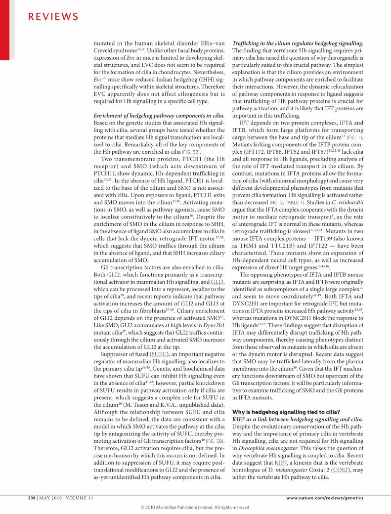
Intraflagellar transportThe cilium is extended and maintained by the trans-port of particles along the axoneme mediated by the IFT machinery (FIG. 1). IFT trafficking from the base to the tip of the cilium depends on the microtubule-plus-end-directed kinesin-2 motor (also known as the KIF3 motor complex), which consists of two kinesin-2 family proteins (KIF3A and KIF3B) and KIF-associated protein 3 (KAP3, also known as KIFAP3) and associ-ates with two IFT protein complexes, IFTA and IFTB. IFTB is essential for anterograde trafficking, whereas IFTA and the minus-end-directed cytoplasmic dynein motor (which consists of cytoplasmic dynein 2 heavy chain 1 (DynC2H1) and cytoplasmic dynein 2 light interme-diate chain 1 (DynC2lI1)) are required for retrograde trafficking1. In all organisms studied, disruption of the kinesin-2 motor or IFTB blocks cilia formation. Perturbation of retrograde trafficking by disruption of the dynein motor or IFTA results in short, bulged cilia1,12–15 (TABLE 1).
Cilia are nucleated by the basal body, which is made up of the mother centriole and associated pericentri-olar proteins. some basal body proteins are required for cilia formation; evidence suggests that some may recruit cargo from the Golgi complex to the nascent ciliary membrane and others may promote loading of cargo into the axoneme16,17 (FIG. 1).
Evidence linking hedgehog signalling to ciliaVertebrate hedgehog signalling requires intraflagellar transport. The first evidence that vertebrate Hh signal-ling depends on cilia came from a phenotype-based screen for mutations that alter the patterning of the mouse embryo. This screen identified several mutants showing morphological and patterning phenotypes that were consistent with altered Hh signalling: these phenotypes included loss of the ventral cell types in the neural tube specified by high levels of sonic hedgehog (sHH)11. The genes disrupted in these mutants encode several components of the IFT machinery, including the IFTB complex components IFT172 and IFT88, as well as DynC2H1, which is a subunit of the IFT-dedicated retrograde motor11,18,19 (FIG. 1). Disruption of the kinesin-2 motor in Kif3a-null embryos also caused similar defects in sHH-dependent neural patterning5 (FIG. 2). Genetic studies showed that IFT proteins act at the heart of the sHH pathway, downstream of the mem-brane proteins patched 1 (PTCH1) and smoothened (smO) and upstream of the Gli transcription factors that implement the pathway11,18 (FIG. 3; TABLE 1).
The role of IFT proteins in Hh signalling is complex, partly because of the complex output of the Hh pathway. In the absence of Hh ligand, Gli transcription factors, which function as effectors of the pathway, are proteo-lytically processed to Gli repressor forms (Glirs) that keep Hh target genes switched off (FIG. 3). In response to Hh ligand, processing of Glirs is blocked and acti-vated Gli transcription factors (GliAs) activate the expression of Hh target genes. IFT is required for the production of GliAs and Glirs18–21, and as a result IFT mutants show loss of Hh phenotypes in some cell
Figure 1 | cilia structure and intraflagellar transport. a | Mammalian nodal cilia at embryonic day 7.75. The long cilia of the mouse node are required for left–right asymmetry. b | Primary mammalian cilia (green) in embryonic fibroblasts. The basal bodies are shown in red and nuclei are stained blue. c | Cargo is transported from the base to the tip of the cilium along the microtubule axoneme by the kinesin‑2 motor together with the intraflagellar transport A (IFTA) and IFTB complexes. The dynein motor (which consists of cytoplasmic dynein 2 heavy chain 1 (DYNC2H1) and cytoplasmic dynein 2 light intermediate chain 1 (DYNC2LI1)) mediates the return of IFT cargo to the base of the cilium1,14. The IFTB proteins IFT20 and IFT54 may also participate in the trafficking of membrane vesicles from the Golgi complex to the ciliary membrane together with small GTPases136. Other small GTPases, including ADP‑ribosylation factor‑like 13B (ARL13B), also localize to cilia. Although its precise trafficking role is not known, ARL13B is required for axoneme structure137. Certain basal body proteins also influence ciliary trafficking. Among these are components of the BBSome, which are named after their association with Bardet–Biedl syndrome (BBS). The precise functions of BBS proteins in cilia formation are unclear as they are not individually required for primary cilia formation. However, they may function to promote loading of cargo to the ciliary axoneme17. Other basal‑body‑associated proteins, such as Meckel syndrome type 1 (MKS1), fantom (FTM, also known as RPGRIP1L), oral‑facial‑digital syndrome 1 (OFD1) and TALPID3, are required for cilia formation, although how they regulate ciliogenesis has not been defined10. BBIP10, BBSome‑interacting protein of 10 kDa; KAP3, KIF‑associated protein 3 (also known as KIFAP3).
R E V I E W S
332 | mAy 2010 | vOlume 11 www.nature.com/reviews/genetics
© 20 Macmillan Publishers Limited. All rights reserved10

AxonemeThe long projection of the cilium into the extracellular space. It is composed of a circular array of nine microtubule doublets.
Anterograde traffickingTransport towards the microtubule plus end (the cilia tip).
Retrograde traffickingTransport towards the microtubule minus end (the cilia base).
Mother centrioleCentrioles are tube-shaped structures composed of nine triplets of microtubules. One of the two centrioles is the mother centriole, which forms the basal body.
Table 1 | Roles of ciliary and basal body genes in development and disease
Mouse gene
Function Mutant phenotype Primary cilia phenotype
Human disorder
Arl13b Small GTPase Hh signalling defects137 Abnormal microtubule structure137
Joubert syndrome143
Bbs1 Basal body protein, BBSome component
Sensory defects, obesity131 Defects in specialized cilia only131
BBS144
Bbs2 Basal body protein, BBSome component
Sensory defects, obesity133 Defects in specialized cilia only133
BBS145
Bbs3 Small GTPase ND ND BBS146
Bbs4 Basal body protein, BBSome component
Sensory defects, male infertility, obesity131,132
Defects in specialized cilia only131,132
BBS147
Bbs5 Basal body protein, BBSome component
ND ND BBS148
Bbs6 Chaperonin‑like Sensory defects, male infertility, obesity
Defects in specialized cilia only
BBS, McKusick– Kaufman syndrome
Bbs7 Basal body protein, BBSome component
ND ND BBS149
Bbs8 Basal body protein, BBSome component
ND ND BBS150
Bbs9 Basal body protein, BBSome component
ND ND BBS151
Bbs10 Chaperonin‑like ND ND BBS152
Bbs11 E3 ubiquitin ligase Muscle defects153 ND BBS154, muscular dystrophy155
Bbs12 Chaperonin‑like ND ND BBS156
Dync2h1 Dynein retrograde motor subunit
Reduced Hh signalling18,19 Bulged18 JATD157
Evc Basal body protein, skeletal specific
IHH signalling defects129 Normal129 EVC33
Ftm Basal body protein Reduced Hh signalling31 Short31 Joubert syndrome type B28, MKS158
Fuz PCP effector Hh signalling defects103,104 Short103,104 ND
Ift122 IFT complex A Increased Hh signalling45 Bulged45 ND
Ift139 IFT complex A Increased Hh signalling15 Bulged, short15 ND
Ift172 IFT complex B Reduced Hh signalling11 Absent11 ND
Ift52 IFT complex B Reduced Hh signalling21 ND ND
Ift57 IFT complex B Reduced Hh signalling43 Absent43 ND
Ift80 IFT complex B ND ND JATD159
Ift88 IFT complex B Reduced Hh signalling11 Absent160 ND
Intu PCP effector Hh signalling defects105 Short105 ND
Kif3a Kinesin‑2 motor subunit, anterograde
Reduced Hh signalling11 Absent161 ND
Kif3b Kinesin‑2 motor subunit ND Absent162 ND
Kif7 Kinesin‑like, COS2 homologue Hh signalling defects40,55,56 Normal40,55 ND
Mks1 Basal body protein Hh defects, skeletal defects, cystic kidneys32
Sparse, short32 MKS163
Ofd1 Basal body protein Skeletal defects, reduces Hh signalling29
Short29 OFD30
Stil Centrosomal protein Reduced Hh signalling164 ND Primary microcephaly165
Arl13b, ADP‑ribosylation factor‑like 13B; BBS, Bardet–Biedl syndrome; COS2, Costal 2; Dync2h1, cytoplasmic dynein 2 heavy chain 1; EVC, Ellis–van Creveld syndrome; Ftm, fantom (also known as Rpgrip1l); Fuz, fuzzy; Hh, hedgehog; IFT, intraflagellar transport; IHH, Indian hedgehog; Intu, inturned; JATD, Jeune asphyxiating thorasic dystrophy; Kif, kinsein superfamily protein; MKS, Meckel syndrome; ND, not determined; OFD, oral‑facial‑digital syndrome; PCP, planar cell polarity; Stil, Scl/Tal1 interrupting locus.
R E V I E W S
nATure revIeWs | Genetics vOlume 11 | mAy 2010 | 333
© 20 Macmillan Publishers Limited. All rights reserved10

Nature Reviews | Genetics
d Anterograde IFT and dynein motor mutants
a Wild type
e Retrograde IFT mutant (IFTA mutant)
b No Hh
f Kif7 mutant
c Excess Hh activity
PAX6 domain Floorplate V3 Motor neurons V1 V0
PolydactylyThe formation of additional digits on the limbs. Additional posterior digits are referred to as postaxial, and additional anterior digits are referred to as preaxial.
Pericentriolar materialA network of fibres and associated proteins that surround the centriole and contain the microtubule-organizing activity of the centrosome.
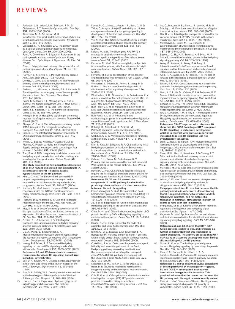
types and gain of Hh phenotypes in others. For exam-ple, GliAs have a central role in neural patterning, and IFT mutant embryos show a loss of Hh signalling in the neural tube. By contrast, Glirs have a central role in limb development, and IFT mutants that survive to later stages of embryogenesis show preaxial polydactyly, which is characteristic of loss of Glirs18,19,21 (FIG. 2).
recent experiments showed that IFT is also required for Hh signalling in zebrafish. Zebrafish that lack both maternal and zygotic ift88 have Hh signalling defects in the neural tube and somites22; however, the patterning defects caused by loss of IFT in zebrafish are slightly different from those seen in mammals. mouse Ift88 mutants lack sHH-dependent ventral neural cell fates11, and zebrafish maternal and zygotic ift88 mutants lack cell fates, such as v3 interneuron progenitors and muscle pioneer cells, that require the highest levels of Hh. However, cell types in the neural tube and somites that are normally specified by lower levels of Hh are expanded22. This difference may be due to a differ-ent balance of GliAs and Glirs in zebrafish compared with mice22.
Basal body proteins required for hedgehog signalling. Additional evidence that cilia are required for Hh sig-nalling came from the analysis of basal body protein mutations. Dozens of proteins are localized to centro-somes and the pericentriolar material, and a subset of these proteins has been shown to be required for cilia formation (TABLE 1). In every case examined so far, these proteins are required for Hh signalling. For example, the chick talpid3 mutation was first identified based on polydactyly23,24 and causes developmental defects con-sistent with disrupted Hh signalling25,26. Talpid3 mutant embryos fail to form cilia, and the affected gene was shown to encode a centrosomal protein27.
mutations disrupting other basal body proteins, such as oral-facial-digital syndrome 1 (OFD1), fantom (FTm, also known as rPGrIP1l), meckel syndrome type 1 (mKs1) and ellis–van Creveld syndrome protein (evC), cause human ciliopathies and affect mammalian Hh sig-nalling. mice mutant for Ofd1, Ftm or Mks1 have abnor-mal or absent cilia and exhibit Hh signalling defects corresponding to the severity of the cilia disruption28–32 (TABLE 1). evC also localizes to the basal body and is
Figure 2 | neural and limb patterning phenotypes in hedgehog pathway and cilia mutants. a | In wild‑type embryos, ventral neural cell fates are specified by a gradient of sonic hedgehog (SHH). The number and identity of digits in the limb is established by an SHH gradient from the posterior limb bud to the anterior limb bud, where the repressor form of GLI3 (GLI3R) inhibits SHH. b | In the absence of hedgehog (Hh) (for example, in smoothened (Smo)–/– embryos) ventral neural cell fates are lost. The limbs of Shh–/– mutants lack digits. c | If the pathway is hyperactive (for example, in patched 1 (Ptch1)–/– embryos), ventral cell types expand in the neural tube. Activation of the pathway within the limb, such as in Gli3–/– mutants (which lack GLI3R), causes the formation of extra digits. d | Anterograde intraflagellar transport (IFT) mutants (for example, Ift88–/– or Ift172–/– embryos) lack cilia: Hh signalling is reduced, and the neural tube is dorsalized. This phenotype is milder than in b because cilia are also required for GLI3R processing, and cell types that require low levels of Hh signalling are specified. Reduced GLI3R results in polydactyly. Dynein motor mutants display a similar phenotype; however, they retain motor neurons in the caudal neural tube. e | IFTA complex mutants (for example, Ift139–/– embryos) exhibit phenotypes consistent with excess Hh signalling. f | Mutations that disrupt the kinesin Kif7 cause a partial activation of the Hh pathway, with a modest expansion of cells that require intermediate levels of Hh. PAX6, paired box 6.
R E V I E W S
334 | mAy 2010 | vOlume 11 www.nature.com/reviews/genetics
© 20 Macmillan Publishers Limited. All rights reserved10

Nature Reviews | Genetics
PTCH1
SUFU
SUFUBasal body
Gli
Gli
Kinases
Kinases
Kinases
SUFU
IFT
Basal body
KIF7
KIF7
GliA
Gli
(+)
PTCH1Hh
SMO
Vesicle
SMO
SMO
SMO
Dynein motor
GliA
(+)
CI
CIA
FU
SU(FU)Kinases
FU
SU(FU)
SUFU
PTC
Aa Unstimulated Ab HH stimulated
Ba Unstimulated Bb Hh stimulated
Target genes
SMO
GliR
GliA
SMO
Vesicle
CIR
CIRCI
CI
COS2
COS2
PTC
HH
Target genes
CIA
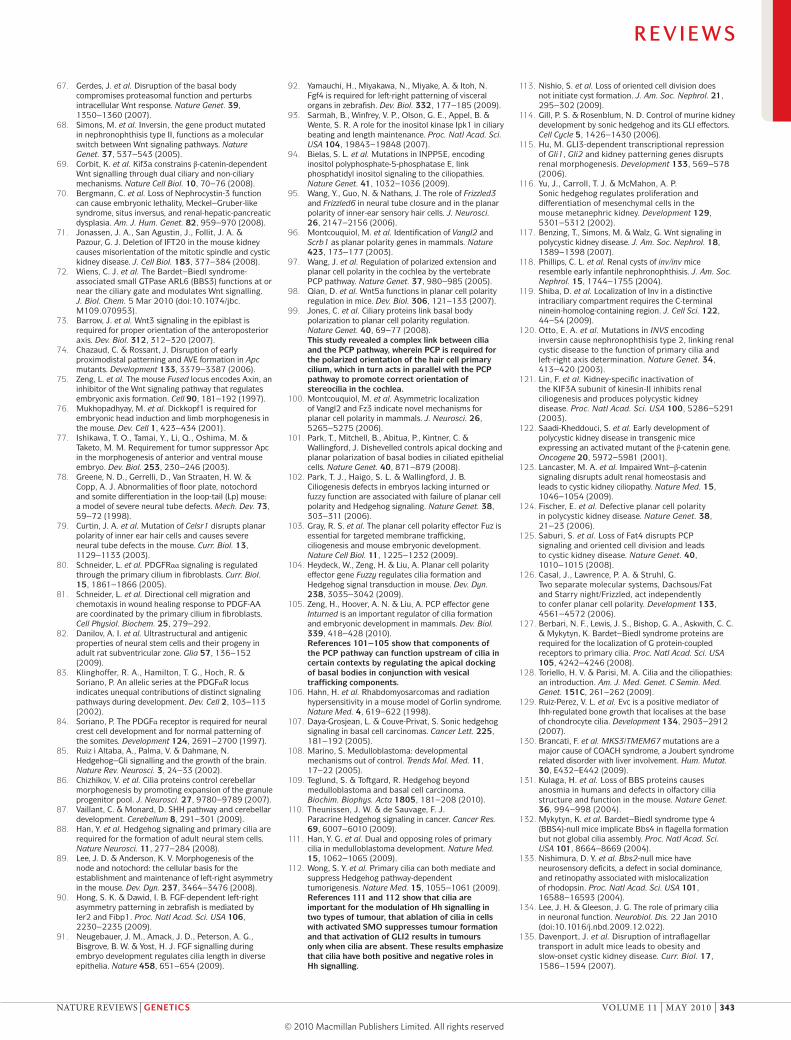
Figure 3 | Localization of hedgehog pathway complexes in Drosophila melanogaster and mammals. A | Modulation of protein complex structure and localization in D. melanogaster by Hedgehog (HH). Aa | In the absence of ligand, Patched (PTC) prevents translocation of Smoothened (SMO) to the plasma membrane. A microtubule‑associated complex including Costal 2 (COS2), Fused (FU), Suppressor of fused (SU(FU)) and Cubitus interruptus (CI) recruits kinases, including Protein kinase A (PKA), Casein kinase I (CKI) and Glycogen synthase kinase 3β (GSK3β), that promote the processing of CI into its repressor form (CIR)138–141. SU(FU) may also associate with full‑length CI to prevent its translocation to the nucleus138. Ab | Upon activation of the pathway, SMO moves to the plasma membrane, and the FU–COS2 complex associates with the carboxy‑terminal tail of SMO, resulting in the release of CI. Pathway activation also inactivates the negative regulator138–141. B | In vertebrate Hh signalling, signal transduction takes place within cilia, but the behaviour of protein complexes may parallel that of protein complexes in D. melanogaster. Ba | In the absence of ligand, PTCH1 localizes to the cilium and is thought to block the entry of SMO into cilia35. The kinesin KIF7 (the COS2 homologue) localizes to the base of the cilium55, where it may form a complex with Gli proteins and other pathway components. KIF7 at the cilium base prevents Gli enrichment within the cilium and promotes processing of GliRs. Bb | After activation of the pathway, SMO moves to the ciliary membrane and KIF7 translocates into the cilium, thereby promoting GLI2 accumulation at the cilium tip40,55. KIF7 at the cilia tip may also block the function of SUFU. Activated Gli is transported out of the cilium by the dynein motor and intraflagellar transport (IFT) particles.
R E V I E W S
nATure revIeWs | Genetics vOlume 11 | mAy 2010 | 335
© 20 Macmillan Publishers Limited. All rights reserved10
mutated in the human skeletal disorder ellis–van Creveld syndrome33,34. unlike other basal body proteins, expression of Evc in mice is limited to developing skel-etal structures, and evC does not seem to be required for the formation of cilia in chondrocytes. nevertheless, Evc–/– mice show reduced Indian hedgehog (IHH) sig-nalling specifically within skeletal structures. Therefore evC apparently does not affect ciliogenesis but is required for Hh signalling in a specific cell type.
Enrichment of hedgehog pathway components in cilia. Based on the genetic studies that associated Hh signal-ling with cilia, several groups have tested whether the proteins that mediate Hh signal transduction are local-ized to cilia. remarkably, all of the key components of the Hh pathway are enriched in cilia (FIG. 3B).
Two transmembrane proteins, PTCH1 (the Hh receptor) and smO (which acts downstream of PTCH1), show dynamic, Hh-dependent trafficking in cilia35,36. In the absence of Hh ligand, PTCH1 is local-ized to the base of the cilium and smO is not associ-ated with cilia. upon exposure to ligand, PTCH1 exits and smO moves into the cilium35,36. Activating muta-tions in smO, as well as pathway agonists, cause smO to localize constitutively to the cilium36. Despite the enrichment of smO in the cilium in response to sHH, in the absence of ligand smO also accumulates in cilia in cells that lack the dynein retrograde IFT motor37,38, which suggests that smO traffics through the cilium in the absence of ligand, and that sHH increases ciliary accumulation of smO.
Gli transcription factors are also enriched in cilia. Both GlI2, which functions primarily as a transcrip-tional activator in mammalian Hh signalling, and GlI3, which can be processed into a repressor, localize to the tips of cilia39, and recent reports indicate that pathway activation increases the amount of GlI2 and GlI3 at the tips of cilia in fibroblasts37,40. Ciliary enrichment of GlI2 depends on the presence of activated smO37. like smO, GlI2 accumulates at high levels in Dync2h1 mutant cilia37, which suggests that GlI2 traffics contin-uously through the cilium and activated smO increases the accumulation of GlI2 at the tip.
suppressor of fused (suFu), an important negative regulator of mammalian Hh signalling, also localizes to the primary cilia tip39,40. Genetic and biochemical data have shown that suFu can inhibit Hh signalling even in the absence of cilia41,42; however, partial knockdown of suFu results in pathway activation only if cilia are present, which suggests a complex role for suFu in the cilium20 (m. Tuson and K.v.A., unpublished data). Although the relationship between suFu and cilia remains to be defined, the data are consistent with a model in which smO activates the pathway at the cilia tip by antagonizing the activity of suFu, thereby pro-moting activation of Gli transcription factors40 (FIG. 3B). Therefore, GlI2 activation requires cilia, but the pre-cise mechanism by which this occurs is not defined. In addition to suppression of suFu, it may require post-translational modifications to GlI2 and the presence of as-yet-unidentified Hh pathway components in cilia.
Trafficking in the cilium regulates hedgehog signalling. The finding that vertebrate Hh signalling requires pri-mary cilia has raised the question of why this organelle is particularly suited to this crucial pathway. The simplest explanation is that the cilium provides an environment in which pathway components are enriched to facilitate their interactions. However, the dynamic relocalization of pathway components in response to ligand suggests that trafficking of Hh pathway proteins is crucial for pathway activation, and it is likely that IFT proteins are important in this trafficking.
IFT depends on two protein complexes, IFTA and IFTB, which form large platforms for transporting cargo between the base and tip of the cilium13 (FIG. 1). mutants lacking components of the IFTB protein com-plex (IFT172, IFT88, IFT52 and IFT57)11,21,43 lack cilia and all response to Hh ligands, precluding analysis of the role of IFT-mediated transport in the cilium. By contrast, mutations in IFTA proteins allow the forma-tion of cilia (with abnormal morphology) and cause very different developmental phenotypes from mutants that prevent cilia formation: Hh signalling is activated rather than decreased (FIG. 2; TABLE 1). studies in C. reinhardtii argue that the IFTA complex cooperates with the dynein motor to mediate retrograde transport1, as the rate of anterograde IFT is normal in these mutants, whereas retrograde trafficking is slowed12,14,44. mutants in two mouse IFTA complex proteins — IFT139 (also known as THm1 and TTC21B) and IFT122 — have been characterized. These mutants show an expansion of Hh-dependent neural cell types, as well as increased expression of direct Hh target genes15,45,46.
The opposing phenotypes of IFTA and IFTB mouse mutants are surprising, as IFTA and IFTB were originally identified as subcomplexes of a single large complex47 and seem to move coordinately48,49. Both IFTA and DynC2H1 are important for retrograde IFT, but muta-tions in IFTA proteins increased Hh pathway activity15,45, whereas mutations in DynC2H1 block the response to Hh ligands18,19. These findings suggest that disruption of IFTA may differentially disrupt trafficking of Hh path-way components, thereby causing phenotypes distinct from those observed in mutants in which cilia are absent or the dynein motor is disrupted. recent data suggest that smO may be trafficked laterally from the plasma membrane into the cilium50. Given that the IFT machin-ery functions downstream of smO but upstream of the Gli transcription factors, it will be particularly informa-tive to examine trafficking of smO and the Gli proteins in IFTA mutants.
Why is hedgehog signalling tied to cilia?KIF7 as a link between hedgehog signalling and cilia. Despite the evolutionary conservation of the Hh path-way and the importance of primary cilia in vertebrate Hh signalling, cilia are not required for Hh signalling in Drosophila melanogaster. This raises the question of why vertebrate Hh signalling is coupled to cilia. recent data suggest that KIF7, a kinesin that is the vertebrate homologue of D. melanogaster Costal 2 (COs2), may tether the vertebrate Hh pathway to cilia.
R E V I E W S
336 | mAy 2010 | vOlume 11 www.nature.com/reviews/genetics
© 20 Macmillan Publishers Limited. All rights reserved10

HydrocephalusThe build-up of cerebrospinal fluid within the ventricles of the brain.
KinomeThe set of protein kinases in the genome of a given organism.
MetazoanMulticellular organisms that, with the exception of sponges, have specialized cell types. Historically referred to as the kingdom ‘Animalia’.
Convergent extensionA morphogenetic movement characterized by the intercalation and elongation of cells, which causes a structure to generally become longer and thinner.
COs2, a key component of the D. melanogaster Hh pathway, is a kinesin-related protein that serves as a scaffold for Hh signalling complexes. COs2 has dual functions in the pathway: it promotes formation of the repressor form of Cubitus interruptus (CI, the D. melanogaster Gli homologue) in the absence of Hh ligand by recruiting kinases that prime CI for processing, and it permits high levels of pathway activation after Hh stim-ulation by antagonizing su(Fu)51–53 (FIG. 3). Although COs2 can bind microtubules, amino acids in its motor domain have diverged from those of other kinesins such that its motor function is disrupted54.
several recent papers showed that zebrafish and mouse KIF7 proteins, like D. melanogaster COs2, both positively and negatively regulate the sHH pathway40,55–57. unlike COs2, the vertebrate KIF7 motor domain retains all of the motifs that are typical of kinesin motors, suggesting that it should act as a motor protein. In the absence of ligand, KIF7 localizes to the base of the primary cilium and moves to the tip of the cilium in response to pathway activation40,55 (FIG. 3). This translocation depends on the KIF7 motor domain, which suggests that KIF7, like the kinesin-2 motor, acts as an anterograde motor in the cilium55.
Conventional kinesins, such as KIF7, carry cargo towards the plus end of microtubules. The minus ends of axonemal and cytoplasmic microtubules are located at the base of the cilium. Because KIF7–enhanced GFP (eGFP) is enriched at the base of the cilium, we proposed that KIF7 might traffic GlI2 away from the cilium in the absence of ligand to prevent Gli activation55 (FIG. 3B). The positive role of KIF7 is presumably coupled to its movement, after pathway activation, to the cilia tip, where suFu and the Gli transcription factors are enriched. KIF7 may promote Gli activation at the tip, perhaps by antagonizing the activity of suFu55. Therefore, the dual roles of KIF7 as a sHH pathway component and ciliary motor could explain why mammalian sHH signalling depends on the primary cilium (FIG. 3B).
Fused is a cilia-associated protein in vertebrates. Fused (Fu) is an important component of the D. melanogaster Hh pathway: it is a serine/threonine kinase that phospho-rylates COs2, su(Fu) and perhaps other components of the pathway, and it is required for activation of CI in response to Hh ligand53. Fu is also important for Hh sig-nalling in zebrafish58, but Hh signalling is normal in mice lacking Fu59,60. recent work has shown that mammalian and zebrafish Fu are required for the construction of spe-cialized motile cilia61, and Fu–/– mutant mice die postna-tally with hydrocephalus, presumably due to dysfunction of motile cilia in brain ventricles59,60. Therefore Fu, like KIF7 or COs2, links Hh signalling with cilia, although the connection to Hh seems to have been lost in mammals. It has been proposed that another unidentified kinase may substitute for Fu in mammalian Hh signalling, and several human kinome screens have been undertaken to identify kinases that are required for Hh signalling62,63. Although the kinases identified in these screens have yet to be characterized in vivo, it will be interesting to deter-mine whether a protein that is functionally homologous to Fu also links sHH signalling to cilia in mammals.
The evolution of KIF7 and Fused. recent work in planaria supports the view that some conserved components of the Hh pathway were associated with cilia before they were associated with Hh signalling. Planaria homologues of the Hh pathway components KIF7, Fu and iguana are required in planaria for formation of motile cilia but not Hh signalling64,65. Planaria are a distinct lineage of ani-mals from both insects and vertebrates. Therefore, the finding that KIF7 and Fu function in cilia in two inde-pendent metazoan lineages suggests that the ancestral role of these proteins was in cilia. The requirement for these cilia-associated proteins in D. melanogaster Hh signalling suggests that Hh signalling was associated with cilia in the common ancestor of D. melanogaster and vertebrates.
Are cilia dedicated to hedgehog signalling?most cells in the mouse embryo have primary cilia, and a relatively small number of cells respond to Hh at any particular stage. This has raised interest in the possibil-ity that other developmental signalling pathways may also depend on cilia. However, the disruption of other developmental signalling pathways, including canonical and non-canonical Wnt, transforming growth factor-β (TGF-β), notch and fibroblast growth factor (Fgf) sig-nalling, causes developmental abnormalities that do not overlap with the IFT mutant phenotypes. nevertheless, cilia could have more subtle roles in other signalling pathways or might be important for signalling at later embryonic stages, after IFT mutants arrest.
Wnt signalling. most attention has focused on the rela-tionship between cilia and Wnt signalling. several groups reported that knockdown of cilia-associated proteins in cultured cells or zebrafish embryos elevates canonical Wnt signalling and/or disrupts processes that depend on non-canonical Wnt signalling, such as convergent extension66–72. The primary cilium was therefore proposed to act as a switch between canonical and non-canonical Wnt signalling pathways68,69.
However, this connection between cilia and Wnt sig-nalling is controversial. mouse IFT mutants do not show the phenotypes that are characteristic of Wnt pathway mutants. For example, reduced canonical Wnt signalling disrupts gastrulation and early patterning73, and inap-propriate activation of the Wnt pathway can cause axis duplications and failure to form anterior structures74–77. Although mammalian non-canonical Wnt pathway mutants fail to close the entire neural tube caudal to the forebrain, neural patterning in these mutants is rela-tively normal78,79. similarly, zebrafish mutants that lack both maternal and zygotic activity of the ift88 gene have defects in Hh signalling but do not show the defects in convergent extension that are associated with disruption of non-canonical Wnt signalling22.
recent work examined the expression of canonical Wnt reporters in vivo in Kif3a, Ift88, Ift172 and Dync2h1 mutant mice and failed to find any alteration in either the domain or levels of Wnt activity38. similarly, mouse embryos homozygous for a mutation in the IFTA pro-tein IFT139 have a neural patterning phenotype that is consistent with the activation of Hh signalling15,46 but do
R E V I E W S
nATure revIeWs | Genetics vOlume 11 | mAy 2010 | 337
© 20 Macmillan Publishers Limited. All rights reserved10

Nature Reviews | Genetics
Lung and airway abnormalities
Liver cysts
Skeletal abnormalities
Pancreatic cysts
Obesity
Renal cysts
Polydactyly
Thoracic skeleton
Cognitive impairment
Craniofacial abnormalities
Cochlea: hearing loss
Neuronal sensory cilia defectMotile cilia defectHh defectBasal body positioning defectDisrupted pathway (unknown or controversial)
Retinal degenerationAnosmia
Sterility
Cerebellum
not show altered canonical Wnt signalling46. Therefore it seems that cilia are not required for canonical or non-canonical Wnt signalling in the first half of vertebrate embryogenesis.
Platelet-derived growth factor receptor-α signalling. Cilia have been found to be important for signalling by platelet-derived growth factor receptor-α (PDGFrΑ) in cultured fibroblasts80, as well as for PDGF-dependent directed migration in these cells81. Additionally, the receptor is localized to primary cilia in vivo in neural stem cells of the adult rat subventricular zone82. loss of PDGFrΑ signalling does not produce any striking phe-notypes in early mouse embryos but is crucial for the development of later tissues, including oligodendrocytes and neural crest-derived craniofacial structures83,84. It will be important to test whether loss of cilia in the sec-ond half of embryogenesis affects PDGFrΑ signalling in these cell types in vivo.
Hedgehog signalling in adult tissues. After birth, sHH signalling continues to have important roles in the growth of the brain and the maintenance of neural progenitors85. Conditional deletion of Ift88 or Kif3a in the brain results in severe hypoplasia of the cerebellum due to the failure of granule cell progenitor proliferation86, a process that depends on sHH signalling85,87. Primary cilia are also needed to modulate the sHH-dependent formation and maintenance of hippocampal granule neuron precur-sors, which are important for maintaining neurogenesis in adults88. Based on the tight association between cilia mutants and specific defects of Hh signalling, we propose that cilia are essential for Hh signalling in all cell types and that, at least in early development, primary cilia in vertebrate embryos are dedicated to Hh signal transduc-tion. The data do not rule out the possibility that cilia may have important roles in other signalling pathways later in development or in specific cell types. For example, although a number of the defects observed in human cili-opathies can be attributed to abnormal Hh signalling, the molecular bases of other features of these diseases remain unknown (BOX 1).
Signalling pathways that regulate ciliogenesisBecause of the importance of primary cilia in embryonic patterning, there is considerable interest in identifying signalling pathways that regulate cilia formation. several transcription factors are known to be required for the formation of motile cilia and node cilia89. However, only recently has evidence emerged about signalling pathways that regulate the formation and position of primary cilia.
Fgf and inositol signalling. recent evidence from zebrafish implicates Fgf signalling in the regulation of cilia length. Knockdown of Fgfr1 or Fgf ligands results in shortened cilia in Kupffer’s vesicle and randomized organ laterality90–92. The expression of ciliogenic transcription factors and ift88 is reduced in these embryos91. It will be interesting to test whether Fgf pathway mutations in mice also affect ciliogenesis, and whether these effects on ciliogenesis alter Hh signal transduction.
Box 1 | Organs affected in human ciliopathies
Numerous pleiotropic human disorders have been attributed to defects in cilia formation10,128 (see the figure). Some aspects of these syndromes, such as the polydactyly in patients with Bardet–Biedl syndrome (BBS) and Meckel syndrome and the skeletal abnormalities that affect the limbs of patients with Ellis–van Creveld syndrome, have been attributed to defective hedgehog (Hh) signalling. Polydactyly results from a loss of the repressor form of GLI3 (GLI3R) and skeletal abnormalities resemble those observed in mutants that lack Indian hedgehog (IHH) signalling129. Sonic hedgehog (SHH) signalling is also required for craniofacial development, and defects in craniofacial structures, such as those observed in Meckel syndrome type 1 (Mks1) mutant mice, are also likely to be due to misregulated Hh signalling32. In addition, patients with a Joubert syndrome-like disorder exhibit ataxia due to cerebellar hypoplasia130. Growth of this tissue is Hh dependent85.
Other attributes of human disorders result from defective specialized cilia. Retinal degeneration results from defects in photoreceptor connecting cilia, which connect the outer light-responsive segment to the cell body. Detection of odorants depends on the primary cilia of sensory neurons in the olfactory epithelium, and patients with BBS often exhibit anosmia131–133. Another sensory deficit, hearing loss, is due to a requirement for the specialized primary cilia of the cochlea downstream of the planar cell polarity pathway in establishing the correct polarity of sensory hair cells99. Infertility observed in patients with ciliopathies is the result of defective sperm flagella and motile oviduct cilia132.
For some of the most severe and common abnormalities associated with ciliopathies, such as cyst formation in the kidneys, liver, biliary duct and pancreas, the underlying molecular causes downstream of the cilium remain unclear. Cyst formation is thought to result from defects in cell proliferation or misorientation of the mitotic spindle. However, whether and how these processes are regulated by cilia remain the subject of active investigation124. In addition, patients with BBS often show obesity and cognitive impairments that are thought to be due to neuronal defects; however, the specific pathways responsible for these attributes in patients with BBS have not been clearly identified134,135.
R E V I E W S
338 | mAy 2010 | vOlume 11 www.nature.com/reviews/genetics
© 20 Macmillan Publishers Limited. All rights reserved10

Subventricular zoneA layer of cells lining the ventricles of the brain in which neurogenesis takes place in adult mammals.
Kupffer’s vesicleA ciliated organ of the fish embryo that serves to generate asymmetry during development, functioning analogously to the mammalian node.
Hair cellsThe sensory cells in the vertebrate auditory system. They are contained within the cochlea.
Organ of CortiThe organ in the mammalian inner ear that contains the hair cells.
CochleaThe portion of the inner ear that contains the sensory organs of hearing.
KinociliumThe specialized primary cilium of the hair cells of the cochlea.
StereociliaThe actin-based sensory organelles that form a polarized chevron-shaped structure in the hair cell.
Components of the phosphatidylinositol signalling cascade also seem to regulate cilia length. In zebrafish, morpholino knockdown of inositol-pentakisphosphate 2-kinase (Ipk1, also known as Ippk) reduced the fre-quency of cilia beating and decreased cilia length93. In humans, inositol polyphosphate 5-phosphatase (InPP5e) is mutated in one form of Joubert syndrome, a ciliopathy. InPP5e is enriched in the ciliary axoneme, and in fibro-blasts from patients with Joubert syndrome, cilia are more labile than wild-type cilia94.
The mechanisms by which the Fgf and phosphatidylino-sitol pathways regulate cilia formation or maintenance remain to be elucidated. It will be informative to investi-gate whether these pathways have a general role in primary cilia formation or act in a subset of specialized cilia.
Planar cell polarity signalling and cilia. recent experi-ments argue that there is a close connection between components of PCP signalling and cilia positioning. An excellent example of this connection is found in the mech-anosensory hair cells in the organ of Corti in the cochlea. The primary cilium of the hair cell, called the kinocilium, is always oriented on the lateral side of the developing cell. The position of the kinocilium determines the polarity of the chevron of stereocilia (actin-based sensory organelles) on the hair cell. Core components of the non-canonical Wnt pathway, a PCP pathway, are required for the polarity of these hair cells95–97,98.
When primary cilia are removed by conditional dele-tion of Ift88, the polarity of the hair cells is disrupted99, similar to the phenotype seen in non-canonical Wnt mutants97. This finding indicated that the presence of the kinocilium is important for the correct orientation of the stereocilia and the organization of the hair cells, and raised the possibility that the primary cilium might regulate the non-canonical Wnt pathway in this tissue. However, the relationship between the kinocilium and planar polarity is more complex. As in D. melanogaster, components of the non-canonical Wnt pathway are pla-nar polarized in hair cells, and that polarity is required for PCP signalling and provides a read-out of effective PCP signalling97,100. In the hair cells of Ift88 conditional mutants, the polarity of PCP proteins is not disrupted. This indicates that IFT88, and presumably cilia, are not required for the activity of the core PCP pathway in this tissue, as in early embryos. Instead, it seems that one out-put of non-canonical Wnt signalling is to control the posi-tion of the basal body and thereby cilia position99 (FIG. 4a). In addition, the findings indicate that IFT88 itself must be needed to reposition the basal body to a polarized posi-tion. The mechanisms by which the position of the basal body is regulated by IFT88 are not known.
studies of the motile cilia on the epidermis of Xenopus laevis embryos support the hypothesis that components of the PCP pathway control polarized organization of cilia. These cells are multiciliated, and the cilia on each cell share a common polarity101. Disruption of the activ-ity of the PCP proteins dishevelled 1 (dvl1), dvl2 and/or dvl3 disrupts the polarity of the cilia on these cells — based on morpholino data targeting, all three dvl proteins localize to the basal bodies of epidermal cells
and dvl morphants have a reduced number of short cilia101. This finding indicates that apical docking of basal bodies, and therefore the ability to form cilia, depends on dvl and possibly other components of the PCP pathway (FIG. 4b).
Although changes in the position of cilia are unlikely to influence their ability to transduce Hh signals, some PCP components do affect Hh signalling. Inturned and Fuzzy (FuZ) are downstream effectors of the non-canonical Wnt pathway in D. melanogaster, and morpholino knockdown of X. laevis inturned (intu) or fuz disrupts the apical actin network and cilia formation102. These morphants fail to undergo normal convergent extension due to defects in PCP and also show defects that are con-sistent with a loss of shh signalling102. similarly, mouse Fuz and Intu mutants have short cilia and disrupted Hh signalling103–105. Therefore, components of the planar polarity pathway can be important for both formation and polarity of cilia (FIG. 4b). However, there is no evidence that other components of the non-canonical Wnt pathway are required for ciliogenesis, which suggests that the roles of InTu and FuZ in cilia formation may be unrelated to their roles in non-canonical Wnt signalling.
Cilia, signalling and diseasenumerous human disorders have now been linked to defects in cilia structure or in cilia-localized proteins. These include autosomal-dominant polycystic kidney disease (PKD), and recessive pleiotropic disorders, such as Bardet–Biedl syndrome, Joubert syndrome, meckel syndrome and ellis–van Creveld syndrome. some aspects of these disorders, such as polydactyly and skel-etal abnormalities, are likely to be due to misregulated Hh signalling, but the molecular bases of other defects, such as cystic kidneys, are not well understood (BOX 1). The deregulated Hh signalling associated with several types of human cancers also depends on cilia. Therefore studies on the relationship between cilia and signal-ling during development have direct implications for human disease.
Hedgehog signalling in tumours. Inappropriate activation of sHH signalling can cause medulloblastomas and rhabdo-myosarcomas (paediatric tumours of the cerebellum and muscle, respectively) and is found in all cases of basal cell carcinoma106–108. In addition, growing evidence indicates that sHH signals promote the growth of other types of tumours109,110. recent studies show that cilia regulate Hh signalling in tumours and that the role of cilia in tumours depends on how the pathway is activated. expression of activated smO in the postnatal mouse brain can cause medulloblastomas, but removal of cilia prevents tumour formation111, consistent with earlier genetic experiments indicating that cilia are required for the activity of the pathway at a step downstream of smO11. Constitutively active GlI2 can also cause medulloblastomas in mice, but only when cilia are removed111. This suggests that GlI2 alone cannot activate tumorigenesis in this cellular context when the cilia-dependent GlI3 repressor is also present. similar results were observed in basal cell carci-nomas in mice harbouring similar activating Hh pathway
R E V I E W S
nATure revIeWs | Genetics vOlume 11 | mAy 2010 | 339
© 20 Macmillan Publishers Limited. All rights reserved10

Directional cues
Nature Reviews | Genetics
RSG1FUZ
Golgi
WT
a
b
PCP mutant
IFT mutant
Directional cues
VANGL2Hair cell Basal body StereociliaDvl Kinocilium
SEC8
DvlSEC8
Dvl
SEC8
Dvl
SEC8
Dvl
FUZRSG1
1 2
3 4
Nucleus
Axoneme Basal body Apical actin network
Ciliary components Golgi or vesicles
Figure 4 | the role of the planar cell polarity pathway in cilia formation. a | In the wild‑type (WT) cochlea, directional cues establish the localization of core planar cell polarity (PCP) pathway components. The kinocilium then directs the basal body towards the medial side of the cells. This in turn directs the orientation of the stereocilia bundles, resulting in the correct orientation of the bundles within the cochlea. In PCP mutants, the initial cell polarity is never established, resulting in the improper positioning of the basal body and stereocilia. In intraflagellar transport (IFT) mutants, planar polarity is established; however, the basal body is not repositioned in the absence of the IFT‑dependent kinocilium99. Red boxes depict the step in the establishment of polarity that is defective in each category of mutants. b | Components of the PCP pathway are implicated in cilia formation. The centrioles — one of which will become the basal body — are initially located away from the cell surface (1)1, where they associate with PCP proteins from the dishevelled (Dvl) family (DVL1, DVL2 or DVL3). A component of the vesicle trafficking machinery, SEC8 (also known as EXOC4), is then recruited to the basal body (2)101. At the cell surface (3), DVL mediates the fusion of the basal‑body‑associated membrane with the cell membrane101,142, and the axoneme then extends through IFT (4). In vivo mouse experiments indicate that the PCP effector fuzzy (FUZ) and Rem/Rab‑similar GTPase 1 (RSG1, also known as C1orf89) also have roles in trafficking membrane vesicles and ciliary components to the basal body103. VANGL2, vang‑like 2.
R E V I E W S
340 | mAy 2010 | vOlume 11 www.nature.com/reviews/genetics
© 20 Macmillan Publishers Limited. All rights reserved10

mutations: activated smO caused tumours only in the presence of cilia, whereas removal of cilia enhanced tum-origenesis due to expression of activated GlI2 (REF. 112). Therefore, in tumours as in development, cilia have both positive and negative effects on the Hh pathway.
Cilia and polycystic kidney disease. A hallmark of many human ciliopathies is the formation of kidney cysts, which often begins during fetal life and is a developmen-tal rather than physiological defect3,113. This raises the question of whether kidney cysts result from a disrup-tion of cilia-dependent developmental signals. Hh sig-nalling is required for normal kidney development114, but kidney cysts have not been reported in mutants that lack either positive or negative Hh regulators115,116. Although a role for Hh signalling in PKD cannot be ruled out, some data suggest that cilia might modulate Wnt signalling in this tissue117.
The connection among cilia, cystic kidneys and Wnt signalling was first raised by analysis of the inversin (Invs) gene. Invs binds microtubules and localizes to the basal body118 and cilium119. mutations in INVS cause kidney disease in humans120 and renal cysts in mice118. Invs interacts with Dvl1, targeting membrane-bound Dvl1 for destruction. Invs has been proposed to act as a switch between canonical and non-canonical Wnt signal-ling, based on cell culture and morpholino knockdown experiments68. However, altered canonical Wnt signalling has not been reported in the kidneys of Invs–/– mice.
mouse overexpression experiments show that increased Wnt signalling can cause kidney cysts, and increased nuclear β-catenin is observed in the cystic kidney tubules of mice in which cilia have been conditionally ablated121,122. However, reduced Wnt sig-nalling has been observed in mice lacking Jouberin (AHI1), a cilia-localized protein mutated in a form of Joubert syndrome. Ahi1–/– mice have cystic kidneys with reduced expression of a Wnt reporter and reduced nuclear β-catenin123. Therefore additional experiments that examine Wnt signalling during kidney development will be needed to reconcile the conflicting data.
recent theories of PKD have focused on the impor-tance of the plane of cell division, under the control of PCP signalling, as a possible underlying defect in kidney cysts124. The elongation of kidney tubules is thought to depend on oriented cell divisions, and this is disrupted in kidney tubule cells of mice with cystic kidneys117,124. supporting this hypothesis, mice lacking the PCP protein FAT4 exhibit polycystic kidneys beginning at embryonic day 16 associated with misorientation of mitotic spin-dles within the renal tubules125. This cyst formation is enhanced by removal of one or both copies of the core PCP pathway component vang-like 2 (Vangl2). moreover, FAT4 localizes to cilia within the kidney, implicating the cilium in the modulation of the kidney PCP pathway125. D. melanogaster FAT, however, acts in a PCP pathway that does not depend on non-canonical Wnt signal-ling126. moreover, mouse mutants of other PCP pathway components, such as vAnGl2 and FuZ, have not been reported to have cystic kidneys, and the polarity of PCP-component localization in kidney tubule cells has not
been assessed in mice lacking renal cilia. recent results suggest that misoriented cell division is neither neces-sary nor sufficient for the formation of kidney cysts113. Therefore it remains to be determined which pathway (or pathways) downstream of primary cilia is the underlying cause of renal cysts.
Conclusions and perspectivesnon-motile primary cilia have vital roles in vertebrate development from early stages of embryonic patterning, when they regulate the activity of the Hh pathway, to organogenesis, when they are important in the develop-ment and homeostasis of numerous tissues. The recent resurgence in interest in primary cilia has raised many new questions about the roles of cilia.
We know very little about the events of Hh signal transduction that occur within cilia. The mechanisms that traffic smO to cilia, traffic PTCH1 out of cilia and modulate the trafficking of Gli proteins within cilia in response to Hh pathway activation are largely unknown. The opposing effects of different IFT components upon the regulation of the Hh pathway suggest that, in addi-tion to providing a compartment in which Hh pathway components are enriched, the IFT machinery has a more complex role in regulating the pathway, but these roles have not been defined. To address these questions, it will be necessary to examine the trafficking of Hh pathway components in real time and to probe their physical asso-ciations with the IFT machinery in wild-type cells, as well as in cells that are mutated for various IFT components.
The dual roles of KIF7 in intraciliary trafficking and in the Hh pathway suggest a reason why vertebrate Hh signalling is tied to cilia, and other proteins may also have dual roles. For example, is there a mammalian kinase that performs functions analogous to those of D. melanogaster Fu? And if so, does it have roles in both ciliogenesis and Hh signalling? Do other components of the Hh signalling pathway affect the dynamics of ciliary trafficking?
Based on the phenotypes of the numerous IFT mutants characterized to date, it seems that during early vertebrate development the cilium functions as an Hh-dedicated organelle. However, this does not preclude a requirement for cilia in modulating other signalling pathways in specific tissues later in development. These may include PDGFrΑ signalling, signalling through G-protein-coupled receptors in specific neurons127 and PCP signalling in the kidney. A particularly interesting question is whether, in specific cell types, cilia are sites at which Hh and other signalling pathways are integrated. As complex crosstalk between pathways is vital in regu-lating cellular responses during development and in dis-ease states such as cancer, understanding the function of the cilium as a signalling centre will be crucial.
Note added in proofrecent work by singla et al.166 describes the mechanism by which the centrosomal protein OFD1 functions to promote ciliogeneis. This paper shows that OFD1 con-trols the length of the centriole and is required for micro-tubule stability. OFD1 is also important in recruiting IFT88 to the centrosome.
R E V I E W S
nATure revIeWs | Genetics vOlume 11 | mAy 2010 | 341
© 20 Macmillan Publishers Limited. All rights reserved10
1. Pedersen, L. B., Veland, I. R., Schroder, J. M. & Christensen, S. T. Assembly of primary cilia. Dev. Dyn. 237, 1993–2006 (2008).
2. Silverman, M. A. & Leroux, M. R. Intraflagellar transport and the generation of dynamic, structurally and functionally diverse cilia. Trends Cell Biol. 19, 306–316 (2009).
3. Lancaster, M. A. & Gleeson, J. G. The primary cilium as a cellular signaling center: lessons from disease. Curr. Opin. Genet. Dev. 19, 220–229 (2009).
4. Patel, V., Chowdhury, R. & Igarashi, P. Advances in the pathogenesis and treatment of polycystic kidney disease. Curr. Opin. Nephrol. Hypertens. 18, 99–106 (2009).
5. Zhou, J. Polycystins and primary cilia: primers for cell cycle progression. Annu. Rev. Physiol. 71, 83–113 (2009).
6. Harris, P. C. & Torres, V. E. Polycystic kidney disease. Annu. Rev. Med. 60, 321–337 (2009).
7. Gerdes, J., Davis, E. E. & Katsanis, N. The vertebrate primary cilium in development, homeostasis, and disease. Cell 137, 32–45 (2009).
8. Badano, J. L., Mitsuma, N., Beales, P. L. & Katsanis, N. The ciliopathies: an emerging class of human genetic disorders. Annu. Rev. Genomics Hum. Genet. 7, 125–148 (2006).
9. Baker, K. & Beales, P. L. Making sense of cilia in disease: the human ciliopathies. Am. J. Med. Genet. C Semin. Med. Genet. 151C, 281–295 (2009).
10. Tobin, J. L. & Beales, P. L. The nonmotile ciliopathies. Genet. Med. 11, 386–402 (2009).
11. Huangfu, D. et al. Hedgehog signalling in the mouse requires intraflagellar transport proteins. Nature 426, 83–87 (2003).
12. Blacque, O. E. et al. The WD repeat-containing protein IFTA-1 is required for retrograde intraflagellar transport. Mol. Biol. Cell 17, 5053–5062 (2006).
13. Cole, D. G. The intraflagellar transport machinery of Chlamydomonas reinhardtii. Traffic 4, 435–442 (2003).
14. Iomini, C., Babaev-Khaimov, V., Sassaroli, M. & Piperno, G. Protein particles in Chlamydomonas flagella undergo a transport cycle consisting of four phases. J. Cell Biol. 153, 13–24 (2001).
15. Tran, P. et al. THM1 negatively modulates mouse sonic hedgehog signal transduction and affects retrograde intraflagellar transport in cilia. Nature Genet. 40, 403–410 (2008).This study provided the first phenotypic description of an IFTA mutant and showed that disrupting IFTA, in contrast to other IFT mutants, causes hyperactivation of the Hh pathway.
16. Kim, J. C. et al. The Bardet–Biedl protein BBS4 targets cargo to the pericentriolar region and is required for microtubule anchoring and cell cycle progression. Nature Genet. 36, 462–470 (2004).
17. Nachury, M. et al. A core complex of BBS proteins cooperates with the GTPase Rab8 to promote ciliary membrane biogenesis. Cell 129, 1201–1213 (2007).
18. Huangfu, D. & Anderson, K. V. Cilia and Hedgehog responsiveness in the mouse. Proc. Natl Acad. Sci. USA 102, 11325–11330 (2005).
19. May, S. R. et al. Loss of the retrograde motor for IFT disrupts localization of Smo to cilia and prevents the expression of both activator and repressor functions of Gli. Dev. Biol. 287, 378–389 (2005).
20. Ocbina, P. J. & Anderson, K. V. Intraflagellar transport, cilia, and mammalian Hedgehog signaling: analysis in mouse embryonic fibroblasts. Dev. Dyn. 237, 2030–2038 (2008).
21. Liu, A., Wang, B. & Niswander, L. A. Mouse intraflagellar transport proteins regulate both the activator and repressor functions of Gli transcription factors. Development 132, 3103–3111 (2005).
22. Huang, P. & Schier, A. F. Dampened Hedgehog signaling but normal Wnt signaling in zebrafish without cilia. Development 136, 3089–3098 (2009).References 20 and 22 demonstrate a conserved requirement for cilia in Hh signalling, but not Wnt signalling, in vertebrates.
23. Ede, D. A. & Kelly, W. A. Developmental abnormalities in the trunk and limbs of the talpid3 mutant of the fowl. J. Embryol. Exp. Morphol. 12, 339–356 (1964).
24. Ede, D. A. & Kelly, W. A. Developmental abnormalities in the head region of the talpid mutant of the fowl. J. Embryol. Exp. Morphol. 12, 161–182 (1964).
25. Lewis, K. E. et al. Expression of ptc and gli genes in talpid3 suggests bifurcation in Shh pathway. Development 126, 2397–2407 (1999).
26. Davey, M. G., James, J., Paton, I. R., Burt, D. W. & Tickle, C. Analysis of talpid3 and wild-type chicken embryos reveals roles for Hedgehog signalling in development of the limb bud vasculature. Dev. Biol. 301, 155–165 (2007).
27. Yin, Y. et al. The Talpid3 gene (KIAA0586) encodes a centrosomal protein that is essential for primary cilia formation. Development 136, 655–664 (2009).
28. Delous, M. et al. The ciliary gene RPGRIP1L is mutated in cerebello-oculo-renal syndrome (Joubert syndrome type B) and Meckel syndrome. Nature Genet. 39, 875–81 (2007).
29. Ferrante, M. et al. Oral-facial-digital type I protein is required for primary cilia formation and left-right axis specification. Nature Genet. 38, 112–117 (2006).
30. Ferrante, M. I. et al. Identification of the gene for oral-facial-digital type I syndrome. Am. J. Hum. Genet. 68, 569–576 (2001).
31. Vierkotten, J., Dildrop, R., Peters, T., Wang, B. & Ruther, U. Ftm is a novel basal body protein of cilia involved in Shh signalling. Development 134, 2569–2577 (2007).
32. Weatherbee, S. D., Niswander, L. A. & Anderson, K. V. A mouse model for Meckel syndrome reveals Mks1 is required for ciliogenesis and Hedgehog signaling. Hum. Mol. Genet. 18, 4565–4575 (2009).
33. Ruiz-Perez, V. L. et al. Mutations in a new gene in Ellis–van Creveld syndrome and Weyers acrodental dysostosis. Nature Genet. 24, 283–286 (2000).
34. Ruiz-Perez, V. L. et al. Mutations in two nonhomologous genes in a head-to-head configuration cause Ellis–van Creveld syndrome. Am. J. Hum. Genet. 72, 728–732 (2003).
35. Rohatgi, R., Milenkovic, L. & Scott, M. Patched1 regulates Hedgehog signaling at the primary cilium. Science 317, 372–376 (2007).
36. Corbit, K. C. et al. Vertebrate Smoothened functions at the primary cilium. Nature 437, 1018–1021 (2005).
37. Kim, J., Kato, M. & Beachy, P. A. Gli2 trafficking links Hedgehog-dependent activation of Smoothened in the primary cilium to transcriptional activation in the nucleus. Proc. Natl Acad. Sci. USA 106, 21666–21671 (2009).
38. Ocbina, P. J., Tuson, M. & Anderson, K. V. Primary cilia are not required for normal canonical Wnt signaling in the mouse embryo. PLoS ONE 4, e6839 (2009).
39. Haycraft, C. et al. Gli2 and Gli3 localize to cilia and require the intraflagellar transport protein polaris for processing and function. PLoS Genet. 1, e53 (2005).References 35, 36 and 39 showed that components of the Hh pathway localize to cilia, providing cellular evidence of a direct connection between cilia and Hh signalling.
40. Endoh-Yamagami, S. et al. The mammalian Cos2 homolog Kif7 plays an essential role in modulating Hh signal transduction during development. Curr. Biol. 19, 1320–1326 (2009).
41. Jia, J. et al. Suppressor of Fused inhibits mammalian Hedgehog signaling in the absence of cilia. Dev. Biol. 330, 452–460 (2009).
42. Chen, M.-H. et al. Cilium-independent regulation of Gli protein function by Sufu in Hedgehog signaling is evolutionarily conserved. Genes Dev. 23, 1910–1928 (2009).
43. Houde, C. et al. Hippi is essential for node cilia assembly and Sonic hedgehog signaling. Dev. Biol. 300, 523–533 (2006).
44. Iomini, C., Li, L., Esparza, J. M. & Dutcher, S. K. Retrograde IFT mutants identify complex A proteins with multiple genetic interactions in Chlamydomonas reinhardtii. Genetics 183, 885–896 (2009).
45. Cortellino, S. et al. Defective ciliogenesis, embryonic lethality and severe impairment of the Sonic Hedgehog pathway caused by inactivation of the mouse complex A intraflagellar transport gene Ift122/Wdr10, partially overlapping with the DNA repair gene Med1/Mbd4. Dev. Biol. 325, 225–237 (2009).
46. Stottmann, R. W., Tran, P. V., Turbe-Doan, A. & Beier, D. R. Ttc21b is required to restrict sonic hedgehog activity in the developing mouse forebrain. Dev. Biol. 339, 166–178 (2009).
47. Cole, D. G. et al. Chlamydomonas kinesin-II-dependent intraflagellar transport (IFT): IFT particles contain proteins required for ciliary assembly in Caenorhabditis elegans sensory neurons. J. Cell Biol. 141, 993–1008 (1998).
48. Ou, G., Blacque, O. E., Snow, J. J., Leroux, M. R. & Scholey, J. M. Functional coordination of intraflagellar transport motors. Nature 436, 583–587 (2005).
49. Qin, H. et al. Intraflagellar transport is required for the vectorial movement of TRPV channels in the ciliary membrane. Curr. Biol. 15, 1695–1699 (2005).
50. Milenkovic, L., Scott, M. P. & Rohatgi, R. Lateral transport of Smoothened from the plasma membrane to the membrane of the cilium. J. Cell Biol. 187, 365–374 (2009).
51. Sisson, J. C., Ho, K. S., Suyama, K. & Scott, M. P. Costal2, a novel kinesin-related protein in the Hedgehog signaling pathway. Cell 90, 235–245 (1997).
52. Wang, G., Amanai, K., Wang, B. & Jiang, J. Interactions with Costal2 and suppressor of fused regulate nuclear translocation and activity of cubitus interruptus. Genes Dev. 14, 2893–2905 (2000).
53. Aikin, R. A., Ayers, K. L. & Therond, P. P. The role of kinases in the Hedgehog signalling pathway. EMBO Rep. 9, 330–336 (2008).
54. Farzan, S. F. et al. Costal2 functions as a kinesin-like protein in the hedgehog signal transduction pathway. Curr. Biol. 18, 1215–1220 (2008).
55. Liem, K. F. Jr, He, M., Ocbina, P. J. & Anderson, K. V. Mouse Kif7/Costal2 is a cilia-associated protein that regulates Sonic hedgehog signaling. Proc. Natl Acad. Sci. USA 106, 13377–13382 (2009).
56. Cheung, H. O. et al. The kinesin protein Kif7 is a critical regulator of Gli transcription factors in mammalian hedgehog signaling. Sci. Signal. 2, ra29 (2009).
57. Tay, S. Y., Ingham, P. W. & Roy, S. A homologue of the Drosophila kinesin-like protein Costal2 regulates Hedgehog signal transduction in the vertebrate embryo. Development 132, 625–634 (2005).References 55–57 together with reference 40 show that the COS2 homologue KIF7 is required for Hh signalling in vertebrate development, which is in contrast with previous reports that indicated its role in Hh signalling was not evolutionarily conserved.
58. Wolff, C., Roy, S. & Ingham, P. W. Multiple muscle cell identities induced by distinct levels and timing of hedgehog activity in the zebrafish embryo. Curr. Biol. 13, 1169–1181 (2003).
59. Chen, M. H., Gao, N., Kawakami, T. & Chuang, P. T. Mice deficient in the fused homolog do not exhibit phenotypes indicative of perturbed hedgehog signaling during embryonic development. Mol. Cell. Biol. 25, 7042–7053 (2005).
60. Merchant, M. et al. Loss of the serine/threonine kinase fused results in postnatal growth defects and lethality due to progressive hydrocephalus. Mol. Cell. Biol. 25, 7054–7068 (2005).
61. Wilson, C. W. et al. Fused has evolved divergent roles in vertebrate Hedgehog signalling and motile ciliogenesis. Nature 459, 98–102 (2009).This paper establishes FU as a link between the Hh pathway and cilia in vertebrates, demonstrating that it is required both for cilia structure and Hh signalling in zebrafish, and for motile cilia formation in mammals, although the link with Hh seems to have been lost in mammals.
62. Evangelista, M. et al. Kinome siRNA screen identifies regulators of ciliogenesis and hedgehog signal transduction. Sci. Signal. 1, ra7 (2008).
63. Varjosalo, M. et al. Application of active and kinase-deficient kinome collection for identification of kinases regulating hedgehog signaling. Cell 133, 537–548 (2008).References 36 and 63 showed that KIF7–GFP fusion proteins localize to cilia, and reference 63 further demonstrated that this localization is ligand-dependent. The authors proposed that KIF7 may act as an accessory anterograde motor linking cilia trafficking with the Hh pathway.
64. Glazer, A. M. et al. The Zn finger protein Iguana impacts Hedgehog signaling by promoting ciliogenesis. Dev. Biol. 337, 148–156 (2009).
65. Rink, J. C., Gurley, K. A., Elliott, S. A. & Sánchez Alvarado, A. Planarian Hh signaling regulates regeneration polarity and links Hh pathway evolution to cilia. Science 326, 1406–1410 (2009).References 64 and 65 show that several components of the Hh pathway in D. melanogaster — Iguana, FU and COS2 — are required in a separate invertebrate lineage for cilia formation. This provided evidence that the connection between the Hh pathway and cilia might be evolutionarily ancient.
66. Ross, A. J. et al. Disruption of Bardet–Biedl syndrome ciliary proteins perturbs planar cell polarity in vertebrates. Nature Genet. 37, 1135–1140 (2005).
R E V I E W S
342 | mAy 2010 | vOlume 11 www.nature.com/reviews/genetics
© 20 Macmillan Publishers Limited. All rights reserved10
67. Gerdes, J. et al. Disruption of the basal body compromises proteasomal function and perturbs intracellular Wnt response. Nature Genet. 39, 1350–1360 (2007).
68. Simons, M. et al. Inversin, the gene product mutated in nephronophthisis type II, functions as a molecular switch between Wnt signaling pathways. Nature Genet. 37, 537–543 (2005).
69. Corbit, K. et al. Kif3a constrains β-catenin-dependent Wnt signalling through dual ciliary and non-ciliary mechanisms. Nature Cell Biol. 10, 70–76 (2008).
70. Bergmann, C. et al. Loss of Nephrocystin-3 function can cause embryonic lethality, Meckel–Gruber-like syndrome, situs inversus, and renal-hepatic-pancreatic dysplasia. Am. J. Hum. Genet. 82, 959–970 (2008).
71. Jonassen, J. A., San Agustin, J., Follit, J. A. & Pazour, G. J. Deletion of IFT20 in the mouse kidney causes misorientation of the mitotic spindle and cystic kidney disease. J. Cell Biol. 183, 377–384 (2008).
72. Wiens, C. J. et al. The Bardet–Biedl syndrome-associated small GTPase ARL6 (BBS3) functions at or near the ciliary gate and modulates Wnt signalling. J. Biol. Chem. 5 Mar 2010 (doi:10.1074/jbc.M109.070953).
73. Barrow, J. et al. Wnt3 signaling in the epiblast is required for proper orientation of the anteroposterior axis. Dev. Biol. 312, 312–320 (2007).
74. Chazaud, C. & Rossant, J. Disruption of early proximodistal patterning and AVE formation in Apc mutants. Development 133, 3379–3387 (2006).
75. Zeng, L. et al. The mouse Fused locus encodes Axin, an inhibitor of the Wnt signaling pathway that regulates embryonic axis formation. Cell 90, 181–192 (1997).
76. Mukhopadhyay, M. et al. Dickkopf1 is required for embryonic head induction and limb morphogenesis in the mouse. Dev. Cell 1, 423–434 (2001).
77. Ishikawa, T. O., Tamai, Y., Li, Q., Oshima, M. & Taketo, M. M. Requirement for tumor suppressor Apc in the morphogenesis of anterior and ventral mouse embryo. Dev. Biol. 253, 230–246 (2003).
78. Greene, N. D., Gerrelli, D., Van Straaten, H. W. & Copp, A. J. Abnormalities of floor plate, notochord and somite differentiation in the loop-tail (Lp) mouse: a model of severe neural tube defects. Mech. Dev. 73, 59–72 (1998).
79. Curtin, J. A. et al. Mutation of Celsr1 disrupts planar polarity of inner ear hair cells and causes severe neural tube defects in the mouse. Curr. Biol. 13, 1129–1133 (2003).
80. Schneider, L. et al. PDGFRαα signaling is regulated through the primary cilium in fibroblasts. Curr. Biol. 15, 1861–1866 (2005).
81. Schneider, L. et al. Directional cell migration and chemotaxis in wound healing response to PDGF-AA are coordinated by the primary cilium in fibroblasts. Cell Physiol. Biochem. 25, 279–292.
82. Danilov, A. I. et al. Ultrastructural and antigenic properties of neural stem cells and their progeny in adult rat subventricular zone. Glia 57, 136–152 (2009).
83. Klinghoffer, R. A., Hamilton, T. G., Hoch, R. & Soriano, P. An allelic series at the PDGFαR locus indicates unequal contributions of distinct signaling pathways during development. Dev. Cell 2, 103–113 (2002).
84. Soriano, P. The PDGFα receptor is required for neural crest cell development and for normal patterning of the somites. Development 124, 2691–2700 (1997).
85. Ruiz i Altaba, A., Palma, V. & Dahmane, N. Hedgehog–Gli signalling and the growth of the brain. Nature Rev. Neurosci. 3, 24–33 (2002).
86. Chizhikov, V. et al. Cilia proteins control cerebellar morphogenesis by promoting expansion of the granule progenitor pool. J. Neurosci. 27, 9780–9789 (2007).
87. Vaillant, C. & Monard, D. SHH pathway and cerebellar development. Cerebellum 8, 291–301 (2009).
88. Han, Y. et al. Hedgehog signaling and primary cilia are required for the formation of adult neural stem cells. Nature Neurosci. 11, 277–284 (2008).
89. Lee, J. D. & Anderson, K. V. Morphogenesis of the node and notochord: the cellular basis for the establishment and maintenance of left-right asymmetry in the mouse. Dev. Dyn. 237, 3464–3476 (2008).
90. Hong, S. K. & Dawid, I. B. FGF-dependent left-right asymmetry patterning in zebrafish is mediated by Ier2 and Fibp1. Proc. Natl Acad. Sci. USA 106, 2230–2235 (2009).
91. Neugebauer, J. M., Amack, J. D., Peterson, A. G., Bisgrove, B. W. & Yost, H. J. FGF signalling during embryo development regulates cilia length in diverse epithelia. Nature 458, 651–654 (2009).
92. Yamauchi, H., Miyakawa, N., Miyake, A. & Itoh, N. Fgf4 is required for left-right patterning of visceral organs in zebrafish. Dev. Biol. 332, 177–185 (2009).
93. Sarmah, B., Winfrey, V. P., Olson, G. E., Appel, B. & Wente, S. R. A role for the inositol kinase Ipk1 in ciliary beating and length maintenance. Proc. Natl Acad. Sci. USA 104, 19843–19848 (2007).
94. Bielas, S. L. et al. Mutations in INPP5E, encoding inositol polyphosphate-5-phosphatase E, link phosphatidyl inositol signaling to the ciliopathies. Nature Genet. 41, 1032–1036 (2009).
95. Wang, Y., Guo, N. & Nathans, J. The role of Frizzled3 and Frizzled6 in neural tube closure and in the planar polarity of inner-ear sensory hair cells. J. Neurosci. 26, 2147–2156 (2006).
96. Montcouquiol, M. et al. Identification of Vangl2 and Scrb1 as planar polarity genes in mammals. Nature 423, 173–177 (2003).
97. Wang, J. et al. Regulation of polarized extension and planar cell polarity in the cochlea by the vertebrate PCP pathway. Nature Genet. 37, 980–985 (2005).
98. Qian, D. et al. Wnt5a functions in planar cell polarity regulation in mice. Dev. Biol. 306, 121–133 (2007).
99. Jones, C. et al. Ciliary proteins link basal body polarization to planar cell polarity regulation. Nature Genet. 40, 69–77 (2008).This study revealed a complex link between cilia and the PCP pathway, wherein PCP is required for the polarized orientation of the hair cell primary cilium, which in turn acts in parallel with the PCP pathway to promote correct orientation of stereocilia in the cochlea.
100. Montcouquiol, M. et al. Asymmetric localization of Vangl2 and Fz3 indicate novel mechanisms for planar cell polarity in mammals. J. Neurosci. 26, 5265–5275 (2006).
101. Park, T., Mitchell, B., Abitua, P., Kintner, C. & Wallingford, J. Dishevelled controls apical docking and planar polarization of basal bodies in ciliated epithelial cells. Nature Genet. 40, 871–879 (2008).
102. Park, T. J., Haigo, S. L. & Wallingford, J. B. Ciliogenesis defects in embryos lacking inturned or fuzzy function are associated with failure of planar cell polarity and Hedgehog signaling. Nature Genet. 38, 303–311 (2006).
103. Gray, R. S. et al. The planar cell polarity effector Fuz is essential for targeted membrane trafficking, ciliogenesis and mouse embryonic development. Nature Cell Biol. 11, 1225–1232 (2009).
104. Heydeck, W., Zeng, H. & Liu, A. Planar cell polarity effector gene Fuzzy regulates cilia formation and Hedgehog signal transduction in mouse. Dev. Dyn. 238, 3035–3042 (2009).
105. Zeng, H., Hoover, A. N. & Liu, A. PCP effector gene Inturned is an important regulator of cilia formation and embryonic development in mammals. Dev. Biol. 339, 418–428 (2010).References 101–105 show that components of the PCP pathway can function upstream of cilia in certain contexts by regulating the apical docking of basal bodies in conjunction with vesical trafficking components.
106. Hahn, H. et al. Rhabdomyosarcomas and radiation hypersensitivity in a mouse model of Gorlin syndrome. Nature Med. 4, 619–622 (1998).
107. Daya-Grosjean, L. & Couve-Privat, S. Sonic hedgehog signaling in basal cell carcinomas. Cancer Lett. 225, 181–192 (2005).
108. Marino, S. Medulloblastoma: developmental mechanisms out of control. Trends Mol. Med. 11, 17–22 (2005).
109. Teglund, S. & Toftgard, R. Hedgehog beyond medulloblastoma and basal cell carcinoma. Biochim. Biophys. Acta 1805, 181–208 (2010).
110. Theunissen, J. W. & de Sauvage, F. J. Paracrine Hedgehog signaling in cancer. Cancer Res. 69, 6007–6010 (2009).
111. Han, Y. G. et al. Dual and opposing roles of primary cilia in medulloblastoma development. Nature Med. 15, 1062–1065 (2009).
112. Wong, S. Y. et al. Primary cilia can both mediate and suppress Hedgehog pathway-dependent tumorigenesis. Nature Med. 15, 1055–1061 (2009).References 111 and 112 show that cilia are important for the modulation of Hh signalling in two types of tumour, that ablation of cilia in cells with activated SMO suppresses tumour formation and that activation of GLI2 results in tumours only when cilia are absent. These results emphasize that cilia have both positive and negative roles in Hh signalling.
113. Nishio, S. et al. Loss of oriented cell division does not initiate cyst formation. J. Am. Soc. Nephrol. 21, 295–302 (2009).
114. Gill, P. S. & Rosenblum, N. D. Control of murine kidney development by sonic hedgehog and its GLI effectors. Cell Cycle 5, 1426–1430 (2006).
115. Hu, M. GLI3-dependent transcriptional repression of Gli1, Gli2 and kidney patterning genes disrupts renal morphogenesis. Development 133, 569–578 (2006).
116. Yu, J., Carroll, T. J. & McMahon, A. P. Sonic hedgehog regulates proliferation and differentiation of mesenchymal cells in the mouse metanephric kidney. Development 129, 5301–5312 (2002).
117. Benzing, T., Simons, M. & Walz, G. Wnt signaling in polycystic kidney disease. J. Am. Soc. Nephrol. 18, 1389–1398 (2007).
118. Phillips, C. L. et al. Renal cysts of inv/inv mice resemble early infantile nephronophthisis. J. Am. Soc. Nephrol. 15, 1744–1755 (2004).
119. Shiba, D. et al. Localization of Inv in a distinctive intraciliary compartment requires the C-terminal ninein-homolog-containing region. J. Cell Sci. 122, 44–54 (2009).
120. Otto, E. A. et al. Mutations in INVS encoding inversin cause nephronophthisis type 2, linking renal cystic disease to the function of primary cilia and left-right axis determination. Nature Genet. 34, 413–420 (2003).
121. Lin, F. et al. Kidney-specific inactivation of the KIF3A subunit of kinesin-II inhibits renal ciliogenesis and produces polycystic kidney disease. Proc. Natl Acad. Sci. USA 100, 5286–5291 (2003).
122. Saadi-Kheddouci, S. et al. Early development of polycystic kidney disease in transgenic mice expressing an activated mutant of the β-catenin gene. Oncogene 20, 5972–5981 (2001).
123. Lancaster, M. A. et al. Impaired Wnt–β-catenin signaling disrupts adult renal homeostasis and leads to cystic kidney ciliopathy. Nature Med. 15, 1046–1054 (2009).
124. Fischer, E. et al. Defective planar cell polarity in polycystic kidney disease. Nature Genet. 38, 21–23 (2006).
125. Saburi, S. et al. Loss of Fat4 disrupts PCP signaling and oriented cell division and leads to cystic kidney disease. Nature Genet. 40, 1010–1015 (2008).
126. Casal, J., Lawrence, P. A. & Struhl, G. Two separate molecular systems, Dachsous/Fat and Starry night/Frizzled, act independently to confer planar cell polarity. Development 133, 4561–4572 (2006).
127. Berbari, N. F., Lewis, J. S., Bishop, G. A., Askwith, C. C. & Mykytyn, K. Bardet–Biedl syndrome proteins are required for the localization of G protein-coupled receptors to primary cilia. Proc. Natl Acad. Sci. USA 105, 4242–4246 (2008).
128. Toriello, H. V. & Parisi, M. A. Cilia and the ciliopathies: an introduction. Am. J. Med. Genet. C Semin. Med. Genet. 151C, 261–262 (2009).
129. Ruiz-Perez, V. L. et al. Evc is a positive mediator of Ihh-regulated bone growth that localises at the base of chondrocyte cilia. Development 134, 2903–2912 (2007).
130. Brancati, F. et al. MKS3/TMEM67 mutations are a major cause of COACH syndrome, a Joubert syndrome related disorder with liver involvement. Hum. Mutat. 30, E432–E442 (2009).
131. Kulaga, H. et al. Loss of BBS proteins causes anosmia in humans and defects in olfactory cilia structure and function in the mouse. Nature Genet. 36, 994–998 (2004).
132. Mykytyn, K. et al. Bardet–Biedl syndrome type 4 (BBS4)-null mice implicate Bbs4 in flagella formation but not global cilia assembly. Proc. Natl Acad. Sci. USA 101, 8664–8669 (2004).
133. Nishimura, D. Y. et al. Bbs2-null mice have neurosensory deficits, a defect in social dominance, and retinopathy associated with mislocalization of rhodopsin. Proc. Natl Acad. Sci. USA 101, 16588–16593 (2004).
134. Lee, J. H. & Gleeson, J. G. The role of primary cilia in neuronal function. Neurobiol. Dis. 22 Jan 2010 (doi:10.1016/j.nbd.2009.12.022).
135. Davenport, J. et al. Disruption of intraflagellar transport in adult mice leads to obesity and slow-onset cystic kidney disease. Curr. Biol. 17, 1586–1594 (2007).
R E V I E W S
nATure revIeWs | Genetics vOlume 11 | mAy 2010 | 343
© 20 Macmillan Publishers Limited. All rights reserved10
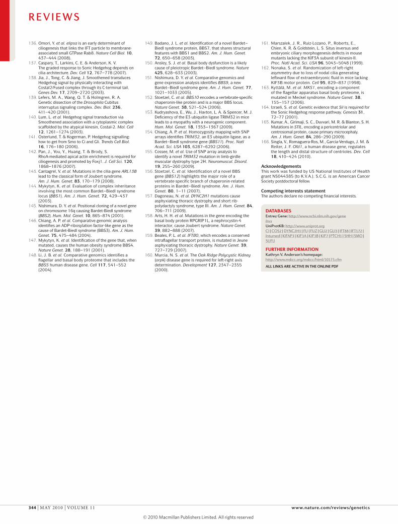
136. Omori, Y. et al. elipsa is an early determinant of ciliogenesis that links the IFT particle to membrane-associated small GTPase Rab8. Nature Cell Biol. 10, 437–444 (2008).
137. Caspary, T., Larkins, C. E. & Anderson, K. V. The graded response to Sonic Hedgehog depends on cilia architecture. Dev. Cell 12, 767–778 (2007).
138. Jia, J., Tong, C. & Jiang, J. Smoothened transduces Hedgehog signal by physically interacting with Costal2/Fused complex through its C-terminal tail. Genes Dev. 17, 2709–2720 (2003).
139. Lefers, M. A., Wang, Q. T. & Holmgren, R. A. Genetic dissection of the Drosophila Cubitus interruptus signaling complex. Dev. Biol. 236, 411–420 (2001).
140. Lum, L. et al. Hedgehog signal transduction via Smoothened association with a cytoplasmic complex scaffolded by the atypical kinesin, Costal-2. Mol. Cell 12, 1261–1274 (2003).
141. Osterlund, T. & Kogerman, P. Hedgehog signalling: how to get from Smo to Ci and Gli. Trends Cell Biol. 16, 176–180 (2006).
142. Pan, J., You, Y., Huang, T. & Brody, S. RhoA-mediated apical actin enrichment is required for ciliogenesis and promoted by Foxj1. J. Cell Sci. 120, 1868–1876 (2007).
143. Cantagrel, V. et al. Mutations in the cilia gene ARL13B lead to the classical form of Joubert syndrome. Am. J. Hum. Genet. 83, 170–179 (2008).
144. Mykytyn, K. et al. Evaluation of complex inheritance involving the most common Bardet–Biedl syndrome locus (BBS1). Am. J. Hum. Genet. 72, 429–437 (2003).
145. Nishimura, D. Y. et al. Positional cloning of a novel gene on chromosome 16q causing Bardet-Biedl syndrome (BBS2). Hum. Mol. Genet. 10, 865–874 (2001).
146. Chiang, A. P. et al. Comparative genomic analysis identifies an ADP-ribosylation factor-like gene as the cause of Bardet-Biedl syndrome (BBS3). Am. J. Hum. Genet. 75, 475–484 (2004).
147. Mykytyn, K. et al. Identification of the gene that, when mutated, causes the human obesity syndrome BBS4. Nature Genet. 28, 188–191 (2001).
148. Li, J. B. et al. Comparative genomics identifies a flagellar and basal body proteome that includes the BBS5 human disease gene. Cell 117, 541–552 (2004).
149. Badano, J. L. et al. Identification of a novel Bardet–Biedl syndrome protein, BBS7, that shares structural features with BBS1 and BBS2. Am. J. Hum. Genet. 72, 650–658 (2003).
150. Ansley, S. J. et al. Basal body dysfunction is a likely cause of pleiotropic Bardet–Biedl syndrome. Nature 425, 628–633 (2003).
151. Nishimura, D. Y. et al. Comparative genomics and gene expression analysis identifies BBS9, a new Bardet–Biedl syndrome gene. Am. J. Hum. Genet. 77, 1021–1033 (2005).
152. Stoetzel, C. et al. BBS10 encodes a vertebrate-specific chaperonin-like protein and is a major BBS locus. Nature Genet. 38, 521–524 (2006).
153. Kudryashova, E., Wu, J., Havton, L. A. & Spencer, M. J. Deficiency of the E3 ubiquitin ligase TRIM32 in mice leads to a myopathy with a neurogenic component. Hum. Mol. Genet. 18, 1353–1367 (2009).
154. Chiang, A. P. et al. Homozygosity mapping with SNP arrays identifies TRIM32, an E3 ubiquitin ligase, as a Bardet–Biedl syndrome gene (BBS11). Proc. Natl Acad. Sci. USA 103, 6287–6292 (2006).
155. Cossee, M. et al. Use of SNP array analysis to identify a novel TRIM32 mutation in limb-girdle muscular dystrophy type 2H. Neuromuscul. Disord. 19, 255–260 (2009).
156. Stoetzel, C. et al. Identification of a novel BBS gene (BBS12) highlights the major role of a vertebrate-specific branch of chaperonin-related proteins in Bardet–Biedl syndrome. Am. J. Hum. Genet. 80, 1–11 (2007).
157. Dagoneau, N. et al. DYNC2H1 mutations cause asphyxiating thoracic dystrophy and short rib-polydactyly syndrome, type III. Am. J. Hum. Genet. 84, 706–711 (2009).
158. Arts, H. H. et al. Mutations in the gene encoding the basal body protein RPGRIP1L, a nephrocystin-4 interactor, cause Joubert syndrome. Nature Genet. 39, 882–888 (2007).
159. Beales, P. L. et al. IFT80, which encodes a conserved intraflagellar transport protein, is mutated in Jeune asphyxiating thoracic dystrophy. Nature Genet. 39, 727–729 (2007).
160. Murcia, N. S. et al. The Oak Ridge Polycystic Kidney (orpk) disease gene is required for left-right axis determination. Development 127, 2347–2355 (2000).
161. Marszalek, J. R., Ruiz-Lozano, P., Roberts, E., Chien, K. R. & Goldstein, L. S. Situs inversus and embryonic ciliary morphogenesis defects in mouse mutants lacking the KIF3A subunit of kinesin-II. Proc. Natl Acad. Sci. USA 96, 5043–5048 (1999).
162. Nonaka, S. et al. Randomization of left-right asymmetry due to loss of nodal cilia generating leftward flow of extraembryonic fluid in mice lacking KIF3B motor protein. Cell 95, 829–837 (1998).
163. Kyttälä, M. et al. MKS1, encoding a component of the flagellar apparatus basal body proteome, is mutated in Meckel syndrome. Nature Genet. 38, 155–157 (2006).
164. Izraeli, S. et al. Genetic evidence that Sil is required for the Sonic Hedgehog response pathway. Genesis 31, 72–77 (2001).
165. Kumar, A., Girimaji, S. C., Duvvari, M. R. & Blanton, S. H. Mutations in STIL, encoding a pericentriolar and centrosomal protein, cause primary microcephaly. Am. J. Hum. Genet. 84, 286–290 (2009).
166. Singla, V., Romaguera-Ros, M., Garcia-Verdugo, J. M. & Reiter, J. F. Ofd1, a human disease gene, regulates the length and distal structure of centrioles. Dev. Cell 18, 410–424 (2010).
AcknowledgementsThis work was funded by US National Institutes of Health grant NS044385 (to K.V.A.). S.C.G. is an American Cancer Society postdoctoral fellow.
Competing interests statementThe authors declare no competing financial interests.
DATABASESEntrez Gene: http://www.ncbi.nlm.nih.gov/geneInvsUniProtKB: http://www.uniprot.orgCI | COS2 | DYNC2H1 | FU | FUZ | GLI2 | GLI3 | IFT88 | IFT172 | Inturned | KIFAP3 | KIF3A | KIF3B | KIF7 | PTCH1 | SHH | SMO | SUFU
FURTHER INFORMATIONKathryn V. Anderson’s homepage: http://www.mskcc.org/mskcc/html/10173.cfm
ALL Links Are Active in tHe onLine PdF
R E V I E W S
344 | mAy 2010 | vOlume 11 www.nature.com/reviews/genetics
© 20 Macmillan Publishers Limited. All rights reserved10