the prodrug of 7,8-dihydroxyflavone development and ... · the bdnf mimetic compound...
TRANSCRIPT

The prodrug of 7,8-dihydroxyflavone development andtherapeutic efficacy for treating Alzheimer’s diseaseChun Chena,b, Zhihao Wangb, Zhentao Zhangb, Xia Liub, Seong Su Kangb, Ying Zhanga, and Keqiang Yea,b,1
aCollege of Biosystems Engineering and Food Science, Zhejiang University, Zhejiang 310058, China; and bDepartment of Pathology and LaboratoryMedicine, Emory University School of Medicine, Atlanta, GA 30322
Edited by Solomon H. Snyder, Johns Hopkins University School of Medicine, Baltimore, MD, and approved December 12, 2017 (received for review October27, 2017)
The BDNF mimetic compound 7,8-dihydroxyflavone (7,8-DHF), apotent small molecular TrkB agonist, displays prominent thera-peutic efficacy against Alzheimer’s disease (AD). However, 7,8-DHFhas only modest oral bioavailability and a moderate pharmacoki-netic (PK) profile. To alleviate these preclinical obstacles, we used aprodrug strategy for elevating 7,8-DHF oral bioavailability andbrain exposure, and found that the optimal prodrug R13 has favor-able properties and dose-dependently reverses the cognitive de-fects in an AD mouse model. We synthesized a large number of7,8-DHF derivatives via ester or carbamate group modification onthe catechol ring in the parent compound. Using in vitro absorp-tion, distribution, metabolism, and excretion assays, combinedwith in vivo PK studies, we identified a prodrug, R13, that prom-inently up-regulates 7,8-DHF PK profiles. Chronic oral administra-tion of R13 activated TrkB signaling and prevented Aβ depositionin 5XFAD AD mice, inhibiting the pathological cleavage of APP andTau by AEP. Moreover, R13 inhibited the loss of hippocampal syn-apses and ameliorated memory deficits in a dose-dependent man-ner. These results suggest that the prodrug R13 is an optimaltherapeutic agent for treating AD.
7,8-dihydroxyflavone | prodrug | pharmacokinetics | TrkB |Alzheimer’s disease
Alzheimer’s disease (AD), the leading cause of dementiaworldwide, is characterized by accumulation of β-amyloid
peptide (Aβ) within the brain, along with hyperphosphorylated andcleaved forms of the microtubule-associated protein Tau. At pre-sent, drugs used for the treatment of AD only slightly delay theinevitable symptomatic progression of the disease and do not affectthe main neuropathological hallmarks of the disease, i.e., senileplaques and neurofibrillary tangles. Reduced acetylcholine neuro-transmission due to loss of neurons in the basal forebrain and de-pletion of choline acetyltransferase is observed in AD pathology (1).Currently, there are two types of medications to treat AD:
cholinesterase inhibitors and N-methyl-D-aspartate receptor(NMDAR) antagonists. The first category of inhibitors blocksthe activity of acetylcholinesterase, thereby increasing acetyl-choline levels in the CNS to restore cognitive functions. Thesecond category inhibits overactivation of NMDA-induced glu-tamate excitotoxicity. These drugs can only partially alleviate thesymptoms of AD (2).The amyloid hypothesis has historically dominated the pro-
posed pathology of AD. However, over the last decade, efforts totarget the traditional pathways have been consistently ineffective(3). Therefore, it is of urgent need to develop more potentagents to manage cognitive impairment and disease-modifyingagents that prevent or delay the onset of AD.Neurotrophins are growth factors that regulate neuronal de-
velopment, differentiation, and survival. Neurotrophins consist offour related proteins: nerve growth factor (NGF), brain-derivedneurotrophic factor (BDNF), neurotrophin-3 (NT-3), andneurotrophin-4 (NT-4/5). Neurotrophins exert the trophic effectsvia the cognate Trk receptors. It has been well documented thatBDNF expression is reduced in the brains of patients with AD (4).
BDNF exerts a protective role against AD pathogenesis, and hasbeen shown to increase learning and memory in demented animals(5). BDNF gene delivery has been reported as a novel potentialtherapy in diverse models related to AD (6). Thus, the preclinicalevidence strongly supports that BDNF might be useful as a thera-peutic agent for a variety of neurologic disorders. The outcomes ofseveral clinical trials using recombinant BDNF are disappointing,however. Presumably, this is due to poor delivery and the shortin vivo half-life of BDNF.To search for TrkB receptor agonists to mimic BDNF, we used
cell-based screening. We found that 7,8-dihydroxyflavone(7,8-DHF) specifically binds to the TrkB receptor extracellulardomain and acts as a selective TrkB agonist, which mimics thephysiological actions of BDNF (7). Remarkably, systemic ad-ministration of 7,8-DHF can activate TrkB receptors in brainsand induce BDNF-like behavioral phenotypes, such as enhancedlearning and memory, and antistress or antidepressant-like ef-fects in rodents in a TrkB-dependent manner (8–10). Recently,several independent studies have shown that 7,8-DHF rescuesmemory deficits in different AD animal models, restoring deficientTrkB signaling without affecting endogenous BDNF levels. In-terestingly, 7,8-DHF represses BACE1 expression and lowers lev-els of Aβ40 and Aβ42. Strikingly, it improves spatial memory andincreases thin spine density in a mouse model of AD-like neuronalloss. Therefore, 7,8-DHF represents a novel oral bioactive thera-peutic agent for treating AD (11–14). Taken together, these studiesindicate that 7,8-DHF displays prominent therapeutic efficacy to-ward AD through its action as a TrkB receptor agonist.Dozens of independent studies have shown that 7,8-DHF
mimics the physiological actions of BDNF in numerous animalmodels (15–17), and chronic treatment with 7,8-DHF has dem-onstrated no detectable toxicity (10). Remarkably, 7,8-DHF
Significance
In this study, we used a prodrug strategy to improve the drugg-ability of 7,8-DHF, which mimics the physiological actions of BDNFin a variety of animal models but with modest oral bioavailability,by improving its PK profiles. In 5XFAD mouse model, we dem-onstrate that the optimal prodrug R13 increases the half-life, oralbioavailability, and brain exposure of 7,8-DHF. Most importantly,R13 robustly displays promising therapeutic efficacy by stronglyactivating TrkB and repressing AEP, which plays a crucial role inthe pathogenesis of Alzheimer’s disease (AD), leading to elimi-nation of senile plaques in the AD mouse brain.
Author contributions: C.C., X.L., and K.Y. designed research; C.C. and Z.W. performedresearch; C.C., Z.Z., X.L., S.S.K., Y.Z., and K.Y. contributed new reagents/analytic tools;C.C. and K.Y. analyzed data; and C.C. and K.Y. wrote the paper.
The authors declare no conflict of interest.
This article is a PNAS Direct Submission.
Published under the PNAS license.1To whom correspondence should be addressed. Email: [email protected].
This article contains supporting information online at www.pnas.org/lookup/suppl/doi:10.1073/pnas.1718683115/-/DCSupplemental.
578–583 | PNAS | January 16, 2018 | vol. 115 | no. 3 www.pnas.org/cgi/doi/10.1073/pnas.1718683115
Dow
nloa
ded
by g
uest
on
June
1, 2
020

penetrates the blood-brain barrier (BBB) and is orally bioactive(18). A structure-activity relationship study showed that the catecholmoiety in 7,8-DHF is critical for its agonistic effect (10), and thatthis compound has suboptimal oral bioavailability and brain expo-sure (18). Taken together, these findings support that 7,8-DHF isefficacious and safe for treating AD chronically and orally.Given 7,8-DHF’s favorable safety and remarkable efficacy in
treating various neurologic diseases, including AD, we believethat the prodrug strategy to improve its oral bioavailability andexposure in circulation system should be an ideal approach. Ourmetabolism study showed that the catechol group is extensivelyconjugated by glucuronidation, sulfation, and methylation in theliver, leading to the poor oral bioavailability (18). In the presentstudy, we synthesized dozens of derivatives to modify the cate-chol group; monitored the prodrugs’ intestine microsomal sta-bility, liver microsomal stability, and plasma stability; andexamined some of the favorable compounds’ chemical stabilityand Caco-2 permeability. We found that prodrug R13 exhibitedgood absorption and was readily hydrolyzed into 7,8-DHF inliver microsomes. Notably, R13 has a very long half-life, and itimproves 7,8-DHF’s oral bioavailability from 4.6% to ∼10.5%.The plasma concentration and brain exposure are also signifi-cantly enhanced. In a 5XFAD mice strain, chronic oral admin-istration of R13 alleviated Aβ deposition, attenuated the loss ofhippocampal synapse, and ameliorated memory deficits in adose-dependent manner. These exciting data provide a ground-work for moving this prodrug into clinical trials.
ResultsSynthesizing Prodrugs of 7,8-Dihydroxyflavone via Medicinal Chemistry.7,8-DHF is orally bioactive and penetrates the BBB (18). Previousstudies have shown that 7,8-DHF is safe for chronic treatment andefficacious for treating AD, at least in animal models (11–13).Thus, this compound itself is good enough for preclinical in-vestigation. Its oral bioavailability is not optimal, however. Thecatechol group makes 7,8-DHF and its lead compounds labile forfast metabolism, including glucuronidation, sulfation, methylation,and so on (18). Since the catechol group is essential for thepharmacophore, we wished to use a prodrug strategy to elevatethe bioavailability. To do so, we synthesized numerous esters orcarbamates by changing the hydroxy groups on the catechol ring in7,8-DHF (Fig. 1A and SI Appendix, Fig. S1). The detailed syn-thetic routes and characterization of derivatives are described inMaterials and Methods.
Identifying the Optimal Prodrug with Appropriate in Vitro PK Profiles.To identify the ideal prodrug, we used the strategy outlined inFig. 1B. The compounds should be stable in both aqueous so-lution and at room temperature for a long shelf life. Moreover,the optimal derivatives should be stable in intestinal microsomesduring absorption, but can be hydrolyzed in liver microsomes or
plasma into 7,8-DHF. In addition, the prodrugs should exhibitfavorable Caco-2 permeability to allow prominent absorption.The derivatives meeting these standards will be subjected to anin vivo PK study in mice, and 7,8-DHF brain exposure will bedetermined after oral administration of the prodrug.On intestinal microsome, liver microsome, and plasma sta-
bility screening, among the 20 synthetic derivatives, only R5, R7,R13, R16, R17, and R18 were relatively stable in intestinal mi-crosomes and hydrolyzable in both liver microsomes and plasma.Unfortunately, R7 barely yielded free 7,8-DHF in human livermicrosomes, and R16 or R17 failed to release any detectable 7,8-DHF, indicating that these two compounds have different hy-drolytic chemistries (SI Appendix, Tables S1–S3). AlthoughR18 was relatively stable under both conditions and was alsohydrolyzed into 7,8-DHF, its Caco-2 permeability (Re of 61.6)was very poor (SI Appendix, Tables S4 and S5). Taken together,the in vitro screening assays support R13 as the sole appropriatecandidate for further investigation.
In Vivo PK and Brain Exposure of R13. We tested the in vivo PK/BBB profiles of 7,8-DHF and R13 after dosing R13 in six mice.Three animals in group 1 were dosed orally with 7,8-DHF at50 mg/kg, and three animals in group 2 were dosed orally withR13 at 36 mg/kg. Blood samples were collected from all animalsup to 8 h after dosing. Concentrations of 7,8-DHF in the plasmasamples were determined using LC-MS/MS. Pharmacokineticanalysis was performed using a noncompartmental method. Fol-lowing a single oral administration of R13 at a dose of 36 mg/kg,the mean Cmax and Tmax values for 7,8-DHF were 129 ng/mL and30 min, respectively, and the mean area under the curve (AUC)(0–t) value was 11,880 min ng/mL. The mean value of oral bio-availability for R13 was 10.5%. (SI Appendix, Table S6). Notably,7,8-DHF T1/2 released from R13 was 219.6 min versus 134 min byparent compound, indicating the prodrug can sustainably release7,8-DHF in the circulation system.For BBB study, plasma samples and the brains were collected
from three animals at each designated postdose time point (3,10, and 30 min and 1, 2, 4, 6, and 8 h). Concentrations of 7,8-DHF and R13 in the plasma samples and brain were determinedby LC-MS/MS. The concentration ratio of brain/plasma wascalculated, and PK analysis was performed using a non-compartmental method. Following a single oral administrationof R13 at a dose of 72.5 mg/kg, the mean Cmax and Tmax valuesfor 7,8-DHF were 56 ng/mL and 2 h, respectively, in plasmasamples, and 46 ng/mL and 4 h, respectively, in brain samples. Invivo BBB study showed that 7,8-DHF plasma concentrationsfrom prodrug R13 were higher than those from the parentcompound (50 mg/kg) on oral administration. Strikingly, 7,8-DHF was detectable in the plasma even at 8 h, indicating theprodrug can sustainably release 7,8-DHF in the circulation sys-tem. Notably, the 7,8-DHF concentration from R13 in the brainwas >5 ng/g even after 240 min (Fig. 2 A–C). Overall, thiscompound has the favorable druggable properties. Of note,R13 readily hydrolyzes into an intermediate T1 under neutralpH, before 7,8-DHF is finally released in vitro, although R13 isvery stable under pH 1.2 (Fig. 2D). Accordingly, we monitoredT1 concentrations in both the plasma and brain on oral adminis-tration of different doses of R13. As expected, T1 was significantlyelevated in the plasma, but not in the brain, in a dose-dependentmanner at 2 h. Its concentrations were much higher than those of7,8-DHF, supporting the concept that R13 is hydrolyzed intoT1 first before it is finally decayed into 7,8-DHF in vivo, inalignment with in vitro observations (Fig. 2E).
Oral Administration of R13 Activates TrkB and Downstream SignalingPathways in Mouse Brain.Given the molecular weights of R13 and7,8-DHF of 368 and 254, respectively, 7.25, 21.8, and 43.6 mg/kgof R13 are equivalent to 5, 15, and 30 mg/kg of 7,8-DHF,
A B Prodrug Organic Synthesis
Aqueous and Room Temperature Stability
Intestine microsome, Liver microsome and Plasma Stability assay
Caco-2 Permeability (absorption)
In vivo Pharmacokinetics and brain exposure (metabolism and distribution)
Fig. 1. (A) R13: 7,8-DHF prodrug chemical structure. (B) In vitro absorption,distribution, metabolism, and excretion screening strategy for prodrugs.
Chen et al. PNAS | January 16, 2018 | vol. 115 | no. 3 | 579
NEU
ROSC
IENCE
CHEM
ISTR
Y
Dow
nloa
ded
by g
uest
on
June
1, 2
020
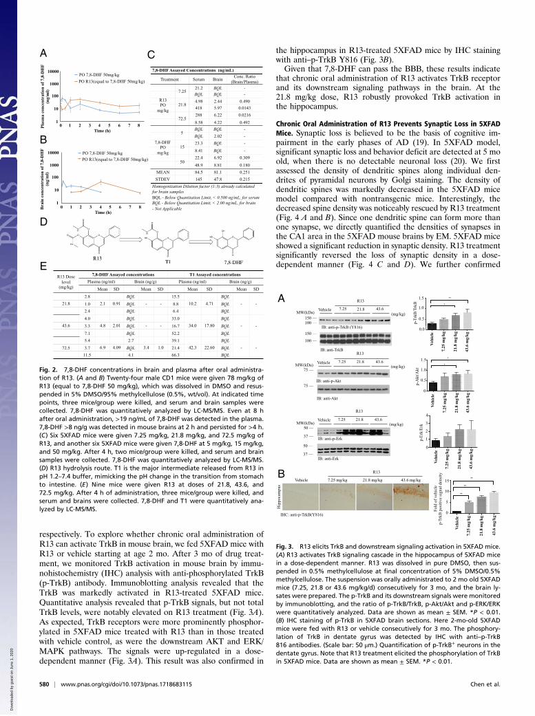
respectively. To explore whether chronic oral administration ofR13 can activate TrkB in mouse brain, we fed 5XFAD mice withR13 or vehicle starting at age 2 mo. After 3 mo of drug treat-ment, we monitored TrkB activation in mouse brain by immu-nohistochemistry (IHC) analysis with anti-phosphorylated TrkB(p-TrkB) antibody. Immunoblotting analysis revealed that theTrkB was markedly activated in R13-treated 5XFAD mice.Quantitative analysis revealed that p-TrkB signals, but not totalTrkB levels, were notably elevated on R13 treatment (Fig. 3A).As expected, TrkB receptors were more prominently phosphor-ylated in 5XFAD mice treated with R13 than in those treatedwith vehicle control, as were the downstream AKT and ERK/MAPK pathways. The signals were up-regulated in a dose-dependent manner (Fig. 3A). This result was also confirmed in
the hippocampus in R13-treated 5XFAD mice by IHC stainingwith anti–p-TrkB Y816 (Fig. 3B).Given that 7,8-DHF can pass the BBB, these results indicate
that chronic oral administration of R13 activates TrkB receptorand its downstream signaling pathways in the brain. At the21.8 mg/kg dose, R13 robustly provoked TrkB activation inthe hippocampus.
Chronic Oral Administration of R13 Prevents Synaptic Loss in 5XFADMice. Synaptic loss is believed to be the basis of cognitive im-pairment in the early phases of AD (19). In 5XFAD model,significant synaptic loss and behavior deficit are detected at 5 moold, when there is no detectable neuronal loss (20). We firstassessed the density of dendritic spines along individual den-drites of pyramidal neurons by Golgi staining. The density ofdendritic spines was markedly decreased in the 5XFAD micemodel compared with nontransgenic mice. Interestingly, thedecreased spine density was noticeably rescued by R13 treatment(Fig. 4 A and B). Since one dendritic spine can form more thanone synapse, we directly quantified the densities of synapses inthe CA1 area in the 5XFAD mouse brains by EM. 5XFAD miceshowed a significant reduction in synaptic density. R13 treatmentsignificantly reversed the loss of synaptic density in a dose-dependent manner (Fig. 4 C and D). We further confirmed
Bra
in c
once
ntra
tion
of 7
,8-D
HF
(ng/
ml)
1
10
1000
100
10000
0 1 2 3 4 5 6 7 8Time (h)
PO 7,8-DHF 50mg/kgPO R13 (equal to 7,8-DHF 50mg/kg)PO 7,8-DHF 50mg/kgPO R13(equal to 7,8-DHF 50mg/kg)
B
A C7,8-DHF Assayed Concentrations (ng/mL)
Treatment Serum Brain Conc. Ratio (Brain/Plasma)
R13 PO
mg/kg
7.2521.2 BQL -BQL BQL -
21.84.98 2.44 0.490418 5.97 0.0143
72.5288 6.22 0.0216
8.58 4.22 0.492
7,8-DHF PO
mg/kg
5BQL BQL -BQL 2.02 -
1523.3 BQL -
8.41 BQL -
5022.4 6.92 0.309
48.9 8.81 0.180MEAN 84.5 81.1 0.251STDEV 145 47.8 0.215
Homogenization Dilution factor (1:3) already calculated for brain samples BQL - Below Quantitation Limit, < 0.500 ng/mL, for serum BQL - Below Quantitation Limit, < 2.00 ng/mL, for brain - Not Applicable
D
ER13 Dose
level (mg/kg)
7,8-DHF Assayed concentrations T1 Assayed concentrationsPlasma (ng/ml) Brain (ng/g) Plasma (ng/ml) Brain (ng/g)
Mean SD Mean SD Mean SD Mean SD
21.82.8
2.1 0.91BQL
- -15.5
10.2 4.71BQL
- -1.0 BQL 8.8 BQL2.4 BQL 6.4 BQL
43.64.0
4.8 2.01BQL
- -33.0
34.0 17.80BQL
- -3.3 BQL 16.7 BQL7.1 BQL 52.2 BQL
72.5
5.46.9 4.09
2.73.4 1.0
39.142.3 22.60
BQL- -3.7 BQL 21.4 BQL
11.5 4.1 66.3 BQL
PO 7,8-DHF 50mg/kg
PO R13 (equal to 7,8-DHF 50mg/kg)
Plas
ma
conc
entr
atio
n of
7,8
-DH
F (n
g/m
l)
1
10
1000
100
10000
0 1 2 3 4 5 6 7 8Time (h)
PO 7,8-DHF 50mg/kgPO R13(equal to 7,8-DHF 50mg/kg)
Fig. 2. 7,8-DHF concentrations in brain and plasma after oral administra-tion of R13. (A and B) Twenty-four male CD1 mice were given 78 mg/kg ofR13 (equal to 7,8-DHF 50 mg/kg), which was dissolved in DMSO and resus-pended in 5% DMSO/95% methylcellulose (0.5%, wt/vol). At indicated timepoints, three mice/group were killed, and serum and brain samples werecollected. 7,8-DHF was quantitatively analyzed by LC-MS/MS. Even at 8 hafter oral administration, >19 ng/mL of 7,8-DHF was detected in the plasma.7,8-DHF >8 ng/g was detected in mouse brains at 2 h and persisted for >4 h.(C) Six 5XFAD mice were given 7.25 mg/kg, 21.8 mg/kg, and 72.5 mg/kg ofR13, and another six 5XFAD mice were given 7,8-DHF at 5 mg/kg, 15 mg/kg,and 50 mg/kg. After 4 h, two mice/group were killed, and serum and brainsamples were collected. 7,8-DHF was quantitatively analyzed by LC-MS/MS.(D) R13 hydrolysis route. T1 is the major intermediate released from R13 inpH 1.2–7.4 buffer, mimicking the pH change in the transition from stomachto intestine. (E) Nine mice were given R13 at doses of 21.8, 43.6, and72.5 mg/kg. After 4 h of administration, three mice/group were killed, andserum and brains were collected. 7,8-DHF and T1 were quantitatively ana-lyzed by LC-MS/MS.
IHC: anti-p-TrkB(Y816)
Hip
poca
mpu
s
Vehicle 21.8 mg/kg 7.25 mg/kg 43.6 mg/kg
R13
A
B
IB: anti-TrkB
IB: anti-p-TrkB (Y816)
Vehicle 7.25 21.8 43.6
R13
MW(kDa) (mg/kg)
0.0
0.5
1.0
**
*
p-Tr
kB/T
rkB
Vehi
cle
7.25
mg/
kg
21.8
mg/
kg
43.6
mg/
kg
1.0
0.5
0.0
1.5
e g g g
*
*
p-A
kt/A
kt
Vehi
cle
7.25
mg/
kg
21.8
mg/
kg
43.6
mg/
kg
1.0
0.5
0
1.5
le g g g
p-Er
k/Er
k
Vehi
cle
7.25
mg/
kg
21.8
mg/
kg
43.6
mg/
kg
3
2
0
4
1
Vehi
cle
7.25
mg/
kg
21.8
mg/
kg
43.6
mg/
kg
Fold
of v
ehic
le
p-Tr
kB p
ositi
ve si
gnal
den
sity
**
**
**
10
5
0
15
75
IB: anti-p-Akt75
IB: anti-Akt
Vehicle 7.25 21.8 43.6
R13
MW(kDa) (mg/kg)
50
37 IB: anti-Erk
50
37 IB: anti-p-Erk
Vehicle 7.25 21.8 43.6
R13
MW(kDa) (mg/kg)
Fig. 3. R13 elicits TrkB and downstream signaling activation in 5XFAD mice.(A) R13 activates TrkB signaling cascade in the hippocampus of 5XFAD micein a dose-dependent manner. R13 was dissolved in pure DMSO, then sus-pended in 0.5% methylcellulose at final concentration of 5% DMSO/0.5%methylcellulose. The suspension was orally administrated to 2 mo old 5XFADmice (7.25, 21.8 or 43.6 mg/kg/d) consecutively for 3 mo, and the brain ly-sates were prepared. The p-TrkB and its downstream signals were monitoredby immunoblotting, and the ratio of p-TrkB/TrkB, p-Akt/Akt and p-ERK/ERKwere quantitatively analyzed. Data are shown as mean ± SEM. *P < 0.01.(B) IHC staining of p-TrkB in 5XFAD brain sections. Here 2-mo-old 5XFADmice were fed with R13 or vehicle consecutively for 3 mo. The phosphory-lation of TrkB in dentate gyrus was detected by IHC with anti–p-TrkB816 antibodies. (Scale bar: 50 μm.) Quantification of p-TrkB+ neurons in thedentate gyrus. Note that R13 treatment elicited the phosphorylation of TrkBin 5XFAD mice. Data are shown as mean ± SEM. *P < 0.01.
580 | www.pnas.org/cgi/doi/10.1073/pnas.1718683115 Chen et al.
Dow
nloa
ded
by g
uest
on
June
1, 2
020

these findings by immunoblotting using presynaptic markers(synaptotagmin) and postsynaptic markers (GluR1, PSD95, andspinophilin). 5XFAD mice displayed a considerable decrease inthese synaptic markers, indicating synaptic degeneration (12).R13 treatment reversed the reduction of synaptic markers (Fig.4E). Electrophysiology analysis demonstrated that R13 increasedlong-term potentiation in a dose-dependent manner (Fig. 4F), inagreement with the findings of augmentation of synapses by R13.These results suggest that activation of the TrkB receptor byR13 inhibits the loss of synapses in 5XFAD mice and improvessynaptic plasticity.
R13 Alleviates Aβ Deposition and Rescues Memory Deficits in 5XFADMice.We further tested the deposition of Aβ by IHC with anti-Aβantibody. The dose-dependent Aβ deposition in both brain re-gions was significantly lower in the R13-treated group than in thecontrol group (Fig. 5 A–C). We further detected the effect ofR13 treatment on senile plaque formation by thioflavin-S stain-ing. 5XFAD mice showed evident plaque deposition in both thecortex and the hippocampus at age 5 mo. Strikingly, the number
of plaques and plaque area fraction were significantly decreasedin both areas in R13-treated mice compared with vehicle control-treated mice (SI Appendix, Fig. S2 A–C). To verify whetherR13 inhibits the production of Aβ, the concentrations of totalAβ42 and Aβ40 were quantitatively determined by ELISA. In-terestingly, Aβ40 concentrations in 5XFAD mice brain were de-creased by R13 at all doses tested, and although Aβ42 concentrationsdisplayed a trend toward reduction with R13, the differences were notstatistically significant (Fig. 5 D and E). Thus, these results suggestthat chronic oral R13 administration may prevent Aβ depositionand Aβ40 production.The hippocampus-dependent spatial memory of 5XFAD mice
was assessed using the Morris water maze test. The average la-tency (Fig. 5 F andG) and swim path length (SI Appendix, Fig. S2D and E) for each of the five acquisition days were calculatedand plotted. A two-way mixed ANOVA (group × training day)on latency revealed a main effect of training day (P < 0.01) andof group (P < 0.01), but not of interaction (Fig. 5F). The AUC oflatency was greater in vehicle-treated 5XFAD mice comparedwith nontransgenic control mice, indicating impaired acquisitionof the spatial learning task; however, the learning impairment in5XFAD mice was attenuated by R13 treatment (Fig. 5G). Amixed two-way ANOVA on swim path distance also revealed asignificant main effect of training day (P < 0.01) and of group(P < 0.01), but not of interaction (SI Appendix, Fig. S2D). TheAUC of the swim path distance was greater in 5XFAD micecompared with nontransgenic control mice, and was decreasedby R13 treatment (SI Appendix, Fig. S2E). The memory recall forthe platform location was assessed in the probe trail when theplatform was removed and the mice were allowed to search for
5XFAD, R13 43.6 mg/kg5XFAD, Vehicle 5XFAD, R13 7.25mg/kg 5XFAD, R13 21.8 mg/kg
IB: anti-spinophilin
100
IB: anti-PSD95
50
37 IB: anti- -actin
75
50IB: anti-synaptotagmin
IB: anti-GluR
Vehicle 7.25 21.8 43.6
R13
(mg/kg)
Vehi
cle
7.25
mg/
kg
21.8
mg/
kg
43.6
mg/
kg
Vehi
cle
7.25
mg/
kg
21.8
mg/
kg
43.6
mg/
kg
**
**
*
Spin
e nu
mbe
r/10μ
m 2015
10
50
**
**
*
Syna
pses
/100μm
2 CA
1 ar
ea
Vehi
cle
7.25
mg/
kg
21.8
mg/
kg
43.6
mg/
kg
60
40
20
0
fEPS
P sl
ope
(% o
f bas
elin
e)
Time (min)
Vehicle7.25 mg/kg21.8 mg/kg43.6 mg/kg
3.0
2.5
2.0
1.5
1.0
0.50 10 20 30 40 50 60 70 80 90
MW(kDa)
B C
D
E F
A
Fig. 4. R13 prevents the synaptic loss in hippocampal CA1 area of 5XFADmice. (A) R13 reversed the synaptic loss in 5XFAD mice. The dendritic spinesfrom apical dendritic layer of the CA1 region were analyzed by Golgistaining. (Scale bar: 5 μm.) (B) Quantitative analysis of spine density. Thedecreased spine density in 5XFAD mice was reversed by R13 in a dose-dependent manner. n = 6 in each group. *P < 0.01. (C) Quantitative analysis ofthe synaptic density in vehicle- and R13-treated 5XFAD mice. 5XFAD miceshow decreased synaptic density, which was reversed by R13. Data are shownas mean ± SEM. *P < 0.01. (D) Representative EM image of the synapticstructures. Red stars indicate the synapses. (Scale bar: 1 μm.) (E) Immuno-blotting analysis of synaptic markers in brain homogenates from mice treatedwith vehicle or R13. R13 treatment increased the expression of synapticmarkers in 5XFAD mice. (F) LTP of field excitatory postsynaptic potentials(fEPSPs) was induced by 3XTBS (theta-burst stimulation) (four pulses at 100 Hz,repeated three times with 200-ms intervals). The traces shown are represen-tative fEPSPs recorded at time points 1 (vehicle-treated 5XFAD mice), 2, 3, and4 (R13-treated 5XFAD mice). The magnitude of LTP in 5XFAD mice is signifi-cantly lower in vehicle-treated transgenic mice, and R13 treatment reversedthe LTP impairment. n = 5 in each group. Data are presented as mean ± SEM.*P < 0.05, vehicle-treated vs. R13-treated mice.
A
F G
Fron
tal
Cor
tex
Hip
poca
mpu
s
IHC: anti-A
Vehicle 21.8 mg/kg 7.25 mg/kg 43.6 mg/kg
R13 B
D E C
**
****
Hippocampus
Num
ber o
f pla
ques
/vis
ual f
ield
Vehi
cle
7.25
mg/
kg
21.8
mg/
kg
43.6
mg/
kg
806040200
Frontal Cortex**
**
**N
umbe
r of p
laqu
es/v
isua
l fie
ld
Vehi
cle
7.25
mg/
kg
21.8
mg/
kg
43.6
mg/
kg
80
60
40
20
0
A42
con
cent
ratio
n (µ
g/g)
Vehi
cle
7.25
mg/
kg
21.8
mg/
kg
43.6
mg/
kg
80
60
40
20
0
**
**
**
A40
con
cent
ratio
n (µ
g/g)
Vehi
cle
7.25
mg/
kg
21.8
mg/
kg
43.6
mg/
kg
1
2345
0
AU
C la
tenc
y
Vehi
cle
7.25
mg/
kg
21.8
mg/
kg
43.6
mg/
kg
Con
trol
200
150
100
50
0
80
60
0
40
20
Training Day
Late
ncy
to m
ount
pl
atef
orm
(+/-
SEM
)
ControlVehicle7.25 mg/kg21.8 mg/kg43.6 mg/kg
% o
f Tim
e in
Pla
tform
Qua
dran
t
Vehi
cle
7.25
mg/
kg
21.8
mg/
kg
43.6
mg/
kg
Con
trol
**
*******80
60
0
40
20
H
1 2 3 4 5
Fig. 5. R13 alleviates Aβ deposition and reduces the concentrations of totalAβ, improving the spatial learning andmemory of 5XFADmice. (A) IHC analysisof Aβ deposits in 5XFAD mice. (Scale bar: 100 μm.) (B and C) Quantitativeanalysis of amyloid plaques. Amyloid deposition in 5XFAD mice was signifi-cantly decreased by orally administrated R13 at all doses. *P < 0.01. (D and E)Aβ42 and Aβ40 ELISA. R13 significantly reduced the concentrations of Aβ40,but not of Aβ42, in mouse brain at all doses. (F–H) R13 improves cognitivefunctions in 5XFAD mice. 5XFAD mice (n = 8–10/group) orally administeredcontrol vehicle or different doses of R13 were trained in the water maze over5 d. Shown are mean ± SEM latency tomount the escape platform (F), the AUCof latency (G), and the percentage of time spent in the target quadrant (H).*P < 0.05 compared with vehicle-treated 5XFAD mice.
Chen et al. PNAS | January 16, 2018 | vol. 115 | no. 3 | 581
NEU
ROSC
IENCE
CHEM
ISTR
Y
Dow
nloa
ded
by g
uest
on
June
1, 2
020

60 s. Compared with nontransgenic control mice, vehicle-treated5XFAD mice spent a significantly lower percentage of their timein the quadrant that formerly contained the hidden platform(Fig. 5H), indicating severe deficits in spatial memory recall. The5XFAD mice treated with R13 spent a significantly higher per-centage of time in the target quadrant, demonstrating rescue ofspatial memory. All groups of mice displayed comparable swimspeeds (SI Appendix, Fig. S2F).
R13 Inhibits AEP Activation and APP and Tau Proteolytic Cleavage in5XFAD Mice. We recently reported that delta-secretase (AEP;asparagine endopeptidase) cleaves both APP and Tau, mediatingAD pathogenesis (21, 22). To explore whether R13 affects AEP’seffect in AD pathologies, we conducted immunoblotting analy-ses, and found that mature and active AEP were dose-dependentlydecreased by R13, correlating with reductions in AEP-cleavedAPP N373, APP N585, and Tau N368 truncates in 5XFADmouse brains (Fig. 6A). To further investigate APP proteolyticcleavage activity by AEP, we conducted immunofluorescent cos-taining of 5XFAD brain sections with anti-APP C585 and MAP2antibodies. With a R13 dose increase, APP C585 immunosignalswere strongly inhibited by R13, inversely coupled withMAP2 augmentation (SI Appendix, Fig. S3A), indicating thatR13 protects against neuronal loss. Furthermore, TauN368 staining was also repressed by R13, coupled with thereduced pathological phosphorylation of Tau: AT-8 (SI Ap-pendix, Fig. S3B). In agreement with these observations, IHCrevealed that AEP expression in both hippocampus and frontal
cortex was progressively attenuated by R13 when its dose wasgradually escalated (Fig. 6 B–D). Enzymatic assays showed thatAEP activity was blocked by R13 at both 21.8 and 43.6 mg/kg (Fig.6E). In alignment with these observations, the inflammatoryfactors, including IL-1β, IL-6, and TNFα, in 5XFADmouse brainswere all repressed by R13. Thus, our data strongly support thatR13 increases 7,8-DHF oral bioavailability and brain exposure andactivates TrkB and its downstream signal pathways, repressingAEP and blocking APP and Tau pathological fragmentation.
DiscussionIn the present study, we used the prodrug strategy to improve thepoor oral bioavailability of parent compound 7,8-DHF. Amongthe synthetic prodrugs, the carbamate prodrug R13 exhibited themost favorable in vitro and in vivo drug metabolism and PKcharacteristics (SI Appendix, Tables S1–S6). Thus, we examinedin vivo PK and found that R13 exhibited ∼10.5% oral bio-availability with a Cmax of 129 ng/mL, Tmax of 0.5 h, and T1/2 fororal administration of 3.66 h. Of note, 7,8-DHF plasma con-centrations released from R13 (oral, 36 mg/kg) were muchhigher than those achieved from oral administration of higherdoses of parent 7,8-DHF (50 mg/kg). The oral bioavailability for7,8-DHF increased from 4.6% with the parent compound to10.5% with R13 (SI Appendix, Table S6). As expected, TrkBreceptor and its downstream p-Akt/p-MAPK signaling are po-tently activated on oral administration of R13, tightly correlatingwith 7,8-DHF concentrations in the brain (Fig. 3). The TrkBactivation fits well with the in vivo PK data, supporting that thereleased 7,8-DHF from R13 prodrug triggers the long-lastingTrkB signaling in mouse brains.5XFAD mice have been shown to develop cerebral amyloid
plaques at 2 mo of age, and show memory impairment at 4–5 moof age (23). It has also been shown that the level of matureBDNF is dramatically reduced in 5XFAD mice, beginning at3 mo of age (11). Given the key roles of BDNF-TrkB signaling inlearning and memory, we propose that prodrug R13 may protectmemory decline in 5XFAD mice. We fed the 2-mo-old 5XFADmice with R13 for 3 mo, and found marked activation of theTrkB receptor by 7,8-DHF in the dentate gyrus. The activationof TrkB downstream Akt and MAPK pathways are coupled toTrkB phosphorylation. Therefore, chronic oral administration ofR13 activates BDNF-TrkB signaling in the brains of 5XFADmice. Furthermore, our results are inconsistent with a previousreport that systematic administration of 7,8-DHF triggers TrkBactivation in a transgenic mice model of AD and in cognitivelyimpaired aged rats (11, 24). Activation of TrkB is required formultiple aspects of neuronal function, including neuronal sur-vival, morphological change of neurons, and synaptic plasticity(4, 25). TrkB signaling promotes the formation of dendriticspines (26). We observed a decrease in dendritic spine density inthe hippocampus in 5XFADmice, and found that R13 increased thespine density in apical dendrites of CA1 neurons of hippocampus. Inagreement with these observations, R13 also exerted a rescue effecton the number of synapses in the CA1 area of the 5XFAD mode.Furthermore, the expression of synaptic markers was also increasedby R13 treatment, supporting that 7,8-DHF released from R13 has aprofound protective effect on synapses in vivo.In the 5XFAD mice model, chronic oral administration of
R13 activates TrkB signaling pathways in brain, attenuates syn-aptic loss, and reverses Aβ deposition. Consequently, R13 rescuesmemory deficits in 5XFAD mice. Therefore, our results suggestthat 7,8-DHF released from prodrug R13 simulates the physio-logical actions of BDNF and prevents the synaptic dysfunction andcognitive deficits in the AD mouse model. A chronic toxicity studyof R13 in C57BL6 mice involving 12 wk of consecutive treatmentwith daily doses of 43.6 mg/kg also showed no toxicity (SI Ap-pendix, Fig. S4). Thus, these studies support that R13 is safe and
AVehicle 7.25 21.8 43.6
R13
MW(kDa)(mg/kg)
37IB: anti-AEP (cleaved)
IB: anti- -actin
50
37
IB: anti-Tau N368
37
IB: anti-Tau 550
75
50IB: anti-APP N373
100
IB: anti-APP N585
FE
R13
Vehicle 21.8 mg/kg 43.6 mg/kg 7.25 mg/kg
Fron
tal
Cor
tex
Hip
poca
mpu
s
IHC: anti-AEP (6E3)
B
C D**
****
Vehi
cle
7.25
mg/
kg
21.8
mg/
kg
43.6
mg/
kgNum
ber o
f -s
ecre
tase
po
sitiv
e ce
ll /
visu
al fi
eld Hippocampus
1500
1000
500
0
Num
ber o
f -s
ecre
tase
po
sitiv
e ce
ll /
visu
al fi
eld
****
**
Vehi
cle
7.25
mg/
kg
21.8
mg/
kg
43.6
mg/
kg
Frontal Cortex
8000
40006000
2000
0
Vehi
cle
7.25
mg/
kg
21.8
mg/
kg
43.6
mg/
kg
e g g g
**
**
AEP
enz
ymat
ic a
ctiv
ity
010002000300040005000
****
**
IL-6
con
cent
ratio
n (p
g/m
l)
Vehi
cle
7.25
mg/
kg
21.8
mg/
kg
43.6
mg/
kg
0
10
20
30**
****
TNF
con
cent
ratio
n (p
g/m
l)
Vehi
cle
7.25
mg/
kg
21.8
mg/
kg
43.6
mg/
kg
0
100200
300400
IL-1
co
ncen
tratio
n (p
g/m
l)
******
***
Vehi
cle
7.25
mg/
kg
21.8
mg/
kg
43.6
mg/
kg
050
100150200
Fig. 6. R13 alleviates inflammation, inhibits AEP activation, and reduces theconcentrations of AEP-derived APP fragments and Tau fragments. (A) West-ern blot showing the processing of APP and Tau by AEP. R13 significantlyinhibited AEP activation by the reduction of AEP cleavage formation, whichattenuated Tau and APP cleavage. (B) R13 repressed AEP expression in5XFAD mice. IHC analysis showing the presence of AEP-positive cells inhippocampus and cortex of both vehicle-treated and R13-treated mice.(Scale bar: 50 μm.) (C and D) Quantitative analysis of AEP-positive cells. AEPexpression in 5XFAD mice was significantly decreased by orally adminis-tered R13 at all doses. *P < 0.01. (E ) AEP enzymatic activity analysis. Dataare presented as mean ± SEM; n = 5, one-way ANOVA. *P < 0.01 comparedwith vehicle-treated mouse brains. (F ) Proinflammatory cytokines, such asIL-1β, IL-6, and TNFα, were determined by ELISA. Chronic R13 treatmentsignificantly decreased the IL-1β, IL-6, and TNFα production in mousebrains compared with age-related vehicle-treated mice (n = 5).
582 | www.pnas.org/cgi/doi/10.1073/pnas.1718683115 Chen et al.
Dow
nloa
ded
by g
uest
on
June
1, 2
020

efficacious for treating AD, and is an ideal clinical candidate fordrug development.Our most recent study reveals that AEP plays a crucial role in
AD and PD pathogenesis by cleaving APP, Tau, and α-synuclein(21, 22, 27). Inhibition of AEP by small molecules showspromising therapeutic efficacy in various AD mouse models (28),supporting that blockade of AEP interferes with AD progression.Here we show that TrkB activation by R13 represses AEP acti-vation, which antagonizes APP N373, APP N585, and TauN368 cleavage by AEP (Fig. 6), indicating that BDNF/TrkBsignaling somehow mediates AEP activation. The molecularmechanisms by which the BDNF/TrkB pathway regulates AEPactivation remain unknown. We recently showed that SRPK2, acell cycle kinase activated in AD patient brains, phosphorylatesAEP on S226 and activates AEP (29). Conceivably, the down-stream kinases may phosphorylate AEP and inhibit its activation.When the design of carrier-linked prodrugs is aimed at im-
proved passive intestinal absorption, the increased lipophilicitymight give rise to counterproductive physicochemical properties,such as insufficient aqueous solubility and a pronounced first-passeffect. Accordingly, R13 is less water-soluble than 7,8-DHF.Nonetheless, it effectively improves 7,8-DHF in vivo PK profiles.Special attention must be given to the metabolic differencesamong animal species, however; for example, rats show signifi-cantly higher rates of esterase-mediated hydrolysis than man.Therefore, PK studies in rats with esterase-activated prodrugsmight overestimate the release of the active principle in man.Moreover, alkyl esters, substituted with basic moieties (e.g., ter-tiary amines), have not only a very short half-life in human plasma(30), but also a decreased chemical stability in buffered solutionsat physiological pH. The toxicity potential of the promoiety alsoshould be evaluated. If possible, promoieties already accepted byregistration authorities or that are known to be nontoxic should beused. Metabolically labile but important pharmacophoric ele-ments can be masked or capped to avoid rapid metabolism. Forinstance, in bambuterol, a prodrug of terbutaline, the phenolicgroups are masked as N,N-dimethylcarbamate. The phenolic al-cohol is protected from phase II metabolism, and the carbamate is
slowly hydrolyzed by nonspecific cholinesterase to release theparent terbutaline. The slow metabolism results in a longer half-life. Bambuterol is dosed once daily, compared with three timesdaily for terbutaline (31). Conceivably, the slowly released 7,8-DHF from R13 displays much longer pharmacology than theparent compound. In summary, this study demonstrates thatchronic oral administration of R13 exerts therapeutic effect in5XFAD mice in a dose-dependent manner. R13 prodrug signifi-cantly improves 7,8-DHF oral bioavailability and increases itsbrain exposure. Our study supports that R13 represents a noveldisease-modifying and neuroprotective pharmaceutical agent forthe treatment of AD.
Materials and MethodsMice and Reagents. 5XFAD mice were obtained from The Jackson Laboratoryand were bred in a pathogen-free environment in accordance with EmoryMedical School guidelines. The 5XFAD mice received vehicle or R13 dissolvedin 5% DMSO/0.5% methylcellulose at a dose of 7.25, 21.8, or 43.6 mg/kg/d.Anti-TrkB antibody was purchased from Biovision. Anti–phospho-TrkBY816 antibody was raised against [H]-CKLQNLAKASPV-pY-LDILG-[OH] (aa806–822; EM437 and EM438) as rabbit polyclonal antibody. Anti-synapto-tagmin, anti-Aβ, and anti-tubulin were purchased from Sigma-Aldrich. Anti-synapsin I, anti-PSD95, anti-spinophilin, anti-Akt, anti-p-Akt, anti-ERK, andanti–phospho-ERK1/2 antibodies were purchased from Cell Signaling Tech-nology. Histostain-SP and Aβ 1–42 ELISA kits were purchased from Invi-trogen. All chemicals not noted above were purchased from Sigma-Aldrich.
EM. Synaptic density was determined by EMas described previously (20). Afterinduction of deep anesthesia, mice were perfused transcardially with 2%glutaraldehyde and 3% paraformaldehyde in PBS. Hippocampal slices werepostfixed in cold 1% OsO4 for 1 h. Samples were prepared and examinedfollowing standard procedures. Ultrathin sections (90 nm) were stained withuranyl acetate and lead acetate and viewed at 100 kV in a JEOL 200CXelectron microscope. Synapses were identified by the presence of synapticvesicles and postsynaptic densities.
ACKNOWLEDGMENTS. This work was supported by a grant from theNational Institutes of Health (R01 DC010204, to K.Y.) and a grant from theNational Natural Science Foundation of China (81771382, to Z.Z.).
1. Schliebs R, Arendt T (2006) The significance of the cholinergic system in the brainduring aging and in Alzheimer’s disease. J Neural Transm (Vienna) 113:1625–1644.
2. Corbett A, Ballard C (2012) New and emerging treatments for Alzheimer’s disease.Expert Opin Emerg Drugs 17:147–156.
3. Giacobini E, Gold G (2013) Alzheimer disease therapy: Moving from amyloid-β to tau.Nat Rev Neurol 9:677–686.
4. Zuccato C, Cattaneo E (2009) Brain-derived neurotrophic factor in neurodegenerativediseases. Nat Rev Neurol 5:311–322.
5. Ando S, et al. (2002) Animal model of dementia induced by entorhinal synaptic damage andpartial restoration of cognitive deficits by BDNF and carnitine. J Neurosci Res 70:519–527.
6. Nagahara AH, et al. (2009) Neuroprotective effects of brain-derived neurotrophicfactor in rodent and primate models of Alzheimer’s disease. Nat Med 15:331–337.
7. Jang SW, et al. (2010) A selective TrkB agonist with potent neurotrophic activities by7,8-dihydroxyflavone. Proc Natl Acad Sci USA 107:2687–2692.
8. Choi DC, et al. (2010) Prelimbic cortical BDNF is required for memory of learned fearbut not extinction or innate fear. Proc Natl Acad Sci USA 107:2675–2680.
9. Andero R, et al. (2011) Effect of 7,8-dihydroxyflavone, a small-molecule TrkB agonist,on emotional learning. Am J Psychiatry 168:163–172.
10. Liu X, et al. (2010) A synthetic 7,8-dihydroxyflavone derivative promotes neurogenesisand exhibits potent antidepressant effect. J Med Chem 53:8274–8286.
11. Devi L, Ohno M (2012) 7,8-dihydroxyflavone, a small-molecule TrkB agonist, reversesmemory deficits and BACE1 elevation in a mouse model of Alzheimer’s disease.Neuropsychopharmacology 37:434–444.
12. Zhang Z, et al. (2014) 7,8-dihydroxyflavone prevents synaptic loss and memory deficitsin a mouse model of Alzheimer’s disease. Neuropsychopharmacology 39:638–650.
13. Castello NA, et al. (2014) 7,8-Dihydroxyflavone, a small-molecule TrkB agonist, im-proves spatial memory and increases thin spine density in a mouse model of Alz-heimer disease-like neuronal loss. PLoS One 9:e91453.
14. Chen C, et al. (2014) 7,8-dihydroxyflavone ameliorates scopolamine-induced Alz-heimer-like pathologic dysfunction. Rejuvenation Res 17:249–254.
15. Blugeot A, et al. (2011) Vulnerability to depression: From brain neuroplasticity toidentification of biomarkers. J Neurosci 31:12889–12899.
16. Wetsel WC, et al. (2013) Disruption of the expression of the proprotein convertasePC7 reduces BDNF production and affects learning and memory in mice. Proc NatlAcad Sci USA 110:17362–17367.
17. English AW, Liu K, Nicolini JM, Mulligan AM, Ye K (2013) Small-molecule trkB agonistspromote axon regeneration in cut peripheral nerves. Proc Natl Acad Sci USA 110:16217–16222.
18. Liu X, et al. (2013) O-methylated metabolite of 7,8-dihydroxyflavone activates TrkBreceptor and displays antidepressant activity. Pharmacology 91:185–200.
19. Shankar GM, Walsh DM (2009) Alzheimer’s disease: Synaptic dysfunction and Abeta.Mol Neurodegener 4:48.
20. Hongpaisan J, Sun MK, Alkon DL (2011) PKC e activation prevents synaptic loss, Aβelevation, and cognitive deficits in Alzheimer’s disease transgenic mice. J Neurosci 31:630–643.
21. Zhang Z, et al. (2014) Cleavage of tau by asparagine endopeptidase mediates theneurofibrillary pathology in Alzheimer’s disease. Nat Med 20:1254–1262.
22. Zhang Z, et al. (2015) Delta-secretase cleaves amyloid precursor protein and regulatesthe pathogenesis in Alzheimer’s disease. Nat Commun 6:8762.
23. Oakley H, et al. (2006) Intraneuronal beta-amyloid aggregates, neurodegeneration,and neuron loss in transgenic mice with five familial Alzheimer’s disease mutations:Potential factors in amyloid plaque formation. J Neurosci 26:10129–10140.
24. Zeng Y, et al. (2012) 7,8-dihydroxyflavone rescues spatial memory and synapticplasticity in cognitively impaired aged rats. J Neurochem 122:800–811.
25. Bekinschtein P, et al. (2008) BDNF is essential to promote persistence of long-termmemory storage. Proc Natl Acad Sci USA 105:2711–2716.
26. Panja D, Bramham CR (2014) BDNF mechanisms in late LTP formation: A synthesis andbreakdown. Neuropharmacology 76:664–676.
27. Zhang Z, et al. (2017) Asparagine endopeptidase cleaves α-synuclein and mediatespathologic activities in Parkinson’s disease. Nat Struct Mol Biol 24:632–642.
28. Zhang Z, et al. (2017) Inhibition of delta-secretase improves cognitive functions inmouse models of Alzheimer’s disease. Nat Commun 8:14740.
29. Wang ZH, et al. (2017) Delta-secretase phosphorylation by SRPK2 enhances its enzymaticactivity, provoking pathogenesis in Alzheimer’s disease. Mol Cell 67:812–825.e5.
30. Bullingham R, Monroe S, Nicholls A, Hale M (1996) Pharmacokinetics and bio-availability of mycophenolate mofetil in healthy subjects after single-dose oral andintravenous administration. J Clin Pharmacol 36:315–324.
31. Tunek A, Svensson LA (1988) Bambuterol, a carbamate ester prodrug of terbutaline,as inhibitor of cholinesterases in human blood. Drug Metab Dispos 16:759–764.
Chen et al. PNAS | January 16, 2018 | vol. 115 | no. 3 | 583
NEU
ROSC
IENCE
CHEM
ISTR
Y
Dow
nloa
ded
by g
uest
on
June
1, 2
020