the protective effects of melittin on propionibacterium acnes-induced inflammatory responses in...
TRANSCRIPT

Woo-Ram Lee, Kyung-Hyun Kim, Hyun-Jin An, Jung-yeon Kim, Young-Chae Chang,
Dahal Ram Hari
Ecology Laboratory of Microorganisms
Kyonggi University
12/05/2015
Woo-Ram Lee, Kyung-Hyun Kim, Hyun-Jin An, Jung-yeon Kim, Young-Chae Chang, Hyun Chung, Yoon-Yub Park, Myeong-Lyeol Lee, and Kwan-kyu Park
Journal of Investigative Dermatology

Isolation of actinobacteria from Soil
Screening of actinobacteria for inhibition of P. acnes and S. epidermidis
Melanin Biosynthesis
Anti-oxidation ability
My study plan
Anti-oxidation ability
Anti-elastase ability
Cytotoxicity
Test in animal model
Topical Application

Contains
Introduction
Materials and Methods
Results Results
Conclusion

Introduction:
Acne is an inflammatory disease of the sebaceous glands caused by P. acnes and S. epidermidis.
It is a common skin disease that induces inflammation on the It is a common skin disease that induces inflammation on the surface of the face, neck, chest and back.
80% of adults are affecting from acne vulgaris.

Source: https://www.google.co.kr/search?q=acne+vulgaris

Antibiotics are typical therapeutic agents for P. acnes-induced inflammatory skin diseases. ( triclosan, benzoyl peroxide, azelaic, retinoid, tetracycline, erythromycin, macrolide and clindamycin).
All of these antibiotics have been known to induce side effects.
Melittin [main component in the venom of the honey bee (Apis mellifera)] has multiple effects including antibacterial, antiviral, and anti-inflammatory activities in various cell types.anti-inflammatory activities in various cell types.
The molecular pathogenesis of anti-inflammatory effects of melittin was investigated in living P. acnes-induced inflammatory skin disease animal models.
So, melittin can be use as alternative agent for inflammatory skin diseases.

Some Terminology
HaCaT Cells: It is a spontaneously transformed aneuploid immortal keratinocyte cell line from adult human skin.
TNF α(Tumor nercosis factor alpha): Cell signaling protein in systemic inflammation.
IL-8(Interleukin): Chemokine produced by macrophages.
IFN- γ(Interferon): Soluble cytokine, adaptive immunity against viral or bacterial infection. viral or bacterial infection.
IL-1β: Lymphocyte activating factor, involves in cell proliferation, differentiation and apoptosis.
INF- α: group of signaling protein, it is released for anti-bacterial defense.
TLR2 & TLR4 (Toll like receptor): Plays role in immune system (pathogen recognition and activation).

MAPK Signaling Pathway (mitogen-activated protein kinase): Involves in various cellular functions including cell proliferation, differentiation and migration.
NF-KB (nuclear factor kappa light chain enhancer of activated B cells): plays key role in regulating the immune response to infection. immune response to infection.
CD14 (Cluster of differentiation): acts as a co-receptor for the detection of bacterial dipolysaccharide. It can bind LPS only in the presence of lipopolysaccharide binding protein.

Materials and Methods
Cells culture: -HaCaT (5x10⁵ cells/ml) cells were seeded in complete medium.
- After 24 hours, cells were changed to serum-free medium.
- after 30 mins, cells were treated with heat-killed P. acnes (1 x 10⁵-7
CFU/ml ).
- culture supernatant of P. acnes (50µl/ml) and lipopolysaccharide (LPS 100ng/ml) were cocultured for 8 hours.
ELISA : -concentrations of cytokines and chemokines were measured with
ELISA kitELISA kit
Western blotting : - western blotting was performed with kits.
Animal model : - eight-week old ICR (institution of cancer research) mice were
randomly subdivided into six groups(5 in each)
- All surgical and experimental procedures used were approved by the IRB (Institutional review board).

Results
Figure 1: Various stimulants induced pro-inflammatory cytokines in keratinocytes. (a) ELISA results with culture medium show that TNF-α, IL-1β, IL-8, and IFN-γ were increased by heat-killed P. acnes, LPS, and Sup treatment. (b) )Western blot analysis demonstrates that TNF-a and IL-1b (17kDa) were increased by heat-killed P. acnes, LPS, and Sup treatment. (c) Western blot analysis demonstrates that TLR2 and 4 were increased by heat-killed P. acnes, LPS, and Sup treatment.

Results
Figure 2: Melittin effectively inhibits pro-inflammatory cytokines and TLRs in HaCaT cells. (a) ELISA results demonstrate that melittin suppressed the secretion of TNF-a, IL-1b, IL-8, and IFN-g in culture medium with HaCaT cells. (b, c) Western blot analysis shows that melittin inhibited the expression of TNF-a and IL-1b (17 kDa), and the regulation of TLR2 and 4. (d) Melittin treatment reduced IL-8 and TLR2 in heat-killed P. acnes–treated HaCaT cells. Immune complexes were detected by anti-mouse FITC (green), anti-rabbit Texas red (red), and nuclei were stained with Hoechst 33342 (blue).

Results
Figure 3. Melittin effectively inhibits the NF-jB and MAPK signaling pathway in P. acnes–treated HaCaT cells. (a) Western blot analysis shows that phosphorylation of IKK, IkB, and NF-kB is suppressed by melittin treatment. (b) Melittin treatment almost completely blocked the phosphorylation of p38 after heat-killed P. acnes treatment of HaCaT cells. (c) Expression levels of TNF-a and IL-1b suppressed by melittin and SB203580 treatment in HaCaT cells. (d) HaCaT cells were transfected with control or specific p38 siRNA for 48 hours and then treated with heat-killed P. acnes for 8 hours. (e) Expression levels of TNF-a and IL-1b suppressed by p38 siRNA and melittin-treated HaCaT cells.
Results
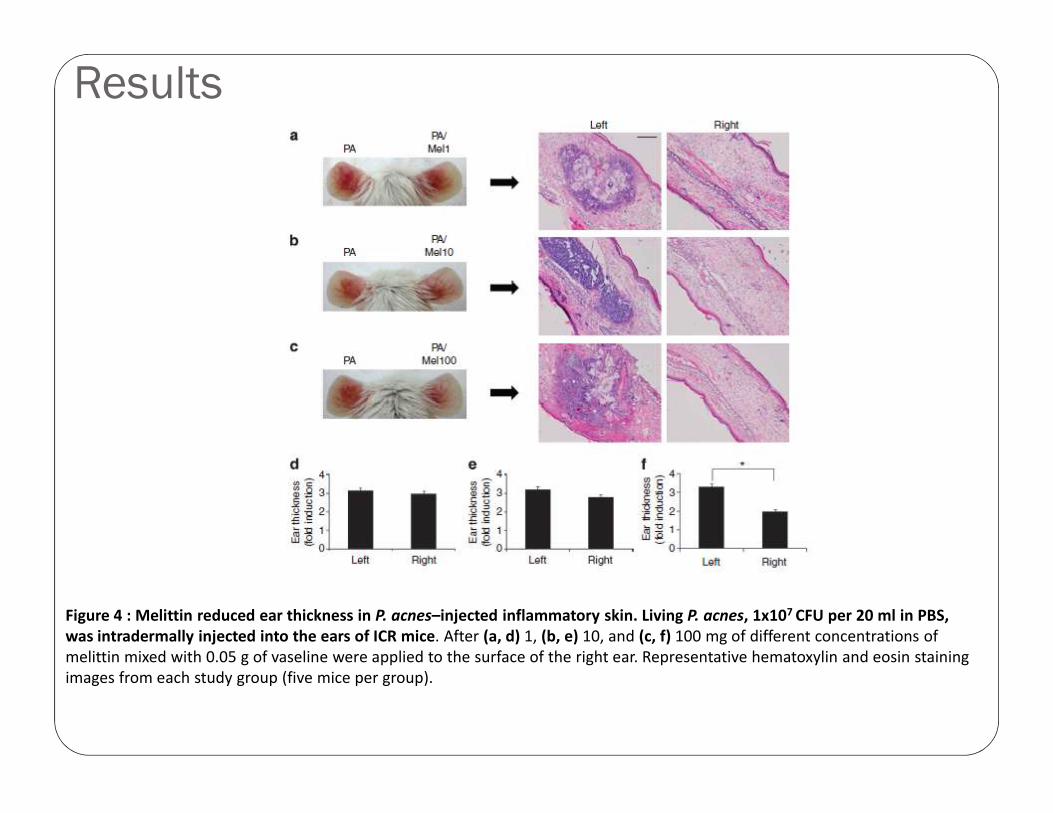
Figure 4 : Melittin reduced ear thickness in P. acnes–injected inflammatory skin. Living P. acnes, 1x10⁷ CFU per 20 ml in PBS, was intradermally injected into the ears of ICR mice. After (a, d) 1, (b, e) 10, and (c, f) 100 mg of different concentrations of melittin mixed with 0.05 g of vaseline were applied to the surface of the right ear. Representative hematoxylin and eosin staining images from each study group (five mice per group).

Discussion
Figure 5. Melittin effectively inhibits the pro-inflammatory cytokine in P. acnes–injected inflammatory skin. (a, b)Immunohistochemical results demonstrate that melittin suppresses the expression of TNF-a and IL-1b. (c, d) Western blot and RT–PCR analyses show that melittin treatment inhibited the expression of TNF-a and IL-1b. (e) Real time–PCR analyses show that melittin treatment inhibited the mRNA levels of TNF-α, IL-1b, IL-8, and IFN-γ.

Discussion
Figure 6: Melittin inhibits the NF-kB signaling and DNA-binding activity. (a) Melittin treatment reduced the phosphorylation of IKK, IkB, and NF-kB. Electrophoretic mobility shift assay results show that melittin treatment suppresses the binding activity of NF-kB (b) and AP-1 (c) in P. acnes–treated inflammatory skin. (d) Representative immunofluorescence images show that melittin treatment suppresses TLR2 and CD14 in the P. acnes–treated inflammatory skin. CD14 and TLR2 immune complexes were detected by anti-mouse FITC (green) and anti-rabbit Texas red (red).

Conclusion
The protective effects of melittin on the P. acnes-induced in vitro and in vivo inflammatory models was demonstrated.
Administration of melittin significantly decreased the expression of various inflammatory cytokines in heat-killed P. acnes-treated keratinocytes.
Melittin exerted anti-inflammatory effects against the living P. acnes- treated animal model.
These results demonstrate the feasibility of applying melittin for the prevention of progression of inflammatory skin diseases induced by P. acnes.

Thank You!!!Thank You!!!