the puromycin-sensitive aminopeptidase pam-1 is...
TRANSCRIPT
DEVELO
PMENT
4281RESEARCH ARTICLE
INTRODUCTIONIn Caenorhabditis elegans, sperm entry into the oocyte triggers thecompletion of meiosis. Before oocyte maturation, the oocytechromosomes are arrested at diakinesis of meiosis I (Greenstein,2005). Maturation cues entry into metaphase I (Greenstein, 2005),but fertilization is required for extrusion of the first polar body andthe second meiotic division (McNally and McNally, 2005). Duringmeiosis, the sperm chromosomes remain condensed near the futureposterior pole. Immediately following extrusion of the second polarbody, the embryo exits meiosis, the sperm and egg chromosomesdecondense, and pronuclei appear as the embryo enters the firstmitotic interphase (Albertson and Thomson, 1993; Schneider andBowerman, 2003).
Near the end of meiosis II or during the first interphase, theanteroposterior (AP) axis is established. During axis formation, thesperm pronucleus/centrosome complex (SPCC) is closely apposedto the cell membrane at the cortex of the zygote (Rappleye et al.,2002), and its position defines the posterior pole (Albertson, 1984;Goldstein and Hird, 1996; Schneider and Bowerman, 2003).Evidence points to the centrosome as the key SPCC component thatorchestrates axis polarization (Cowan and Hyman, 2004b; Hamill etal., 2002; O’Connell et al., 2000; Sadler and Shakes, 2000;Sonneville and Gönczy, 2004). The centrosome, by an unknownmechanism, appears to destabilize the cortical actomyosin networkin the posterior (Munro et al., 2004). This destabilization results ina flow of cortical F-actin and nonmuscle myosin to the anterior pole,
along with the PDZ-domain polarity proteins PAR-3 and PAR-6(Cuenca et al., 2003; Munro et al., 2004). Following this initialpolarization, the ring-finger protein PAR-2 becomes enriched at theposterior cortex, where it stabilizes polarity (Cuenca et al., 2003;Munro et al., 2004). These reciprocal domains are then maintaineddue to interactions between the PAR proteins themselves (Cuenca etal., 2003). The PAR proteins are required for all but the initial APasymmetries, which occur in response to the centrosome, includingposterior displacement of the first mitotic spindle and the polarizeddistribution of the germline P granules and developmentaldeterminants (Cowan and Hyman, 2004a; Lyczak et al., 2002;Schneider and Bowerman, 2003).
One process that is important for axis formation is cell cycleprogression, as polarity establishment occurs immediately aftermeiotic exit. Moreover, many mutants with meiotic defects also havepolarity defects, although these are in some cases separable (Liu etal., 2004; Rappleye et al., 2002; Shakes et al., 2003; Sonneville andGönczy, 2004). For example, the scaffolding protein CUL-2 and itsputative adaptor ZYG-11 are components of a cullin-based E3 ligaserequired for progression through meiosis and AP axis formation,most likely through degradation of different targets (Liu et al., 2004;Sonneville and Gönczy, 2004). Nevertheless, proper progressionthrough meiosis may be important for axis formation. Mutants witha partial loss of the anaphase-promoting complex (APC) sometimescompletely bypass meiosis II, and when this bypass occurs thesemutants exhibit AP axis defects (Shakes et al., 2003). These resultssuggest that either cell cycle progression is important for AP polarityestablishment, or that the machineries governing cell cycleprogression and cell polarity share some components.
If common machinery links meiotic completion and axisestablishment, it may be proteolytic. All the proteins mentionedabove are components of either the APC or SCF E3 ubiquitin ligase
The puromycin-sensitive aminopeptidase PAM-1 is requiredfor meiotic exit and anteroposterior polarity in the one-cellCaenorhabditis elegans embryoRebecca Lyczak1,2,*, Lynnsey Zweier1, Thomas Group1, Mary Ann Murrow1, Christine Snyder1,Lindsay Kulovitz1, Alexander Beatty1, Kristen Smith1 and Bruce Bowerman2
In the nematode Caenorhabditis elegans, sperm entry into the oocyte triggers the completion of meiosis and the establishment ofthe embryonic anteroposterior (AP) axis. How the early embryo makes the transition from a meiotic to a mitotic zygote andcoordinates cell cycle changes with axis formation remains unclear. We have discovered roles for the C. elegans puromycin-sensitiveaminopeptidase PAM-1 in both cell cycle progression and AP axis formation, further implicating proteolytic regulation in theseprocesses. pam-1 mutant embryos exhibit a delay in exit from meiosis: thus, this peptidase is required for progression to mitoticinterphase. In addition, the centrosomes associated with the sperm pronucleus fail to closely associate with the posterior cortex inpam-1 mutants, and the AP axis is not specified. The meiotic exit and polarity defects are separable, as inactivation of the B-typecyclin CYB-3 in pam-1 mutants rescues the meiotic exit delay but not the polarity defects. Thus PAM-1 may regulate CYB-3 duringmeiotic exit but presumably targets other protein(s) to regulate polarity. We also show that the pam-1 gene is expressed bothmaternally and paternally, providing additional evidence that sperm-donated gene products have important roles during earlyembryogenesis in C. elegans. The degradation of proteins through ubiquitin-mediated proteolysis has been previously shown toregulate the cell cycle and AP axis formation in the C. elegans zygote. Our analysis of PAM-1 requirements shows that a puromycin-sensitive aminopeptidase is also required for proteolytic regulation of the oocyte to embryo transition.
KEY WORDS: C. elegans, Anteroposterior axis, Meiotic exit, PAM-1, CYB-3, Aminopeptidase
Development 133, 4281-4292 (2006) doi:10.1242/dev.02615
1Department of Biology, Ursinus College, Collegeville, PA 19426, USA. 2Institute ofMolecular Biology, University of Oregon, Eugene, OR 97403, USA.
*Author for correspondence (e-mail: [email protected])
Accepted 6 September 2006

DEVELO
PMENT
4282
complexes, proposed to target specific substrates for degradationduring the cell cycle (Bowerman and Kurz, 2006; Koepp et al.,1999). Furthermore, CUL-2-based ECS E3 ligase(s) have beenshown to regulate cell polarity by degrading developmentaldeterminants in the anterior or posterior cytoplasm during and afterthe first asymmetric division of the one-cell zygote (DeRenzo et al.,2003). Acting downstream of these E3 ubiquitin ligases is the 26Sproteasome. Intriguingly, a puromycin-sensitive aminopeptidase(PSA) appears to colocalize with the 26S proteosome and toparticipate in proteolytic events to regulate the cell cycle inmammalian cells (Constam et al., 1995). PSA family members aremetalloproteases that hydolyze N-terminal amino acids and aremembers of the M1 metalloprotease family, characterized by theHEXXH(X)18E metal coordination site and an upstream GAMENmotif (Laustsen et al., 2001). These peptidases are widelyconserved and include a human family member (Taylor, 1993).Caenorhabditis elegans has one PSA homolog, PAM-1, acytoplasmic aminopeptidase localized to neurons and intestinalcells in larvae and adults (Brooks et al., 2003). While PSA has beenimplicated in cell cycle regulation in mammals, its requirementsremain largely unstudied. Here we show that C. elegans PAM-1 iscontributed by both the sperm and the egg, and is required fortimely exit from meiosis and AP axis specification. These twoprocesses appear to be independently regulated by PAM-1. Wepropose that PAM-1 is part of proteolytic machinery in the earlyembryo used to trigger meiotic completion and trigger axisformation through regulated protein degradation.
MATERIALS AND METHODSStrains and nematode cultureCaenorhabditis elegans were cultured as described (Brenner, 1974). N2Bristol was used for wild-type and CB4856 for SNP mapping. The followingalleles and balancers were used: LG I dpy-5(e61); LG II rol-6(e187); LG IIIunc-32(e189); LG IV unc-5(e53), bli-6(sc16), unc-8(e49), pam-1(or282ts),pam-1(or403ts), pam-1(or347ts), pam-1(or547ts), pam-1(or370ts), unc-24(e138), dpy-20(e1282), him-8(e1489), DnT1 (LG IV/V translocationbalancer); LGV him-5(e1409), dpy-11(e224), fog-2(q71), unc-51(e1189);X lon-2(e678).
GFP lines: JH227 pie-1::GPF axEx73[pJH3.92;pRF4] (Reese et al.,2000); WH204 pie-1::GFP::�-TUBULIN (Strome et al., 2001); and AZ212pie-1::GFP::H2B (Praitis et al., 2001) were used to construct strains EU929pam-1(or282ts)/DnT1 IV; +/DnT1 V; pie-1::GFP axEx73[pJH3.92;pRF4],EU881 pam-1(or282ts) IV; pie-1::GFP::�-TUBULIN, and EU947 pam-1(or282ts)/DnT1 IV; +/DnT1 V; pie-1::GFP::H2B. The CYB-3::GFP strainwas provided by P. Gönczy (Sonneville and Gönczy, 2004).
Homozygous temperature-sensitive strains were propagated at thepermissive temperature of 15°C. L4 larvae were shifted to 25°C overnightbefore analysis of the mutant embryos.
Isolation and characterization of pam-1 allelesFive pam-1 alleles (or282ts, or347ts, or370ts, or403ts, or547ts) wereidentified in a screen for temperature-sensitive embryonic lethal mutants(Encalada et al., 2000). This locus was originally named scu-1 (sperm cueabnormal) but subsequently changed to pam-1 after gene identity wasdetermined. Complementation tests were performed for each allele withor282ts. All alleles showed weak temperature sensitivity for embryoniclethality when homozygous hermaphrodites were shifted to 25°C as L4s.pam-1(or282ts) was 85.3% embryonic lethal at 15°C (n=495), and 97.0%embryonic lethal at 25°C (n=396). Similarly, pam-1(or403ts) was 88.7%embryonic lethal at 15°C (n=1029), and 98.5% embryonic lethal at 25°C(n=470). All phenotypic characterization was performed with these twoalleles.
To test for a zygotic requirement, hermaphrodites of strain pam-1(or282ts) unc-24(e138)/+ or pam-1(or403)/dpy-13(e184) unc-24(e138)were allowed to make embryos at 25°C: nearly all embryos hatched(705/716; 425/428, respectively). To test for paternal requirements, fog-2
females from strain CB4108 were mated at 25°C with EU983 pam-1(or403); him-5 mutant males, which had been raised at 25°C from the L1stage. To test for maternal requirements, pam-1(or403); unc-51 fog-2females were crossed with N2 males at 25°C. From each cross, females wereremoved after mating for embryo analysis. As a control, CB4108 fog-2females were crossed with N2 males.
For deficiency analysis, pam-1(or282ts) males were mated intohermaphrodites from the following strains: RW1324 fem-1(e1991) unc-24(e138) unc-22(s12)/stDf7 IV and RW1333 fem-1(e1991) unc-24(e138) unc-22(s12)/stDf8 IV at the permissive temperature. F1animals were shifted to 25°C and embryos from pam-1(or282ts)/stDf7(n=3) or pam-1(or282ts)/stDf8 (n=3) worms were imaged using DICmicroscopy.
Positional cloningLinkage group and 3-factor mapping (Brenner, 1974) were used toposition the or282ts allele to +3.39 on chromosome IV. Single nucleotidepolymorphism (SNP) mapping (Wicks et al., 2001) was then done usingSNPs identified from The University of Washington Genetics andGenome Sequencing Center. Unc-nonPam recombinants were found fromstrains heterozygous for CB4856 DNA and either unc-8(e49) pam-1(or282) or pam-1(or282) unc-24(e138). PCR-amplified DNA wasisolated from recombinants using a kit from Gentra Systems and scoredfor the SNPs to narrow the region to between cosmids R05G6 and C06A6.
The entire coding region of candidate genes in the region were sequenceddirectly from PCR products produced from purified DNA for each allele. Foreach sequencing reaction, four independent PCR reactions were combinedand purified using the QUIAquick PCR purification kit (Quiagen) andsequenced at the Nucleic Acid/Protein Research Core Facility at theChildren’s Hospital of Philadelphia. Sequences and chromatograms werealigned and compared to the published sequence using DNASIS MAXsoftware (Hitachi). All potential sequences were confirmed via a secondaryPCR and sequencing reaction. The pam-1 gene was amplified in twoseparate reactions using primers f49e8.3a: CAAAATTGACGAGAGGGGwith f49e8.3b: GTGATCCAGGAGTCACG and f49e8.3c: GCCAAAGA-TCAGTCCACC with f49e8.3d: AAGCAAGATGATGCCACG.
Mutations found in all five alleles are detailed below. Nucleotide numbersare based on position in the confirmed sequence for F49E8.3a. In or370, aG to A substitution was found at nucleotide 1022 and resulted in an A278Tchange. In or347, a C to T substitution was at nucleotide 1023 and resultedin A278W. In or403 a G to A change was observed at nucleotide 1849 andresulted in W538X. In or547 a C to T substitution at nucleotide 2027 wasobserved, which resulted in Q598X. In or282 a deletion from nucleotide 283to 873 was found in the genomic DNA. This deletion resulted in an aminoacid sequence change following K56 to NNFSX.
Microscopy and immunoflorescenceFor time-lapse imaging, embryos were placed on a 3% agarose cushionunder a coverslip. DIC images were captured every 5 seconds using a DageMT1 VE1000 camera and Scion Image software, or Nikon Eclipse E600 andSPOT Basic software. Cortical flows were time-lapsed as previouslydescribed (Severson et al., 2002). PIE-1::GFP and TUBULIN::GFP imageswere acquired using epifluorescence and a Micromax EBF512 cooled CCDcamera (Roper Scientific) under the control of Metamorph imaging software(Universal Imaging). HISTONE::GFP images were captured similarlyexcept on a spinning disk confocal (Perkin Elmer) or a Nikon C1 ConfocalImaging System. For time-lapse GFP imaging, a Z-series of 5-15 frames wasacquired at 1.5 �m intervals every 20-60 seconds and in some experimentsone center-plane DIC image was captured. Imaging of meiosis wasperformed in utero with worms anesthetized in 0.1% tricane and 0.01%tetramisole (McCarter et al., 1999). Measurements of pronuclear meetingposition and centrosome movements were made using Object-Imagesoftware.
The following primary antibodies and dilutions were used: rabbit anti-PAR-2 (1:5), rabbit anti-PAR-3 (1:20), both a gift from K. Kemphues, rabbitanti-PGL-1 (1:10,000), a gift from S. Strome, mouse anti-tubulin (1:250)(Sigma clone DM 1A), and mouse anti-actin (1:200) (ICN clone C4).Immunostaining was done using a methanol fix protocol (Severson et al.,
RESEARCH ARTICLE Development 133 (21)

DEVELO
PMENT
2002). The DNA was stained with 0.2 �m TOTO3 (Molecular Probes) orDAPI in Antifade mounting media (Invitrogen). Embryos were imaged ona BioRad MRC 1024 laser scanning confocal microscope or a Nikon C1confocal microscope system.
RNA interferenceFeeding RNAi was used to inactivate cyb-3 as described (Kamath et al.,2003). Bacteria were grown on plates containing 1 mmol/l IPTG and 75�g/�l carbenacillin and placed at 37°C overnight. L4 worms were picked tothe plates and grown at 25°C for 24 hours before analysis.
RESULTSIdentification of mutations in the pam-1 geneIn a screen for maternal-effect embryonic lethal mutants, weidentified five partially conditional recessive alleles of a gene wedetermined to be pam-1 (puromycin-sensitive aminopeptidase; seebelow). Our phenotypic analysis was performed with two alleles,pam-1(or282ts) and pam-1(or403ts). Unless the allele is specified,we refer hereafter to embryos produced by homozygous pam-1(or282 or or403) mutant hermaphrodites raised at the restrictivetemperature as pam-1 mutants.
To identify the molecular lesion in pam-1 mutants, the gene wasmapped to a small interval (see Materials and methods). Wesequenced candidate genes in the region and found, in allfive mutant alleles, mutations in the puromycin-sensitiveaminopeptidase gene called pam-1 (Fig. 1). This gene is closelyrelated to mouse and human PSA genes (Fig. 1). Two of the allelescontained missense mutations. The A278T mutation in pam-1(or370) and the A278V mutation in pam-1(or347) both alteredthe GAMEN motif that is conserved in all M1 aminopeptidasesand has been shown in other systems to be important for fullenzyme activity (Laustsen et al., 2001). Two of the allelesencoded nonsense mutations. Both the W538X mutation in pam-1(or403) and the Q598X mutation in pam-1(or547) are predictedto truncate the protein downstream of the active site (Fig. 1). A590 bp deletion was found in pam-1(or282), resulting in a changefollowing K56 to NNFSX and thus a truncated protein product.All five alleles are recessive and pam-1(or282) is most likely anull, as this deletion results in a frameshift and stop codon beforethe active site. Additionally, embryos produced by hemizygousmothers (with one mutant copy of pam-1 in trans to achromosomal deficiency that removes pam-1) wereindistinguishable from those produced by homozygous pam-1(or282) mothers (data not shown).
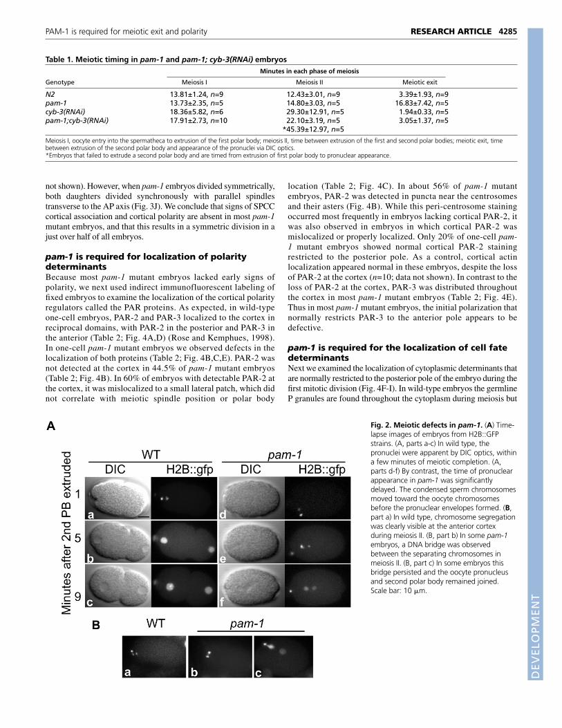
pam-1 embryos exhibit a meiotic exit defectThe first defects observed in pam-1 mutant embryos occurred duringmeiosis. The timing of meiosis was compared in wild-type and pam-1 embryos in utero using a HISTONE::GFP line (H2B::GFP) tofluorescently label chromosomes in live embryos (Table 1). Wedetected no significant difference in the timing of completion ofeither meiosis I or II in pam-1(or282) mutants, compared to wild-type embryos. For these experiments we defined the end of meiosisas the time when polar body extrusion was evident. However, we didobserve meiosis II defects in 33% (4/12) of pam-1(or282) embryos.In these embryos, a DNA bridge connected the separating anaphasechromosomes (Fig. 2B).
In contrast to the incomplete penetrance of the meioticchromosome segregation defects, all pam-1(or282) embryosexhibited a significant delay in exiting meiosis II. Here we definemeiotic exit as the time between extrusion of the second polarbody and the appearance of pronuclei as detected usingdifferential interference contrast (DIC) microscopy. Shortly after
extrusion of the second polar body in wild type, both the oocyteand sperm chromosomes began to decondense, and pronucleibecame evident (Fig. 2A, parts a-c; Table 1). However, in pam-1mutants the chromosomes remained condensed after polar bodyextrusion, and oocyte and sperm pronuclear envelope formationwas delayed (Fig. 2A, parts d-f). In wild-type embryos pronucleiwere detectable on average 3.39 minutes after polar bodyextrusion, but in pam-1 mutant embryos, pronuclei were visibleafter an average of 16.86 minutes (Table 1). In some cases, theoocyte pronucleus never became visible using DIC optics (4/17;data not shown). These severe delays indicate that pam-1 mutantembryos are defective in the regulation or execution of meioticexit.
Meiotic exit defects are rescued by loss of cyclinB3Previous studies have shown that inactivation of B-type cyclins isnecessary for exit from meiosis in several organisms. Forexample, reduction of the cyclin encoded by cyb-3 in C. eleganshas been shown to rescue meiotic exit delays in zyg-11 mutants(Sonneville and Gönczy, 2004). We therefore asked if cyb-3 wasrequired for the meiotic exit delay in pam-1 mutants, using RNAito deplete CYB-3. As previously shown (Sonneville and Gönczy,2004), we found that cyb-3(RNAi) embryos exhibit a prolongedmeiosis II, but a timely exit from meiosis (Table 1). In pam-1(or282); cyb-3(RNAi) embryos that completed meiosis II andextruded a second polar body, we observed a marked decrease inthe time of meiotic exit, similar to wild type (Table 1). Thusdepletion of CYB-3 rescues the meiotic exit defect in pam-1mutants, suggesting that PAM-1 may promote CYB-3 degradationto permit exit from meiosis.
Interestingly, in some pam-1(or282);cyb-3(RNAi) embryos, asecond polar body was never extruded. Embryos entered intometaphase II, but never progressed into a clear anaphase stagebefore chromosome decondensation became evident. In theseembryos, the timing from meiosis II to appearance of thepronuclei appeared to be lengthened even over cyb-3(RNAi)alone (Table 1), suggesting a possible role for PAM-1 in meiosisII.
pam-1 embryos lack early signs of AP polarityBecause polarity defects are associated with meiotic progressiondefects in other C. elegans mutants, we next examined AP axisformation in pam-1 mutant and wild-type embryos using DICoptics. As the SPCC contacts the posterior cortex and polarizesthe AP axis in wild type, cortical changes are apparent in whichthe posterior cortex smoothens and the anterior cortex ruffleswhile cortical flows occur (Fig. 3A). The oocyte pronucleus thenmigrates to meet the sperm pronucleus in the posterior (Fig. 3B).Together the pronuclei and associated centrosomes move to thecenter of the embryo and set up the first mitotic spindle, whichdisplaces posteriorly during anaphase, resulting in an asymmetricdivision (Fig. 3C,D). The two daughters differ in size, cell-cycletiming and spindle orientation (Fig. 3E) (Hyman and White, 1987;Sulston et al., 1983).
pam-1 mutant embryos failed to exhibit the earliest signs of APaxis polarization. In most embryos the SPCC failed to make a tightassociation with the cortex (54/60) and was often near the center ofthe embryo when it first became visible (46/60; Fig. 3F). In addition,SPCC-cued polarity was not apparent. The cortex did not smoothenat either pole, membrane ruffling persisted around the entireperiphery, and a pseudocleavage furrow failed to form (54/60; Fig.
4283RESEARCH ARTICLEPAM-1 is required for meiotic exit and polarity

DEVELO
PMENT
4284
3F). Furthermore, cortical flows were absent (n=6; data not shown).Thus it seems that the cortex did not polarize in pam-1 mutantembryos.
pam-1 mutant embryos also lacked polarity downstream of theinitial sperm cue. Based on time-lapse DIC analysis, the pronucleimet closer to the center of the embryo (Fig. 3G,K) and the firstmitotic spindle was often mis-positioned. Posterior placement of the
spindle was observed in all embryos that exhibited normal SPCCcortical association (6/6). However, in embryos lacking SPCCassociation, most divided symmetrically (32/54; Fig. 3H,I), butsurprisingly some divided asymmetrically with the spindle orientedeither toward the posterior (17/54) or toward the anterior (5/54).When pam-1 mutant embryos divided asymmetrically, the daughtercells exhibited further asymmetries, as in wild-type embryos (data
RESEARCH ARTICLE Development 133 (21)
C. elegansmousehuman
C. elegansmousehuman
C. elegansmousehuman
C. elegansmousehuman
C. elegansmousehuman
C. elegansmousehuman
C. elegansmousehuman
C. elegansmousehuman
111
226116
8212176
142181136
201241196
261301256
321361316
381421376
---------------------------------------MAACGNPSAAVKFERLPTFAEPMWLAAAVPSLARRLLLLGPPPPPLLLLLSRSSRRRRRLHSLGLAAMPEKRPFERLPAEVSP---------------------------------------------MPEKRPFERLPADVSP
PTHYNVRLSPCLNQFSFDGHATIDVTIKEATDVLKVHAQSLLIQSVSLITQPGDASKSLETPINYSLCLKPDLLDFTFEGKLEAAAQVRQATNQIVMNCADIDIITASYAPEGDEEIHATGFPINYSLCLKPDLLDFTFEGKLEAAAQVRQATNQIVMNCADIDIITASYAPEGDEEIHATGF
TSYDDKLNILTIKLPTTMQPQKVQLDFKFVGELNDKMRGFYRSQYKDKNGTEKFLASTQFEFNYQNEDEKVTLSFPSTLQTGTGTLKIDFVGELNDKMKGFYRSRYTTPAGEVRYAAVTQFEFNYQNEDEKVTLSFPSTLQTGTGTLKIDFVGELNDKMKGFYRSKYTTPSGEVRYAAVTQFE
ESTYARYAFPCFDEPIYKATFDVTLEVENHLTALSNMNVISETPTADGK-RKAVTFATSPKEATDARRAFPCWDEPAIKATFDISLVVPKDRVALSNMNVIDRKPYPDDENLVEVKFARTPVEATDARRAFPCWDEPAIKATFDISLVVPKDRVALSNMNVIDRKPYPDDENLVEVKFARTPV
KMSSYLVAFAVGELEYISAQTKSGVEMRVYTVPGKKEQGQYSLDLSVKCIDWYNEWFDIKYVMSTYLVAFVVGEYDFVETRSKDGVCVRVYTPVGKAEQGKFALEVAAKTLPFYKDYFNVPYVMSTYLVAFVVGEYDFVETRSKDGVCVRVYTPVGKAEQGKFALEVAAKTLPFYKDYFNVPY
YPLPKCDLIAIPDFSMGAMENWGLVTYREIALLVDPGVTSTRQKSRVALVVAHELAHLWFGYPLPKIDLIAIADFAAGAMENWGLVTYRETALLIDPKNSCSSSRQWVALVVGHELAHQWFGYPLPKIDLIAIADFAAGAMENWGLVTYRETALLIDPKNSCSSSRQWVALVVGHELAHQWFG
GNLVTMKWWTDLWLKEGFASFMEYMFVGANCPEFKIWLHFLNDELASGMGLDALRNSHPIEGNLVTMEWWTHLWLNEGFASWIEYLCVDHCFPEYDIWTQFVSADYTRAQELDALDNSHPIEGNLVTMEWWTHLWLNEGFASWIEYLCVDHCFPEYDIWTQFVSADYTRAQELDALDNSHPIE
EVEIDNPNELDEIYDSITYAKSNSVNRMLCYYLSEPVFQKGLRLYLKRFQYSNAVTQDLWTEVSVGHPSEVDEIFDAISYSKGASVIRMLHDYIGDKDFKKGMNMYLTKFQQKNAATEDLWEEVSVGHPSEVDEIFDAISYSKGASVIRMLHDYIGDKDFKKGMNMYLTKFQQKNAATEDLWE
C. elegansmousehuman
C. elegansmousehuman
C. elegansmousehuman
C. elegansmousehuman
C. elegansmousehuman
C. elegansmousehuman
C. elegansmousehuman
C. elegansmousehuman
441481436
500541496
560601556
620661616
680719674
740779734
799838793
859890845
TALSEASGQNVNELMSGWTQQMGFPVLKVSQRQDGNNRILTVEQRRFISDGGEDPKNS-QWESLESASGKPIAAVMNTWTKQMGFPLIYVEAEQVEDDRVLKLSQKKFCASGPYGGEDCPQWESLENASGKPIAAVMNTWTKQMGFPLIYVEAEQVEDDRLLRLSQKKFCAGGSYVGEDCPQW
WQVPITVAVGSSPSDVKARFLLKEKQQEFTIEGVAPGEWVKLNSGTTGFYRVEYSDEMLTAWMVPITISTSEDPNQAKLKILMDKPEMSVVLKNVKPDQWVKLNLGTVGFYRTQYSSAMLESWMVPITISTSEDPNQAKLKILMDKPEMNVVLKNVKPDQWVKLNLGTVGFYRTQYSSAMLES
AMLPDIASRRMPVLDRFGLINDLSALLNTGRVSIAQFVQVAASSAKEDEYVVWGAIDEGMSSLLPGIRDLSLPPVDRLGLQNDLFSLARAGIISTVEVLKVMEAFVNEPNYTVWSDLSCNLGSLLPGIRDLSLPPVDRLGLQNDLFSLARAGIISTVEVLKVMEAFVNEPNYTVWSDLSCNLG
SKLLACAREMSEDTLKSAKQLVVKMFEQTGAELGFAEQAGEDSQKMMLRSLVQARLARAGHGILSTLLSHT--DFYEEIQEFVKDVFSPIGERLGWDPKPGEGHLDALLRGLVLGKLGKAGHGILSTLLSHT--DFYEEIQEFVKDVFSPIGERLGWDPKPGEGHLDALLRGLVLGKLGKAGH
HQPTIDKFTQMFNDFLEKGTPIHPDIRLATFGVVARYGGKEGFDKLMNLRETTTFQEIERQHKATLEEARRRFKEHVEGKQILSADLRSPVYLTVLKHGDGATLDIMLKLHKQADMQEEKNRHKATLEEARRRFKDHVEGKQILSADLRSPVYLTVLKHGDGTTLDIMLKLHKQADMQEEKNR
QTMVAMSQTPEESLLAQLFEYGFEKNKVRPQDQLYLFLG-TGATHMGQQYAWKYFCEHIKERIERVLGATLSPELIQKVLTFALS-EEVRPQDTVSVIGGVAGGSKHGRKAAWKFIKDNWEERIERVLGATLLPDLIQKVLTFALS-EEVRPQDTVSVIGGVAGGSKHGRKAAWKFIKDNWEE
EFLDKYGGANSSLFQRCLKFAGESFGNEKRAVEFQDFFCNCNVLSDTDRQTLARPIGQTVEELHNRYQGG--FLISRLIKLSVEGFAVDKMAGEVKAFFESHPAPS------AERTIQQCCEELYNRYQGG--FLISRLIKLSVEGFAVDKMAGEVKAFFESHPAPS------AERTIQQCCE
EAIRLNARLLESNRQIIENLLKQSNV-----..............................ENILLNAAWLKRDADSIHQYLLQRKTSPPSV..............................ENILLNAAWLKRDAESIHQYLLQRKASPPTV..............................
*
Fig. 1. pam-1 encodes apuromycin-sensitiveaminopeptidase. Alignment ofPAM-1 amino acid sequenceshowing conservation with mouse(GenBank AAH86798) and human(GenBank NP_006301) PSA (seealso Brooks et al., 2003). Coloredblocks mark amino acid identity.Overlined area denotes region ofhomology with 26S proteasomesubunits (Constam et al., 1995).The underlined GAMEN motif ischaracteristic of M1 familymetalloproteases and the site oftwo of the missense mutationsA278W in pam-1(or347) andA278T in pam-1(or370) (markedwith *). The boxed amino acids inthe HEXXH(X)18E motif arenecessary for zinc binding. The stardenotes the site of deletion in pam-1(or282). The open circle denotesthe position of the stop codonpresent in pam-1(or403), W538X,while the closed circle denotes theposition of the stop codon presentin pam-1(or547), Q598X.
DEVELO
PMENT
not shown). However, when pam-1 embryos divided symmetrically,both daughters divided synchronously with parallel spindlestransverse to the AP axis (Fig. 3J). We conclude that signs of SPCCcortical association and cortical polarity are absent in most pam-1mutant embryos, and that this results in a symmetric division in ajust over half of all embryos.
pam-1 is required for localization of polaritydeterminantsBecause most pam-1 mutant embryos lacked early signs ofpolarity, we next used indirect immunofluorescent labeling offixed embryos to examine the localization of the cortical polarityregulators called the PAR proteins. As expected, in wild-typeone-cell embryos, PAR-2 and PAR-3 localized to the cortex inreciprocal domains, with PAR-2 in the posterior and PAR-3 inthe anterior (Table 2; Fig. 4A,D) (Rose and Kemphues, 1998).In one-cell pam-1 mutant embryos we observed defects in thelocalization of both proteins (Table 2; Fig. 4B,C,E). PAR-2 wasnot detected at the cortex in 44.5% of pam-1 mutant embryos(Table 2; Fig. 4B). In 60% of embryos with detectable PAR-2 atthe cortex, it was mislocalized to a small lateral patch, which didnot correlate with meiotic spindle position or polar body
location (Table 2; Fig. 4C). In about 56% of pam-1 mutantembryos, PAR-2 was detected in puncta near the centrosomesand their asters (Fig. 4B). While this peri-centrosome stainingoccurred most frequently in embryos lacking cortical PAR-2, itwas also observed in embryos in which cortical PAR-2 wasmislocalized or properly localized. Only 20% of one-cell pam-1 mutant embryos showed normal cortical PAR-2 stainingrestricted to the posterior pole. As a control, cortical actinlocalization appeared normal in these embryos, despite the lossof PAR-2 at the cortex (n=10; data not shown). In contrast to theloss of PAR-2 at the cortex, PAR-3 was distributed throughoutthe cortex in most pam-1 mutant embryos (Table 2; Fig. 4E).Thus in most pam-1 mutant embryos, the initial polarization thatnormally restricts PAR-3 to the anterior pole appears to bedefective.
pam-1 is required for the localization of cell fatedeterminantsNext we examined the localization of cytoplasmic determinants thatare normally restricted to the posterior pole of the embryo during thefirst mitotic division (Fig. 4F-I). In wild-type embryos the germlineP granules are found throughout the cytoplasm during meiosis but
4285RESEARCH ARTICLEPAM-1 is required for meiotic exit and polarity
Table 1. Meiotic timing in pam-1 and pam-1; cyb-3(RNAi) embryosMinutes in each phase of meiosis
Genotype Meiosis I Meiosis II Meiotic exit
N2 13.81±1.24, n=9 12.43±3.01, n=9 3.39±1.93, n=9pam-1 13.73±2.35, n=5 14.80±3.03, n=5 16.83±7.42, n=5cyb-3(RNAi) 18.36±5.82, n=6 29.30±12.91, n=5 1.94±0.33, n=5pam-1;cyb-3(RNAi) 17.91±2.73, n=10 22.10±3.19, n=5 3.05±1.37, n=5
*45.39±12.97, n=5
Meiosis I, oocyte entry into the spermatheca to extrusion of the first polar body; meiosis II, time between extrusion of the first and second polar bodies; meiotic exit, timebetween extrusion of the second polar body and appearance of the pronuclei via DIC optics.*Embryos that failed to extrude a second polar body and are timed from extrusion of first polar body to pronuclear appearance.
Fig. 2. Meiotic defects in pam-1. (A) Time-lapse images of embryos from H2B::GFPstrains. (A, parts a-c) In wild type, thepronuclei were apparent by DIC optics, withina few minutes of meiotic completion. (A,parts d-f) By contrast, the time of pronuclearappearance in pam-1 was significantlydelayed. The condensed sperm chromosomesmoved toward the oocyte chromosomesbefore the pronuclear envelopes formed. (B,part a) In wild type, chromosome segregationwas clearly visible at the anterior cortexduring meiosis II. (B, part b) In some pam-1embryos, a DNA bridge was observedbetween the separating chromosomes inmeiosis II. (B, part c) In some embryos thisbridge persisted and the oocyte pronucleusand second polar body remained joined.Scale bar: 10 �m.

DEVELO
PMENT
4286
then localize posteriorly before the first mitosis (Hird et al., 1996;Strome and Wood, 1982). Similarly, in fixed, wild-type embryos(n=19), we observed posterior localization in one-cell embryos andlocalization to the P1 cell at the two-cell stage (Table 3; Fig. 4F,F�).By contrast, P granules were not localized to one pole in 67% offixed pam-1 mutant one-cell embryos and were also mislocalized inlater stage embryos (Table 3; Fig. 4G,G�).
To see if proper determinant localization correlated with anasymmetric first cleavage, we followed localization of thecytoplasmic and nuclear transcription factor PIE-1 in live embryosexpressing PIE-1 fused to green fluorescent protein (PIE-1::GFP).In wild-type embryos, PIE-1 is enriched in posterior cytoplasmbefore the completion of P0 mitosis and segregated to the posteriordaughter P1 after the first mitosis (Fig. 4H,H�; n=3) (see Reese et al.,2000). By contrast, PIE-1::GFP was distributed throughout thecytoplasm in 41% of one-cell pam-1 embryos (7/17; Fig. 4I).Of the embryos with mislocalized PIE-1::GFP, some dividedasymmetrically (3/7) and others divided symmetrically (4/7) duringthe first mitosis. By contrast, the majority of embryos that properlysegregated PIE-1::GFP went on to divide asymmetrically (data notshown; 7/10). By the late two-cell stage, most asymmetric embryosexhibited normal PIE-1::GFP localization (12/13), and allsymmetric embryos showed diffuse mislocalized PIE-1::GFP(16/16; Fig. 4I). We conclude that PAM-1 is necessary to promoteproper segregation of developmental determinants along the AP axisduring the first mitotic division.
pam-1 polarity defects are separable from meioticexit defectsAs meiotic exit and axis polarization occur in rapid successionin the one-cell embryo, we asked if the polarity defects in pam-1 mutants might be a secondary consequence of the meiotic exitdefect. As described above, CYB-3/Cyclin B depletion rescuedthe meiotic exit defect in pam-1 mutants. We next asked whetherthe AP polarity defects were also rescued in CYB-3-depletedpam-1 mutant embryos. pam-1; cyb-3(RNAI) embryos wereobserved through the first cell division for signs of polarity,such as pseudocleavage and asymmetric spindle positioning.While all cyb-3(RNAi) embryos observed showed normalpseudocleavage and a posteriorly displaced spindle (n=7), manypam-1; cyb-3(RNAi) embryos lacked pseudocleavage (n=16/23)and had spindles positioned toward the anterior (n=5/20) orcenter (n=6/20) (Fig. 5A-D). As only some pam-1; cyb-3(RNAi)embryos exit meiosis promptly, due to meiosis II defects inothers (see above), we examined whether embryos with rescuedmeiotic exit were additionally rescued for polarity. We saw nosuch correlation; pam-1; cyb-3(RNAi) embryos that promptlyexited meiosis showed no signs of polarity 55% of the time(n=9). As an additional marker, we examined P granulelocalization in cyb-3(RNAi) and in pam-1(or403); cyb-3(RNAi)embryos (Table 3; Fig. 5). All cyb-3(RNAi) embryos examinedshowed normal posterior localization of the P granules duringthe first mitosis (Fig. 5E,G; Table 3) (see also Sonneville andGönczy, 2004). By contrast, even though pam-1(or282); cyb-3(RNAi) embryos often exhibited meiotic exit timings similar towild type (Table 1), P granule localization was still severelydefective in many embryos (Fig. 5F-I; Table 3). As a control todetermine if CYB-3 levels could be eliminated effectively byRNAi treatment, we exposed CYB-3::GFP-expressing worms(Sonneville and Gönczy, 2004) to cyb-3 RNAi. While alluntreated worms (n=6), displayed strong oocyte GFPexpression, we detected no GFP in all treated worms (n=8),
RESEARCH ARTICLE Development 133 (21)
Fig. 3. pam-1 embryos lack early signs of AP polarity. Images froma time-lapse DIC series of a single embryo. (A) In wild type, the oocyteand sperm pronuclei appeared at opposite ends of the embryo, withthe sperm pronucleus at the posterior pole. The SPCC cued axispolarization and caused changes in the cortex called pseudocleavage.(B) The oocyte pronucleus migrated to meet the sperm pronucleus inthe posterior of the embryo. (C,D) The first spindle displaced towardthe posterior and the first cleavage was asymmetric, with a largeranterior cell, AB, and a smaller posterior cell, P1. (E) These cells thendiffered in their cell cycle timing and spindle orientations during thenext division. (F) In pam-1 embryos, polarization of the cortex wasabsent and the SPCC did not contact the posterior cortex as thepronuclei first appeared close to the center of the embryo. (G) Thepronuclei met in the center of the cell. (H,I) In about 60% of pam-1embryos, the first mitotic spindle remained in the center of the embryo,resulting in an equivalent cleavage. (J) When this occurred, the twodaughter cells divided synchronously with parallel spindle orientations.(K) The position of pronuclear meeting was plotted for five wild-typeand ten pam-1 embryos. On average, the pronuclei in pam-1 embryosmet closer to the center of the embryo than in wild type. o, oocytepronucleus; s, sperm pronucleus. Scale bar: 10 �m.

DEVELO
PMENT
suggesting that incomplete elimination of CYB-3 does notaccount for the persistent polarity defects (data not shown). Weconclude that the polarity defects in pam-1 mutants occur evenin the absence of meiotic exit defects.
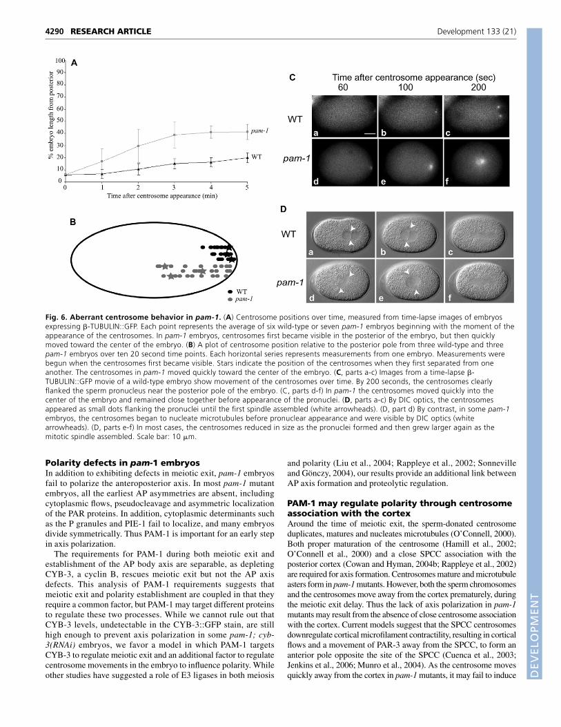
Aberrant centrosome positioning in pam-1mutantsThe abnormally late appearance of pronuclei and the centralappearance of the pronuclei in pam-1 mutant embryos suggested thatthe SPCC that normally specifies the posterior pole might not bemature or associated with the cortex at the time needed for properaxis specification. We therefore monitored centrosome maturationand position in wild-type and pam-1(or282) mutant embryos, usingtransgenic �-TUBULIN::GFP lines (Fig. 6A-C). When centrosomesfirst became visible in wild type, they were together and in closeassociation with both the sperm pronucleus and the posterior cortex(n=6; Fig. 6A,B,C, part a). In the first few minutes after centrosomescould be detected, they separated from one another and increased insize as they began to nucleate more microtubules (Fig. 6C, parts b,c).During this time, wild-type centrosomes remained in the posterior25% of the embryo (Fig. 6A-C). By contrast, pam-1 mutant embryosexhibited multiple defects in centrosome positioning. In all embryosexamined, the centrosomes first appeared normally near thepresumptive posterior cortex, but then rapidly moved toward thecenter of the embryo (n=13; Fig. 6A,B,C, parts d-f). As a result, thetime centrosomes spent near the cortex in pam-1 mutant embryoswas dramatically decreased (Fig. 6A). In addition, in most embryos,this centrosome movement occurred prior to the appearance of thesperm pronucleus, suggesting that centrosome movement occurredduring the delayed meiotic exit stage (n=10/13). This aberrantcentrosome movement was also observed in pam-1; cyb-3(RNAi)embryos (n=6; data not shown). It is possible that this change incentrosome localization in pam-1 mutants prevents polarization ofthe AP axis, through a failure of the centrosomes to influencecortical actomyosin and trigger axis formation.
We next used a combination of DIC and TUBULIN::GFP time-lapse videomicroscopy to track both centrosome-associatedmicrotubules and prouclei at the same time in live embryos. Whileall pam-1 embryos showed rapid centrosome movement from thecortex, we observed an additional defect in 32% of pam-1 mutantembryos (10/31). In these embryos, centrosomes nucleated robustmicrotubule asters before the appearance of pronuclei.Centrosomes enlarged until they were visible using DICmicroscopy and resembled mitotic centrosomes (Fig. 6D, part d).In most cases the enlarged centrosomes diminished in size as thepronuclei became visible (Fig. 6D, part e), but subsequentlynucleated more microtubules once again before and during the firstmitotic division (n=7/10; Fig. 6D, part f). There was no correlationof this premature microtubule nucleation with the subsequentpolarity of the division.
Oocyte- or sperm-supplied PAM-1 is sufficient fordevelopmentAs pam-1 mutant embryos are defective in events triggered byfertilization, we asked if there might be any paternal contributionof PAM-1 from sperm. We crossed pam-1(or403) feminizedhermaphrodites and wild-type males, or wild-type feminizedhermaphrodites and pam-1(or403) males, recorded subsequenthatching rates, and observed early embryonic cell divisions usingtime-lapse videomicroscopy (Table 4). When pam-1 feminizedhermaphrodites were mated with wild-type males, about 32% ofthe embryos hatched, a significant increase over pam-1 mutanthermaphrodite self-progeny hatching rates (1.5%). Furthermore,25% of embryos observed by DIC optics exhibited earlydevelopment that was indistinguishable from wild type (3/12),suggesting that paternal PAM-1 is sufficient in some embryos fornormal development. The remaining embryos showed phenotypeslike those produced by pam-1 mutant hermaphrodites (n=9/12;data not shown). Thus, embryos produced from mutant mothersand wild-type males either appeared normal, or exhibited fullyexpressed pam-1 mutant phenotypes. When feminized pam-1(+)hermaphrodites were mated to pam-1(or403) mutant males,nearly all embryos hatched and exhibited normal early embryoniccell divisions (12/13; data not shown), indicating that maternallysupplied PAM-1 is sufficient for embryogenesis. Thus while thepaternal contribution can partially rescue pam-1 mutant oocytes,it is not necessary for wild-type oocytes to develop normally afterfertilization.
DISCUSSIONWe have shown that the puromycin-sensitive aminopeptidase, PAM-1, is required for meiotic exit and AP axis formation in the one-cellstage C. elegans embryo. PAM-1 controls meiotic exit throughregulation of the B-type cyclin CYB-3, and appears to controlpolarity through other target(s). Thus our data implicate proteindegradation or processing by PAM-1 in multiple processes.Intriguingly, PAM-1 is supplied both maternally and paternally. Thesperm contribution and the coupling of these events through PAM-1 regulation in the one-cell embryo may help to coordinate cell cycleprogression and the establishment of AP polarity in the C. eleganszygote.
pam-1 encodes a puromycin-sensitiveaminopeptidasePAM-1 is the only C. elegans member of the puromycin-sensitiveaminopeptidase (PSA) family. This protein is highly conserved,being 36-37% identical to human PSA, with key domains showinghigher levels of homology (Brooks et al., 2003). PSA is a memberof the M1 class of metalloproteases, which cleave N-terminal aminoacids from a variety of substrates (Taylor, 1993). These cleavageevents have been implicated in inactivation of neuropeptides in
4287RESEARCH ARTICLEPAM-1 is required for meiotic exit and polarity
Table 2. PAR-2 and PAR-3 localization in one-cell pam-1 mutants% Embryos with PAR localization patterns in each category
PAR-2 n Posterior No cortical localization Lateral patch
N2 25 100% 0% 0%pam-1 45 22.2% (1/10) 44.5% (16/20) 33.3% (8/15)
PAR-3 n Anterior Around whole periphery
N2 24 100% 0%pam-1 17 35.3% 64.7%
Parentheses represent the number of embryos in each class that also exhibit PAR-2 cytoplasmic puncta.

DEVELO
PMENT
4288
mammalian systems (Hui et al., 1995) and processing of MHC classI peptides (Saric et al., 2001; Stoltze et al., 2000). However, few invivo substrates are known. This family of proteases include a zinc-binding motif preceded by the GAMEN sequence, which isnecessary for protease specificity and enhanced activity (Laustsenet al., 2001). The recessive mutations identified in our alleles affectconserved residues or truncate the protein to reduce or eliminatefunction. Two mutations affect the GAMEN alanine residue,highlighting its importance.
Loss-of-function studies in mice have implicated PSA in maleand female fertility, as well as in growth and pain response (Osadaet al., 1999; Osada et al., 2001a; Osada et al., 2001b).Additionally, in Drosophila, males with only one wild-type copyof dPSA show reduced fertility (Schulz et al., 2001). InArabidopsis, the single PSA homolog is essential for meiosis
(Sanchez-Moran et al., 2004). Less is known about C. elegansPSA, although a previous study showed that PAM-1 is expressedin intestinal cells and in neurons of the head, and in the male tail(Brooks et al., 2003). However, this study did not addressmaternal, paternal or early embryonic expression, due to germlinesilencing of the transgene reporter (Brooks et al., 2003). Theaminopeptidase activity of C. elegans PAM-1 is similar to otherPSA molecules, except that it is less sensitive to puromycin(Brooks et al., 2003). When RNAi was used to reduce pam-1function in adult hermaphrodites, 30% embryonic lethalityresulted, although early embryos were not examined (Brooks etal., 2003). We conclude that RNAi does not diminish pam-1function as effectively as the mutations we identified, and ourstudies reveal new roles for this protease in the regulation ofmeiotic exit and the establishment of the AP body axis.
RESEARCH ARTICLE Development 133 (21)
Fig. 4. Polarity markers are disrupted inpam-1. (A-E) Laser scanning confocalimages of PAR-2 and PAR-3 in fixedembryos using indirectimmunofluorescence. DNA was stained withTOTO. (A,A�,D,D�) Before and during thefirst cell division in wild type, PAR-2 localizedto the posterior cortex of the embryo andPAR-3 to the anterior cortex. (B,B�) In manyone-cell pam-1 embryos, PAR-2 did notlocalize to the cortex, but was found insteadon cytoplasmic puncta around thepronuclei. (C,C�) When PAR-2 did localize tothe cortex, it was often at a lateral position.(E,E�) In roughly half of pam-1 embryos,PAR-3 localized around the entire periphery.(F-G��) PGL-1 antibody staining of the Pgranules in fixed embryos using indirectimmunofluorescence. (H-I��) Fluorescentimages of PIE-1::GFP taken in living embryosat the one- and two-cell stage. (F,F�) Duringthe one-cell stage in wild type, the germlineP granules localized to the posterior poleand were segregated into the posteriordaughter cell P1. (H,H�) Similarly, thetranscription factor PIE-1 localized to theposterior and was found in the cytoplasmand nucleus of the P1 cell. (G,G�) In pam-1embryos, the P granules were frequentlymislocalized during the first division andoften found in both cells at the two-cellstage. (I,I�) PIE-1 failed to become properlypartitioned during the first division, and wasabsent from both daughter cells in embryosthat divided symmetrically. Scale bar: 10�m.

DEVELO
PMENT
A role for PAM-1 in meiotic exitThe most penetrant early defect in pam-1 mutant embryos was adelayed exit from meiosis II. During meiotic exit in wild-type embryos, the sperm and oocyte chromosomes rapidlydecondensed and nuclear envelopes formed, followed by entryinto S phase before the first mitosis (Edgar and McGhee, 1988;
Vidwans and Su, 2001). In pam-1 mutant embryos, chromosomedecondensation and formation of the pronuclei were delayed afterextrusion of the second polar body. Surprisingly, in some pam-1mutant embryos, centrosome maturation and microtubulenucleation were not delayed, and thus the centrosomes maturedand nucleated large microtubule asters before prouclearformation. Mitotic exit was not delayed in pam-1 mutants (datanot shown), suggesting that the mechanisms governing meioticand mitotic exit in the early embryo are at least partially distinct.However, PAM-1 might act redundantly with other factors toregulate mitotic exit.
Ubiquitin-mediated targeting of proteins to the proteasome fordegradation is widely used to regulate progression throughmeiosis and mitosis, and many other processes (reviewed byBowerman and Kurz, 2006; Deshaies, 1999). Specifically, theAPC and cullin-based SCF and ECS E3 ligase complexes arerequired for progression through meiosis or mitosis in manyspecies. For example, studies in fission yeast have implicated mfr1as a meiotic-specific activator of the APC necessary to ensuremeiotic exit and sporulation following meiosis (Blanco et al.,2001). Similarly, in C. elegans, the APC regulates the metaphaseto anaphase transition of meiosis I (Golden et al., 2000), andhypomorphic alleles of APC components sometimes exhibitmeiotic exit abnormalities (Shakes et al., 2003). Also in C.elegans, inactivation of the ubiquitin E3 ligase scaffold, a cullincalled CUL-2, or a potential adaptor component for this E3 ligasecalled ZYG-11, delays the metaphase to anaphase transitionduring meiosis II, resulting in elevated levels of B-type cyclinsand meiotic exit defects (Liu et al., 2004; Sonneville and Gönczy,2004). Timely meiotic exit is restored in cul-2 and zyg-11 mutantswhen RNAi is used to deplete the B-type cyclins, implicating thisE3 ligase in targeting B-type cyclins for degradation and meioticexit (Liu et al., 2004; Sonneville and Gönczy, 2004).
Our results indicate that the aminopeptidase PAM-1 also actsupstream of CYB-3 to regulate meiotic exit. However, PAM-1may act more specifically than the known ubiquitin E3 ligasesto regulate meiotic exit. Most pam-1 embryos advance throughmeiosis normally, with the first defect appearing during meioticexit. This is in contrast to many ubiquitin E3 ligase mutants,which display more severe meiotic defects. How PAM-1 andubiquitin-mediated proteolysis interact to influence cell cycleprogression and other processes, and whether PAM-1 directlydegrades B-type cyclins, are important topics for further study.However, the potential colocalization of PSA, the mammalianPAM-1 homolog, to the 26S proteasome suggests a roledownstream of the E3 ligases, perhaps in conjunction with theproteasome (Constam et al., 1995). Studies of a mouse PSAhave also implicated these aminopeptidases in mitotic exit(Constam et al., 1995). Thus regulation of cell cycle exitmay be a widely conserved role for puromycin-sensitiveaminopeptidases.
4289RESEARCH ARTICLEPAM-1 is required for meiotic exit and polarity
Table 3. P granule localization in pam-1 and pam-1; cyb-3(RNAi) embryosGenotype/treatment n Posterior Mislocalized Posterior bias
N2 19 100% 0% 0%pam-1 24 29.2% 66.6% 4.2%cyb-3(RNAi) 26 100% 0% 0%pam-1; cyb-3(RNAi) 36 16.7% 47.2% 36.1%
Shown is the percentage of embryos with PGL-1 staining in each category during first cell division.Posterior, embryos with all P granules near the posterior pole; mislocalized, embryos with P granules throughout cell; posterior bias, embryos with many P granules toward theposterior pole but significant numbers in other parts of the cell.
Fig. 5. Polarity defects in pam-1 mutants occur in the absence ofmeiotic exit defects. (A-D) DIC images of cyb-3(RNAi) (A,C) and pam-1; cyb-3(RNAi) (B,D) embryos. cyb-3(RNAi) embryos show normalpseudocleavage (A) and a posteriorly displaced spindle (C), while manypam-1; cyb-3(RNAi) embryos lack pseudocleavage (B) and have acentrally positioned spindle (D). (E-I) Triple-stained confocal images withPGL-1 staining of P granules in red, tubulin staining in green and DAPIDNA staining in blue. While P granules were not localized in meioticembryos of cyb-3(RNAi) and pam-1; cyb-3(RNAi) (E-F), they werelocalized to the posterior in all cyb-3(RNAi) embryos during the firstmitosis (G). P granules failed to localize posteriorly in most pam-1; cyb-3(RNAi) embryos, being distributed either throughout the cytoplasm (H)or with a slight but incomplete posterior bias (I). Scale bar: 10 �m.
DEVELO
PMENT
4290
Polarity defects in pam-1 embryosIn addition to exhibiting defects in meiotic exit, pam-1 embryosfail to polarize the anteroposterior axis. In most pam-1 mutantembryos, all the earliest AP asymmetries are absent, includingcytoplasmic flows, pseudocleavage and asymmetric localizationof the PAR proteins. In addition, cytoplasmic determinants suchas the P granules and PIE-1 fail to localize, and many embryosdivide symmetrically. Thus PAM-1 is important for an early stepin axis polarization.
The requirements for PAM-1 during both meiotic exit andestablishment of the AP body axis are separable, as depletingCYB-3, a cyclin B, rescues meiotic exit but not the AP axisdefects. This analysis of PAM-1 requirements suggests thatmeiotic exit and polarity establishment are coupled in that theyrequire a common factor, but PAM-1 may target different proteinsto regulate these two processes. While we cannot rule out thatCYB-3 levels, undetectable in the CYB-3::GFP stain, are stillhigh enough to prevent axis polarization in some pam-1; cyb-3(RNAi) embryos, we favor a model in which PAM-1 targetsCYB-3 to regulate meiotic exit and an additional factor to regulatecentrosome movements in the embryo to influence polarity. Whileother studies have suggested a role of E3 ligases in both meiosis
and polarity (Liu et al., 2004; Rappleye et al., 2002; Sonnevilleand Gönczy, 2004), our results provide an additional link betweenAP axis formation and proteolytic regulation.
PAM-1 may regulate polarity through centrosomeassociation with the cortexAround the time of meiotic exit, the sperm-donated centrosomeduplicates, matures and nucleates microtubules (O’Connell, 2000).Both proper maturation of the centrosome (Hamill et al., 2002;O’Connell et al., 2000) and a close SPCC association with theposterior cortex (Cowan and Hyman, 2004b; Rappleye et al., 2002)are required for axis formation. Centrosomes mature and microtubuleasters form in pam-1 mutants. However, both the sperm chromosomesand the centrosomes move away from the cortex prematurely, duringthe meiotic exit delay. Thus the lack of axis polarization in pam-1mutants may result from the absence of close centrosome associationwith the cortex. Current models suggest that the SPCC centrosomesdownregulate cortical microfilament contractility, resulting in corticalflows and a movement of PAR-3 away from the SPCC, to form ananterior pole opposite the site of the SPCC (Cuenca et al., 2003;Jenkins et al., 2006; Munro et al., 2004). As the centrosome movesquickly away from the cortex in pam-1 mutants, it may fail to induce
RESEARCH ARTICLE Development 133 (21)
Fig. 6. Aberrant centrosome behavior in pam-1. (A) Centrosome positions over time, measured from time-lapse images of embryosexpressing �-TUBULIN::GFP. Each point represents the average of six wild-type or seven pam-1 embryos beginning with the moment of theappearance of the centrosomes. In pam-1 embryos, centrosomes first became visible in the posterior of the embryo, but then quicklymoved toward the center of the embryo. (B) A plot of centrosome position relative to the posterior pole from three wild-type and threepam-1 embryos over ten 20 second time points. Each horizontal series represents measurements from one embryo. Measurements werebegun when the centrosomes first became visible. Stars indicate the position of the centrosomes when they first separated from oneanother. The centrosomes in pam-1 moved quickly toward the center of the embryo. (C, parts a-c) Images from a time-lapse �-TUBULIN::GFP movie of a wild-type embryo show movement of the centrosomes over time. By 200 seconds, the centrosomes clearlyflanked the sperm pronucleus near the posterior pole of the embryo. (C, parts d-f) In pam-1 the centrosomes moved quickly into thecenter of the embryo and remained close together before appearance of the pronuclei. (D, parts a-c) By DIC optics, the centrosomesappeared as small dots flanking the pronuclei until the first spindle assembled (white arrowheads). (D, part d) By contrast, in some pam-1embryos, the centrosomes began to nucleate microtubules before pronuclear appearance and were visible by DIC optics (whitearrowheads). (D, parts e-f) In most cases, the centrosomes reduced in size as the pronuclei formed and then grew larger again as themitotic spindle assembled. Scale bar: 10 �m.

DEVELO
PMENT
the cortical changes. This hypothesis is consistent with the lack ofposterior PAR-2 localization, and the presence of PAR-3 throughoutthe entire cortex, in most pam-1 mutant embryos.
Paternal contribution of PAM-1The pam-1 gene product is contributed to the zygote by both thesperm and the egg. Egg contribution is necessary in nearly allembryos for viability and is also fully sufficient for normaldevelopment. By contrast, the sperm contribution of PAM-1 is notnecessary for embryo viability but is sufficient in many cases. It isunlikely that the rescue of pam-1 mutant oocytes by wild-type spermis due to zygotic expression of pam-1, as the rescued processes occurshortly after fertilization, before the zygotic genome is activated(Seydoux and Fire, 1994). Thus PAM-1 is partially contributed as apaternal gene product, although this contribution appearsnonessential. It is interesting to note that, although cues from thesperm are known to promote resumption of meiosis and axispolarization, only one true paternal-effect mutation, spe-11, has beenidentified to date in C. elegans (Browning and Strome, 1996). Despitethis, many mutants that affect early development of the embryo,including pam-1, show both maternal and paternal contributions ofthe gene product (Gönczy et al., 1999; Lee et al., 2001; O’Connell etal., 1998). Perhaps the addition of multiple sperm-contributedproteins at fertilization helps to produce a threshold concentration offactors necessary for the progression into embryogenesis.
Possible mechanisms for PAM-1 actionWhile in vivo substrates of puromycin-sensitive aminopeptidases arepoorly understood, these enzymes have been implicated in a varietyof processes, including neuropeptide inactivation (Hui et al., 1995),MHC class I peptide processing (Saric et al., 2001; Stoltze et al.,2000) and cell cycle regulation in conjunction with the proteasome(Constam et al., 1995). The role of PAM-1 role in the early embryomay be complex, causing the activation of some peptides through N-terminal trimming and the degradation of other proteins, perhaps inconjunction with the proteasome. One intriguing possibility is thatPAM-1 may trigger peptides for degradation through the N-end rule,with removal of N-terminal residues by PAM-1 exposing aminoacids that target the protein for ubiquitin-mediated proteolysis(Varshavsky, 1996).
Our study provides new insights into the PAM-1 aminopeptidaseand its roles in early development. Because PAM-1 is a memberof the highly conserved PSA gene family, its role in cell cycleprogression and cell polarity may provide important new insightsinto these processes in other systems.
We thank K. Kemphues and S. Strome for antibodies, P. Gönczy, G. Seydoux,C. Malone and J. Austin for GFP strains. Other strains were supplied by the C.elegans Genetics Center, which is funded by the NIH National Center forResearch Resources. We also thank C. Trzepacz for sharing data prior to
publication. R.L. was supported by a postdoctoral grant from the AmericanCancer Society (PF-00-097-010-MBC), an NIH-AREA grant (1 R15 GM72523-01) and institutional grants from Ursinus College (HHMI and VanSant). M.M.was supported in part by a Merck-SURF grant, and B.B. by R01-GM49869from the NIH.
ReferencesAlbertson, D. G. (1984). Formation of the first cleavage spindle in nematode
embryos. Dev. Biol. 101, 61-72.Albertson, D. G. and Thomson, J. N. (1993). Segregation of holocentric
chromosomes at meiosis in the nematode, Caenorhabditis elegans.Chromosome Res. 1, 15-26.
Blanco, M. A., Pelloquin, L. and Moreno, S. (2001). Fission yeast mfr1 activatesAPC and coordinates meiotic nuclear division with sporulation. J. Cell Sci. 114,2135-2143.
Bowerman, B. and Kurz, T. (2006). Degrade to create: developmentalrequirements for ubiquitin-mediated proteolysis during early C. elegansembryogenesis. Development 133, 773-784.
Brenner, S. (1974). The genetics of Caenorhabditis elegans. Genetics 77, 71-94.Brooks, D. R., Hooper, N. M. and Isaac, R. E. (2003). The Caenorhabditis
elegans orthologue of mammalian puromycin-sensitive aminopeptidase has rolesin embryogenesis and reproduction. J. Biol. Chem. 278, 42795-42801.
Browning, H. and Strome, S. (1996). A sperm-supplied factor required forembryogenesis in C. elegans. Development 122, 391-404.
Constam, D. B., Tobler, A. R., Rensing-Ehl, A., Kemler, I., Hersh, L. B. andFontana, A. (1995). Puromycin-sensitive aminopeptidase. Sequence analysis,expression, and functional characterization. J. Biol. Chem. 270, 26931-26939.
Cowan, C. R. and Hyman, A. A. (2004a). Asymmetric cell division in C. elegans:cortical polarity and spindle positioning. Annu. Rev. Cell Dev. Biol. 20, 427-453.
Cowan, C. R. and Hyman, A. A. (2004b). Centrosomes direct cell polarityindependently of microtubule assembly in C. elegans embryos. Nature 431, 92-96.
Cuenca, A. A., Schetter, A., Aceto, D., Kemphues, K. and Seydoux, G. (2003).Polarization of the C. elegans zygote proceeds via distinct establishment andmaintenance phases. Development 130, 1255-1265.
DeRenzo, C., Reese, K. J. and Seydoux, G. (2003). Exclusion of germ plasmproteins from somatic lineages by cullin-dependent degradation. Nature 424,685-689.
Deshaies, R. J. (1999). SCF and Cullin/Ring H2-based ubiquitin ligases. Annu. Rev.Cell Dev. Biol. 15, 435-467.
Edgar, L. G. and McGhee, J. D. (1988). DNA synthesis and the control ofembryonic gene expression in C. elegans. Cell 53, 589-599.
Encalada, S. E., Martin, P. R., Phillips, J. B., Lyczak, R., Hamill, D. R., Swan, K.A. and Bowerman, B. (2000). DNA replication defects delay cell division anddisrupt cell polarity in early Caenorhabditis elegans embryos. Dev. Biol. 228,225-238.
Golden, A., Sadler, P. L., Wallenfang, M. R., Schumacher, J. M., Hamill, D. R.,Bates, G., Bowerman, B., Seydoux, G. and Shakes, D. C. (2000). Metaphaseto anaphase (mat) transition-defective mutants in Caenorhabditis elegans. J. CellBiol. 151, 1469-1482.
Goldstein, B. and Hird, S. N. (1996). Specification of the anteroposterior axis inCaenorhabditis elegans. Development 122, 1467-1474.
Gönczy, P., Schnabel, H., Kaletta, T., Amores, A. D., Hyman, T. and Schnabel,R. (1999). Dissection of cell division processes in the one cell stageCaenorhabditis elegans embryo by mutational analysis. J. Cell Biol. 144, 927-946.
Greenstein, D. (2005). Control of oocyte meiotic maturation and fertilization. InWormBook (ed. The C. elegans Research Community), pp. 1-23.doi/10.1895/wormbook.1.53.1, http://www.wormbook.org.
Hamill, D. R., Severson, A. F., Carter, J. C. and Bowerman, B. (2002).Centrosome maturation and mitotic spindle assembly in C. elegans require SPD-5, a protein with multiple coiled-coil domains. Dev. Cell 3, 673-684.
Hird, S. N., Paulsen, J. E. and Strome, S. (1996). Segregation of germ granules
4291RESEARCH ARTICLEPAM-1 is required for meiotic exit and polarity
Table 4. Maternal and paternal contributions to pam-1 embryonic lethality at 25°CHermaphrodite n % Embryos hatching
pam-1 407 1.5%pam-1/dpy-13 unc-24 428 99.3%
Female Male n % Embryos hatching
fog-2 N2 605 99.7%fog-2 pam-1 2161 94.3%*fog-2; pam-1 N2 198 32.3%
All scoring done with pam-1(or403).n, number of embryos examined from each cross.fog-2 is a feminized strain (Schedl and Kimble, 1988).*No significant difference by Student’s t-test when compared with control cross of fog-2 females and N2 males.
DEVELO
PMENT
4292
in living Caenorhabditis elegans embryos: cell-type-specific mechanisms forcytoplasmic localisation. Development 122, 1303-1312.
Hui, M., Budai, E. D., Lajtha, A., Palkovits, M. and Hui, K. S. (1995). Changesin puromycin-sensitive aminopeptidases in postmortem schizophrenic brainregions. Neurochem. Int. 27, 433-441.
Hyman, A. A. and White, J. G. (1987). Determination of cell division axes in theearly embryogenesis of Caenorhabditis elegans. J. Cell Biol. 105, 2123-2135.
Jenkins, N., Saam, J. R. and Mango, S. E. (2006). CYK-4/GAP provides alocalized cue to initiate anteroposterior polarity upon fertilization. Science 313,1298-1301.
Kamath, R. S., Fraser, A. G., Dong, Y., Poulin, G., Durbin, R., Gotta, M.,Kanapin, A., Le Bot, N., Moreno, S., Sohrmann, M. et al. (2003). Systematicfunctional analysis of the Caenorhabditis elegans genome using RNAi. Nature421, 231-237.
Koepp, D. M., Harper, J. W. and Elledge, S. J. (1999). How the cyclin became acyclin: regulated proteolysis in the cell cycle. Cell 97, 431-434.
Laustsen, P. G., Vang, S. and Kristensen, T. (2001). Mutational analysis of theactive site of human insulin-regulated aminopeptidase. Eur. J. Biochem. 268, 98-104.
Lee, J., Jee, C., Lee, J. I., Lee, M. H., Koo, H. S., Chung, C. H. and Ahnn, J.(2001). A deubiquitinating enzyme, UCH/CeUBP130, has an essential role in theformation of a functional microtubule-organizing centre (MTOC) during earlycleavage in C. elegans. Genes Cells 6, 899-911.
Liu, J., Vasudevan, S. and Kipreos, E. T. (2004). CUL-2 and ZYG-11 promotemeiotic anaphase II and the proper placement of the anterior-posterior axis in C.elegans. Development 131, 3513-3525.
Lyczak, R., Gomes, J. E. and Bowerman, B. (2002). Heads or tails: cell polarityand axis formation in the early Caenorhabditis elegans embryo. Dev. Cell 3, 157-166.
McCarter, J., Bartlett, B., Dang, T. and Schedl, T. (1999). On the control ofoocyte meiotic maturation and ovulation in Caenorhabditis elegans. Dev. Biol.205, 111-128.
McNally, K. L. and McNally, F. J. (2005). Fertilization initiates the transition fromanaphase I to metaphase II during female meiosis in C. elegans. Dev. Biol. 282,218-230.
Munro, E., Nance, J. and Priess, J. R. (2004). Cortical flows powered byasymmetrical contraction transport PAR proteins to establish and maintainanterior-posterior polarity in the early C. elegans embryo. Dev. Cell 7, 413-424.
O’Connell, K. F. (2000). The centrosome of the early C. elegans embryo:inheritance, assembly, replication, and developmental roles. Curr. Top. Dev. Biol.49, 365-384.
O’Connell, K. F., Leys, C. M. and White, J. G. (1998). A genetic screen fortemperature-sensitive cell-division mutants of Caenorhabditis elegans. Genetics149, 1303-1321.
O’Connell, K. F., Maxwell, K. N. and White, J. G. (2000). The spd-2 gene isrequired for polarization of the anteroposterior axis and formation of the spermasters in the Caenorhabditis elegans zygote. Dev. Biol. 222, 55-70.
Osada, T., Ikegami, S., Takiguchi-Hayashi, K., Yamazaki, Y., Katoh-Fukui, Y.,Higashinakagawa, T., Sakaki, Y. and Takeuchi, T. (1999). Increased anxietyand impaired pain response in puromycin-sensitive aminopeptidase gene-deficient mice obtained by a mouse gene-trap method. J. Neurosci. 19, 6068-6078.
Osada, T., Watanabe, G., Kondo, S., Toyoda, M., Sakaki, Y. and Takeuchi, T.(2001a). Male reproductive defects caused by puromycin-sensitiveaminopeptidase deficiency in mice. Mol. Endocrinol. 15, 960-971.
Osada, T., Watanabe, G., Sakaki, Y. and Takeuchi, T. (2001b). Puromycin-sensitive aminopeptidase is essential for the maternal recognition of pregnancyin mice. Mol. Endocrinol. 15, 882-893.
Praitis, V., Casey, E., Collar, D. and Austin, J. (2001). Creation of low-copyintegrated transgenic lines in Caenorhabditis elegans. Genetics 157, 1217-1226.
Rappleye, C. A., Tagawa, A., Lyczak, R., Bowerman, B. and Aroian, R. V.(2002). The anaphase-promoting complex and separin are required forembryonic anterior-posterior axis formation. Dev. Cell 2, 195-206.
Reese, K. J., Dunn, M. A., Waddle, J. A. and Seydoux, G. (2000). Asymmetricsegregation of PIE-1 in C. elegans is mediated by two complementarymechanisms that act through separate PIE-1 protein domains. Mol. Cell 6, 445-455.
Rose, L. S. and Kemphues, K. J. (1998). Early patterning of the C. elegansembryo. Annu. Rev. Genet. 32, 521-545.
Sadler, P. L. and Shakes, D. C. (2000). Anucleate Caenorhabditis elegans spermcan crawl, fertilize oocytes and direct anterior-posterior polarization of the 1-cellembryo. Development 127, 355-366.
Sanchez-Moran, E., Jones, G. H., Franklin, F. C. and Santos, J. L. (2004). Apuromycin-sensitive aminopeptidase is essential for meiosis in Arabidopsisthaliana. Plant Cell 16, 2895-2909.
Saric, T., Beninga, J., Graef, C. I., Akopian, T. N., Rock, K. L. and Goldberg, A.L. (2001). Major histocompatibility complex class I-presented antigenic peptidesare degraded in cytosolic extracts primarily by thimet oligopeptidase. J. Biol.Chem. 276, 36474-36481.
Schedl, T. and Kimble, J. (1988). fog-2, a germ-line-specific sex determinationgene required for hermaphrodite spermatogenesis in Caenorhabditis elegans.Genetics 119, 43-61.
Schneider, S. Q. and Bowerman, B. (2003). Cell polarity and the cytoskeleton inthe Caenorhabditis elegans zygote. Annu. Rev. Genet. 37, 221-249.
Schulz, C., Perezgasga, L. and Fuller, M. T. (2001). Genetic analysis of dPsa, theDrosophila orthologue of puromycin-sensitive aminopeptidase, suggestsredundancy of aminopeptidases. Dev. Genes Evol. 211, 581-588.
Severson, A. F., Baillie, D. L. and Bowerman, B. (2002). A formin homologyprotein and a profilin are required for cytokinesis and Arp2/3-independentassembly of cortical microfilaments in C. elegans. Curr. Biol. 12, 2066-2075.
Seydoux, G. and Fire, A. (1994). Soma-germline asymmetry in the distributionsof embryonic RNAs in Caenorhabditis elegans. Development 120, 2823-2834.
Shakes, D. C., Sadler, P. L., Schumacher, J. M., Abdolrasulnia, M. and Golden,A. (2003). Developmental defects observed in hypomorphic anaphase-promoting complex mutants are linked to cell cycle abnormalities. Development130, 1605-1620.
Sonneville, R. and Gönczy, P. (2004). Zyg-11 and cul-2 regulate progressionthrough meiosis II and polarity establishment in C. elegans. Development 131,3527-3543.
Stoltze, L., Schirle, M., Schwarz, G., Schroter, C., Thompson, M. W., Hersh, L.B., Kalbacher, H., Stevanovic, S., Rammensee, H. G. and Schild, H. (2000).Two new proteases in the MHC class I processing pathway. Nat. Immunol. 1,413-418.
Strome, S. and Wood, W. B. (1982). Immunofluorescence visualization of germ-line-specific cytoplasmic granules in embryos, larvae, and adults ofCaenorhabditis elegans. Proc. Natl. Acad. Sci. USA 79, 1558-1562.
Strome, S., Powers, J., Dunn, M., Reese, K., Malone, C. J., White, J.,Seydoux, G. and Saxton, W. (2001). Spindle dynamics and the role of gamma-tubulin in early Caenorhabditis elegans embryos. Mol. Biol. Cell 12, 1751-1764.
Sulston, J. E., Schierenberg, E., White, J. G. and Thomson, J. N. (1983). Theembryonic cell lineage of the nematode Caenorhabditis elegans. Dev. Biol. 100,64-119.
Taylor, A. (1993). Aminopeptidases: structure and function. FASEB J. 7, 290-298.Varshavsky, A. (1996). The N-end rule: functions, mysteries, uses. Proc. Natl.
Acad. Sci. USA 93, 12142-12149.Vidwans, S. J. and Su, T. T. (2001). Cycling through development in Drosophila
and other metazoa. Nat. Cell Biol. 3, E35-E39.Wicks, S. R., Yeh, R. T., Gish, W. R., Waterston, R. H. and Plasterk, R. H.
(2001). Rapid gene mapping in Caenorhabditis elegans using a high densitypolymorphism map. Nat. Genet. 28, 160-164.
RESEARCH ARTICLE Development 133 (21)