the qrs axis: the isoelectric lead method · o normal qrs duration, but rsr pattern in the anterior...
TRANSCRIPT
This document was created by Alex Yartsev ([email protected]); if I have used your data or images and forgot to reference you, please email me.
From “the ECG made easy”, by Hampton (2003), and ECGs shamelessly stolen from Life in The Fastlane without any sort of permission, but in the non-commercial spirit of free education.
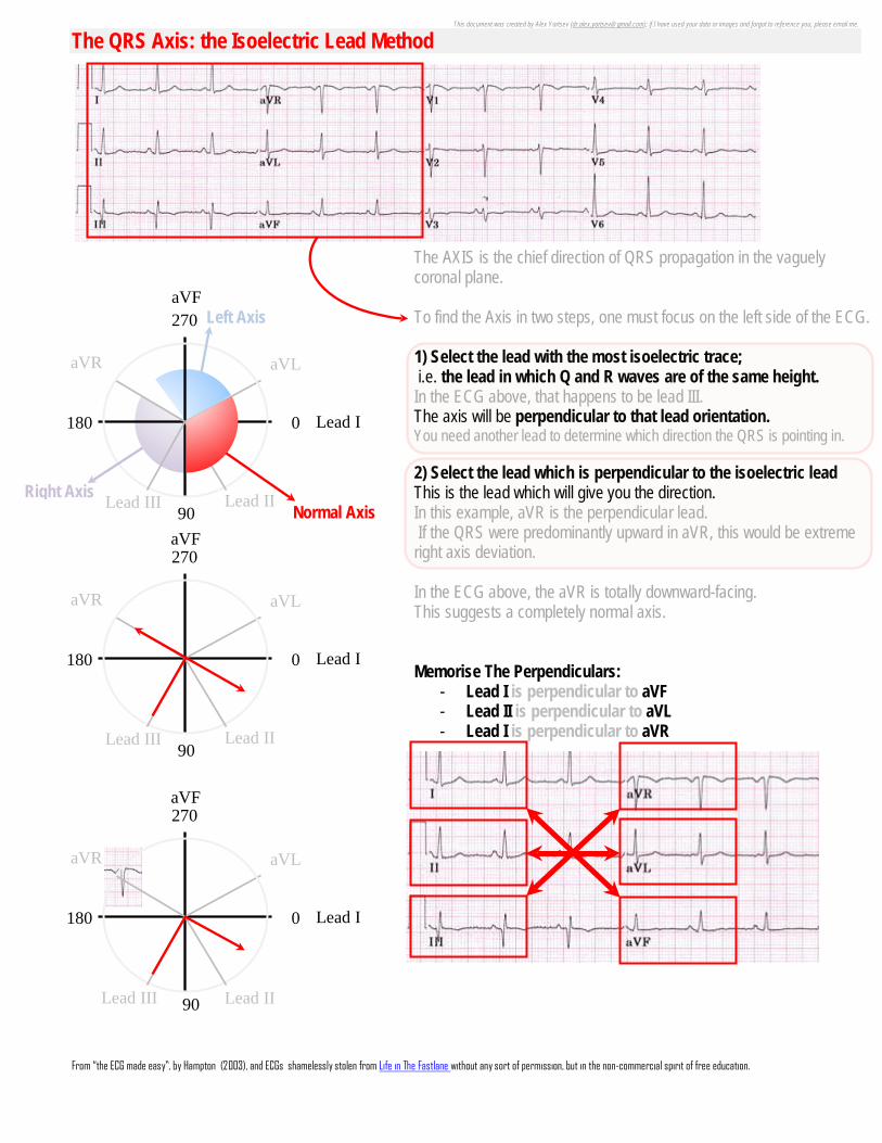
The QRS Axis: the Isoelectric Lead Method
270
90
0 180
aVF
Lead I
The AXIS is the chief direction of QRS propagation in the vaguely coronal plane. To find the Axis in two steps, one must focus on the left side of the ECG. 1) Select the lead with the most isoelectric trace; i.e. the lead in which Q and R waves are of the same height. In the ECG above, that happens to be lead III. The axis will be perpendicular to that lead orientation. You need another lead to determine which direction the QRS is pointing in. 2) Select the lead which is perpendicular to the isoelectric lead This is the lead which will give you the direction. In this example, aVR is the perpendicular lead. If the QRS were predominantly upward in aVR, this would be extreme right axis deviation. In the ECG above, the aVR is totally downward-facing. This suggests a completely normal axis. Memorise The Perpendiculars:
- Lead I is perpendicular to aVF - Lead II is perpendicular to aVL - Lead I is perpendicular to aVR
aVL aVR
Lead III Lead II
270
90
0 180 Lead I
aVL aVR
Lead III Lead II
aVF
Right Axis
Left Axis
Normal Axis
270
90
0 180 Lead I
aVL aVR
Lead III Lead II
aVF
This document was created by Alex Yartsev ([email protected]); if I have used your data or images and forgot to reference you, please email me.
From “the ECG made easy”, by Hampton (2003), and ECGs shamelessly stolen from Life in The Fastlane without any sort of permission, but in the non-commercial spirit of free education.
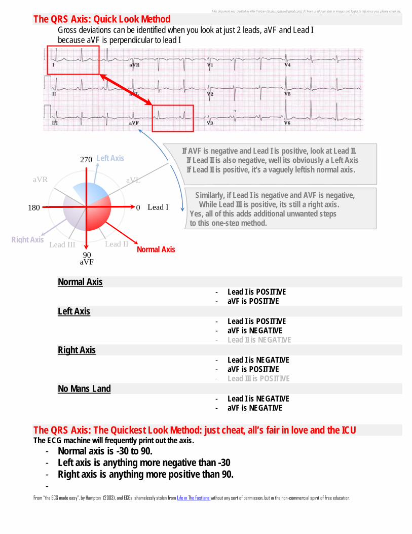
The QRS Axis: Quick Look Method Gross deviations can be identified when you look at just 2 leads, aVF and Lead I because aVF is perpendicular to lead I Normal Axis
- Lead I is POSITIVE - aVF is POSITIVE
Left Axis - Lead I is POSITIVE - aVF is NEGATIVE - Lead II is NEGATIVE
Right Axis - Lead I is NEGATIVE - aVF is POSITIVE - Lead III is POSITIVE
No Mans Land - Lead I is NEGATIVE - aVF is NEGATIVE
The QRS Axis: The Quickest Look Method: just cheat, all’s fair in love and the ICU The ECG machine will frequently print out the axis.
- Normal axis is -30 to 90. - Left axis is anything more negative than -30 - Right axis is anything more positive than 90. -
270
90
0 180 Lead I
aVL aVR
Lead III Lead II
aVF
Right Axis
Left Axis
Normal Axis
If AVF is negative and Lead I is positive, look at Lead II. If Lead II is also negative, well its obviously a Left Axis If Lead II is positive, it’s a vaguely leftish normal axis.
Similarly, if Lead I is negative and AVF is negative, While Lead III is positive, its still a right axis. Yes, all of this adds additional unwanted steps to this one-step method.
This document was created by Alex Yartsev ([email protected]); if I have used your data or images and forgot to reference you, please email me.
From “the ECG made easy”, by Hampton (2003), and ECGs shamelessly stolen from Life in The Fastlane without any sort of permission, but in the non-commercial spirit of free education.
“Heart Block” – Atrioventricular block
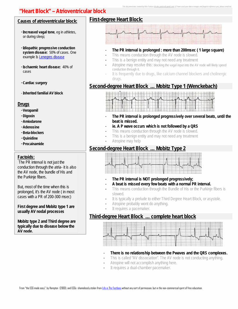
First-degree Heart Block:
- The PR interval is prolonged : more than 200msec ( 1 large square) - This means conduction through the AV node is slowed. - This is a benign entity and may not need any treatment - Atropine may resolve this: blocking the vagal input into the AV node will likely speed
conduction through it. - It is frequently due to drugs, like calcium channel blockers and cholinergic
drugs.
Second-degree Heart Block … Mobitz Type 1 (Wenckebach)
- The PR interval is prolonged progressively over several beats, until the beat is missed.
- ie. A P wave occurs which is not followed by a QRS - This means conduction through the AV node is slowed. - This is a benign entity and may not need any treatment - Atropine may help
Second-degree Heart Block … Mobitz Type 2
- The PR interval is NOT prolonged progressively; - A beat is missed every few beats with a normal PR interval. - This means conduction through the Bundle of His or the Purkinje fibers is
slowed. - It is typically a prelude to either Third Degree Heart Block, or asystole. - Atropine probably wont do anything. - It requires a pacemaker.
Third-degree Heart Block … complete heart block
- There is no relationship between the Pwaves and the QRS complexes. - This is called “AV dissociation”. The AV node is not conducting anything. - Atropine will not accomplish anything here. - It requires a dual-chamber pacemaker.
Causes of atrioventricular block:
- Increased vagal tone, eg in athletes, or during sleep;
- Idiopathic progressive conduction system disease: 50% of cases. One example Is Lenegres disease
- Ischaemic heart disease: 40% of
cases
-Cardiac surgery
- Inherited familial AV block
Drugs -Verapamil -Digoxin -Amiodarone -Adenosine -Beta-blockers -Quinidine -Procainamide
Factoids: The PR interval is not just the conduction through the atria- it is also the AV node, the bundle of His and the Purkinje fibers. But, most of the time when this is prolonged, it’s the AV node ( in most cases with a PR of 200-300 msec) First degree and Mobitz type 1 are usually AV nodal processes Mobitz type 2 and Third degree are typically due to disease below the AV node.

This document was created by Alex Yartsev ([email protected]); if I have used your data or images and forgot to reference you, please email me.
From “the ECG made easy”, by Hampton (2003), and ECGs shamelessly stolen from Life in The Fastlane without any sort of permission, but in the non-commercial spirit of free education.
Right Bundle Branch Block
- Broad QRS - M-shaped QRS pattern in V1 – V3 ( “RSR” waves) - Wide S-wave in lateral leads ( Lead I, V5-6, aVL)
- There will also be T wave inversion in the anterior leads - There may be an extra R wave in the anterior leads ( R’),
or there may just be a broad slurred QRS. - The depolarization of the right ventricle is delayed - The left ventricle activates normally: the early part of the QRS remains unchanged - Axis is unchanged because left ventricular activation is normal.
INCOMPLETE Right Bundle Branch Block o Normal QRS duration, but RSR pattern in the anterior leads o This is a normal variant
Causes of Right Bundle Branch Block
- Acute pulmonary embolism - Right ventricular hypertrophy / cor pulmonale the right bundle is vulnerable to stretching trauma - Ischaemia - Myocarditis or cardiomyopathy - Congenital septal defect
Consequences of Right Bundle Branch Block
- Independent predictor of increased mortality in patients with coronary artery disease - Dyssynchronous left ventricle: thus, decreased ejection fraction
Management options for Right Bundle Branch Block
- In absence of other conduction abnormalities, with normal ejection fraction: do nothing. - Isolated RBBB is very rarely a problem. - If LVEF is poor (<35%) or there is some other heart block, or the patient has episodes of
syncope, go with A PERMANENT PACEMAKER. - These people typically don’t respond as well to pacing, as do the LBBBs
RSR: M-shaped broadened
QRS complexes
Broad slurred S wave
Right hand down
Brugada Syndrome also gives you anterior RSR.
This document was created by Alex Yartsev ([email protected]); if I have used your data or images and forgot to reference you, please email me.
From “the ECG made easy”, by Hampton (2003), and ECGs shamelessly stolen from Life in The Fastlane without any sort of permission, but in the non-commercial spirit of free education.
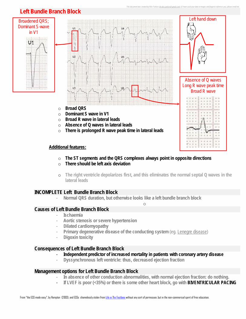
Left Bundle Branch Block
o Broad QRS o Dominant S wave in V1 o Broad R wave in lateral leads o Absence of Q waves in lateral leads o There is prolonged R wave peak time in lateral leads
Additional features:
o The ST segments and the QRS complexes always point in opposite directions o There should be left axis deviation
o The right ventricle depolarizes first, and this eliminates the normal septal Q waves in the
lateral leads INCOMPLETE Left Bundle Branch Block
- Normal QRS duration, but otherwise looks like a left bundle branch block o
Causes of Left Bundle Branch Block - Ischaemia - Aortic stenosis or severe hypertension - Dilated cardiomyopathy - Primary degenerative disease of the conducting system (eg. Lenegre disease) - Digoxin toxicity
Consequences of Left Bundle Branch Block
- Independent predictor of increased mortality in patients with coronary artery disease - Dyssynchronous left ventricle: thus, decreased ejection fraction
Management options for Left Bundle Branch Block
- In absence of other conduction abnormalities, with normal ejection fraction: do nothing. - If LVEF is poor (<35%) or there is some other heart block, go with BIVENTRICULAR PACING
Broadened QRS; Dominant S-wave
in V1
Absence of Q waves Long R wave peak time
Broad R wave
Left hand down
This document was created by Alex Yartsev ([email protected]); if I have used your data or images and forgot to reference you, please email me.
From “the ECG made easy”, by Hampton (2003), and ECGs shamelessly stolen from Life in The Fastlane without any sort of permission, but in the non-commercial spirit of free education.
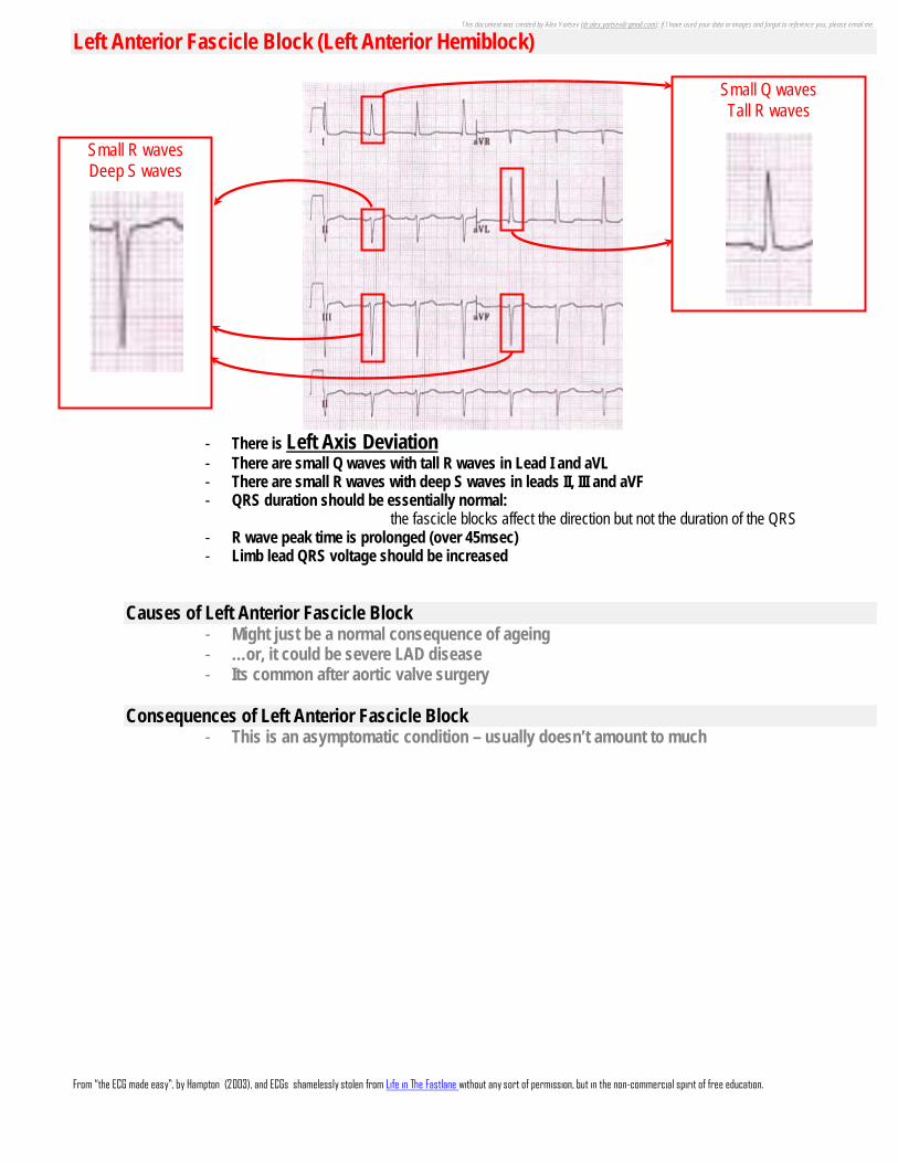
Left Anterior Fascicle Block (Left Anterior Hemiblock)
- There is Left Axis Deviation - There are small Q waves with tall R waves in Lead I and aVL - There are small R waves with deep S waves in leads II, III and aVF - QRS duration should be essentially normal:
the fascicle blocks affect the direction but not the duration of the QRS - R wave peak time is prolonged (over 45msec) - Limb lead QRS voltage should be increased
Causes of Left Anterior Fascicle Block - Might just be a normal consequence of ageing - …or, it could be severe LAD disease - Its common after aortic valve surgery
Consequences of Left Anterior Fascicle Block
- This is an asymptomatic condition – usually doesn’t amount to much
Small R waves Deep S waves
Small Q waves Tall R waves
This document was created by Alex Yartsev ([email protected]); if I have used your data or images and forgot to reference you, please email me.
From “the ECG made easy”, by Hampton (2003), and ECGs shamelessly stolen from Life in The Fastlane without any sort of permission, but in the non-commercial spirit of free education.
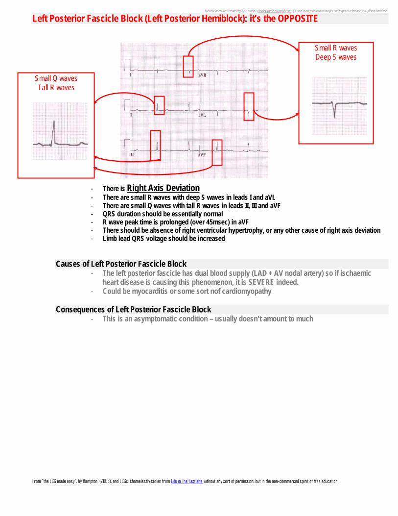
Left Posterior Fascicle Block (Left Posterior Hemiblock): it’s the OPPOSITE
- There is Right Axis Deviation - There are small R waves with deep S waves in leads I and aVL - There are small Q waves with tall R waves in leads II, III and aVF - QRS duration should be essentially normal - R wave peak time is prolonged (over 45msec) in aVF - There should be absence of right ventricular hypertrophy, or any other cause of right axis deviation - Limb lead QRS voltage should be increased
Causes of Left Posterior Fascicle Block - The left posterior fascicle has dual blood supply (LAD + AV nodal artery) so if ischaemic
heart disease is causing this phenomenon, it is SEVERE indeed. - Could be myocarditis or some sort nof cardiomyopathy
Consequences of Left Posterior Fascicle Block
- This is an asymptomatic condition – usually doesn’t amount to much
Small R waves Deep S waves
Small Q waves Tall R waves

This document was created by Alex Yartsev ([email protected]); if I have used your data or images and forgot to reference you, please email me.
From “the ECG made easy”, by Hampton (2003), and ECGs shamelessly stolen from Life in The Fastlane without any sort of permission, but in the non-commercial spirit of free education.
Bifascicular Block a combination of RBBB and either left anterior hemiblock and left posterior hemiblock
- The ventricles depolarize from the single remaining fascicle. - This is a sign of extensive conducting system disease - The example above is RBBB + LAFB:
o Small Q waves and tall R waves in Lead I and aVL o Small R waves and deep S waves in Lead II, Lead III and aVF
But wait! … Isnt Left Bundle Branch Block (LBBB) a bi-fascicular block? Both the anterior and posterior fascicles are blocked! Yes. Yes it is. In fact the guidelines from the European Society of cardiology include LBBB in their guidelines for management of bifascicular block.
“Trifascicular” Block
Its Bifascicular Block – with the important addition of a prolonged PR interval (1st degree AV block).
How does this endanger my patient? - It may progress to complete heart block, and kill them ( 1% per year progress this way)
Management options - If this ECG presents with a history of syncope, most would argue in favour of a pacemaker. - - In fact, if there is no reversible cause, a pacemaker is ideal. - If a pacemaker is needed, make it a dual chamber
Small R waves Deep S waves
Small Q waves Tall R waves
Positive QRS in Lead I, Negative QRS in aVF = Left axis deviation
Widened QRS Prominent R wave in V1
RSR pattern in V1-V3
Slurred S wave in V5-V6
This document was created by Alex Yartsev ([email protected]); if I have used your data or images and forgot to reference you, please email me.
From “the ECG made easy”, by Hampton (2003), and ECGs shamelessly stolen from Life in The Fastlane without any sort of permission, but in the non-commercial spirit of free education.
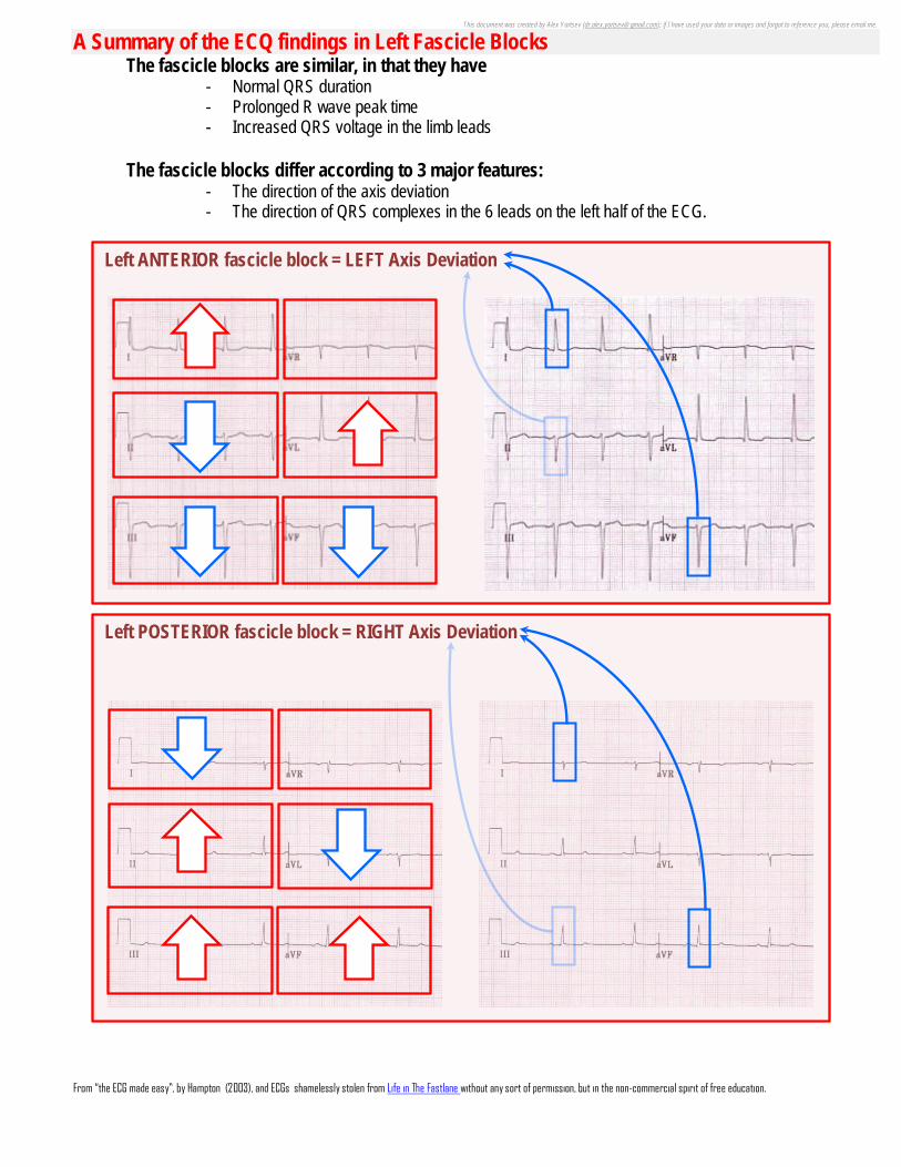
A Summary of the ECQ findings in Left Fascicle Blocks The fascicle blocks are similar, in that they have
- Normal QRS duration - Prolonged R wave peak time - Increased QRS voltage in the limb leads
The fascicle blocks differ according to 3 major features:
- The direction of the axis deviation - The direction of QRS complexes in the 6 leads on the left half of the ECG.
Left ANTERIOR fascicle block = LEFT Axis Deviation
Left POSTERIOR fascicle block = RIGHT Axis Deviation