the qsec sensor kinase: a bacterial adrenergic receptor
TRANSCRIPT
The QseC sensor kinase: A bacterialadrenergic receptorMarcie B. Clarke*, David T. Hughes*, Chengru Zhu†, Edgar C. Boedeker†, and Vanessa Sperandio*‡
*Department of Microbiology, University of Texas Southwestern Medical Center, Dallas, TX 75390-9048; and †Center for Vaccine Development,University of Maryland School of Medicine, Baltimore, MD 21201
Communicated by Steven L. McKnight, University of Texas Southwestern Medical Center, Dallas, TX, May 27, 2006 (received for review February 14, 2006)
Quorum sensing is a cell-to-cell signaling mechanism in whichbacteria respond to hormone-like molecules called autoinducers(AIs). The AI-3 quorum-sensing system is also involved in inter-kingdom signaling with the eukaryotic hormones epinephrine�norepinephrine. This signaling activates transcription of virulencegenes in enterohemorrhagic Escherichia coli O157:H7. However,this signaling system has never been shown to be involved invirulence in vivo, and the bacterial receptor for these signals hadnot been identified. Here, we show that the QseC sensor kinase isa bacterial receptor for the host epinephrine�norepinephrine andthe AI-3 produced by the gastrointestinal microbial flora. We alsofound that an �-adrenergic antagonist can specifically block theQseC response to these signals. Furthermore, we demonstratedthat a qseC mutant is attenuated for virulence in a rabbit animalmodel, underscoring the importance of this signaling system invirulence in vivo. Finally, an in silico search found that the periplas-mic sensing domain of QseC is conserved among several bacterialspecies. Thus, QseC is a bacterial adrenergic receptor that activatesvirulence genes in response to interkingdom cross-signaling. Weanticipate that these studies will be a starting point in understand-ing bacterial–host hormone signaling at the biochemical level.Given the role that this system plays in bacterial virulence, furthercharacterization of this unique signaling mechanism may be im-portant for developing novel classes of antimicrobials.
AI-3 � enterohemorrhagic Escherichia coli � epinephrine �quorum sensing � two-component systems
The gastrointestinal (GI) tract is the site of the most complexheterogeneous environment in the mammalian host, where the
majority of the microbial flora reside in the large intestine (1). It isestimated that the microbial population within the human GI tractexceeds the total number of mammalian cells by at least an orderof magnitude (1). The bacterial flora is beneficial to the host byfacilitating nutrient assimilation and immune competence (2).Conversely, adverse interactions between pathogenic or opportu-nistic bacteria and the host can lead to disease. The high density anddiversity of bacteria within the GI tract suggests that the membersof this community communicate among themselves as well as withthe host to coordinate any number of advantageous processes. Littleis currently known about the communicative relationship(s) amongthe normal flora, bacterial pathogens, and the host. It has beenwidely held that bacteria must sense and recognize the hostenvironment to express genes essential for colonization and�orvirulence expression. Along those lines, quorum sensing is acell-to-cell signaling mechanism wherein bacteria respond to chem-ical hormone-like molecules called autoinducers (AIs). When anAI reaches a critical concentration threshold, the bacteria recognizeand respond to this signal by altering their patterns of geneexpression. A signaling system has been identified in which abacterial chemical signal (AI-3) is produced and used for inter-kingdom signaling between prokaryotic and eukaryotic cells.Namely, AI-3 cross-signals with the eukaryotic hormones epineph-rine (epi) and�or norepinephrine (NE) (3).
Enterohemorrhagic Escherichia coli (EHEC) 0157:H7 colonizesthe human colon, resulting in the development of often fatal
hemorrhagic colitis and hemolytic uremic syndrome (4). EHECexploits the AI-3�epi�NE signaling system to activate its virulencegenes (3). These signals may be sensed by histidine sensor kinases(HKs) in the membrane of EHEC that relay this information to acomplex regulatory cascade (3). HKs are, arguably, among the mostwidely used sensors of all of the signal transduction enzymes innature, being present in bacteria, archaea, and eukarya (5, 6).Although there are no known HKs present in animals, eukaryotessuch as yeast, fungi, plants, and protozoa use HKs to regulatehormone-dependent developmental processes (6). Thus, it has beensuggested that HKs originated in bacteria and were later transferredinto eukaryotes and archaea (7). Of relevance to EHEC, QseB�QseC comprise a two-component system, in which QseC is thepredicted HK and QseB the predicted response regulator. QseB�QseC activate transcription of the flagella regulon responsible forswimming motility in EHEC (8). An EHEC mutant unable toproduce AI-3 activates transcription of the flagella�motility genesand, consequently, swimming motility in response to both AI-3 andepi given exogenously. However, a qseC (sensor mutant) is unableto activate expression of these genes in response to both thesesignals (3).
In this study, we demonstrate that QseC specifically senses thebacterial AI-3 signal and the host epi�NE hormones. QseC directlybinds to these signals, and this binding can be blocked by the�-adrenergic antagonist phentolamine (PE). The role of QseC inpathogenesis has also been defined by using a rabbit infectionanimal model, demonstrating that a qseC mutant is attenuated forvirulence. Taken together, these results suggest that QseC is abacterial adrenergic receptor that is crucial for interkingdomsignaling.
ResultsQseC Senses the Host Hormones Epi and�or NE. We have previouslyreported that a qseC mutant did not activate expression of theflagella and motility genes in response to AI-3, epi, and�or NE (3).These results led us to hypothesize that QseC could be the sensorfor these signals and may act as a bacterial adrenergic receptor forthese compounds. We tested this hypothesis at the molecular levelby expressing and purifying MycHis-tagged QseC under nativeconditions, and performing in vitro autophosphorylation assays.Because most HKs, including QseC, are membrane-bound, wereconstituted QseC into liposomes. This system can be used to studysignal transduction and transmembrane signaling, which dependson the membrane-intrinsic portions of the protein linking theperiplasmic sensory and cytoplasmic kinase domains (9, 10). Asdepicted in Fig. 1A, QseC adopts an inside-out orientation, in which
Conflict of interest statement: No conflicts declared.
Freely available online through the PNAS open access option.
Abbreviations: AI, autoinducer; EHEC, enterohemorrhagic E. coli; epi, epinephrine; HK,histidine sensor kinase; NE, norepinephrine; PE, phentolamine; PO, propranolol; REPEC,rabbit enteropathogenic E. coli.
‡To whom correspondence should be addressed at: Department of Microbiology, Univer-sity of Texas Southwestern Medical Center, 5323 Harry Hines Boulevard, Dallas,TX 75390-9048. E-mail: [email protected].
© 2006 by The National Academy of Sciences of the USA
10420–10425 � PNAS � July 5, 2006 � vol. 103 � no. 27 www.pnas.org�cgi�doi�10.1073�pnas.0604343103

the periplasmic signal-recognition domain is inside the liposomes,whereas the kinase domain is outside. Orientation of proteins inliposomes has been established (10). In the case of QseC as well ason previous studies (9, 10), the inside-out orientation can beconcluded from the accessibility of ATP to the kinase site withoutdisruption of the liposomes. To verify QseC protein incorporationinto the liposomes, Western blot analysis using anti-Myc antibodywas performed. The recognition of the Myc-tag by the anti-Mycantibody without disruption of the liposomes further confirms theinside-out orientation of QseC (data not shown). QseC autophos-phorylation was weak in the liposomes alone (Fig. 1B).
To test whether QseC could sense epi, QseC-loaded liposomeswere treated with the physiologically intestinal-relevant concen-tration of 5–50 �M epi (11). Addition of epi to QseC-loadedliposomes during the freeze–thaw step enabled this signal to gainaccess to the internal space of these liposomes (containing theperiplasmic sensing domain of QseC). QseC then increased itslevel of autophosphorylation dramatically (compared with lipo-somes to which no epi was added) (Fig. 1B). The addition of 5�M NE also increased the autophosphorylation of QseC (Fig. 2D and F). Thus, QseC directly responds to the presence ofepi�NE. Of note, DcuS (E. coli fumarate and succinate sensor)loaded into liposomes increased its autophosphorylation inresponse to 20 mM fumarate, as expected, but not to 50 �M epi(Fig. 2 A), confirming that QseC and DcuS each sense andrespond to different signaling compounds.
We did not observe autophosphorylation of full-length, solubleQseC in free solution under various buffer conditions (data notshown). This is not inordinate; however, given that autophosphor-ylation studies of most other HKs have been performed by usingtruncated proteins containing only the cytoplasmic kinase domain.An important caveat is that these studies preclude signal-recognition assessment by HKs, because the recombinant proteinsdo not contain the periplasmic domain necessary for signal recog-nition (12–18). In order for QseC to respond to epi�NE, thesesignals had to be loaded within the liposomes, where the periplas-mic domain is located; loading these signals from outside yielded noresponse (data not shown). Thus, QseC is one of the rare HKswhose signals have been biochemically determined.
To further assess the signal specificity of QseC, we tested whetherQseC could sense and respond to other intestinal hormones. QseCautophosphorylation did not increase in response to other intestinalhormones, such as gastrin, secretin, or galanin (Fig. 2B). In fact, the
basal level of QseC autophosphorylation in response to any of thesehormones was not significantly different from the negative control,where no signal was added, indicating that these other gastrichormones, unlike epi�NE do not play any role in signaling throughQseC.
The neuronal response to epi�NE in the intestinal tract can beblocked by �- and �-adrenergic receptor antagonists, such aspropranolol (PO) or PE, respectively (19). Because QseC auto-phosphorylation increases in response to epi�NE, we investigatedwhether we could block this response using PO or PE. The additionof PO or PE alone to the liposomes did not have a significant effecton QseC autophosphorylation, whereas the addition of 5 �M epiagain induced QseC autophosphorylation (Fig. 2C). To addresswhether PO and PE could act as antagonists of epi for QseCautophosphorylation, liposomes were loaded with 5 �M epi and anexcess concentration (50 �M) of PO or PE. Autophosphorylationof QseC was increased by epi in the presence of PO (Fig. 2C),suggesting that PO cannot antagonize the recognition of epi byQseC. However, in the presence of PE, QseC did not respond to epi(Fig. 2C), suggesting that this �-adrenergic antagonist can block therecognition of epi by QseC.
QseC Binds to NE. Because QseC autophosphorylation increases inresponse to epi�NE, we reasoned that these compounds may bedirectly interacting with the membrane-bound QseC to stimulatekinase activity. To study binding, 5 or 10 �M of [3H]NE, alone orin conjunction with 50 �M PE, were loaded into QseC liposomes.In the presence of [�32P]dATP, QseC autophosphorylation in-creased with the addition of [3H]NE, as expected (Fig. 2 D and F).The addition of 5 �M [3H]NE plus 50 �M PE did not show asignificant increase in QseC autophosphorylation when comparedwith liposomes without signal added. These results confirm that PEantagonizes NE recognition. To determine whether the [3H]NEwas binding to QseC, we excised the bands that contained phos-phorylated QseC protein, suspended them in scintillation fluid, andcounted, which allows the differential counting of [3H]NE (to assessbinding) and �[32]ATP (to assess autophosphorylation). (Fig. 2 Eand F). The addition of 5 �M [3H]NE to the QseC liposomeresulted in a 2.25-fold increase in the amount of [3H] bound toQseC, which correlated with the increase in QseC autophosphor-ylation (Fig. 2 D and E). The addition of 10 �M [3H]NE to theseliposomes resulted in a 4-fold increase in the amount of [3H]NEbound to QseC and also an increase in QseC autophosphorylation.
Fig. 1. QseC increases autophosphorylation in re-sponse to epi. (A) Graphic depiction of the inside-outorientation of QseC in the liposome. QseC has twotransmembrane domains and a HK domain, indicat-ing that its membrane location may allow autophos-phorylation upon sensing its specific environmentalcue. QseC also contains an ATPase domain, whichallows it to exhibit phosphatase activity toward QseB,and a conserved EAL domain that may confer cyclicdiguanylate phosphodiesterase activity. (B) QseCweakly autophosphorylates in the liposome, but in-creases its autophosphorylation in response to epi.
Clarke et al. PNAS � July 5, 2006 � vol. 103 � no. 27 � 10421
MIC
ROBI
OLO
GY

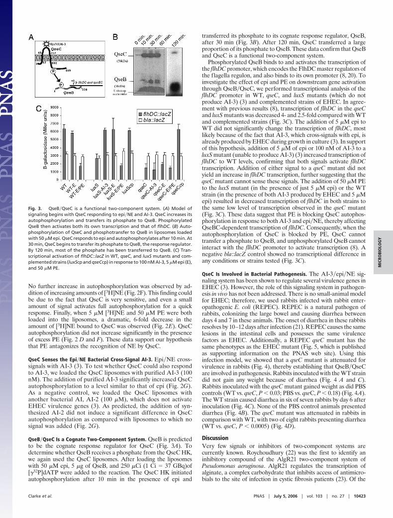
Fig. 2. QseC specifically senses epi�NE�AI-3. Each graph represents results from three separate experiments. Student’s t test was performed to determinewhether the results were statistically significant as compared with the control (no signal added). (A) DcuS increases autophosphorylation in response to fumaratebut not to epi. (B) QseC autophosphorylation in the presence of 5 �M epi, 50 �M gastrin, 50 �M galanin, and 50 �M secretin. (C) QseC autophosphorylation inthe presence of 5 �M epi, 50 �M PO, 50 �M PE, and 5 �M epi with 50 �M PE or PO. (D) QseC autophosphorylation in the presence of 5 and 10 �M [3H]NE and5 �M NE with 50 �M PE. NE induces QseC autophosphorylation, and NE-induced autophosphorylation is also inhibited by PE. (E) Graphical representation of theamount of [3H]NE bound to QseC after incubation with 5 and 10 �M [3H]NE alone or in the presence of 50 �M PE. (F) Graphical representation of the amountof QseC autophosphorylation (cpm measurement of P32) after incubation with 5 and 10 �M [3H]NE, alone or in the presence of 50 �M PE. (G) QseC significantlyincreases autophosphorylation in response to AI-3 but not to AI-2 (negative control).
10422 � www.pnas.org�cgi�doi�10.1073�pnas.0604343103 Clarke et al.
No further increase in autophosphorylation was observed by ad-dition of increasing amounts of [3H]NE (Fig. 2F). This finding couldbe due to the fact that QseC is very sensitive, and even a smallamount of signal activates full autophosphorylation for a quickresponse. Finally, when 5 �M [3H]NE and 50 �M PE were bothloaded into the liposomes, a dramatic, 6-fold decrease in theamount of [3H]NE bound to QseC was observed (Fig. 2E). QseCautophosphorylation did not increase significantly in the presenceof excess PE (Fig. 2 D and F). These data support our hypothesisthat PE antagonizes the recognition of NE by QseC.
QseC Senses the Epi�NE Bacterial Cross-Signal AI-3. Epi�NE cross-signals with AI-3 (3). To test whether QseC could also respondto AI-3, we loaded the QseC liposomes with purified AI-3 (100nM). The addition of purified AI-3 significantly increased QseCautophosphorylation to a level similar to that of epi (Fig. 2G).As a negative control, we loaded the QseC liposomes withanother bacterial AI, AI-2 (100 �M), which does not activateEHEC virulence genes (3). As predicted, the addition of syn-thesized AI-2 did not induce a significant difference in QseCautophosphorylation as compared with liposomes to which nosignal was added (Fig. 2G).
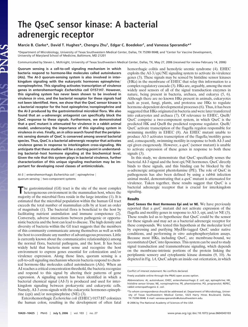
QseB�QseC Is a Cognate Two-Component System. QseB is predictedto be the cognate response regulator for QseC (Fig. 3A). Todetermine whether QseB receives a phosphate from the QseC HK,we again used the QseC liposomes. After loading the liposomeswith 50 �M epi, 5 �g of QseB, and 250 �Ci (1 Ci � 37 GBq)of[�32P]dATP were added to the reaction. The QseC HK initiatedautophosphorylation after 10 min in the presence of epi and
transferred its phosphate to its cognate response regulator, QseB,after 30 min (Fig. 3B). After 120 min, QseC transferred a largeproportion of its phosphate to QseB. These data confirm that QseBand QseC is a functional two-component system.
Phosphorylated QseB binds to and activates the transcription ofthe flhDC promoter, which encodes the FlhDC master regulators ofthe flagella regulon, and also binds to its own promoter (8, 20). Toinvestigate the effect of epi and PE on downstream gene activationthrough QseB�QseC, we performed transcriptional analysis of theflhDC promoter in WT, qseC, and luxS mutants (which do notproduce AI-3) (3) and complemented strains of EHEC. In agree-ment with previous results (8), transcription of flhDC in the qseCand luxS mutants was decreased 4- and 2.5-fold compared with WTand complemented strains (Fig. 3C). The addition of 5 �M epi toWT did not significantly change the transcription of flhDC, mostlikely because of the fact that AI-3, which cross-signals with epi, isalready produced by EHEC during growth in culture (3). In supportof this hypothesis, addition of 5 �M of epi or 100 nM of AI-3 to aluxS mutant (unable to produce AI-3) (3) increased transcription offlhDC to WT levels, confirming that both signals activate flhDCtranscription. Addition of either signal to a qseC mutant did notyield an increase in flhDC transcription, further suggesting that theqseC mutant cannot sense these signals. The addition of 50 �M PEto the luxS mutant (in the presence of just 5 �M epi) or the WTstrain (in the presence of both AI-3 produced by EHEC and 5 �Mepi) resulted in decreased transcription of flhDC in both strains tothe same low level of transcription observed in the qseC mutant(Fig. 3C). These data suggest that PE is blocking QseC autophos-phorylation in response to both AI-3 and epi�NE, thereby affectingQseBC-dependent transcription of flhDC. Consequently, when theautophosphorylation of QseC is blocked by PE, QseC cannottransfer a phosphate to QseB, and unphosphorylated QseB cannotinteract with the flhDC promoter to activate transcription (8). Anegative bla::lacZ control showed no transcriptional difference inany conditions or strains tested (Fig. 3C).
QseC Is Involved in Bacterial Pathogenesis. The AI-3�epi�NE sig-naling system has been shown to regulate several virulence genes inEHEC (3). However, the role of this signaling system in pathogen-esis in vivo has not been addressed. There is no small-animal modelfor EHEC; therefore, we used rabbits infected with rabbit enter-opathogenic E. coli (REPEC). REPEC is a natural pathogen ofrabbits, colonizing the large bowel and causing diarrhea betweendays 4 and 7 in these animals. The onset of diarrhea in these rabbitsresolves by 10–12 days after infection (21). REPEC causes the samelesions in the intestinal cells and possesses the same virulencefactors as EHEC. Additionally, a REPEC qseC mutant has thesame phenotypes as the EHEC mutant (Fig. 5, which is publishedas supporting information on the PNAS web site). Using thisinfection model, we showed that a qseC mutant is attenuated forvirulence in rabbits (Fig. 4), thereby establishing that QseB�QseCare involved in pathogenesis. Rabbits inoculated with the WT straindid not gain any weight because of diarrhea (Fig. 4 A and C).Rabbits inoculated with the qseC mutant gained weight as did PBScontrols (WT vs. qseC, P � 0.03; PBS vs. qseC, P � 0.18) (Fig. 4A).The WT strain caused diarrhea in six of seven rabbits by day 6 afterinoculation (Fig. 4C). None of the PBS control animals presenteddiarrhea (Fig. 4B). The qseC mutant was attenuated in rabbits incomparison with WT, with two of eight rabbits presenting diarrhea(WT vs. qseC, P � 0.0005) (Fig. 4D).
DiscussionVery few signals or inhibitors of two-component systems arecurrently known. Roychoudhury (22) was the first to identify aninhibitory compound of the AlgR21 two-component system ofPseudomonas aeruginosa. AlgR21 regulates the transcription ofalginate, a complex carbohydrate that inhibits access of antimicro-bials to the site of infection in cystic fibrosis patients (23). Of the
Fig. 3. QseB�QseC is a functional two-component system. (A) Model ofsignaling begins with QseC responding to epi�NE and AI-3. QseC increases itsautophosphorylation and transfers its phosphate to QseB. PhosphorylatedQseB then activates both its own transcription and that of flhDC. (B) Auto-phosphorylation of QseC and phosphotransfer to QseB in liposomes loadedwith 50 �M epi. QseC responds to epi and autophosphorylates after 10 min. At30 min, QseC begins to transfer its phosphate to QseB, the response regulator.By 120 min, most of the phosphate has been transferred to QseB. (C) Tran-scriptional activation of flhDC::lacZ in WT, qseC, and luxS mutants and com-plemented strains (luxScp and qseCcp) in response to 100 nM AI-3, 5 �M epi (E),and 50 �M PE.
Clarke et al. PNAS � July 5, 2006 � vol. 103 � no. 27 � 10423
MIC
ROBI
OLO
GY

25,000 synthetic and natural compounds screened, only 15 inhibi-tors were identified, many of which contained aromatic rings (22).Additionally, extensive screening assays have identified severalinhibitors, including several for the Bacillus subtilis KinA�SpoOFtwo-component system (24, 25). Although a few compounds havebeen reported as inhibitors of two-component systems, their mech-anisms of action and true stimulatory signals are largely unknown(26). Even less is known about the signaling compounds thatspecifically activate HKs. One of the few systems for which a definedsignal has been identified is the DcuSR two-component system ofE. coli, which controls the expression of genes of C (4)-dicarboxylatemetabolism (27). Janausch et al. (9) have shown that the autophos-phorylation of liposome-reconstituted DcuS is stimulated by thesignals fumarate and succinate. Here, we show that both the AI-3signal produced by the microbial flora and the epi�NE signalsproduced by the host are sensed by the QseC HK. Upon sensingthese signals, QseC autophosphorylates and then transfers itsphosphate to QseB, which then regulates both is own transcriptionand the transcription of the flagella and motility genes (8, 20).EHEC likely is then able to exploit its motility apparatus to swimto the intestinal epithelium and initiate infection. The observationthat a qseC mutant is attenuated for virulence in a rabbit infectionmodel (Fig. 4), further underscores the role of this signaling systemin bacterial pathogenesis.
QseC acts a bacterial adrenergic receptor and links cross-kingdom signaling by sensing a bacterial hormone-like compound(AI-3) and the host hormones epi and�or NE. It is noteworthy thatQseC does not share primary sequence homology with adrenergicreceptors; hence, it may serve as a functional analog, not homolog,of these G protein-coupled receptors. An in silico search using theperiplasmic (signal-sensing) domain of QseC reveals a high degreeof conservation among different bacterial species (Fig. 6, which ispublished as supporting information on the PNAS web site). TheQseC sensor is found in Shigella sp., Salmonella sp., Erwiniacarotovora, Haemophilus influenzae, Pasteurella multocida, Acti-nobacillus pleuropneumoniae, Chromobacterium violaceium, Rubri-vivax gelatinosus, Thiobacillus denitrificans, Ralstonia eutropa, Ral-stonia metallidurans, and Psychrobacter sp. This search also revealedhomology to a fungal protein of unknown function from Aspergillusnidulans. Taken together, these findings suggest that QseC mayhave an ancient evolutionary history.
One of the daunting medical challenges at present surrounds theissue of microbial antibiotic resistance. It once was believed thatmodern medicine conquered infectious diseases through the dis-covery of both first- and second-generation antimicrobials. How-ever, very few novel antibiotics have been discovered in the past 30years, and, during this period, profound selective pressure throughboth justified and indiscriminant use of treatment has allowedbacteria to quickly evolve mechanisms of resistance to virtually allknown antibiotics. Hence, diseases such as tuberculosis are againdeveloping into significant public health problems and, thus, areconsidered to be reemerging infectious diseases. Many other in-fections, like the hemorrhagic colitis and hemolytic uremic syn-drome caused by EHEC, comprise newly emerging infectiousdiseases. These combined health threats constitute a compellingargument for the development of novel classes of antimicrobialcompounds. Our combined data, reported herein, in addition topublished data (3), indicate that adrenergic antagonists can inhibitthe AI-3�epi�NE signaling cascade in EHEC and render it unableto induce its virulence genes (in response to these signals), sug-gesting that antagonists targeting this signaling cascade mightconstitute a novel class of antimicrobials (3). Furthermore, theseantimicrobials will possibly be useful against other human patho-gens, including enteropathogenic E. coli, Salmonella, Shigella, andYersinia, etc., all of which ostensibly harbor this signaling cascade.A broad understanding of the QseB�QseC signaling cascade will beinstrumental for investigating further the intriguing concept ofinterkingdom sensory signaling.
Materials and MethodsStrains and Plasmids. E. coli strains were grown in LB at 37°C. TheEHEC WT strain, 86-24, was isolated from an outbreak in theUnited States (28). The EHEC isogenic qseC (VS138), luxS(VS94), and complemented strains (VS179 and VS95, respectively)have been described (29, 30). The REPEC qseC mutant (VS243)was generated by allelic exchange using vector pVS132 as described(29). The qseC mutant (VS243) was complemented with plasmidpVS178 (29), generating strain VS247.
Inoculation of Rabbits. Bacterial strains were inoculated in2-month-old New Zealand White rabbits. Three groups ofanimals (seven to eight in each group) were inoculated with the
Fig. 4. A qseC mutant is attenuated for virulence in rabbits. (A) Cumulative weight gain of rabbits inoculated with WT, qseC, or PBS by day 6. Developmentof diarrhea by rabbits inoculated with PBS (B), WT (C), and qseC (D). The WT caused diarrhea in six of seven rabbits by day 6 after inoculation. None of the PBScontrol animals presented diarrhea. The qseC mutant was attenuated in rabbits in comparison with WT, with two of eight rabbits presenting diarrhea.
10424 � www.pnas.org�cgi�doi�10.1073�pnas.0604343103 Clarke et al.

WT, the qseC mutant (VS243) strain, or PBS (negative control).Bacteria were cultured in LB broth, washed once, suspended insterile PBS, and adjusted to OD600 � 1. Subsequent dilution ofthis bacterial suspension at 1:100 was made to obtain �1 � 107
colony-forming units per ml for inoculation. Bacterial viablecounts were determined by plating. These experiments wererepeated twice with two different groups of animals to ensuretheir reproducibility.
Before inoculation, rabbits were starved overnight. Rabbits wereinoculated intragastrically via a pediatric feeding tube with 10 ml of10% bicarbonate solution, followed by a 3-ml inoculum. Rabbitswere weighed daily and observed for stool characteristics andclinical signs of illness for 13 days. Stools were graded as normal(hard pellets no diarrhea) or diarrhea (completely liquid). Rabbitswere killed 13 days after inoculation. Differences in weight gainbetween experimental groups were analyzed by the Student t test.Differences in diarrheal disease between the experimental groupswere analyzed by using a �2 test.
�-Galactosidase Assays. The lacZ fusions ( flhDC::lacZ frompVS182 and bla::lacZ from pVSAP) were constructed as described,and assayed for �-galactosidase activity by using o-nitrophenyl-�-D-galactopyranoside as a substrate (29).
Reconstitution of QseC-His into Liposomes. As described in ref. 8, E.coli strains containing either pVS154 (QseB-MycHis) or pVS155(QseC-MycHis) were induced with 0.2% arabinose, and purifiedthrough nickel columns according to the manufacturer’s instruc-tions (Qiagen).
Liposomes were reconstituted as described by Janausch et al.(31). Briefly, 50 mg of E. coli phospholipids (20 mg�ml inchloroform; Avanti Polar Lipids) were evaporated and thendissolved into 5 ml of potassium phosphate buffer containing 80mg of N-octyl-�-D-glucopyranoside. The solution was dialyzedovernight against potassium phosphate buffer. The resultingliposome suspension was subjected to freeze–thaw in liquid N2.Liposomes were then destabilized by the addition of 26.1 mg ofdodecylmaltoside, and 2.5 mg of QseC-MycHis was added,followed by stirring at room temperature for 10 min. Twohundred-sixty milligrams of Biobeads were then added to re-move the detergent, and the resulting solution was allowed toincubate at 4°C overnight. The supernatant was then incubatedwith fresh Biobeads for 1 h in the morning. The resultingliposomes containing reconstituted QseC-MycHis were frozen inliquid N2 and stored at �80°C until used. Orientation of HKs inthe liposome system has been established by other groups (12)and can be concluded from the accessibility of ATP to the kinase
site and anti-Myc antisera to the C-terminal QseC-MycTagwithout disruption of the liposomes.
Phosphorylation of QseC-MycHis in Liposomes. Twenty microliters ofthe liposomes containing QseC-MycHis were adjusted to 10 mMMgCl2 and 1 mM DTT, and various concentrations of agonist orantagonist, frozen and thawed rapidly in liquid N2, and kept at roomtemperature for 1 h (this allows for the signals to be loaded withinthe liposomes). [�32P]dATP (0.625 �l) (110 TBq�mmol) was addedto each reaction. To some reactions, 10 �g of QseB-MycHis wasadded. At each time point (0, 10, 30, 60, or 120 min), 20 �l of SDSloading buffer was added. For all experiments involving QseCalone, a time point of 10 min was used. The samples were run onSDS�PAGE without boiling and visualized via PhosphorImager.The bands were quantitated by using IMAGEQUANT version 5.0software (Amersham Pharmacia).
Agonists and Antagonists. Various concentrations of agonist orantagonist (Sigma) were added to each of the liposome experi-ments, resulting in final concentrations as follows: 5 �M or 50 �Mepi, 5 �M or 10 �M NE, 50 �M PE, 50 �M PO, 50 �M gastrin, 50�M galanin, and 50 �M secretin. Synthetic AI-2 (�100 �M) andpurified AI-3 (�100 nM) were obtained as described (3). AI-3 wasa gift from B. Sangras and J. R. Falck (University of TexasSouthwestern Medical Center) and was purified from EHEC strain86-24 as described (3). Tritiated NE was obtained from AmershamPharmacia Biosciences and used at a final concentration of 5 or10 �M.
Determination of Tritiated Ligand Binding. To determine the con-centration of tritiated NE that was bound to QseC-MycHis in theliposomes, 20 �l of the liposome containing QseC-MycHis wereadjusted to 10 mM MgCl2, 1 mM DTT, and 5 �M tritiated NE, 10�M tritiated NE, or 5 �M tritiated NE plus 50 �M PE. Theliposomes were frozen and thawed rapidly in liquid N2 and kept atroom temperature for 1 h. [�32P] (0.625 �l) dATP (110 TBq�mmol)was added to each reaction. After 10 min, SDS loading dye wasadded, and the samples were run on SDS�PAGE and visualized byPhosphorImager. The bands containing phosphorylated QseC-Hiswere excised and counted in a scintillation counter.
We thank G. Unden (Johannes Gutenberg-Universitat, Mainz, Ger-many) for the DcuS protein; J. R. Falck for the AI-3; Hernan Rios andKatherine Davis for assistance with the rabbits’ infections; and MichaelGale, Jr., Lora Hooper, James B. Kaper, Michael Norgard, Kim Orth,Melissa Kendall, and David Rasko for critical reading of the manuscript.This work was supported by National Institutes of Health (NIH) GrantAI053067 and an Ellison Foundation award. M.B.C. was supportedthrough NIH Training Grant 5-T32-AI007520-07.
1. Berg, R. D. (1996) Trends Microbiol. 4, 430–435.2. Hooper, L. V. & Gordon, J. I. (2001) Science 292, 1115–1118.3. Sperandio, V., Torres, A. G., Jarvis, B., Nataro, J. P. & Kaper, J. B. (2003) Proc. Natl.
Acad. Sci. USA 100, 8951–8956.4. Nataro, J. P. & Kaper, J. B. (1998) Clin. Microbiol. Rev. 11, 142–201.5. Grebe, T. W. & Stock, J. B. (1999) Adv. Microb. Physiol. 41, 139–227.6. Koretke, K. K., Lupas, A. N., Warren, P. V., Rosenberg, M. & Brown, J. R. (2000) Mol.
Biol. Evol. 17, 1956–1970.7. Wolanin, P. M., Thomason, P. A. & Stock, J. B. (2002) Genome Biol. 3, REVIEWS3013.8. Clarke, M. B. & Sperandio, V. (2005) Mol. Microbiol. 57, 1734–1749.9. Janausch, I. G., Garcia-Moreno, I. & Unden, G. (2002) J. Biol. Chem. 277, 39809–39814.
10. Jung, K., Tjaden, B. & Altendorf, K. (1997) J. Biol. Chem. 272, 10847–10852.
11. Eldrup, E. & Richter, E. A. (2000) Am. J. Physiol. Endocrinol. Metab. 279, E815–E822.12. Gilles-Gonzalez, M. A., Ditta, G. S. & Helinski, D. R. (1991) Nature 350,
170–172.13. Lois, A. F., Weinstein, M., Ditta, G. S. & Helinski, D. R. (1993) J. Biol. Chem. 268,
4370–4375.14. Hill, B., Sandstrom, G., Schroder, S., Franzen, C. & Tarnvik, A. (1990) Scand. J. Infect.
Dis. 22, 95–99.15. Tzipori, S., Wachsmuth, I. K., Chapman, C., Birden, R., Brittingham, J., Jackson, C. &
Hogg, J. (1986) J. Infect. Dis. 154, 712–716.16. Emmerich, R., Panglungtshang, K., Strehler, P., Hennecke, H. & Fischer, H. M. (1999)
Eur. J. Biochem. 263, 455–463.17. Georgellis, D., Kwon, O. & Lin, E. C. (1999) J. Biol. Chem. 274, 35950–35954.
18. Roberts, D. L., Bennett, D. W. & Forst, S. A. (1994) J. Biol. Chem. 269, 8728–8733.
19. Horger, S., Schultheiss, G. & Diener, M. (1998) Am. J. Physiol. 275, G1367–G1376.
20. Clarke, M. B. & Sperandio, V. (2005) Mol. Microbiol. 58, 441–455.21. Heczko, U., Carthy, C. M., O’Brien, B. A. & Finlay, B. B. (2001) Infect. Immun. 69,
4580–4589.22. Roychoudhury, S., Zielinski, N. A., Ninfa, A. J., Allen, N. E., Jungheim, L. N., Nicas,
T. I. & Chakrabarty, A. M. (1993) Proc. Natl. Acad. Sci. USA 90, 965–969.23. Kato, J. & Chakrabarty, A. M. (1991) Proc. Natl. Acad. Sci. USA 88, 1760–1764.24. Barrett, J. F., Goldschmidt, R. M., Lawrence, L. E., Foleno, B., Chen, R., Demers, J. P.,
Johnson, S., Kanojia, R., Fernandez, J., Bernstein, J., et al. (1998) Proc. Natl. Acad. Sci.USA 95, 5317–5322.
25. Macielag, M. J., Demers, J. P., Fraga-Spano, S. A., Hlasta, D. J., Johnson, S. G., Kanojia,R. M., Russell, R. K., Sui, Z., Weidner-Wells, M. A., Werblood, H., et al. (1998) J. Med.Chem. 41, 2939–2945.
26. Matsushita, M. & Janda, K. D. (2002) Bioorg. Med. Chem. 10, 855–867.27. Zientz, E., Bongaerts, J. & Unden, G. (1998) J. Bacteriol. 180, 5421–5425.28. Griffin, P. M., Ostroff, S. M., Tauxe, R. V., Greene, K. D., Wells, J. G., Lewis, J. H. &
Blake, P. A. (1988) Ann. Intern. Med. 109, 705–712.29. Sperandio, V., Torres, A. G. & Kaper, J. B. (2002) Mol. Microbiol. 43, 809–821.30. Sperandio, V., Torres, A. G., Giron, J. A. & Kaper, J. B. (2001) J. Bacteriol. 183,
5187–5197.31. Janausch, I. G., Garcia-Moreno, I., Lehnen, D., Zeuner, Y. & Unden, G. (2004)
Microbiology 150, 877–883.
Clarke et al. PNAS � July 5, 2006 � vol. 103 � no. 27 � 10425
MIC
ROBI
OLO
GY