the reaction timesmy current project is to expand this imaging system to include structure- ... the...
TRANSCRIPT

The Reaction TimesVOLUME II DECEMBER 2018 Fall Edition
UMassAmherst College of Natural SciencesDepartment of Chemistry
Inaugural Chemistry Cultural Potluck Luncheon
The impetus for The Reaction Times newsletter was a series of discussions members of the Equity & Diversity Committee had with undergraduates, graduate students, postdocs, faculty, and staff, who indicated that they would like to learn more about others in the UMass Chemistry community. The cultural luncheon, held on November 20th, was another extension of our effort to share our experiences and learn from each other. It was such a success that we plan to have more events soon. Editorial staff:Amanda Bennett, Christie L. C. Ellis, Ricardo Metz, and Mahdieh Yazdani Design and editing: Brigette McKenna

Building BridgesThe Building Bridges Exhibit featured artists from across campus, including chemistry’s own Marvin Ellin, Karen Hakala and Lisa Korpiewski (now in IALS).
Marv Ellin: “I picked up a love of wildlife photography from my dad. It takes a lot of patience to wait for the right picture. But it is very rewarding when you are successful. Particularly, I love observing wading birds as they patiently wait to grab a fish. Their majestic, slow steps as they look for food feels like I’m watching a dance program on stage.”
1. Cedar Waxwing, taken at a heron rookery in Venice, Florida. 2. Black Crowned Night Heron, taken from a canoe during a deluge on a fresh water spring near Ocala, Florida. 3. Roseate Spoonbill, taken after I jumped out of the car and chased it for several minutes (in Sarasota, Florida). 4. Meerkat at the San Diego Zoo, one of my favorite places. 5. Sandhill Crane, in someone’s front yard in Sarasota, Florida.
Karen Hakala: “I enjoy working with mixed media because it allows for a variety of techniques that encourage my creative side. Though I am a beginner, I find the process of doing art brings me joy and helps me better weather the stresses of life. I am particularly interested in the intersection of art and spirituality. This piece came about as a result of a talk I heard about St. Jane Frances de Chantal and her memoir in which she discusses the ‘Martyrdom of Love.’ I am grateful to artists who are staff members in the chemistry department for sharing their knowledge and inspiration.”
Lisa Korpiewski: “Three small floral tiles: I have always depicted my floral paintings by magnifying a section of the flower, capturing the details. These tiles were painted using an alcohol-based ink which provides vibrant colors, but less control of your medium, making each piece truly unique.
Mixed Media: Mixed media art allows for a very open form of expression by building up in layers. I use paints, waxes, papers, found and handmade objects, and miscellaneous elements to create my altered art and handmade cards. In creating art I get totally lost in the process, and am always surprised by the finished product.”
1. 2.
4. 5.3.

Undergraduate Research
Designing RNA Molecules for Intracellular Detectionand Quantification of Small Molecules by Mark Anthony Leon-Duque (You lab)
Being honored as a Rising Researcher for my work gives me a great sense of accomplishment. I spend countless hours in the lab to become a better student and a better scientist, receiving the award shows that hard work really does pay off. I hope to use this recognition to continue learning and to continue making contributions to science in graduate school. The following is a snapshot of the work I have done as an undergraduate to earn this award. There are great challenges to overcome to image small molecules within a cell. The molecules are present at low concentrations, the complexity of the cellular matrix can cause issues, and the delivery of an imaging system through the cellular membrane is difficult. To overcome these challenges, an imaging system must be easily delivered, be robust in the cellular environment, and provide quantification of its target. My research in the You lab is to design such an imaging system using RNA. The advantage of RNA is that it is genetically encodable, it reacts readily within a cell, and it won’t be immediately degraded while there. We have made strides of success in developing a catalytic hairpin assembly reaction that is genetically encoded (CHARGE). This imaging system detects small RNA molecules by using them as a catalyst to generate fluorescent RNAs. Despite CHARGE’s success, it is only limited to detecting small RNA molecules. My current project is to expand this imaging system to include structure-switching RNA aptamers so that CHARGE can also be used to detect small molecules within cells.
DNA Origami by Dominique “Kiki” Carey (Thompson lab)
Chemotaxis is the process by which bacteria sense chemicals in their environment. This is done through the use of chemoreceptors to sense a chemical gradient that they can follow towards higher concentrations of food or away from higher concentrations of poisons or other unfavorable conditions. The Tar chemoreceptor is involved with the sensing of aspartate, a common amino acid, by binding aspartate in the extracellular portion of the protein and then propagating a signal down the receptor to activate a pathway to alter movement.
Understanding how signals are propagated in chemotaxis would be incredibly helpful in the fight against antibiotic resistance. Being able to control bacterial movement could allow a treatment to be engineered to move bacteria either towards antibiotics, therefore reducing the necessary dosage, or away from food or nutrients, effectively starving the bacteria. In addition, being able to use bacteria as carriers for drugs could also be a novel drug delivery technique. My project in the Thompson group involves using a DNA tetrahedron as a scaffold for the Tar chemoreceptor complex in vitro. In this model, receptor dimers are attached at three vertices of the DNA tetrahedron to make the native trimer of dimers structure seen in vivo. At the other end of the receptor, two proteins are shown: CheA, a kinase, shown in blue, and CheW, a coupling protein, shown in cyan. The protein receptor dimer is attached to the tetrahedron using NTA-functionalized DNA. This means that the DNA has an NTA, or nitrilotriacetic acid, is able to coordinate with nickel ions, shown in green, which is also able to coordinate with histidines. The Tar chemoreceptor has six histidines added to the N-terminus of the protein in vitro, which should be able to coordinate with the nickel ion as well, creating a coordination complex. One of the monomers, shown in black, in each dimer is coordinated with the nickel atom.
This assembly method will allow this receptor to be investigated in ways not previously possible. These ways include expanding the trimer of dimers by changing the size of the DNA tetrahedron as well as having more control of dimer position in the trimer of dimers. With these new experiments possible, more information can be gained about bacterial movement.
Schematic of CHARGE technique, and resulting image
Tar chemorecepetor
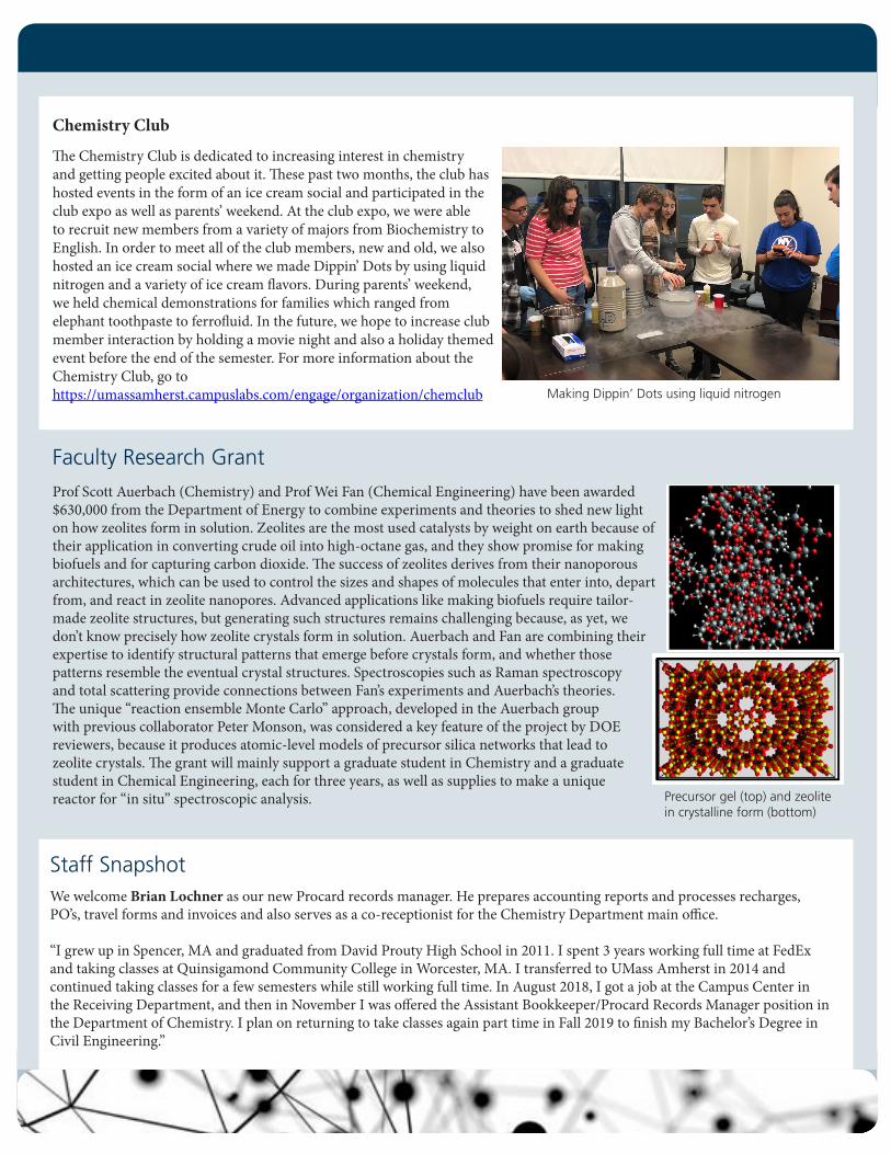
Chemistry Club
The Chemistry Club is dedicated to increasing interest in chemistry and getting people excited about it. These past two months, the club has hosted events in the form of an ice cream social and participated in the club expo as well as parents’ weekend. At the club expo, we were able to recruit new members from a variety of majors from Biochemistry to English. In order to meet all of the club members, new and old, we also hosted an ice cream social where we made Dippin’ Dots by using liquid nitrogen and a variety of ice cream flavors. During parents’ weekend, we held chemical demonstrations for families which ranged from elephant toothpaste to ferrofluid. In the future, we hope to increase club member interaction by holding a movie night and also a holiday themed event before the end of the semester. For more information about the Chemistry Club, go to https://umassamherst.campuslabs.com/engage/organization/chemclub Making Dippin’ Dots using liquid nitrogen
Prof Scott Auerbach (Chemistry) and Prof Wei Fan (Chemical Engineering) have been awarded $630,000 from the Department of Energy to combine experiments and theories to shed new light on how zeolites form in solution. Zeolites are the most used catalysts by weight on earth because of their application in converting crude oil into high-octane gas, and they show promise for making biofuels and for capturing carbon dioxide. The success of zeolites derives from their nanoporous architectures, which can be used to control the sizes and shapes of molecules that enter into, depart from, and react in zeolite nanopores. Advanced applications like making biofuels require tailor-made zeolite structures, but generating such structures remains challenging because, as yet, we don’t know precisely how zeolite crystals form in solution. Auerbach and Fan are combining their expertise to identify structural patterns that emerge before crystals form, and whether those patterns resemble the eventual crystal structures. Spectroscopies such as Raman spectroscopy and total scattering provide connections between Fan’s experiments and Auerbach’s theories. The unique “reaction ensemble Monte Carlo” approach, developed in the Auerbach group with previous collaborator Peter Monson, was considered a key feature of the project by DOE reviewers, because it produces atomic-level models of precursor silica networks that lead to zeolite crystals. The grant will mainly support a graduate student in Chemistry and a graduate student in Chemical Engineering, each for three years, as well as supplies to make a unique reactor for “in situ” spectroscopic analysis.
Faculty Research Grant
Staff SnapshotWe welcome Brian Lochner as our new Procard records manager. He prepares accounting reports and processes recharges, PO’s, travel forms and invoices and also serves as a co-receptionist for the Chemistry Department main office.
“I grew up in Spencer, MA and graduated from David Prouty High School in 2011. I spent 3 years working full time at FedEx and taking classes at Quinsigamond Community College in Worcester, MA. I transferred to UMass Amherst in 2014 and continued taking classes for a few semesters while still working full time. In August 2018, I got a job at the Campus Center in the Receiving Department, and then in November I was offered the Assistant Bookkeeper/Procard Records Manager position in the Department of Chemistry. I plan on returning to take classes again part time in Fall 2019 to finish my Bachelor’s Degree in Civil Engineering.”
Precursor gel (top) and zeolite in crystalline form (bottom)

Graduate Student Awards
Francesca Anson – Kuhn Fellowship (Hardy lab)Apoptosis, or programmed cell death, maintains tissue homeostasis. Hijacking dysregulated apoptotic pathways contributing to unregulated cell proliferation is an attractive and powerful therapeutic strategy for a myriad of diseases. To utilize the potential therapeutic roles of caspases, the executioners of apoptosis, to induce cell death in a targeted population, the Hardy and Thayumanavan labs developed a nanogel platform for intracellular caspase-3 delivery. As a joint student between the groups, I am working towards investigating caspase cargos beyond caspase-3. Further, we are exploring possibilities of caspase-nanogel and pro-survival antagonist nanogel combinatorial delivery. To maximize the potential of this synergistic platform, we are further probing caspase-nanogel conjugate self-assembly and delivery to elucidate mechanisms of cellular uptake, cargo release and potency.
Kingshuk Dutta – Rausch Fellowship (Thayumanavan lab) Designing Polymeric Delivery Kits for the Cytosolic Traceless Release of TherapeuticsBio-therapeutics have gained recent momentum in the post-genomic era due to their promise in controlling human disease. Proteins, small interfering RNAs (siRNAs), enzymes, and antibodies are classical examples of biomacromolecules studied under the class of biologics. In comparison to small molecule drugs, biologics offer higher specificity and potency with fewer side effects, leading to more predictable therapeutic outcomes. However, the dynamic and complex architecture of biologics makes them hard to stabilize in formulation and deliver to the target sites in active form. The loss of structural integrity/denaturation
during systemic circulation or upon storage presents one of the major hurdles in dealing with biologics. During my graduate study in the laboratory of Prof. S. Thayumanavan, I perceived these challenges as opportunities to interrogate and design stimuli-responsive polymeric nanogel systems for the delivery of such sensitive biomacromolecules. We envisaged the possibility of encapsulating biologics through both covalent and complementary electrostatic interactions. Recently, we have
successfully demonstrated intra-cellular delivery of proteins via a covalent self-immolative redox sensitive strategy by utilizing lysine moieties abundant in >85% of available proteins. Apart from designing protein delivery vehicles, I am investigating strategies for siRNA delivery that will ensure high encapsulation efficacy with low/no toxicity compared to conventional systems. In summary, the developed strategies could provide simple and robust delivery platforms potentially applicable to a broad range of bio-therapeutics.
Kristen Sikora – PPG Fellowship (Vachet lab)
Functionalized nanomaterials are attractive in applications such as sensing and drug delivery due to their tunable physical and chemical properties. My research in the Vachet lab focuses on developing novel measurement tools to monitor how the chemical and physical properties of functionalized nanomaterials dictate their uptake and distributions in vivo. With the support of the PPG fellowship, I will be working to develop new mass spectrometry imaging methods that simultaneously monitor how nanomaterial properties affect their distributions in vivo and how these properties influence the underlying biochemistry of the biological system. Developing these methods will provide important structure/function relationships that will lead to the design of better nanomaterials for biological applications.

Graduate and Postdoc Research
Quantitative Understanding of Lipid-mediated DNA Probe Modification on Mammalian Cell Membranes by Yousef Bagheri (You lab)
Modification of functional probes on live cell membranes have found a range of applications in bioanalysis, nanobiotechnology, cell biology, and medicine. Among different approaches, lipid-mediated cell membrane insertion of DNA probes has become popular for studying cell membrane signaling, intercellular interactions and tissue engineering. Lipid-mediated DNA probe works based on hydrophobic-hydrophobic interaction of the probe’s lipid tail with cell membrane phospholipids. This interaction provides efficient, biocompatible, and relatively simple probe modification of the cell membrane. However, one major limitation of this method is the short persistence time of the probe on the cell membrane. After insertion into the membrane, probes are internalized into the cell so they are no longer useful for membrane studies. Here, we systematically studied probe internalization pathways and have developed two methods to enhance probe persistence on cell membranes for long-term bioanalysis and regulation. By using these methods we are able to extend the probe's persistence on the cell membrane up to 12 hours.
BK Channel: The Mystery of Gating Mahdieh Yazdani (Jianhan Chen lab)
“I love this quote from Richard Feynman ‘...if we were to name the most powerful assumption of all, which leads one on and on in an attempt to understand life, it is that all things are made of atoms, and that everything that living things do can be understood in terms of the jigglings and wigglings of atoms’” says Mahdieh, a graduate student in Prof. Jianhan
Chen’s lab. She adds, “Our research involves simulating biomolecules which would let us look at the jigglings and wigglings of atoms closely! Recently we were able to crack a secret that had eluded science for many years: the gating mechanism of big potassium channels known as BK channels.” This ion channel is involved in many important physiological functions such as neuronal excitation and muscle contraction. Over the past 30 years, biophysicists were puzzled by the fact that the large central pore in BK channels seems to remain widely open in both activated and deactivated states. Similar ion channels usually contain structures that physically occlude the pore and prevent the passage of ions. Mahdieh explained, “Upon performing molecular dynamic simulation on the recently solved atomistic structures, Zhiguang Jia, a postdoctoral researcher in the lab, and I discovered how subtle changes in the channel’s central cavity lead to a change in hydrophobicity. As a consequence, the pore cavity becomes depleted of water, blocking the passage of potassium ions.” Now in Nature Communications, they demonstrate that this “hydrophobic dewetting” mechanism is how BK channels are regulated. This new finding provides knowledge toward developing new therapies to target the channel.
This discovery was awarded as one of the best poster presentations at the 15th Annual North-Eastern Structure Symposium at UConn Health and the 8th Annual Life Science Graduate Student Symposium at UMass Amherst.
Efficient modification of the MDCK cell membrane with lipid mediated DNA; effective inhibition of probe internalization up to 12 hours
The central pore of BK channels, (yellow), can become depleted of water, (blue), to block the passage of potassium ions, (pink spheres)

Push It, Pull It by Bin Zhao (You lab)
Mechanical forces play critical roles in a variety of biological processes including cell signaling, cell migration, and differentiation. One particularly interesting example is the recognition of antigen by T-cells in which the T-cell receptors use
mechanical contact-push and pull forces to make decisions about whether or not the cells they encounter are threats. Another interesting example is notch signaling activation where pull forces are exerted onto the notch receptors. Tensile forces are also involved in collective cell behaviors such as wound healing and embryonic development. The measurement of intercellular forces at cell-cell interfaces has been attracting remarkable attention; however, few techniques are available due to difficulty in modifying and controlling functional probes at complex cell-cell interfaces. We reported a novel strategy to construct membrane DNA tension probes (MDTPs) to visualize tensile forces at cell junctions (J. Am. Chem. Soc., 2017, 139 (50), pp 18182–18185). These lipid-modified probes can self-assemble onto cell membranes with high efficiency and stability. Upon experiencing tensile forces generated by neighboring cells, unfolding of the probes leads to a large increase in the fluorescence intensity. Compatible with
readily accessible fluorescence microscopes, these easy-to-use membrane DNA tension probes can be broadly used to measure intercellular tensile forces. We will further explore the applications of MDTPs in notch signaling and T-cell recognition.
Intracellular Imaging of the Magic Spot by Zhining “Jennings” Sun (You lab) I am a second-year chemistry graduate student working on developing RNA-based sensors in Professor You’s lab. My current project is to develop an RNA-based sensor that can detect and image guanosine tetraphosphate (ppGpp, also known as the “magic spot”) in live bacterial cells. This magic spot compound is a widespread signaling molecule involved in the stringent response, which is what bacteria cells do when they are under harsh conditions such as nutrient depletion. Under nutrient depletion, bacteria are facing severe threat of death, so they need to figure out a way to redistribute their limited nutrients from reproduction into survival. Now, this is where the magic spot comes in. As an alarmone, the magic spot will be synthesized in the cells soon after bacteria realize they are running out of “fuel,” and then the magic spot will start to regulate the cell’s transcriptional profile to promote survival until the environment improves.
There is currently no way to track these magic spots in living cells. To realize real-time in-cell imaging of the magic spot, I have designed a series of fluorescent RNA sensors, each containing a recognition unit, where the magic spot can bind to, and a reporting system, where a dye can bind and give off green fluorescence. Only after the magic spot binds to the recognition unit will the reporting system properly function, so when I see the bright green fluorescence coming out of the cells, I know there’s a a large number of magic spots produced, and the bacteria are starving.
Green Channel Bright Field Merged image
Highly Cited Prof. Vince Rotello and his former graduate student Chaekyu Kim (Ph.D. 2012) were recognized as “Highly Cited Researchers” by Clarivate Analytics for being in the top 1% of researchers in the world in their field in terms of the number of times their papers were cited last year. Dr. Kim is currently a Research Professor at Ulsan National Institute of Science and Technology in Korea. Two of their joint papers, review articles on gold nanoparticles for delivery applications and for chemical and biological sensing, have each been cited more than 1000 times!

Graduate OutreachA Story of Science Outreach by the Colombian Diaspora by Laura Castellanos (Vachet lab)A recurrent concern among researchers in Colombia is the small investment in science and technology, which leads to the general perception of success in science being unachievable. The numbers are highly discouraging. While in the USA around 200 PhDs per one million habitants graduate each year, in Colombia only 8 per one million do. Having such scarce numbers of researchers has created a common belief in Colombia that scientists are a rare and particular species, and that studying science
is not worthwhile. As a way to prove this myth wrong, a group of Colombian scientists in Massachusetts decided to create a program in the summer of 2015, and Clubes de Ciencia Colombia (Science Clubs Colombia) was created. This group aimed to design an intensive one-week course, or club, in an applied area of science and technology. The idea was that professional researchers (PhD students, postdocs, and professors) would travel to Colombia and work in local universities with a national researcher and a group of 20 high school students ranging from 14 to 17 years old. Through funding from the Colombian government, the program aimed to cover tickets, accommodations, transportation for the instructors, and a small budget for materials.
In 2015, these clubs became a reality, as the group in Massachusetts successfully obtained funding from Colciencias, which is the Colombian administrative department of science and technology, and SENA, which is the National Education Service, for 18 clubs in two Colombian cities. The first edition of the program was a complete success, leading the group’s partners to increase the budget to include 78 clubs in 2016, and more in 2017 and 2018. To date, the program has organized 247 clubs in 14 cities in Colombia. They have worked with almost 5000 students and have brought 265 researchers from all over the world to work with Colombian students during the one-week science clubs. I joined the group in the beginning of 2016, a few months after starting my PhD program in chemistry at UMass, and so far this has been an amazing experience. In 2016, I taught a club on intelligent textiles, using my previous knowledge and experience in nanoparticle (NP) synthesis. During the week-long club, I taught students how to synthesize and incorporate NPs in fabrics and showed them that these fabrics have antibacterial activity. For the 2017 and 2018 clubs, I moved into a coordinator role, as a new member of the board of directors. I traveled to Colombia both years to help coordinate the logistics of the clubs in my home city of Bucaramanga. Being in Bucaramanga each year has allowed me to witness the growth of the program and the progress and desire of some of our students, such as Edilmer, Alonso, and Brayan, who were students at my very first club in 2016. They each live about eight hours away from Bucaramanga in a rural town named Carcasí, and they have attended all three editions of the science clubs in Bucaramanga. Due to all their great efforts with their teacher Bernardo Rey, Edilmer, Alonso, and Brayan have developed a compelling research project that uses microalgae for the decontamination of cheese whey in Carcasí wastewater. Carcasí, their hometown, is known in Colombia for dairy production, and cheese whey contamination is an environmental problem in their rural community. Their research has led to them being awarded the “Samsung - Ideas for the Future Prize,” which gave them the opportunity to travel to Los Angeles, CA in 2017 to represent Colombia in the Intel-ISEF (International Science and Engineering Fair). In 2018 Edilmer, Alonso, and their teacher Bernardo represented Colombia in Mostratec, the biggest Latin-American science fair, hosted each year in Brazil. They won the first prize in the environmental science category, which was the first time in history a team from Colombia won first prize. Upon returning to Colombia, a special ceremony was held in their honor in Bucaramanga, and Guy Djoken, the CEO of the UNESCO Center for Peace, awarded them a special prize. Edilmer, Alonso, and Bernardo will be travelling to Washington DC in the summer of 2019 to attend a science summer camp. Our group is working hard to find the resources to bring them to Massachusetts so that they can learn more about the research environment here in the USA. Edilmer, Alonso and Brayan’s story is just one example of all the stories we have encountered over the time the science clubs have been running. It is very gratifying to see that our vision for bringing the Colombian diaspora back to our country is a reality, but we have been even more surprised to see how much we have learned from our students. We, as a group, hope to continue to learn from these amazing students and will continue to strive to make science accessible to more and more students in Colombia.

Annual Metawampe Hikeby Craig Martin
The 111th Metawampe Hike over Mount Toby on November 4th found beautiful weather and a great turnout this year (more than 50 hikers)! Led by organizers Dave Adams (class of 1967 and retired Senior Lecturer in Chemistry) and Bob Sabola, students, faculty and staff across the university were joined this year by some very friendly folks from a Connecticut hiking club! This UMass tradition, begun in 1908, traverses about five miles from Leverett to Sunderland, and starts off with the ascent of (a very full this year) waterfall. It then continues with a stop at the Metawampe Cabin, built in 1921 by UMass faculty, including Chemistry Professor Charles Peters (class of 1897) and then President Kenyon Butterfield. Although the cabin is now in significant decay, the stop provides an important remembrance of those who have gone before us. The hike peaks with a stop at the Mount Toby fire tower, offering views to UMass and beyond. Although the weather was dry (and just the right temperature for a Fall hike!), the very wet year preceding left unusually beautiful rivulets and streams for the hikers to traverse and enjoy. Keeping with tradition, the day culminated with an all you can eat chicken pot pie dinner, served up by members of the Sunderland Congregational Church — very New England!
The editorial staff thanks those who sent in accomplishments and story ideas. Please email comments, achievements, and ideas for our next issue to [email protected].