the reciprocal infection of ducks and chickens with...
TRANSCRIPT

The Reciprocal Infection of Ducks and Chickens with Tumor- Inducing Viruses*
iv. Duran-Reynals, M. D.**
With notes on histopathological findings by Henry Bunting, M. D.
(From the Departments o/ Bacteriology and Pathology, Yale University School o/ Medicine, New Haven, Conn.)
(Received for publication February 8, 1942 )
T h e present report considers the variat ions w h i c h take place in the causal virus fo l lowing inject ion of duckl ings wi th the Rous sarcoma of chickens, and wi th fur ther variat ions fo l lowing the infection of chickens wi th viruses f rom the resul t ing strains of duck tumors . T h e s tudy of the h e m o r r h a g i c lesions often developing in these infections will be t aken up in detail in ano the r paper. T h e grea t susceptibili ty of chicks to ch icken t u m o r viruses injected intra- venously (4, 5) contrasted wi th the resistance of adul t chickens to s imilar injections (3, 27) suggested the present invest igat ion. A p re l iminary note on the subject has been publ i shed (6) .
In I912 , Murphy and Rous (2i) grew chicken tumor I in the membranes of developing duck embryos, but transplan- tation of the tumors obtained into adult ducks failed. Purdy (26) also failed to infect Khaki Campbell ducklings and ducks by intramuscular injection of large amounts of extracts from Rous tumors and Mill Hill 2 endothelioma, and Des Ligneris (2) had the same experience with adult ducks and geese injected with minced tumor tissue. However, Purdy (26) propagated the Rous sarcoma through 5 serial generations of Khaki Campbell ducklings (and presumably could have con- tinued this propagation indefinitely) by injecting large amounts of minced tumor tissue (o.5 cc.). The age of the host was a very important factor. In i-day-old ducklings, transplantation was invariably successful, and apparently the same was true for 4-day-old ducks; only half of the ducks 11 to 14 days old were found susceptible, while adult ducks were wholly re- sistant. The Mill Hill 2 endothelioma, also injected under similar conditions, induced tumors which regressed after one week of growth. Fujinami and Hatano in i929 (7) re- ported the successful transplantation of the Fujinami sarcoma through 40 generations of ducks and obscrved that the tumor grew remarkably well in such hosts. Presumably cells were used for transplantation in these experiments. Gye (I4) con- firmed these observations on 20 Aylesbury ducklings. He further induced tumors in half-grown ducks by means of a filtrate, but these tumors always regressed. Using the Khaki Campbell breed the same author (13, 14) propagated the Fujinami tumor through I8 serial generations of ducklings by means of filtrates or ceil suspensions. Half-grown or o!dcr ducks formed tumors in response to injection of cell suspen- sions or filtrates, but these tumors were always followed by
e This investigation was aided by a grant from The Jane Coffin Childs Memorial Fund for Medical Research.
** With the technical assistance of Mr. J. Patti.
regression. Purdy (24) , by using larger doses (2 cc. of minced tumor) than Gye did, found it possible to propagate the tumor through an indefinite number of generations of adult ducks. In some cases, however, the tumors regressed and tile host recovered. These ducks werc found later to be resistant to reinoculation with the same tumor, but no resistance against the Fujinami tumor was found in ducklings in which the Rous tumor grew for a time and later regressed (25).
T w o points concern ing these d u c k tumors should be emphas ized . T h e first is tha t in no case, even after being carr ied for 4o genera t ions in ducks, did the virus fail to induce sarcomas in adu l t chickens. T h e second is tha t a genera l i zed neoplastic disease was never observed in ducks; the only evident mani - festation of the virus activity was a rapidly g r o w i n g t u m o r w h i c h ki l led the host in such a short t ime tha t Gye suspected a toxic action (~4)" These facts indi- cate that var ia t ion of the ch icken virus was not ob- served in any instance.
MATERIALS AND METHODS
Both Rous and F u j i n a m i sarcomas were employed in the present invest igat ion. T h e Rous sarcoma was used rout inely, the F u j i n a m i only occasionally. Both tumors were serially g r o w n in adu l t Bar red P l y m o u t h Rock chickens. Passages were m a d e by in jec t ing a i : 5 suspension of t u m o r cells in 0.85 per cent saline solut ion into the breast. N o t u m o r older t han Io days was ever employed. Berkefe ld N candles were used for f i l t rat ion of the t u m o r extracts. D u c k s of the P e k i n breed were genera l ly used, o ther breeds being employed only in a f ew exper iments . T h e y all
came f rom the same f a r m ? T h e in t ravenous injec-
tions in ducks and chicks were m a d e by way of the
j ugu la r vein; subcutaneous and in t r amuscu la r injec- tions by way of the breast. Da t a on the dosage and
s t rength of the t u m o r p r e p a r a t i o n s in jected wil l be
g iven in the text. Tissue sections were stained wi th
hematoxy l in and eosin unless o therwise indicated.
1 The cooperation of the Messrs. Robinson of the Carman River Duck Farm, Brookhaven, L. I., in securing these breeds of ducks is acknowledged with pleasure.
343
Research. on May 8, 2018. © 1942 American Association for Cancercancerres.aacrjournals.org Downloaded from
344 Cancer Research
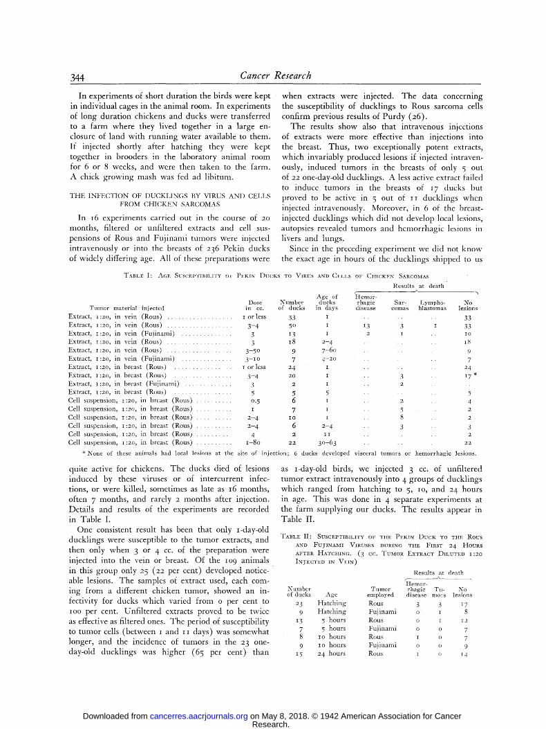
In experiments of short duration the birds were kept in individual cages in the animal room. In experiments of long duration chickens and ducks were transferred to a farm where they lived together in a large en- closure of land with running water available to them. If injected shortly after hatching they were kept together in brooders in the laboratory animal room for 6 or 8 weeks, and were then taken to the farm. A chick growing mash was fed ad libitum.
T H E I N F E C T I O N O F D U C K L I N G S BY VIRUS A N D C E L L S
F R O M C H I C K E N S A R C O M A S
In i6 experiments carried out in the course of 20 months, filtered or unfiltered extracts and cell sus- pensions of Rous and Fuj inami tumors were injected intravenously or into the breasts of 236 Pekin ducks of widely differing age. All of these preparations were
Tumor
Extract , 1:20,
Extract , 1:20~
Extract , 1:20,
Extract , 1:20,
Extract , 1:2(3,
Extract , 1:20,
Extract , 1:20, in
Extract , I :20 , in
Extract , I :20 , in
Extract , I :20 , in
Cell suspension,
Cell suspension,
Cell suspension,
Cell suspension,
Cell suspension,
Cell suspension,
when extracts were injected. The data concerning the susceptibility of ducklings to Rous sarcoma cells confirm previous results of Purdy (26).
The results show also that intravenous injections of extracts were more effective than injections into the breast. Thus, two exceptionally potent extracts, which invariably produced lesions if injected intraven- ously, induced tumors in the breasts of only 5 out of 22 one-day-old ducklingsl A less active extract failed to induce tumors in the breasts of 17 ducks but proved to be active in 5 out of i i ducklings when injected intravenously. Moreover, in 6 of the breast- injected ducklings which did not develop local lesions, autopsies revealed tumors and hemorrhagic lesions in livers and lungs.
Since in the preceding experiment we did not know the exact age in hours of the ducklings shipped to us
TABLE I: AGE SUSCEPTIBILITY OF PEKIN DUCKS TO VIRUS AND CELLS OF CHICKEN SARCOiXlAS
.Age of Dose Number ducks
material injected in cc. of ducks in days
in vein (Rous) . . . . . . . . . . . . . . . . . . I or less 33 x
in vein (Rous) . . . . . . . . . . . . . . . . . . 3 - 4 50 :E
in vein ( F u j i n a m i ) . . . . . . . . . . . . . . 3 13 I
in vein (Rous) . . . . . . . . . . . . . . . . . . 3 I8 2 - 4
in vein (Rous) . . . . . . . . . . . . . . . . . . 3 - 5 o 9 7 - 6 0
in ve in ( F u j i n a m i ) . . . . . . . . . . . . . . 3 - I o 7 4 - 2 o
breast (Rous) . . . . . . . . . . . . . . . I or less 24 I
breast (Rous) . . . . . . . . . . . . . . . 3 - 4 20 I
breast ( F u j i n a m i ) . . . . . . . . . . . . . 3 2 I
breast (Rous) . . . . . . . . . . . . . . . . 5 5 5
1:20, in breast (Rous) . . . . . . . . . . 0. 5 6 I
1:20, in breast (Rous) . . . . . . . . . . I 7 I
1 : 2 0 , in breast (Rous) . . . . . . . . . . 2 - 4 I0 I
1:2o, in breast (Rous) . . . . . . . . . . 2 - 4 6 2 - 4
1:20, in breast (Rous 2 . . . . . . . . . 4 2 11
1:20, in breast (Rous) . . . . . . . . . . 1 -8o :22 3 0 - 6 3
r FI emor- rhagic
disease
�9 .
I3 3 2 I
�9 .
Results at death &
Sar- l ,ympho- comas blastomas
]2~To lesions
33
33 IO
I8
9
7 24
i 7 " �9 .
5
4 2
2
3 2
22
None of these animals had local lesions at the site of inject ion; 6 ducks developed visceral tumors or hemorrhagic lesions.
quite active for chickens. The ducks died of lesions induced by these viruses or of intercurrent infec- tions, or were killed, sometimes as late as i6 months, often 7 months, and rarely 2 months after injection. Details and results of the experiments are recorded in Table I.
One consistent result has been that only I-day-old ducklings were susceptible to the tumor extracts, and then only when 3 or 4 cc. of the preparation were injected into the vein or breast. Of the io 9 animals in this group only :z 5 ( 2 2 per cent) developed notice- able lesions. The samples of extract used, each com- ing from a different chicken tumor, showed an in- fectivity for ducks which varied from o per cent to IOO per cent. Unfiltered extracts proved to be twice as effective as filtered ones. The period of susceptibility to tumor cells (between I and i i days) was somewhat longer, and the incidence of tumors in the 23 one- day-old ducklings was higher (65 per cent) than
as >day-old birds, we injected 3 cc. of unfiltered tumor extract intravenously into 4 groups of ducklings which ranged from hatching to 5, io, and 2 4 hours in age. This was done in 4 separate experiments at the farm supplying our ducks. The results appear in Table II.
TABLE II : SUSCEPTIBILITY OF THE PEKIN DUCK TO THE Rous
AND FU)-INAMI VIRUSES DURING THE FIRST 24 HouRs
AFTER HATCtIING. (3 CC. TUMOR EXTRACT DILUTED 1:20
INyECTED IN VEIN)
Results at death
ri•Iemor- Number Tumor rhagic Tu- No of ducks Age employed disease mors lesions
23 H a t c h i n g Rous 3 3 17
9 H a t c h i n g F u j i n a m i o 1 8
I3 5 hours Rous o I 12
7 5 hours F u j i n a m i o o 7
8 lO hours Rous 1 o 7
9 Io hours F u j i n a m i o o 9
15 24 hours Rous , o I4
Research. on May 8, 2018. © 1942 American Association for Cancercancerres.aacrjournals.org Downloaded from

Duran-Reynals--Reciprocal Infection of Ducks and Chickens 345
I t is c lear tha t the g rea te s t suscept ibi l i ty of the d u c k s
to the t u m o r vi ruses was f o u n d at h a t c h i n g a n d di-
m i n i s h e d r ap id ly d u r i n g the first hou r s of life.
T h e lesions i n d u c e d by the in jec t ion into d u c k l i n g s
of extracts f r o m the Rous a n d F u j i n a m i t u m o r s w e r e
e i the r h e m o r r h a g i c or neoplas t ic , a n d in d e s c r i b i n g
each g r o u p a d i s t inc t ion will be m a d e b e t w e e n im- mediate a n d late lesions.
either grossly or microscopically. The animals died from 34 to lOI days after injection. In 8 other cases, recorded as nega- tive in Tables I and II, indications of healed hemorrhagic lesions were found.
Neoplastic ledons.--In 7 ducks, immediate sarcomas de- veloped which killed the animals in io to 2o days without obvious generalized lesions. Late tumors developed in 4 ap- parently healthy ducks which were killed 4o days to 7 months after injection. The tumors induced by the injection of cells fell within the former group. These tumors often killed their
Fro. i. Hemorrhagic disease in a duckling injected intravenously I day after hatching with extract of Rous tumor, and dying 14 days later. Note minute tumor nodules in the pancreas and neck.
Fie. 2. Section of periosteal giant cell sarcoma from a bone of the face in a duckling injected intravenously I day after hatching with filtrate of Rous tumor, and killed 40 days later. Mag. X i2o.
fAll photographs were taken by Mr. H. J. Reynolds.]
Hemorrhagic tesions.--An immediate and acute type of hemorrhagic disease was observed in I5 ducklings. The disease was similar to that described in chicks (4), although differing from it in minor features (Fig. I) . Small viscid tumor nodules, mostly around the injected vein, were noticed in 5 cases. Death occurred in from IO to 25 days after injection. The microscopic study showed that blebs were in most cases without any associa- tion with neoplastic tissue. A late type of hemorrhagic disease, never observed in chicks, occurred in 5 ducks. The liver was the only organ affected; it was enlarged, showed pronounced gross and microscopic signs of degeneration, and large organized clots bulged from its surface. No tumor tissue was apparent
hosts from 5 to 20 days after injection usually without general- ized lesions, thus confirming a previous finding by Gye (14). In some cases, however, invasion of the liver by direct exten- sion of the growth was observed.
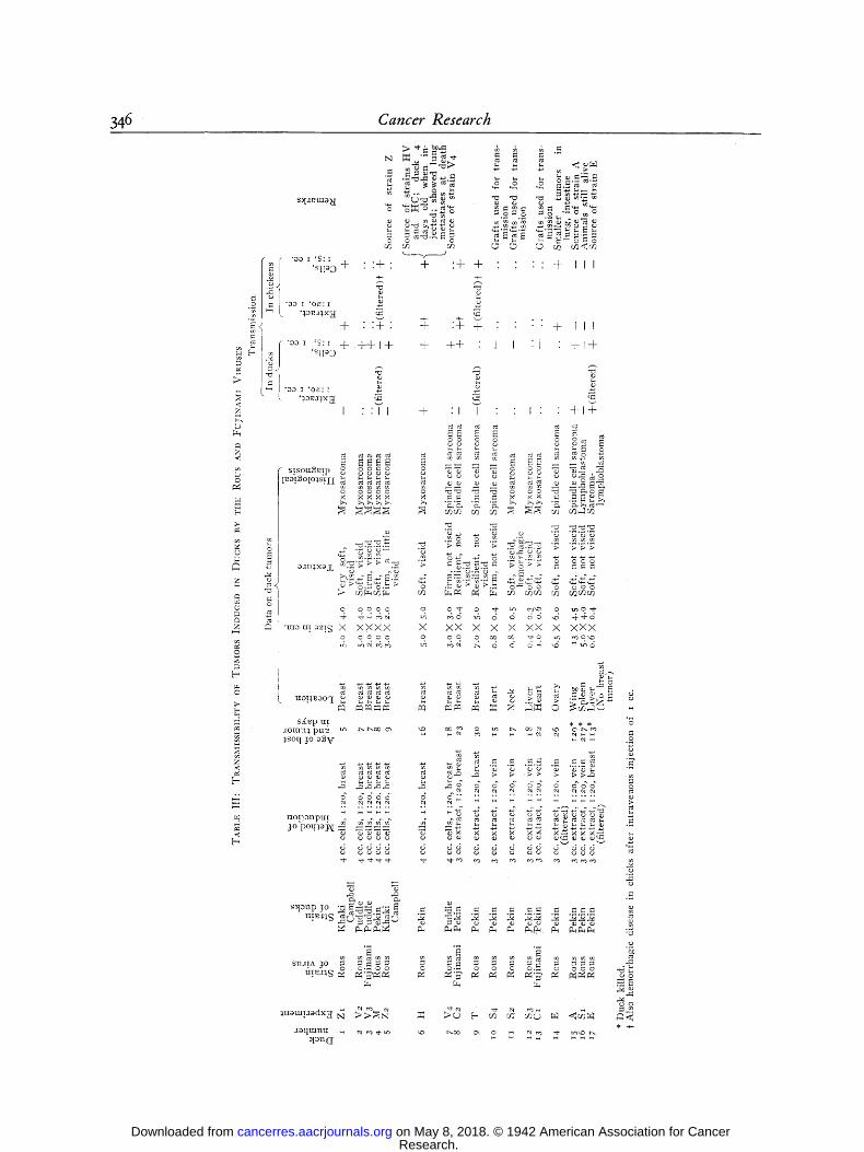
D a t a c o n c e r n i n g the t u m o r s in w h i c h t r a n s m i s s i o n
was a t t e m p t e d a re g i v e n in T a b l e III . T h e f e w re-
m a i n i n g t u m o r s we re , w i t h o n e excep t ion , i m m e d i a t e
m y x o - or c o m p a c t sp ind le cell s a r c o m a s c o r r e s p o n d i n g
w i t h those of T a b l e III . T h e excep t ion was a t u m o r
in a d u c k of e x p e r i m e n t E , w h i c h was in jec ted in t ra -
Research. on May 8, 2018. © 1942 American Association for Cancercancerres.aacrjournals.org Downloaded from
346 Cancer Research
o ~ ~ , - ~ . ~ ~ ~ �9 ,..,_,.~,-~ ~ = o �9
f ( " a o r ' ~ : [ u " - - " v - ' - - ~ .
| = I . ! . [ ~ 0 + " : + ' + : 4 - + �9 �9 " + I I
. -
~ ) + : : + : + : + + " 4- I I
§ § ++ " ' ' " + §
�9 o o ~ ,o~:~ ~ ~
• I " " f I + " i J " " l " +i+
~ " ~ ~ "~ ~ .~ -~.~-~ ~ ~r~ .~ ~ = ~ ..~.--= .~ .~.~.~
- ~>~.~'~.~ ~o . ~ > ~ > . ~ o -= ~ o o o o o
o o o o o o o ' ~ - o "~- 1-o c o ' o o i n o ,,~'-
i "mau[~z~.~ X X X X X X X X X X X X X X X X X c r o o o o o 0 o c ,,a'- o w~ ~.,~ o , , ~
o~ ,,-, , ~ d 4 4 ~ 4 4 .~ 6 d 6,.-: ~ " - ' ~ d
5 sXep m % * *
e4 ~l m N ~ c ~ ~i c 4 ~ i ~
. . ~ ,,...q
�9 .-m'-' ~ ~'-~..m "-~ ~ o ca r r ~ ~ r ~ r r 0J t~
o
~ o
: , t a n ( I
Research. on May 8, 2018. © 1942 American Association for Cancercancerres.aacrjournals.org Downloaded from

Duran-Reynals--Reciprocal Infection of Ducks and Chickens 347
venously at the age of i day with 4 cc. of filtered extract of Rous sarcoma. The growth was a small tumor, histologically a giant cell sarcoma (Fig. 2), apparently arising from the bone of the face. It was found when the animal was killed 4 ~ days after the injection and was in all probability a late lesion.
The results concerning the transmissibility of the duck tumors to adult chickens, to chicks and ducklings are also summarized in Table III. The animals in- jected in the different groups have been listed by their age which, excepting duck No. 6 of experiment H, is the same as the age of their tumors. The material for each injection was tested by way of the vein of the breast on a m i n i m u m of 3 ducklings, 3 to 5 days old, and 2 chickens. Chicks were occasionally injected.
It is clear from Table III that extracts from im- mediate tumors (virus or cell-induced tumors which grew in ducks for 3 ~ days or less) did not induce tumors in ducks whereas they effectively induced tumors in chickens or hemorrhagic disease in chicks. It will also be noticed that most of the immediate duck tumors had the gross and microscopic features of the original Rous sarcoma. An exception to this rule was duck tumor H, which differed from the others in that the animal was 4 days old when injected and showed metastases at death. It was because of this deviation that transmission was attempted.
On the other hand, extracts from the late sarcomas from ducks Nos. 15 and 17 of experiments A and E (which grew in their hosts for 121 and 113 days, respectively) were active for ducks but not for chick- ens. Lymphoblastoma of duck No. i6 of experiment S I has not been successfully transmitted to either. These 3 tumors and the giant cell sarcoma of experi- ment E had features which distinguished them com- pletely from the original Rous tumors. Extreme youth of the host and large virus dosage, factors indispensable for the infection of the first generation of ducks, were no longer required for the infection of the second generation. All of the r 4 ducks injected with filtrates of tumors A and E (from ducks 15 and I7) were from 3 to 5 days of age, and the amount of extract injected was only i c c . Yet all developed rapidly growing generalized tumors. Furthermore, the route of infec- tion was no longer an important factor, since the extracts were equally effective when injected in the vein or in the skin.
Table II[ also shows that transmission by cell suspen- sion was easy for all tumors regardless of their age. Following the inoculation of 9 out of i i immediate and late tumors, 2o out of 3 o ducks injected developed tumors. Transmission failed with 3 immediate tumors when graft ing was resorted to. The special conditions of transmission of tumor E of duck No. 17 will be taken up later.
The results of experiment E warrant special attention. Here ducks of the same batch, infected with the same filtrate of Rous virus, developed the following varieties of tumors: spindle cell sarcoma of the ovary; giant cell sarcoma of the bone; and a sarcoma-lymphoblas- toma of the liver. Moreover, other ducks of the same series developed a typical hemorrhagic disease, and the few tumors accompanying the hemorrhagic lesions were myxosarcomas. Therefore, 5 varieties of lesions were induced by the same virus preparation.
STUDY OF THE DUCK STRAINS OF TIlE Rovs TUMOR
As indicated in Table III, tumors from 5 ducks gave origin to 5 strains of duck tumors after serial passage through ducks of the same or other breeds. Strain H was split into strains H C and HV. The latter strains were the most thoroughly studied.
In Fig. 3 are expresscd the conventional signs which will be used in the graphic representation of the passages of the strains
[ ~ ADULT CHICKEN [ ] @ DISCRETE GENERALIZATION] .
I Fo~ I T U ~o~,.
O ADULT DUCK I ~ GENERA . . . . . . . . WITH | BONE INVOLVEMENT
C) DUCKLING [ ] ~ H EMOR-I~HAGIC DISEASE
J--~ @ PRIMAI~Y TUMOR ~ @ LEUCOSIS
[ ] PRIMARY TUMOP_ N OSTEOPETROSI5 FOLLOWED BY REGRESSION
Fw.. 3.--Conventional signs used in transmission diagrams and descriptive charts. See text.
either in ordinary or in three dimensional charts. In some cases signs representing two different sorts of lesions will be combined in the same individual. The small figure underneath the signs indicates the number of days subsequent to injection at which the animal died or was killed. Some charts contain additional pertinent indications. With a few exceptions tumor extracts at i:2o--filtered or not--and cell suspensions at 1:5 were employed for transmission, the former being injected in the vein and the latter in the breast. Both materials were pre- pared in saline solution and I cc. was injected routinely.
Strain d . - - T h e source tun-mr (No. 15 of Table III) developed in the wing of a Pekin duck injected intravenously wi thin the first 2 4 hours after hatching with 3 cc. of unfiltered extract of Rous tumor diluted i :2o with saline. The animal was killed 12o days after injection, and a large, rapidly growing spindle cell sarcoma composed of nodules of cells arranged in whorls and separated from each other by large bands of collagenous tissue (Figs. 4 and 5) was found.
The tumor was carried through two more generations of ducklings, all of which developed a generalized disease. Fol- lowing this, passages were purposely discontinued. A graphic summary of the tests carried out on ducks and chickens is given in Fig. 6. The gross and microscopic lesions induced
Research. on May 8, 2018. © 1942 American Association for Cancercancerres.aacrjournals.org Downloaded from

348 Cancer Research
in ducklings of the 2nd and 3rd passages were identical with those induced by Strains H V . and t tC to be studied later, and will therefore be described with them. It will suffice to say here that filtrates injected in the vein induced widespread tu- mors, together with hemorrhagic lesions, mostly in the skin
resistant, 4 developed slowly growing sarcomas which later regressed, and I developed a progressively growing sarcoma which was followed by generalization. The two other chickens died 17 and ioo days after injection. The former showed signs of an acute infection and the latter presented a large
F~c;. 4.--Collagenous tumor from the wing of a duck injected intravenously I day after hatching with extract of Rous tumor and killed 4 months later. Source of strain A. Masson stain. Mag. X 2o.
FIG. 5.--Section of the same tumor as in Fig. 4- Mag. X 3 ~
S T R A I N A
CELLS IN B R ~ [ BREAST
lSO EXTRACT IN EXTRACT IN
~EAST r~.jEAST EXTR, b, CTIIN BR,
[ I I ] 1 I1 1 NNF .37 17 .~ 75 7S
F I L T g ; N ; I N
2Z~14 IO IS I ~ FILTRATE IN ~" FILTRATE IN ~ T
FILTRATE I'M 10o 14o 30
90 JO0 J40 I00 I00
FIG. 6
and digestive tract, but also in the skull. Cell suspensions in- jected in the breast induced large collagenous tumors followed by the same generalized lesions. Death occurred from I o to 27 clays after injection.
Of the t7 adult chickens injected in the breast with ex- tracts or cells from the 2nd and 3rd passages, i0 were found
liver in which microscopic examination disclosed a very heavy periportal infiltration by lymphoblast-like cells. Chicks injected in the breast with cells developed rapidly growing but localized and well encapsulated tumors, which killed their hosts in 2 to 3 weeks. Those injected with filtrates in the vein showed no lesions when killed 22 days later.
Research. on May 8, 2018. © 1942 American Association for Cancercancerres.aacrjournals.org Downloaded from

Duran'Reynals--Reciprocal ln/ection o/ Ducks and Chickens 3 4 9
Comment.--Despite the i ncomple t enes s of t he s t udy
it is c lear tha t the source tun-mr b red t rue h is to logica l ly
or in ra te o f g r o w t h . I t is also c lear t ha t t he abi l i ty
of the v i rus to i n d u c e i m m e d i a t e sa rcomas in ch i ckens
and ch icks was to a l a rge ex t en t lost. Ana lys i s of
the da ta indicates tha t the possibil i ty exists t ha t the
virus i n d u c e d l y m p h o m a t o s i s in one ch i cken .
Strain E . - - T h e source t u m o r ( N o . 17 of T a b l e I I I )
was a smal l g r o w t h tha t was f o u n d b u r i e d in the
l iver of a P e k i n d u c k ki l led i i 3 days a f te r in jec t ion
wi th 3 cc. of f i l trate of Rous t u m o r ~:2o in the breas t
2 4 h o u r s a f te r h a t c h i n g . T h e r e was no breas t t u m o r .
T h e l iver g r o w t h was soft, no t viscid, a n d well en-
capsula ted , a n d the pancreas s h o w e d several small , flat, neoplas t ic nodu les .
and that others developed an acute disease of doubtful etiology (6th passage B) may have had some influence on the results. lqut it may well be that, as is often observed in tumor trans- plantatian, something inherent in the tumor or virus itself was the cause of the failure.
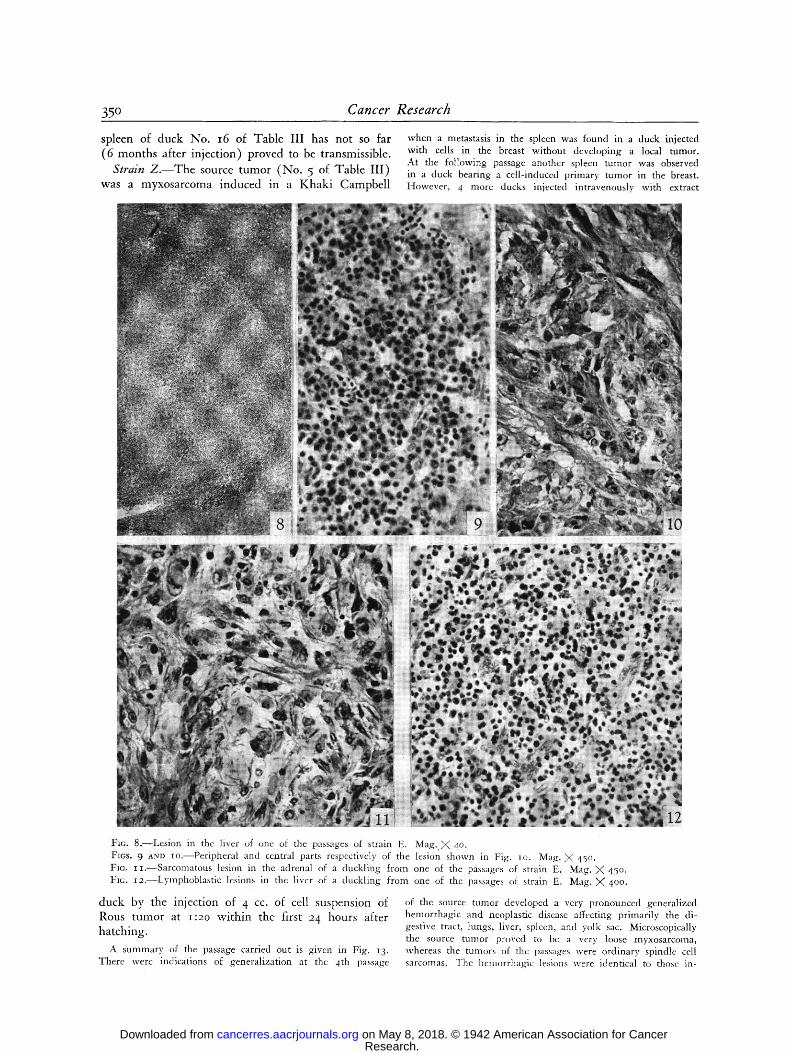
No histological study was made of the primary tumor be- cause it was used in its entirety for transmission. However, the nodules from the pancreas consisted of zones of fibroblastic cells surrounded by a ring of lymphoid and lymphoblastic cells. The same mixed lesions were observed in the passages eithcr in the viscera (Figs. 8 to Ia) or in the skin in the only case where a tumor developed in this organ. Moreover, nodules of lymphoid cells such as those occurring in lymphomatosis, without any fibroblasts, were present in viscera (Fig. ]2) while in one duck of the 6th passage a tumor consisting of sarcoma ceils was observed in one adrenal (Fig. i i). Polymorpho- nuclcars and myelocytcs either scattered or in a small cluster were also present in the mixed lesions.
STRAIN E.
PASS I ?. 3 4
VEIN
Tj~I3 I VEIN BI~EAS ,30
~ 9 0 [ FI LTRATi
5ouncE TU MOP..
~ 154 BI~F.AS'I"
B REASTt IrsJ 75
5 6 [ A ] -, [ou~Ksxo~ o~o-J bu~ 7s ~o~oa
-~2o
| 1 7 6 vE'N D2, i
-G 20 s Z 2 BREAST TUMOR
F]G. 7
CELLS IH BREAST
EXTRACT IN VEIN
EXTRACT IN VEIN
t 6 ~ UCKS 3 O.OLD]
~ $A I~COIV~A |N ADRENAL
0 2 o 3 cr 6 ?
EXTRACT J ~,../6 .~ Gz4 r~ YE3~ - ~ 3 3 7cc'L_~S3 -~7o M.JS~
-J~7o -[~7o
A summary of the passagcs is given in Fig. 7- Throughout the passages intravenous injections of tumor filtrates induced a disease affecting the spleen in every case. The liver was fre- quently affected while the lung, intestine, and pancreas showed only occasional lcsions. An adrenal tumor was found in one instancc. The organs heavily involved were studded with whit- ish, soft, nonviscid nodules from less than I mm. to 7 ram. in diameter and had rather diffuse boundaries. Throughout the passages, filtrates or cell suspensions injected into the breast never induced a local disease except in one duck. This bird was one of II ducks used in the 5th passage. Eight ducks in ocher passages injected in the breast developed visceral lesions. With few exceptions the tumors grew very slowly. Passage ducks rarely died of the tumor growths, but were sacrificed from time to time for passage material. However, in a few ducks killed as soon as 8 days after intravenous injection, small growths had already appeared in the spleen. Filtrates were in- effective in inducing lesions in chickens or chicks. The strain was lost at the 6th passage due to obscure reasons. The fact that some of the ducks were killed too soon (6th passage A)
Comment.--The t u m o r b red t rue ( a ) h is to logical ly ,
( b ) in its rate of g r o w t h , a n d ( c ) in its inabi l i ty to
i n d u c e local g r o w t h s , the s u b c u t a n e o u s rou t e b e i n g
as effect ive as t he i n t r a v e n o u s one in p r o d u c i n g typica l
visceral lesions. T h e t u m o r , w i t h o u t a n y doub t , con-
t a i n e d bo th s a r c o m a t o u s a n d l y m p h o b l a s t o m a t o u s ele-
men t s , one of t h e m occas iona l ly t a k i n g the u p p e r h a n d
w i t h c o m p l e t e exc lus ion of the o the r . W h e t h e r these
cells w e r e i n d e p e n d e n t or d e r i v e d f r o m each o t h e r
c a n n o t be dec ided . T h e p o i n t o f in teres t , h o w e v e r , is
t ha t the Rous v i rus has g i v e n rise to b o t h types of
neoplas t ic g r o w t h in t he s a m e a n i m a l . T h e poor
t r ansp lan t ab i l i t y of the t u m o r w o u l d be in l ine w i t h
w h a t is k n o w n a b o u t t h e dif f icul ty in t r a n s m i t t i n g
l y m p h o b l a s t o m a t o u s t u m o r s . I t is p e r t i n e n t to recal l
in this c o n n e c t i o n t h a t the l y m p h o b l a s t o m a f r o m the
Research. on May 8, 2018. © 1942 American Association for Cancercancerres.aacrjournals.org Downloaded from
35 ~ Cancer Research
spleen of duck No. i6 of Table III has not so far (6 months after injection) proved to be transmissible.
Strain Z. - -The source tumor (No. 5 of Table III) was a myxosarcoma induced in a Khaki Campbell
when a metastasis in the spleen was found in a duck injected with cells in the breast without developing a local tumor. At the following passage another spleen tumor was observed in a duck bearing a cell-induced primary tumor in the breast. However , 4 more ducks injected intravenously with extract
Fro. 8 . - -Lesion in thc liver of one of the passages of strain E. Mag. X 40. Fins. 9 AND Io.- -Per ipheral and central parts respectively of the lesion shown in Fig. Io. Mag. X 45o. Fro. I I . - -Sarcomatous lesion in the adrenal of a duckling f rom one of the passages of strain E. Mag. X 45o. FIc. ~2.--Lymphoblast ic lesions in the liver of a duckling from one of the passages o~ strain E. Mag. ),( 4oo.
duck by the injection of 4 cc. of cell suspension of R o u s t un -mr a t I : 2 0 w i t h i n t h e f i r s t 24 h o u r s a f t e r
h a t c h i n g .
A summary of the passage carried out is given in Fig. 13. There were indications of generalization at the 4th passage
of the source tumor developed a very pronounced generalized hemorrhagic and neoplastic disease affecting primarily the di- gestive tract, lungs, liver, spleen, and yolk sac. Microscopically the source tumor proved to be a very loose myxosarcoma, whereas the tumors of the passages were ordinary spindle cell sarcomas. The hemorrhagic lesions were identical to those in-
Research. on May 8, 2018. © 1942 American Association for Cancercancerres.aacrjournals.org Downloaded from

Duran-Reynals--Reciprocal Infection of Ducks and Chickens 351
duced by the Rous virus in ducklings. The series was pur- posely stopped at the 5th passage.
Comment.--Since exper ience g a i n e d w i t h the o the r
strains ob ta ined by serial cell passages has s h o w n tha t the occur rence of gene ra l i za t ion is a clear i nd ica t ion
of the t r a n s f o r m a t i o n of the ch icken strain into a d u c k strain, we believe tha t such t r a n s f o r m a t i o n has t aken place in the p resen t case. In v iew of the fa i lure of P u r d y (:26) to infect K h a k i C a m p b e l l d u c k l i n g s w i t h extracts of Rous t u m o r it is p e r t i n e n t to po in t ou t tha t genera l i za t ion in ou r s t ra in was ob ta ined in P e k i n
ducks since K h a k i Campbe l l s were no t available at the t ime.
Strain V . - - T h e source t u m o r ( N o . 7 of T ab l e I l l ) was a sp indle cell sa rcoma i n d u c e d in a P u d d l e d u c k by the in jec t ion in the breast of 4 cc. of cell suspens ions of Rous t u m o r at i : ao w i t h i n the first 2 4 hours after ha tch ing .
STRAIN Z PASS I 2 3 4 5 KHAKI KHAK I KHAKI P E K I N PEKI N CAMPBELL CAMPBELL CAM.PBELL
PEKIN
14- CELLS N EXTRACT CELL$I 5 1 ~ 2
EXTRACT - IN VEIN NO BREAST TUMOR CE'LLS - IN BR.EAST SPLEENTUIV~OR
SPLEEN TUN~,OR
-Co CELLS ~ 1 6
�9 . - ' ( ~ 1 1
FIG. 13
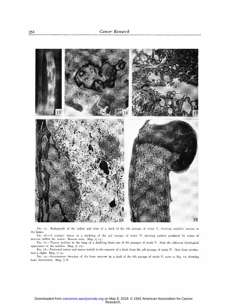
proliferation of new vessels. Microscopically the visceral blebs again showed their independence from neoplasia, and a more complete study of them will be reported elsewhere. The tumors of the soft tissues were composed of either typical spindle- shaped fibroblasts or of sheets of large round cells. In the lung both types of tumors were observed side by side (Fig. I7). Within the marrow cavity were collections of tumor cells in sheet-like arrangement. These cells possessed a large amount of pale cytoplasm which at times appeared to be vacuolated. The periosteal and endostcal tumors were not accompanied by significant new bone formation (Figs. 18 and I9); on the con- trary, osteoclastic activity as well as necrosis of trabeculae gave evidence of bone destruction.
Comment.--Changes in the disease sugges t i ng trans- f o r m a t i o n of the ch i cken strain into a d u c k s t ra in; e. g., d e v e l o p m e n t of a bone t u m o r and special p a t t e r n
of the p r i m a r y t um or s , occu r red at the :znd passage. A l t h o u g h it is qu i t e possible tha t the var ie ty of d u c k s used m a y have e n h a n c e d this t r an s fo rm a t ion , it shou ld
STRAIN V PASS I 2 3 4 5 6 7 PEKIN PEKIN PUDDLE PUDDLE PUDDLEpEKIN PEKIN PEKIN ~40 D. OLO~
F I LTRATE
Soup.r I '~'J3Zm.T.I ~ 1 9 'rUMO~ ~ " 1 - ~
FILTRATE - IN VEIN CELLS - - IN fiREA.~
FIG. 14
-[•.o ?~"%'
[--]60
The results of the passages carried out are given in Fig. 14. The primary growths were very characteristic. They consisted of a tumor flatly spread over the breast integrated by small round nodules of different sizes separated by a stroma. Some areas of the tumor were viscid. The generalized lesions pro- duced either by metastasis of the primary growth or by intra- venous injections of filtrates were of two main types: I. hemorrhagic lesions in viscera and bone marrow, and 2. peri- osteal and endosteal tumors. These lesions appeared separately or combined. The hemorrhagic lesions, most numerous in the liver and spleen, consisted of blebs from a few millimeters to several centimeters in diameter. These sometimes dissected the whole organ because of their large size. No association of these lesions with tumor tissue was grossly found in any case. Periosteal tumors were sometimes solitary in the affected bone, were thinly spread over a good length of it, or were multiple. In the ducks of the 6th passage practically every bone showed multiple tumors which gave a typical radiological image (Fig. 15).
Microscopically the nodules composing the primary tumors consisted of whorls of fibroblastic cells surrounded by delicate strands of collagen. The presence of these collagenous bands together with the necrotic areas gave the tumor a very typical appearance (Fig. 16). In some cases there was a conspicuous
be p o i n t e d ou t tha t the t u m o r s g r e w in the ducks of
the 2 first gene ra t ions for a total of 7 ~ days . T h e abil i ty
to i n d u c e bo th peri- a n d endos tea l t u m o r s a n d p u r e
h e m o r r h a g i c lesions, even in ducks 4 ~ days old w h e n
in jected (6 th passage) , we re dis t inct ive traits of the
strain. Passages are still be ing con t inued , and a com-
plete descr ip t ion of the results wil l be g iven in a n o t h e r
publ ica t ion .
In s u m m a r y , desp i te the fact t ha t only an i n c o m p l e t e
inves t iga t ion was m a d e of the strains of d u c k t u m o r s
ob ta ined , one can state t ha t each of t h e m s h o w e d
typical and cons t an t character is t ics w h e r e b y they cou ld
easily be d i f fe ren t i a ted f r o m the others . In the 3 s trains
w h e r e the p o i n t was s tud ied , a c o m m o n nega t ive char-
acteristic was f o u n d ; n a m e l y , the inabi l i ty s h o w n by
extracts of the t u m o r s to induce , in m o s t instances,
sa rcomas in a d u l t ch ickens . In the 2 o the r s trains
w h i c h wil l be descr ibed, a t h o r o u g h s tudy was m a d e
of the disease in ducks as wel l as in chicks. T h e s tudy
Research. on May 8, 2018. © 1942 American Association for Cancercancerres.aacrjournals.org Downloaded from
352 Cancer Research
Fro. I5.--Radiograph of the radius and ulna of a duck of the 6th passage of strain V, showing multiple tumors in the bones.
FIG. I6 . - -A primary tumor in a duckling of the 3rd passage of strain V, showing pattern produced by zones of necrosis within the tumor. Masson stain. Mag. X 13-
Fro. 17.--Tumor nodules in the lung of a duckling from one of the passages of strain V. Note the different histological appearance of the nodules. Mag. X 15 O.
FIo. I8.--Endosteal tumor and tumor nodule in the marrow of a duck from the 4th passage of strain V. New bone produc- tion is slight. Mag. X 5o.
FIG. I9.--Sarcomatous invasion of the bone marrow in a duck of the 6th passage of strain V, same as Fig. 15, showing bone destruction. Mag. X 8.
Research. on May 8, 2018. © 1942 American Association for Cancercancerres.aacrjournals.org Downloaded from

Duran-Reynals--Reciprocal Infection of Duck s and Chickens 353
of the disease in ch icks is the basis of t he second pa r t of this repor t .
Strains HC and H V . - - T h e source t u m o r ( N o . 6 of
T a b l e I I I ) was o r i g i n a t e d by the in jec t ion of 4 cc. of
cell suspens ion at i : 2 o in the breas t of a 4-day-old
P e k i n d u c k . T h e a n i m a l d i ed i6 days la ter w i t h a
l a rge viscid t u m o r in the breas t a n d a f e w smal l
n o d u l e s in the lung . Cel l suspens ions a n d extracts at
i : 2 o d i lu t ion of the breas t t u m o r w e r e ob ta ined . T h e
cells w e r e passed to a second g e n e r a t i o n of ducks , a n d
the passages w e r e c o n t i n u e d by this m e t h o d . T h i s is
s t ra in H C , the last le t te r i n d i c a t i n g tha t cells on ly w e r e
used t h r o u g h o u t the t ransp lan ts . T w o cc. of f i l t rate of
this t u m o r was in jec ted i n t r a v e n o u s l y in to each of 3
d u c k s also 4 days old. O n e of t h e m d e v e l o p e d a t u m o r
3-5 X 3.5 cm., p lus s o m e o t h e r s of a smal le r size, in the
sk in of the neck." T h e d u c k was k i l led 17 days la ter
a n d cell suspension of its t u m o r s in jec ted in to the
breas t of o the r d u c k s i n d u c e d r a p i d l y g r o w i n g t u n m r s
fo l l owed by g e n e r a l i z a t i o n . T h e passages w e r e con-
t i n u e d by inocu la t ion of cell suspens ions . T h i s is s t ra in
H V , the last le t ter i n d i c a t i n g tha t v i rus was the cause
of the first t u m o r of the series.
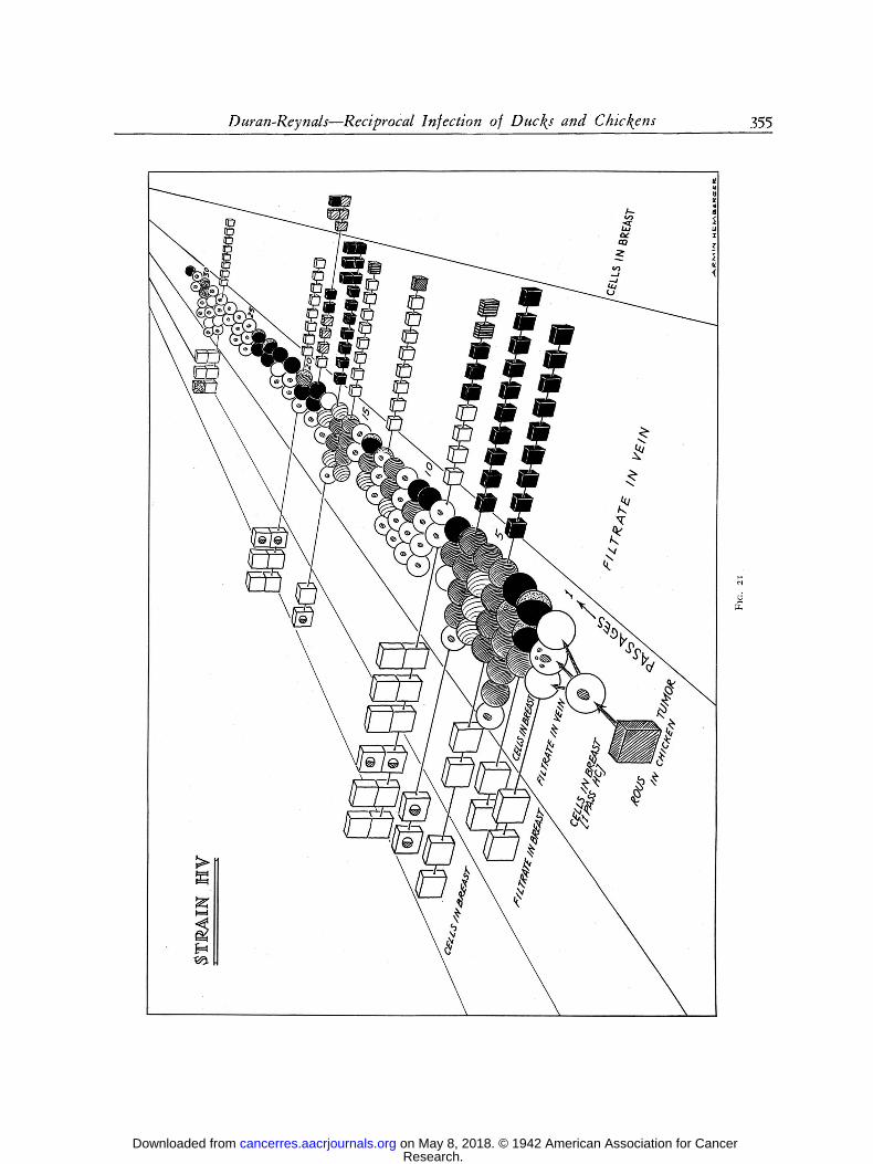
A th ree d i m e n s i o n a l r e p r e s e n t a t i o n o f the passages
a n d e x p e r i m e n t s ca r r ied ou r w i t h the t w o s trains is
g i v e n in Figs. 2o a n d 2 i . T h e desc r ip t ion tha t fo l lows
refers to the disease obse rved in s t ra in H V since the
2nd passage, in s t ra in H C since the 9 th passage, a n d
also in s t ra in A. T h e lesions w e r e iden t ica l in all the
s t ra ins a l t h o u g h the i r i nc idence var ied .
The primary tumors were firm, resilient, translucent, showed bands of necrosis, and had a very characteristic structurc (Fig. 22). Necrosis increased in the later passages and this resulted in the formation of cavities and less cohesion of the growth. Vis- cidity, so characteristic of the original Rous tumor, was absent or very scant in these tumors, and this indicates a profound functional difference between the malignant fibroblasts of chicken and duck tumors, a The distinctive features of the generalized lcsions were the involvement of the skin, whole digestive tract, and bones (Fig. 23). The involvement of the skin and that of the preventriculus and small intestine was frequently massive. That of the skull and ribs was moderate. Tumors in the long bones were in naost cases symmetrical, and in a few cases generalized to all the extremities (such tumors were not observed in strain A). Metastases in the spleen, liver, and lung were frequently found while metastases
e The frequency with which tumors deveIoped in the anterior part of the neck skin in ducklings injected with tumor viruses which may well be called dermotropic is due to the localiza- tion of blood-carried virus in an area which is constantly subjected to friction while the animal feeds. This was very frequently observed in strains A, HC, and HV.
a The viscidity of the Rous sarcoma, and other tumors as well, is due to the secretion by the fibroblasts of a mucin largely composed of hyaluronic acid which is the substrate acted upon by the spreading factors from tissues and other sources. This mucin is analogous to that existing between normal fibroblasts, in synovial fluid, etc. The findings concerning the changes from mucin to collagen may be of interest from the point of view of the histogenesis of the latter.
in the muscles, heart, kidney, pancreas, trachea, peritoneum, etc. occurred less frequently. The tumors in viscera differed widely in size and if they were large enough one could recognize in them the typical gross pattern of the primary tumors. Vis- cidity was observed in some of them, whereas bone tumors never showed it.
A curious occurrence only occasionally met with was the development of solitary tumors in different parts of the body in the absence of primary tumors. One of these, a fungating sarcoma in the auditory canal, is reproduced in Fig. 24. It de- veloped in a duck injected at the age of 77 days with cells of thc 3rd passage of series V. The bird was killed 7 weeks after injection. The way these tumors were induced can be compared to those induced by the original Rous virus injected into newborn ducks.
Hemorrhagic lesions similar to thosc shown in Fig. 23, de- veloped in the same locations where tumors were found. In many cases the blebs were secn grossly free of ncoplastic tissue, an observation confirmed by microscopic study. The fndings of large blood clots surrounding the livcr, of free blood in the peritoneal cavity, and the replacement of the spleen by a large clot were a common occurrence. Hcnrorrhagic lesions were most frequently observed in strain HC, and, in some ducklings in- jected with this strain, they developed with complete exclusion of secondary tumors. Another important finding was the great hypertrophy of the liver and spleen in the absence of neoplasia or hemorrhage within these organs, and occasionally, in older ducks, in the absence of gencralized lesions.
Microscopically the source tumor lacked any special pattern, and consisted of large fibroblasts loosely arranged much as in thc same tumor growing in chickens. The lung metastases had the same structure as the primary tumor but some of the growths were surrounded by a ring of actively dividing lympho- blastic cells very much like the lesions of strain E.
Aftcr the tumor had become adaptcd to the ducks, the primary growths showed a very typical pattern, much the same as in Fig. I6: whorls and nodules of fibroblasts separated by bands of collagenous tissue alternating with zones of necrosis. The generalized lesions were also spindle cell sarcomas with a varying degree of cellularity; in the intestine they were mostly subserosal. In the bones they were periosteal and endosteal with little or no proliferation of new bone (Fig. 25). Giant cell sarcomas were sometimes observed. Some of the tumors were largely collagenous and one could dctect in them the same pattern as in the primary tumors, whilc in other cases they contained a large number Of hemorrhagic foci (Fig. 26). In the liver, in those cases showing hypertrophy there was a pro- nounced vacuolization of the cord cells, and a discrete periportaI infiltration of lymphoblastic cells actively multiplying.
Separa te tests s h o w e d tha t f i l t rates in the b reas t in-
d u c e d the s a m e local a n d g e n e r a l i z e d disease as w h e n
cells w e r e in jec ted , and , if i n t r o d u c e d in to t he vein ,
t he s a m e g e n e r a l i z e d lesions as those s e c o n d a r y to
breas t in jec t ions fo l lowed . T h i s sugges ts t ha t in y o u n g
ducks , as in y o u n g ch i ckens in jec ted w i t h the o r ig ina l
Rous t u m o r s , me tas tases m a y be i n d u c e d by v i rus as
wel l as by ceils.
In the course of t he passages it was obse rved t h a t t he
age of the d u c k was in inverse re la t ion w i t h t he in-
t e n s i t y of the disease, c o i n c i d i n g w i t h an a n a l o g o u s
f i n d i n g w i t h c h i c k e n s in jec ted w i t h the Rous v i rus (4 ) -
I n 8 e x p e r i m e n t s in w h i c h v i rus f r o m b o t h s t ra ins was
in jec ted in p r o p o r t i o n a t e a m o u n t s in to d u c k s f r o m
Research. on May 8, 2018. © 1942 American Association for Cancercancerres.aacrjournals.org Downloaded from

354 Cancer Research
%
k.-
~0
taa
Research. on May 8, 2018. © 1942 American Association for Cancercancerres.aacrjournals.org Downloaded from
Duran-Reynals--Reciprocal Infection of Ducks and Chickens 355
\
\
...../
ta.a f , j
Research. on May 8, 2018. © 1942 American Association for Cancercancerres.aacrjournals.org Downloaded from

356 Cancer Research
i day to 3 rnonths of age the point was confirmed. The rare resistant individuals found were in general older than 2 months. On the contrary, death occurred in most ducks from Io to 2o days after injection regard- less of the presence or absence of generalization, those with a single primary tumor dying just as fast as those
ducks, and tumor cells from each passage were used to secure the next, the primary tumors underwent a progressive change from the noncollagenous, viscid, soft chicken type to the collagenous, nonviscid, firm duck type. Typical examples of such tumors were irregularly observed in the 9th and Ioth passage but
Fro. 22.--Series of sections through a tumor of a duck from the 6th passage of strain HV. Note area of ulceration and underlying necrotic tissue.
FIo. 23.--Generalized disease in a duckling injected intravenously with virus from the 2nd passage of strain HV. FIe. 24.--Sarcoma in the auditory canal in a duck injected in the breast with a cell suspension from a tumor of the 3rd
passage of strain HV. No local tumor developed.
with widespread metastases. Death was not attribut- able to bacterial infection as cultures showed.
Comment and additional experiments.--On compar- ing strains H V and HC in Figs. 20 and 2i, one notices first the differences in the occurrence of primary col- lagenous tumors and of generalized lesions according to whether filtrates in vein or cell suspension in breast was used for transmission. In strain HC where cells from a chicken tumor were injected into the breast of
constantly observed from the I Ith passage on. General- ization was also progressively accomplished; one metas- tasis in the spleen appeared in one duck at the 5th passage; a few skin blebs in another duck of the 6th passage; discrete intestinal tumors in another of the 7th; and widespread lesions with involvement of the ribs at the 9 t h . However, invasion of the liver, probably by direct extension of the breast growth, was frequently observed from the 2nd passage on.
Research. on May 8, 2018. © 1942 American Association for Cancercancerres.aacrjournals.org Downloaded from
Duran-Reynals--Reciprocal ln/ection of Ducks and Chickens 357
On the contrary, in strain HV, a strain derived from a tumor induced by a cell-free extract, generalization took place at once at the 2nd passage in the 3 ducks injected intravenously, but typical collagenous tumors were not clearly present until the 8th passage.
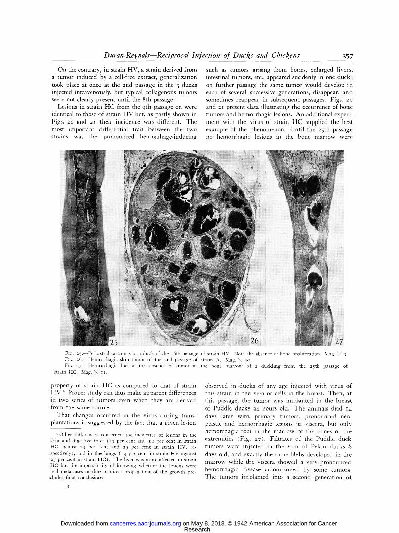
Lesions in strain HC from the 9th passage on were identical to those of strain H V but, as partly shown in Figs. 20 and 2I their incidence was different. The most important differential trait between the two strains was the pronounced hemorrhage-inducing
such as tumors arising from bones, enlarged livers, intestinal tumors, etc., appeared suddenly in one duck; on further passage the same tumor would develop in each of several successive generations, disappear, and sometimes reappear in subsequent passages. Figs. 20 and 21 present data illustrating the occurrence of bone tumors and hemorrhagic lesions. An additional experi- ment with the virus of strain HC supplied the best example of the phenomenon. Until the 25th passage no hemorrhagic lesions in the bone marrow were
Fro. 25.--Periosteal sarcomas in a duck of the I6th passage of strain HV. Note the absence of bone proliferation. Mag. X 9. Fro. 26.--Hemorrhagic skin tumor of the 2nd passage of strain A. Mag. X 4o. Fro. 27.--Hemorrhagic foci in the absence of tumor in the bone marrow of a duckling from the 25th passage of
strain HC. Mag. X II.
property of strain HC as compared to that of strain HV.* Proper study can thus make apparent differences in two series of tumors even when they are derived from the same source.
That changes occurred in the virus during trans- plantations is suggested by the fact that a given lesion
4 Other differences concerned the incidence of lesions in the skin and digestive tract (z9 per cent and z4 per cent in strain HC against 39 per cent and 29 per cent in strain HV, re- spectively), and in the lungs (i 3 per cent in strain HV against 25 per cent in strain HC). The liver was more affected in strain HC but the impossibility of knowing whether the lesions were real metastases or due to direct propagation of the growth pre- cludes final conclusions.
observed in .ducks of any age injected with virus of this strain in the vein or cells in the breast. Then, at this passage, the tumor was implanted in the breast of Puddle ducks 24 hours old. The animals died 14 days later with primary tumors, pronounced neo- plastic and hemorrhagic lesions in viscera, but only hemorrhagic foci in the marrow of the bones ot the extremities (Fig. 27). Filtrates of the Puddle duck tumors were injected in t h e vein of Pekin clucks 8 days old, and exactly the same blebs developed in the marrow while the viscera showed a very pronounced hemorrhagic disease accompanied by some tumors. The tumors implanted into a second generation of
Research. on May 8, 2018. © 1942 American Association for Cancercancerres.aacrjournals.org Downloaded from

358 Cancer Research
Pekin ducks induced large primary growths followed again by blebs in the marrow. After this, no more blebs in marrow were observed.
On several occasions injectio n in other ducks of extracts from the hemorrhagic lesions, in the absence of tumors, a n d of the enlarged livers was carried out. No lesions of any kind were ever induced. The reasons for the failure in transmitting the hemorrhagic lesions will be considered in another paper. The failure of the liver extracts to induce any othcr lesions would fit with the lymphomatous character suspected in the lesions present in the organ.
Results obtained in the infection of adult chickens reflect the adaptation, sudden or gradual, of the chicken virus to the new host. Figs. 2o and 21 s h o w
that the capacity of the virus to induce immediate tumors in chickens is only progressively lost in strain HC as the adaptation to the duck is progressively accomplished; whereas, in strain H V it is totally lost at the first passage. The same holds true to a large extent when ceils are injected, al though in both strains there may be left in them a residual capacity to induce tumors. In strain HV these tumors always regressed and the same happened in strain HC in ~ out of ~2 cases when suspensions of tumor ceils of the ioth and later passages were employed. It is noteworthy that, in this strain, whenever progressively growing tumors developed, the time of death was retarded from ~7 days in the first passage to 46 in the ,oth when cells were injected, and from 45 days in the xst passage to 75 in the 3rd when extracts were injected. Also, the tumors induced by materials from the early passages resembled the original chicken tumors whereas those of later passages resembled duck tumors. Passages of these tumors were attempted in 3 instances. The pro- cedure followed and results obtained are summarized in Table IV.
The results show the absolute lack of transplanta- bility to other adult chickens of tumor resulting from the growth of duck tumor cells in chickens. This important point will be fully developed in the follow- ing section.
THE INFECTION OF CHICKS BY VIRUSES AND CELLS FROM DUCK TUMORS
Analysis of the phenomena attending the infection of chicks by the viruses from the duck tumor strains HC and H V to be described now showed that despite some differences, they ~ were essentially the same as those attending the infection of ducklings by chicken tumor viruses and, accordingly, the same order in the description of the results will be followed. To all intents and purposes the duck variants of the Rous virus behaved toward chickens as if they were genuine duck tumors, but a check on this point has so far been impossible because we could never secure the latter.
2 .~<
m
0
Z
a2
Z
�9
[-
Z o
Z
g
. � 9 4.a
r]?
~ ~ ~'~ , t'l
g
b ,
r
m ~
g: m
= r r ;>
�9 o �9 -cj ,.~= 0o m
c a . ~
g~0 . ~ 0o
m ~ ,+~ ca o-~
~
o 0a
+a
~4
o - ~
o ~
, x Z ~
ca
ca .v g
O ~
Research. on May 8, 2018. © 1942 American Association for Cancercancerres.aacrjournals.org Downloaded from

Duran-Reynals--Reciprocal Infection of Ducks and Chickens 359
T h e principal characteristics o f this infect ion are graphical ly represented in Figs. 2o and 2i . T h e figures show that in 24 instances filtrates ( I cc. of an extract at I :2o) f rom tumors of different passages were in-
jected in t ravenously into 175 chicks f rom I to 3 days o f age. A l t h o u g h not indicated in the figures, 2 ducks were similarly injected in every instance as controls of the presence of virus in the filtrates. It is clear f rom the Figs. 2o and 2i that the infectivity for chicks of the virus f rom the different passages was ext remely variable. Lesions developed in 42 per cent of the animals . Yet the virus showed its usual infectivity for ducks because all of t hem died with genera l ized lesions.
T w o addi t ional sets of exper iments dealt wi th the impor tance of the age of host and the dose of virus as factors d e t e r m i n i n g infection. It was found that chicks 6 days old injected in the vein wi th the usual a m o u n t of virus were as susceptible as younger chicks similarly injected, but only i ou t of IO chicks 9 days old devel- oped lesions. For reasons to be given later it is impor- tant to point out that the results hold t rue as far as the induc t ion of h e m o r r h a g i c or sarcomatous lesions is concerned. Tha t such lesions were not i nduced in adul t chickens injected wi th duck viruses was already men t ioned . It was also found that all of io chicks one day old developed sarcomas w h e n injected intra- venous ly wi th i c c . of t u m o r filtrate at i :2oo but none of 5 chicks of the same age similarly injected wi th the filtrate d i lu ted at i :2ooo developed lesions.
It is seen f rom these results that as in the ch icken virus-duck sequence the age of the host, the dose of virus, and an inheren t proper ty of the latter concern ing its infectiousness for the heterologous host are factors of p r ime impor tance in i nduc ing infection.
T h e impor tance of the route of inocula t ion was not directly investigated. It m a y be said, however , that in exper iments devised for o ther purposes, out of 34 chicks injected in the breast ( sk in and musc le ) wi th 3 t imes the a m o u n t of virus used in the in t ravenous injections, the incidence of infect ion was also 42 per cent. T h e tumors were well c i rcumscr ibed and were not fo l lowed by genera l iza t ion . Moreover , in 6 of t hem regression occurred, whi le no regression of the tumors fo l lowing in t ravenous injections was ever observed.
Figs. 2o and 21 also conta in data per ta in ing to the
effect of cells f rom d u c k tumors injected into chicks.
T h e 13 animals were I day old and they were injected
wi th i CO. Of t u m o r cell suspension at 1:5 in the breast. T h e y all developed rapidly g r o w i n g tumors fol lowed
by genera l iza t ion and dea th f rom 15 tO 25 days after injection.
F r o m some of the tumors in chicks injected wi th
filtrates, sublines were started employ ing a total of
203 chicks. These animals developed lesions identical to those of the animals injected direct ly wi th filtrates or cells f rom the duck tumors . T h e descr ipt ion of the lesions tha t follows applies "to both groups of animals , the total n u m b e r ana lyzed being 39 I. H e r e again the lesions will be classified as immediate and late. T h e i m m e d i a t e lesions were observed in 232 animals f rom 9 to Ioo days after injection, and they were ei ther h e m o r r h a g i c or neoplastic.
T h e neoplastic lesions were sarcomatous, and in one instance a myelo id t u m o r of the ovary was present a long wi th the sarcoma. It will be seen later that this could be considered a late lesion.
Late lesions were observed in 14 animals . T h e y did not appear previous to ioo days after inject ion. These birds were sacrificed w h e n still in good health, f rom 4 to 7 m o n t h s after injection, and the lesions found to be lymphoblas tomas and osteopetroses.
Of the 145 animals w h i c h did not develop lesions, l o 3 were ki l led f rom 4 to 7 months ; the r e m a i n i n g 42, 2 m o n t h s or less after injection. It is thus possible that the incidence of late lesions wou ld have been h igher if the animals had been ma in t a ined for longer periods of t ime.
Hemorrhagic lesions.--Thcse occurred only in chicks in- iccted with filtrates from strain tIC. From the ISt to the 6th passage, the disease induced in chicks by virus from this strain was generalized and identical to that induced by tile original chicken virus (5)- The animals died 7 to I7 days after in- jcction, and no gross neoplastic lesions werc apparent. In the later passages, after the injected virus had become completely adapted to the duck, the hemorrhagic lesions were always lo- calized in the liver, and appeared less frequently in other or- gans. Large clots around the liver were typically found, and in a few cases the spleen had the appearance of a clot. Frequently, blebs were the only lesions found in the affected organs or, occasionally, in the whole chick. This observation was con- firmed microscopically. When tumors in either bones or viscera were present, no association of tumors and hemorrhagic lesions was apparent at gross examination. Some new features of the hemorrhagic disease of the chick induced by duck viruses will be described elsewhere. The diseased animals characteristically presented pure hemorrhagic lesions in some viscera, and tumors arising from bone.
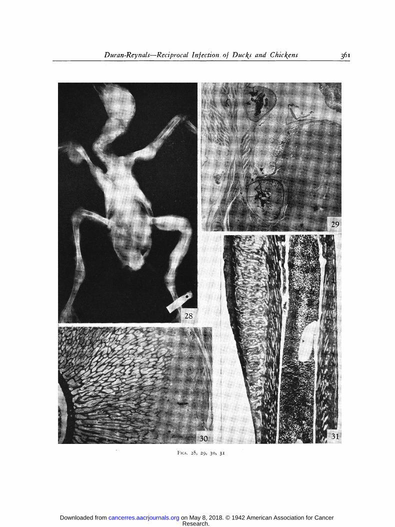
Neoplastic" lesions.--They were all sarcomas which took origin either from the bones or the soft tissues. Bone tumors were the more frequent and were a distinctive feature of the infec- tion of chicks by duck viruses. Most were periosteal, some were cndosteal, and a few were a combination of these two types. They were firm, not viscid, and not hemorrhagic. They arose from practically every bone (Fig. 28) and the most pronounced involvement was found in chicks injected with virus of the HV strain. Here bones were often exclusively involved, while this occurred but rarely with the virus of strain HC. The older bone tumors were in general larger, firmer and less widespread. A secondary formation of new bone was often grossly noticeable in these lesions. Soft tissues were sometimes invaded by growing bone tumors but at other times growth arose directly from them. Muscle involvement was second to that of bone, but tumors were rare in viscera. Of the latter, a char- acteristic lesion was the replacement of part of the preventriculus by tumor tissue. Large tumors m the neck sometimes de-
Research. on May 8, 2018. © 1942 American Association for Cancercancerres.aacrjournals.org Downloaded from

360 Cancer Research
veloped when the tissues of the region were soiled by virus in the process of intravenous injection. All these tumors were compact, firm, and well circumscribed. Cases of diffuse malig- nant invasion of organs by loose myxosarcomatous tissue such as commonly occurs in chicks injected with the Rous virus were never observed. However, when tumors reached a very large size, some viscidity was present. The large growths that developed after cell suspensions were injected in the breast, were often accompanied in the samc animal by tumors in bones. The carlicst death attributable to tumor growth, and not to hemorrhagic lesions, was observcd 15 days after injection.
One of the chicks that dicd 45 days after the injection showed in addition to wi(Icspread tumors arising from bones a 2 ~ 2 lnln. growth m the ovary which proved to be a tumor composed of immature m)cloid cells. Thc histological nature of the tumor and facts concerning their transmission to be de- scribed later indicate that wc wcre dealing with a late lesion at the beginning of its dcvclopment; time: death of the animal was attributable to the bone tumors.
Microscopically the tumors in the bones were periostcaI or endosteal spindle cell sarcomas like tbose in ducks, except that giant cell tumors were n(:vcr observed. A vcry important additional lesion in chicks was pronounced formation of new bonc by thc inner layer of the periosteum nruch the same as observed in the physioldgical intramcmbranous ossification from thc fetal periosteum. This lesion occurred, always in associa- tion with tumor growth (Fig. 29) , either when tim growth had pierced the periosteum and invaded the bone (Fig. 3o), or in the absence of this invasion, tumor growth and bone growth being then histologically independent (Figs. 31 and 32). However, bone prolifcration without sarcoma growth was never observed. In addition to the bone proliferation there was also a pronounced fibrosis of the marrow spaces frequently present which can easily be seen in Figs. 3 ~ and 32. Both lesions were more pronounced in older than in young tumors, and it is important to point out that these lesions are the same as those described by Junghcrr and l ,andauer ( t7 ) in the early stages of ostcopctrosis. This resemblance strongly suggests that this condition as observed by us rcprescnts a further stage of thcse early lesions.
The tumors f rom muscles and viscera were spindle cell sar- comas of the ordinary type. Intravascular growth was observed only occasionally.
Leul(otic tumors and leu/~oses.--These lesions were observed in 4 cases. In all of them there was a tumor in the ovary as a maior lesion while one of the chickens showed paralysis. Two of the animals were killed 3�89 and 4 months after in- jection while the others died.
Microscopically 3 of the tumors were of tile lymphoid type consisting of cithcr large or sma]l lymphoblasts. The tu- ,nor of the 4th animal was composed of myeloid cells; and it was found in an animal that died 45 days after injection with widesprcad tumors in bones. For reasons indicated before,
although chronologically this tumor falls within the group of the immediate lesions, it is considered as a typical late lesion. In all the 3 cases there was a leukotic infiltration of the viscera of varying intensity.
Osteopetrosis.--Before describing our results it is pertinent to summarize the studies of lungherr and Landauer (17) on the subject.
These authors described the condition in 1938 under the term osteopetrosis gallinarum. It was associated with an epi- demic outbreak of lymphomatosis; ~ the authors thoroughly reviewed related osteopathies in other animals and man (Pagct's disease, osteodystrophia fibrosa, etc.) and emphasized time fre- quent association between some of these conditions and a variety of hcnaopathics. They injected 61 newborn chicks with blood, bone marrow, and lymphomatous tissue from florid cases of ostcopetrosis and obtained in thc course of 4 passages 6 gross lesion cases of ostcopctrosis, 6 gross lesion cases of osteopetro- sis, 6 gross lcsion cases of osteopetrosis associatcd with lympho- matosis, and 23 cases of lymphomatosis. The animals were kept for 25o clays, and those that became infected did not die until from ~oo to 189 days after injection. Stubbs and Furth (29) also observcd bone changes resembling those of osteitis fibrosa in chickens infected with erythroleukosis virus.
In our experiments the condition was obscrvcd in IO of the animals, 7 of which were injected a few days after hatching, 2 at the age of 4 montlns. The disease was the same as de- scribed by Junghcrr and Landauer (17) being characterized by diaphyseal thickcning and hardening Inostly of the wing and leg boncs (Fig. 33) with total or partial occlusion of th~ marrow cavity. In the florid cases the affccted areas sbowed increased surface tempcrature, and the pcriosteum was thick- ened and hcmorrhagic. ]n no case was there evidence of sar- COlnatosis either in time bones or elsewhcre. Parathyroids were not enlarged. The gross lesions wcre not clearly noticeable until from 4 to 7 months had elapscd after the animals were injected.
B e c a u s e o f t h e n u m b e r o f e x p e r i m e n t s i n v o l v e d , t h e
v a g a r i e s o f t h e d i seases , a n d b e c a u s e t h e c h i c k e n s w e r e
n o t k e p t l o n g e n o u g h , i t is h a r d to e s t i m a t e f r o m o u r
d a t a t h e i n c i d e n c e o f l e u k o s e s a n d o s t e o p e t r o s i s . I f
c a l c u l a t e d o n t h e s t r i c t r e s u l t s o b t a i n e d , t h e i n c i d e n c e
w o u l d be 3-5 p e r c e n t .
5 TI!e outbreak occurred in Connecticut in the years 1933 and I934. The chickens showed big liver discase, paralysis, and still other manifestations of lymphomatosis. There were 7 ~ recognizable cases of osteopetrosis among 4,5oo chickens from 7 strains, while many other chickens showed slight bone changes. The earliest lesions were noticed at the age of 6 weeks. Only 5 sporadic cases of ostcopetrosis were observed in the rou- tine autopsies carried out during 7 years and comprising ~6,949 chickens.
I )ESCRIPTION OF
FIG. 28.--X-ray picture of a chick injected intravenously with filtrate from a duck tumor of the 4th passage, strain HV. Note periosteal tumors in almost every bone. Viscera were wholly free of tumor.
FIG. 29.--Sarcoma in the thoracic wall of a chick injected intravenously with a filtrate from a tumor derived from the 4th passage of strain HV. Note subperiosteal bone proliferation in the rib surrounded but not invaded by sarcoma, and absence of bone proliferation in adjacent rib not lying within the tumor.
FIGURES 28 TO 31
Fro. 3o.--Invasion of bone by periosteal sarcoma, pronounced formation of new bone, and fibrosis of marrow spaces in the tibia of a chick injected intravenously with virus from a tumor derived from the 4th passage of strain HV. Note original dense cortical bone. The animal died 44 days after injection. Mag. X 17.
FIG. 3I . - - -Humerus of the same chick. Note pronounced formation of new bone and fibrosis despite the fact that the bone was not invaded by the tumor. Mag. >< 12.
Research. on May 8, 2018. © 1942 American Association for Cancercancerres.aacrjournals.org Downloaded from
Duran-Reynals--Reciprocal Infection o/ Ducks and Chickens 36i
Fins. 28, 29, 30, 3I
Research. on May 8, 2018. © 1942 American Association for Cancercancerres.aacrjournals.org Downloaded from

362 Cancer Research
Microscopically the osteopetrotic bones showed the same pathognomonic changes as described by Jungherr and Landauer (I7) of which formation of strongly hyperchromatic new bone, marrow fibrosis, and partial or total disappearance of tile mar- row spaces were the most conspicuous (Figs. 34 and 35)-
No definite sarcoma was observed. In only one case was it possibly present. Histological evidence from the cases of osteo- petrosis indicates that the inner layer of the periosteum is the source (or one of the sources) of the new bone, and this opinion is strengthened by the study of the bone proliferation observed in association with the periosteal sarcomas (compare Figs. 3 o and 32 with Figs. 34 and 35). Whether a previous sarcomatons lesion was indispensable for the development of osteopetrosis or whether this condition can develop from the inner layer of the pcriostcum as a rcaction to the virus entirely indcpcndent of the ncoplastic reaction of thc outer layer is not known as yet. A decisive factor in favor of the latter alterna- tive would be of course the presence in chicks of early bone proliferation in tile absence of neoplasia. Although we did observe histological independence of both lesions in the samc bone, proliferation in those animals was always associated with sarcoma growth. The question is then raised whether the cases of osteopetrosis represent these lcsions at an advanced stage of their evolution. On the other hand that osteopetrosis can develop in the absence of demonstrable sarcomas is shown by the work of Jungherr and Landauer (*7) and by our own experiments to be rcported later.
THE NATURE OF THE IMMEDIATE LESIONS INDUCED iN
CHICKS BY DUCK VIRUSES
It will be remembered that in the chicken virus-duck sequence, the immediate tumors were solitary sarcomas looking much as the original Rous tumor does, and that the virus extracted from them was still a chicken virus because it induced tumors in chickens but not in ducks. Much the same thing occurred in comparable tumors in the duck virus-chick sequence. The analo- gies fundamental ly present between the disease in chicks and that in ducks will be first considered.
A very important trait common to strains H C and H V ; namely, the development of periosteal and endo- steal sarcoma, was faithfully reproduced in the infec- tion of chicks, but the incidence of tumors induced by each strain in ducks and chicks was reversed. Strain H V induced 14 per cent of bone tumors in ducks and 45 per cent in chicks, whereas strain H e , from the 8th to the last passage, induced 4i per cent of bone tumors in ducks, 20 per cent in chicks. Moreover, there was in chicks a marked formation of new bone not observed in ducks. Also, in chicks injected with the strain H V the tumors always involved many bones whereas in those injected with strain H C the bone involvement was more moderate. A second important trait C~bmmon to both strains of duck tumors; namely, the involvement of the digest ivetract , was also repro- duced but only to a limited extent in the infection of chicks, for only about I o per cent of them injected either with the virus from ducks or with material from chick tumors of the subsequent passages developed
lesions in the intestinal tract, mostly in the preven- triculus and gizzard. In strain H C these lesions were often purely hemorrhagic. A third important trait common to both duck strains, the involvement of the skin, was practically lost in the infection of the chick, these lesions being found in less than i per cent of the injected chicks. On the other hand, the incidence of a pronounced hemorrhage-inducing power in strain HC, in contrast to its absence in strain HV, was directly reflected in the disease of chicks; the former strain induced 6i per cent of hemorrhagic lesions in ducks and 32 per cent in chicks, and the latter strain induced io per cent of such lesions in ducks and o per cent in chicks. The latter point is also brought up in Figs. 20 and 2i. These observations emphasize the relative importance of factors either from the host or from the virus in determining the clinical manifesta- tions of the neoplastic disease.
The transmissibility to both chickens and ducks of the immediate lesions shown by chicks injected with duck viruses was next taken up. Hemorrhagic lesions could not be transmitted in any instance, even when newly hatched animals were used. The subject will be developed elsewhere.
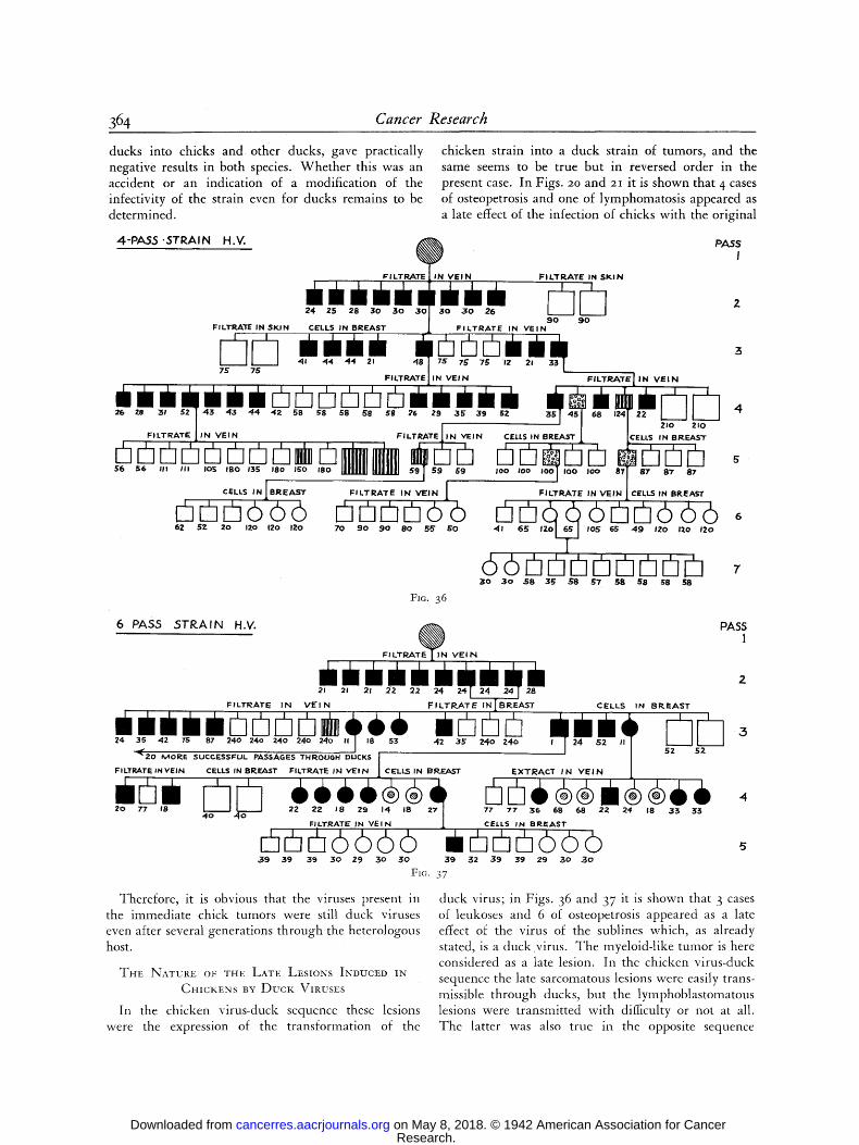
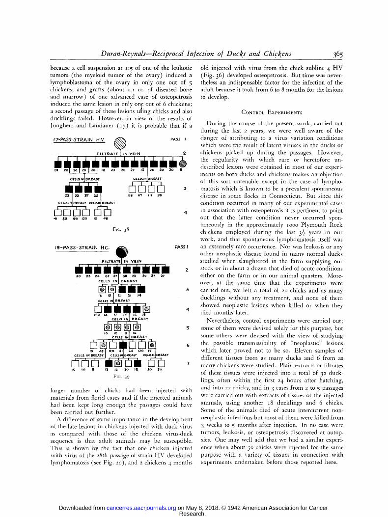
The following procedure was followed using the neoplastic l e s ions . Sublines of the tumors in chicks were started from the 4th, 6th, and I7th passages of strain H V and from the i9th passage of strain H C and carried out through both chicks and ducks. To secure good material for transmission the donors were killed and filtrates at i :2o and cell suspensions at I: 5 were immediately obtained from tissue wholly free of necrosis. Details of the experiments and results ob- tained are represented in Figs. 36 to 39. In these figures we are now only concerned with the induction of immediate lesions--sarcomas and hemorrhage. Signs in the figures indicating leukoses or osteopetrosis will be considered later.
It is clear from Figs. 36 to 38 that none of the sub- lines from strain H V could be perpetuated beyond 3 generations of chicks while in subline 19 H C (Fig. 39) propagation was not accomplished beyond 6 gener- ations. The age of the tumors used for transmission did not influence the results, The induction of lesions came to an end either gradually or abruptly. In the sub!ine 19 H C (Fig. 39) the development of large primary tumors without metastases preceded the com- plete disappearance of the power to induce tumors. As shown in subline 6 H V (Fig. 37) ducklings injected with materials from the first chick passage developed a typical generalized disease which could be transmitted to other ducks for a presumably indefinite number of generations and the same was probably true after 2 chick passages. However a 3rd passage, at- tempted with a variety of materials from one of these
Research. on May 8, 2018. © 1942 American Association for Cancercancerres.aacrjournals.org Downloaded from

Duran-Reynals--Reciprocal Infection of Ducks and Chickens 363
Fro. 32.--Section from another bone of same chick (see Fig. 28) showing sarcoma, at top, external to the intact perios- teum, and marked proliferation of the inner layers of the periosteum with formation of new bony trabeculae. Mag. )K I25.
Fro. 33.--Ostcopetrosis of the leg bones of a chicken injected intravenously at the age of I day with virus f rom a tumor derived from the 4th passage of strain HV and killed I26 days later. The opposite leg was equally involved. Compare with bones of a normal chicken of the same age, shown on the right.
Fins. 34 AND 35.--Longi tudinal and transverse scctions of the tibia of chickcn with osteopctrosis. Mag. )K 4o and 4-5.
Research. on May 8, 2018. © 1942 American Association for Cancercancerres.aacrjournals.org Downloaded from
364 Cancer Research
ducks into chicks and other ducks, gave practically negative results in both species. Whether this was an accident or an indication of a modification of the infectivity of the strain even for ducks remains to be determined.
4 - P A 5 5 " S T R A I N H . V .
FILTP-~TE IN SKIN
chicken strain into a duck strain of tumors, and the same seems to be true but in reversed order in the present case. In Figs. 20 and 21 it is shown that 4 cases of osteopetrosis and one of lymphomatosis appeared as a late effect of the infection of chicks with the original
@
, , , i ' i / i ; ' i , �9 +i 24 ,25 28 30 3 o 3 o I 3 o 3 0 26
9 0 9 0 / CELLS IN BREAST l F ILTRATE IN VEIN
P A S S
I
FILTRATE l IN VEI N
/ ' 4 M + I ' + , ' i ~ ' m * @ ~ p r L - ' ~ + L - ' * i ~ ~ ' ' i r a m ' * ' ' ,~ + ' + + ' +~'+'+. +'+ +' +~ + ' " +' " + '+ +" + + " + Z , , + + ' + + ' + c .o '+- i I++,. ~ + ' " ' % , h . " - - ' ..++..+,,+N + ,_L.++++ + +L' + + + +""'-L+ N @ + + n + $6 $6 III III Io5 180 135 180 150 180 59 69 JOO IoO I00 Ioo 87 B7 87
CELLS IN I B R E A S T
@@@666 62 5Z 20 12o 12o 120
/ FILTRATE IN VEIN ICELLS IN BREAST
r-'-i r-h ~ 6 r-h r"-I 6 6 6 -41 65 I IO5 65 49 IZO 120 12o
6 6 rh Pl l-h rh rh @ rh i~ 3o 30 38 3~; $8 57 58 58 ~;8 58
FILTRATE IN VE IN 1
@r~• 70 90 9 0 80 56" ~0
Fro. 36
6_ ~___2~ ~.____2'_. ~.~. @ ~
21 21 21 22 22 2 4 2 4 1 2 4 2 4 128 FILTRATE IN V E ~ _ ~ ~ _ _ ~ CELLS IN BR.EAST
42 3s z4o z+o ~ 5 " 2 o ~ o m ~ S u c c E s s f u L ~ASSA~ES VHROU~H DUCKS I ' I -- -
FILTRATE IN VEIN CELLS IN BREAST I~ILTRATE IN VE IN jl CELLS IN BREAST EXTRACT IN V E I N [I
[email protected] ++ 22 22 29 14 18 77 77' 36 68 08 22. 24 18 33 35
4 o 4 0 FILTRATE IN VF--,IN CELLS IN BREAST
@ r~ r-h 6 6 6 6 m rh r-h ~ <:5 6 <:5 ,39 39 39 2;0 29 30 30 39 32 3 9 39 29 .30 3 0
F1G. 37
Therefore, it is obvious that the viruses present in the immediate chick tumors were still duck viruses even after several generations through the heterologous host.
THE NATURE O1; THE LATE LESIONS INDUCH) xN CHICKENS BY D u c k VIRusEs
In the chicken virus-duck sequence these lesions were the expression of the transformation of the
duck virus; in Figs. 36 and 37 it is shown that 3 cases of leukoses and 6 of osteopetrosis appeared as a late effect of the virus of the sublines which, as already stated, is a duck virus. The myeloid-like tumor is here considered as a late lesion. In the chicken virus-duck sequence the late sarcomatous lesions were easily trans- missible through ducks, but the lymphoblastomatous lesions were transmitted with difficulty or not at all. The latter was also true in the opposite sequence
Research. on May 8, 2018. © 1942 American Association for Cancercancerres.aacrjournals.org Downloaded from
Duran-Reynds--Reciprocal In/ection o/Ducks and Chickens 365
because a cell suspension at i :5 of one of the leukotic tumors (the myeloid tumor of the ovary) induced a lymphoblastoma of the ovary in only one out of 5 chickens, and grafts (about o.I cc. of diseased bone and marrow) of one advanced case of osteopetrosis induced the same lesion in only one out of 6 chickens; a second passage of these lesions U~ing chicks and also ducklings failed. However, in view of the results of Jungherr and Landauer ( i7 ) it is probable that if a
17-PASS.STRAIN HM K~,,~ PASS I
F I L T R A T E J IN V E I N 2
. 2 . 2 . , 3 , 2 0 zo 2 0 .
I I C E L L S 1~ B R.EAST CELLS/N[BR-EAST
22 1 22 257 ZZ / 58 67 I l l $ 9
L$ I~1i BREAST CELL$1~ BREAST
4t II 9 120 120 15 4 8
FIG. 3 8
1 9 - P A S S " S T R A I N H C . ~ PASSI
FILTRATEI IN VE IN
20 23 2 4 67
CELLS 38 23 20 27
BREAST
16 i s I 21 21 14 CELLS INI BREAST
~5o 14 e~' 14 ! IS 16 CELLS INJ BREAST
13 12 18 CE'LLS II
IS 4 3 CgLL$ IN BREAST
24 e20 1'7 [ I
~.EAST CELLS I~IBR~~$T
15 I 0 9 13 fT. 20 12 2 0 20
FIG. 39
l a rge r number of chicks had been injected with materials from florid cases and if the injected animals had been kept long enough the passages could have been carried out further.
A difference of some importance in the development of the late lesions in chickens injected with duck virus as compared with those of the chicken virus-duck sequence is that adult animals may be susceptible. This is shown by the fact that one chicken injected with virus of the 28th passage of strain H V developed lymphomatosis (see Fig. 2o), and 2 chickens 4 months
old injected with virus from the chick subline 4 H V (Fig. 36) developed osteopetrosis. But time was never- theless an indispensable factor for the infection of the adult because it took from 6 to 8 months for the lesions to develop.
CONTROL EXVERIMI~:NrS
Dur ing the course of the present work, carried out during the last 2 years, we were well aware of the danger of attributing to a virus variation conditions which were the result of latent viruses in the ducks or chickens picked up during the passages. However, the regularity with which rare or heretofore un- described lesions were obtained in most of our experi- ments on both ducks and chickens makes an objection of this sort untenable except in the case of lympho- matosis which is known to be a prevalent spontaneous disease in some flocks in Connecticut. But since this condition occurred in many of our experimental cases in association with osteopetrosis it is pertinent to point out that the latter condition never occurred spon- taneously in the approximately ~ooo Plymouth Rock chickens employed during the last 3�89 years in our work, and that spontaneous lymphomatosis itself was an extremely rare occurrence. Nor was leukosis or any other neoplastic disease found in many normal ducks studied when slaughtered in the farm supplying our stock or in about 2 dozen that died of acute conditions either on the farm or in our animal quarters. More- over, at the same time that the experiments were carried out, we left a total of 2o chicks and as many ducklings without any treatment, and none of them showed neoplastic lesions when killed or when they died months later.
Nevertheless, control experiments were carried out; some of them were devised solely for this purpose, but some others were devised with the view of studying the possible transmissibility of "neoplastic" lesions which later proved not to be so. Eleven samples of different tissues from as many ducks and 6 from as many chickens were studied. Plain extracts or filtrates of these tissues were injected into a total of 32 duck- lings, often within the first 24 hours after hatching, and into 22 chicks, and in 3 cases from 2 to 5 passages were carried out with extracts of tissues of the injected animals, using another I8 ducklings and 6 chicks. Some of the animals died of acute intercurrent non- neoplastic infections but most of them were killed from 3 weeks to 5 months after injection. In no case were tumors, leukosis, or osteopetrosis discovered at autop- sies. One may well add that we had a similar experi- ence when about 5 ~ chicks were injected for the same purpose with a variety of tissues in connection with experiments undertaken before those reported here.
Research. on May 8, 2018. © 1942 American Association for Cancercancerres.aacrjournals.org Downloaded from

366 Cancer Research
DISCUSSION
The essence of the findings is that the injection of ducklings with the virus of the Rous sarcoma of chickens- induced two types of lesions: immediate tumors, which killed the host in a few weeks, and late tumors, which in most cases were not noticeable until several months later. The former infection in- volved no variation of the virus which remained a chicken virus, whereas the latter infection involved such variation, the virus passing to be a duck virus and inducing new types of tumors. Reversing the above sequence, the infection of chickens with duck variants of the Rous virus resulted, too, in the produc- tion of immediate and late tumors, the former involv- ing no variation in the virus and the latter involving further variations with the production of still other types of tumors. Therefore, the infection that resulted in the production of immediate tumors was a heterolo- gous infection, whereas that which resulted in the production of late lesions was a homologous infection. A summary of the results is given in Table V.
The hetarologous in~action by tumor viruses.--The main characteristic of this type of infection was its being strongly conditioned by several factors. Two of them; namely, immaturi ty of the host and high doses of the virus, were experimentally controllable; a third factor depended on inherent characteristics of the sample of virus used, and was uncontrollable within the limits of our experiments. These 3 factors were found indispensable in practically all cases. A fourth factor, the route of inoculation, was found important, but not indispensable. The conditioning was stronger in the infection of ducks by chicken viruses than in the opposite sequence. The fact that, within the limits of our experiments, only ducks within the first :z 4 hours after hatching were found susceptible, and that there was a detectable hourly increase in the resistance of the ducklings to the virus during this time was striking and can only be compared to the sudden
acquisition of resistance by avian embryos in the last days of their development to heterologous viruses (~, 12) and tumors (20). The implications of the findings are obvious if one considers that the effects of this early infection cannot be manifested until later in life when the animal has reached maturity.
A second important characteristic of the heterologous in fec t ion was that the tumors retained in the heterolo- gous host most of the features; e.g., morphology, texture, and tissue affinities, that they showed in the homologous host. The hetarologous tumors were generally localized in the chicken virus-duck infection, and generally widespread in the opposite sequence, but heterogenicity of the infection by no means implied mildness because practically all of our animals, ducks or chickens died of their tumors.
The third characteristic of the heterologous infection was that it could not be perpetuated, or only through a few generations, through the heterologous host, if cell-free extracts were used. If cells were used, then perpetuation of the heterologous duck tumors was possible, but these passages eventually led to variation of the virus; that is, to homologous infection. In chicks, the heterologous tumors died out after a few, generations without ever reverting to the original Rous tumor. On the contrary, if virus or cells from the heterologous tumors were injected in the opposite host; that is, the host homologous to the tumor, then the original disease, capable of indefinite transmission, was restored.
Data concerning the length of the experimental heterologous infections, where large amounts of virus were injected, are indicated in Table V. One may well speculate on the possibility of spontaneous tumors occurring in nature following a comparable infection with smaller amounts of virus, and on the time a tumor can grow in a heterologous host without its virus undergoing variation. The problem then would be to find the host homologous to the tumor so that the etiological agent would be brought up.
TABLE V: HOMOLOGOUS AND HETEROLOGOUS INFECTION OF DUCKS AND CI-IICKENS BY 'I'UMOR VIRUSES
Hemorrhagic disease Lesions obtained~Myxosarcomas
[.Spindle cell sarcomas
Spindle cell sarcomas (collagenous) Giant cell sarcoma of bone Peri- and endosteal spindle cell sarcoma Sarcoma-lymphoblastoma Lymphoblastoma
9-3o days 4o-2I 5 days or longer Rous virus > ducklings > --
Immediate lesions Late lesions [No virus variation] [Virus variation]
9-Ioo days 45-215 days or longer Duck virus > chicks >
I Hemorrliagic diseasc Myeloblastoma Lesions obtained~ Spindle ccll sarcomas (collagenous) Lymphoblastoma
[`Peri- and endosteal spindle cell sarcoma Osteopetrosis
Research. on May 8, 2018. © 1942 American Association for Cancercancerres.aacrjournals.org Downloaded from

Duran-Reynals~Reciprocal Injection o[ Duck s and Chickens 367
The age resistance of ducks and chickens to cells from heterologous hosts is only slightly inferior to that to the virus, and as it happened with the latter, the conditioning by age of the infection of ducks by chicken tumor cells was more pronounced than in the inverse sequence. Allowance being made for these differences, it is clear that, whatever the defensive forces of the host are, they are equally directed against both tumor virus and tumor cells.
Variation o/ t,~e tumor viruses and homologous infection.--Oncc the tumor viruses have implanted themselves in the hcterologous host, time was found to be the only additional conditioning factor for the virus to vary and the infection to become homologous2 ~ Rapidity of growth of the tumors was not indispensable because some of the late tumors, clue to the variant virus, were several months old and yet they were quite small. It is obvious that homologous infection began when heterologous infection ended. But in the strains obtained by repeated passages of cells from the Rous tumor through ducks one could observe a gradual transition of the chicken type to the duck type, the new variant taking finally the upper hand. These tumors can well be considered as experimentally in- duced "mixed" tumors, and it is not inconceivable that some spontaneous tumors may contain a n-fixture of both homologous and heterologous viruses.
Is maturity of the host indispensable for the virus to vary and induce late lesions or are those the result of an evolution of the immediate lesions? Variation of the Rous virus into a duck virus has been repeatedly accomplished, by means of cell passages, in young ducks. But we do not know whether variation would have eventually occurred in all the tumors induced by the Rous virus in ducks had not the animals died of the toxic-like effect from the growth or whether the late tumors were induced by special and rare units of virus endowed with a potential ability to vary. Nor do we know for certain, al though the second alterna- tive seems far more probable, whether late lesions in chickens developed from unnoticeable immediate sar- comatous lesions or whether they developed from healthy tissue. Nevertheless, since in many cases a relatively long sojourn of the tun-mr in the heterologous host was indispensable for variation of the virus to occur, it is clear that such phenomenon is most likely to take place in maturity.
When the preliminary note (6) on the subject was pub- lished only the immediate hemorrhagic lesions and the late sarcomas had been studied for transmissibility to adult chickens, and negative results were obtained in both cases. This led to the partly erroneous statement that apparently the change in the virus takes place as soon as the chicken virus "has in- fected the duck cells. Nor was it then known that the infection of chicks by the duck variants may result in further virus variations and induction of late lesions.
The variant virus showed important changes which were best shown in the chicken virus-duck sequence resulting in late sarcomas. Besides the obvious morpho- logical differences and the acquisition of new host affinities there was an acquisition of new tissue affini- ties. Thus, the original Rous virus shows a selective affinity for several viscera while bones, skin, and diges- tive tract are never affected, whereas most strains of duck viruses showed a selective affinity for the latter structures while still inducing tumors in viscera affected by the original virus. The entirely new texture of the duck sarcomas compared to that of the original Rous tumor reflects a profound alteration in the metabolic activity of the fibroblasts. To all these differences one should add others concerning the antigenic consti- tution of the chicken and duck viruses, but this will be taken up in another publication.
As for the transmissibility of the late lesions through the new host, now the homologous host, the following may be said. There was a varifible capacity in the tumors to permit transmission depending on some inherent quality of the virus or cells. Sarcomas were most easily transmitted, and some of the strains are now at their 4oth generation; next was the intermediate sarcoma-lymphoblastoma; and lastly, the lymphoblas- ton-ms, the myeloid tumor, and the osteopetrosis. Since only late lesions of the latter type were obtained in the duck virus-chicken sequence one may erroneously infer that the poor transplantability was somehow connected with the way these tumors originated, but lympho- blastoma in the chicken virus-duck sequence has also proved to be not transmissible so far. However, follow- ing the findings of Pappenheimer and Seidlin (23) it is known that at least some strains of lymphomatosis are transmissible, a l though far less readily than most sarcomas, provided certain requirements are fulfilled (8, i6, i7, i9) . Al though inherent characteristics (alluded to above) of the growths may be at play in our case, it is probable that better results would have been obtained by improving the methods of transmis- sion. This is now done..
In the chicken virus-duck sequence, as the virus varied and the infection became homologous, the fac- tors condi t ioning the heterologous infection became of much less importance. Ducks of practically all ages were infected with the passage virus al though young individuals were more susceptible; the amounts of virus required were much smaller, and injections into the breast were as effective as intravenous injections. In the duck virus-chicken sequence our data are too scant to allow generalization. If we judge by the results obtained by ourselves and other authors in the transmission of lymphomatosis and osteopetrosis, it may well be that some of the conditioning factors are
Research. on May 8, 2018. © 1942 American Association for Cancercancerres.aacrjournals.org Downloaded from
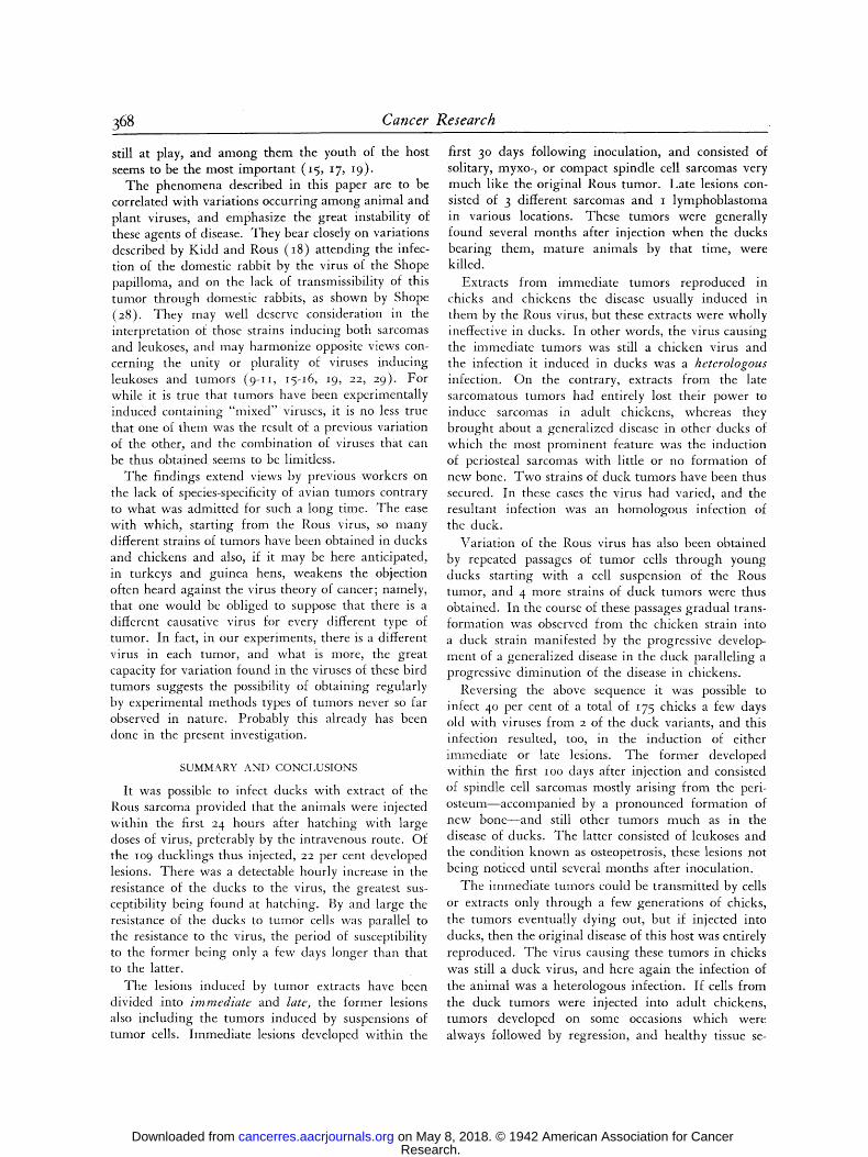
368 Cancer Research
still at play, and among them the youth of the host seems to be the most important (I 5, 17, I9).
The phenomena described in this paper are to be correlated with variations occurring among animal and plant viruses, and emphasize the great instability of these agents of disease. They bear closely on variations described by Kidd and Rous (18) attending the infec- tion of the domestic rabbit by the virus of the Shope papilloma, and on the lack of transmissibility of this tumor through domestic rabbits, as shown by Shope (28). They may well deserve consideration in the interpretation of those strains inducing both sarcomas and leukoses, and may harmonize opposite views con- cerning the unity or plurality of viruses inducing leukoses and tumors (9-II, I5-I6 , ~9, 22, 29). For while it is true that tumors have been experimentally induced containing "mixed" viruses, it is no less true that one of them was the result of a previous variation of the other, and the combination of viruses that can be thus obtained seems to be limitless.
The findings extend views by previous workers on the lack of species-specificity of avian tumors contrary to what was admitted for such a long time. The ease with which, starting from the Rous virus, so many different strains of tumors have been obtained in ducks and chickens and also, if it may be here anticipated, in turkeys and guinea hens, weakens the objection often heard against the virus theory of cancer; namely, that one would bc obliged to suppose that there is a different causative virus for every different type of tumor. In fact, in our experiments, there is a different virus in each tumor, and what is more, the great capacity for variation found in the viruses of these bird tumors suggests the possibility of obtaining regularly by experimental methods types of tumors never so far observed in nature. Probably this already has been done in the present investigation.
SUMMARY AN[) CONCI .USIONS
It was possible to infect ducks with extract of the Rous sarcoma provided that the animals were injected within the first 24 hours after hatching with large doses of virus, preferably by the intravenous route. Of the Io 9 ducklings thus injected, 22 per cent developed lesions. There was a detectable hourly increase in the resistance of the ducks to the virus, the greatest sus- ceptibility being found at hatching. By and large the resistance of the ducks to tumor cells was parallel to the resistance to the virus, the period of susceptibility to the former being only a few days longer than that to the latter.
The lesions induced by tumor extracts have been divided into immediate and late, the former lesions also including the tumors induced by suspensions of tumor cells. Immediate lesions developed within the
first 3 ~ days following inoculation, and consisted of solitary, myxo-, or compact spindle cell sarcomas very much like the original Rous tumor. Late lesions con- sisted of 3 different sarcomas and i lymphoblastoma in various locations. These tumors were generally found several months after injection when the ducks bearing them, mature animals by that time, were killed.
Extracts from immediate tumors reproduced in chicks and chickens the disease usually induced in them by the Rous virus, but these extracts were wholly ineffective in ducks. In other words, the virus causing the immediate tumors was still a chicken virus and the infection it induced in ducks was a keterologous infection. On the contrary, extracts from the late sarcomatous tumors had entirely lost their power to induce sarcomas in adult chickens, whereas they brought about a generalized disease in other ducks of which the most prominent feature was the induction of periosteal sarcomas with little or no formation of new bone. Two strains of duck tumors have been thus secured. In these cases the virus had varied, and the resultant infection was an homologous infection of the duck.
Variation of the Rous virus has also been obtained by repeated passages of tumor cells through young ducks starting with a cell suspension of the Rous tumor, and 4 more strains of duck tumors were thus obtained. In the course of these passages gradual trans- formation was observed from the chicken strain into a duck strain manifested by the progressive develop- ment of a generalized disease in the duck paralleling a progressive diminution of the disease in chickens.
Reversing the above sequence it was possible to infect 4 ~ per cent of a total of I75 chicks a few days old with viruses from 2 of the duck variants, and this infection resulted, too, in the induction of either immediate or late lesions. The former developed within the first ioo days after injection and consisted of spindle cell sarcomas mostly arising from the peri- osteum--accompanied by a pronounced formation of new bone--and still other tumors much as in the disease of ducks. The latter consisted of leukoses and the condition known as osteopetrosis, these lesions not being noticed until several months after inoculation.
The immediate tumors could be transmitted by cells or extracts only through a few generations of chicks, the tumors eventually dying out, but if injected into ducks, then the original disease of this host was entirely reproduced. The virus causing these tumors in chicks was still a duck virus, and here again the infection of the animal was a heterologous infection. If cells from the duck tumors were injected into adult chickens, tumors developed on some occasions which were always followed by regression, and healthy tissue se-
Research. on May 8, 2018. © 1942 American Association for Cancercancerres.aacrjournals.org Downloaded from
Duran-Reynals--Reciprocal Infection of Ducks and Chickens 369
cured by biopsies from these tumors failed to grow in other chickens.
The late lesions could be transmitted only to another generation of chicks, and also to a few adult chickens, the failure of indefinite propagation being attributable to causes which are analyzed. Injections of leukotic lesions induced leukoses but also osteopetrosis, and the reverse was also true.
Practically all the tumors in ducks and chickens r e su l t i ng f r o m e i the r he t e ro logous or h o m o l o g o u s in-
fec t ion g r e w s teadi ly unt i l t h e y ki l led the i r hosts.
D e a t h also resu l ted f r o m the h e m o r r h a g i c lesions
w h i c h qu i t e f r e q u e n t l y deve loped t o g e t h e r w i t h the
neoplas t ic lesions, bu t this subject was o n l y s tud ied in passing.
T h e g r ea t var iabi l i ty , f r o m o per cen t to ion per
cent , in the in fec t iv i ty of c h i c k e n a n d d u c k viruses
for he t e ro logous hosts even w h e n all the r e q u i r e m e n t s
for successful i n fec t ion w e r e fulf i l led, t h e g r e a t var ie ty
of t u m o r s ob ta ined , a n d still o the r facts descr ibed ,
e m p h a s i z e the ins tabi l i ty a n d the g r e a t capac i ty for
va r i a t i on of the viruses i n d u c i n g the t u m o r s he re
s tud ied ; a n d the impl i ca t ions of the f i nd ings in the t u m o r p r o b l e m are discussed.
REFERENCES
~. A~'~ERSON, K. Pathogenesis of Herpes Simplex Virus In- fection in Chick Embryos. Am. ]. Path., 16:137-155. 194 o.
2. Des LmxERis, M. J. A. On the Transplantation of" Rous Fowl Sarcoma No. I into Guinea-Fowls and Tinkeys. Am. J. Cancer, I6:3o7-32i. 1932.
3. DOi.~RR, R., g. Bt.~.'rHq and G. W. ScH~,~m'r. ()bcr das Verhalten des Virus des Rous-Sarkolns in der Blut- zirculation refraktSrer und empfiinglicher Tiere. Ztschr. f. Krebsforsch., 36:256-275. i932.
4. DURAN-REYNALS, F. A Hemorrhagic Disease Occurring in Chicks Inoculated with the Rous and Fujinami Viruses. Yale J. Biol. & Med., 13:77-98. 194o.
5. DVaAN-REY_,,ALS, F. Neutralization of Tumor Viruses by the Blood of Normal Fowls of Different Ages. Yale J. Biol. a Med., 13:61-76. 194o.
6. DURAN-RFNNALS, F. Age Susceptibility of Ducks to the Virus of the Rous Sarcoma and Variations of the Virus in the Duck. Science, 93:5Ol-5O2. I94I.
7. FUJINAMI, A., and S. HATANO. Quoted by Gye (13). 8. FURTH, E. L., and J. FUleTH. The Relation of Leukosis to
Sarcoma of Chickens. I. Sarcoma and Erythroleukosis (Strain i3). J. Exper. Med., 6I:593-615. 1935.
9. FURTH, J. Lymphomatosis, Myelomatosis, and Endothelio- ma of Chickens Caused by a Filterable Agent. II. Mor- phologic Characteristics of the Endotheliomata Caused by this Agent. J. Exper. Med., 89:5oi-517. I934 .
IO. FURTH, J. Lymphomatosis in Relation to Fowl Paralysis. Arch. Path., 20:379-428. I935.
i i. FURTH, J. The Relation of Leukosis to Sarcoma of Chick- ens. II. Mixed Osteochondrosarcoma and Lymphomato- sis. J. Exper. Med., 6I : I27-I55. I935 .
12. GOODPASTURG E. W. Virus Infection of the Chick Embryo. Ann. Int. Med., I 3 : I - I I . I939.
13 . GYE, W. E. A Note on the Propagation of the Fujinami Fowl Myxo-Sarcoma in Ducks. Brit. J. Exper. Path., I2:93-97. I93I.
14. GYE, W. E. The Propagation of Fujinami's Fowl Myxo- Sarcoma in Ducklings. Brit. J. Exper. Path., I3:458-46o. I932 �9
I5. JOHNSOX, E. P. A Study of I~ymphomatosis of Fowls (Fowl Paralysis). Virginia Agricultural Experiment Station. Technical Bullctin, 44:i-22. 1932.
16. Ju~,-CH~:a,, E. Studies on Fowl Paralysis. II. Transmission Experiments. Storrs Agr. Exper. Sta. Bull., 218:1-47. 1937.
17. JI:NGHEP.R, E., and W. I~ANDAUER. Studies on Fowl Paraly- sis. A Condition Resclnblii~g Osteopetrosis (Marble Bone) in the Common Fowl. Storrs Agr. Exper. Sta. Bull., :122:1-34. 1938.
18. Klrm, ]. G., and P. Rous. Cancers Deriving from the Virus Papillomas of Wild Rabbits Under Natural Conditions. J. Exper. Med., 2I:469-494. ~94 o.
I9. LEJ~, C. D., H. L. WI1.CKE, C. MURRAY, and E. W. tIEN- DERSON. Fowl Leukosis. 1. Infect. Dis., 61:I-2o. ~937.
2o. MI:RPI-IY, J. B. The Lymphocyte i n Resistance to Tissue Grafting, Malignant Disease, and Tuberculous Infection. An Experimental Study. Monograph Rockefeller Insti- tute, No. 21, 1926.
21. MuRPm-, J. B., and P. Rous. The Behavior of Chicken Sarcoma Implanted in the Developing Embryo. J. Exper. Med., I8:1 i9-i32. 1912.
22. OBERI.ING, C., and M. Gur Lesions tumorales en rapport avec la leucernie transn-lissible des poules. Bull. Assoc. frang, p. l'dtude du cancer, 2;1:i80-214. 1933.
23. PAPPENHEIMER, A. M., and M. SEmL*N. A Study of Fowi Paralysis (Neurolynlphomatosis Gallinarium). II. Trans- mission Experiments. Storrs Agr. Exper. Sta. Bull., 143: 87-1o2. I926.
24. PURDY, W. J. Thc Fujinami Myxo-Sarconla in Ducks; Resistance to Re-infection. Brit. J. Exper. Path, 14: 297-3 ~ ~933.
25. P~:RD',', W. J. The Propagatioo of the Fujinami Fowl Myxo-Sarcoma in Adult Ducks. Brit. J. Exper. Path., 13:467-472. I932.
26. PUgDY, W. J. The Propagation of the Rous Sarcoma NO. I in Ducklings. Brit. J. Exper. Path., 13:473-479. 1932.
0- 7. Rovs, P., I- B. Mut~mn,, and W. H. TYTLr.R. The RSle of Injury in the Production of a Chicken Sarcoma by a Filterable Agent, I.A,M.A, 83:1751-1752. I912.
28. SI-IOpG R. E . Infectious Papill0matosis of Rabbits. J . Exper. Med., 88:6077624. 1933.
29. STUBBS, E. L., and ]. FURTH. Anemia and Erythrolcukosis Occurring Spontaneously in the Common Fowl. J'. AID_. Vet. M. A., 8I:209-222. 1932.
Research. on May 8, 2018. © 1942 American Association for Cancercancerres.aacrjournals.org Downloaded from
1942;2:343-369. Cancer Res F. Duran-Reynals Tumor-Inducing VirusesThe Reciprocal Infection of Ducks and Chickens with
Updated version
http://cancerres.aacrjournals.org/content/2/5/343.citation
Access the most recent version of this article at:
E-mail alerts related to this article or journal.Sign up to receive free email-alerts
Subscriptions
Reprints and
To order reprints of this article or to subscribe to the journal, contact the AACR Publications
Permissions
Rightslink site. Click on "Request Permissions" which will take you to the Copyright Clearance Center's (CCC)
.http://cancerres.aacrjournals.org/content/2/5/343.citationTo request permission to re-use all or part of this article, use this link
Research. on May 8, 2018. © 1942 American Association for Cancercancerres.aacrjournals.org Downloaded from