the relationship between oral health and
TRANSCRIPT

The Relationship Between Oral Health and
Chronic Obstructive Pulmonary Disease Exacerbations
A THESIS
SUBMITTED TO THE FACULTY OF THE
UNIVERSITY OF MINNESOTA
BY
Arianne Katrina Baldomero, M.D.
IN PARTIAL FULFILLMENT OF THE REQUIREMENTS
FOR THE DEGREE OF
MASTER OF SCIENCE IN CLINICAL RESEARCH
Chris H. Wendt, M.D. and Ken M. Kunisaki, M.D., M.S.
May 2019

©2019
Arianne Katrina Baldomero, M.D.

i
Acknowledgements
First and foremost, I would like to thank my mentors, Chris H. Wendt, M.D. and
Ken M. Kunisaki, M.D., M.S. of the Pulmonary, Critical Care, and Sleep Medicine Section
at the Minneapolis VA Health System and the University of Minnesota for their ongoing
support and encouragement.
I would also like to thank my collaborators/co-investigators for this project: Mariam
Siddiqui, B.D.S, Chi-Yin Lo, D.D.S, Susan Johnson, L.P.N., Ashley Petersen, Ph.D, Alexa
A, Pragman, M.D., Ph.D, and John E. Connett, Ph.D.
We are tremendously grateful to the Veterans who participated in this project.
This research was supported by the National Heart, Lung and Blood Institute
(NHLBI) grant T32 HL007741-23 (AKB), Flight Attendant Medical Research Institute
(CHW), and Veterans Affairs Career Development Award 1IK2CX001095 (AAP). This
material is also the result of work supported with resources of the Minneapolis VA Health
Care System.
The views expressed in this article are those of the authors and do not necessarily
represent the views of the Minneapolis VA Health Care System, the U.S. Department of
Veterans Affairs, the National Institutes of Health, the U.S. Government, or the authors’
affiliated academic institutions. The funders had no role in the conduct, analysis, writing,
or decision to submit for publication, for either the original studies or this current analysis.
This work was originally published by Dove Medical Press Limited. Full citation
is provided below:
Baldomero AK, Siddiqui M, Lo C-Y, Petersen A, Pragman AA, Connett JE,
Kunisaki KM, Wendt CH. The Relationship Between Oral Health and COPD

ii
Exacerbations. International Journal of Chronic Obstructive Pulmonary Disease.
2019;14:881-892.
Available from https://www.dovepress.com/the-relationship-between-oral-health-
and-copd-exacerbations-peer-reviewed-article-COPD

iii
Abstract
Introduction: Poor oral health has been implicated as an independent risk factor for the
development of chronic obstructive pulmonary disease (COPD), but few studies have
evaluated the association between oral health and COPD exacerbations. We aimed to
determine if poor oral health is associated with COPD exacerbations and/or worse
respiratory health.
Methods: We performed a case-control study of oral health among COPD exacerbators
and non-exacerbators. Cases (exacerbators) had experienced ≥1 exacerbation in the
previous 12 months, while controls (non-exacerbators) had no exacerbations in the
previous 24 months. We excluded those with <4 teeth. We evaluated the global oral health
assessment, Oral Health Impact Profile (OHIP-5), dental symptoms/habits, and St.
George’s Respiratory Questionnaire (SGRQ). In a subset, we performed blinded dental
exams to measure bleeding on probing, probing depth, clinical attachment loss,
periodontitis severity, plaque index, gingival index, and carries risk. We evaluated
associations between oral health and COPD exacerbations using logistic regression. Linear
regression was used to assess relationships between oral health and SGRQ scores.
Results: Screened non-exacerbators (n=118) were significantly more likely to have <4
teeth, compared to screened exacerbators (n=100) (44% vs. 30% respectively; p=0.046).
After excluding those with <4 teeth there were 70 cases and 66 controls. Self-reported oral
health and objective dental exam measures did not vary significantly between cases vs.
controls. However, the odds of severe COPD exacerbations requiring hospitalizations

iv
and/or emergency department visits trended higher in those with worse dental exam
compared to those with better dental exam. Worse OHIP-5 was strongly associated with
worse SGRQ scores.
Conclusions: Oral health status was not related to COPD exacerbations, but was associated
with self-reported respiratory health. Larger studies are needed to address oral health as a
potential method to improve respiratory health in patients with COPD.

v
Table of Contents
List of Tables
vi
List of Figures vii
List of Abbreviations viii
Introduction
1
Methods 2
Study Participants 2
Questionnaires 3
Statistical Analysis 4
Sample Size Calculation
6
Results
7
Discussion
9
Conclusion
12
References
13
Appendix 16
Supplementary Material 25
vi
List of Tables
Table 1. Baseline characteristics by COPD
exacerbation status
16
Table 2. Oral health questionnaire responses based on
COPD exacerbation status
19
Table 3. Dental exam measurements comparing
COPD exacerbators vs. non-exacerbators
20
Table 4. St. George’s Respiratory Questionnaire
(SGRQ) mean (SD) scores based on COPD
exacerbation status.
21
Table 5. Association between oral health measures
and COPD exacerbation status.
22
Table 6. Association between dental exam measures
and COPD exacerbation status.
23
Table 7. Association between oral health-related
quality of life (OHIP-5) and respiratory health (total
SGRQ score).
25
Supplementary Material
Table 1S. Oral health questionnaire
26
Table 2S. Periodontitis severity based on probing
depth (PD) and clinical attachment loss (CAL)
27

vii
List of Figures
Figure 1. Study participant flow diagram.
17
Figure 2. Percent of screened participants with <4
teeth vs. 4 teeth, by COPD exacerbation status.
18
Figure 3. Association between dental exam measures
and severity of COPD exacerbations.
24

viii
List of Abbreviations
BOP Bleeding on probing
CAL Clinical attachment loss
CAT COPD Assessment Test
CI Confidence interval
COPD Chronic obstructive pulmonary disease
CRA Carries risk assessment
GI Gingival index
GOLD Global Initiative for Chronic Obstructive Lung Disease
mMRC Modified Medical Research Council
OHIP Oral Health Impact Profile
OHRQoL Oral health-related quality of life
OR Odds ratio
PD Probing depth
PI Plaque index
SGRQ St. George’s Respiratory Questionnaire
VA Veterans Affairs

1
Introduction
Chronic obstructive pulmonary disease (COPD) exacerbations are a major cause
of morbidity, seriously impair quality of life, and can result in irreversible loss of lung
function.1-3 A severe COPD exacerbation requiring hospitalization is associated with high
mortality both in the hospital and after discharge.2 Prevention of COPD exacerbations is
an important aspect of COPD management.4 Therefore, exploration of risk factors and
identification of patients who are susceptible to COPD exacerbations is needed.
Approximately 50% of COPD exacerbations are attributed to bacterial infections.
Published studies have demonstrated increased microbial biomass and microbial diversity
in COPD patients compared to healthy adults.5-7 In addition, oral and nasal bacteria have
been identified in the COPD lung tissue microbiota, suggesting aspiration of oral
secretions as a major source of the COPD lung microbiota.5-7 This is consistent with
observations that COPD patients are prone to aspiration of oral secretions due to reduced
laryngotracheal mechanosensitivity8 and decreased airway clearance from impaired
mucociliary function.9 These findings highlight the potential impact of oral health in
COPD. Thus, we hypothesize that COPD exacerbations are likely associated with poor
oral health status.
Periodontal disease and poor oral health have been associated with a number of
systemic diseases, including COPD. A meta-analysis evaluating 14 observational studies
demonstrated an association between periodontal disease and COPD10, but few studies
have evaluated the association between oral health and COPD exacerbations.11-14 In this
study we compared the oral health status of COPD exacerbators and non-exacerbators to
determine if COPD exacerbations are related to worse oral health.

2
Methods
Using a case-control design, we administered questionnaires via phone interview
or in-person visits at the Minneapolis Veterans Affairs Health Care System.
Comprehensive dental examination was performed on a subset of participants.
Study participants
Participants with COPD were recruited from the Minneapolis Veterans Affairs
Health Care System. Inclusion criteria included participants between 40-80 years of age
with COPD. We defined COPD in standard fashion as recommended by the American
College of Physicians, American College of Chest Physicians, and American Thoracic
Society15 – respiratory symptoms with spirometry-confirmed airflow obstruction defined
as FEV1/FVC <0.70 in those with ≥10 pack-year smoking history – and further included
only those with FEV1 <70% of predicted normal. Exclusion criteria included history of
asthma, presence of <4 teeth, and undergoing active treatment with chemotherapy for
malignancy (exceptions: radiation or hormonal therapy for prostate or breast cancer, and
non-metastatic skin cancer). Cases were defined as having at least one COPD
exacerbation in the previous 12 months. COPD exacerbations were defined as taking
antibiotics and/or oral corticosteroids for respiratory symptoms, or hospitalization or
emergency department visit for respiratory illness. Severe exacerbations were defined as
requiring an emergency department visit and/or hospitalization for the exacerbation.
Controls had COPD but with no exacerbations in the previous 24 months. Case or control
status was verified by medical chart review and participant interviews.
3
The study was approved by the Minneapolis VA Institutional Review Board. All
study participants provided informed consent before participating in the study.
Participants who completed the questionnaires by phone provided verbal informed
consent, while participants who had dental examinations and in-person visits provided
written informed consent. Both methods of informed consent were reviewed and
approved by the Minneapolis VA Institutional Review Board.
Questionnaires
Oral Health Questionnaire
Our oral health questionnaire assessed demographic information, history of
COPD exacerbations, inhaler use, dental symptoms, dental care habits, a 1-item global
oral health status assessment, and the 5-item version of the Oral Health Impact Profile
(OHIP) (Table 1S). OHIP is the most widely used oral health-related quality of life
(OHRQoL) instrument to assess the impact of oral health disorders and dental
interventions.16 OHIP captures four correlated aspects of patient-perceived OHRQoL:
oral function, orofacial pain, orofacial appearance, and psychological impact. The
responses were classified using the Likert scale with five choices ranging from “never”
(1) to “very often” (5), with a score of 5 reflecting the most severe oral health for each
item, and the overall score ranging from 4 to 20. There is no established minimal
clinically important difference for the OHIP.

4
St. George’s Respiratory Questionnaire (SGRQ)
The SGRQ is a 50-item questionnaire developed to assess respiratory health status
in patients with obstructive lung diseases. The questions evaluate three domains:
symptoms, activity, and impact. The SGRQ is scored on a scale of 0 to 100, with higher
scores reflecting worse respiratory health status. The minimum clinically important
difference in the SGRQ is widely accepted as 4 units.17
Dental examination
All participants were offered a comprehensive dental examination.
Comprehensive dental examinations were performed on a subset (27 cases and 29
controls) of participants by two dentists. The dentists were blinded to the case-control
status of the participants. Assessments included periodontitis severity, bleeding on
probing (BOP), gingival index (GI), plaque index (PI), and carries risk assessment
(CRA).
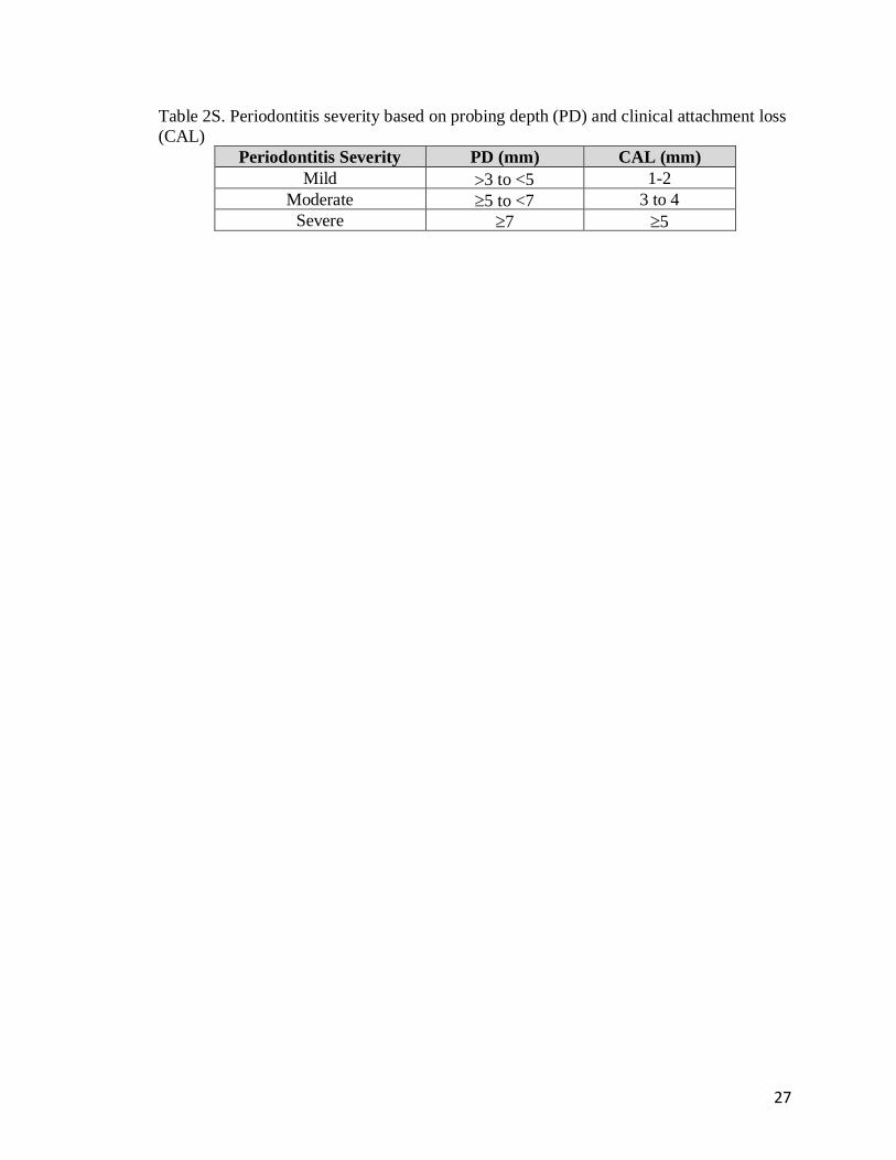
Periodontitis severity (mild, moderate, and severe) was determined by probing
depth (PD, scored as 3, 3 to <5, 5 to <7, and 7+ mm) and clinical attachment loss
(CAL, scored as <1, 1 to 2, or 5+ mm) based on the involvement of at least 30% of the
entire dentition, according to the American Academy of Periodontology Task Force
Report (Table 2S).18 PD evaluations were carried out by measuring pocket depths using a
periodontal probe at six points per tooth. The probe, positioned parallel to the long axis of
the tooth at each site, is inserted until the probe tip encounters the resistance of the
junctional epithelium. The probe is then moved up and down in short strokes and forward
in 1-mm increments. CAL was the measured distance between cemento-enamel junction
5
and the free gingival margin.19 PD and CAL measurements were rounded up to the
nearest millimeter. The higher category of severity was used if PD and CAL were in two
different categories.
BOP was assessed by probing gently along the wall of soft tissue of the gingival
sulcus and was scored as yes or no for presence or absence of bleeding, respectively.
GI was used to assess evidence of inflammation in the gingival tissues
characterized by redness, swelling, and BOP. Each of the four gingival areas (buccal,
mesial, distal, and lingual) of the tooth was scored from 0 to 3 based on Löe’s Gingival
Index System: 0 – normal gingiva, 1 – mild inflammation, 2 – moderate inflammation,
and 3 – severe inflammation.20 The GI score for the tooth was calculated by adding the
four scores then dividing by four. The final GI recorded was the sum of all values from
each tooth divided by the number of teeth examined.20
PI indicates soft deposits and calculi at the gingival margin and interproximally.
The Modified Plaque Scoring System was used to record PI measurements and was
scored as 0 – no plaque, 1 – separate flecks of plaque at the cervical margin of the tooth,
2 – a thin continuous band of plaque up to 1 mm at the cervical margin of the tooth, 3 – a
band of plaque wider than 1 mm covering less than one-third of the crown of the tooth, 4
– plaque covering at least one-third but less than two-thirds of the crown of the tooth, and
5 – plaque covering two-thirds or more of the crown of the tooth.21
The American Dental Association Carries Risk Assessment Form was used to
evaluate the overall dental carries risk categorized as low, moderate, or high.

6
Statistical analysis
We used Chi-square tests for categorical variables and two-sample t-tests for
continuous variables to test the differences in demographic and clinical characteristics
between cases and controls. We used logistic regression models to estimate the
associations between oral health and COPD exacerbation (case:control) status, adjusting
for potential confounders including inhaler use (inhaled corticosteroids and
anticholinergic inhalers) and FEV1 % predicted. The variables used for adjustment in the
models were based on the baseline characteristics that were found to be significantly
different between exacerbators and non-exacerbators (Table 1). Additionally, we fit a
logistic regression model with the outcome of severe vs. mild exacerbation for each
dental exam measure (periodontitis severity, BOP, PD, CAL PI, GI, and CRA). All but
BOP were ordinal scales where higher values indicated worse oral health. We modeled
these scales as continuous predictors to assess for trends between worse oral health and
exacerbation severity. For this association between dental exam measures and severity of
COPD exacerbations, only unadjusted logistic regression analyses were performed due to
the small sample size. Multivariate linear regression model was used to assess
relationships between oral health and SGRQ scores with adjustment for the same
covariates.
Sample size calculation
We initially planned to enroll 360 participants (120 cases and 240 controls), to
provide 83% power to detect an odds ratio of 2.0 at a two-sided significance level of
0.05. We anticipated that 80% of screened participants would be eligible and provide

7
complete data, so we planned to screen 150 cases and 300 controls. However, due to an
unexpectedly high proportion of participants being ineligible due to having <4 teeth,
especially amongst potential controls, we decided to perform an interim analysis of our
primary outcome OHIP-5 data and re-evaluate feasibility of this observational study. This
analysis indicated futility, so we halted enrollment at 70 cases and 66 controls.
Results
We screened 100 COPD exacerbators and 118 non-exacerbators (Figure 1). Of
these, only 70 (70%) and 66 (56%), respectively, met the inclusion criteria of having ≥4
teeth (p=0.046 for difference in proportion with this exclusion criteria between cases and
controls) (Figure 2). Dental examinations were performed on 27 (39%) exacerbators and
29 (44%) non-exacerbators.
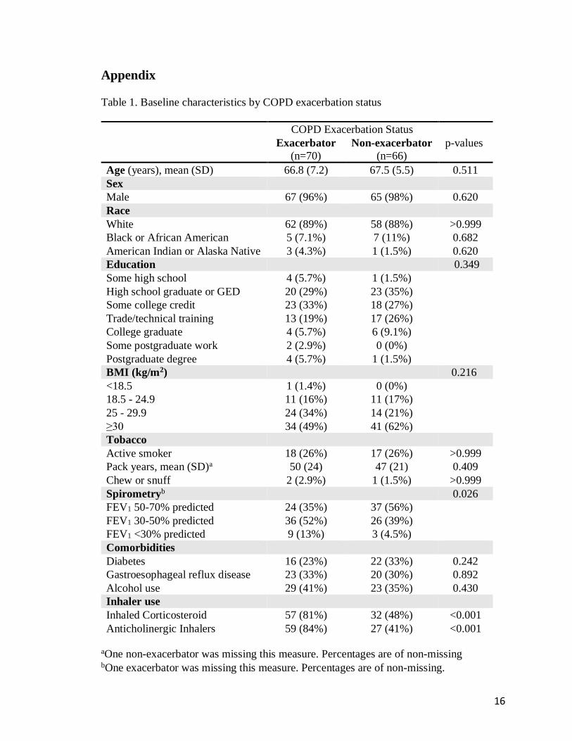
The baseline characteristics of the participants that met eligibility are summarized
in Table 1 according to COPD exacerbation status. As expected, there were significant
differences between cases and controls in inhaler use (both inhaled corticosteroids and
anticholinergics were more commonly used in cases) and FEV1 % predicted, therefore
inhaler use and FEV1 % predicted were included as covariates in adjusted models.
Exacerbators tended to have greater inhaler use and lower FEV1 % predicted compared to
non-exacerbators, consistent with previously published studies.3,4,22-24 No other
statistically significant differences were found between cases and controls including the
oral health questionnaire responses, dental exam measurements, and SGRQ scores,
shown in Table 2, Table 3, and Table 4, respectively.

8
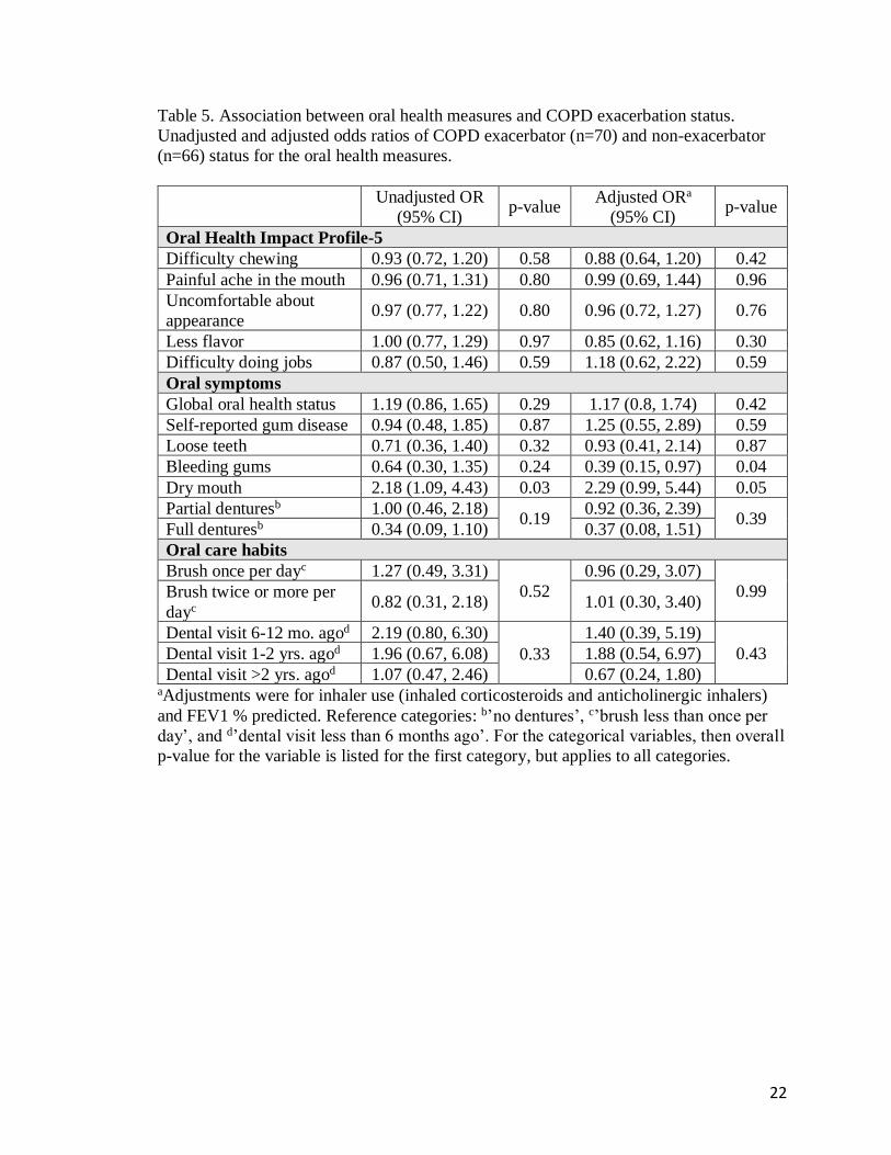
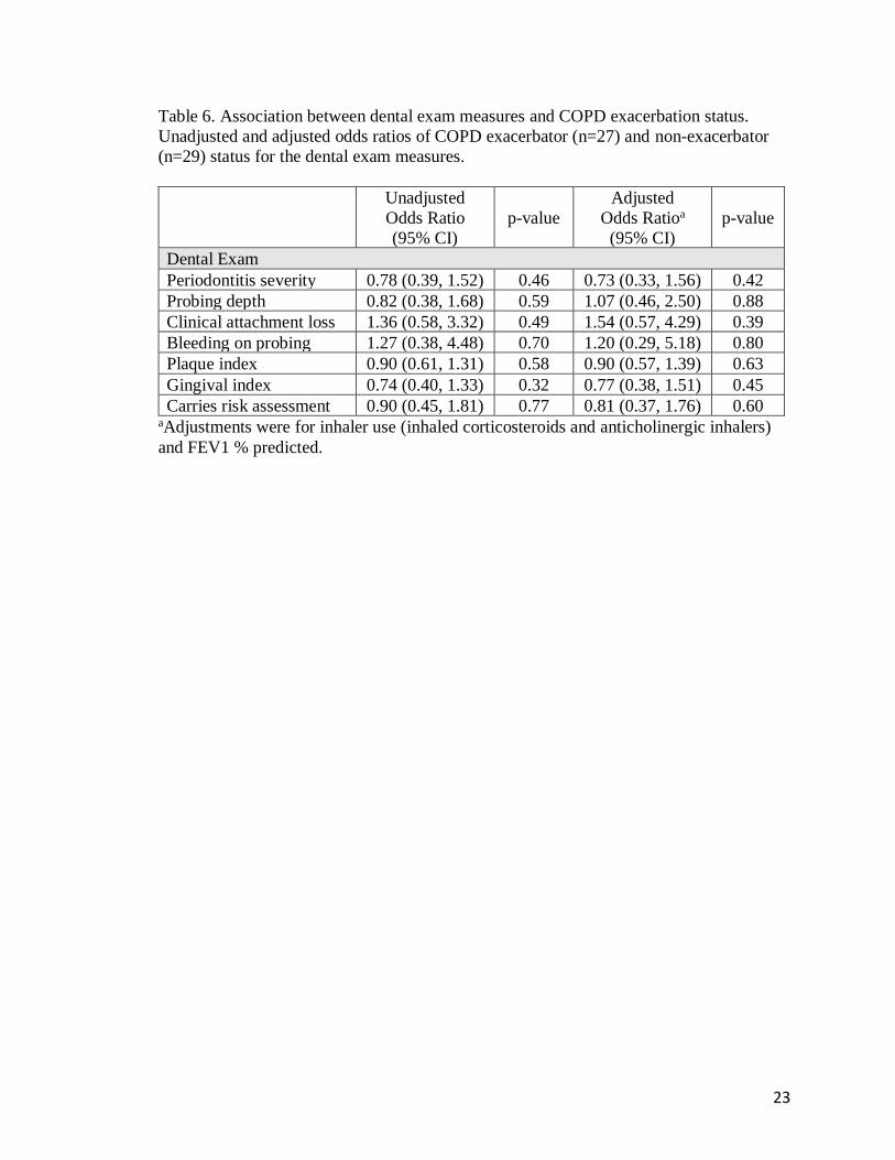
The unadjusted and adjusted odds ratios (OR) for self-reported oral health status
(Table 5) and dental exam measures (Table 6) did not vary significantly between
exacerbators and non-exacerbators. However, there was a trend towards higher odds of
exacerbations in those with ‘dry mouth’ in both unadjusted and adjusted models
(unadjusted OR 2.18; 95% CI 1.09 to 4.43; p-value 0.03 and adjusted OR 2.29; 95% CI
0.99 to 5.44; p-value 0.05). Self-reported ‘bleeding gums’ was less likely among
exacerbators compared to non-exacerbators (adjusted OR 0.39; 95% CI 0.15 to 0.97; p-
value 0.04).
The associations between dental exam measurements (Table 3) and severe COPD
exacerbations are shown in Figure 3. Of the exacerbators who underwent dental exam,
19/27 had severe exacerbations (requiring emergency department visit and/or
hospitalization), while 7/27 had mild exacerbations. The unadjusted odds ratios of severe
exacerbations relative to mild exacerbations trended higher in those with worse
categories of measurements for periodontitis severity (mild, moderate severe), PD (≤3, >3
to <5, ≥5 to <7, ≥7), CAL (<1, 1 to 2, 3 to 4, ≥5), BOP (yes/no), PI (score 0, 1, 2, 3, 4, 5),
GI (score 0, 1, 2, 3), and CRA (low, moderate, high) compared to those with better dental
exam measurements. However, statistical significance was not reached. Adjustment for
covariates was not performed due to the small sample size.
The correlations between OHRQoL and respiratory health are shown in Table 7.
The adjusted estimates reflect the difference in mean total SGRQ scores between the
exacerbators and non-exacerbators with a 1-point difference in OHIP. Worse OHRQoL
was strongly associated with worse respiratory health scores. In addition, ‘painful ache in
9
the mouth’ and ‘difficulty doing jobs’ were also clinically significantly associated with
worse respiratory health scores.
Discussion
We aimed to determine if COPD exacerbation status (exacerbator vs. non-
exacerbator) is associated with oral health. In this study, we did not find that poor oral
health, as determined by self-report or by objective dental examination, was associated
with COPD exacerbations. We did find a relationship between poor OHRQoL and worse
respiratory health status and a possible trend towards worse dental exam measures among
those with severe COPD exacerbations.
There are several observational studies linking periodontitis with COPD. A meta-
analysis of 14 observational studies by Zeng et al showed an association between
periodontitis and COPD diagnosis with a pooled OR of 2.09 (95% CI: 1.48 to 2.91).10
Published studies also show that COPD patients have fewer remaining teeth compared
with healthy controls.25-28 However, few studies have evaluated the relationship between
COPD exacerbation rates with periodontitis and dentition status.11-14
One cross-sectional study by Liu et al. evaluating the association between oral
hygiene and periodontal health in COPD exacerbations in China showed increased COPD
exacerbations in those with fewer remaining teeth.11 In contrast, we found that fewer
teeth were associated with non-exacerbator status. However, the definitions of ‘fewer’
teeth and COPD exacerbation status varied between these two studies. Liu et al defined
fewer teeth as ≤25 teeth vs. >25 teeth while we compared <4 teeth vs. ≥4 teeth (most of
our patients with <4 teeth were edentate).11 Additionally, Liu et al. did not find

10
significant differences in periodontal health indices including PD, CAL, and bleeding on
probing between exacerbators and non-exacerbators, similar to our findings.11
We found a trend towards more severe COPD exacerbations requiring emergency
room visits and/or hospitalizations in those with worse periodontal health indices,
although this association did not reach statistical significance, possibly due to small
sample size. Shen et al. found a strong positive correlation between incident periodontal
disease and frequency of emergency room visits and hospitalizations for COPD
exacerbation, which support our findings.12
We theorize that the oral microbiota and oral inflammation play key roles in this
relationship between dentition status and COPD exacerbations.5-7 The oral microbiota is a
major source of the lung microbiota. 5-7 Lung inflammation may be mediated by
aspiration of inflammatory cytokines from the oral cavity, or aspiration of a dysbiotic oral
microbiota may lead to dysbiosis of the lung microbiota. The presence of diseased teeth
allows for extensive biofilm formation, which then could be aspirated into the lungs,
thereby contributing to COPD exacerbations.29 Full-mouth teeth extraction has been
found to significantly reduce the burden of periodontopathogens.19 Absence of any teeth
is likely associated with a lower burden of periodontal pathogens compared to those with
the presence of potentially diseased teeth30,31 and may explain our observation that non-
exacerbators were more likely to have fewer or no teeth.
In this study, we also show a relationship between poor oral health-related quality
of life as measured by OHIP-5 and respiratory health status (SGRQ total score),
regardless of the COPD exacerbation status. We used the OHIP, the most widely used
oral health-related quality of life instrument, to assess the impact of oral health.16 Prior

11
studies have demonstrated good correlation of the OHIP with clinical oral examination
among the Veteran population.32 However, limitations related to the OHIP-5 are the
absence of an established minimal clinically important difference. Similarly, Zhou et al
report an association between poor periodontal health and low quality of life in COPD
patients.33 Correlation of our current findings with the Global Initiative for Chronic
Obstructive Lung Disease (GOLD) ABCD stages that incorporates both spirometric
severity and symptom burden would have been interesting, however, we did not collect
data for the Modified Medical Research Council (mMRC) Dyspnea Scale and COPD
Assessment Test (CAT) scores. The observed relationship between OHIP-5 and SGRQ
total score suggest the need for studies to address oral health as a potential method to
improve respiratory health status in patients with COPD.
Our study has several limitations. First, case-control studies are subject to
selection bias and information bias, and therefore directional causality cannot be
established. In addition, questionnaire studies are susceptible to response bias. This study
was also conducted in a single center, predominantly white male population with a
relatively modest sample size. Larger studies with more diverse populations may provide
further insight regarding the role of oral health in COPD outcomes and quality of life. We
also did not meet our original target sample size, possibly limiting statistically significant
findings, although we chose to close the study after interim analyses suggested that larger
sample sizes would be futile for detecting differences in oral health between cases and
controls. Lastly, we assessed comorbidities that are most likely to affect oral health status
such as diabetes, GERD, and alcohol use, but we acknowledge that other comorbidities
that we did not assess could also play a role.

12
Conclusions
Oral health status was not related to COPD exacerbation status, but was
associated with patient-reported respiratory health status. Larger studies are needed to
address oral health as a potential method to improve respiratory health status in patients
with COPD.
13
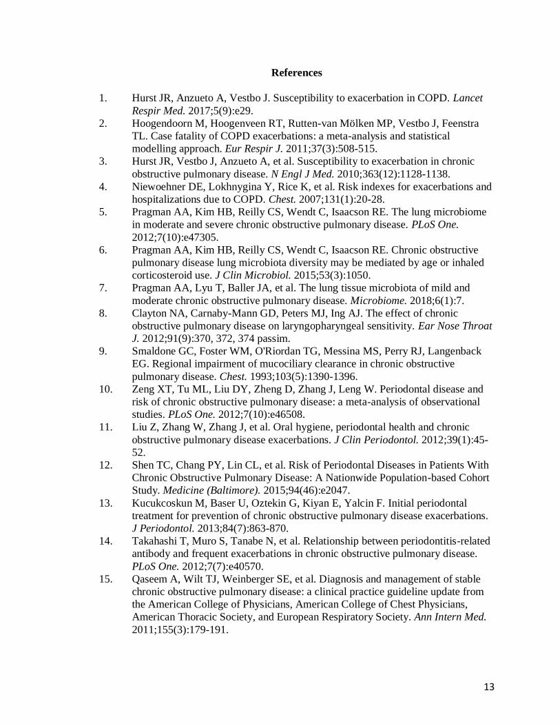
References
1. Hurst JR, Anzueto A, Vestbo J. Susceptibility to exacerbation in COPD. Lancet
Respir Med. 2017;5(9):e29.
2. Hoogendoorn M, Hoogenveen RT, Rutten-van Mölken MP, Vestbo J, Feenstra
TL. Case fatality of COPD exacerbations: a meta-analysis and statistical
modelling approach. Eur Respir J. 2011;37(3):508-515.
3. Hurst JR, Vestbo J, Anzueto A, et al. Susceptibility to exacerbation in chronic
obstructive pulmonary disease. N Engl J Med. 2010;363(12):1128-1138.
4. Niewoehner DE, Lokhnygina Y, Rice K, et al. Risk indexes for exacerbations and
hospitalizations due to COPD. Chest. 2007;131(1):20-28.
5. Pragman AA, Kim HB, Reilly CS, Wendt C, Isaacson RE. The lung microbiome
in moderate and severe chronic obstructive pulmonary disease. PLoS One.
2012;7(10):e47305.
6. Pragman AA, Kim HB, Reilly CS, Wendt C, Isaacson RE. Chronic obstructive
pulmonary disease lung microbiota diversity may be mediated by age or inhaled
corticosteroid use. J Clin Microbiol. 2015;53(3):1050.
7. Pragman AA, Lyu T, Baller JA, et al. The lung tissue microbiota of mild and
moderate chronic obstructive pulmonary disease. Microbiome. 2018;6(1):7.
8. Clayton NA, Carnaby-Mann GD, Peters MJ, Ing AJ. The effect of chronic
obstructive pulmonary disease on laryngopharyngeal sensitivity. Ear Nose Throat
J. 2012;91(9):370, 372, 374 passim.
9. Smaldone GC, Foster WM, O'Riordan TG, Messina MS, Perry RJ, Langenback
EG. Regional impairment of mucociliary clearance in chronic obstructive
pulmonary disease. Chest. 1993;103(5):1390-1396.
10. Zeng XT, Tu ML, Liu DY, Zheng D, Zhang J, Leng W. Periodontal disease and
risk of chronic obstructive pulmonary disease: a meta-analysis of observational
studies. PLoS One. 2012;7(10):e46508.
11. Liu Z, Zhang W, Zhang J, et al. Oral hygiene, periodontal health and chronic
obstructive pulmonary disease exacerbations. J Clin Periodontol. 2012;39(1):45-
52.
12. Shen TC, Chang PY, Lin CL, et al. Risk of Periodontal Diseases in Patients With
Chronic Obstructive Pulmonary Disease: A Nationwide Population-based Cohort
Study. Medicine (Baltimore). 2015;94(46):e2047.
13. Kucukcoskun M, Baser U, Oztekin G, Kiyan E, Yalcin F. Initial periodontal
treatment for prevention of chronic obstructive pulmonary disease exacerbations.
J Periodontol. 2013;84(7):863-870.
14. Takahashi T, Muro S, Tanabe N, et al. Relationship between periodontitis-related
antibody and frequent exacerbations in chronic obstructive pulmonary disease.
PLoS One. 2012;7(7):e40570.
15. Qaseem A, Wilt TJ, Weinberger SE, et al. Diagnosis and management of stable
chronic obstructive pulmonary disease: a clinical practice guideline update from
the American College of Physicians, American College of Chest Physicians,
American Thoracic Society, and European Respiratory Society. Ann Intern Med.
2011;155(3):179-191.
14
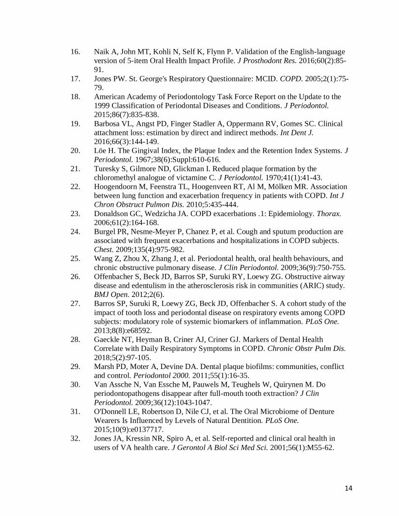
16. Naik A, John MT, Kohli N, Self K, Flynn P. Validation of the English-language
version of 5-item Oral Health Impact Profile. J Prosthodont Res. 2016;60(2):85-
91.
17. Jones PW. St. George's Respiratory Questionnaire: MCID. COPD. 2005;2(1):75-
79.
18. American Academy of Periodontology Task Force Report on the Update to the
1999 Classification of Periodontal Diseases and Conditions. J Periodontol.
2015;86(7):835-838.
19. Barbosa VL, Angst PD, Finger Stadler A, Oppermann RV, Gomes SC. Clinical
attachment loss: estimation by direct and indirect methods. Int Dent J.
2016;66(3):144-149.
20. Löe H. The Gingival Index, the Plaque Index and the Retention Index Systems. J
Periodontol. 1967;38(6):Suppl:610-616.
21. Turesky S, Gilmore ND, Glickman I. Reduced plaque formation by the
chloromethyl analogue of victamine C. J Periodontol. 1970;41(1):41-43.
22. Hoogendoorn M, Feenstra TL, Hoogenveen RT, Al M, Mölken MR. Association
between lung function and exacerbation frequency in patients with COPD. Int J
Chron Obstruct Pulmon Dis. 2010;5:435-444.
23. Donaldson GC, Wedzicha JA. COPD exacerbations .1: Epidemiology. Thorax.
2006;61(2):164-168.
24. Burgel PR, Nesme-Meyer P, Chanez P, et al. Cough and sputum production are
associated with frequent exacerbations and hospitalizations in COPD subjects.
Chest. 2009;135(4):975-982.
25. Wang Z, Zhou X, Zhang J, et al. Periodontal health, oral health behaviours, and
chronic obstructive pulmonary disease. J Clin Periodontol. 2009;36(9):750-755.
26. Offenbacher S, Beck JD, Barros SP, Suruki RY, Loewy ZG. Obstructive airway
disease and edentulism in the atherosclerosis risk in communities (ARIC) study.
BMJ Open. 2012;2(6).
27. Barros SP, Suruki R, Loewy ZG, Beck JD, Offenbacher S. A cohort study of the
impact of tooth loss and periodontal disease on respiratory events among COPD
subjects: modulatory role of systemic biomarkers of inflammation. PLoS One.
2013;8(8):e68592.
28. Gaeckle NT, Heyman B, Criner AJ, Criner GJ. Markers of Dental Health
Correlate with Daily Respiratory Symptoms in COPD. Chronic Obstr Pulm Dis.
2018;5(2):97-105.
29. Marsh PD, Moter A, Devine DA. Dental plaque biofilms: communities, conflict
and control. Periodontol 2000. 2011;55(1):16-35.
30. Van Assche N, Van Essche M, Pauwels M, Teughels W, Quirynen M. Do
periodontopathogens disappear after full-mouth tooth extraction? J Clin
Periodontol. 2009;36(12):1043-1047.
31. O'Donnell LE, Robertson D, Nile CJ, et al. The Oral Microbiome of Denture
Wearers Is Influenced by Levels of Natural Dentition. PLoS One.
2015;10(9):e0137717.
32. Jones JA, Kressin NR, Spiro A, et al. Self-reported and clinical oral health in
users of VA health care. J Gerontol A Biol Sci Med Sci. 2001;56(1):M55-62.

15
33. Zhou X, Wang Z, Song Y, Zhang J, Wang C. Periodontal health and quality of
life in patients with chronic obstructive pulmonary disease. Respir Med.
2011;105(1):67-73.
16
Appendix
Table 1. Baseline characteristics by COPD exacerbation status
aOne non-exacerbator was missing this measure. Percentages are of non-missing bOne exacerbator was missing this measure. Percentages are of non-missing.
COPD Exacerbation Status
Exacerbator
(n=70)
Non-exacerbator
(n=66)
p-values
Age (years), mean (SD) 66.8 (7.2) 67.5 (5.5) 0.511
Sex
Male 67 (96%) 65 (98%) 0.620
Race
White 62 (89%) 58 (88%) >0.999
Black or African American 5 (7.1%) 7 (11%) 0.682
American Indian or Alaska Native 3 (4.3%) 1 (1.5%) 0.620
Education 0.349
Some high school 4 (5.7%) 1 (1.5%)
High school graduate or GED 20 (29%) 23 (35%)
Some college credit 23 (33%) 18 (27%)
Trade/technical training 13 (19%) 17 (26%)
College graduate 4 (5.7%) 6 (9.1%)
Some postgraduate work 2 (2.9%) 0 (0%)
Postgraduate degree 4 (5.7%) 1 (1.5%)
BMI (kg/m2) 0.216
<18.5 1 (1.4%) 0 (0%)
18.5 - 24.9 11 (16%) 11 (17%)
25 - 29.9 24 (34%) 14 (21%)
≥30 34 (49%) 41 (62%)
Tobacco
Active smoker 18 (26%) 17 (26%) >0.999
Pack years, mean (SD)a 50 (24) 47 (21) 0.409
Chew or snuff 2 (2.9%) 1 (1.5%) >0.999
Spirometryb 0.026
FEV1 50-70% predicted 24 (35%) 37 (56%)
FEV1 30-50% predicted 36 (52%) 26 (39%)
FEV1 <30% predicted 9 (13%) 3 (4.5%)
Comorbidities
Diabetes 16 (23%) 22 (33%) 0.242
Gastroesophageal reflux disease 23 (33%) 20 (30%) 0.892
Alcohol use 29 (41%) 23 (35%) 0.430
Inhaler use
Inhaled Corticosteroid 57 (81%) 32 (48%) <0.001
Anticholinergic Inhalers 59 (84%) 27 (41%) <0.001

17
Figure 1. Study participant flow diagram

18
Figure 2. Percent of screened participants with <4 teeth vs. 4 teeth, by COPD
exacerbation status. Non-exacerbators (n=118) were significantly more likely to have <4
teeth compared to exacerbators (n=110) (p=0.046).
30
70
44
56
0
10
20
30
40
50
60
70
80
<4 teeth ≥4 teeth
% P
art
icip
an
ts
Exacerbators
Non-exacerbators

19
Table 2. Oral health questionnaire responses based on COPD exacerbation status
COPD Exacerbation Status
Exacerbator
(n=70)
Non-exacerbator
(n=66)
p-value
Global oral health status 0.30
Poor 17 (24%) 21 (32%)
Fair 24 (34%) 25 (38%)
Good 22 (31%) 12 (18%)
Very good 3 (4.3%) 6 (9.1%)
Excellent 4 (5.7%) 2 (3.0%)
Self-reported gum disease 34 (49%) 33 (50%) 0.94
Oral symptoms
Loose teeth 28 (40%) 32 (48%) 0.37
Bleeding gums 17 (24%) 22 (33%) 0.30
Dry mouth 48 (69%) 33 (50%) 0.02
Dentures
Partial denture 20 (29%) 17 (26%) 0.09
Full denture (upper or lower) 4 (5.7%) 10 (15%) 0.49
Frequency of teeth
brushing or denture
cleaninga
0.75
Never 1 (1.5%) 1 (1.5%)
Less than once a day 11 (16%) 11 (17%)
Once a day 33 (49%) 26 (39%)
Two times per day 21 (31%) 24 (36%)
More than two times per day 2 (2.9%) 4 (6.1%)
Time since last dental visitb 0.36
<6 months ago 24 (36%) 30 (45%)
6-12 months ago 14 (21%) 8 (12%)
1-2 years ago 11 (16%) 7 (11%)
>2 years ago 18 (27%) 21 (32%)
Teeth/denture cleaning
Toothbrush 66 (94%) 60 (91%) 0.46
Dental floss 38 (54%) 32 (48%) 0.56
Mouthwash 36 (51%) 28 (42%) 0.33
Denture cleanser solution 16 (23%) 16 (24%) 0.81
aTwo frequent exacerbators are missing this measure. Percentages are of non-missing. bThree frequent exacerbators are missing this measure. Percentages are of non-
missing.

20
Table 3. Dental exam measurements comparing COPD exacerbators vs. non-exacerbators
COPD Exacerbation Status
Exacerbator
(n=27)
Non-exacerbator
(n=29) p-value
Probing Depth (mm)a 0.36
≤3 12 (46%) 14 (48%)
>3 and <5 12 (46%) 10 (34%)
≥5 and <7 2 (7.7%) 4 (14%)
≥7 0 (0%) 1 (3.4%)
Bleeding on Probinga 20 (77%) 21 (72%) 0.71
Clinical Attachment
Loss (mm)a 0.65
<1 0 (0%) 0 (0%)
1 to 2 1 (3.8%) 3 (10%)
3 to 4 12 (46%) 13 (45%)
≥5 13 (50%) 13 (45%)
Severity of
Periodontitisa 0.68
Mild 7 (27%) 5 (17%)
Moderate 8 (31%) 10 (34%)
Severe 11 (42%) 14 (48%)
Plaque Indexa 0.54
Score 0 2 (7.7%) 2 (6.9%)
Score 1 4 (15%) 4 (14%)
Score 2 7 (27%) 5 (17%)
Score 3 5 (19%) 5 (17%)
Score 4 5 (19%) 12 (41%)
Score 5 3 (12%) 1 (3.4%)
Gingival Indexa 0.38
Score 0 2 (7.7%) 3 (10%)
Score 1 11 (42%) 6 (21%)
Score 2 8 (31%) 12 (41%)
Score 3 5 (19%) 8 (28%)
Caries Risk
Assessment 0.69
Low 7 (26%) 5 (17%)
Moderate 9 (33%) 13 (45%)
High 11 (41%) 11 (38%)
aData for one exacerbator is missing. Percentages are of non-missing.

21
Table 4. St. George’s Respiratory Questionnaire (SGRQ) mean (SD) scores
based on COPD exacerbation status.
COPD Exacerbation Status
Exacerbator
(n=70)
Non-exacerbator
(n=66)
SGRQ (mean, SD)
Symptom 56 (23) 49 (22)
Activity 70 (22) 65 (22)
Impact 40 (20) 30 (19)
Total 52 (18) 44 (18)
22
Table 5. Association between oral health measures and COPD exacerbation status.
Unadjusted and adjusted odds ratios of COPD exacerbator (n=70) and non-exacerbator
(n=66) status for the oral health measures.
Unadjusted OR
(95% CI) p-value
Adjusted ORa
(95% CI) p-value
Oral Health Impact Profile-5
Difficulty chewing 0.93 (0.72, 1.20) 0.58 0.88 (0.64, 1.20) 0.42
Painful ache in the mouth 0.96 (0.71, 1.31) 0.80 0.99 (0.69, 1.44) 0.96
Uncomfortable about
appearance 0.97 (0.77, 1.22) 0.80 0.96 (0.72, 1.27) 0.76
Less flavor 1.00 (0.77, 1.29) 0.97 0.85 (0.62, 1.16) 0.30
Difficulty doing jobs 0.87 (0.50, 1.46) 0.59 1.18 (0.62, 2.22) 0.59
Oral symptoms
Global oral health status 1.19 (0.86, 1.65) 0.29 1.17 (0.8, 1.74) 0.42
Self-reported gum disease 0.94 (0.48, 1.85) 0.87 1.25 (0.55, 2.89) 0.59
Loose teeth 0.71 (0.36, 1.40) 0.32 0.93 (0.41, 2.14) 0.87
Bleeding gums 0.64 (0.30, 1.35) 0.24 0.39 (0.15, 0.97) 0.04
Dry mouth 2.18 (1.09, 4.43) 0.03 2.29 (0.99, 5.44) 0.05
Partial denturesb 1.00 (0.46, 2.18) 0.19
0.92 (0.36, 2.39) 0.39
Full denturesb 0.34 (0.09, 1.10) 0.37 (0.08, 1.51)
Oral care habits
Brush once per dayc 1.27 (0.49, 3.31)
0.52
0.96 (0.29, 3.07)
0.99 Brush twice or more per
dayc 0.82 (0.31, 2.18) 1.01 (0.30, 3.40)
Dental visit 6-12 mo. agod 2.19 (0.80, 6.30) 1.40 (0.39, 5.19)
0.43 Dental visit 1-2 yrs. agod 1.96 (0.67, 6.08) 0.33
1.88 (0.54, 6.97)
Dental visit >2 yrs. agod 1.07 (0.47, 2.46) 0.67 (0.24, 1.80) aAdjustments were for inhaler use (inhaled corticosteroids and anticholinergic inhalers)
and FEV1 % predicted. Reference categories: b’no dentures’, c’brush less than once per
day’, and d’dental visit less than 6 months ago’. For the categorical variables, then overall
p-value for the variable is listed for the first category, but applies to all categories.
23
Table 6. Association between dental exam measures and COPD exacerbation status.
Unadjusted and adjusted odds ratios of COPD exacerbator (n=27) and non-exacerbator
(n=29) status for the dental exam measures.
Unadjusted
Odds Ratio
(95% CI)
p-value
Adjusted
Odds Ratioa
(95% CI)
p-value
Dental Exam
Periodontitis severity 0.78 (0.39, 1.52) 0.46 0.73 (0.33, 1.56) 0.42
Probing depth 0.82 (0.38, 1.68) 0.59 1.07 (0.46, 2.50) 0.88
Clinical attachment loss 1.36 (0.58, 3.32) 0.49 1.54 (0.57, 4.29) 0.39
Bleeding on probing 1.27 (0.38, 4.48) 0.70 1.20 (0.29, 5.18) 0.80
Plaque index 0.90 (0.61, 1.31) 0.58 0.90 (0.57, 1.39) 0.63
Gingival index 0.74 (0.40, 1.33) 0.32 0.77 (0.38, 1.51) 0.45
Carries risk assessment 0.90 (0.45, 1.81) 0.77 0.81 (0.37, 1.76) 0.60 aAdjustments were for inhaler use (inhaled corticosteroids and anticholinergic inhalers)
and FEV1 % predicted.

24
Figure 3. Association between dental exam measures and severity of COPD
exacerbations. Unadjusted odds ratios of severe (n=19) vs. mild (n=7) COPD
exacerbations for the dental exam measures. Categories of scales – a: mild, moderate,
severe; b: ≤3, >3 to <5, ≥5 to <7, ≥7; c: <1, 1 to 2, 3 to 4, ≥5; d: yes/no; e: score 0, 1, 2, 3,
4, 5; f: score 0, 1, 2, 3; and g: low, moderate, high.

25
Table 7. Association between oral health-related quality of life (OHIP-5) and respiratory
health (total SGRQ score). Unadjusted and adjusted ß coefficients reflect the difference
in mean SGRQ total scores between groups with a 1-point difference in the OHIP
measure.
Unadjusted ß
Coefficient
(95% CI)
p-value
Adjusted ß
Coefficienta
(95% CI)
p-value
OHIP-5
Difficulty chewing 2.36 (0.11, 4.61) 0.042 2.57 (0.39, 4.75) 0.023
Painful ache in the
mouth 5.26 (2.57, 7.95) <0.001 5.43 (2.84, 8.02) <0.001
Uncomfortable about
appearance 2.95 (0.88, 5.03) 0.006 3.17 (1.15, 5.19) 0.003
Less flavor 3.89 (1.58, 6.20) 0.001 3.53 (1.11, 5.94) 0.005
Difficulty doing jobs 5.79 (1.20, 10.38) 0.015 7.31 (3.08, 11.54) <0.001 aAdjustments were for inhaler use and FEV1 % predicted.

26
Supplementary Material
Table 1S. Oral health questionnaire
Global oral health assessment
Overall, how would you rate the health of your teeth and gums?
Excellent Very Good
Good Fair Poor
OHIP-5
In the last month: Never Hardly Ever
Occasion-ally
Fairly Often
Very Often
Have you had difficulty chewing any foods because of problems with your teeth, mouth, dentures, or jaw?
Have you had painful aching in your mouth?
Have you felt uncomfortable about the appearance of your teeth, mouth, dentures, and jaws?
Have you felt that there has been less flavor in your food because of problems with your teeth, mouth, dentures, or jaws?
Have you had difficulty doing your usual jobs because of problems with your teeth, mouth, dentures, or jaws?
Dental symptoms
Yes No
Do you think you might have gum disease? (Symptoms of gum disease include bad breath that won't go away, red or swollen gums, tender or bleeding gums, painful chewing, loose teeth, sensitive teeth, and receding gums or longer appearing teeth).
Have you ever had any teeth become loose on their own, without an injury?
Do your gums bleed after you brush your teeth?
Do you have dry mouth?
Dental habits
Do you have removable dentures? Yes No
If yes, are your dentures: Partial
denture Full
denture
How often do you brush or clean your teeth or dentures?
Never <1X per
day 1X per
day 2X per
day >2X per
day
How long has it been since you last saw a dental specialist (dentist or orthodontist)?
<6 months ago
6 to 12 months
ago
1 to 2 years ago
>2 years ago
Never
Do you use any of the following to clean your teeth or dentures? (Check all that apply)
Tooth-brush
Dental floss
Mouth-wash
Denture cleanser
27
Table 2S. Periodontitis severity based on probing depth (PD) and clinical attachment loss
(CAL)
Periodontitis Severity PD (mm) CAL (mm)
Mild 3 to <5 1-2
Moderate 5 to <7 3 to 4
Severe 7 5