the relationship between retinopathy based on direct ... filetest did not show any significant...
TRANSCRIPT

The Relationship between Retinopathy Based
on Direct Ophthalmoscope Examination with
Cognitive Impairment in Hypertensive Patients
Yetty Ramli1, Eva Dewati2, Mellia Ambarningrum 3*, Elvioza4
1,2,3Departement of Neurology, Faculty of Medicine of University of Indonesia/
Cipto Mangunkusumo Hospital, Jakarta 4Departement of Ophthalmology, Faculty of Medicine of University of Indonesia/
Cipto Mangunkusumo Hospital, Jakarta
*Correspondence: [email protected]
ABSTRACT
Background: Hypertension is a chronic disease which is characterised by increased systolic
blood pressure ≥ 130 mmHg and diastolic blood pressure ≥ 90mmHg. Uncontrolled blood
pressure could later result to the narrowing of the arteriole wall, which can interfere any
organ vascularization. One of the complications of hypertension is retinopathy, which could
affect cognitive function. The only blood vessel that we can observe directly from direct
ophthalmoscope examination is the branches of artery or vein centralis retina. Objectives:
This study assesses the correlation between retinopathy and cognitive impairment in
hypertensive patients. Method: This was an assosiative study with a cross sectional design
which involved subjects who had minimum experience of 5 years chronic hypertension. They
were between ≥ 18 years old and ≤ 65 years old, who came to the renal hypertension or
neurology clinic at Cipto Mangunkusumo hospital in the period of September - November
2016. The subjects must not have had any history of diabetes melitus, stroke, intracranial
infection or tumor, head injury, parkinson, epilepsy and depresion. We used the MoCA-INA,
TMT A&B and the Grooved pegboard for cognitive function testing. Retinopathy was
assessed by using Heine mini 3000 ophtalmoscope. Result: A total of 47 subjects met the
inclusion study consist of 27 (57.4%) woman and 20 (42.6%) man, and the median age was
57 years. The proportion of cognitive impairment were 45 (95.7%) subjects, predominantly
woman 26 (55%). Memory was the most affected domain of cognitive impairment, with 44
(93,6%) subjects affected. Many subjects had mild retinopathy which were characterized by
arteriole-venule narrowing and arteriole-venule nicking. Statistical analysis using the Fisher
test did not show any significant correlation between retinopathy and cognitive impairment in
hypertension patients (p= 1,000). Conclusion: There were no significant correlation between
retinopathy with cognitive impairment in hypertensive patients.
Keywords: Hypertension, retinopathy, cognitive impairment
INTRODUCTION
Hypertension is a chronic disease which is characterised by increased systolic blood
pressure ≥ 130 mmHg and diastolic blood pressure ≥ 90mmHg (Madhur, 2014; Kaplan,
2006). Based on the result of health research data base of health ministry of Republic of
Indonesia year 2013 that prevalence of hypertension in adult which have been diagnosed by
health worker equal to 9.4%. As much 63.2% of cases of hypertension in the community are
undiagnosed (Departemen Kesehatan Republik Indonesia, 2013). In 30% of patients will be
at risk of atherosclerosis and 50% of patients will experience organ damage within 8-10 years
47
Health Science International Conference (HSIC 2017)Advances in Health Sciences Research (AHSR), volume 2
This is an open access article under the CC BY-NC license (http://creativecommons.org/licenses/by-nc/4.0/).
Copyright © 2017, the Authors. Published by Atlantis Press.

after the onset (Madhur, 2014). Uncontrolled blood pressure could later result to the
narrowing of the arteriole wall, which can interfere any organ vascularization, such as eyes,
kidneys and brain (Østergaard, Engedal, Moreton, Hansen, Wardlaw, Dalkara, et al., 2015;
Theng, 2014). Ocular disorders in hypertensive patients is due to changes in the structure of
blood vessels. Retinal arteries and its capillaries have an anatomical resemblance to blood
vessels in the brain that exhibit autoregulation and tight junctions to maintain blood ocular
barrier. So that the retinal blood vessels can be considered as a reflection of the brain’s
vasculature (Theng, 2014; Patton, Aslam, MacGillivray, Pattie, Deary, & Dhillon, 2005). One
of the complications of hypertension is retinopathy, which could affect cognitive function
(Wolf, 2007; Galetta, Balcer, & Liu, 2008; Waldstein, 2003) The only blood vessel that we
can observe directly from direct ophthalmoscope examination is the branches of artery or
vein centralis retina (Liew, Wang, Mitchell, & Wong, 2008; Patton, Aslam, MacGillivray,
Pattie, Deary, & Dhillon, 2005). Cognitive function is a complex function involving various
circuits in the brain that include orientation, attention, memory, language, visuospatial and
executive functions. The presence of a disturbance in the function illustrates the presence of
brain dysfunction.This study assesses the correlation between retinopathy and cognitive
impairment in hypertensive patients.
METHOD
This was an assosiative study with a cross sectional design which involved subjects
with chronic hypertension,who had minimum experience of 5 years. The age of the subjects
were between ≥ 18 years old and ≤ 65 years old, who came to the renal hypertension or
neurology clinic at Cipto Mangunkusumo Hospital in the period of September - November
2016. The inclusion criteria for this study were that the subjects must not have had any
history of the following illnesses: diabetes melitus, stroke, intracranial infection or tumor,
head injury, parkinson, epilepsy and depresion. The subjects were examined by anamnesis,
general and neurological physical examination, cognitive function test and funduscopy. We
used the Montreal Cognitive Assesment Indonesia (MoCA-INA), Trail Making Test A and B
(TMT A&B) and the Grooved pegboard for cognitive function testing. Cognitive impairment
is established if the score of MoCA-INA < 26 or the score of TMT A or TMT B or Grooved-
pegboard were under the normal score by age group from the preliminary study. Whereas
retinopathy was assessed by using HEINE mini 3000 ophtalmoscope. Retinopathy was
established when any of these criterias were observed: arteriol-venule narrowing, arteriole-
venul nicking, crossing sign, retinal hemorrhage, microaneurysm, exudate or optic disc
edema. The correlation between retinopathy and cognitive impairment was analyzed with
Fisher test because the Chi-Square test requirement was not fulfilled.
RESULT AND DISCUSSION
A total of 47 subjects met the study criteria (Table 1) consist of 27 (57.4%) woman and
20 (42.6%) man, and the median age was 57 years. The proportion of cognitive impairment
by MoCA-INA or TMT A or TMT B or Grooved pegboard were 45 (95.7%) subjects,
predominantly woman 26 (55%). Memory was the most affected domain of cognitive
impairment, with 44 (93,6%) subjects affected, followed by language with 27 (57,4%)
subjects affected and psychomotor functions with 26 (55,3%) subjects affected. Many
subjects had mild retinopathy which were characterized by arteriole-venule narrowing and
arteriole-venule nicking. Statistical analysis using the Fisher test did not show any significant
correlation between retinopathy and cognitive impairment in hypertensive patients (p=
1,000).
48
Advances in Health Sciences Research (AHSR), volume 2
Among the 47 subjects which were involved in the study, 45 of them had retinopthy
(95,7%). 43 (91.4%) subjects with retinopathy produced a low score in cognitive
examinations. The result of this study is similar with the Atherosclerosis Risk in Communities
Study by Wong et al, 1987, which concluded that retinopathy is correlated to low cognitive
scores. Subjects with retinopathy showed lower Delayed Word Recall score, OR 2.6 and
95% confidence interval: 1.3 - 2.91, microaneurisma had OR 3.00 (95% confidence interval:
1.81 – 4.98), retinal bleeding had OR 3.39 (95% confidence interval: 1.99-5.78) and soft
exudat had OR 3.07 (95% confidence confidence interval: 1.53-6.17).
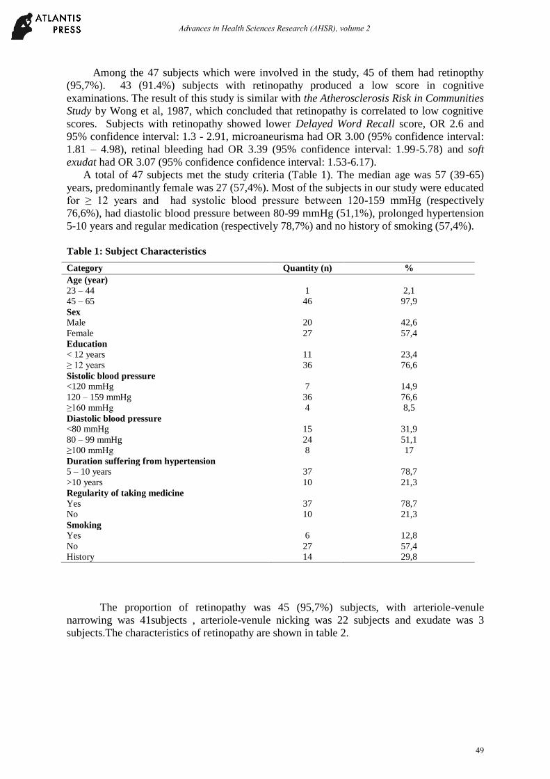
A total of 47 subjects met the study criteria (Table 1). The median age was 57 (39-65)
years, predominantly female was 27 (57,4%). Most of the subjects in our study were educated
for ≥ 12 years and had systolic blood pressure between 120-159 mmHg (respectively
76,6%), had diastolic blood pressure between 80-99 mmHg (51,1%), prolonged hypertension
5-10 years and regular medication (respectively 78,7%) and no history of smoking (57,4%).
Table 1: Subject Characteristics
The proportion of retinopathy was 45 (95,7%) subjects, with arteriole-venule
narrowing was 41subjects , arteriole-venule nicking was 22 subjects and exudate was 3
subjects.The characteristics of retinopathy are shown in table 2.
Category Quantity (n) %
Age (year)
23 – 44 1 2,1
45 – 65 46 97,9
Sex
Male 20 42,6
Female 27 57,4
Education
< 12 years 11 23,4
≥ 12 years 36 76,6
Sistolic blood pressure
<120 mmHg 7 14,9
120 – 159 mmHg 36 76,6
≥160 mmHg 4 8,5
Diastolic blood pressure
<80 mmHg 15 31,9
80 – 99 mmHg 24 51,1
≥100 mmHg 8 17
Duration suffering from hypertension
5 – 10 years 37 78,7
>10 years 10 21,3
Regularity of taking medicine
Yes 37 78,7
No 10 21,3
Smoking
Yes 6 12,8
No 27 57,4
History 14 29,8
49
Advances in Health Sciences Research (AHSR), volume 2

Table 2: Retinopathy Characteristics
The proportion of cognitive impairment was 45 (95,7%) subjects, predominantly
female was 26 (57,8%). Cognitive impairment based on MoCA-INA examination was found
in 24 (51%) subjects, TMT A examination was 40 (85,1%), TMT B examination was 42
(89,4%) and Grooved pegboard was 26 (55,3%). There were many cognitive impairment
domain based on MoCA-INA examination, the most was memory domain 44, language was
27, executive was 23, visuospatial was 21 and attention deficits was 12 subjects. Distribution
of cognitive impairment are shown in table 3.
Table 3: Distribution of Cognitive Impairment Domain Based on MoCA-INA
A total of 43 subjects (91.4%) with retinopathy are suffered cognitive impairment.
Whereas 2 subjects (4.3%) without retinopathy have cognitive impairment. Through fisher
test obtained p value = 1,000. In statistical analysis there were no significant correlation
between retinopathy with cognitive impairment.The relationship between retinopathy with
cognitive impairment is shown in table 4.
Table 4: Relationship Between Retinopathy with Cognitive Impairment
Cognitive
impairment
Normal Total p
Retinopathy Yes 43 (91,4) 2 (4,2) 45 (95,7) 1,000*
No 2 (4,3) 0 (0) 2 (4,3)
Total 45 (95,7) 2 (4,3) 47 (100) *Fisher Test
CONCLUSION
There were no significant correlation between retinopathy and cognitive impairment
in hypertensive patients.
Category Quantity (n)
Arteriole-venule narrowing 41
Arteriole-venule nicking 22
Exudat 3
Cognitive domain Quantity (n)
Memory 44
Language 27
Executive 23
Visuospatial 21
Atention 12
Cognitive examination
50
Advances in Health Sciences Research (AHSR), volume 2

REFERENCES
Departemen Kesehatan Republik Indonesia. (2013). Laporan hasil riset kesehatan dasar
Indonesia (Riskesdas). Badan Penelitian dan Pengembangan Kesehatan Kementerian
Kesehatan Republik Indonesia.
Galetta, S.L., Balcer, L.J., & Liu, G.T. (2008). Neuro-ophthalmologic anatomy and
examination techniques. In : Kidd DP, Newman NJ, Blousse V, editor. Neuro-
Kaplan, N.M. (2006). Primary hypertension : Pathogenesis. In: Caplan’s clinical
hypertension. 9th edition. Philadelphia: Lippincott Williams & Wilkins.
Liew ,G, Wang, J.J., Mitchell, P, & Wong, T.Y. (2008). Retinal vascular imaging a new tool
in microvascular disease research. ahajournals. 1:156-61.
Madhur, M.S. (2014). Hypertension: practice essentials, background, pathophysiology. Sept
30 [accessed on March15, 2016].
Available in: http://emedicine.medscape.com/article/241381-overview#a1
Østergaard L, Engedal TS, Moreton F, Hansen MB, Wardlaw JM, Dalkara T, et all. (2015).
Cerebral small vessel disease : Capillary pathways to stroke and cognitive decline.
JCBFM. July 30.
Patton, N., Aslam, T., MacGillivray, T., Pattie, A., Deary, I.J., & Dhillon, B. (2005). Retinal
vascular image analysis as a potential screening tool for cerebrovascular disease: a
rationale based on homology between cerebral and retinal microvasculatures. J
Anat.;206:319-48.
Theng K. (2014) Ophtalmologic manifestations of hypertension: acute and chronic changes
to the eyes. Mar 21 [accessed on March 15, 2016. Available in:
http://emedicine.medscape.com/article/1201779-overview//showall
Waldstein, SR. (2003). The relation of hypertension to cognitive function. Available in: www
psychologicalscience.orgophthalmology. Philadelphia: Butterworth Heinemann
Elsevier. h. 12-15.
Wolf S. (2007). Hyprtensive retinopathies. In : Joussen AM, Gardner TW, Kirchhof B, Ryan
SJ. editor. Retinal vascular disease. Germany: Springer; h. 717-19.
Wong, T.Y., Klein, R., Sharrett, A.R., Couper, D.J., Klein, B.E., Liao, D.P., et al. (2002).
Cerebral white matter lesions, retinopathy, and incident clinical stroke.
JAMA.;288:1.
51
Advances in Health Sciences Research (AHSR), volume 2