the relationship between the range of moton of the...
TRANSCRIPT
160
SummaryIntroduction. A knee injury, especially anterior cruciate ligament, has recently become more common significantly affecting the life standard. There are many factors that cause an injury of the ante-rior cruciate ligament, and one of them is limited range of motion in the hip joint. This study has been aimed at finding a relationship between the range of motion in the hip joint and the anterior cru-ciate ligament injury. Material and Methods. Of 88 male athletes included in the study sample in 2014, 60 (68%) had ruptured knee anterior cruciate ligament and 28 (32%) were without an injury. There was no significant difference in sex, height, weight, age and time of injury between the two groups. Results. Significant diffe-rences were found in the range of motion between the left and right leg in both groups. The athletes with a ruptured anterior cruciate ligament had an abduction limit of the hip joint (p=0.007) and a wider range of motion of the knee joint (p=0.002) than the athletes without the injury. Conclusion. Data obtained in this study suggest a possible relationship between a limited hip abduction of range of motion in athletes and an increased risk of anterior cruciate li-gament injury.Key words: Anterior Cruciate Ligament; Knee Injuries; Hip Joint; Range of Motion, Articular; Athletes; Athletic Injuries; Risk Factors; Age Factors; Sex Factors
SažetakUvod. Povrede kolena, a naročito prednjeg ukrštenog ligamenta, dešavaju se sve češće poslednjih godina i značajno utiču na kvalitet života. Postoji veliki broj faktora koji utiču na povredu prednjeg ukrštenog ligamenta, a jedan od mogućih jeste ograničenje pokre-ta u zglobu kuka. Cilj našeg rada bio je pronaći povezanost obima pokreta u zgobu kuka sa kidanjem prednjeg ukrštenog ligamenta kolena. Materijal i metode. U ispitivanju, sprovedenom 2014. go-dine, učestvovalo je 88 ispitanika muškog pola, od toga 60 (68%) su bili sportisti sa pokidanim prednjim ukrštenim ligamentom ko-lena i 28 (32%) sportisti bez povrede. Između posmatranih grupa nije bilo značajne razlike prema polu, visini, težini, starosti i vre-menu proteklom od povrede. Rezultati. Kod ispitanika obe grupe se uočavaju značajne razlike u obimu pokreta između leve i desne noge. Kod sportista sa pokidanim prednjim ukrštenim ligamentom postoji ograničenje odvođenja u zglobu kuka (p = 0,007) i veći obim pokreta u zglobu kolena (p = 0,002) u odnosu na sportiste bez po-vrede. Zaključak. Dobijeni podaci ukazuju na moguću povezanost ograničenja odvođenja u zglobu kuka sa kidanjem prednjeg ukršte-nog ligamenta.Ključne reči: prednji ukršteni ligament; povrede kolena; zglob kuka; opseg pokreta zgloba; sportisti; sportske povrede; fakto-ri rizika; uzrast; pol
Introduction
More than 400 million years of evolution of qua-drupeds resulted in the human knee, which is the most complex of the joints of the human body. Its anatomy, which is almost perfect, enables the static as well as huge functional features that distinguish it from other joints [1]. Normal functioning of all its parts allows the harmony of movement, stability, or-thograde posture and constant protection against injuries. A knee injury, especially of the anterior cru-ciate ligament (ACL), has become more frequent in recent years as a result of increased sport activity, work engagement and involvement in traffic acci-
dents [1]. Because of the additional damage to the soft tissue and bony structures of the knee and early development of secondary degenerative changes, an injury of ACL is the “beginning of the end of the knee” [2]. These injuries are a significant epidemio-logical problem, especially because they usually occur to young people who are sport and work active [2]. The nature of ACL injuries is multifactorial and the risk factors are divided into external and internal, as well as into changing and unchanging factors. External factors include the environmental circum-stances such as the type of surface, type of footwear, weather conditions and the type of sport [3]. The group of internal factors consists of anatomical fac-
Clinical Center of Vojvodina, Novi Sad Professional articleDepartment of Orthopedic Surgery and Traumatology1 Stručni članakDepartment of Vascular and Transplantation Surgery2 UDK 616.728.2/.3-001University of Novi Sad, Faculty of Medicine3 DOI: 10.2298/MPNS1606160B Department for Health Care4
THE RELATIONSHIP BETWEEN THE RANGE OF MOTON OF THE HIP JOINT WITH RUPTURED ANTERIOR CRUCIATE LIGAMENT
POVEZANOST OBIMA POKRETA ZGLOBA KUKA SA KIDANJEM PREDNJEG UKRŠTENOG LIGAMENTA KOLENA
Zlatko BUDINSKI1, Slavko BUDINSKI2, Miodrag VRANJEŠ1,3, Mirko OBRADOVIĆ1,3, Milena MIKIĆ3,4 and Miroslav MILANKOV1,3
Corresponding Author: Dr Zlatko Budinski, Klinički centar Vojvodine, Klinika za ortopedsku hirurgiju i traumatologiju21000 Novi Sad, Hajduk Veljkova 1-7, E-mail: [email protected]
Budinski Z, et al. Ruptured ACL and Motion of the Hip Joint

tors; then anthropometric differences between the sexes, height, weight, body mass index (BMI); hor-monal influences, primarily the effect of estrogen on the incidence of the injury and the strength of the ligaments; static parameters, the size of the “Q” an-gle, i.e. the pelvis width being the most important; neuromuscular factors with a focus on the relation-ship of agonists and antagonists; biomechanical fac-tors and, finally, a previous injury and the age of the patient as a potential risk factor [4].
Identification of risk factors in ACL injury is an important step in the development of the injury pre-vention. The analysis of etiological factors is primari-ly aimed at defining their share in the incidence of the injury, but at the same time at defining the factors that can influence it in terms of prevention. Today there is no complete agreement on the most important factor that is the most accountable for ACL rupture [2]. The-refore, it is very important to assess the risk factors to reduce the incidence of the injury because this injury does not only restrict sports activity, but also affects the quality of life [5]. One of the possible causes of the frequent ACL injury is the difference in range of mo-tion (ROM) of the hip and knee according to the stu-dies of Tainaka et al. [6] and Gomes et al. [7]. The aim of our study is to determine the relationship between ACL injury and our patients’ hip and knee ROM.
Material and Methods
Having been given the permission by the Ethics Committee of Clinical Center a prospective study was conducted at the Department of Orthopaedic Surgery and Traumatology. It included 60 male athletes with a torn ACL prior to the surgery in the time period from March 28 to July 10 2014 and 28 male athletes without an injury to ACL who agreed to participate in the study. The measurement was conducted within the first six months after the patients had suffered the injury. The patients who had refused to participate in the study as well as those who had reported previous injuries and diseases of the hip and knee were exclu-ded from the study. The study included only men in
order to exclude hormonal influences and hormonal differences between the sexes.
In the group of athletes with the torn ACL there were 60 men, whose average age was 24.86 years (15-46 years), average body weight was 82 kg (53-110 kg) and average height was 181 cm (155-203 cm). According to sport activities, the majority of them were football players (34), then handball players (10), volleyball players (5), basketball players (4) and other athletes (7). Of the total num-ber of athletes, 31 had the left knee injury, while 29 athletes had the right knee injury. The meniscus was damaged in 40% of athletes and the cartilage lesions were found in 10% of the athletes.
The control group of athletes with no torn ACL included 28 men, whose average age was 23.16 years (15-35), average weight and height was 81 kilograms (50-108 kg) and 181.16 cm (155-194 cm), respective-ly. There were 20 football players, 6 handball players and 2 athletes. The right leg was dominant in over 90% of the participants from both groups.
AbbreviationsACL – anterior cruciate ligamentROM – range of motion
Figure 1. Measuring ROM in the hip with goniometer. A. Flexi-on, B. Extension, C. External rotationSlika 1. Merenje obima pokreta u kuku uglomerom. A. Savija-nje, B. Ispružanje, C. Spoljašnja rotacija
Figure 2. Measuring ROM in the hip with goniometer. A. In-ternal rotation, B. Adduction, C. AbductionSlika 2. Merenje obima pokreta u kuku uglomerom. A. Unu-trašnja rotacija, B. Privođenje, C. Odvođenje
Figure 3. Measuring ROM in the knee with goniometer. A. The active knee extension test, B. The passive knee extension testSlika 3. Merenje obima pokreta u kolenu uglomerom. A. Aktiv-no ispružanje kolena, B. Pasivno ispružanje kolena
Med Pregl 2016; LXIX (5-6): 160-166. Novi Sad: maj-juni.
162
According to the obtained data from the group of patients with ACL rupture and the control group, there were no significant differences in the average age, height, weight and dominant hand, so the two groups were comparable. The measurement was done on a flat surface using a standard goniometer, the values were expressed in degrees. The maximal movement of the hip joint (flexion, extension, exter-nal rotation, internal rotation, adduction, abduction) and knee (the active and passive knee extension test) was measured.
The hip joint flexion (Figure 1A) was measured while the participant was lying on his back with the maximally flexed leg at the hip joint to the body wit-hout the help of hands. The center of goniometer was placed in the region of the hip, the fixed part of gonio-meter was placed horizontally and the mobile part fo-
llowed the movement of the thigh. The hip joint exten-sion (Figure 1B) was measured while the participant was lying down on stomach, stretching out his legs.
The hip external rotation (Figure 1C) was measured when the center of goniometer was placed on the knee in the level of the shaft of the femur bone, the fixed part was in the level of the line connecting the hip joint while the dynamic part of goniometer followed the mo-vement of the lower leg. The hip joint internal rotation (Figure 2A) was measured in the same way except that the lower leg rotated laterally.
The hip joint adduction (Figure 2B) was measured when the center of goniometer was set on the region of spina iliaca anterior superior, the fixed part was in the bispinal line while the dynamic part followed the upper leg. The hip joint abduction (Figure 2C) was measured
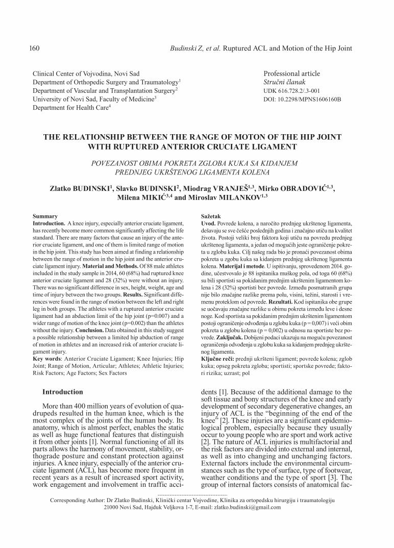
Table 1. Movements of the hip joint between the left and right leg of patients with torn ACLTabela 1. Pokreti u zglobu kuka između leve i desne noge kod pacijenata sa pokidanim prednjim ukrštenim ligamentom
Movements of the hip joint Pokreti u zglobu kuka
Side/Strana Average valueSrednja vrednost
Standard deviationStandardna devijacija
p
Internal rotation/Unutrašnja rotacija Left/Levo 31.29 11.151Right/Desno 30.88 9.303 0.761
External rotation/Spoljašnja rotacija Left/Levo 39.06 11.664Right/Desno 40.62 10.569 0.140
Flexion/Savijanje Left/Levo 122.16 9.306Right/Desno 121.80 8.446 0.627
Extension/Ispružanje Left/Levo 10.09 7.294Right/Desno 9.32 6.529 0.266
Abduction/Odvođenje Left/Levo 56.48 11.749Right/Desno 54.52 12.186 0.0378 *
Adduction/Privođenje Left/Levo 25.78 6.343Right/Desno 30.35 6.519 0.000 **
* Abduction of the left hip vs. abduction of the right hip/Odvođenje u levom kuku vs. odvođenje u desnom kuku, p = 0.0378; ** Adduction of the left hip vs. adduction of the right hip/Privođenje u levom kuku vs. privođenje u desnom kuku, p = 0.000
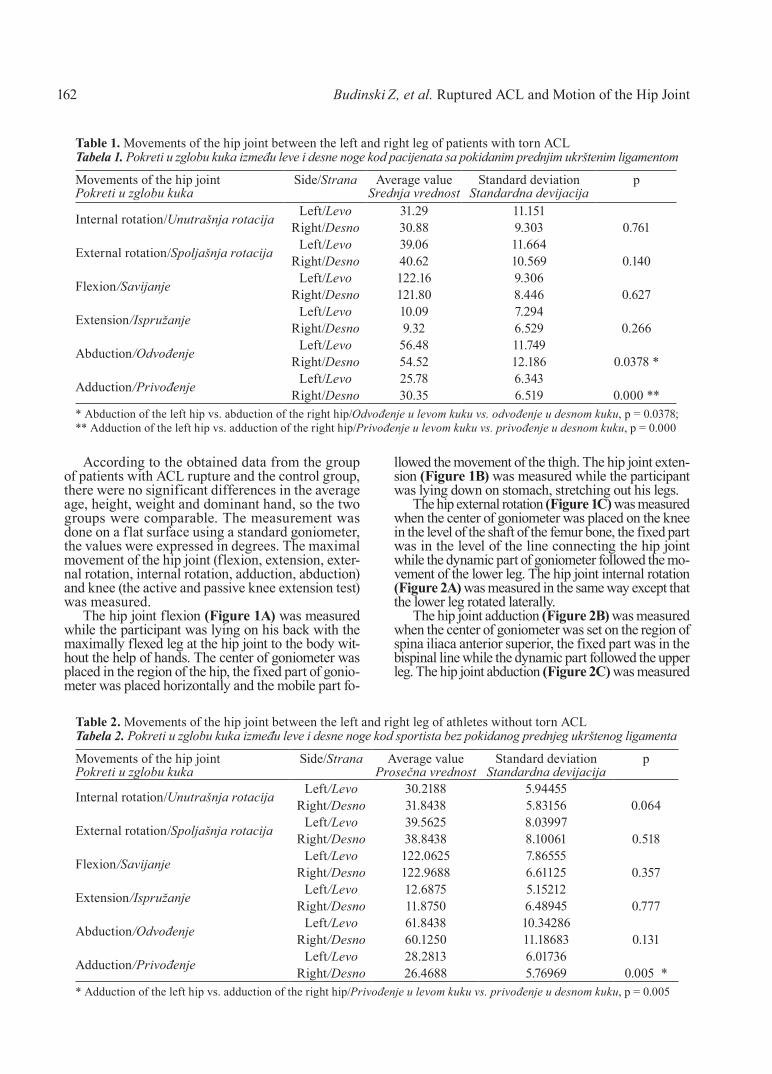
Table 2. Movements of the hip joint between the left and right leg of athletes without torn ACLTabela 2. Pokreti u zglobu kuka između leve i desne noge kod sportista bez pokidanog prednjeg ukrštenog ligamenta
Movements of the hip jointPokreti u zglobu kuka
Side/Strana Average valueProsečna vrednost
Standard deviationStandardna devijacija
p
Internal rotation/Unutrašnja rotacija Left/Levo 30.2188 5.94455Right/Desno 31.8438 5.83156 0.064
External rotation/Spoljašnja rotacija Left/Levo 39.5625 8.03997Right/Desno 38.8438 8.10061 0.518
Flexion/Savijanje Left/Levo 122.0625 7.86555Right/Desno 122.9688 6.61125 0.357
Extension/Ispružanje Left/Levo 12.6875 5.15212Right/Desno 11.8750 6.48945 0.777
Abduction/Odvođenje Left/Levo 61.8438 10.34286Right/Desno 60.1250 11.18683 0.131
Adduction/Privođenje Left/Levo 28.2813 6.01736Right/Desno 26.4688 5.76969 0.005 *
* Adduction of the left hip vs. adduction of the right hip/Privođenje u levom kuku vs. privođenje u desnom kuku, p = 0.005
Budinski Z, et al. Ruptured ACL and Motion of the Hip Joint
Med Pregl 2016; LXIX (5-6): 160-166. Novi Sad: maj-juni. 163
in the same way; however, in this movement the parti-cipant moved the leg laterally, away from the body.
The active knee extension test (Figure 3A) was done when the participant was lying on his back with the leg flexed at the hip at the angle of 90 degrees and stretching his lower leg in the knee joint to the maxi-mum of his free will. The center of goniometer was placed in the region of the knee joint, the fixed part followed the axis of the thigh and the dynamic part of goniometer followed the lower leg. The passive knee extension test (Figure 3B) was done in a similar way, except that the examiner stretched out the lower leg of the participant while he rested and relaxed the muscles of the legs.
The Statistical Package for the Social Sciences (SPSS) version 19.0 for Windows was used in the anal-ysis. The results are presented in tables expressed as mean values and standard deviations. In all compari-sons, p<0.05 was considered a statistically significant difference.
Results
The comparison of the ROM between the left and right leg of the patients with a torn ACL revealed statistically significant differences in abduction and adduction of the hip (Table 1). The range of abduc-tion was higher for the left leg (for 3±1 degree;
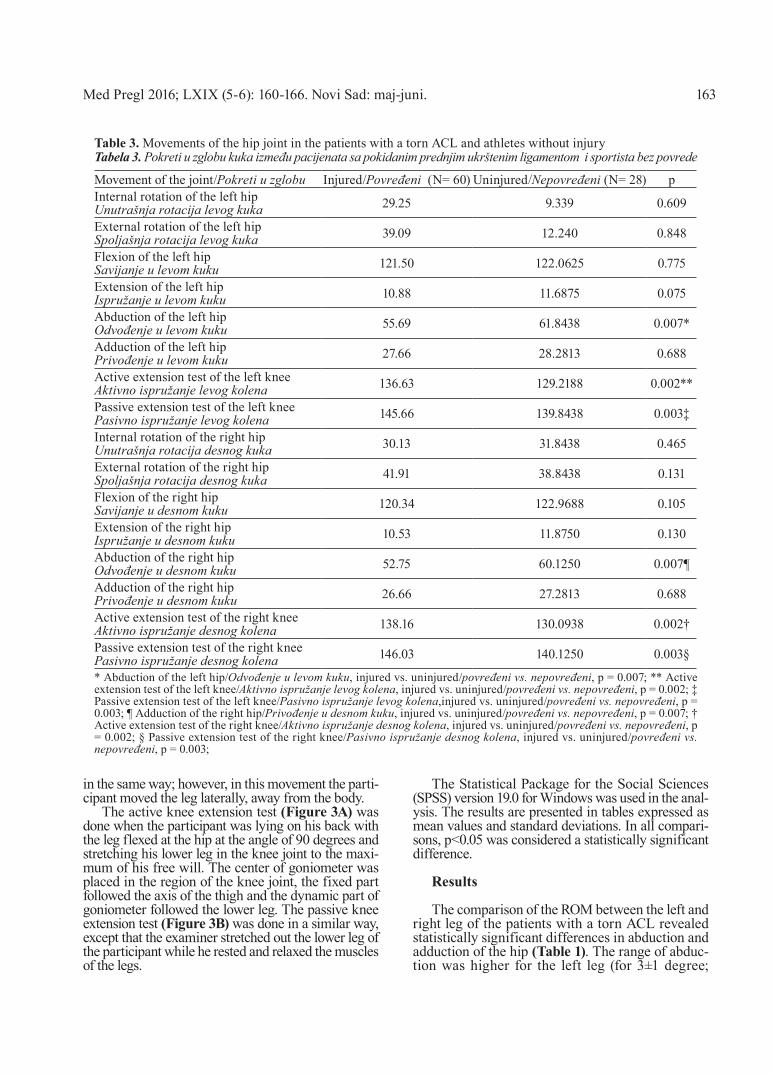
Table 3. Movements of the hip joint in the patients with a torn ACL and athletes without injuryTabela 3. Pokreti u zglobu kuka između pacijenata sa pokidanim prednjim ukrštenim ligamentom i sportista bez povrede
Movement of the joint/Pokreti u zglobu Injured/Povređeni (N= 60) Uninjured/Nepovređeni (N= 28) pInternal rotation of the left hipUnutrašnja rotacija levog kuka 29.25 9.339 0.609
External rotation of the left hipSpoljašnja rotacija levog kuka 39.09 12.240 0.848
Flexion of the left hipSavijanje u levom kuku 121.50 122.0625 0.775
Extension of the left hipIspružanje u levom kuku 10.88 11.6875 0.075
Abduction of the left hipOdvođenje u levom kuku 55.69 61.8438 0.007*
Adduction of the left hipPrivođenje u levom kuku 27.66 28.2813 0.688
Active extension test of the left kneeAktivno ispružanje levog kolena 136.63 129.2188 0.002**
Passive extension test of the left kneePasivno ispružanje levog kolena 145.66 139.8438 0.003‡
Internal rotation of the right hipUnutrašnja rotacija desnog kuka 30.13 31.8438 0.465
External rotation of the right hipSpoljašnja rotacija desnog kuka 41.91 38.8438 0.131
Flexion of the right hipSavijanje u desnom kuku 120.34 122.9688 0.105
Extension of the right hipIspružanje u desnom kuku 10.53 11.8750 0.130
Abduction of the right hipOdvođenje u desnom kuku 52.75 60.1250 0.007¶
Adduction of the right hipPrivođenje u desnom kuku 26.66 27.2813 0.688
Active extension test of the right kneeAktivno ispružanje desnog kolena 138.16 130.0938 0.002†
Passive extension test of the right kneePasivno ispružanje desnog kolena 146.03 140.1250 0.003§
* Abduction of the left hip/Odvođenje u levom kuku, injured vs. uninjured/povređeni vs. nepovređeni, p = 0.007; ** Active extension test of the left knee/Aktivno ispružanje levog kolena, injured vs. uninjured/povređeni vs. nepovređeni, p = 0.002; ‡ Passive extension test of the left knee/Pasivno ispružanje levog kolena,injured vs. uninjured/povređeni vs. nepovređeni, p = 0.003; ¶ Adduction of the right hip/Privođenje u desnom kuku, injured vs. uninjured/povređeni vs. nepovređeni, p = 0.007; † Active extension test of the right knee/Aktivno ispružanje desnog kolena, injured vs. uninjured/povređeni vs. nepovređeni, p = 0.002; § Passive extension test of the right knee/Pasivno ispružanje desnog kolena, injured vs. uninjured/povređeni vs. nepovređeni, p = 0.003;

164
p=0.0378), while the range of adduction was higher for the right leg (5 degrees; p=0.000).
Statistically significant differences in the hip adduction between the left and right leg were obser-ved in the control group of athletes without a torn ACL (Table 2). The range of adduction was higher for the left leg (for 3±1 degree; p=0.005).
The comparison of the ROM of the hip and knee between the patients with ruptured ACL and the participants without injuries (Table 3) revealed sta-tistically significant differences in the hip joint ab-duction as well as the active and passive knee exten-sion. Abduction of the left and right hip in the injured athletes was smaller than abduction in the athletes without the injury (an average of 8 degrees; p=0.007). The injured athletes showed higher ROM during the active (average of 8 degrees; p=0.002) and passive knee extension (average of 6 degrees; p=0.003) than the athletes without an ACL injury.
Discussion
Anterior cruciate ligament injuries are injuries that are commonly found in athlete population. According to the available epidemiological data in the United States, it is estimated that the annual incidence of ACL injuries varies from 100,000 to 250,000 injuries, and more than 70% of these injuries happen to the popu-lation who engage in sports [9, 10]. In Vojvodina, whi-ch has about two million people, about 400 reconstruc-tions of the ACL (2 reconstructions per 10,000 resi-dents) are performed per year. As for the European countries, the most accurate data are those from De-nmark, where three injuries happen to 10,000 resi-dents per a year, the frequency of occurrence being higher among athletes [11]. These data are very simi-lar to the data from the United States, where it is estimated that injuries occur in one out of every 3,000 people in the general population [9]. The rea-son for this epidemiological situation certainly lies in the fact that the number of participants in the sport is constantly growing on the global level. Sports ac-tivities are becoming an important part of modern life; however, more and more people spend their free time in recreation and entertainment. The number of men and women who become members of fitness and various sports clubs is increasing [12]. All this matches with the fact that a significant increase in injuries of ACL was recorded in last 10-15 years [13].
Another reason for a higher incidence of knee and ACL injuries is the growing popularity of extreme sports such as snowboarding, skateboarding and extreme cycling [14]. However, in our country these sports still do not have a high number of fans. Since the most popular sports in our country are ball sports, the injuries of ACL are most common in football, basketball, handball and volleyball [3].
An ACL injury is affected by a number of external and internal factors such as gender, condition of the knee joint, ground conditions, condition of surface, part of the training, the ranking of the competition [15,
16]. Many researchers have made an effort to find potential risk factors that can predispose towards injury of ACL [17, 18]. Detection of risk factors is essential for finding specific training programs in or-der to reduce the incidence of injury [10]. Hewett et al. [15] included contact with an opponent, the effect of wearing corsets on the knee and contact sports shoes with the ground in their study on the group of external factors. It is often thought that artificial surface is a reason for development of a large number of injuries [19], but data from many studies do not support this statement because serious injuries occur on natural surfaces as well as on the parquet floor [19].
Anterior cruciate ligament injuries are more com-mon among women but since the proportion of men engaged in sport is higher, the number of injuries of ACL is much higher in males [17, 18, 20]. Measure-ments in our study were performed only in males due to a small number of women who were operated during this period and to avoid the influence of hormonal changes during the pre-ovulatory period as a potential confounding risk factor for an ACL injury. Gomes et al. [7] also performed measurements only in males in their research. Of 44 participants included in the study performed by Tainaka et al. [6], 23 were men and 42 were women. Reurink et al. [21] included 50 partici-pants in their research on injury of the biceps thighs, 46 men and 4 women.
In our study, the average age was 24.86 (ranging between 15 and 46 years), that being in accordance with the data from previous studies which suggest that knee injuries happen most frequently to men and wo-men between the ages of 20 and 29 years [10]. There were 28 males, whose average age was 25.8±4.4 years, in the study done by Gomes et al. [7]. The average age of the participants in the study conducted by Reurink et al. [21] was 28 years. The structure of sports that our patients were involved in has only confirmed ear-lier epidemiological studies (Swärd et al. [22]), which recognize that intensive sports such as football and volleyball involving movements of rotation, valgus knee and anterior translation are risky and related to the occurrence of injuries of ACL.
Anatomical variations of the proximal tibia and in-tercondilar groove are considered to be risk factors in an ACL injury [23]. A statistically significant differen-ce in the value of the posterior tibial slope was observed between the groups with and without torn ACL [24]. Tibial articular surfaces in the knee joint form, with the axis of the tibia, the angle of the posterior slope of the tibia of 7-10 degrees. A larger posterior tibial slope cre-ates a larger anterior displacement of the tibia [25]. Sin-ce ACL is the main stabilizer of this motion, this results in its increased workload. This leads to increased inter-nal rotation of tibia and greater acting force on the ACL [26]. Shelbourne et al. [27] observed the relationship between intercondilar groove and noncontact ACL injury and concluded that when the groove was 15 mm wide or less, there was a higher incidence of contrala-teral ACL tear than when it was 16 or more mm wide.
Budinski Z, et al. Ruptured ACL and Motion of the Hip Joint

Med Pregl 2016; LXIX (5-6): 160-166. Novi Sad: maj-juni. 165
Anatomical variations are one of the risk factors for injuries of the ACL [28]. In the group of athletes with a torn ACL, significant differences can be obser-ved in the range of abduction (p=0.037) and adducti-on (p=0.000) of the hip joint between the left and right leg. Adduction of the right leg is higher (3±2 degrees) than adduction of the left leg, while abduc-tion is higher for the left leg (5±2 degrees).
In the control group consisting of athletes wit-hout an injury, there was a statistically significant difference only in adduction of the hip joint between the left and right leg. ROM of the left hip was usu-ally by 3 ± 2 degrees (p = 0.005) wider than of the right hip, the probable reason being that the left leg was the jumping leg in most of our patients going in for jum-ping sports. Baltaci et al. [29] in their study compared the range of flexion of the left and right hip joint at different positions of the body and the results showed that there was a difference between the left and right leg depending on the flexibility of the muscles.
The comparison of the ROM in the controls and the patients with a torn ACL revealed statistically si-gnificant differences. The following statistically signi-ficant difference was observed in the range of hip abduction (p=0.007): ROM was smaller in the athletes with a ruptured ACL, i.e. the muscles of internal part of the thigh were less stretched, which could be a po-ssible risk factor for ACL injury.
Hartig et al. [28] have reported that flexibility of posterior thigh muscles is very important in sports and that argument has been confirmed by the results of their research which shows that the number of injuri-es of the lower extremities decreases with proper and good flexibility of posterior thigh muscles. A statisti-cally significant difference was observed in the active knee extension test (p=0.002) and in the passive knee extension test (p=0.003), where the patients with torn ACL had wider ROM showing a good muscle flexi-bility of posteior part of thigh. Tainaka et al. [6] found a limit in the external and internal rotation of the hip joint in the athletes with noncontact ACL rupture. In their study they compared the ROM of the hip joint between the group of 44 athletes with torn ACL and
the control group of 123 athletes without injury of ACL. They have observed that the risk of rupture is rapidly increasing with a decrease in ROM, in external and internal rotation of the hip. Gomes et al. [7] mea-sured the ROM of the hip joint and the connection with the re-tearing the ACL where limited external and internal rotation of the hip joint was determined.
In our study no statistically significant difference was found in the external and internal rotation of the hip joint between the athletes without injuries and those with a torn ACL. A possible cause for different results is that our participants had higher average body weight (82kg), height (181cm) and possibly different motives to participate in the study.
Limitations of this study is that it did not include all patients with torn ACL who were in hospital during the study and we did not have an magnetic resonance imagfing or X-ray of the hip and knee joint of each patient we measured. The biggest limitation of this study is that this kind of measuring the patients is a completely subjective method so its results can be si-gnificantly affected by the will and motivation of exa-minees and examiners.
Conclusion
According to the results of this study, there is a statistically significant difference in the range of mo-tion of the hip and knee joint between the athletes with a torn anterior cruciate ligament and those wi-thout an injury. The athletes with a torn anterior cru-ciate ligament were found to have a wider range of motion for active and passive knee extension (greater muscle flexibility of posterior part of thigh) and ab-duction limit of the hip joint. These results suggest a possible connection between an anterior cruciate li-gament injury and the size of abduction of the hip joint. It is therefore necessary to increase the muscle flexibility of internal part of thigh during training time in order to increase the range of motion of ab-duction of leg so as to reduce the risk of anterior cruciate ligament injury.
References1. Ristić V, Ninković S, Harhaji V, Stanković M, Savić D,
Milankov M. Reconstruction of anterior cruciate ligament by using two different techniques. Med Pregl. 2010;63(11):845-50.
2. Milankov M, Kecojević V, Ninković S, Gajdobranski Đ. Patella fracture following anterior cruciate ligament reconstruc-tion: a case report. Med Pregl. 2003;56(11-12):574-7.
3. Ristić V, Ninković S, Harhaji V, Milankov M. Causes of anterior cruciate ligament injuries. Med Pregl. 2010;63(7-8):541-5.
4. Griffin L, Agel J, Albohm M, Arendt E, Dick R, Garrett W, et al. Noncontact anterior cruciate ligament injuries: risk factors and prevention strategies. J Am Acad Orthop Surg. 2000;8(3):141-50.
5. Banović D. Povrede kolena: Traumatologija koštano-zglobnog sistema. U: Povrede kolena. Gornji Milanovac: Dečje novine; 1989. p. 474-506.
6. Koji T, Tsuyoshi T, Hiroyuki K, Masakazu U. Limited hip rotation and non-contact cruciate ligament injury: a case–control study. Knee. 2014;21(1):86-90.
7. Joa˜o L. Ellera G, Humberto MP, Roberto R. Influence of hip restriction on noncontact ACL rerupture. Knee Surg Sports Traumatol Arthrosc. 2014;22:188-91.
8. Bahr R, Krosshaug T. Understanding injury mechanisms: a key component of preventing injuries in sport. Br J Sports Med. 2005;39:324-9.
9. Silvers HJ, Mandelbaum BR. Prevention of anterior cru-ciate ligament injury in the female athlete. Br J Sports Med. 2007;41:52-9.
10. Bradley JP, Klimkiewicz JJ, Rytel MJ, Powell JW. An-terio Cruciate Injuries in the National football Leauge: epide-milology and current treatment trend among physicians. Ar-throscopy. 2002;18:502-9.

166
11. Kvist J. Rehabilitation following anterior cruciate liga-ment injury. Sports Med. 2004;34:269-82.
12. Majewski M, Nabelt S, Steinbruck K. Epidemiology of athletic knee injuries: a 10-year study. Knee. 2006;13:184-8.
13. Olsen OE, Myklebust L, Engerbretsen I, Holme R, Bahr R. Relationship between floor type and risk of ACL injury I team handball. Scand J Med Sci Sports. 2003;13:299-304.
14. Steinbruck K. Epidemiology of sport injuries- 25-year analysis of sports orthopedic-traumatologie ambulatory care. Sportverletz Sportshaden. 1999;13:38-52.
15. Hewett T, Myer G, Ford K. Anterior cruciate ligament injuries in female athletes: Part 1, mechanisms and risk factors. Am J Sports Med. 2006;34(2):299-311.
16. Myer GD, Ford KR, Paterno MV, Schranz PJ. The ef-fects of generalized joint laxity on risk of anterior cruciate li-gament injury in young female athletes. Am J Sports Med. 2008;36:1073-80.
17. Arendt E, Dick R. Knee injury patterns among men and women in collegiate basketball and soccer. Am J Sports Med. 1995;23:694-701.
18. Powell J. Incidence of injury associated with playing surfaces in the National Football League 1980-1985. Athl Tra-ining. 1987;22:202-6.
19. Nigg BM, Segesser B: The influence of playing surfaces on the load of the locomotor system and on football and tennis injuries. Sports Med. 1988;5:375-85.
20. Brandon ML, Haynes PT, Bonamo JR, et al. The asso-ciation between posterior-inferior tibial slope and anterior cru-ciate ligament insufficiency. Arthroscopy. 2006;22:894-9.
21. Gustaaf R, Gert Jan G, Henricus GO, Maarten HM, Johannes LT, Jan ANV, Adam W. Reliability of the Active and
Passive Knee Extension Test in Acute Hamstring Injuries. Am J Sports Med. 2013;41:17-57.
22. Swärd P, Kostogiannis I, Roos H. Risk factors for a contralateral anterior cruciate ligament injury. Knee Surg Sports Traumatol Arthrosc. 2010;18(3):277-91.
23. Zeng C, Cheng L, Wei J, Gao S, Yang T, Luo W, et al. The influence of the tibial plateau slopes on injury of the ante-rior cruciate ligament: a meta-analysis. Knee Surg Sports Tra-umatol Arthrosc. 2014;22:53-65.
24. Ristić V, Maljanović MC, Pericin B, Harhaji V, Milankov M. The relationship between posterior tibial slope and anterior cruciate ligament injury. Med Pregl. 2014;67(7-8):216-21.
25. Dejour H, Bonnin M. Tibial translation after anterior cruciate ligament rupture: two radiological tests compared. J Bone Joint Surg Br. 1994;76:745-9.
26. McLean SG, Lucey SM, Rohrer S, Brandon C. Knee joint anatomy predicts high-risk in vivo dynamic landing knee biomechanics. Clin Biomech. 2010;25:781-8.
27. Shelbourne KD, Facibene WA, Hunt JJ. Radiographic and intraoperative intercondilar notch width measurements in men and women with unilateral and bilateral anterior cruciate liga-ment tears. Knee Surg Sports Traumatol Arthrosc. 1997;5:229-33.
28. Hartig E, Henderson M. Increasing hamstring flexibility decreases lower extremity overuse injuries in military basic tra-inees. Am J Sports Med. 1999.
29. Baltaci G, Un N, Tunay V, Besler A, Gerçeker S. Com-parison of three different sit and reach tests for measurement of hamstring flexibility in female university students. Br J Sports Med. 2003;37:59-61.
Rad je primljen 13. XI 2015.Recenziran 17. XI 2015.Prihvaćen za štampu 22. II 2016.BIBLID.0025-8105:(2016):LXIX:5-6:160-166.
Budinski Z, et al. Ruptured ACL and Motion of the Hip Joint