the replication cycle of varicella-zoster virus: …jvi.asm.org/content/83/8/3904.full.pdfthe...
TRANSCRIPT

JOURNAL OF VIROLOGY, Apr. 2009, p. 3904–3918 Vol. 83, No. 80022-538X/09/$08.00�0 doi:10.1128/JVI.02137-08Copyright © 2009, American Society for Microbiology. All Rights Reserved.
The Replication Cycle of Varicella-Zoster Virus: Analysis of theKinetics of Viral Protein Expression, Genome Synthesis, and
Virion Assembly at the Single-Cell Level�
Mike Reichelt,* Jennifer Brady, and Ann M. ArvinDepartments of Pediatrics and Microbiology & Immunology, Stanford University School of Medicine, Stanford, California 94305
Received 9 October 2008/Accepted 23 January 2009
Varicella-zoster virus (VZV) is a human alphaherpesvirus that is highly cell associated in cell culture.Because cell-free virus yields are too low to permit the synchronous infections needed for time-resolvedanalyses, information is lacking about the sequence of events during the VZV replication cycle. To address thischallenge, we differentially labeled VZV-infected inoculum cells (input) and uninfected (output) cells withfluorescent cell dyes or endocytosed nanogold particles and evaluated newly infected cells by confocal immu-nofluorescence or electron microscopy (EM) at the single-cell level at defined intervals. We demonstrated thespatiotemporal expression of six major VZV proteins, ORF61, IE62, IE63, ORF29, ORF23, and gE, represent-ing all putative kinetic classes, for the first time. Newly synthesized ORF61, as well as IE62, the major VZVtransactivator, appeared within 1 h, and they were targeted to different subnuclear compartments. Theformation of VZV DNA replication compartments started between 4 and 6 h, involved recruitment of ORF29to putative IE62 prereplication sites, and resulted in large globular nuclear compartments where newlysynthesized viral DNA accumulated. Although considered a late protein, gE accumulated in the Golgi com-partment at as early as 4 h. ORF23 capsid protein was present at 9 h. The assembly of viral nucleocapsids andmature enveloped VZ virions was detected by 9 to 12 h by time-resolved EM. Although syncytium formation isa hallmark of VZV infection, infection of neighboring cells did not require cell-cell fusion; its occurrence from9 h is likely to amplify VZV replication. Our results define the productive cycle of VZV infection in a single cellas occurring in 9 to 12 h.
Varicella-zoster virus (VZV) is a ubiquitous human alpha-herpesvirus that causes varicella (chickenpox) during pri-mary infection, can establish latency in sensory ganglia, andmay reactivate to cause herpes zoster (shingles) (11, 24).VZV is related to herpes simplex virus types 1 and 2 (HSV-1and HSV-2) and simian varicella virus and has a linear DNAgenome of 125 kbp that has at least 70 open reading frames(ORFs) encoding known or predicted viral proteins (11).
Like those of other herpesviruses, VZV particles are pre-sumed to enter cells by fusion of the virion envelope withthe plasma membrane or by endocytosis followed by thetransport of capsids and associated virion tegument proteinsto the cell nucleus (11, 46). The major VZV transactivatingprotein, referred to as immediate-early 62 (IE62) is a tegu-ment component, as are other VZV regulatory proteins,including IE4, ORF10, IE63, and the viral kinases ORF47and ORF66 (11, 29, 30, 50). As has been demonstrated incells infected with HSV and other herpesviruses, VZV genetranscription is believed to occur in a cascade that leads tothe synthesis of viral proteins that are classified as immedi-ate-early, early, and late, based on the time course of theirexpression after virus entry (11). Studies using VZV-in-fected cells to inoculate uninfected cells in conjunction withmetabolic pulse-labeling of newly synthesized proteins and
Western blot analysis have indicated that viral proteins areexpressed by 4 to 6 h after infection (1, 2). However, be-cause VZV is so highly cell associated in cultured cells,experiments that reveal the timing of gene transcription orthe spatiotemporal characteristics of VZV protein expres-sion in single cells within one infectious cycle have not beenperformed (11). Achievable titers of cell-free VZV are toolow to permit synchronous infections of cultured cells, as isdone to define the kinetics of viral mRNA and proteinsynthesis for HSV-1 and other herpesviruses (10, 13, 25, 26).Therefore, information is lacking, and there is some contro-versy about when and where VZV proteins are expressed innewly infected cells, how the assembly of VZV nuclear rep-lication compartments is orchestrated, the time required tocomplete one infectious cycle, and the role of cell-cell fusionin VZV propagation, which is of interest, given the extensivesyncytium formation that characterizes VZV replication (11,12, 26, 52).
VZV experiments are usually done by adding an infected-cell inoculum of human fibroblasts or melanoma (MeWo)cells to a monolayer of uninfected cells. Initial events duringreplication are assessed by using low numbers of infectedinoculum cells as a means to enrich for newly infected cells.Infection is then monitored for 24 to 72 h to demonstrateviral spread within the monolayer and to allow enough newVZV protein synthesis for detection by Western blotting,confocal microscopy, or other methods. Since VZV is notreleased into media, secondary plaque formation does notoccur during the 72-h interval. Many important parametersof VZV genome replication, protein expression, and virus-
* Corresponding author. Mailing address: Stanford UniversitySchool of Medicine, 300 Pasteur Dr., Grant Bldg., Room S356, Stan-ford, CA 94305. Phone: (650) 723-6353. Fax: (650) 725-8040. E-mail:[email protected].
� Published ahead of print on 4 February 2009.
3904
on June 5, 2018 by guesthttp://jvi.asm
.org/D
ownloaded from

host cell interactions have been defined by using this ap-proach (11). However, these experimental conditions arenot compatible with generating an accurate time-resolvedanalysis of events in the VZV replication cycle because theinfected cells are a mixed population that reflect differentstages of viral infection.
Overcoming the experimental challenges to studies of theVZV replication cycle requires a strategy that permits the useof high numbers of infected inoculum cells so that enough cellsare infected to evaluate the earliest time points while allowingunequivocal discrimination of the inoculum and newly infectedcells at sequential time points. To address these challenges, weused methods to label either the input cells (VZV-infectedinoculum cells) or the output cells (uninfected cells) and in-vestigated the VZV infectious cycle, including the VZV ge-nome and viral protein synthesis, by confocal immunofluores-cence (IF) or by standard electron microscopy (EM). Analysesusing confocal microscopy were done by labeling VZV-in-fected inoculum cells with fluorescent cell dyes. In EM exper-iments, the cells in the uninfected monolayer were preincu-bated with protein A-gold (PA-gold) beads before beinginoculated with VZV-infected cells, allowing their identifica-tion as newly infected cells based on detection of the PA-goldparticles in the cytoplasm (38).
With these methods, we demonstrated the time course ofexpression of major VZV proteins of all putative kineticclasses, as well as their cellular localization patterns, for thefirst time. Among other observations, we found that ORF61, aswell as IE62, is expressed very early, less than 1 h after infec-tion, in human fibroblasts and that these proteins are targetedto different subnuclear compartments. We tracked the forma-tion of VZV DNA replication compartments, showing theaccumulation of newly synthesized viral DNA by 4 h afterinfection, and demonstrated the assembly of viral nucleocap-sids and mature enveloped VZ virions by 9 to 12 h afterinfection. These experiments have also documented that VZVinfection of neighboring cells does not require cell-cell fusionbut that cell-cell fusion beginning at 9 h is likely to substantiallyamplify VZV replication.
MATERIALS AND METHODS
Cells and viruses. VZV (rOka) was propagated in human embryonic lungfibroblast (HELF) cells by using standard methods (53). Inoculum titers weredetermined by infectious focus assay; inoculum titers of cells labeled with fluo-rescent dyes (see below) were determined after labeling (53).
Fluorescent cell tracker labeling. For IF experiments, infected inoculum cellswere labeled with green BODIPY CellTracker (Invitrogen) according to themanufacturer’s instructions. Green BODIPY CellTracker labels the cytoplasmwith bright green fluorescence that is retained within the labeled cells and is notreleased into the medium. In cell-cell fusion experiments, the uninfected outputcells were also labeled, using orange CellTracker (CMRA; Invitrogen). Cell-cellfusion is demonstrated when the cytoplasmic mixing of red and green fluores-cence produces yellow cytoplasmic fluorescence.
Cell labeling by endocytosis of PA-gold particles. In EM experiments, theoutput cells were preincubated with PA-gold particles (CMC, Utrecht, TheNetherlands) before unlabeled VZV-infected inoculum cells were added (38).The PA-gold was diluted 1:50 into growth medium and was added to HELF cellsat 3 h before infection to allow endocytosis. PA-gold-labeled output cells werethen washed several times with medium and chased for 45 min to removePA-gold from the medium and to allow further uptake of surface-bound PA-gold. The PA-gold accumulated in lysosomes, where it served as a readily visibleelectron-dense marker to identify output cells by standard EM.
Time course experiments. For IF time course experiments, uninfected HELF(output) cells were seeded onto sterile glass coverslips, grown to 90% confluence,and inoculated with green-BODIPY-CellTracker-labeled inoculum cells (input)at a ratio of 1 infected cell per 50 uninfected cells. Inoculum cells were allowedto settle on the monolayer during a 30-min incubation on ice. The coverslips weretransferred to 37°C to initiate infection. Cells were fixed with 4% paraformal-dehyde at 0 h, 2 h, 4 h, 6 h, 9 h, or 12 h; in some experiments, samples were alsoprepared at 30, 60, and 90 min, or 3 h, 8 h, 10 h, and 24 h after inoculation. Atleast three coverslips were examined at each time point in all experiments, andat least two independent time course experiments were performed to evaluatethe kinetics of expression of each VZV protein. For time-resolved EM experi-ments, infected cells were fixed at 3 h, 6 h, 9 h, and 12 h.
Antibodies and IF staining. Cells on coverslips were permeabilized for 15 minwith 0.5% Triton X-100 in phosphate-buffered saline, washed, and blocked indigoxigenin (DIG) blocking solution (Roche) for 30 min. Primary antibodieswere diluted in blocking solution, and incubation was generally for 1 h at roomtemperature in a humid chamber. The primary antibodies used included rabbitpolyclonal antibodies against ORF61 generated in our laboratory (L. Wang,unpublished data), IE62 (a gift from Paul Kinchington, University of Pittsburgh),IE63 (a gift from William Ruyechan, University of Buffalo), ORF29 (a gift fromS. Silverstein, Columbia University, New York), and ORF23 (9) and monoclonalantibodies against gE (Chemicon, Temecula, CA), anti-DIG (Roche), and anti-BrdU-biotin conjugate (Invitrogen). Secondary detection was done with TexasRed-conjugated (goat) anti-rabbit or anti-mouse antibodies (Jackson Immuno-Research). Additional antibodies used for some double-staining experimentsincluded polyclonal rabbit anti-promyelocytic leukemia (PML) (Santa Cruz Bio-tech), monoclonal mouse anti-PML (Santa Cruz Biotech) with secondary re-agents, including (goat) anti-mouse-Alexa Fluor 488 (Invitrogen), (donkey) anti-rabbit-fluorescein isothiocyanate (FITC), (donkey) anti-rabbit or anti-mouseAlexa Fluor 647 (Invitrogen), and streptavidin-FITC and streptavidin-Texas Red(Jackson ImmunoResearch). Hoechst 22358 was used as a nuclear counterstainin all experiments.
VZV DNA in situ hybridization. The VZV-specific DNA probe was preparedby random priming, using a DIG DNA-labeling kit (Roche Diagnostics,Penzberg, Germany) as described previously (45). One milliliter of the hybrid-ization mix contained 50% formamide, 10% dextran sulfate, 1� SSC (0.15 MNaCl plus 0.015 M sodium citrate), 1� Denhardt’s solution, 0.5 mg salmonsperm DNA, and 0.01 ml DIG-labeled VZV DNA probe. Cells on coverslipswere permeabilized as for IF and treated with RNase A (1 mg/ml) for 20 min at37°C. After being dehydrated with 70%, 90%, and 100% ethanol for 2 min each,coverslips were air dried and transferred onto 15-�l drops of preheated hybrid-ization mix on a clean glass slide. Simultaneous heat denaturation of the probeand sample DNA was achieved by placing the slide on a heat block (98°C) for 10min; slides were incubated at 37°C in a humid chamber for 12 h. Coverslips werethen washed twice in 2� SSC and 0.1� SSC (10 min each) and blocked in DIGblocking solution (Roche) for 30 min. Sections were incubated with anti-DIGmonoclonal antibody (Roche) for 1 h. Secondary detection was with anti-mouseDIG-conjugated antibody (Roche) and sheep anti-DIG-Fab fragments conju-gated with rhodamine (Roche) for 1 h. When DNA-fluorescence in situ hybrid-ization (FISH) was combined with BrdU or protein detection, the hybridizedDNA probe was detected directly with rhodamine-conjugated anti-DIG-Fabfragments.
Drug inhibition experiments. Uninfected cells grown on coverslips were eitheruntreated or were pretreated by incubation with cycloheximide (100 �g/ml),actinomycin D (4 �g/ml), or phosphonoacetic acid (PAA; 0.5 mg/ml and 1mg/ml) for 30 min at 37°C. All these drugs were purchased from Sigma-Aldrich.The uninfected output cells were then inoculated with green-labeled infectedcells and incubated for 4 h at 37°C in the presence of the drugs at the sameconcentrations. The 4-h incubation was chosen to allow infection to proceed toa time at which newly synthesized IE62 or ORF61 would be detected easily, asdetermined in IF kinetics experiments. The cells were then fixed and stained forIE62 or ORF61.
Confocal microscopy. IF analysis was performed with a Leica TCSSP2 confocallaser scanning microscope (Heidelberg, Germany) and a 63�/1.4 Plan Apochro-mat. Images were scanned at 1,024 � 1,024 pixels with at least 4� frameaveraging and the pinhole adjusted to 1 airy unit. Brightness and contrast wereadjusted using iPhoto (Apple) and Photoshop CS3 (Adobe). For each time pointin the time course experiments, 50 fields with 30 to 50 output cells were scanned;this method allowed the analysis of the staining pattern for each VZV protein orfor VZV DNA in at least 20 to 50 newly infected single cells for each time point.
Electron microscopy. HELF cells were grown and infected on a 10-cm Petridish. At specified time points, cells were trypsinized, gently pelleted, and fixed in4% paraformaldehyde and 2% glutaraldehyde in phosphate buffer (0.1 M [pH
VOL. 83, 2009 THE VZV REPLICATION CYCLE IN SINGLE CELLS 3905
on June 5, 2018 by guesthttp://jvi.asm
.org/D
ownloaded from

7.2]). Cells were postfixed with 1% osmium tetroxide (2 h) and incubated in 1%aqueous uranyl acetate overnight. The samples were dehydrated in a series ofincreasing ethanol concentrations followed by a final propylenoxide step. Thesamples were embedded in Embed 812 (Electron Microscopy Sciences, FortWashington, PA). Ultrathin sections (60 nm) were prepared with a diamondknife (Diatome) and an ultramicrotome (Ultracut, Leica). Sections were stainedwith 3.5% aqueous uranyl acetate for 10 min and with 0.2% lead citrate for 3min. The sections were analyzed using a Jeol 1230 transmission electron micro-scope at 80 kV, and digital photographs were taken with a Gatan Multiscan 701digital camera. In the time-resolved EM studies, only cells that showed noPA-gold clusters in lysosomes were included in the morphological analysis.
RESULTS
Differentiation of cells newly infected with VZV from in-fected inoculum cells. Initial experiments were done to evalu-ate the feasibility of using fluorescent-dye labeling of VZV-infected inoculum cells (input cells) to investigate the kineticsof expression of VZV proteins in newly infected HELF cells(output cells) (Fig. 1). Expression of ORF61, IE62, IE63,ORF29, gE, and ORF23 proteins was examined at 0, 2, 4, 6, 9,and 12 h after inoculation, using well-characterized antibodiesto these proteins, and was compared to that of the uninfectedHELF controls. The accumulation of VZV DNA over 2 to 12 hwas followed by in situ hybridization, using a DIG-labeledprobe. The technique allowed the discrimination of newly in-fected cells from inoculum cells at all time points, as illustratedby examples showing ORF61 and IE62 at 4 h, ORF29 and IE63at 6 h, and gE and ORF23 at 9 h (Fig. 1). The cells identifiedas newly infected by the presence of VZV protein or DNA (redsignal) and the absence of the green-fluorescence cell labelwere observed adjacent to or in close proximity to VZV-in-fected inoculum cells, as illustrated in a newly infected cellexpressing IE62 at 4 h (Fig. 1, upper right panel). In manycases, several newly infected cells were observed adjacent toone infected inoculum cell.
Spatiotemporal expression of selected VZV proteins andviral genomic DNA in the first 12 h after infection. When thedifferential-labeling protocol and time course conditions hadbeen established and staining with VZV-specific antibodieswas optimized, this method was used to define the temporalpattern of expression of six major VZV proteins and the char-acteristics of their spatial locations within newly infected cellsat intervals of 2, 4, 6, 9, and 12 h after inoculation (Fig. 2). Theproteins included IE62 and IE63, which have been designatedputative immediate-early proteins, based on the kinetics ofexpression of their HSV orthologs; the ORF61 gene product,which is related to HSV-1 ICP0; and ORF29, the single-stranded DNA binding protein. gE and the ORF23 capsidprotein were evaluated as putative late gene products (3, 11,14, 22, 31, 39, 41, 46). This analysis was done by staining foreach protein separately to avoid potential cross-reactions and“bleed through”; fifty microscopic fields were examined foreach time point, which allowed a single-cell assessment ofprotein expression in 20 to 50 newly infected cells at each timepoint after inoculation. Protein expression patterns within thenewly infected cells at the same time point were quite similarduring the interval before 6 h after infection. For example, allnewly infected cells showed a similar punctate nuclear local-ization of IE62 at 4 h (Fig. 1, upper right panel; Fig. 2).Patterns of protein expression in the newly infected cells be-came more heterogeneous when examined at 6, 9, or 12 h.
Most cells showed increased levels and changes in intracellulardistribution of the VZV proteins at the later time points, butsome cells showed patterns consistent with those observed at2 h and 4 h. These observations are consistent with the ex-pected continued transfer of virus from input cells into unin-fected cells over the 12-h time course. However, because thepattern of expression had been established for each VZV pro-tein in the analyses done at 2 h and 4 h, it was possible todifferentiate the infected cells in the 6-to-12-h specimens thatshowed progression of infection from those that exhibited thepattern that was characteristic of early infection.
In these comparative kinetics experiments, ORF61 was de-tected at the earliest 2 h time point in a punctate nucleardistribution (Fig. 2). This pattern was maintained, but morepunctae were observed by 4 h. The punctae appeared in arelatively uniform distribution in nuclei and did not show en-richment along the nuclear margins, in contrast to IE62. Thepunctae became progressively less distinct, so that the punctatepattern was almost completely replaced at 6 h by a diffusenuclear expression that increased in intensity but remainednuclear up to 12 h.
Like ORF61, IE62 was detected at 2 h and, at 2 h and 4 h,appeared as distinct nuclear punctae, which were located pre-dominantly along the inner nuclear rim (Fig. 2). In many newlyinfected cells at these time points, IE62 punctae were enrichedat the nuclear rim closest to the adjacent VZV-infected inputcell. By 4 h, the diameter of the punctae had increased to formglobular structures. By 6 h, these globular IE62-expressingdomains occupied up to 25% of the nuclear space, enlarging toan estimated 50% by 9 h. IE62 expression covered 75% to 90%of the nuclei by 12 h, but the IE62 nuclear domains retainedtheir globular shape, with distinct boundaries, throughout thetime course. Limited but discernible levels of IE62 appeared inthe cytoplasm at 12 h.
IE63 became minimally detectable only at 4 h at low levelsand was present in a diffuse distribution in the nuclei of newlyinfected cells (Fig. 2). By 6 h, the intensity of IE63 nuclearexpression had increased markedly and continued to increase,generating extensive nuclear expression at 12 h. In contrast toORF61 and IE62, nuclear IE63 remained diffuse without lo-calizing to punctae or distinct globular domains. Weak cyto-plasmic expression of IE63 was detectable by 9 h and hadincreased at 12 h.
ORF29, the single-stranded DNA binding protein, was firstapparent by 4 h as a very weak diffuse nuclear signal along witha few (1 to 5) tiny but distinct punctae (Fig. 2). By 6 h, moreand larger ORF29 punctae were visible and formed widelydistributed clusters in the nuclei of infected cells. These clus-ters of ORF29 punctae formed a pattern with contours like theIE62 nuclear compartments and also increased in size over 9 to12 h, suggesting that ORF29 and IE62 were present in thesame nuclear domains.
Sufficient newly synthesized VZV DNA had accumulated tobe detected unequivocally by 4 h and was localized in smallpunctae, usually near the nuclear rim (Fig. 2). By 6 h, VZVDNA was observed within globular domains that increased insize over 9 to 12 h, in a pattern similar to that in the IE62 andORF29 nuclear compartments.
Although gE has been considered a late gene product, it wasdetectable by 4 h as a low intensity but distinct signal in a
3906 REICHELT ET AL. J. VIROL.
on June 5, 2018 by guesthttp://jvi.asm
.org/D
ownloaded from

Golgi-compartment-like cytoplasmic distribution, and it in-creased at this location by 6 h. gE was not apparent on plasmamembranes at 4 h or 6 h (Fig. 2). By 9 h, gE was present notonly in Golgi-compartment regions but also in a patchy cyto-plasmic distribution and as an intense signal on plasma mem-branes. This pattern was maintained at 12 h.
The ORF23 capsid protein, the ortholog of HSV VP26, wasdetectable in the nuclei of newly infected cells at all time points(Fig. 2). However, careful analysis using optical sections indifferent focal planes (Z stacks) revealed that tiny discretepunctae visible at 2 h and 4 h were probably incoming virionsthat had reached the outer envelope of the nuclear membrane
FIG. 1. Differentiation of cells newly infected with VZV from the infected-cell inoculum. The labeling method and selective analysis of newlyinfected (output) cells are outlined in the upper left panel. Examples of the spatiotemporal expression patterns of several viral proteins in newlyinfected cells (white stars) are shown. Unlabeled and uninfected (output) HELF cells were seeded on coverslips and infected with green-CellTracker-labeled and infected inoculum cells. The cells were then fixed at various time points and stained with specific antibodies for VZVproteins (red) IE62 and ORF61 (fixation at 4 h), IE63 and ORF29 (fixation at 6 h), or gE and ORF23 (fixation at 9 h) and Texas Red-conjugatedsecondary antibodies. Viral DNA (vDNA; red) was detected by DNA in situ hybridization (fixation at 6 h). The cell nuclei (blue) werecounterstained with Hoechst 22358. The white arrow in the panel for ORF23 marks a newly infected cell nucleus that is also shown at highermagnification (inset). Scale bars are 20 �m.
VOL. 83, 2009 THE VZV REPLICATION CYCLE IN SINGLE CELLS 3907
on June 5, 2018 by guesthttp://jvi.asm
.org/D
ownloaded from
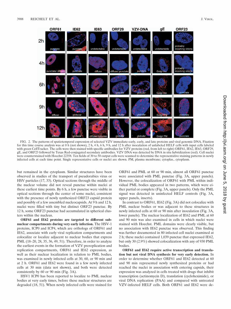
but remained in the cytoplasm. Similar structures have beenobserved in studies of the transport of pseudorabies virus orHSV particles (17, 33). Optical sections through the middle ofthe nuclear volume did not reveal punctae within nuclei atthese earliest time points. By 6 h, a few punctae were visible inoptical sections through the center of some nuclei, consistentwith the presence of newly synthesized ORF23 capsid proteinand possibly of a few assembled nucleocapsids. At 9 h and 12 h,nuclei were filled with tiny but distinct ORF23 punctae. By12 h, some ORF23 punctae had accumulated in spherical clus-ters within the nucleus.
ORF61 and IE62 proteins are targeted to different sub-nuclear compartments during very early infection. The HSV-1proteins, ICP0 and ICP4, which are orthologs of ORF61 andIE62, associate with early viral replication compartments andcolocalize or localize adjacent to nuclear bodies that expressPML (18–20, 28, 35, 36, 49, 51). Therefore, in order to analyzethe earliest events in the formation of VZV prereplication andreplication compartments, ORF61 and IE62 expression, aswell as their nuclear localization in relation to PML bodies,was examined in newly infected cells at 30, 60, or 90 min andat 2 h. ORF61 and IE62 were found in a few newly infectedcells at 30 min (data not shown), and both were detectedconsistently by 60 or 90 min (Fig. 3A).
HSV1 ICP0 has been reported to localize to PML nuclearbodies at very early times, before these nuclear structures aredegraded (18, 51). When newly infected cells were stained for
ORF61 and PML at 60 or 90 min, almost all ORF61 punctaewere associated with PML punctae (Fig. 3A, upper panels).However, the colocalization of ORF61 with PML within indi-vidual PML bodies appeared in two patterns, which were ei-ther partial or complete (Fig. 3A, upper panels). Only the PMLsignal was detected in uninfected HELF controls (Fig. 3A,upper panels, inserts).
In contrast to ORF61, IE62 (Fig. 3A) did not colocalize withPML nuclear bodies or was adjacent to these structures innewly infected cells at 60 or 90 min after inoculation (Fig. 3A,lower panels). The nuclear localization of IE62 and PML at 60and 90 min was also examined in cells in which nuclei werestained with Hoechst. PML domains were clearly visible, butno association with IE62 punctae was observed. This findingwas further documented in 80 infected cell nuclei examined at2 h; these nuclei contained 1,039 punctae that expressed IE62,but only 30 (2.9%) showed colocalization with any of 930 PMLbodies.
ORF61 and IE62 require active transcription and transla-tion but not viral DNA synthesis for very early detection. Inorder to determine whether ORF61 and IE62 detected at 60and 90 min represented newly synthesized proteins or hadreached the nuclei in association with entering capsids, theirexpression was analyzed in cells treated with drugs that inhibittranscription (actinomycin D), translation (cycloheximide), orviral DNA replication (PAA) and compared with untreatedVZV-infected HELF cells. Both ORF61 and IE62 were de-
FIG. 2. The patterns of spatiotemporal expression of selected VZV immediate-early, early, and late proteins and viral genomic DNA. Fixationfor this time course analysis was at 0 h (not shown), 2 h, 4 h, 6 h, 9 h, and 12 h after inoculation of unlabeled HELF cells with input cells labeledwith green CellTracker. The cells were then stained with specific antibodies for VZV proteins (red, from left to right) ORF61, IE62, IE63, ORF29,gE, and ORF23 followed by Texas Red-conjugated secondary antibodies. VZV DNA was detected by DNA in situ hybridization (red). Cell nucleiwere counterstained with Hoechst 22358. Ten fields of 30 to 50 output cells were scanned to determine the representative staining patterns in newlyinfected cells at each time point. Single representative cells or nuclei are shown. PM, plasma membrane; cytoplas., cytoplasm.
3908 REICHELT ET AL. J. VIROL.
on June 5, 2018 by guesthttp://jvi.asm
.org/D
ownloaded from

tected at 4 h in newly infected cells adjacent to green-labeledinoculum cells with or without PAA treatment (Fig. 3B), andtheir intracellular localization was similar to that observed inthe time course experiments (Fig. 1 and 2). The size and
morphology of the nuclear compartments that expressed IE62were not altered in PAA-treated cells at 4 h. PAA concentra-tions of either 0.5 mg/ml (data not shown) or 1 mg/ml did notreduce the expression of either ORF61 or IE62. However, no
FIG. 3. ORF61 and IE62 proteins are targeted to different subnuclear compartments and require active transcription and translation but notviral DNA synthesis for very early expression. (A) Unlabeled HELF cells were infected and fixed after 60 min (left panels) or 90 min (right panels).The cells were then double stained with either a combination of rabbit polyclonal anti-ORF61 antibody (upper panels, red) and anti-PML (blue;monoclonal mouse) or with rabbit polyclonal anti-IE62 (lower panels, red) and anti-PML (blue; monoclonal mouse). Secondary antibodies weredonkey anti-rabbit Alexa Fluor 546 and donkey anti-mouse Alexa Fluor 647. Uninfected control cells (insets) were stained with the same antibodycombinations. The small images in the ORF61 panels show single-PML (blue)- or ORF61 (red)-staining patterns that completely overlap (pink)in the larger overlay images. The two images shown in the IE62 panels (at 60 min or 90 min) depict the same nucleus. In the images on the left,nuclear staining (Hoechst 22358) is shown with the IE62 (red) and PML (blue) signals to clearly mark the nuclear areas. In the images on the right,the nuclear staining was left away for better visibility of the IE62 punctae (red; white arrows) that do not overlap with the PML signal (blue). Scalebars are 10 �m. (B) Uninfected HELF cells were pretreated for 30 min with cycloheximide, actinomycin D, or PAA or left untreated (control)and then inoculated with infected, untreated, green-labeled input cells. Incubation occurred in the presence of the same drugs and was stoppedby fixation at 4 h. Cells were then stained for either ORF61 (red, upper panels) or IE62 (red, lower panels), and nuclei (blue) were counterstainedwith Hoechst 22358. Output cells adjacent to infected inoculum cells (green cytoplasmic staining plus red nuclear staining of ORF61 or IE62) wereanalyzed for nuclear expression of ORF61 or IE62 (red). Nuclear ORF61 or IE62 (red) is visible in several newly infected cells (white stars) ineither the control (far left panels) or the PAA-treated cells (far right panels) but not in the cycloheximide- or actinomycin D-treated cells (middlepanels). Scale bars are 20 �m.
VOL. 83, 2009 THE VZV REPLICATION CYCLE IN SINGLE CELLS 3909
on June 5, 2018 by guesthttp://jvi.asm
.org/D
ownloaded from

ORF61 or IE62 was detected in cells treated with actinomycinD or cycloheximide. None of the punctate signals seen at 60 or90 min in the absence of these drugs were detected in cellsadjacent to the inoculum cells, indicating that ORF61 andIE62 detected at these times represented newly synthesizedVZV proteins (Fig. 3A).
The ORF29 single-stranded DNA binding protein is tar-geted to nuclear domains that express IE62 and transforminto viral replication compartments. HSV-1 ICP4, which isrelated to IE62, and ICP8, the single-stranded DNA bindingprotein, are involved in the formation and function of replica-tion compartments in infected cell nuclei (15, 44). Analyses ofthe stages in their formation indicate a stepwise recruitment ofICP8 and other viral replication proteins to ICP4 in generatingactive replication compartments (7, 15, 32). To explore thetiming and define the steps in the creation of VZV replicationcompartments, the spatiotemporal expression of IE62 andORF29 was analyzed, along with the localization of nascentVZV DNA, detected by BrdU pulse-labeling, and of accumu-lated VZV DNA, detected by DNA-FISH. The 2- to 12-hkinetics experiments showed that IE62, ORF29, and viralDNA accumulated in globular nuclear domains over time (Fig.2). These experiments also revealed that both viral DNA andORF29 were detectable unequivocally only at about 4 h afterinfection, whereas IE62 was detectable within 1 h (Fig. 2 and 3).These observations suggested that ORF29 might be recruited tonuclear compartments containing IE62 to form active viral repli-cation sites, where VZV genomic DNA accumulated.
IE62 punctae were prominent at 4 h, whereas ORF29 ex-pression was limited and diffuse at this time point (Fig. 4A,panels a and a’), confirming the observations shown in Fig. 2.By 6 h, single ORF29 punctae (Fig. 4A, panels b and b’) orclusters of ORF29 punctae (Fig. 4A, panels c and c’) that wereclearly associated with the periphery of small or more globularIE62 compartments (Fig. 4A, panels b’ and c’) were observed.However, ORF29 was only partially colocalized with IE62. By8 h, both ORF29 and IE62 were present within globular com-partments, where they remained in a pattern of partial colo-calization (Fig. 4A, panel d’).
Whether viral DNA replication was associated within theseglobular compartments that expressed IE62 and ORF29 wasthen determined by labeling nascent VZV DNA with BrdUand by detecting accumulated VZV DNA, using DNA-FISH.Newly infected cells were examined at 12 h after infection,because staining protocols to detect incorporated BrdU orDNA-FISH together with immunofluorescence for viral pro-teins were usually much less sensitive than those to detectVZV DNA or proteins separately. As shown in Fig. 2, largerand more intensely stained nuclear IE62, ORF29, or VZVDNA domains were present at 12 h than at 6 h, making ex-periments using BrdU detection or DNA-FISH in combinationwith antibodies to detect VZV proteins more robust and easierto interpret at this time point.
At 12 h, cells were pulse-labeled with BrdU for 30 minbefore being fixed. First, single stainings for incorporatedBrdU, VZV DNA, ORF29, or IE62 were performed (Fig. 4B).As expected from the time course experiments, each singlestaining revealed large globular nuclear compartments thatwere of similar size and shape, suggesting that incorporatedBrdU, VZV DNA, ORF29, and IE62 resided in the same
nuclear domains at this time point. To confirm this observa-tion, double staining of newly infected cells from the sameexperiment were performed (Fig. 4C). Incorporated BrdU andVZV DNA signals exhibited colocalization, demonstratingthat nascent VZV DNA was within the same compartment asaccumulated viral DNA (Fig. 4C, panels a through c). Todemonstrate that ORF29 was present with nascent viral DNA,infected cells were stained for incorporated BrdU and ORF29,which showed colocalization (Fig. 4C, panels d through f).Dual staining for ORF29 and IE62 confirmed that both pro-teins resided in the same nuclear compartments at 12 h (Fig.4C, panels g through i). Control staining for ORF29 and VZVDNA in uninfected cells or for BrdU in untreated cells dem-onstrated the specificity of the staining protocols (Fig. 4C,panels j through l).
The harsh conditions of the BrdU-labeling and DNA-FISHprotocols prevented the combined visualization of VZV DNAwith ORF29 or IE62 at 4 h or 8 h. Nevertheless, these resultssuggest that IE62 and ORF29 accumulated together with nas-cent and accumulated VZV DNA within the same nucleardomains and that these domains represent VZV replicationcompartments. Both ORF29 and VZV DNA signals wereweak at 4 h but were clearly recognizable at 6 h in single-staining experiments (Fig. 2), indicating that the first activeVZV replication compartments form between 4 and 6 h innewly infected HELF.
VZV nucleocapsid formation and plasma membrane expres-sion of glycoprotein gE. Having observed that VZV DNAreplication occurs well before 9 h in VZV-infected HELF cells,we analyzed later events in the VZV replication cycle, includ-ing nucleocapsid assembly, glycoprotein expression on plasmamembranes, and virion envelopment and egress. The kineticsexperiments revealed that gE was expressed as early as 4 h andaccumulated in a Golgi-compartment-like pattern in the cyto-plasm from 4 to 6 h (Fig. 2). However, gE association withplasma membranes was minimal at the early times comparedto 9 h, when it was expressed extensively on plasma membranesas well as in the cytoplasm (Fig. 2 and 5A). These experimentsalso revealed that distinct ORF23 punctae were very rarewithin the nucleus at 6 h but had become abundant by 9 h (Fig.2 and 5A). However, confocal microscopy did not revealwhether ORF23 is associated with assembled nucleocapsids.
EM experiments were done to assess whether VZV nucleo-capsid assembly, nuclear egress of virions into the cytoplasm,and secondary envelopment of cytoplasmic virions had oc-curred by 9 h after infection. In order to distinguish betweeninoculum cells and newly infected cells, the uninfected cellswere labeled by fluid-phase endocytosis of PA-gold particlesbefore infection. The accumulation of the electron-dense10-nm PA-gold particles within lysosomes differentiated theoutput cells from VZV-infected input cells. Assessment ofVZV-induced changes in the ultrathin sections of these sam-ples was done by examining only these labeled cells. Cellswithin multinucleated syncytia were excluded from this analysisbecause fusion of an infected input cell with a labeled outputcell could result in the presence of virion structures that weremade in the inoculum cells rather than in the newly infectedcell. Only cells that had a single nucleus and a large area ofcytoplasm with at least two lysosomes containing electron-dense PA-gold clusters were evaluated.
3910 REICHELT ET AL. J. VIROL.
on June 5, 2018 by guesthttp://jvi.asm
.org/D
ownloaded from

FIG. 4. The single-stranded DNA binding protein ORF29 is targeted to IE62 nuclear domains that transform into viral replication compartments.(A) HELF cells were seeded on glass coverslips and infected with green-labeled inoculum cells for 4 h, 6 h, or 8 h and were double stained with anti-IE62 (green;mouse monoclonal antibody) and anti-ORF29 (red; polyclonal rabbit antibody) followed by secondary anti-mouse Alexa Fluor 488 or anti-rabbit TexasRed-conjugated secondary antibodies. Nuclei (upper panels a through d; blue) were counterstained with Hoechst 22358. Newly infected cells were identified bythe absence of green cytoplasmic staining and were analyzed for IE62 and ORF29 expression by confocal microscopy. The upper panels (a through d) show anoverview of the nuclei with merged blue, green, and red channels. Areas of the same nuclei (within the white squares in the upper panels) are shown at highermagnification in lower panels a’ through d’ (for better visualization of red and green signals, no blue channels are shown). White arrows point to red ORF29punctae associated with green-stained IE62 nuclear domains. Scale bars are 10 �m. (B) HELF cells were seeded and infected and then additionally treated at12 h after infection with a 30-min pulse of 0.1 mM BrdU (in medium) before being fixed at 12.5 h. Cells were then stained with anti-ORF29 (red) or -ORF62(green; polyclonal antibody) and Texas Red (ORF29)- or FITC (IE62)-conjugated secondary antibodies, respectively, processed for DNA in situ hybridization(red; DIG-labeled VZV DNA probe), or stained for incorporated BrdU (red; biotinylated anti-BrdU monoclonal antibody and streptavidin-Texas Redconjugate for secondary detection). Scale bars are 10 �m. Each of these single stainings reveals similarly shaped globular nuclear compartments. (C) Cells fromthe same experiment as for panel B were double stained for either BrdU (a, red) with viral DNA (vDNA; b, green), BrdU (d, red) with ORF29 (e, green), orORF29 (g, red) with IE62 (h, green). The merged channels are shown in panels c, f, and i. Control stainings for ORF29, viral DNA, and BrdU are shown inpanels j, k, and l. Scale bars are 10 �m.
3911
on June 5, 2018 by guesthttp://jvi.asm
.org/D
ownloaded from

FIG. 5. VZV nucleocapsids and surface expression of glycoprotein gE are detectable at 9 h after infection. (A) Uninfected HELF cells were inoculated withgreen-labeled inoculum cells and fixed after 9 h. Unpermeabilized cells were stained with anti-gE (left panel, red; monoclonal antibody) and a secondaryanti-mouse Texas Red antibody. White arrows mark areas of plasma membrane-expressed gE protein (red). Other cells were permeabilized and stained withrabbit polyclonal anti-ORF23 (right panel, red) and a secondary anti-rabbit Texas Red-conjugated antibody. Newly infected cells are marked with a white star.Scale bars are 30 �m. (B) Time-resolved standard EM analysis of newly infected HELF cells. (a) Overview of an infected HELF cell 9 h after inoculation. TheGolgi-compartment area (b, white square) is shown at higher magnification in the inset panel b’, which shows a nucleocapsid (arrow) undergoing envelopmentwith a Golgi-compartment-derived membrane. The white arrows labeled c through f in the overview image point to structures that are shown at much highermagnification in the images on the right (c through f). (c and d) Nuclear viral nucleocapsids are indicated by arrows. (e and f) Lysosomes with clusters ofinternalized 10-nm gold particles (arrows) that were used to discriminate newly infected cells from infected inoculum cells are shown.
3912 REICHELT ET AL. J. VIROL.
on June 5, 2018 by guesthttp://jvi.asm
.org/D
ownloaded from

EM analysis at 2 h and 6 h did not reveal morphologicalchanges in the PA-gold-labeled cells compared to unlabeledcells, indicating that the PA-gold particles did not alter cellularstructures. No evidence of VZV infection was detectable, al-though small numbers of incoming virions would not be likelyto be observed in these ultrathin sections. Importantly, PA-gold particles were not present on cell surfaces, between cells,or in the heavily infected inoculum cells at these early timepoints, showing that PA-gold particles were not transferredfrom labeled output cells to VZV-infected inoculum cells,which would interfere with interpretation of the results. By 9 h,ultrathin sections revealed VZV nucleocapsids in the nuclei ofnewly infected cells (Fig. 5B, panels a, c, and d) that wereidentified by PA-gold clusters in lysosomes (Fig. 5B, panels a,e, and f). Several virions were also present in the cytoplasmadjacent to Golgi-compartment-derived membranes (Fig. 5B,panel b, inset b’). The apparent density of nucleocapsids in theEM sections was less than might be expected, given the extentof ORF23 expression observed at 9 h by confocal analysis, butit is explained by the evaluation of a much smaller cell volumein ultrathin sections (60 nm) than in a confocal optical section(500 to 1,000 nm). Although nucleocapsids and cytoplasmicvirions were observed, cytoplasmic vacuoles containing numer-ous enveloped VZ virions were essentially absent at 9 h afterinfection, as were extracellular virions.
The kinetics of cell-cell fusion after VZV infection. The timecourse experiments showed that gE was expressed in a Golgi-compartment-like pattern as early as 4 h and was enriched on
plasma membranes by 9 h. Since these experiments relied ondistinguishing inoculum cells by their green-fluorescent-dyelabeling, only newly infected cells that had not fused withinoculum cells were analyzed. These conditions demonstratedthat very early expression of VZV proteins, e.g., ORF61 andIE62, nascent VZV DNA synthesis, and expression of laterproteins, e.g., gE and ORF23, could occur in the absence ofcell-cell fusion. However, cell-cell fusion and the formation ofmultinucleated cells is a hallmark of VZV replication in cul-tured cells, as well as in the pathogenesis of VZV skin infection(11, 12). Therefore, to examine this characteristic of VZVreplication, fluorescent-dye labeling was used to determine thetime course of cell-cell fusion in VZV-infected HELF cells.VZV-infected and mock-infected cells were labeled with greenBODIPY CellTracker, and uninfected output cells were la-beled with orange CellTracker (CMRA). The cell populationswere washed thoroughly with medium before the inoculumcells were added to the uninfected-cell monolayer. As ex-pected, control experiments demonstrated that the two cellpopulations could be distinguished by confocal microscopywithout “bleed-through” of the red fluorescence into the greenchannel or vice versa (Fig. 6a through c). In order to followcell-cell fusion events, cells were also stained with anti-gEantibody and a secondary Alexa Fluor 647 (far red)-conjugatedantibody and with nuclear Hoechst 22358 stain to facilitate thedetection of syncytia (Fig. 6d through l).
Multinucleated cells that exhibited only green fluorescencewere detected at each time point, consistent with the presence
FIG. 6. Demonstration of cell-cell fusion events at 9 h after VZV infection. HELF cells were seeded on glass coverslips and stained with orangeCellTracker (CMRA), washed, and then inoculated with green-CellTracker-labeled uninfected HELF cells (a through c) or infected HELF cells(d through l). Cells were fixed at 4 h (d through f) or 9 h (g through i and j through l) after infection and stained for gE protein (blue; monoclonalantibody and secondary anti-mouse Alexa Fluor 647 conjugate). Nuclei were stained with Hoechst 22358 (d through l, gray). Cells were analyzedfor the mixing of green and red fluorescent signals in the cytoplasm that would indicate fusion of newly infected cells with an input cell. The thinwhite arrows in panels g through i point to a cell (red and green cytoplasm) that seems to have partially fused with fused input cells. The adjacentoutput cell (arrowhead) has not yet fused (only red fluorescence). Scale bars are 50 �m.
VOL. 83, 2009 THE VZV REPLICATION CYCLE IN SINGLE CELLS 3913
on June 5, 2018 by guesthttp://jvi.asm
.org/D
ownloaded from

of fused cells in the inoculum (data not shown). No clearevidence of cell-cell fusion between green- and red-labeledcells was detected from 2 to 6 h. In an example at 4 h, aninfected inoculum cell was adjacent to output cells that wereeither not yet infected or did not yet express gE (Fig. 6dthrough f). In this case, distinct red or green cytoplasmic stain-ing was apparent with no evidence of a yellow-orange colorthat would indicate cytoplasmic mixing. However, by 9 h, manysyncytia with yellow fluorescence were observed, indicatingfusion of inoculum and output cells (Fig. 6g through i and jthrough l). Some cells adjacent to a syncytium showed someevidence of red/green cytoplasmic mixing but still seemed to bepartially separated, suggesting that only partial fusion had oc-curred (Fig. 6g through i). However, other cells adjacent to asyncytium exhibited only red fluorescence, indicating that nofusion had occurred (Fig. 6g through i and k through l). Theseexperiments indicate that cell-cell fusion is usually a relativelylate event in the VZV replication cycle, becoming prominentat 9 h, and that it is not necessary for the spread of VZV toadjacent uninfected cells.
Detection of VZV nucleocapsids, enveloped virions in cyto-plasmic vacuoles, and extracellular enveloped virions. VZVgE and the ORF23 capsid protein were expressed extensivelyby 9 h after infection, as determined by confocal analysis (Fig.2 and 5A), and time-resolved EM revealed that nucleocapsidsand cytoplasmic virions were present in newly infected cells by9 h (Fig. 5B). However, since few virions in cytoplasmic trans-port vacuoles or extracellular virions were detected at 9 h,further EM analyses of newly infected cells were done at 12 h,again assessing only cells with a single nucleus and endocytosedPA-gold particles within at least two lysosomes. Figure 7 showsdetailed ultrastructural images of two newly infected cells at12 h. Cell 1, shown at lower magnification in Fig. 7A, hasnumerous nucleocapsids within the nucleus (Fig. 7B), cytoplas-mic virions in the Golgi-compartment area (Fig. 7C), and mod-ified Golgi-compartment membranes with putative tegumentprotein (Fig. 7D and E), all within the same cell. Extracellularvirions were also associated with this cell (Fig. 7C, inset).
Cell 2 (Fig. 7G) has several nucleocapsids within the nucleus(Fig. 7H) and enveloped virions within cytoplasmic vacuoles(Fig. 7H). Both cells contained 10-nm PA-gold particles withinlysosomes, as shown in Fig. 7F and I. Thus, by 12 h afterinfection, VZV-infected cells exhibited an abundance of nu-cleocapsids, extensively modified Golgi-compartment mem-branes, cytoplasmic vacuoles with many virions with secondaryenvelopment, and completely enveloped virions in the cyto-plasm, along with numerous extracellular virions on plasmamembranes.
DISCUSSION
In this study, the kinetics of major events during the first 12 hof the VZV replication cycle was established, using novel strat-egies to differentiate virus-infected inoculum cells from newlyinfected cells in time-resolved confocal IF and EM analyses ofindividual human fibroblasts after VZV entry. Since VZV ishighly cell associated, studies of its replication are done withinfected-cell or low-titer, cell-free virus inocula that cannotproduce uniform, synchronous infections (11). In confocal mi-croscopy experiments, this obstacle to evaluating the spatio-
temporal expression of VZV proteins and viral DNA synthesiswas overcome by labeling VZV-infected inoculum cells withfluorescent dyes that are nontoxic and do not diffuse into themedium. These fluorescent dyes are commonly employed totrace cell lineages over intervals of hours up to several days indevelopmental biology experiments; the characterization ofentosis, a form of nonapoptotic cell death, is a recent applica-tion of the method (43).
In our investigations of VZV replication, green fluorescencelabeling of inoculum cells and detection of viral proteins andDNA with Texas Red-conjugated reagents proved to be themost effective. Even though the inoculum cells were heavilyinfected, the green CellTracker dye was not released, and thelabeled cells retained their infectivity for uninfected fibro-blasts. Any effect of the fluorescent dye on the VZV replicationcycle was excluded because the cell tracer was not available tobe taken up by newly infected cells. This approach allowed anelucidation of the kinetics of synthesis and intracellular local-ization of VZV proteins and genomic DNA and provided asemiquantitative and reproducible assessment of events inVZV replication based on examining a defined number ofindividual infected cells at each time point. The method alsopermitted studies of different VZV proteins in parallel at eachinterval, the effects of drugs that inhibit various stages of rep-lication, and associations between viral proteins, VZV DNA,and cell proteins in samples from the same assay.
As a complementary strategy for examining VZ virion as-sembly, intracellular trafficking, and egress in a time-resolvedanalysis of events at the ultrastructural level, the uninfectedcell population was labeled with PA-gold particles by fluid-phase endocytosis before inoculation and EM analysis (38).The newly infected cells, identifiable by the presence of goldparticles within lysosomes, showed no ultrastructural differ-ences from unlabeled cells and remained susceptible to pro-ductive VZV infection. Both strategies should be applicablefor investigating the replication cycle of other cell-associatedviruses. Differential labeling of inoculum and newly infectedcells with fluorescent dyes can also be used for sorting by flowcytometry or selective harvest of newly infected cells by lasercapture at specific intervals, either of which may facilitatelarge-scale, time-resolved profiling of virus and host cell genetranscription and of the virus or host cell proteome for VZV orother viruses. In addition, the two cell-labeling techniquescould be combined in multiscale correlative IF/EM experi-ments to examine rare events in virus-infected cells (45).
Events during the first 12 h after VZV infection were de-fined by tracking the spatiotemporal expression of IE62 andIE63, tegument proteins that regulate VZV transcription;OR61, a poorly characterized regulatory protein; ORF29, theVZV single-stranded DNA binding protein; gE, the majorVZV glycoprotein; and ORF23, a nucleocapsid protein andortholog of HSV VP26 (11). These experiments were done inparallel with ultrastructural examination of virion assemblyand egress. Of these six VZV proteins, only ORF61 and IE62were detected at the earliest time point and were newly syn-thesized within 1 h. VZV DNA accumulated with IE62 innuclear domains as early as 4 h, and these domains enlarged tooccupy 75 to 90% of the nuclear compartment by 12 h. VZVgE and ORF23 expression was extensive by 9 h, and consistentwith these observations, nucleocapsids and cytoplasmic virions
3914 REICHELT ET AL. J. VIROL.
on June 5, 2018 by guesthttp://jvi.asm
.org/D
ownloaded from

in the Golgi-compartment region were detected at 9 h. By 12 h,newly infected cells contained many nucleocapsids, envelopedcapsids in the Golgi compartment, many VZ virions with sec-ondary envelopes in intracellular vacuoles, and extracellularenveloped virions associated with plasma membranes. Theseexperiments demonstrated that completion of the VZV repli-cation cycle within individual cells, leading to the release ofprogeny virions, takes only 9 to 12 h in human fibroblasts. Thetime course established with these six VZV proteins provides a
point of reference for grouping other VZV proteins with re-gard to the timing of their synthesis after viral entry. In addi-tion, this information about the normal VZV replication cyclewill be useful in defining disruptions resulting from mutationsof the VZV genome, revealing when the functions of the mu-tated gene or promoter region are important in the course ofthe VZV replication cycle. These observations also confirmthat VZV replication is comparatively slower than that ofHSV-1, which produces new genomic DNA within 2 h after
FIG. 7. Detection of VZV nucleocapsids, intracellular enveloped virions in cytoplasmic vacuoles, and extracellular enveloped virions at 12 hafter infection. Areas of two cells (A through F, cell 1; G through I, cell 2) that were fixed after 12 h of infection are shown at the ultrastructurallevel after processing for time-resolved standard EM. (A) Overview of cell 1 with black squares indicating the areas that are shown in panels Bthrough D at a higher magnification. (B) Demonstration of many nucleocapsids (arrows) in the nucleus. (C) Stack of Golgi cisternae (G) withadjacent nucleocapsids (small arrows) and one enveloped nucleocapsid (arrowhead). The inset shows extracellular virions associated with theplasma membrane of cell 1. (D and E) Another Golgi stack (G) in the same cells with modified Golgi cisternae (D, arrows) and enrichment ofelectron-dense putative tegument proteins (E, arrowheads) at the concave sides of these cisternae. (F) A multilamellar lysosome (L) is shown witha cluster of internalized 10-nm gold particles that was used to identify cell 1 as a newly infected cell (see Materials and Methods). (G) Overviewof cell 2. The black square marks an area shown at higher magnification in panel H. (H) A nucleus with nucleocapsids (left side) is visible, andthree cytoplasmic vacuoles (v) that contain VZV particles (arrows) can be seen. An extracellular virion is also marked (right side, arrow). (I) Threelysosomes are marked (L), one of which harbors a cluster of 10-nm gold particles (white square) that are clearly visible at a higher magnification(arrow in inset).
VOL. 83, 2009 THE VZV REPLICATION CYCLE IN SINGLE CELLS 3915
on June 5, 2018 by guesthttp://jvi.asm
.org/D
ownloaded from
infection. Large replication compartments are formed in HSV-infected cells by 6 h, compared to between 9 and 12 h inVZV-infected cells (8, 15).
IE62, the predominant VZ virion tegument protein, is pre-dicted to be an important immediate-early protein because itfunctions as the most potent and promiscuous transactivator ofVZV gene promoters, including its own, and is essential forVZV replication (47, 48). The functions of ORF61 are muchless well defined, although it also has regulatory activity intransient expression experiments (42). Using drug inhibitionprotocols and spatiotemporal analyses of individual newly in-fected cells, we demonstrated synthesis and nuclear transloca-tion of both ORF61 and IE62 within 1 h after VZV infection.Of interest, ORF61 and IE62 were targeted to different nu-clear subcompartments. ORF61 was consistently associatedwith ND10 bodies, identified by PML expression, whereasIE62 was associated with fewer than 3% of PML-expressingnuclear domains. The strong association of ORF61 with PMLis consistent with observations about ICP0, which is its HSV-1ortholog (40, 41). ICP0 localization to ND10 bodies and itsubiquitin ligase function is essential for their molecular rear-rangement and degradation in HSV-1-infected cells (6, 18).ORF61 has an N-terminal ring finger domain that shares ap-proximately 26% amino acid identity with the ICP0 ring fingermotif, and ORF61 complements HSV-1-ICP0 deletion mu-tants (11, 40, 41). These observations suggest that ORF61 maycompensate for the absence of ICP0-mediated degradation ofND10 bodies in cells infected with these mutants. WhetherORF61 shares the other functions of ICP0 that are necessaryto inhibit the host cell antiviral response during initial replica-tion is not known.
At the earliest time points, IE62 tended to be enriched at theinner nuclear rim in the newly infected cell that was nearest toan infected inoculum cell. While IE62 has important differ-ences, both IE62 and HSV1 ICP4 have DNA binding domainsand the capacity to interact with cellular transactivators toregulate transcription from viral gene promoters (21, 46, 47).Functionally, IE62 can complement HSV-1 mutants that lackICP4 (22). Since ICP4 has been demonstrated to mark thelocation of HSV-1 prereplication compartments (19, 20, 32), itis likely that the first detection of IE62 punctae at the innernuclear rim adjacent to an attached VZV-infected input cellrepresented its localization with incoming VZV genomes. Asnoted, IE62 showed essentially no localization with PML nu-clear bodies during the first 2 h after VZV entry. In contrast,although earlier reports differ, recent studies indicate thatICP4 associated with these structures within 30 min afterHSV-1 entry (32).
The time-resolved assessment of IE62, ORF29, the single-stranded DNA binding protein, and newly synthesizedgenomic DNA provides the first information that addresses theformation of VZV replication compartments at very early timepoints. IE62, ORF29, and VZV DNA began to colocalize inputative replication compartments between 4 to 6 h after in-fection. However, ND10 components, represented by PML,did not seem to be deposited in these locations within infectedcell nuclei at these early time points (data not shown). Thestepwise assembly of active HSV-1 replication compartmentsfrom prereplicative sites has been demonstrated by trackingthe intracellular localization of ICP4; ICP8, which is the
HSV-1 single-stranded DNA binding protein; and incorpo-rated BrdU as a marker for nascent viral DNA (7, 8, 15, 32, 44,46). Although delayed compared to that for HSV-1-infectedcells, a similar sequence of events was observed in VZV-in-fected cells in that IE62 was first observed at less than 4 h innuclear punctae that did not yet contain ORF29, the VZVsingle-stranded DNA binding protein, and probably repre-sented prereplicative sites. ORF29 was first detectable at lowlevels in a diffuse nuclear distribution at 4 h. IE62-expressingdomains grew into larger globular nuclear structures andORF29 began to appear at these sites from 4 to 6 h afterinfection. When evaluated at 4 h and 8 h, small ORF29 punc-tae were localized specifically and exclusively at the margins ofIE62 punctae, and small, although somewhat more globularIE62 sites, followed at later times by a distribution togetherwith IE62 in larger globular compartments. For comparison,HSV-1 ICP4 and ICP8 colocalization was observed at 3 h, andsmall ICP8 punctae were associated with the periphery ofICP4-positive domains; colocalization of the helicase/primaseand viral polymerase complexes was also shown (32). Similarly,ORF29 seems to be recruited to sites where IE62 has accumu-lated and that may serve as platforms for the assembly ofproteins required for VZV replication. Although we did notanalyze VZV helicase/primase or polymerase proteins, theirrecruitment would be expected, since the basic replication ma-chinery is predicted to be highly conserved among herpesvi-ruses (46).
The VZV ORF63 gene product is a tegument phosphopro-tein with some regulatory functions, e.g., enhancing IE62transactivation of some VZV promoters. It is related to theHSV-1 US1.5 protein, which is expressed colinearly withICP22 (3). IE63 has been designated an immediate-early pro-tein based on its presumed kinetics (16). However, IE63 nu-clear expression was detectable only as a weak, diffuse signal at4 to 6 h, which was substantially later than that of IE62 andORF61. A difference in the sensitivity of IE63 detection isunlikely, because the anti-IE63 antibody is a high-potency re-agent (34) and the intensity of the IE63 signal showed a rapidincrease when evaluated between 6 to 8 h after infection.However, because IE63 was diffusely expressed and was notenriched in discrete compartments like the IE62 signal, earlierexpression of IE63 might escape detection. IE63 expressionremained diffuse throughout the nuclei, suggesting that thisprotein may be present within IE62-positive replication com-partments but is not specifically enriched at these sites eventhough IE63 binds to IE62 by residues in the IE63 N terminus(3). IE63 was predominantly nuclear throughout the 9- to 12-hVZV replication cycle and remained so up to 24 h, but IE63expression in the cytoplasm increased beginning at 9 h. Thisextensive cytoplasmic expression of IE63 at later times may beimportant for its role as an antiapoptotic factor, as has beendemonstrated in cultured neurons (27).
VZV gE is the most abundant glycoprotein in infected cellsand is a prominent envelope glycoprotein. In contrast to otheralphaherpesviruses which replicate in the absence of gE, VZVrequires gE for viral replication (11, 39). While it has beenconsidered to be a late gene product, our time course experi-ments revealed that gE synthesis occurred quite early in newlyinfected cells in a pattern similar to that described for HSV gB(4, 5). gE appeared as a weak but readily detectable signal in a
3916 REICHELT ET AL. J. VIROL.
on June 5, 2018 by guesthttp://jvi.asm
.org/D
ownloaded from

Golgi-compartment-like distribution as early as 4 h, progress-ing significantly in intensity and extent at this site by 6 h. Thisearly gE expression may serve to rearrange the Golgi compart-ment and prepare Golgi-compartment-derived membranes forVZ virions exiting the nucleus by accumulating tegument pro-teins such as IE62 on membrane-bound gE (11, 23, 54). Be-cause gE was absent from the plasma membrane and did notshow a punctate endosome-like cytoplasmic pattern at earlytime points, it is unlikely that we detected endocytosed gE.ORF23, which is a VZV capsid protein related to HSV-1VP26, became enriched within nuclei at 9 h, suggesting thatviral nucleocapsids could become assembled for release fromthe nucleus and envelopment in the premodified Golgi-com-partment area by 6 to 9 h after infection. In contrast to itsGolgi-compartment localization, substantial gE expression onplasma membranes was evident only at 9 h.
While syncytium formation is a hallmark of VZV replicationin cultured cells, our analyses of the VZV replication cyclewere done by tracking events during the infection of individualcells that were located next to an infected inoculum cell buthad no evidence of green-fluorescent-dye transfer into thecytoplasm. These experiments suggested that VZV infectionwas initiated without cell-cell fusion and could be attributed tothe transfer of extracellular virions on surfaces of inoculumcells, as visualized by EM. This explanation was supported bythe observation that newly infected cells were consistentlyfound adjacent to inoculum cells. The inoculum cells were alsowashed extensively to eliminate any unabsorbed fluorescentdye, which also removed the original culture medium and anycell-free VZ virions it may have contained. Furthermore, it isknown that secondary plaques attributable to virus release donot appear in VZV-infected monolayers; in addition, extracel-lular VZ virions are quite labile, whereas enveloped VZVparticles are abundant on the surfaces of heavily infected fi-broblasts and are also present in intracellular vacuoles near theplasma membranes (11). When the inoculum cell was attachedin proximity to the uninfected cell, the space between theplasma membranes appeared to be minimal, therefore placingsurface virions next to the uninfected cell membrane and po-tential entry receptors of the uninfected cell and limiting thedispersal of virions. These conditions would favor entry ofcell-associated extracellular virus particles without requiringcell-cell fusion (Fig. 8). Of interest, VZV gE may promotethese conditions, since gE enhances the formation of tightjunctions between plasma membranes (37).
Although VZV infection occurred immediately after theexposure of uninfected cells to the infected cell inoculum with-out the formation of polykaryocytes, the kinetics experimentsdone using two cell dyes with different emission wavelengths(red and green fluorescence) to label inoculum and uninfectedcells revealed that abundant cell-cell fusion and syncytium for-mation occurred by 9 h after infection. Some earlier fusion mayhave been undetected if a syncytium consisting of severalgreen-labeled inoculum cells had fused with one red-labeledoutput cell, diluting the red fluorescence signal. In any case,while viral entry into uninfected cells occurs without it, cell-cellfusion at 9 h should amplify VZV replication substantially,since this time point coincides with the detection of envelopedcytoplasmic virions and precedes their appearance in large
numbers on the surfaces of plasma membranes, which becameprominent at 12 h.
In summary, this kinetic analysis of the VZV replicationcycle in individual fibroblasts demonstrated the spatiotemporalexpression of six VZV proteins, newly synthesized viral DNA,and virion morphogenesis. The earliest event after infectionwas new IE62 and ORF61 synthesis within 1 h. VZV DNAreplication was observed at 4 h, and virion assembly and en-velopment were detected at 9 h. Our results reveal that onecomplete VZV replication cycle leading to a new generation ofinfectious VZV particles takes between 9 and 12 h.
ACKNOWLEDGMENTS
This work was supported by NIH grants (AI053846, AI20459, andCA049605) to A.M.A. and by a postdoctoral fellowship to M.R. from theDeutsche Forschungsgemeinschaft (DFG, Germany; GeschaeftszeichenRE2716/1-1).
Sample preparation, sectioning, and IF-EM analyses were per-formed in the CSIF/Stanford University Medical School.
REFERENCES
1. Asano, Y., and M. Takahashi. 1979. Studies on the polypeptides of varicella-zoster (V-Z) virus. 1. Detection of varicella-zoster virus polypeptides ininfected cells. Biken J. 22:81–89.
2. Asano, Y., and M. Takahashi. 1980. Studies on the polypeptides of varicella-zoster (V-Z) virus. II. Syntheses of viral polypeptides in infected cells. BikenJ. 23:95–106.
3. Baiker, A., C. Bagowski, H. Ito, M. Sommer, L. Zerboni, K. Fabel, J. Hay, W.Ruyechan, and A. M. Arvin. 2004. The immediate-early 63 protein of vari-cella-zoster virus: analysis of functional domains required for replication invitro and for T-cell and skin tropism in the SCIDhu model in vivo. J. Virol.78:1181–1194.
FIG. 8. Model of the cell-to-cell spread of VZV before cell fusion invitro. Mature VZV particles are transported in an export vacuole to theplasma membrane of an infected inoculum cell (gray cell on left).(A) VZV particles released by exocytosis adhere to the plasma membraneat the interface with culture media. Infectious VZV particles are notreleased into the media or rapidly lose infectivity, since secondary plaquesdo not form in VZV-infected monolayers. (B) Some VZV particles arereleased from the inoculum cell in close proximity to the plasma mem-brane of the adjacent uninfected cell (white cell on right). (C) Envelopeglycoproteins of extracellular VZV particles at these sites have an in-creased probability of binding to cell surface receptors on the plasmamembrane of the uninfected cell. Entry into the adjacent cell occurs byendocytosis (D) or direct entry (E), which is followed by the release ofpartially tegumented capsids into the cytoplasm of the newly infected cell.Our model predicts that the spread of cell-associated extracelluar VZvirions in vitro is determined by the proximity of infected and uninfectedcells and does not require cell fusion.
VOL. 83, 2009 THE VZV REPLICATION CYCLE IN SINGLE CELLS 3917
on June 5, 2018 by guesthttp://jvi.asm
.org/D
ownloaded from

4. Beitia Ortiz de Zarate, I., L. Cantero-Aguilar, M. Longo, C. Berlioz-Torrent,and F. Rozenberg. 2007. Contribution of endocytic motifs in the cytoplasmictail of herpes simplex virus type 1 glycoprotein B to virus replication andcell-cell fusion. J. Virol. 81:13889–13903.
5. Beitia Ortiz de Zarate, I., K. Kaelin, and F. Rozenberg. 2004. Effects ofmutations in the cytoplasmic domain of herpes simplex virus type 1 glyco-protein B on intracellular transport and infectivity. J. Virol. 78:1540–1551.
6. Boutell, C., S. Sadis, and R. D. Everett. 2002. Herpes simplex virus type 1immediate-early protein ICP0 and its isolated RING finger domain act asubiquitin E3 ligases in vitro. J. Virol. 76:841–850.
7. Burkham, J., D. M. Coen, C. B. Hwang, and S. K. Weller. 2001. Interactionsof herpes simplex virus type 1 with ND10 and recruitment of PML toreplication compartments. J. Virol. 75:2353–2367.
8. Burkham, J., D. M. Coen, and S. K. Weller. 1998. ND10 protein PML isrecruited to herpes simplex virus type 1 prereplicative sites and replicationcompartments in the presence of viral DNA polymerase. J. Virol. 72:10100–10107.
9. Chaudhuri, V., M. Sommer, J. Rajamani, L. Zerboni, and A. M. Arvin. 2008.Functions of varicella-zoster virus ORF23 capsid protein in viral replicationand the pathogenesis of skin Infection. J. Virol. 82:10231–10246.
10. Chen, J. J., Z. Zhu, A. A. Gershon, and M. D. Gershon. 2004. Mannose6-phosphate receptor dependence of varicella zoster virus infection in vitroand in the epidermis during varicella and zoster. Cell 119:915–926.
11. Cohen, J., S. Straus, and A. Arvin (ed.). 2007. Varicella-zoster virus repli-cation, pathogenesis, and management, 5th ed., vol. 2. Lippincott Williams &Wilkins, Philadelphia, PA.
12. Cole, N. L., and C. Grose. 2003. Membrane fusion mediated by herpesvirusglycoproteins: the paradigm of varicella-zoster virus. Rev. Med. Virol. 13:207–222.
13. Cook, M. L., and J. G. Stevens. 1968. Labile coat: reason for noninfectiouscell-free varicella-zoster virus in culture. J. Virol. 2:1458–1464.
14. Davison, A. J., and J. E. Scott. 1986. The complete DNA sequence ofvaricella-zoster virus. J. Gen. Virol. 67:1759–1816.
15. Debrus, S., C. Sadzot-Delvaux, A. F. Nikkels, J. Piette, and B. Rentier. 1995.Varicella-zoster virus gene 63 encodes an immediate-early protein that isabundantly expressed during latency. J. Virol. 69:3240–3245.
16. de Bruyn Kops, A., and D. M. Knipe. 1988. Formation of DNA replicationstructures in herpes virus-infected cells requires a viral DNA binding protein.Cell 55:857–868.
17. Dohner, K., K. Radtke, S. Schmidt, and B. Sodeik. 2006. Eclipse phase ofherpes simplex virus type 1 infection: efficient dynein-mediated capsid trans-port without the small capsid protein VP26. J. Virol. 80:8211–8224.
18. Everett, R. D., and G. G. Maul. 1994. HSV-1 IE protein Vmw110 causesredistribution of PML. EMBO J. 13:5062–5069.
19. Everett, R. D., G. Sourvinos, C. Leiper, J. B. Clements, and A. Orr. 2004.Formation of nuclear foci of the herpes simplex virus type 1 regulatoryprotein ICP4 at early times of infection: localization, dynamics, recruitmentof ICP27, and evidence for the de novo induction of ND10-like complexes.J. Virol. 78:1903–1917.
20. Everett, R. D., G. Sourvinos, and A. Orr. 2003. Recruitment of herpessimplex virus type 1 transcriptional regulatory protein ICP4 into foci juxta-posed to ND10 in live, infected cells. J. Virol. 77:3680–3689.
21. Faber, S. W., and K. W. Wilcox. 1986. Association of the herpes simplex virusregulatory protein ICP4 with specific nucleotide sequences in DNA. NucleicAcids Res. 14:6067–6083.
22. Felser, J. M., P. R. Kinchington, G. Inchauspe, S. E. Straus, and J. M.Ostrove. 1988. Cell lines containing varicella-zoster virus open reading frame62 and expressing the “IE” 175 protein complement ICP4 mutants of herpessimplex virus type 1. J. Virol. 62:2076–2082.
23. Gershon, A. A., D. L. Sherman, Z. Zhu, C. A. Gabel, R. T. Ambron, andM. D. Gershon. 1994. Intracellular transport of newly synthesized varicella-zoster virus: final envelopment in the trans-Golgi network. J. Virol. 68:6372–6390.
24. Gilden, D., R. Mahalingam, S. Deitch, and R. Cohrs (ed.). 2006. Varicella-zoster virus neuropathogenesis and latency. Caister Academic Press, Nor-wich, United Kingdom.
25. Harper, D. R., N. Mathieu, and J. Mullarkey. 1998. High-titre, cryostablecell-free varicella zoster virus. Arch. Virol. 143:1163–1170.
26. Harson, R., and C. Grose. 1995. Egress of varicella-zoster virus from themelanoma cell: a tropism for the melanocyte. J. Virol. 69:4994–5010.
27. Hood, C., A. L. Cunningham, B. Slobedman, A. M. Arvin, M. H. Sommer,P. R. Kinchington, and A. Abendroth. 2006. Varicella-zoster virus ORF63inhibits apoptosis of primary human neurons. J. Virol. 80:1025–1031.
28. Ishov, A. M., and G. G. Maul. 1996. The periphery of nuclear domain 10(ND10) as site of DNA virus deposition. J. Cell Biol. 134:815–826.
29. Kinchington, P. R., D. Bookey, and S. E. Turse. 1995. The transcriptionalregulatory proteins encoded by varicella-zoster virus open reading frames(ORFs) 4 and 63, but not ORF 61, are associated with purified virus parti-cles. J. Virol. 69:4274–4282.
30. Kinchington, P. R., J. K. Hougland, A. M. Arvin, W. T. Ruyechan, and J.
Hay. 1992. The varicella-zoster virus immediate-early protein IE62 is a majorcomponent of virus particles. J. Virol. 66:359–366.
31. Kinchington, P. R., G. Inchauspe, J. H. Subak-Sharpe, F. Robey, J. Hay, andW. T. Ruyechan. 1988. Identification and characterization of a varicella-zoster virus DNA-binding protein by using antisera directed against a pre-dicted synthetic oligopeptide. J. Virol. 62:802–809.
32. Livingston, C. M., N. A. DeLuca, D. E. Wilkinson, and S. K. Weller. 2008.Oligomerization of ICP4 and rearrangement of heat shock proteins may beimportant for herpes simplex virus type 1 prereplicative site formation.J. Virol. 82:6324–6336.
33. Lyman, M. G., B. Feierbach, D. Curanovic, M. Bisher, and L. W. Enquist.2007. Pseudorabies virus Us9 directs axonal sorting of viral capsids. J. Virol.81:11363–11371.
34. Lynch, J. M., T. K. Kenyon, C. Grose, J. Hay, and W. T. Ruyechan. 2002.Physical and functional interaction between the varicella zoster virus IE63and IE62 proteins. Virology 302:71–82.
35. Maul, G. G., H. H. Guldner, and J. G. Spivack. 1993. Modification ofdiscrete nuclear domains induced by herpes simplex virus type 1 immediateearly gene 1 product (ICP0). J. Gen. Virol. 74:2679–2690.
36. Maul, G. G., A. M. Ishov, and R. D. Everett. 1996. Nuclear domain 10 aspreexisting potential replication start sites of herpes simplex virus type-1.Virology 217:67–75.
37. Mo, C., E. E. Schneeberger, and A. M. Arvin. 2000. Glycoprotein E ofvaricella-zoster virus enhances cell-cell contact in polarized epithelial cells.J. Virol. 74:11377–11387.
38. Mobius, W., E. van Donselaar, Y. Ohno-Iwashita, Y. Shimada, H. F. Hei-jnen, J. W. Slot, and H. J. Geuze. 2003. Recycling compartments and theinternal vesicles of multivesicular bodies harbor most of the cholesterolfound in the endocytic pathway. Traffic 4:222–231.
39. Montalvo, E. A., R. T. Parmley, and C. Grose. 1985. Structural analysis of thevaricella-zoster virus gp98-gp62 complex: posttranslational addition of N-linked and O-linked oligosaccharide moieties. J. Virol. 53:761–770.
40. Moriuchi, H., M. Moriuchi, and J. I. Cohen. 1994. The RING finger domainof the varicella-zoster virus open reading frame 61 protein is required for itstransregulatory functions. Virology 205:238–246.
41. Moriuchi, H., M. Moriuchi, H. A. Smith, S. E. Straus, and J. I. Cohen. 1992.Varicella-zoster virus open reading frame 61 protein is functionally homol-ogous to herpes simplex virus type 1 ICP0. J. Virol. 66:7303–7308.
42. Moriuchi, H., M. Moriuchi, S. E. Straus, and J. I. Cohen. 1993. Varicella-zostervirus (VZV) open reading frame 61 protein transactivates VZV gene promotersand enhances the infectivity of VZV DNA. J. Virol. 67:4290–4295.
43. Overholtzer, M., A. A. Mailleux, G. Mouneimne, G. Normand, S. J. Schnitt,R. W. King, E. S. Cibas, and J. S. Brugge. 2007. A nonapoptotic cell deathprocess, entosis, that occurs by cell-in-cell invasion. Cell 131:966–979.
44. Quinlan, M. P., L. B. Chen, and D. M. Knipe. 1984. The intranuclearlocation of a herpes simplex virus DNA-binding protein is determined by thestatus of viral DNA replication. Cell 36:857–868.
45. Reichelt, M., L. Zerboni, and A. M. Arvin. 2008. Mechanisms of varicella-zoster virus neuropathogenesis in human dorsal root ganglia. J. Virol. 82:3971–3983.
46. Roizman, B., D. M. Knipe, and R. J. Whitley. 2007. Herpes simplex viruses.In D. M. Knipe, P. M. Howley, D. E. Griffin, R. A. Lamb, M. A. Martin, B.Roizman, and S. E. Straus (ed.), Fields virology, 5th ed., vol. 1. LippincottWilliams & Wilkins, Philadelphia, PA.
47. Sato, B., H. Ito, S. Hinchliffe, M. H. Sommer, L. Zerboni, and A. M. Arvin.2003. Mutational analysis of open reading frames 62 and 71, encoding thevaricella-zoster virus immediate-early transactivating protein, IE62, and ef-fects on replication in vitro and in skin xenografts in the SCID-hu mouse invivo. J. Virol. 77:5607–5620.
48. Sato, B., M. Sommer, H. Ito, and A. M. Arvin. 2003. Requirement of vari-cella-zoster virus immediate-early 4 protein for viral replication. J. Virol.77:12369–12372.
49. Sourvinos, G., and R. D. Everett. 2002. Visualization of parental HSV-1genomes and replication compartments in association with ND10 in liveinfected cells. EMBO J. 21:4989–4997.
50. Stevenson, D., K. L. Colman, and A. J. Davison. 1994. Characterization ofthe putative protein kinases specified by varicella-zoster virus genes 47 and66. J. Gen. Virol. 75:317–326.
51. Tavalai, N., and T. Stamminger. 2008. New insights into the role of thesubnuclear structure ND10 for viral infection. Biochim. Biophys. Acta.
52. Yamanishi, K., Y. Matsunaga, T. Ogino, M. Takahashi, and A. Takamizawa.1980. Virus replication and localization of varicella-zoster virus antigens inhuman embryonic fibroblast cells infected with cell-free virus. Infect. Immun.28:536–541.
53. Zerboni, L., C. C. Ku, C. D. Jones, J. L. Zehnder, and A. M. Arvin. 2005.Varicella-zoster virus infection of human dorsal root ganglia in vivo. Proc.Natl. Acad. Sci. USA 102:6490–6495.
54. Zhu, Z., M. D. Gershon, Y. Hao, R. T. Ambron, C. A. Gabel, and A. A.Gershon. 1995. Envelopment of varicella-zoster virus: targeting of viral gly-coproteins to the trans-Golgi network. J. Virol. 69:7951–7959.
3918 REICHELT ET AL. J. VIROL.
on June 5, 2018 by guesthttp://jvi.asm
.org/D
ownloaded from