the restoration of the action potential by thiamine
TRANSCRIPT

Yale UniversityEliScholar – A Digital Platform for Scholarly Publishing at Yale
Yale Medicine Thesis Digital Library School of Medicine
1973
The restoration of the action potential by thiamineJoseph W. EichenbaumYale University
Follow this and additional works at: http://elischolar.library.yale.edu/ymtdl
This Open Access Thesis is brought to you for free and open access by the School of Medicine at EliScholar – A Digital Platform for ScholarlyPublishing at Yale. It has been accepted for inclusion in Yale Medicine Thesis Digital Library by an authorized administrator of EliScholar – A DigitalPlatform for Scholarly Publishing at Yale. For more information, please contact [email protected].
Recommended CitationEichenbaum, Joseph W., "The restoration of the action potential by thiamine" (1973). Yale Medicine Thesis Digital Library. 2554.http://elischolar.library.yale.edu/ymtdl/2554
brought to you by COREView metadata, citation and similar papers at core.ac.uk
provided by Yale University


YALE
MEDICAL LIBRARY
(TITLE OF THESIS)
for the purpose of individual scholarly consultation or reference is hereby
granted 6y the author. This permission is not to be interpreted as affect-
ing publication of this work or otherwise placing it in the public domain,
and the author reserves all rights of ownership guaranteed under common
law protection of unpublished manuscripts.
Signature of Author


THE RESTORATION OF THE ACTION POTENTIAL BY THIAMINE
Submitted by Joseph Walter Eichenbaum, B.A., A.A.
This thesis is in partial fulfillment of the requirements for the
degree of Doctor of Medicine, Department of Pharmacology
Yale University School of Medicine
1973

Digitized by the Internet Archive in 2017 with funding from
The National Endowment for the Humanities and the Arcadia Fund
https://archive.org/details/restorationofactOOeich
ACKNOWLEDGMENTS:
In the course of my work several individuals graciously provided
me with their time and assistance. Many thanks are in order to
Dr. Murdoch Ritchie who taught me his desheathing technique and left
a number of instruments from his laboratory at my disposal. To
Dr. Don McAfee I also wish to express my gratitude for his efforts
in giving me a background in the electronically related portions of
this project. Dr. Robert Roth's guidance was invaluable in using the
Aminco-Bowman Spectrophotometer to ascertain the presence of a
radiation contaminant. Of course, I would like to thank Mrs. Kathy
Piros, Dr. Victor Nadler, Dr. William Wilson, and Mr. Phillip Horowitz
all of whom provided periodic assistance and advice in performing
various lab procedures.
Most of all, however, I am indebted to Dr. Jack Cooper, who
patiently advised me throughout this project as well as served as
a teacher and a friend. His sharp mind, quick wit, and fantastic
drive somehow coupled to an uncanny ability to pause and relax
simultaneously to enjoy his life with his wife and family will serve
as a model for me in the years ahead.
Last, but not least, I wish to thank my dear wife, Annette, for
her technical assistance many nights in the laboratory as well as
her abiding moral support.


ABBREVIATIONS USED IN THIS TEXT:
TPP
TTP
TMP
ATP
ADP
SNE
Thiamine Pyrophosphate or Thiamine Diphospha
Thiamine Triphosphate
Thiamine Monophosphate
Adenosine Triphosphate
Adenosine Diphosphate
Subacute Necrotizing Encephalomyelopathy


1
ABSTRACT:
Desheathed rabbit vagi were irradiated with an ultraviolet lamp
while being superfused with oxygenated Locke solution, until the
action potential disappeared. Fluorometric assays for thiamine
content in irradiated nerves in parallel experiments indicated
negligible levels as compared to those determined for the non-irradiated,
desheathed rabbit vagi. One vagus nerve of the pair was then placed
in a solution of thiamine and Locke after which it consistently
regained its action potential within about 1.5 hours. The second
nerve, remaining in regular Locke solution, never regained its con¬
ductive activity.


2
INTRODUCTION:
In approaching the problem of conduction in nervous tissue, two
main aspects have grown to influence the thinking of investigators.
The first involves the nature of ionic movements. The second, as
yet more obscure, consists of the energy considerations which are
involved in restoration of ionic balance after the passage of impulses
along an axon. Through electrophysiological and pharmacological
experimentation, the inter-relationships of the two processes and the
behavior of the membrane and its constituents have suggested various
mechanisms, e.g,. , a carrier molecule which is involved in the Na-K
ionic shifts between membrane and extracellular fluid and a Na-K
ATPase pump which maintains a high K but low intracellular Na concen¬
tration in the neurone's resting state.
However, in addition to such postulated mechanisms, a number of
findings of concomitant phenomena seem to challenge our understanding
of the actual details in the course of events in nerve conduction.
One significant group of recent findings implicate vitamin in
this sequence of events. This role of thiamine has been suggested
to be a neurophysiological one quite distinct from its well-known
function as a coenzyme; i^e. drastic alterations in nerve conduction
do not apparently always reduce thiamine-dependent metabolic enzyme
levels (transketolase and pyruvate dehydrogenase and alpha-ketoglutarate
dehydrogenase) below physiological ranges. For example, an anti¬
metabolite of thiamine, pyrithiamine, which produces polyneuritis in
vivo, alters the electrical activity of isolated nervous tissue by
displacing thiamine from the nerve membrane. However, the activity of
the thiamine-dependent enzymes remained unchanged. Thus, the question

• '
3
arises: how critical then is the presence of thiamine to the entire
process of nerve conduction? That is to say, apart from its already
defined metabolic role, of what significance is its neurophysiological
role? This, essentially, was the problem we addressed ourselves to.
HISTORICAL BACKGROUND:
When it was discovered that nerves were sensitive to ultra¬
violet (UV) irradiation, it was theorized that absorbed photons
produced chemical lesions which disrupted the normal sequence of "free
2 3 energy transfer". 5 Using UV light then as a tool, one could attempt
to implicate some of the putative neurochemicals underlying the
process of nervous tissue conduction.
2 Audiat in 1931, first investigating the effect of UV light on
peripheral nerves, found that when he irradiated a whole nerve while
in a bath of Ringer's solution, there was an increase in the minimum
threshold voltage required for excitation. Simultaneously the action
potential amplitude decreased and gradually disappeared. This effect
was reversible and the time needed to restore excitability depended
on the time and the intensity of irradiation. When he used a filter
which absorbed all wavelengths below 310 mu, no effect on the action
2 potential was obtained.
3 The work of Hutton-Rudolph with a single motor fiber generally
corroborated the discoveries of Audiat in whole nerves. UV irradiation
increased the minimum excitability at the node quite rapidly. But
excitability ceased within two to eight minutes. When the internodal
region of the fiber was irradiated, there was an initial phase of


"over excitability" with a decrease in the required minimum threshold
voltage, which then again rose rapidly. In this case, excitability
was curtailed after fifteen to twenty minutes. Again, with the use
of filters to pass wavelengths greater than 300 mq these effects were
not observed. Maximum effect was obtained with wavelengths below
280 mq.3
In 1950 Booth et a_l. ^ studied the relationship between wave¬
length and intensity of monochromatic UV light producing these effects.
They found that only UV light below 320 mq. had photochemical action
on nodes in the frog sciatic preparations. Each wavelength studied
below this value had its specific activity and the activity curve
between 320 mq and 248 w/j. had three apparent maxima; at 297 mq,
285 iqu and 265 mq. They pointed out that their results could be
discussed in light of the theory of saltatory transmission and K/Na
exchanges. They stated that it was probable that the substances in
the node which were photochemically affected were related to a Na+
shift during excitation. Thiamine might be one of them.^
It has been demonstrated that thiamine has an absorption maxima
at 265 mq and is destroyed by UV light at this wavelength in about
one half hour. This finding has been employed for the destruction
of thiamine in various tissue preparations.
25 Bachoffer, who studied the electrophysiological effects of UV
irradiation on single, isolated nerve fibers of the earthworm found
that the initial enhancement of activity (increases in conduction
velocity, spike amplitude, and rate of rise of spike) was not due to
the synergistic action of UV energy and that of the nerve fiber;


5
nerves continued to respond in an enhanced manner without concomitant
irradiation. He concluded that "UV light produces a change in the
nerve which is not reversible, at least not without further treatment".
Von Muralt showed a significant difference in UV absorption
at 220 and 265 m<u between extracts from excited and unexcited nerves.
Extracts from excited nerves contained a greater amount of absorbing
material and therefore manifested higher peaks at these wavelengths.
A relationship between thiamine and the nervous system goes back
38 39 to Eijkman and Grijns . They recognized that it was the absence
of a certain dietary factor in rice bran which produced avian poly¬
neuritis and beri-beri. A number of experiments of more recent years
have pointed out a possible distinct neurophysiological role for
thiamine quite apart from its coenzyme function. Minz in 1938, von
Muralt^"* in 1947, Gurtner^ in 1961, and Cooper et_ a_l. ^ in 1963 have
all demonstrated in a variety of nervous tissue preparations that
electrical stimulation results in the release of the vitamin. An
antimetabolite of thiamine, pyrithiamine, which produces polyneuritis
in vivo, has been shown by Kunz/ and Armett and Cooper _in vitro
to alter electrical activity of isolated nervous tissue as mentioned
earlier. In this latter case, the action of the antimetabolite was
to displace thiamine from the nerve, rather than serve as an inhibitor
9 10 of the thiamine-dependent metabolic enzymes. *
The polyneuritis resulting from administration of a thiamine
on
deficient diet is mimicked by the administration of pyrithiamine.
One of pyrithiamine's analogs, which is ten times more potent in
producing polyneuritis in animals, has also been shown to be at least
ten times more potent than pyrithiamine in producing bizarre electro-
25

,
.

6
g physiological effects on the vagus nerve. Oxythiamine and other
antimetabolites of thiamine which do not produce polyneuritis in
vivo, however, did not have any effect on the action potential or
post tetanic hyperpolarization in whole bundle nerve fibers of rabbit
g vagi. Apparently, the polyneuritis associated with beri-beri is
related to the effect of the pyrithiamine antimetabolite group.
Similarly, the polyneuritis related to a dietary deficiency of thiamine
does not always correlate with inhibition of pyruvate dehydrogenase,
alpha-ketoglutarate dehydrogenase, or transketolase.^ ^ This
would also suggest an additional, non-metabolic role for thiamine
in nervous tissue conduction.
15 Based on the above studies Tanaka and Cooper modified a pro¬
cedure for the fluorescence histochemical localization of thiamine
developed by von Muralt. In various peripheral nervous tissue
preparations they reacted freeze-dried preparations with cyanogen
bromide and ammonia to convert thiamine to thiochrome, the fluorescent
product. With this fluorescent microscopic technique, thiamine was
shown to be localized only in nerve membranes and not in axoplasm.^
32 In the squid axon, Nachmansohn and Steinbach using an enzymatic
assay for TPP found TPP to be primarily within the sheath rather than
in axoplasm.
Subsequent investigation has revealed an enzyme, TPPase, with an
absolute specificity for TPP as substrate among thiamine phosphates and
which catalyzes the hydrolysis of TPP to TMP. The enzyme has been
1 /: i-7
specifically localized by electron microscopy in membrane structures. ’
33 Studies of Novikoff and Goldfischer have shown that TPPase is
localized in Golgi apparatus and is currently a marker for this structure.

While the major portion of thiamine in nervous tissue is in the
form of TPP, some 4-107o is comprised of TTP. This form of the vitamin
has received considerable attention recently because of its complete
absence in a post mortem examination of patients with the fatal
genetic neurological disease, SNE, in contrast to the normal brain.
An enzyme system catalyzing the synthesis of TTP and ADP from TPP and
ATP has been isolated from rat brain. This phosphoryl uransferase
is specifically inhibited by blood, spinal fluid, and urine extracts
1 ft from patients with SNE.
In the face of the accumulated information, and their most recent
19 work, Itokawa and Cooper theorized that presumably this same TTP
might serve in the process of conduction in nervous tissue (rather
than as a neurotransmitter) by virtue of its relationship to an
alteration in membrane permeability during excitation. Rats and frogs
35 were injected with S-thiamine and subsequently spinal cords and
sciatic nerves were isolated and perfused. They monitored labeled
thiamine efflux from the perfused nerve preparations subsequent to
the addition of neuroactive drugs in an attempt to ascertain this
activity. Acetylcholine, tetrodotoxin, ouabain, and lysergic acid
diethylamide all released thiamine. Agents such as choline and sodium
chloride had no effect. In brain subfractions this neuroactive drug
specificity was particularly striking. The vast majority of labeled
thiamine was found in the mitochondrial fraction of the brain with
the membrane fraction comprising only about 107,. Yet acetylcholine
and tetrodotoxin released thiamine essentially from the membrane
fraction and had virtually no effect on mitochondrial-bound vitamin.
Trypsin and snake venom, which served as non-specific agents, however,

8
were found to release about 15% of the labeled thiamine from each
subfraction, regardless of whether it was in membrane, synaptosomes,
or mitochondria. in this same work, the released material consisted
mainly of free thiamine and TMP. Also, after electrical stimulation
of a peripheral nerve, only the release of free thiamine and TMP has
been observed. Similarly, in some earlier work von Muralt had
postulated a shift from bound to free thiamine" subsequent to electri¬
cal excitation.37 Thus these drugs that cause a change in ion move¬
ments m nerve, as well as electrical stimulation, also are associated
with the dephosphorylation of thiamine phosphate esters.
The fact that sodium chloride and choline were ineffectual in
releasing thiamine confers a certain specificity to neuroactive drugs
which were able to cause a change in ion movements. Kunz,7 who used
pyrithiamme to partially ina-ctivate the sodium transport system,
Iso pointed out the apparent connection between sodium movement and
thiamine in nervous tissue. in contrast, Petropulos,24 who employed
a "complex forming" type of thiamine antimetabolite, whose effect
was reversible by addition of excess thiamine in single myelinated
nerve fibers from frog sciatic, postulated that the action of the
antimetabolite was to decrease the number of active Na carriers. He
showed that the height of the action potential was reduced after the
addition of antimetabolite. He theorized that since the effect of
the antimetabolite was reversible, a loose carrier mechanism involving
thiamine and sodium was possible.3^
In view of the finding that the dephosphoryiation process
accompanies the release of thiamine from the membrane, Ttokawa and
„ 34 ooper suggested that either (1) ion movement is directly coupled
9
to the dephosphorylation of TTP or TPP; or (2) that ion movements
somehow displace TPP or TTP from the membrane where it undergoes
hydrolysis. As the evidence stands to date it seems likely that
thiamine plays a role in nerve membrane transport, albeit unclear as
yet.
In line with these findings of the destructive power of UV
light on thiamine in nervous tissue preparations and the effect of
UV light on the action potential, we thought it would be of interest
to produce a photochemical lesion in a nerve with UV light, depleting
it of its thiamine, and destroying its action potential. Then, we
might observe if subsequent replacement of thiamine could restore
the action potential.

10
MATERIALS AND METHODS:
Rabbits were killed by an overdose of ether anesthesia and the
vagi were rapidly dissected. A length of about 50-60 mm was excised.
Immediately after removal, the vagi were suspended in oxygenated
Locke solution having the following composition in mMoles/liter:
NaCl, 156; KCl, 5.6; CaC^j 2.2; D-glucose, 5.0; Tris or phosphate
buffer at pH 7.0, 2.0-8.0.
The nerves were desheathed according to a procedure of Armett
21 and Ritchie. Under a dissecting microscope, magnification 30x,
the nerve was stretched out in the bottom of a large plastic petri
dish containing Locke solution. The nerve was fastened to the dish
by small bulldog clamps overlying the thread coming from the tie at
each end of the nerve. The entire nerve near the tie was circum¬
scribed with fine tweezer points and delicately the sheath and nerve
fiber bundles were separated. A fine, sharpened eye scissor then
gently cut along the margin between the retracted sheath and nerve
bundle along the length of the nerve in its entirety.
Action potentials were then recorded diphasically from whole
bundles of nerve fibers in a chamber similar to that used by R. M.
22 Eccles. The nerve was placed in a chamber over five platinum
electrodes (two for stimulating, one for ground, and two for recording).
Locke solution was infused over the nerve and the electrodes before
a slide cover, rimmed about its periphery with an air tight gel, was
placed over the chamber.
23 According to the findings of Evans and Murray, onLy 137, of the
fibers of the cervical vagi are myelinated. Thus, we were primarily


11
working with nonmyelinated C fibers. The electrical stimulus used
was supramaximal for the nonmyelinated fibers. It was 0.5 msec in
duration and about 150 mv in amplitude. This stimulus resulted in
activation of the myelinated B fibers as well as the nonmyelinated C
fibers. Frequently, the initial small elevation, the B fiber activity,
merged with the stimulus artifact. However, the main elevation
21 represents the C fiber activity.
After a nerve was found to have an action potential, it was
irradiated and superfused simultaneously. It was vertically sus¬
pended by an attached thread some 3 cm from above into the center of
a cylindrical uv lamp with 3 coils (PCQ-Xl-photochemical lamp, Ultra¬
violet Products, San Gabriel, Calif.). The thread suspending the
nerve was secured through a rubber dropper fastened around the tip
of the burette. The nerve was superfused dropwise at a constant rate
with cold oxygenated Locke solution dripping from the burette onto
the thread. Thereby, the nerve received steady and adequate perfusion
dropwise along its entire length. With a second, lower thread tied
to the bottom of the nerve, the superfusate continued vertically along
its course which led into a beaker positioned beneath the cylindrical
lamp.
The UV lamp which has 3 circular coils arranged vertically
had an irradiation area of 3 inches in diameter and 5 inches in
height. In the axis of the cylindrical cavity, according to the
manufacturer's specifications, the intensity of the lamp is
2 30,000 qW/cm of 254 m/u wavelength.
It was ascertained that about 2 hours of radiation were required
to completely destroy the action potential in most instances.


12
Sometimes, however, with shorter periods of irradiation, e.g.,
20 min, even in freshly removed nerves, the action potential seemed
to disappear. But it was always restored within the hour just by
allowing the nerve to sit in Locke solution (viz. historical back¬
ground: findings of Audiat and Hutton-Rudolph).
Compounds used were Locke solution (as described above) and
thiamine hydrochloride (Sigma Chemical Company).
Thiamine was assayed in nerves by the fluorometric method of
44 Fujiwara and Matsui, modified for microdetermination in the Turner
fluorometer and also in the Aminco-Bowman spectrophotometer. Thiamine
was extracted by homogenization of the nerves with 0.5 ml 5% TCA
followed by centrifugation.
ATP levels were assayed by an enzymatic fluorometric technique
27 developed by Lowry et al.


13
RESULTS :
(A) The destruction of the action potential:
Desheathed rabbit vagus nerves were irradiated for a period of
about two hours under UV light (_i.£. , until the action potential was
abolished), while being simultaneously superfused with oxygenated
Locke solution. It should be noted, however, that with irradiation
periods of 0.5 hour, the action potential was observed to disappear
sometimes. The nerve was then removed from under the UV lamp.
Spontaneously, the perfused nerve regained its action potential within
an hour. Also, during these brief irradiation periods, with the
nerves in a chamber isolated from further UV exposure, the minimum
excitability voltage initially decreased and then increased after
about twenty minutes (viz. discussion on Hutton-Rudolph, p. 3). At
the beginning of these irradiation periods, the nerve's spike ampli-
25 tude was also increased with irradiation. Similarly, Bachoffer
reported this phenomenon in single nerve fibers in earthworms. By
discontinuing UV irradiation shortly after the nerve was responding
in an enhanced manner, it was observed that the enhanced activity
(increased spike amplitude, rate of rise of spike, and conduction
velocity) was retained. However, after about twenty minutes the
25 above increases were markedly reversed.
Successive trials in attempting to monitor restored conduction in
these nerves after a 2-hour irradiation by returning them to Locke
solutions were unsuccessful. At points in time ranging from immediately
after the irradiation period up to twenty-four hours after, the B as
well as the C fiber potential had been completely abolished (Fig. I,
nerve #2). Perfused control (unirradiated) nerves maintained their

14
action potential some six to seven hours after being desheathed.
(B) The return of the action potential.
Employing the same procedure in the rabbit's second vagus, after
the irradiation period we suspended action potential-deficient nerves
in Locke solution containing 1 mM thiamine (Fig. I, nerve #1). In
these thiamine-treated nerves we were able to restore both the B and
C fiber activity in about one and one half hours to approximately 30-70%
of its spike amplitude in 8 out of 9 experiments. Periodic monitorings
at various intervals during the course of the experiment are illustrated
in the figure below. The increasing amplitude with time in the B and
C fiber potentials after irradiation in thiamine-treated samples may
be noted.
NERVE "l
In Lock* solution
Non - irradiated Irradiated Irrodiated + thiamine nerve 1 hr 2 hrt 1.5 hre
In Locke solution
+ thiamine 2 5 hrs
In Locke solution
♦ thiamine 4 hrs
In Locke solution
♦ thiomine 4 5 hrs
20 ■sec
In Locke
Non-irradiated Irradiated Irrodiated solution nerve 1 hr 2 hrs 1 5 hrs
In Locke
solution
2 5 hrs
In Lock# In Locke
solution solution
4 hre 4.5 hrs
NERVE *2
Fig. I.
UV-irradiated
Effect of thiamine in restoring the action
rabbit vagus nerves. Details in text.
potential in

15
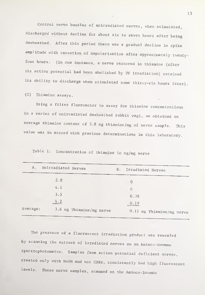
Control nerve bundles of unirradiated nerves, when stimulated,
discharged without decline for about six to seven hours after being
desheathed. After this period there was a gradual decline in spike
amplitude with cessation of depolarization after approximately twenty-
four hours. (In one instance, a nerve restored in thiamine (after
its action potential had been abolished by UV irradiation) retained
its ability to discharge when stimulated some thirty-six hours later).
(C) Thiamine assays.
Using a filter fluorometer to assay for thiamine concentrations
in a series of unirradiated desheathed rabbit vagi, „e obtained an
average thiamine content of 3.8 ng thiamine/mg of nerve sample. This
value was in accord with previous determinations in this laboratory.
Table 1: Concentration of thiamine in ng/mg nerve
A. Unirradiated Nerves B Irradiated Nerves
2.8 0
4.1 0
3.5 0.38
4.2 0.19
Average; 3.6 ng Thiamine/mg nerve 0.11 ng Thiamine/mg nerve
The presence of a fluorescent irradiation product was revealed
by scanning the extract of irradiated nerves on an Aminco-Bowman
spectrophotometer. Samples from action potential deficient nerves,
treated only with NaOH and not CNBr, consistently had high fluorescent
levels. These nerve samples, scanned on the Aminco-Bowman


16
spectrophotofluorometer, showed an emission spectrum with a maximum
at about 465 my which is well within the thiamine range. Thiamine
fluoresces at about 435 my. Thus, we had evidence of a contaminant
derived from radiation. This was corrected for in the assay as
follows: one aliquot of the extract was assayed in the usual manner
and this figure represented the fluorescence of both thiamine and the
unknown material. In a second aliquot the addition of CNBr and NaOH
was reversed and this figure reflected only the unknown fluorescent
material (thiamine is destroyed by NaOH). By subtraction, then, one
could determine fluorescence due to thiamine alone.
(D) ATP assays.
ATP levels were assayed in (i) control desheathed nerves, (ii)
irradiated nerves, and (iii) irradiated nerves followed by thiamine
treatment to restore the action potential. m the normal nerves, the
ATP concentration was 2.48 mM but wide variations were found in
irradiated nerves, both untreated and treated. Irradiated nerves,
not in thiamine, had ATP concentrations ranging from 0.61 to 2.74 mM
with a mean of 1.67 mM; irradiated, thiamine-treated nerves had values
ranging from 0.05 to 5.45 mM with a mean of 1.98 mM. With these
extreme variations, mean values are of little value. However, within
each experiment using 2 vagi from the same rabbit, no significant
difference in ATP content was observed regardless of whether the
irradiated nerve was treated with the vitamin. This finding is
compatible with the findings of unaltered thiamine dependent enzyme
levels despite drastic changes in conductive ability.9,10
I 17
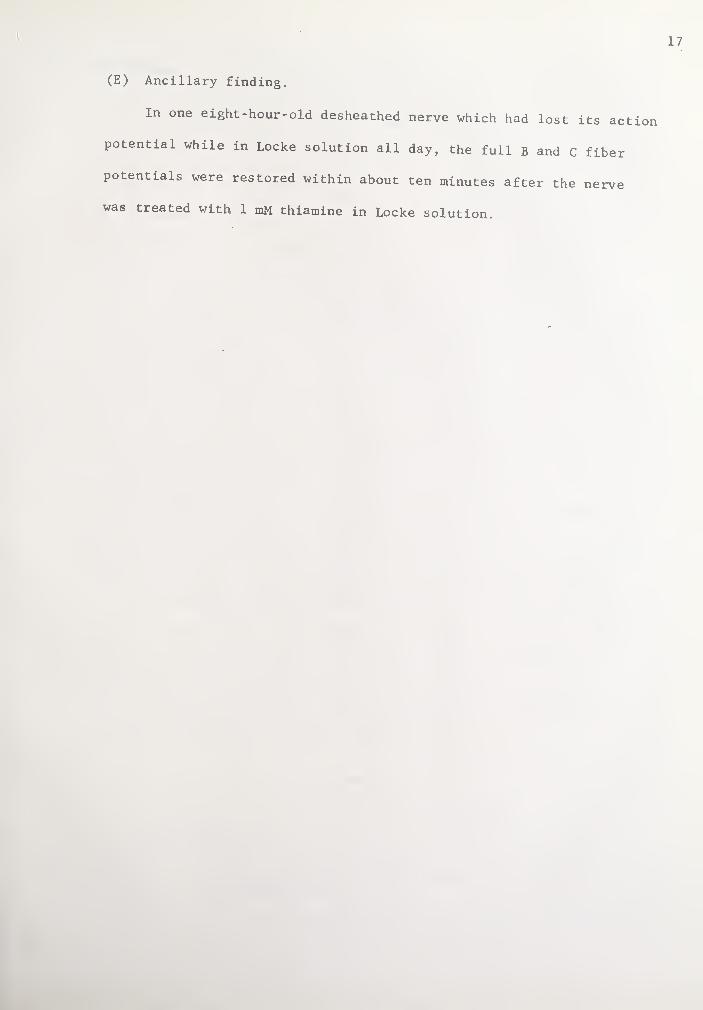
(E) Ancillary finding.
In one eight-hour-old desheathed nerve which had lost its action
potential while in Locke solution all day, the full B and C fiber
potentials were restored within about ten minutes after the nerve
was treated with 1 mM thiamine in Locke solution.
18
DISCUSSION:
Our findings demonstrate that thiamine is essential to nerve con¬
duction. UV irradiation during the course of about two hours resulted
in the destruction of thiamine in the nerve membrane. Simultaneously,
by fluorometric assay, thiamine concentrations were negligible as
compared to those of control, unirradiated nerves. Only subsequent
thiamine-treated nerves went on to conduct approximately 1.5 hours
after irradiation. Control preparations in Locke solution only, failed
to manifest any restoration of activity. Although the action potential
of the thiamine-treated preparation rarely returned to its pre-irradia¬
tion level, even a partial return is significant as compared to the con
trol nerve and in view of the manipulations that are involved in this
procedure. Since no significant difference was observed in ATP levels
between irradiated nerves in the presence and absence of thiamine this
would imply that the thiamine effect had nothing to do with metabolism
of the nerve but was strictly involved in the conduction process.
Various mechanisms have been suggested to explain the chemical
basis of permeability changes which would implicate thiamine. Based
upon his extensive work along these lines, von Muralt,37 the originator
of the idea that thiamine also acts neurophysiologically in nerves,
presented the following scheme in 1958;
Bound Thiamine
Free Thiamine + x
—} Excitation
i Free Thiamine
I_ ■^Recovery
Bound Thiamine Phosphates Excitation

19
Thiamine is pictured, somehow, to commute between a "free" phase and
3. bound phase. The free thiamine and some unknown entity, , are
a consequence of excitation. They ultimately result in additional
''bound" thiamine phosphates with recovery, and in released thiamine
phosphates with excitation. In essence, the mechanism was pointing
to a phosphorylation and dephosphorylation process involving thiamine
during the course of membrane depolarization and repolarization.
Along these latter lines in 1960 Petropulos,24 based upon the
ionic hypothesis of electrical activityf'Heasoned as follows:
The rate of rise of the action potential, dx/dt, is a theoretical
measure of the influx of Na+ ions into the membrane. The S curve,
(i.e., dx/dt plotted against membrane potential), obtained for a
single nerve fiber treated with a thiamine antimetabolite, shows a
decrease in the height in the upper plateau. This suggested to the
uthor a decrease in the "number of active Na carriers." Since this
reduction in the height of the action potential is abolished by
addition of excess thiamine, "a loose carrier mechanism may be
9 / postulated for thiamine."
Itokawa and Cooper,20 in view of the evidence summarized in the
introduction and in their correlation of the effect of neuroactive
drugs on the release of thiamine from nervous tissue, postulated "a
carrier role for TPP or TTP involving a successive dephosphorylation
and rephosphorylation of the vitamin as ion exchange takes place
across the membrane." Complexes binding TPP and TTP with Na+ and
Ca2+ have been described by Hoffman et al.42 A second possibility
Which they offered, links thiamine with conformational changes in
the membrane. In this case a shift of charged particles, similar to

■
.
20
the hypothesis of Baker, 43 -would induce a conformational change in
the protein-lipid-thiamine phosphate mosaic of the membrane to displace
the thiamine phosphate and permit a rapid influx of Na+ and Ca2+. "2°
All of the above mechanisms implicate thiamine in nervous tissue
conduction but further work is necessary to dissect the events in
conduction at a molecular level.

REFERENCES:
1. Booth, J., A. von Muralt, and R. Stampfli: Helv. Physiol. Pharmacol.
Acta, 8, 110 (1950).
2. Audiat, J.; C. R. Soc. Biol., 107, 931 (1931).
3. Hutton-Rudolph, M.: Helv. Physiol. Acta, 1, C15 (1943)
4. Minz, B.: Cr. Seanc. Soc. Biol., 127, 1251 (1938).
5. Gurtner, H. P.: Helv. Physiol. Pharmac. Acta, Suppl. XI (1961).
6. Cooper, J. R., R. h. Roth, and M. M. Kini: Nature, London, 199,
609 (1963).
7. Kunz, H. A.: Helv. Physiol. Pharmac. Acta, 14, 411 (1956).
8. Armett, C. J., and J. R. Cooper; j. Pharmacol. Exp. Ther., 148,
137 (1965).
9. Cooper, J. R., and J. H. Pincus: Thiamine Deficiency; Biochemical
Lesions and Their Clinical Significance, Ciba Foundation Study
. Group #28, p. 112, Churchill, London (1967).
10. Cooper, J. R. : Biochem. Biophys. Acta, 156, 368 (1968).
11. Lof land , H. B, ' ’ H. D. Goodman, T. B. Clarkson, and R. W. Pritchard
J. Nutr. , _79, 188 (1963).
12. Koeppe, R. E., ' R- M- O'Neil, and C. H. Hahn; J. Neurochem., 11,
695 (1964).
13. Dreyfus, P. M. , and G. Hauser; Biochem. Biophys. Acta, 104, 78
(1965).
14. Brin, M.: M. Nutr., 78, 179 (1962).
15. Tanaka, C., and J. R. Cooper; J. Histochem. Cytochem., 16,
362 (1968).
16. Cooper, j. R.: Methods in Enzymology, 18, p. 616 (1970).
17. Tanaka, C., R. J. Barrnett, and J. R. Cooper; Unpublished data.


18. Cooper, J. R., Y. Itokawa, and J. H. Pincus: Science, N.Y., 164,
74 (1969).
19. Itokawa, Y., and J. R. Cooper: Biochem. Biophys. Acta, 158, 180
(1968).
20. Itokawa, Y., and J. R. Cooper; Biochem. Pharmacol., 19, 985
(1970).
21. Armett, C. J., and J. M. Ritchie: j. Physiol., 152, 141 (1960).
22. Eccles, R. M.: J. Physiol., 117, 181 (1952).
23. Evans, E. H. L. , and J. G. Murray; j. Anat., London,' 88, 320
(1954).
24. Petropulos, S. F.: J. Cell. Comp. Physiol., 56, 7 (1960).
25. Bachoffer, C. S.: Arch. Biochem. Biophys., 88, 333 (1960).
26. von Muralt, A.: Pflugers Arch. Ges. Physiol., 247, 1 (1943).
27. Lowry, 0. H., J. V. Passonneua, D. W. Schulz, and M. K. Rock:
J. Biol. Chem., 236? 2746 (1961).
28. Pierce, S., and A. G. Giese; j. Cell. Compar. Physiol., 49,
303 (1957).
29. Cooper, J. R., F. E. Bloom, and R. H. Roth; The Biochemical
Ba_sis of Neuropharmacology. Oxford Univ. Press, N.Y. , (1970)
pp. 5,6.
30. Koedam, J. C. : Biochim. Biophys. Acta, _29, 333 (1958).
31. Armett, C. J. , and J. R. Cooper; Experientia, 2_1, 605 (1965).
32. Nachmansohn, D. and H. B. Steinbach: j. Neurophysiol., 5, 109 (1942)
33. Novikoff, A., and S. Goldfischer: Proc. Nat. Acad. Sci. U.S.A.,
47, 802 (1961).
34. Itokawa, Y., and J. R. Cooper; Biochim. Biophys. Acta, 196,
274 (1970).

35. von Muralt, A.: Vitamins and Hormones, _5, 93 (1947).
36. Udenfriend, S.: Fluorescence Assay in Biology and Medicine,
Academic Press (1962).
37. von MuraIt, A.: Exp. Cell Res., Suppl. 5, 72 (1958).
38. Eijkman, C., Arch. Path. Anat. (Virchow's), 148, 523 (1897).
39. Grijns, G.: Tigdschr. Ned. Ind., 41, 3 (1901).
40. Hodgkin, A. L. : Biol. Rev., 26, 339 (1951).
41. Hodgkin, A. L., and A. F. Huxley: J. Physiol., 116, 497 (1952).
42. Hoffman, H. , T. Eckert, and W. Miifous: Z. Physiol. Chem. , 335.,
156 (1964).
43. Baker, P. F.: Br. Med. Bull., 24, 179 (1968).
44. Fujiwara, M., and Matsui, L.: Anal. Chem., 25, 810 (1953).


YALE MEDICAL LIBRARY
Manuscript Theses
, . Unpublished theses submitted for the Master's and Doctor’s degrees and
eposited m the Yale Medical Library are to be used only with due re-nifd to the rights of the authors. Bibliographical references may be noted, but passages
must not be copied without permission of the authors, and without proper credit bcmg given m subsequent written or published work. P
This thesis by-
used by the following persons, above restrictions.
has been whose signatures attest their acceptance of the
NAME AND ADDRESS DATE
