the revised who classification of … p hasserjian, md associate professor massachusetts general...
TRANSCRIPT

Robert P Hasserjian, MD
Associate Professor
Massachusetts General Hospital and Harvard Medical School
The Revised WHO Classification of Myeloproliferative Neoplasms

Myeloproliferative neoplasms
• Clonal hematopoietic stem cell disorders
• “Overexuberant” production of one or more hematopoietic cell types
• Erythroid and granulocytic elements generally appear normal, without dysplasia
• Treated differently from other myeloid neoplasms

Myeloproliferative neoplasms (WHO 2016)
• Chronic myeloid leukemia, Ph+ • Polycythemia vera • Essential thrombocythemia • Primary myelofibrosis • Rare entities
– Chronic neutrophilic leukemia – Chronic eosinophilic leukemia/hypereosinophilic syndrome – Myeloproliferative neoplasm, unclassifiable
Genetically defined eosinophilic neoplasms
BCR-ABL
CSF3R
JAK2 MPL CALR
PDGFRA PDGFRB
FGFR1 PCM1-JAK2

Diagnostic issues with MPN
• Distinguishing MPN from reactive conditions that can produce elevated counts
• Separating MPN from other myeloid neoplasms (MDS and MDS/MPN)
• Providing a specific diagnosis
– Requires integration of clinical and molecular genetic data with morphology
– Important in predicting prognosis and dictating therapy
• Recognizing signs of progression

CML
• Hematopoietic stem cell neoplasm associated with BCR-ABL1 fusion gene
• Patients present with neutrophilic leukocytosis with morphologically normal and maturing granulocytic elements
• Natural history is that of genetic instability, with progressive accumulation of blasts culminating in acute leukemia

The Philadelphia chromosome
Y412
p230
p210
p190
BCR-ABL fusion proteins

CML peripheral blood

CML bone marrow aspirate

CML bone marrow biopsy

Natural course of CML
• Patients may survive many years with relatively few symptoms
• Inexorably progress to an acute leukemia with loss of differentiation
– Termed ‘Blast crisis’ or ‘Blast phase’
–Blast crisis phenotype
• 70% myeloid (≥20% BM/PB myeloblasts)
• 30% B-lymphoid (any BM/PB lymphoblasts raises strong supsicion)

CML in blast crisis

Faderl S et al. N Engl J Med 1999;341:164-172, Goldman J and Melo J. N Engl J Med 2003;349:1451-1464
Tyrosine kinase inhibitors

CML in the 21st century
• Treated very effectively with tyrosine kinase inhibitors (TKI)
– Imatinib, nilotinib, dasatinib, bosutinib, ponatinib
• Disease progression no longer inevitable
• Patterns of disease evolution are closely linked to responsiveness versus resistance to TKI therapy

Assessing response to TKI Time on therapy
Optimal response “Warning” Failure
3 months Hematologic remission ≤35% Ph+ metaphases ≤10% BCR-ABL1
36-95% Ph+ metaphases or >10% BCR-ABL1
No hematologic remission or >95% Ph+ metaphases
6 months No Ph+ metaphases* <1% BCR-ABL1
1-35% Ph+ metaphases or 1-10% BCR-ABL1
>35% Ph+ metaphases or >10% BCR-ABL1
1 year ≤0.1% BCR-ABL1** >0.1-1% BCR-ABL1 Any Ph+ metaphases or >1% BCR-ABL1
Later ≤0.1% BCR-ABL1** Clonal chromosomal abnormalities in Ph- cells
Loss of hematologic remission New Ph+ metaphases Loss of major molecular response on 2 consecutive tests
*Lack of Ph+ metaphases is considered “complete cytogenetic response” **≤0.1% BCR-ABL1 is considered “major molecular response”
Baccarani M et al. Blood 2013;122:872-884

CML accelerated phase (WHO 2016) • Accelerated phase
definition – BM or PB blasts 10-19%
– Basophils ≥20%
– Platelets >1000 x 109/L or <100 x 109/L
– WBC >10 x 109/L
– Persistent or increasing splenomegaly
– Clonal evolution
– Second Ph, +8, i(17q), +19, complex karyotype, or 3q26.2 abnormalities at diagnosis
• Provisional TKI-response criteria – Hematologic resistance to
first TKI therapy
– Hematologic, cytogenetic, or molecular evidence of resistance to 2 sequential TKI therapies
– 2 or more ABL1 mutations developing during TKI therapy
~10% of CML patients present in accelerated or blast phase

Role of pathology in the current era of CML management
• At the time of initial diagnosis of CML –Get the diagnosis right!
–Provide prognostic information • Accurate blast count in blood and marrow
aspirate, reticulin fibrosis grade
• At later timepoints, assess for progression and evaluate for other pathologic processes while on therapy

Caveats with CML diagnosis
• Increased erythroid proliferation in bone marrow, especially in patients with hemoglobinopathies
• Prominent thrombocytosis mimicking ET – Associated with p230 BCR-ABL1
• Minimal or no myeloid left-shift in blood • Monocytosis mimicking CMML
– Associated with p190 BCR-ABL1
• Blast crisis mimicking AML or ALL

Erythroid-rich CML

Differential diagnosis of neutrophilia
Disease Peripheral counts Neutrophil morphology
Genetics
CML, BCR-ABL1+ ↑Granulocytes with left-shift Eosinophilia Basophilia
Normal t(9;22); BCR-ABL1
Atypical CML, BCR-ABL1-
↑Granulocytes with left-shift
Dysplastic SETBP1 or ETNK1 mutation (30%)
Chronic neutrophilic leukemia
↑Granulocytes without left-shift
Normal CSF3R mutation (90%)
Primary myelofibrosis
Leukoerythroblastic Normal JAK2, MPL, or CALR mutations (90%)
The pathologist has a critical role in evaluating smear and biopsy morphology and integrating these findings with clinical and genetic features to make the diagnosis!

Atypical CML, BCR-ABL1-
• Misnomer
Atypical CML is a distinct MDS/MPN, not a variant of CML (which is a pure MPN)
• Features mimic CML, except:
–No BCR-ABL translocation by definition
–Prominent granulocytic dysplasia
–No or minimal basophilia
• Poor prognosis, not helped by TKI

Atypical CML, BCR-ABL1- bone marrow biopsy

Atypical CML, BCR-ABL1- peripheral smear

Atypical CML, BCR-ABL1-bone marrow aspirate

Chronic neutrophilic leukemia
• Rare MPN with leukocytosis (>25 x 109/L) –No dysplasia (hypogranulation) of
neutrophils
– Splenomegaly
– <10% immature myeloid cells in blood
–No BCR-ABL1 rearrangement
–No significant basophilia or eosinophilia
• 83-89% have CSF3R mutation
Gotlib J et al. Blood 2013;122:1707, Pardanani A et al. Leukemia 2013;27:1870, Maxson JE et al. NEJM 2013;368:1781

Chronic neutrophilic leukemia: peripheral blood smear
WBC 36.7 x 10⁹/L HCT 40.0% (MCV 98 fL) PLT 253 x 109/L 82% polys, 14% lymphs, 2% metas, 2% myelos

Chronic neutrophilic leukemia: peripheral blood smear

Chronic neutrophilic leukemia: bone marrow

Algorithm for workup of persistent neutrophilia
Possible reactive causes excluded?
BCR-ABL1 +
CML
BCR-ABL1 -
Significant granulocytic left-
shift and dysplasia
Atypical CML, BCR-ABL1-
JAK2 mutation and typical bone
marrow findings
Primary myelofibrosis
CSF3R mutation and no left-shift or
dysplasia
Chronic neutrophilic
leukemia
Reactive neutrophilia
Usually self-limited
Treat with TKI immediately to prevent progression
Poor prognosis, difficult to treat
Several treatment options
May respond to ruxolitinib
Pathologist has critical role in evaluating smear and biopsy morphology and to integrate these findings with clinical and genetic findings
Karyotype, FISH, and/or RT-PCR

Eosinophilia
• Reactive
– Allergy, drug, parasitic or other infections
• Paraneoplastic (non-neoplastic eosinophils stimulated by tumor cytokines)
– Lymphomas, mastocytosis, rare solid tumors
• Eosinophils part of a myeloid neoplasm
– CML
– AML with inv(16)
– Genetically-defined eosinophilias
– Chronic eosinophilic leukemia, NOS

Myeloid/lymphoid neoplasms with eosinophilia and abnormalities of
PDGFRA, PDGFRB, FGFR1, or PCM1-JAK2
• Share similar molecular and biologic features – Appear to involve pluripotent stem cell with both
lymphoid and myeloid differentiation capacity
– Translocations activate genes encoding tyrosine kinases
– Eosinophilia is characteristic
• Most entities respond to targeted tyrosine kinase inhibitors

Genetically-defined eosinophilic neoplasms
Disease Presentation Genetics Treatment
PDGFRA Eosinophilia ↑Serum tryptase ↑ Mast cells
Cryptic deletion at 4q12 FIP1L1-PDGFRA or other partners
TKI
PDGFRB Eosinophilia Monocytosis
t(5;12)(q32;p12) ETV6-PDGFRB or other partners
TKI
FGFR1 Eosinophilia Often presents as T-ALL/LBL or AML
Translocations of 8p11 FGFR1 with various partners
PCM1-JAK2 Eosinophilia Left-shifted erythroids
t(8;9)(p22;p24.1) PCM1-JAK2
JAK2 inhibitor

45 year old woman with leukocytosis. WBC 100 x 109/L 34% polys, 26% bands, 6% lymphs, 9% eos, 3% metas, 9% myelos.
Bone marrow biopsy

Inguinal lymph node biopsy
Karyotype of both bone marrow and lymph node: 46, XX, t(8;13)(p12;q12)(ZNF198-FGFR1) Diagnosis: Myeloid and lymphoid neoplasm with FGFR1 rearrangement

Myeloid neoplasms with t(8;9)(p22;q24); PCM1-JAK2
• Eosinophilia, erythroid predominance with left-shift, prominent lymphoid aggregates
• Fibrosis often present, mimicking PMF
• Can rarely present as T- or B-ALL
• Respond to JAK2 inhibitor ruxolitinib
• Added as a provisional entity to the group of genetically defined eosinophilic leukemias
Bousquet M et al. Oncogene 2005;24:7248, Dargent JL et al. Eur J Haematol 2011;86:87, Masselli E et al. Br J Haematol 2013;162:563

Chronic eosinophilic leukemia (CEL), not otherwise specified
• Persistent blood eosinophilia >1,500/mm3 with increased bone marrow eosinophils and end-organ damage by eosinophils
• Exclusion of all reactive, paraneoplastic, and specific cytogenetic causes of eosinophilia
• Evidence of clonality – Clonal cytogenetic abnormality present
– Increased bone marrow (>5%) or peripheral blood (>2%) blasts (but <20% blasts)
• Classified as hypereosinophilic syndrome if clonality cannot be proven

Chronic eosinophilic leukemia, NOS

Chronic eosinophilic leukemia, NOS

Algorithm for workup of persistent eosinophilia >1.5 x 10⁹/L
Reactive eosinophilia
-Abnormal T-cell clone
-Other lymphoma with eosinophilia
-Clonal eosinophilia due to CML or AML
-Systemic mastocytosis
Gotlib J. Curr Opin Hematol 2010;17;117, Wang SA et al. Mod Pathol 2016 (Epub) Courtesy of Tracy George, University of New Mexico
Screen for secondary causes of eosinophilia +
Evaluate peripheral blood & bone marrow
Myeloid/lymphoid neoplasm with PDGFRA, PDGFRB, or FGFR1
Other clonal abnormality or increased blasts
CEL HES
-
-
Cytogenetics
+ -
+ -
+

The JAK2-associated MPNs • Essential thrombocythemia (ET)
– Increased platelet count
– Only rarely progress to a fibrotic phase or AML
• Polycythemia vera (PV)
– Increased red cell production
– May progress to a fibrotic phase or rarely AML
– Can have thrombotic complications
• Primary myelofibrosis (PMF)
– Often thrombocytosis at presentation
– Progressive splenomegaly and marrow fibrosis

The spectrum of megakaryocyte morphology
Normal/Reactive
Myeloproliferative MDS and CML

The non-CML MPN: Deregulation of the JAK/STAT pathway
Nature Reviews Cancer; 2007; Rampal R et al. Blood 2014;123:3123-33; Chachoua I et al. Blood 2016;127:1325
Cytokines
Cyclin D1
FGFB, VEGF
2005
2007
2016
CSF3R CALR 2013 2013
EPOR TPOR

Distribution of mutation types in the non-CML MPN
Klampfl T et al. NEJM 2013;369:2379

Essential thrombocythemia (ET) 2016 WHO Criteria
•Platelet count ≥450 x 109/L
•Bone marrow biopsy showing typical morphology of ET and no or (rarely) minor increase in reticulin fibers.
•Does not meet WHO criteria for another myeloid neoplasm
AND
•Presence of JAK2, CALR or MPL mutation or other proof of clonality (cytogenetic abnormality or other mutation)*
*Must rigorously exclude reactive thrombocytosis if there is no proof of clonality

Other myeloid neoplasms that can present with thrombocytosis
• Other MPNs
– Polycythemia vera
– Primary myelofibrosis (early stages)
– Chronic myeloid leukemia
• Myelodysplastic syndrome with isolated del(5q) or inv(3)/t(3;3)
• MDS/MPNs
– Refractory anemia with ring sideroblasts and thrombocytosis (RARS-T)

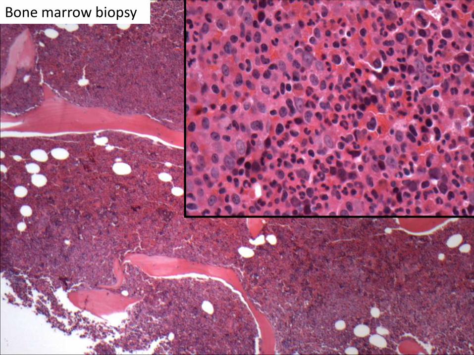
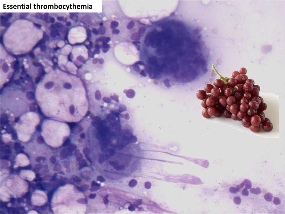
Typical ET morphology
• Normocellular for age
• Increased megakaryocytes with minimal clustering
• Megakaryocytes are large forms
–Predominantly ‘staghorn’ nuclei with complex lobation and abundant cytoplasm
• Reticulin is not increased

Essential thrombocythemia

Essential thrombocythemia
Essential thrombocythemia

Polycythemia vera (PV) 2016 WHO Criteria
• Evidence of increased red cell production
• Bone marrow showing typical PV histology – Hypercellular for age
– Panmyelosis with increased erythroids and megakaryocytes
– Spectrum of small, medium, and large megakaryocytes with bulbous and hyperlobated nuclei
AND
• JAK2 mutation*
*Must have subnormal EPO level in the rare cases (<2%) lacking JAK2 mutation

Polycythemia vera

Polycythemia vera

“Masked” polycythemia vera
• Some patients have bone marrow findings typical of PV, but do not meet required 2008 WHO hemoglobin levels –Male ≥18.5 g/dL, Female ≥16.5 g/dL)
• These patients mimic ET in their clinical presentation (thrombocytosis), but have PV-like clinical behavior –Can progress to fibrotic phase of disease
–Risk of thrombosis
Barbui T et al. Am J Hematology 2013;89:52, Gianelli U et al. Am J Clin Pathol 2008;130:336, Thiele J et al. Acta Hematol 2005;113:213

Masked polycythemia vera
65 year-old woman WBC 14.2 x 10⁹/L HGB 16 g/dL PLT 744 x 109/L
Courtesy of Sa Wang, MDACC

Polycythemia vera(PV) 2016 WHO Criteria
• Male HGB > 16.5 g/dL or HCT > 49%, Female HGB > 16.0 g/dL or HCT> 48%
• Bone marrow showing typical PV histology – Hypercellular for age
– Panmyelosis with increased erythroids and megakaryocytes
– Spectrum of small, medium, and large megakaryocytes with bulbous and hyperlobated nuclei
AND
• JAK2 mutation*
*Must have subnormal EPO level in the rare cases (<2%) lacking JAK2 mutation

Primary myelofibrosis (PMF) 2016 WHO Criteria
• Bone marrow showing typical PMF histology – Hypercellular for age – Increased M:E ratio, atypical megakaryocytes
AND • Presence of JAK2, CALR or MPL mutation or other
proof of clonality (cytogenetic abnormality or other mutation)*
AND • Anemia, WBC ≥ 11 x 109/L, splenomegaly,
increased LDH, or leukoerythroblastosis
*Must exclude possible reactive causes of marrow fibrosis if there is no clonal marker

Stages of Primary Myelofibrosis
WHO Classification of Tumors of Hematopoietic and Lymphoid Tissues; 4th edition

Early/prefibrotic primary myelofibrosis

Early/prefibrotic primary myelofibrosis

ET PMF (early-prefibrotic stage)
no or only slight increase in age-matched cellularity
marked increase in age-matched cellularity
no significant increase in granulo- and erythropoiesis
pronounced proliferation of granulopoiesis and reduction of erythroid precursors
prominent large to giant mature megakaryocytes with hyperlobulated or deeply folded nuclei, dispersed or loosely clustered in the marrow space
dense or loose clustering and frequent endosteal translocation of medium sized to giant megakaryocytes showing hyperchromatic, hypolobulated, bulbous, or irregularly folded nuclei and an aberrant nuclear/cytoplasmic ratio
no or very rarely minor increase in reticulin fibers
no or no significant increase in reticulin fibers
Courtesy of Hans-Michael Kvasnicka, University Hospital, Frankfurt

Primary myelofibrosis, fibrotic phase

Primary myelofibrosis, fibrotic phase

MF-0 MF-1
MF-2 MF-3

Advanced fibrosis on trichrome stain

Survival in the non-CML MPN (n=826)
Tefferi et al. Blood 2014;124:2507-2513
ET PV
PMF
Courtesy of Hans-Michael Kvasnicka, University Hospital, Frankfurt

Importance of accurate diagnosis of MPN to inform prognosis and guide
therapy
PV ET PMF
Leukemic transformation
3% at 10 years 1% at 10 years 12-30% at 10 years
Fibrosis progression 15-25% Rare 100%
Thrombosis, per 100 patients/year
5.5 1-3 2
Initial treatment Phlebotomy +/- HU None , aspirin +/- HU
Allo-SCT, JAK inhibitors,
chemotherapy
Courtesy of Olga Pozdnyakova, BWH

Summary • Myeloproliferative neoplasms have distinctive
morphologies and distinctive genetic aberrations
– Important to correctly diagnose the various MPN diseases, which have different patterns of progression and are treated differently
– Pathologists must utilize a combination of information from morphology, clinical features, and cytogenetics/molecular genetics to
• Eosinophilic myeloid disorders are characterized by recurrent genetic abnormalities and some are amenable to targeted therapies with TKIs