the ribosome: some hard facts about its structure and hot
TRANSCRIPT

10.1101/sqb.2009.74.032Access the most recent version at doi: published online December 2, 2009Cold Spring Harb Symp Quant Biol
V. Ramakrishnan about Its EvolutionThe Ribosome: Some Hard Facts about Its Structure and Hot Air
References http://symposium.cshlp.org/content/early/2009/11/25/sqb.2009.74.032.refs.html
This article cites 58 articles, 21 of which can be accessed free at:
P<P Published online December 2, 2009 in advance of the print volume.
serviceEmail alerting
click heretop right corner of the article orReceive free email alerts when new articles cite this article - sign up in the box at the
articles must include the digital object identifier (DOI) and date of initial publication. Advance online articles have not yet appeared in the print volume. Citations to Advance online
http://symposium.cshlp.org/subscriptions go to: Cold Spring Harbor Symposia on Quantitative BiologyTo subscribe to
Copyright © 2009, Cold Spring Harbor Laboratory Press
Cold Spring Harbor Laboratory Press on January 25, 2010 - Published by symposium.cshlp.orgDownloaded from
Ribosomes from all species consist of approximatelytwo-thirds RNA and one-third protein. Ribosomes frommammalian mitochondria are an exception, with the ratio ofprotein and RNA reversed (see Sharma et al. 2003). Allribosomes consist of two subunits, termed 50S and 30S inbacteria or 60S and 40S in eukaryotes. Together, they com-prise the 70S ribosome in bacteria or the 80S ribosome ineukaryotes (Fig. 1A). The mRNA containing the genetictemplate binds in a cleft in the small subunit. The aminoacids themselves are brought into the ribosome by amino-ac ylated tRNA substrates. The ribosome has three bindingsites for tRNA: the A (aminoacyl) site that brings the newaminoacyl tRNA, the P (peptidyl) site that holds the nascentpeptide chain, and the E (exit) site to which the deacylatedP-site tRNA moves after peptide bond formation (Fig. 1B).Translation in all species can be divided into three
stages (Fig. 2) (for review, see Schmeing and Ram a krish -nan 2009). During initiation, the small subunit of the ribo-some binds mRNA at the start site of the coding sequence,in a precise manner that puts the start codon in the P site.This requires three initiation factors and a special initiatortRNA that binds to the P site. Initiation is followed by the elongation cycle, which
consists of three important steps: decoding, peptidyltransfer, and translocation. During decoding, the correctaminoacyl tRNA, which is delivered to the A site of theribosome as a ternary complex with elongation factor Tu(EF-Tu) and GTP, is selected based on the codon on themRNA in the A site. Selection of the tRNA leads tohydrolysis of GTP by EF-Tu and release of the factorfrom the ribosome. The aminoacyl end of the selectedtRNA then swings into the peptidyl transferase center(PTC) in the 50S subunit of the ribosome, where peptidebond formation occurs rapidly and spontaneously.
Peptidyl transfer leaves the P-site tRNA deacylated, withthe A-site tRNA now containing a nascent peptide chainthat has been extended by one residue. The 3′ ends of theA- and P-site tRNAs then move first with respect to the50S subunit to form an intermediate or hybrid state of theribosome, followed by movement of the mRNA and tRNAswith respect to the 30S subunit, which requires the actionof EF-G, another GTPase factor. This leaves the ribosomewith an empty A site with a new mRNA codon ready toaccept the next aminoacyl tRNA.The elongation cycle continues until a stop codon is
reached in the A site. The so-called class I release factors(RF1 or RF2 in bacteria, eRF1 in eukaryotes) recognizethe stop codon and catalyze the cleavage of the polypep-tide chain from the P-site tRNA. Finally, a factor knownas ribosome recycling factor (RRF), with the help of EF-G, disassembles the ribosome so that a new round of pro-tein synthesis can begin.Most of these aspects of translation are common to all
kingdoms of life. In eukaryotes, initiation is far morecomplex and involves a specifically modified mRNAwith a 5′ cap and a poly(A) tail at the 3′ end, as well asalmost a dozen factors, many of which are large multi-subunit complexes themselves (Kapp and Lorsch 2004).Recent structural and biochemical work has shed light
on many aspects of translation. In particular, the high-res-olution structures of the ribosomal subunits (Ban et al.2000; Wimberly et al. 2000) were useful in the molecularinterpretation and/or phasing of all subsequent structures,including a lower-resolution crystal structure of the 70Sribosome with mRNA and tRNA ligands at 5.5-Å resolu-tion (Yusupov et al. 2001), more recent higher-resolutionstructures of the empty 70S ribosome from Escherichiacoli (Schuwirth et al. 2005), and the 70S ribosome withmRNA and tRNAs from Thermus thermophilus (Selmeret al. 2006). These basic structures have been followed byhigh-resolution structures of the ribosome with proteinfactors, most notably with release factors (Laurberg et al.
The Ribosome: Some Hard Facts about Its Structure and Hot Air about Its Evolution*
V. RAMAKRISHNANMRC Laboratory of Molecular Biology, Cambridge CB2 0QH, United Kingdom
Correspondence: [email protected]
By translating genetically encoded information to synthesize proteins, the ribosome has a central and fundamental role in themolecular biology of the cell. Virtually every molecule made in every cell was made either directly by the ribosome or byenzymes made by the ribosome. Although the ribosome was discovered half a century ago, progress in the field of translationhas been revolutionized by the atomic structures of the ribosomal subunits determined in 2000. These structures paved theway not only for more sophisticated biochemical and genetic experiments, but also for the phasing and/or molecular inter-pretation of all subsequent structures of the ribosome by crystallography or cryoEM (cryo-electron microscopy). In additionto facilitating our understanding of ribosome function, these structures also shed light on the evolution of the ribosome.
Cold Spring Harbor Symposia on Quantitative Biology,Volume LXXIV. ©2009 Cold Spring Harbor Laboratory Press 978-087969870-6 1
*The title is a paraphrase of one by the late eminent crystallographerDavid M. Blow: “Hard facts on structure: Hot air about mobility”(Nature [1982] 297: 454–455).
Cold Spring Harbor Laboratory Press on January 25, 2010 - Published by symposium.cshlp.orgDownloaded from

2 RAMAKRISHNAN
Elongation
Initiation
Release
Recycling
Ternary complex binding
Codon recognition
GTP hydolysis Accomodation
EF-Tu release
PeptidylTransfer
Hybrid statesformation
EF-Gbinding
GTP hydrolysisTranslocation
EF-G release
Initiation factors,tRNA binding
Subunitjoining
Stop Codonin A site
RF binding
Hydrolysis Nacent peptide
release
RF3 Binding
GTP hydrolyisRF release
EF-G, RRFbinding
GTP hydrolysisSubunit dissociation
IF3 bindingmRNA, tRNAdissociation
GTP hydrolysisIF dissociation
30S
50SinitiatortRNAIF3
IF2
IF1mRNA
IF3IF2
IF1
EF-Tu
aa-tRNA
EF-Tu
deacyl-tRNA
EF-G
EF-G
RF1/2
RF1/2RF3
new protein
deacyl-tRNA
RF3
EF-G
RRF
EF-GRRF
deacyl-tRNA
mRNA
Figure 2. Overview of the translational pathway showing the phases of initiation, elongation, release, and recycling. (Reprinted, withpermission, from Schmeing and Ramakrishnan 2009 [©Nature Publishing Group].)
50S
30S
A-tRNAP-tRNA
E-tRNA
mRNA
L7/L12L1
3’
5’ headbody
L7/L12
L1
CP
A-tRNA
P-tRNA
E-tRNA
5S
50SPTC
E-tRNA
P-tRNA
A-tRNA
head
body30S
DC
beak
spur
mRNA3’ GTPase factor
binding region
A
B C Figure 1. Structure of the ribosome. (A) Overview of thebacterial 70S ribosome with the 50S subunit on top andthe 30S subunit on the bottom. The mRNA (dark gray) isshown wrapped round the neck of the 30S subunit.(Magenta) A-site, (green) P-site, (yellow) E-site tRNAs.(B) The 30S subunit showing the decoding center (DC)where codon–anticodon interactions are monitored dur-ing tRNA selection. (C) The 50S subunit showing theGTPase-factor-binding region and the PTC where peptidebond formation is catalyzed. (Reprinted, with permission,from Schmeing and Ramakrishnan 2009 [©Nature Pub -lishing Group].)
Cold Spring Harbor Laboratory Press on January 25, 2010 - Published by symposium.cshlp.orgDownloaded from

2008; Weixlbaumer et al. 2008) and more recently withelongation factors EF-Tu and EF-G (Gao et al. 2009;Schmeing et al. 2009). In addition, many cryoEM struc-tures of the ribosome represent different functional statesat varying resolutions.
THE RIBOSOME AS AN RNA-BASED MACHINE
The ribosome itself is a large and complex assembly ofRNA and more than 50 proteins. In addition, translationrequires a host of protein factors and aminoacyl tRNA sub-strates. Thus, understanding the evolution of the ribosomeposes a difficult challenge. To begin with, the system posesthe standard “chicken or egg” question: If the ribosomeconsists of both RNA and protein, and is needed to makeprotein, how did it come about? The first attempt to addressthis was Crick, who presciently wrote, “It is tempting towonder if the primitive ribosome could have been madeentirely of RNA” (original italics) (Crick 1968). To myknowledge, this was the first idea that RNA could be bothan information carrier and able to perform catalysis, andcan be thought of as the origin of the “RNA world hypoth-esis,” which postulates a primordial world consisting ofreplicating RNA molecules before the advent of proteins.How ever, in the absence of any known examples of cataly-sis by RNA, not even Crick could imagine that catalysis inthe current ribosome would be RNA based.It is clear that protein factors could have evolved later to
make translation more efficient, because even today it ispossible to get inefficient and limited translation withoutthem. For instance, factor-free protein synthesis in vitro wasdemonstrated by Spirin and coworkers (Gavrilova andSpirin 1974; Gavrilova et al. 1976). But what about the ribo-some itself? What are the relative roles of protein and RNA?The earliest work on ribosome function focused on pro-
teins for two reasons. Partly, proteins were thought to bethe molecules responsible for catalytic function. Second,because the standard laboratory organism E. coli containsseven genes for rRNA, it was difficult to isolate RNAmutants, and many of the early mutations, such as thosefor anti biotic resistance, mapped to ribosomal proteins.How ever, there were hints from quite early on that RNAhad a more important role than just providing a scaffold-ing for functional proteins. For instance, it was shown thatchemical modification of rRNA but not proteins wouldabolish binding of tRNA to 30S subunits (Noller andChaires 1972). In the absence of any prior evidence forthe catalytic properties of RNA, the results were taken tosuggest that tRNA-binding sites must therefore consist ofboth protein and RNA. Subsequent work on the ribo some,notably by Noller and coworkers, continued to provideevidence for the importance of rRNA, but in the absenceof an intellectual frame work in which RNA catalysis wasa real possibility, it was hard to make definite progress.This situation changed dramatically when catalysis by
RNA was discovered in the context of the group I intron(Zaug et al. 1983) or RNase P (Guerrier-Takada et al.1983). With evidence that RNA could in principle per-form catalysis, the ribosome community was far readier
to accept that rRNA might have crucial functions, and thisprospect renewed interest in the field (Moore 1988). Sub -sequently, an experiment showed that 50S subunits fromThermus aquaticus treated extensively with proteinase Kin the presence of SDS nevertheless preserved their pep-tidyl transferase activity (Noller et al. 1992). This experi-ment was a major step forward, but not conclusive prooffor a variety of reasons. E. coli 50S subunits did not main-tain activity with such protease treatment, nor did in-vitro-transcribed 23S RNA show activity. Moreover, in theThermus 50S subunit, a large number of protein fragmentsremained bound after protease treatment, and indeed, sub-sequent work showed that the treatment left three proteinsessentially intact (Khaitovich et al. 1999). A subsequenteffort to provide conclusive proof of the role of RNA inpeptidyl transfer using in vitro transcription of 23S RNAand its individual domains appeared to have narroweddown the activity to the RNA domain containing the pep-tidyl transferase center (Nitta et al. 1998), but this workwas retracted a year later (Nitta et al. 1999). Thus, al thoughwork on the group I intron and RNase P showed that catal-ysis by RNA was certainly possible, conclusive evidencefor a similar role in the ribosome proved to be difficult toobtain by purely biochemical means. It is striking that thislimitation was recognized very early on by Crick, whosaid, “Without a detailed knowledge of the structure ofpresent-day ribosomes it is difficult to make an informedguess” (Crick 1968).High-resolution structures of ribosomal subunits and
the whole ribosome have revealed in stunning detail theenvironment of the PTC-, tRNA-, and mRNA-bind ingsites, the intersubunit interface, and many other func tion-ally important regions of the ribosome. These structuresat long last allow us to provide conclusive insights intomany aspects of ribosome function and, in particular,show unambiguously how widespread the role of RNA isin the contemporary ribosome.
DECODING BY tRNA
A crucial event in protein synthesis is the selection ofthe correct tRNA corresponding to the codon on mRNA.At a fundamental level, this involves base pairingbetween the codon and the anticodon on tRNA. However,the free energy of base pairing between codon and anti-codon is not sufficient to explain the relatively low errorrate of the ribosome (Ogle and Ramakrishnan 2005).Kinetic experiments show that binding of the correcttRNA leads to induced conformational changes in theribosome that accelerate GTP hydrolysis by EF-Tu andtRNA selection (see Rodnina and Wintermeyer 2001).
Minor Groove Recognition by RNA
Discrimination against incorrect tRNA ultimatelydepends on recognizing mismatched base pairs. Becausebase pairing alone is insufficient to explain the accuracyof decoding, and the ribosome appears to have an activerole, what is the ultimate nature of this discrimination?Experiments showed that the binding of a cognate tRNA
RIBOSOME STRUCTURE: IMPLICATIONS FOR ITS EVOLUTION 3
Cold Spring Harbor Laboratory Press on January 25, 2010 - Published by symposium.cshlp.orgDownloaded from
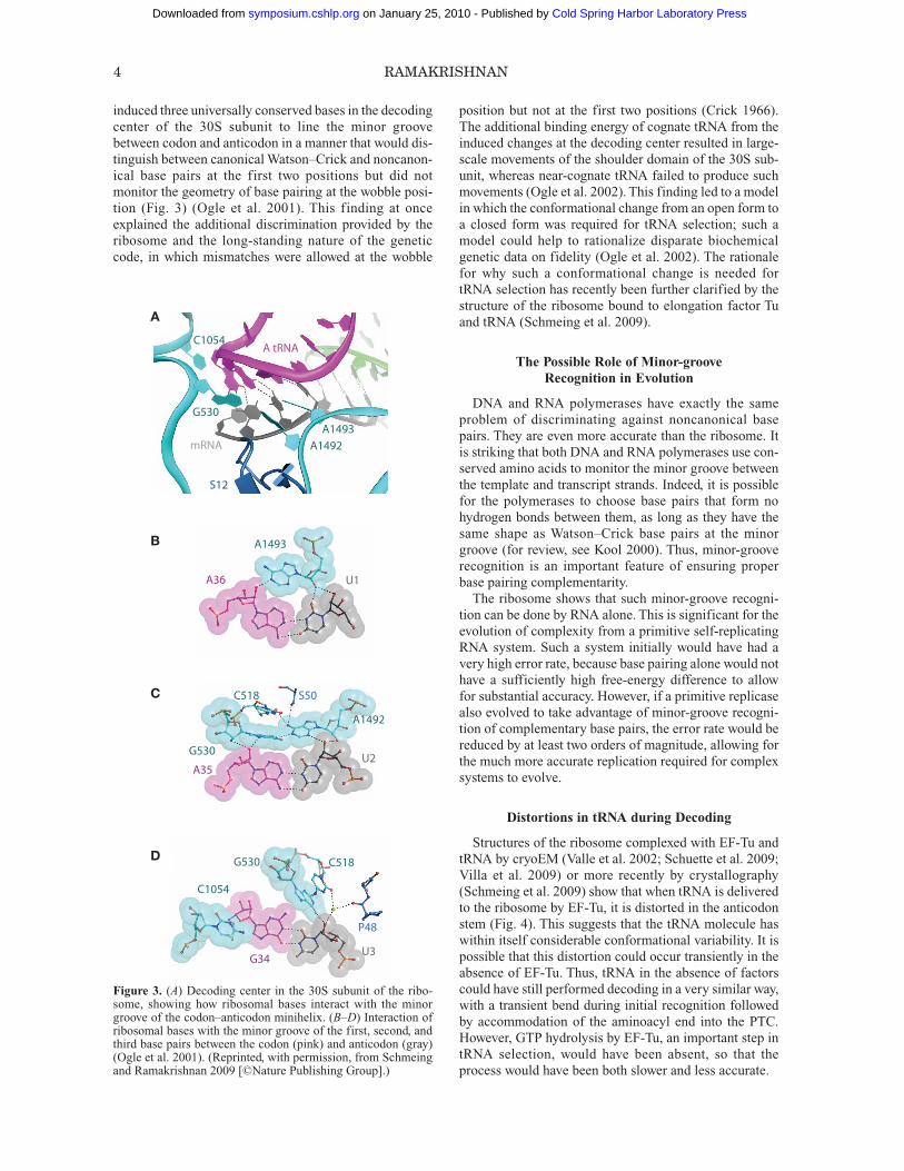
induced three universally conserved bases in the decodingcenter of the 30S subunit to line the minor groovebetween codon and anticodon in a manner that would dis-tinguish between canonical Watson–Crick and noncanon-ical base pairs at the first two positions but did notmonitor the geometry of base pairing at the wobble posi-tion (Fig. 3) (Ogle et al. 2001). This finding at onceexplained the additional discrimination provided by theribosome and the long-standing nature of the geneticcode, in which mismatches were allowed at the wobble
position but not at the first two positions (Crick 1966).The additional binding energy of cognate tRNA from theinduced changes at the decoding center resulted in large-scale movements of the shoulder domain of the 30S sub-unit, whereas near-cognate tRNA failed to produce suchmovements (Ogle et al. 2002). This finding led to a modelin which the conformational change from an open form toa closed form was required for tRNA selection; such amodel could help to rationalize disparate biochemicalgenetic data on fidelity (Ogle et al. 2002). The rationalefor why such a conformational change is needed fortRNA selection has recently been further clarified by thestructure of the ribosome bound to elongation factor Tuand tRNA (Schmeing et al. 2009).
The Possible Role of Minor-groove Recognition in Evolution
DNA and RNA polymerases have exactly the sameproblem of discriminating against noncanonical basepairs. They are even more accurate than the ribosome. Itis striking that both DNA and RNA polymerases use con-served amino acids to monitor the minor groove betweenthe template and transcript strands. Indeed, it is possiblefor the polymerases to choose base pairs that form nohydrogen bonds between them, as long as they have thesame shape as Watson–Crick base pairs at the minorgroove (for review, see Kool 2000). Thus, minor-grooverecognition is an important feature of ensuring properbase pairing complementarity.The ribosome shows that such minor-groove recogni-
tion can be done by RNA alone. This is significant for theevolution of complexity from a primitive self-replicatingRNA system. Such a system initially would have had avery high error rate, because base pairing alone would nothave a sufficiently high free-energy difference to allowfor substantial accuracy. However, if a primitive replicasealso evolved to take advantage of minor-groove recogni-tion of complementary base pairs, the error rate would bereduced by at least two orders of magnitude, allowing forthe much more accurate replication required for complexsystems to evolve.
Distortions in tRNA during Decoding
Structures of the ribosome complexed with EF-Tu andtRNA by cryoEM (Valle et al. 2002; Schuette et al. 2009;Villa et al. 2009) or more recently by crystallography(Schmeing et al. 2009) show that when tRNA is deliveredto the ribosome by EF-Tu, it is distorted in the anticodonstem (Fig. 4). This suggests that the tRNA molecule haswithin itself considerable conformational variability. It ispossible that this distortion could occur transiently in theabsence of EF-Tu. Thus, tRNA in the absence of factorscould have still performed decoding in a very similar way,with a transient bend during initial recognition followedby accommodation of the aminoacyl end into the PTC.However, GTP hydrolysis by EF-Tu, an important step intRNA selection, would have been absent, so that theprocess would have been both slower and less accurate.
4 RAMAKRISHNAN
AC1054
G530
A1492A1493
mRNA
S12
A tRNA
B
C
D
A1493
A36 U1
G530
A1492
S50C518
C1054
P48
C518G530
U2
U3
A35
G34
A
B
C
D
Figure 3. (A) Decoding center in the 30S subunit of the ribo-some, showing how ribosomal bases interact with the minorgroove of the codon–anticodon minihelix. (B–D) Interaction ofribosomal bases with the minor groove of the first, second, andthird base pairs between the codon (pink) and anticodon (gray)(Ogle et al. 2001). (Reprinted, with permission, from Schmeingand Ramakrishnan 2009 [©Nature Publishing Group].)
Cold Spring Harbor Laboratory Press on January 25, 2010 - Published by symposium.cshlp.orgDownloaded from

PEPTIDYL TRANSFER
The central chemical event in translation is the forma-tion of the peptide bond between the nascent polypeptidechain on P-site tRNA and the incoming amino acid on A-site tRNA. This catalysis occurs in the PTC of the ribo-some that is located in the 50S subunit. As we havediscussed above, the question of whether catalysis isRNA-based or whether proteins are involved could not besettled by purely biochemical experiments.It was therefore striking that the structure of the 50S
subunit in complex with a transition-state analog thatdefined the catalytic site showed that there were no pro-teins within 18 Å of the active site (Nissen et al. 2000).Although an acid-base catalytic mechanism involvingspecific ribosomal bases was proposed, this was dis-proved by subsequent biochemical work (for review, seeRodnina et al. 2006). Interestingly, none of the ribosomalbases appear to have a catalytic role in the chemical senseof contributing or accepting electrons or protons. Rather,the ribosome’s contribution appears to be primarilyentropic, by holding the substrates in the proper orienta-tion (Sievers et al. 2004). The one moiety that appears tohave a chemical role is the 2′-OH of the terminal adeno-sine of P-site tRNA itself, showing that the ribosome is anexample of substrate-assisted catalysis (Weinger et al.2004).One interesting role for the ribosome is that of exposing
the ester bond on the P-site tRNA to nucleophilic attack byan induced conformational change in the PTC on A-sitetRNA binding (Schmeing et al. 2005b). This is an elegantway of protecting the nascent chain from hydrolysis bywater except when the proper A-site substrate is bound,when certain conserved bases move to expose the esterbond that links the nascent chain to P-site tRNA. As in thecase of decoding, the induced conformational changes allinvolve the RNA component of the ribosome.
Although no proteins were found near the active site ofthe archaeal PTC (Nissen et al. 2000), studies in bacteriahave long implicated specific proteins in peptidyl trans-fer. In particular, two proteins, L16 (Moore et al. 1975)and L27 (Wower et al. 1998), were shown to aid peptidyltransfer. Of these, L27 was known to cross-link to the 3′ends of both A- and P-site tRNAs, thus placing it right atthe PTC (Wower et al. 1998), and just deletion of the firstfew amino-terminal residues reduced both the cross-link-ing yield and the rate of peptidyl transfer (Maguire et al.2005).The role of these proteins has recently been clarified by
crystallography, in which it was shown that L16 becomesordered and helps to stabilize A-site tRNA, whereas L27has a long extension that places its amino terminus rightat the PTC where it interacts with both A- and P-sitetRNAs (Fig. 5) (Voorhees et al. 2009). Thus, at least in thebacterial ribosome, the PTC does have a protein compo-nent that has some role in facilitating peptidyl transfer bystabilizing tRNA substrates. In so doing, its role is notfundamentally different from that of rRNA. However, oneshould bear in mind that it is possible to delete L27 with-out affecting viability, but the deletion of many conservedRNA residues near the PTC is lethal. So the notion thatthe ribosome is fundamentally an RNA-based ma chine isunchanged, but clearly in viewing the contemporary ribo-some, we are seeing a snapshot in evolution in which pro-teins are beginning to play a supporting role.
PEPTIDE RELEASE
The process of termination has analogies with bothdecoding and peptidyl transfer because the stop codonmust be recognized and catalysis of the release of the pep-tide chain must take place at the PTC.A significant advance has been made recently, owing to
the crystal structures of both RF1 and RF2 bound to the
RIBOSOME STRUCTURE: IMPLICATIONS FOR ITS EVOLUTION 5
50S
30S
EF-Tu
A/T-tRNAP-tRNA
E-tRNA
A/A
A/TEF-Tu
DC
ASL
A B
Figure 4. Structure of the complex of EF-Tu and aminoacyl tRNA bound to the ribosome. (A) Overview showing EF-Tu and amino -acyl tRNA (purple) bound to the ribosome. (B) Comparison of the conformation of the tRNA in the distorted state when it is boundto EF-Tu (purple) with the more canonical form after it has swung into the peptidyl transferase center (dark blue). (A, Reprinted, withpermission from Schmeing and Ramakrishnan 2009 [©Nature Publishing Group]; B, reprinted, with permission from Schmeing et al.2009 [©AAAS].)
Cold Spring Harbor Laboratory Press on January 25, 2010 - Published by symposium.cshlp.orgDownloaded from

ribosome (Korostelev et al. 2008; Laurberg et al. 2008;Weixlbaumer et al. 2008). These structures shed light onboth the mechanism of codon recognition by these factorsand the role of a conserved GGQ motif in catalysis. Inparticular, an induced fit of the same three nucleotidesinvolved in decoding by tRNA is required for properrecognition of the stop codon by release factors. More -over, a similar induced fit on the binding of the GGQmotif in the PTC is seen on release factor binding as wasseen for tRNA binding, except in this case, instead of anucleophilic attack by the amine on A-site tRNA, there ispresumably an attack by a water molecule that leads tohydrolysis of the nascent chain.It is striking that bacterial and eukaryotic release fac-
tors have no sequence or structural homology. This sug-gests that despite their common GGQ motif at thecatalytic site, they evolved independently after the diver-gence of the three kingdoms. If this is true, it is likely thatthe role of termination was originally played by a tRNA.
Presumably, such a tRNA had anticodons complementaryto a stop codon so that decoding could occur, but no syn-thetase was associated with them, so that they bound inthe deacylated form by the more inefficient factor-freeroute, rather than as a complex associated with EF-Tu.They could then still induce a change in the PTC thatwould expose the ester bond to nucleophilic attack bywater. This hypothesis is supported by the fact that evenin the contemporary ribosome, deacylated tRNA pro-motes peptide release but not as efficiently as release fac-tors (Zavialov et al. 2002). Presumably, release factors, inparticular their properly positioned GGQ motif, moreoptimally coordinate a water for hydrolysis of the nascentpeptide chain. They may also be more efficient at stopcodon discrimination, because they have a very low errorrate without the proofreading present in normal decoding(Freistroffer et al. 2000). Nevertheless, the structural andbiochemical data clearly suggest how a protein factor hastaken over a role once likely performed by tRNA.
6 RAMAKRISHNAN
B
A-site tRNA P-site tRNA
L27
AA
A-site tRNA
P-site tRNA
L16
P-site tRNA L27A-site tRNA
A76
C74
C75
Phe (A site)
U2554
U2506 A2451
G2251
G2252A2602
A
C
B
Figure 5. The peptidyl transferase center of the ribosome. (A,B) Location of two proteins that aid peptidyl transfer activity by stabi-lizing tRNA substrates. (C) Details of the bacterial PTC with A-site tRNA (green), P-site tRNA (red), and 23S RNA (cyan). A super-position with the archaeal peptidyl transferase center (1VQN, from Schmeing et al. 2005a) with the tRNAs in a slightly darker shadeand the 23S RNA (orange) shows that bacterial and archaeal PTC are virtually identical. However, in bacteria, the amino-terminal tailof protein L27 is right at the PTC. (A and B, Reprinted, with permission, from Voorhees et al. 2009 [©Nature Publishing Group].)
Cold Spring Harbor Laboratory Press on January 25, 2010 - Published by symposium.cshlp.orgDownloaded from
TRANSLOCATION
The sequential nature of protein synthesis requires thatthe ribosome be able to move relative to mRNA and tRNAafter each round of addition of an amino acid to the grow-ing protein chain. This process, translocation, is highlycomplex and involves large-scale movements that mustresult in the precise movement by one codon to preservethe reading frame.The idea that all ribosomes have two subunits because
they need to move relative to one another was proposed along time ago (Bretscher 1968; Spirin 1968). One of theseproposed that the tRNAs move first relative to one subunitand only then with respect to the other to generate hybridstates (Bretscher 1968), an idea that was borne out almosttwo decades later in a landmark experiment using chemicalfootprinting of rRNA (Moazed and Noller 1989). Morerecent cryoEM experiments have shown that the ribosomalsubunits “ratchet” or rotate relative to one another duringtranslocation (Frank and Agrawal 2000; Valle et al. 2003),and the formation of hybrid states is indeed directly relatedto this ratcheting movement (Ermolenko et al. 2007).Strikingly, the interface between the two subunits con-
sists mainly of RNA. This suggests that the featuresrequired to ratchet as part of translocation may haveexisted even in a primordial protein-free ribosome. Thefinding that factor-free translation, however inefficient,can occur under certain conditions is in keeping with thisidea (Gavrilova and Spirin 1974).
Energy Stored in tRNA
If GTP hydrolysis by EF-G is not strictly required fortranslocation, what determines the directionality of themovement? The progression of tRNA from A to P to Esites involves a progression of changes in its chemicalstate, from aminoacyl to peptidyl to deacylated. As hasbeen pointed out previously (Spirin 1985; Noller 2005),the affinity of the various sites has evolved so thatchanges in the chemical state of tRNA would allow it toprogress to the next site on thermodynamic groundsalone. Moreover, Noller has pointed out that the energyfrom peptide bond formation could be used to drive theprocess even in the absence of GTP hydrolysis by transla-tional factors (Noller 2005).Interestingly, it has been observed that the P-site tRNA
is distorted relative to free tRNA in solution (Selmer et al.2006). If tRNA is allowed to relax, e.g., after peptide bondformation when it becomes deacylated, the direction ofrelaxation would be such as to move it toward the E site.Therefore, some of the energy required may be stored inthe distortion in P-site tRNA itself.
E Site: Conservation and Role
A particular role of the E site in this process could beto trap the intermediate state of translocation by bindingthe 3′ end of the tRNA that has moved from the P site ofthe 50S subunit, resulting in a hybrid P/E tRNA. By sta-bly trapping this intermediate, the E site would facilitate
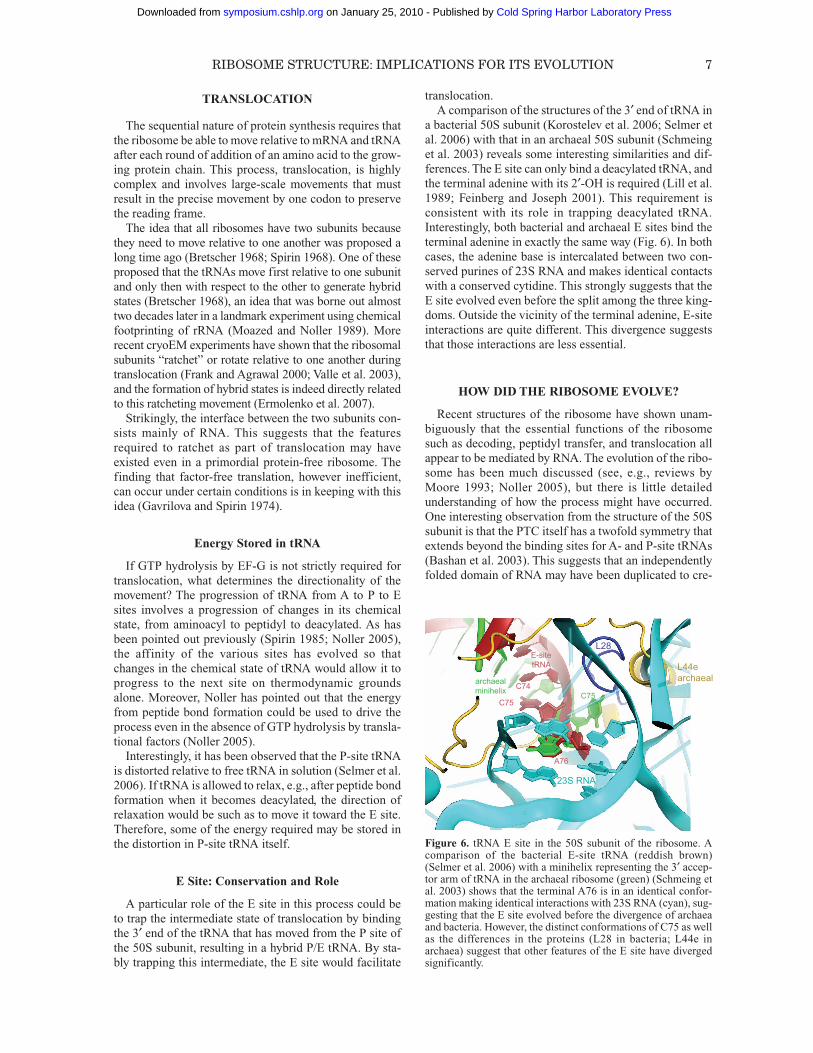
translocation.A comparison of the structures of the 3′ end of tRNA in
a bacterial 50S subunit (Korostelev et al. 2006; Selmer etal. 2006) with that in an archaeal 50S subunit (Schmeinget al. 2003) reveals some interesting similarities and dif-ferences. The E site can only bind a deacylated tRNA, andthe terminal adenine with its 2′-OH is required (Lill et al.1989; Feinberg and Joseph 2001). This requirement isconsistent with its role in trapping deacylated tRNA.Interestingly, both bacterial and archaeal E sites bind theterminal adenine in exactly the same way (Fig. 6). In bothcases, the adenine base is intercalated between two con-served purines of 23S RNA and makes identical contactswith a conserved cytidine. This strongly suggests that theE site evolved even before the split among the three king-doms. Outside the vicinity of the terminal adenine, E-siteinteractions are quite different. This divergence suggeststhat those interactions are less essential.
HOW DID THE RIBOSOME EVOLVE?
Recent structures of the ribosome have shown unam-biguously that the essential functions of the ribosomesuch as decoding, peptidyl transfer, and translocation allappear to be mediated by RNA. The evolution of the ribo-some has been much discussed (see, e.g., reviews byMoore 1993; Noller 2005), but there is little detailedunderstanding of how the process might have occurred.One interesting observation from the structure of the 50Ssubunit is that the PTC itself has a twofold symmetry thatextends beyond the binding sites for A- and P-site tRNAs(Bashan et al. 2003). This suggests that an independentlyfolded domain of RNA may have been duplicated to cre-
RIBOSOME STRUCTURE: IMPLICATIONS FOR ITS EVOLUTION 7
A76
C75
C74C75
23S RNA
L28E-sitetRNA
archaealminihelix
L44earchaeal
Figure 6. tRNA E site in the 50S subunit of the ribosome. Acomparison of the bacterial E-site tRNA (reddish brown)(Selmer et al. 2006) with a minihelix representing the 3′ accep-tor arm of tRNA in the archaeal ribosome (green) (Schmeing etal. 2003) shows that the terminal A76 is in an identical confor-mation making identical interactions with 23S RNA (cyan), sug-gesting that the E site evolved before the divergence of archaeaand bacteria. However, the distinct conformations of C75 as wellas the differences in the proteins (L28 in bacteria; L44e inarchaea) suggest that other features of the E site have divergedsignificantly.
Cold Spring Harbor Laboratory Press on January 25, 2010 - Published by symposium.cshlp.orgDownloaded from

ate the precursor of the PTC. It has been noticed that inde-pendent modules that are duplicated and held by tertiarycontacts involving precisely the same type of minor-groove interactions as found in decoding could be thebasis for the evolution of RNA in the contemporary 50Ssubunit (Bokov and Steinberg 2009). The evolution of the30S subunit and coded synthesis involving tRNA andmRNA is even less well understood despite decades ofspeculation. What is clear is that although the contempo-rary ribosome appears to be a highly complex assembly ofRNA and protein, and additionally involves many differ-ent protein factors, the high-resolution structures of theribosome provide strong support for the idea that theessential functions of the ribosome are mediated by RNAand that the ribosome evolved from a primordial RNAworld. In so doing, it appears to have been a Trojan horsethat accelerated the transformation of that world into theprotein world that we know today.
ACKNOWLEDGMENTS
Work in the author’s laboratory is supported by theMedical Research Council (U.K.), the Wellcome Trust,the Agouron Institute, and the Louis-Jeantet Foundation.I thank T. Martin Schmeing for making Figures 1–4 inconnection with other publications.
REFERENCES
Ban N, Nissen P, Hansen J, Moore PB, Steitz TA. 2000. The com-plete atomic structure of the large ribosomal subunit at 2.4 Åresolution. Science 289: 905–920.
Bashan A, Agmon I, Zarivach R, Schluenzen F, Harms J, BerisioR, Bartels H, Franceschi F, Auerbach T, Hansen HA, et al. 2003.Structural basis of the ribosomal machinery for peptide bondformation, translocation, and nascent chain progression. MolCell 11: 91–102.
Bokov K, Steinberg SV. 2009. A hierarchical model for evolutionof 23S ribosomal RNA. Nature 457: 977–980.
Bretscher MS. 1968. Translocation in protein synthesis: A hybridstructure model. Nature 218: 675–677.
Crick FHC. 1966. Codon-anticodon pairing: The wobble hypoth-esis. J Mol Biol 19: 548–555.
Crick FHC. 1968. The origin of the genetic code. J Mol Biol 38:367–379.
Ermolenko DN, Spiegel PC, Majumdar ZK, Hickerson RP, CleggRM, Noller HF. 2007. The antibiotic viomycin traps the ribo-some in an intermediate state of translocation. Nat Struct MolBiol 14: 493–497.
Feinberg JS, Joseph S. 2001. Identification of molecular interac-tions between P-site tRNA and the ribosome essential fortranslocation. Proc Natl Acad Sci 98: 11120–11125.
Frank J, Agrawal RK. 2000. A ratchet-like inter-subunit reorgani-zation of the ribosome during translocation. Nature 406: 318–322.
Freistroffer DV, Kwiatkowski M, Buckingham RH, Ehrenberg M.2000. The accuracy of codon recognition by polypeptiderelease factors. Proc Natl Acad Sci 97: 2046–2051.
Gao Y-G, Selmer M, Dunham CM, Weixlbaumer A, Kelley AC,Ramakrishnan V. 2009. The structure of the ribosome withelongation factor G trapped in the post-translocational state.Science 326: 694–699.
Gavrilova LP, Spirin AS. 1974. “Nonenzymatic” translation.Methods Enzymol 30: 452–462.
Gavrilova LP, Kostiashkina OE, Koteliansky VE, Rutkevitch NM,Spirin AS. 1976. Factor-free (“non-enzymic”) and factor-
dependent systems of translation of polyuridylic acid byEscherichia coli ribosomes. J Mol Biol 101: 537–552.
Guerrier-Takada C, Gardiner K, Marsh T, Pace N, Altman S. 1983.The RNA moiety of ribonuclease P is the catalytic subunit ofthe enzyme. Cell 35: 849–857.
Kapp LD, Lorsch JR. 2004. The molecular mechanics of eukary-otic translation. Annu Rev Biochem 73: 657–704.
Khaitovich P, Mankin AS, Green R, Lancaster L, Noller HF. 1999.Characterization of functionally active subribosomal particlesfrom Thermus aquaticus. Proc Natl Acad Sci 96: 85–90.
Kool ET. 2000. Synthetically modified DNAs as substrates forpolymerases. Curr Opin Chem Biol 4: 602–608.
Korostelev A, Trakhanov S, Laurberg M, Noller HF. 2006. Crystalstructure of a 70S ribosome-tRNA complex reveals functionalinteractions and rearrangements. Cell 126: 1065–1077.
Korostelev A, Asahara H, Lancaster L, Laurberg M, Hirschi A,Zhu J, Trakhanov S, Scott WG, Noller HF. 2008. Crystal struc-ture of a translation termination complex formed with releasefactor RF2. Proc Natl Acad Sci 105: 19684–19689.
Laurberg M, Asahara H, Korostelev A, Zhu J, Trakhanov S, NollerHF. 2008. Structural basis for translation termination on the70S ribosome. Nature 454: 852–857.
Lill R, Robertson JM, Wintermeyer W. 1989. Binding of the 3′ ter-minus of tRNA to 23S rRNA in the ribosomal exit site activelypromotes translocation. EMBO J 8: 3933–3938.
Maguire BA, Beniaminov AD, Ramu H, Mankin AS, Zim mer -mann RA. 2005. A protein component at the heart of an RNAma chine: The importance of protein L27 for the function of thebacterial ribosome. Mol Cell 20: 427–435.
Moazed D, Noller HF. 1989. Intermediate states in the movementof transfer RNA in the ribosome. Nature 342: 142–148.
Moore PB. 1988. The ribosome returns. Nature 331: 223–227.Moore PB. 1993. Ribosomes and the RNA world. In The RNA
world (ed. RF Gesteland and JF Atkins), pp. 119–136. ColdSpring Harbor Laboratory Press, Cold Spring Harbor, NY.
Moore VG, Atchison RE, Thomas G, Moran M, Noller HF. 1975.Identification of a ribosomal protein essential for peptidyltransferase activity. Proc Natl Acad Sci 72: 844–848.
Nissen P, Hansen J, Ban N, Moore PB, Steitz TA. 2000. The struc-tural basis of ribosome activity in peptide bond synthesis.Science 289: 920–930.
Nitta I, Ueda T, Watanabe K. 1998. Possible involvement ofEscherichia coli 23S ribosomal RNA in peptide bond forma-tion. RNA 4: 257–267.
Nitta I, Kamada Y, Noda H, Ueda T, Watanabe K. 1999. Peptidebond formation: Retraction. Science 283: 2019–2020.
Noller HF. 2005. Evolution of ribosomes and translation from anRNA world. In The RNA world, 3rd ed. (ed. RF Gesteland etal.), pp. 287–307. Cold Spring Harbor Laboratory Press, ColdSpring Harbor, NY.
Noller HF, Chaires JB. 1972. Functional modification of 16S ribo-somal RNA by kethoxal. Proc Natl Acad Sci 69: 3115–3118.
Noller HF, Hoffarth V, Zimniak L. 1992. Unusual resistance ofpeptidyl transferase to protein extraction procedures. Science256: 1416–1419.
Ogle JM, Ramakrishnan V. 2005. Structural insights into transla-tional fidelity. Annu Rev Biochem 74: 129–177.
Ogle JM, Brodersen DE, Clemons WM Jr, Tarry MJ, Carter AP,Ramakrishnan V. 2001. Recognition of cognate transfer RNAby the 30S ribosomal subunit. Science 292: 897–902.
Ogle JM, Murphy FV, Tarry MJ, Ramakrishnan V. 2002. Selectionof tRNA by the ribosome requires a transition from an open toa closed form. Cell 111: 721–732.
Rodnina MV, Wintermeyer W. 2001. Fidelity of aminoacyl-tRNAselection on the ribosome: Kinetic and structural mechanisms.Annu Rev Biochem 70: 415–435.
Rodnina MV, Beringer M, Wintermeyer W. 2006. Mechanism ofpeptide bond formation on the ribosome. Q Rev Biophys 39:203–225.
Schmeing TM, Ramakrishnan V. 2009. What recent ribosomestructures have revealed about the mechanism of translation.Nature 461: 1234–1242.
Schmeing TM, Moore PB, Steitz TA. 2003. Structures of deacy-
8 RAMAKRISHNAN
Cold Spring Harbor Laboratory Press on January 25, 2010 - Published by symposium.cshlp.orgDownloaded from
lated tRNA mimics bound to the E site of the large ribosomalsubunit. RNA 9: 1345–1352.
Schmeing TM, Huang KS, Kitchen DE, Strobel SA, Steitz TA.2005a. Structural insights into the roles of water and the 2′hydroxyl of the P site tRNA in the peptidyl transferase reaction.Mol Cell 20: 437–448.
Schmeing TM, Huang KS, Strobel SA, Steitz TA. 2005b. Aninduced-fit mechanism to promote peptide bond formation andexclude hydrolysis of peptidyl-tRNA. Nature 438: 520–524.
Schmeing TM, Voorhees RM, Kelley AC, Gao Y-G, Murphy FVIV, Weir JR, Ramakrishnan V. 2009. The crystal structure of theribosome bound to EF-Tu and aminoacyl-tRNA. Science 326:688–694.
Schuette JC, Murphy FV IV, Kelley AC, Weir JR, Giesebrecht J,Connell SR, Loerke J, Mielke T, Zhang W, Penczek PA, et al.2009. GTPase activation of elongation factor EF-Tu by theribosome during decoding. EMBO J 28: 755–765.
Schuwirth BS, Borovinskaya MA, Hau CW, Zhang W, Vila-Sanjurjo A, Holton JM, Cate JH. 2005. Structures of the bacte-rial ribosome at 3.5 Å resolution. Science 310: 827–834.
Selmer M, Dunham CM, Murphy FV IV, Weixlbaumer A, Petry S,Kelley AC, Weir JR, Ramakrishnan V. 2006. Structure of the70S ribosome complexed with mRNA and tRNA. Science 313:1935–1942.
Sharma MR, Koc EC, Datta PP, Booth TM, Spremulli LL,Agrawal RK. 2003. Structure of the mammalian mitochondrialribosome reveals an expanded functional role for its componentproteins. Cell 115: 97–108.
Sievers A, Beringer M, Rodnina MV, Wolfenden R. 2004. The ribo-some as an entropy trap. Proc Natl Acad Sci 101: 7897–7901.
Spirin AS. 1968. How does the ribosome work? A hypothesisbased on the two subunit construction of the ribosome. CurrMod Biol 2: 115–127.
Spirin AS. 1985. Ribosomal translocation: Facts and models. ProgNucleic Acid Res Mol Biol 32: 75–114.
Valle M, Sengupta J, Swami NK, Grassucci RA, Burkhardt N,Nierhaus KH, Agrawal RK, Frank J. 2002. Cryo-EM reveals an
active role for aminoacyl-tRNA in the accommodation process.EMBO J 21: 3557–3567.
Valle M, Zavialov A, Sengupta J, Rawat U, Ehrenberg M, Frank J.2003. Locking and unlocking of ribosomal motions. Cell 114:123–134.
Villa E, Sengupta J, Trabuco LG, LeBarron J, Baxter WT, ShaikhTR, Grassucci RA, Nissen P, Ehrenberg M, Schulten K, FrankJ. 2009. Ribosome-induced changes in elongation factor Tuconformation control GTP hydrolysis. Proc Natl Acad Sci 106:1063–1068.
Voorhees RM, Weixlbaumer A, Loakes D, Kelley AC, Rama krish -nan V. 2009. Insights into substrate stabilization from snapshotsof the peptidyl transferase center of the intact 70S ribosome.Nat Struct Mol Biol 16: 528–533.
Weinger JS, Parnell KM, Dorner S, Green R, Strobel SA. 2004.Substrate-assisted catalysis of peptide bond formation by theribosome. Nat Struct Mol Biol 11: 1101–1106.
Weixlbaumer A, Jin H, Neubauer C, Voorhees RM, Petry S,Kelley AC, Ramakrishnan V. 2008. Insights into translationaltermination from the structure of RF2 bound to the ribosome.Science 322: 953–956.
Wimberly BT, Brodersen DE, Clemons WM Jr, Morgan-WarrenRJ, Carter AP, Vonrhein C, Hartsch T, Ramakrishnan V. 2000.Structure of the 30S ribosomal subunit. Nature 407: 327–339.
Wower IK, Wower J, Zimmermann RA. 1998. Ribosomal proteinL27 participates in both 50 S subunit assembly and the peptidyltransferase reaction. J Biol Chem 273: 19847–19852.
Yusupov MM, Yusupova GZ, Baucom A, Lieberman K, EarnestTN, Cate JH, Noller H.F. 2001. Crystal structure of the ribo-some at 5.5 Å resolution. Science 292: 883–896.
Zaug AJ, Grabowski PJ, Cech TR. 1983. Autocatalytic cyclizationof an excised intervening sequence RNA is a cleavage-ligationreaction. Nature 301: 578–583.
Zavialov AV, Mora L, Buckingham RH, Ehrenberg M. 2002.Release of peptide promoted by the GGQ motif of class 1release factors regulates the GTPase activity of RF3. Mol Cell10: 789–798.
RIBOSOME STRUCTURE: IMPLICATIONS FOR ITS EVOLUTION 9
Cold Spring Harbor Laboratory Press on January 25, 2010 - Published by symposium.cshlp.orgDownloaded from