the role of ccaat/enhancer-binding protein in the differential
TRANSCRIPT

THE JOURNAL OF BIOLOGICAL CHEMISTRY (c) 1991 by The American Society for Biochemistry and Molecular Biology, Inc Vol. 266, No. 18, Issue of June 25, pp. 11594-11603,1991
Printed in U. S. A.
The Role of CCAAT/Enhancer-binding Protein in the Differential Transcriptional Regulation of a Family of Human Liver Alcohol Dehydrogenase Genes*
(Received for publication, November 27, 1990)
Mark J. Stewart#, Mary Lou Shean, Bryan W. Paeper, and Gregg Duesterj From the Department of Biochemistry, Colorado State University, Fort Collins, Colorado 80523
The transcription factor CCAAT/enhancer-binding protein (C/EBP) was found to selectively trans-acti- vate one member of the human class I alcohol dehydro- genase (ADH) gene family. A comparison of the pro- moters for the three human class I ADH genes A D H l , ADH2, and ADH3 indicated a very similar pattern of binding sites (sites A-F) for rat liver nuclear proteins located between -10 and -210 base pairs (bp). In all three promoters site A consisted of two binding sites for the transcription factor C/EBP closely flanking both sides of the TATA box, but C/EBP bound with much greater affinity to site A of ADH2. C/EBP also bound at two locations which coincide with site D (-120 bp) and site E (-160 bp) of all three promoters. Cotransfection studies of human hepatoma cells using ADH-cat fusions and a C/EBP expression plasmid in- dicated that the human ADH2 promoter responded well to C/EBP trans-activation whereas the human ADHl and ADH3 promoters, which bind C/EBP weakly, responded poorly. Individual mutations in sev- eral ADH2 nuclear factor-binding sites allowed the identification of four functional C/EBP-binding sites, Le. two in site A as well as one each in sites D and E. Also, the ADH2 TATA box was found to be dispensable for C/EBP induction. Compared to ADH2 and ADH3, site A in ADHl contains four extra base pairs between the two C/EBP motifs, and deletion of these nucleotides increased the C/EBP responsiveness of ADHl presum- ably by changing the spacing of the two C/EBP motifs. Thus, sequence divergence of human class I ADH gene family members has led to forms which vary in their responsiveness to C/EBP. We suggest that C/EBP con- tributes to liver-specific expression of the human class I ADH gene family by selectively inducing the ADH2 gene via a TATA-independent mechanism during liver development.
* This work was supported in part by National Institute on Alcohol Abuse and Alcoholism Grant AA07261 and a National Institutes of Health Biomedical Research Support Grant (to G. D.). The costs of publication of this article were defrayed in part by the payment of page charges. This article must therefore be hereby marked “aduer- tisement” in accordance with 18 U.S.C. Section 1734 solely to indicate this fact.
+Present address: Howard Hughes Medical Institute, MSRBII Rm. 4544, University of Michigan Medical Center, Ann Arbor, MI 48109- 0650.
Recipient of Research Scientist Development Award KO2 AA00119 from the National Instltute on Alcohol Abuse and Alcohol- ism. To whom correspondence should be addressed.
Human class I alcohol dehydrogenase (ADH)’ is composed of three isozymes (a, p, and y) encoded by distinct genes (ADHI, ADH2, and ADH3, respectively) which all display large increases in expression during liver development (Mur- ray and Motulsky, 1971; Pikkarainen and Raiha, 1969; Smith et al., 1971). Lower, but significant expression of ADH3 also occurs in kidney, intestine, and stomach (Smith et al., 1972). Mouse class I ADH is encoded by a single gene Adh-I, homologous to all three human class I ADH genes (Ceci et al., 1987; Zhang et al., 1987), which is expressed in nearly the same locations as human ADH (Holmes et al., 1983). The large abundance of ADH in adult liver correlates with the knowledge that it catalyzes not only ethanol oxidation (Li, 1977), but the reversible oxidation/reduction of numerous alcohols and aldehydes (Smith et al., 1973; Wagner et al., 1983). ADH catalyzes steps in several metabolic pathways important in postnatal liver development including bile acid synthesis (Okuda and Okuda, 1983), norepinephrine and do- pamine metabolism (Mlrdh and Vallee, 1986; MIrdh et al., 1985), and retinol metabolism (Zachman and Olson, 1961; Mezey and Holt, 1971; Leo et al., 1987). One very important reaction catalyzed by ADH is retinol oxidation which is nec- essary for degradation of excess retinol as well as for synthesis of retinoic acid, the active form of vitamin A required for normal vertebrate growth and development (Roberts and Sporn, 1984). A developmental increase in liver ADH produc- tion postnatally may facilitate the establishment of retinoid homeostasis in the neonate which can no longer rely on maternal retinoid metabolism. Since the liver and the other major sites of ADH production (i.e. kidney, intestine, stom- ach, and lung) possess epithelial cell populations which re- quire retinoic acid for their differentiation (Roberts and Sporn, 1984), ADH in these tissues may enable the local synthesis of retinoic acid.
The human a, /3, and y class I ADH isozymes have very similar substrate specificities (Wagner et al., 1983) and share 95% amino acid sequence identity (von Bahr-Lindstrom et al., 1986). Since rodents possess only one class I ADH isozyme expressed from a single locus Adh-I, the three genes for class I ADH in humans most likely arose through gene duplication. The three class I ADH gene loci with their unique control regions may expand the possibilities for fine-tuning the pro- duction of class I ADH in the developing human liver and other tissues. Indeed, differential expression of the three human class I ADH genes has been observed. Starch gel analysis of human liver extracts indicates that a low level of
’ The abbreviations used are: ADH, alcohol dehydrogenase; A D H , human gene encoding ADH; bp, base pairs; CAT, chloramphenicol acetyltransferase; cat, gene encoding CAT; C/EBP, CCAAT/en- hancer binding protein; HEPES, 4-(2-hydroxyethyl)-l-piperazineeth- anesulfonic acid.
11594

Regulation of ADH Gene Expression by CIEBP 11595
0-ADH is produced during early fetal development with ad- ditional low level production of @-ADH during late fetal development, and y-ADH several months postnatally (Smith et al., 1971, 1972). The total amount of a-, is-, and y-ADH activity in the human liver increases by more than an order of magnitude postnatally between birth and 5 years of age when adult levels of all three are achieved (Pikkarainen and Raiha, 1967; Smith et al., 1971). ADH accounts for about 2- 3% of the soluble protein in adult human liver (Ditlow et al., 1984). Human adult hepatomas often revert to production of a low level of ADH, primarily the a subunit (Smith, 1986), suggesting there is a reversion to the type of ADH gene expression seen in early fetuses. In contrast to the situation seen in liver, in human kidney, intestine, and stomach it is y- ADH which is observed prenatally and @-ADH which is observedpostnatally, with a-ADH absent (Smith et al., 1972). The significance of the differential expression of class I ADH genes in various tissues is yet to be determined, but may relate to the role of ADH in human retinoid metabolism.
Northern blot experiments using ADH cDNA probes as well as gene-specific oligonucleotide probes have indicated that there is at least an order of magnitude increase in the level of all three class I ADH mRNAs in human liver between the 20-week fetal stage and adulthood (Bilanchone et al., 1986; Ikuta and Yoshida, 1986). This suggests that as devel- opment proceeds there is increased transcription of all three human class I ADH genes in liver. The study of liver-specific gene expression has centered recently around the transcrip- tion factors which make hepatic specificity possible. One protein, in particular, which has been implicated as playing an important role in liver-specific gene transcription is CCAAT/enhancer-binding protein (C/EBP) (Landschulz et al., 1988, 1989). C/EBP is expressed primarily in liver and fat tissue in rodents, and in the liver is restricted to fully differ- entiated parenchymal cells (Birkenmeier et al., 1989). Liver C/EBP undergoes a developmental increase in production just prior to birth, and it has been suggested that this stimu- lates a postnatal increase in production of other liver proteins such as albumin (Birkenmeier et al., 1989). The nucleotide sequences for theADH1, ADHZ, andADH3 promoters as well as their transcription start sites have been determined (Dues- ter et al., 1986; Stewart et al., 1990a). We have previously shown that the ADHZ promoter is transcriptionally activated in hepatoma cells by C/EBP molecules bound at sites closely flanking both sides of the TATA box (Stewart et al., 1990b). We have now analyzed the differential transcriptional regu- lation of human ADHl, ADH2, and ADH3 genes by C/EBP.
EXPERIMENTAL PROCEDURES
Plasmid Constructions and Site-directed Mutagenesis-Fusion of various lengths of the 5'-flanking sequences of the human A D H l , ADHZ, and ADH3 genes to the chloramphenicol acetyltransferase (cat) gene in the plasmid vector pA,,CAT, has been described (Stew- art et al., 1990a). The genomic sequences were derived from hADH26 for ADHZ, XADH15 for ADHZ, and XADH8 for ADH3 (Duester et al., 1986). Convenient restriction sites were used to excise portions of the 5'-flanking DNA for subsequent fusion to cat (Fig. 5).
Site-directed mutations were constructed as described previously (Kunkel et al., 1987). The ADH promoter regions to be mutated were cloned into an M13mplO phage which was used to infect a midlog culture of Escherichia coli CJ236 (dut-, ung-) to prepare uracil- containing template DNA. Mutant oligonucleotides were constructed in which the central region contained the six-bp recognition sequence for a restriction enzyme in place of ADH sequence, and the ends consisted of 8-16 bases complementary to the wild-type sequence to allow hybridization. Oligonucleotides were phosphorylated with T4 polynucleotide kinase and hybridized to the appropriate ADH pro- moter region in the single-stranded form of the Ml3mplO vector which had been passed through E. coli CJ236. Second strand synthesis
and ligation was carried out with T4 DNA polymerase and T4 DNA ligase in a one-step reaction. The product was transfected into E. coli JM109, and the presence of a new restriction site in M13 replicative form plasmid DNA was used for screening clones which contained the mutation. The mutant ADH DNA was then isolated from the M13 clone and inserted back into the pAloCAT2 vector. The presence of the appropriate mutation was also confirmed by DNA sequence analysis (Maxam and Gilbert, 1980), and the sequences of the various mutations we prepared are summarized in Table I. The construction of mutations in ADH2 at sites A-A and A-B were previously described (Stewart et al., 1990b). All plasmids were purified through two cycles of CsC1-ethidium bromide gradients (Sambrook et al., 1989).
Transfection Conditions-The differentiated human hepatoma cell line HepG2 (Knowles et al., 1980) was cultured in minimal essential medium containing Earle's salts and nonessential amino acids and was supplemented with 10% fetal bovine serum. DNA transfections were carried out by the calcium-phosphate coprecipitate method described previously (Graham and van der Eh, 1973). HepG2 cells were seeded at a density of IO6 cells/lOO-mm tissue culture dish and were transfected 24 h later with a mixture of DNA including 15 pg of a reporter CAT plasmid, 20 pg of carrier plasmid pUC18, and 5 pg of an internal control plasmid pCH110. For C/EBP cotransfections, 20 pg of a C/EBP mammalian expression plasmid pMSV-C/EBP (Fried- man et al., 1989) replaced the carrier pUC18 to keep the total amount of DNA constant a t 40 pg. The internal control plasmid pCHllO contains the SV40 early promoter fused to the lacZ gene and was used to correct for plate-to-plate differences in transfection efficiency (Herbomel et al., 1984). The DNA precipitate (40 pg total) was added to the cells for 16 h, after which time the cells were washed and supplemented with fresh media. The cells were harvested 48 h after DNA addition, and cell extracts were prepared by repeated freeze/ thaw treatment as described (Gorman et al., 1982). Cell extracts were first assayed for P-galactosidase activity, then normalized amounts of extract were used in the CAT assay (Herbomel et al., 1984). CAT assay reactions were incubated for 2 h a t 37 "C, and additional acetyl- CoA was added after 1 h. The amount of CAT activity (% acetylation of chloramphenicol) was quantitated by cutting out chloramphenicol (C) substrate spots and acetylated chloramphenicol (AC) product spots from the thin layer chromatograms and counting the radioac- tivity by liquid scintillation. The results of three to eight experiments were averaged to give the reported values of CAT activity for each ADH-cat fusion tested.
DNA Binding Studies-DNase I footprint analysis was performed as described previously (Galas and Schmitz, 1978) using modifications for rat liver nuclear extracts (Lichtsteiner et al., 1987). The binding reaction was carried out in a 50 pl volume with 0.1 pmol of 5'-end- labeled ADH DNA. The standard reaction consisted of the following components: 50 pg/ml poly(d1-dC) carrier DNA, 25 mM Tris-HC1, pH 7.9, 6.25 mM MgC12, 0.1 mM EDTA, 50 mM KCl, 0.5 mM dithio- threitol, and 10% glycerol. Various amounts of rat liver nuclear extract or C/EBP expressed in E. coli were preincubated with poly(d1- dC) for 15 min a t 0 "C to eliminate nonspecific binding, after which the end-labeled fragment was added and the mixture was incubated for an additional 75 min at 0 "C. DNase I was added and digestion was allowed to proceed for 60 s at 25 "C. The amount of DNase I was adjusted for each extract to produce an even pattern of partial cleavage products. Reactions were stopped by the addition of 100 pl of a solution containing 0.6 M sodium acetate, 0.4% sodium dodecyl sulfate, and 250 pg/ml yeast transfer RNA. Proteinase K (10 p ) was added, and the reactions were incubated for 1 h at 42 "C. Nucleic acids were extracted twice with phenol/chloroform/isoamyl alcohol (25:24:1 by volume), then precipitated with ethanol. The pellet was dissolved in formamide, heated at 90 "C for 3 min, and subjected to electrophoresis on 6% polyacrylamide, 50% urea gels. A purine- specific cleavage reaction (G+A) was used as a sequencing standard for footprinting reactions (Maxam and Gilbert, 1980).
prepared essentially as described (Ohlsson and Edlund, 1986). All Preparation of Rat Liver Nuclear Extracts-Nuclear extracts were
procedures were performed a t 4 "C. Minced adult rat liver tissue was suspended in an equal volume of a buffer containing 10 mM HEPES, pH 7.8, 15 mM KC1, 2 mM MgCl,, 0.1 mM EDTA, 1 mM dithiothreitol, and 1 mM phenylmethylsulfonyl fluoride, then homogenized in a Potter-Elvehjem tissue grinder fitted with a motor-driven Teflon pestle. The homogenate was filtered through cheesecloth and centri- fuged at 10,000 rpm for 10 min in a Beckman JA-20 rotor to pellet nuclei. The nuclei were then resuspended in 3 ml of the above buffer/ lo9 nuclei and lysed by adding 4 M (NH&S04 to a final concentration of 0.3 M. After 30 min of gentle mixing, the lysate was centrifuged in
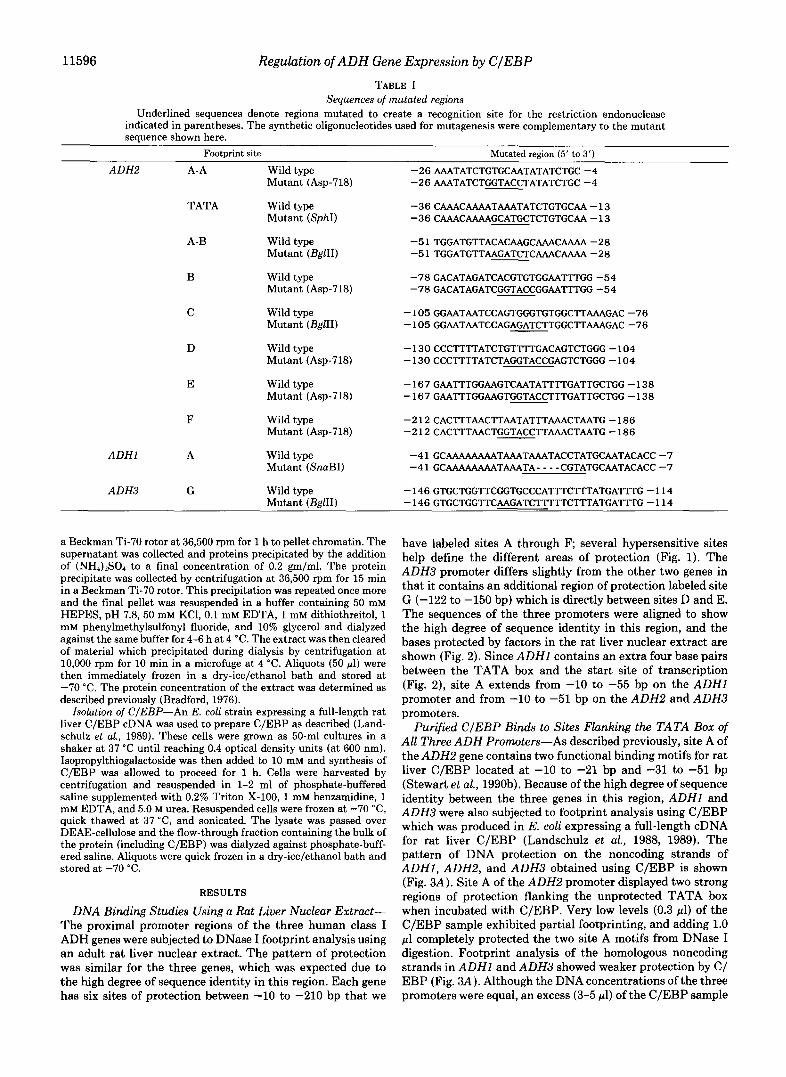
11596 Regulation of ADH Gene Expression by CIEBP TABLE I
Sequences of mutated regions Underlined sequences denote regions mutated to create a recognition site for the restriction endonuclease
indicated in parentheses. The synthetic oligonucleotides used for mutagenesis were complementary to the mutant sequence shown here.
Footprint site Mutated region (5' to 3')
ADH2 A-A Wild type -26AAATATCTGTGCAATATATCTGC-4 Mutant (Asp-718) -26AAATATCTGGTACCTATATCTGC-4
TATA Wild type -36CAAACAAAATAAATATCTGTGCAA-13 Mutant (SphI) -36CAAACAAAAGCATGCTCTGTGCAA-13
A-B Wild type Mutant (BglII)
-5lTGGATGTTACACAAGCAAACAAAA-28 -51 TGGATGTTAAGATCTCAAACAAAA -28
B Wild type -78GACATAGATCACGTGTGGAATTTGG-54
C Wild type -105GGAATAATCCAGTGGGTGTGGCTTAAAGAC-76
D Wild type -130CCCTTTTATCTGTTTTGACAGTCTGGG-104
Mutant (Asp-718) -78GACATAGATCGGTACCGGAATTTGG-54
Mutant (EglII) -105GGAATAATCCAGAGATCTTGGCTTAAAGAC -76
Mutant (Asp-718) -130CCCTTTTATCTAGGTACCGAGTCTGGG -104
E Wild type -167GAATTTGGAAGTCAATATTTTGATTGCTGG-138 Mutant (Asp-718) -167GAATTTGGAAGTGGTACCTTTGATTGCTGG-138
F Wild type -212CACTTTAACTTAATATTTATAATG-186 Mutant (Asp-718) -212CACTTTAACTGGTACCTTAAACTAATG-186
ADHl A Wild type -41 GCAAAAAAAATAAATAAATACCTATGCAATACACC -7 Mutant (SnaBI) -4 1 GCAAAAAAAATAAATA - - - - CGTATGCAATACACC -7
ADH3 G Wild type -146GTGCTGGTTCGGTGCCCATTTCTTTATGATTTG-114 Mutant (BglII) -146GTGCTGGTTCAAGATCTTTTTCTTTATGATTTG-114
a Beckman Ti-70 rotor at 36,500 rpm for 1 h to pellet chromatin. The supernatant was collected and proteins precipitated by the addition of (NH,),SO, to a final concentration of 0.2 gm/ml. The protein precipitate was collected by centrifugation at 36,500 rpm for 15 min in a Beckman Ti-70 rotor. This precipitation was repeated once more and the final pellet was resuspended in a buffer containing 50 mM HEPES, pH 7.8, 50 mM KCl, 0.1 mM EDTA, 1 mM dithiothreitol, 1 mM phenylmethylsulfonyl fluoride, and 10% glycerol and dialyzed against the same buffer for 4-6 h at 4 "C. The extract was then cleared of material which precipitated during dialysis by centrifugation at 10,000 rpm for 10 min in a microfuge at 4 "C. Aliquots (50 pl) were then immediately frozen in a dry-ice/ethanol bath and stored at -70 "C. The protein concentration of the extract was determined as described previously (Bradford, 1976).
Isolation of CIEBP-An E. coli strain expressing a full-length rat liver C/EBP cDNA was used to prepare C/EBP as described (Land- schulz et al., 1989). These cells were grown as 50-ml cultures in a shaker at 37 "C until reaching 0.4 optical density units (at 600 nm). Isopropylthiogalactoside was then added to 10 mM and synthesis of C/EBP was allowed to proceed for 1 h. Cells were harvested by centrifugation and resuspended in 1-2 ml of phosphate-buffered saline supplemented with 0.2% Triton X-100, 1 mM benzamidine, 1 mM EDTA, and 5.0 M urea. Resuspended cells were frozen at -70 "C, quick thawed at 37 "C, and sonicated. The lysate was passed over DEAE-cellulose and the flow-through fraction containing the bulk of the protein (including C/EBP) was dialyzed against phosphate-buff- ered saline. Aliquots were quick frozen in a dry-ice/ethanol bath and stored at -70 "C.
RESULTS
DNA Binding Studies Using a Rat Liver Nuclear Extract- The proximal promoter regions of the three human class I ADH genes were subjected to DNase I footprint analysis using an adult rat liver nuclear extract. The pattern of protection was similar for the three genes, which was expected due to the high degree of sequence identity in this region. Each gene has six sites of protection between -10 to -210 bp that we
have labeled sites A through F; several hypersensitive sites help define the different areas of protection (Fig. 1). The ADH3 promoter differs slightly from the other two genes in that it contains an additional region of protection labeled site G (-122 to -150 bp) which is directly between sites D and E. The sequences of the three promoters were aligned to show the high degree of sequence identity in this region, and the bases protected by factors in the rat liver nuclear extract are shown (Fig. 2). Since ADHl contains an extra four base pairs between the TATA box and the start site of transcription (Fig. 2), site A extends from -10 to -55 bp on the ADHl promoter and from -10 to -51 bp on the ADH2 and ADH3 promoters.
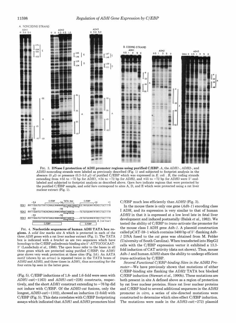
Purified CIEBP Binds to Sites Flanking the TATA Box of All Three ADH Promoters-As described previously, site A of the ADH2 gene contains two functional binding motifs for rat liver C/EBP located at -10 to -21 bp and -31 to -51 bp (Stewart et al., 199Ob). Because of the high degree of sequence identity between the three genes in this region, ADHl and ADH3 were also subjected to footprint analysis using C/EBP which was produced in E. coli expressing a full-length cDNA for rat liver C/EBP (Landschulz et al., 1988, 1989). The pattern of DNA protection on the noncoding strands of ADHl, ADH2, and ADH3 obtained using C/EBP is shown (Fig. 3A) . Site A of the ADH2 promoter displayed two strong regions of protection flanking the unprotected TATA box when incubated with C/EBP. Very low levels (0.3 pl) of the C/EBP sample exhibited partial footprinting, and adding 1.0 p1 completely protected the two site A motifs from DNase I digestion. Footprint analysis of the homologous noncoding strands in ADHl and ADH3 showed weaker protection by C/ EBP (Fig. 3A) . Although the DNA concentrations of the three promoters were equal, an excess (3-5 pl) of the C/EBP sample

Regulation of ADH Gene Expression by CIEBP 11597
A ALPHA
0"
0" A
C G A Y Y A
m , t
, -
A
FIG. 1. DNase I protection of ADH promoter regions using a rat liver nuclear extract. A, the noncoding strand of the ADHZ proximal promoter region extending from +55 to -228 bp was 5'- end-labeled and digested with DNase I in the absence (0 pg) or presence (20-200 pg) of crude rat liver nuclear extract. B, the non- coding strand of the ADH2 promoter extending from +34 to -272 bp was 5"end-labeled and digested with DNase I in the absence (0 pg) or presence (160 pg) of rat liver nuclear extract. C, the noncoding strand of the ADH3 promoter extending from +55 to -328 bp was treated as in R. Brackets indicate regions of protection labeled A through G, and arrouls indicate DNase I-hypersensitive cleavage sites. The area labeled G for the ADH3 footprint (C) is shown with an asterisk to indicate that it is unique to the ADH3 promoter. G+A, purine-specific cleavage reaction.
was needed to exhibit protection of ADHl and ADH3 se- quences. For ADHl, the sequence downstream of the TATA box was partially protected using C/EBP, whereas the up- stream sequence was not protected. The footprinting pattern obtained with the ADH3 promoter using C/EBP resembled that of the ADH2 promoter in that protection of both sites flanking the TATA box occurred, but only when using high levels of C/EBP. Footprint analysis of the coding strands for the three genes using C/EBP also indicated stronger binding for the A D H 2 promoter region, and once again showed that the TATA box was unprotected (Fig. 3B). The coding strand of ADHl did show weak protection of the sequence just upstream of the TATA box (Fig. 3B). A comparison of the sequences of the TATA box regions of the three ADH genes is shown (Fig. 4), with an indication of the relationship of the footprints observed with a rat liver nuclear extract and C/ EBP.
Upstream C/EBP-binding Sites-In ADHl, ADH2, and ADH3, additional sequences upstream of site A were also footprinted with C/EBP. ADHI has a bipartite region of
A D H I 5'-~RRTTTGRCRGRTGAGGRflTfl------R~GTGflGGRGRflGflGflflRflTGRTTflflGCTTTflTCflCTTT
R D H Z 5'-GRRTTCCAGRGGCCGGGGGGGGGTGGGflflGTGRGGRflRflGRGRflflGTGRTTflCRRTTTRTCflCTTT
ADH3 5'-GARCTTGRGAGA------------------GGRGGflflGflGflGflflRGTRflTTRRRRTGTflTCGTT~T *** ******** ****** ***.***.. *.****** **** tt.
**** ** ** ********* .***.*** ****** *****.****.
- 1 4 7 CTGGTTCRTTGCCCTCTTCTTTRTGflTTTGRCRGTCTGTGRflTflflTCTRflTGGGTGTGGCTTRflRG I******* * **** ** * ** ************ ******** * ****************
- 1 4 1 CTGGTTCRGTflCCCTTTT--RTCTGTTTTGRCflGTCTGGGRflTflflTCCRGTGGGTGTGGCTTRflRG I****** .***** *.$**.********** ****** .** tt* * * .*.***********
-143 CTGGTTCGGTGCCCRTTTCTTTflTGRTTTGRTRGTCTGRGRflGflRTRCGflCGGGTGTGGCTTRflflfl ClEEP D -t-t-
ADHl-rprciOc G C
-81 RCCTflGflTCRTGTGTGGRRCTGGflflTTGGGTGTTflTTCRRGCflflRflRRRflTflRRTRflflTflCCTflTG
-77 RCRTAGATCACGTGTGGRflTTGGflRTTGGRTGTTflCflCRRGCflflRCRRflflTflRRTR----TCTGTG ** ****a** *****.** ********* ***** ******** ********** ** **
******t**t**~*** I ****tt.**.*.**8*** ***************** ****** -77 ~ T C R C G T G T G T R G T T G G f l f l T T G G G T G T T f l T R T G R G C f l f l R C R f l f l f l T f l R R T R - - - - C C T G T G
' B ' ClEBP A - C l E B P TPITA- box
+! -15 CAATRCRCCTGCTTT~TGCACTTGf lGCf lGGGRf lGf l f l f lTCCRC~f lGGRCTC~CCf lGTCTCCTGGlCT-3 '
-15 CflflTRTATCTGCTTT~TGCflCTC~flGC~GflGRRGflRflTCCRCflflflGflCTCR-CflGTCTGCTGGTGG-3' ***** * *.*.********** ***.* ..*****.*.*..* *****. I..*.. I****
-15 C~TRCCTGCTTTRTGCflCTCR~GCflGf lGf lRGf lRf lTCCf lCRf lGTf lCTCRCCflGCCTCCTGGTCT-3 ' *** . . . . . . . . . . . . . . . . . . . . . . . . . . . . . . . . . . . . . . . . . *****.*** *.**.**.*.
FIG. 2. ADH nucleotide sequences protected by a rat liver nuclear extract. The ADHI, ADHZ, andADH3 promoter sequences were previously determined (Stewart et al., 1990a) and have been aligned to show regions of identity. Asterisks are placed when matches occur between two of the genes. Dashes indicate gaps which were introduced in order to properly align the sequences. The start site of transcription is labeled + I bp as determined by S1 nuclease mapping (Duester et al., 1986) and primer extension analysis (Stewart et al., 1990a) and the TATA box is displayed by a bracket. The footprinted regions shown in Fig. 1 which are present in all three genes (sites A to F) are underlined by a solid bar, and the ADH3-specific footprint (site G ) is indicated by a cross-hatched bar. Sites that bind purified C/EBP (Fig. 3) are labeled.
protection (-152 to -161 bp and -164 to -171 bp; Fig. 3A) which corresponds to site E seen with a rat liver nuclear extract (-152 to -171 bp; Fig. lA), with the exception that C/EBP did not protect the central two base pairs. The ADH2 and ADH3 promoters also exhibited C/EBP protection of their site E sequences near -160 bp, as well as an additional C/EBP footprint centered over their site D sequences near -120 bp (Fig. 3A). The ADHl site D sequence did not bind C/EBP very well as evidenced by only a minor effect on the DNase I protection of the sequences just downstream of site E (Fig. 3A).
Extent of Trans-actiuation of ADH Promoters by C/EBP- To determine the effect of C/EBP on the transcriptional activity of the three A D H promoters, cotransfection studies were done in which 5"flanking A D H sequences fused to the gene encoding chloramphenicol acetyltransferase (cat) were introduced into HepG2 human hepatoma cells along with the C/EBP expression vector pMSV-C/EBP. It has been shown that the level of C/EBP in HepG2 cells is only 5% of that seen in adult rat liver and that transfection of HepG2 with pMSV-C/EBP can significantly increase that level (Friedman et al., 1989). We have previously shown that C/EBP cotrans- fection led to a 4-fold induction of cat activity for the con- struction ADH2-cat(-272) containing 272 bp of ADH2 5'- flanking DNA fused upstream of cat (Stewart et al., 1990b). The C/EBP induction of ADH2-cat(-272) is shown as a control for several ADHl-cat and ADH3-cat fusions which exhibit very little or no effect of C/EBP on cat expression
11598 Regulation of ADH Gene Expression by CIEBP
A. NONCODING STRAND AOH 1
0 3.0 5 0
b r
Ann7
0:: I E "-129 I
nl/l
6. CODING STRAND
0.5 I 3 5 0 ADH I
n"'
Y
(I * I :
0.5 I 3 5 o ADH2 ADH3
-9
-33
FIG. 3. DNase I protection of ADH promoter regions using purified C/EBP. A , the ADHI-, ADH2-, and ADH3-noncoding strands were labeled as previously described (Fig. 1) and subjected to footprint analysis in the absence (0 pl) or presence (0.3-5.0 pl) of purified C/EBP which was expressed in E. coli . B, the coding strands extending from +55 to -15 bp for ADHI, +34 to -12 bp for ADH2, and +55 to -12 bp for ADH3 were 5"end- labeled and subjected to footprint analysis as described above. Open bars indicate regions that were protected by the purified C/EBP sample, and solid bars correspond to sites A, D, and E which were protected using a rat liver nuclear extract (Fig. 1).
ClEBF TATA-box ClEBP -54 "34- ,-,-IO ~DHJ A R T T C G G T G T T A T T C A A C C A A R R R R A R T ~ R R T ~ A R T ~ C C T A T G C R R T R ~ R C C T G C T T T R
+ I
R&S RATTGGATGTTACACARGCRAACRRART~RAT~-- - -TCTGTGCAATRTRTCTGCTTTA
~3 A A T T ~ G C T G T T R T A T G A C C R R R C R ~ A R T ~ R R T ~ - - - - C C T G T G C R ~ C R T R C C T G C T T T A
I C/EBP I I C/EBP I
-50 - 30 -50 -30
A (rat liver)
FIG. 4. Nucleotide sequences of human ADH TATA box re- gions. A solid line marks site A which is protected in each of the three ADH genes with a rat liver nuclear extract (Fig. 1). The TATA box is indicated with a bracket as are two sequences which have homology to the C/EBP palindromic binding site 5"ATTGCGCAAT- 3' (Landschulz et al., 1988). The open boxes refer to the bases in all three genes which are protected using purified C/EBP; the ADHl gene shows very weak protection a t these sites (Fig. 3A) . An AATA motif (shown by an arrow) is repeated twice in the TATA boxes of ADH2 and ADH3, and three times in ADHl , thus accounting for the four extra bp seen in the latter gene.
(Fig. 5). C/EBP inductions of 1.8- and 1.6-fold were seen with ADHl-cat(-1163) and ADHl-cat(-228) constructs, respec- tively, and the short ADHl construct extending to -76 bp did not induce with C/EBP. Of the ADH3-cat fusions, only the longest, ADH3-cat(-1102), showed an induction (2-fold) with C/EBP (Fig. 5). This data correlates with C/EBP footprinting assays which indicated that ADHl and ADH3 promoters bind
C/EBP much less efficiently than ADH2 (Fig. 3). In the mouse there is only one gene (Adh-1) encoding class
I ADH, and its expression is very similar to that of human ADH2 in that it is expressed at a low level late in fetal liver development and induced postnatally (Balak et al., 1982). We tested the ability of C/EBP to trans-activate the promoter for the mouse class I ADH gene Adh-1. A plasmid construction called pCAT-59-1 which contains 3400 bp of 5"flankingAdh- 1 DNA fused to the cat gene was obtained from M. Felder (University of South Carolina). When transfected into HepG2 cells with the C/EBP expression vector it exhibited a 13.3- fold induction of CAT activity (data not shown). Thus, mouse Adh-1 and humanADH2 share the ability to undergo efficient trans-activation by C/EBP.
Several Functionul C/EBP-binding Sites in the ADH2 Pro- moter-We have previously shown that mutations of either C/EBP-binding site flanking the ADH2 TATA box blocked C/EBP induction (Stewart et al., 1990b). These mutations are both present in site A defined above as a region of protection by rat liver nuclear proteins. Since rat liver nuclear proteins and C/EBP bind to several additional sequences in the ADH2 promoter in uitro, a series of site-directed mutations were constructed to determine which sites effect C/EBP induction. The mutations were made in the ADH2-&(-272) plasmid

Regulation of ADH Gene Expression by CIEBP 11599
C rat liver nuclear extract or C/EBP were comparable to the Rclotive CAT k t l v l t y VEEP n
8.rnHI -I 163 Transient transfection assays were performed to compare
+ I
19 - c-7 wild-type template (data not shown).
the basal level and C/EBP-induced expression of the various ADH2 mutant promoters (Fig. 8A). None of the mutations
in the basal level. The wild-type ADH2-cat(-272) promoter exhibited a 4-fold C/EBP induction. Mutations of either C/ EBP motif in site A (i.e. motifs A-A or A-B) created ADH2 promoters which were unresponsive to C/EBP, and mutations of sites D or E led to reductions in C/EBP responsiveness to 1.8- and 2.7-fold, respectively (Fig. 8A). The mutations in sites D and E led to 2-fold higher basal levels than wild-type, and from these higher basal levels there were lower fold inductions by C/EBP compared to wild-type. Thus, maximal C/EBP induction requires C/EBP binding a t both motifs in site A as previously shown (Stewart et al., 1990b), as well as binding at both sites D and E. The mutants with altered sites B, C, or F still responded to C/EBP induction with values ranging from 3.9- to 4.7-fold (Fig. 8A), despite the fact that they did eliminate or alter the binding of rat liver nuclear
which bind these latter sites are not needed for C/EBP induction of ADH2 in HepG2 cells. Surprisingly, a TATA box mutant exhibited a 2-fold increase in basal level expression comparable to the site E mutation but was still able to be induced quite nicely (5.6-fold) with C/EBP (Fig. 8A). This suggests that C/EBP induction of ADH2 may function via a TATA-independent mechanism.
Deletion Mutation in Site A of ADHl-As shown above, ADHl responds very weakly to C/EBP despite the fact that its promoter proximal region is very similar in sequence to ADH2 which is very responsive. However, a major difference
tions with a C/EBP expression vector in HepG2 cells. Results existence of an extra four bp present in site A of ADHl (Fig.
ADH2-cat(-272). Relative CAT activity and autoradiograms are dis- 4) which we reasoned may inhibit C/EBP induction due to are shown for ADHI-cat andADH3-catconstructs, as well as a control
played next to each construct for transfections done in the presence increased spacing of the two C/EBP motifs flanking the
Hind111 eliminated basal level expression, and some led to increases
EcoRl -328 * factors as shown in Fig. 6B. This may indicate that the factors
52 - * * -72 +55
[loo -4 ADH2-crt(-272) 401
FIG. 5. COtranSfeCtiOn Of ADH-cat 5"deletion COnStrUC- between the site A sequences of ADHl and ADH2 is the

11600 Regulation of ADH Gene Expression by CIEBP
A Mutant A-A Mutant D Mutant E 1 1 1 I I
A 0 0 0 0 0 0 0 0 0 4 6 0 4 8
" " .
"
B Mutant 6 Mutant C G I 1 G 1 1 A 0 0 0 0 2 0 8 8 0 " 6 0 4
G Mutant F
L 6 A ! , A 0 0 0 0
-en 4 "-+ .., .Y.
- ". A
FIG. 6. Binding of rat liver nuclear factors to mutant ADH2 promoters. Various ADH2 promoter mutants were subjected to DNase I footprint analysis as described in Fig. 1B using from 0 to 200 pg of a rat liver nuclear extract. The solid burs indicate sites of protection, except for those accompanied by parallel hatched burs which indicate the sites that were not motected relative to the wild-type template. A, mutants of sites A-A, D, and E are shown. R, mutants of sites B, C,*and F are shown
unresponsive to C/EBP in a transfection assay. The wild- type ADH3-cat(-328) exhibited a C/EBP effect of 0.8-fold and the site G mutant exhibited a C/EBP effect of 1.1-fold, and there was only a slight effect effect on the basal level expression (Fig. 8C). DNase I footprint analysis of the site G mutant verified that the rat liver nuclear footprint which normally exists a t site G was missing (data not shown). Thus, it appears that site G does not play a role in decreasing the C/EBP responsiveness of the ADH3 promoter relative to ADH2.
DISCUSSION
In both humans and rodents, the adult liver is by far the richest source for class I ADH, whereas much lower levels are seen in the intestine, kidney, and lung (Smith et al., 1971, 1972; Holmes et al., 1983). In rodents, high levels of mRNA encoding the transcription factor C/EBP have been observed in brown fat and liver with significantly lower levels seen in the intestine, lung, and kidney (Friedman et al., 1989). The tissue distribution of C/EBP in rodents thus parallels class I ADH distribution in humans and rodents. These observations led us to hypothesize a role for C/EBP in human class I ADH gene expression. Since humans possess three class I ADH genes which are expressed sequentially during liver develop- ment, we have sought to examine the transcriptional regula- tion of all three including the role of C/EBP.
There is an overall 85% sequence identity between the promoter proximal regions (-250 to +1 bp) for the ADHl, ADH2, andADH3 genes. This suggests that these genes might share common cis-acting elements necessary for transcrip- tional activation. The footprinting pattern, using a rat liver nuclear extract, for these closely related ADH genes indicates an almost identical pattern of six binding sites (sites A to F) between -10 and -210 bp with the exception that the ADH3 promoter exhibits an extra region of protection, site G, be- tween -122 and -150 bp that is not seen in the ADHl and ADH2 promoters. We have shown that C/EBP binds well to two distinct sequences within site A of the ADH2 promoter and with lower affinity to the corresponding sequences in ADHl and ADH3. Also, C/EBP binds in all three promoters to sequences which correlate to sites D and E detected with a rat liver nuclear extract. Inspection of the nucleotide se- quences of the various C/EBP-binding sites in ADHI, ADH2, and ADH3 indicates that those in site A of ADH2 have the closest identity with the palindromic C/EBP binding site, followed by those in site A of either ADHl or ADH3 (Fig. 9). The further upstream C/EBP motifs in all three promoters a t sites D and E have very limited homology to the palin- dromic C/EBP-binding site (Fig. 9).
HepG2 cells provide a good system for studying the affects of C/EBP on the transcription of a liver-specific gene. C/EBP is present at a much lower concentration in cultured HepG2

Regulation of ADH Gene Expression by CIEBP 11601
1 3 0 Mutant D
-165
L 6 I E
ID
-56
;!il - **
ilrl
-51
A
-10
1 3 0 Mutant E
-129
L o 4
56
32
-2 1
- I (
-51
A
-10
FIG. 7. Binding of C/EBP to mutant ADH2 promoters. The noncoding strands of ADH2 promoter templates with mutations in sites D or E were labeled as described in Fig. 1B and subjected to DNase I footprint analysis using from 0 to 3 ~1 C/EBP expressed in E . coli. Open bars indicate regions that were protected by the purified C/EBP sample in the mutants, and solid bars correspond to sites which are normally protected in the wild-type template.
cells relative to adult liver. The albumin gene, which depends on the action of C/EBP for its expression, has previously shown a clear induction when the C/EBP levels were elevated within HepG2 cells (Friedman et al., 1989). Our results from transient transfection studies of various ADH-cut fusions in HepG2 indicate that the human ADH2 gene can also be truns- activated by C/EBP. Also, the single class I ADH gene in mouse (Aclh-1) was shown to be very responsive to C/EBP, thus indicating a more general role for C/EBP in regulating mammalian ADH gene expression. Since the ADH2 promoter was much more responsive to C/EBP than the ADHl and ADH3 promoters, this functional data correlates well with the footprint analysis using purified C/EBP in which site A of ADH2 was able to bind C/EBP with greater affinity than the
0 5 6
B 4.1 rl
C ADH 3 . - . . C/EBP
i nduci ion ( f o l d )
1 .1
F E C 0 C B A-B T A T A A-A # + I
.a" G-mutant -328 I
wi I d t ,,ne
-1 r e I at i ve -C/EBP loo
FIG. 8. Transfection analysis of mutant ADH promoters in HepG2. CAT activity results are shown for several wild-type and mutant ADH-cat fusions transfected into HepGP cells with or without the C/EBP expression vector pMSV-C/EBP. The CAT activity val- ues shown for each panel are relative to that for the wild-type promoter which was given the value of 100. The C/EBP fold induc- tions for cells cotransfected with pMSV-C/EBP (+C/ERP) uersus no C/EBP (-C/EBP) are reported as open bars (wild-type promoter) or hatched bars (mutant promoters). A, ADH2 wild-type promoter and mutants in several sites; R, ADHl wild-type promoter and a deletion mutant; C, ADH3 wild-type promoter and a mutant in site G.
C/EBP Palindrome 5 ' - A T T G C G C A A T -3'
ADH 1
::!:> site-E -161 E E A A E T C A A T -152 - 1 6 7 m A A A fl T C A A T -158
ADH3 - 1 6 3 m A A A G T a C A A G -154
FIG. 9. Nucleotide sequence homology of C/EBP-binding sites in human ADH promoters. Sequences in ADHI, ADH2, and ADH3 which bind C/EBP are compared to the C/EBP perfect pal- indrome-binding site (Landschulz et al., 1988). Boxed sequences match those shown for the C/EBP palindrome.
corresponding sites in ADHl or ADH3. This indicates that C/EBP is potentially involved in the differential regulation of the three human class I ADH genes.
To determine the functional significance of rat liver nuclear factor binding sites A-F within the ADH2 promoter, site- directed mutants were made in which these sites were no longer able to bind either E. coli-expressed C/EBP or adult rat liver nuclear proteins. Several sites necessary for trans-

11602 Regulation of ADH Gene Expression by CIEBP
activation of ADH2 by C/EBP in cultured hepatoma cells were uncovered. These results together indicate a direct affect of C/EBP on ADH2 transcription and indicate that several C/EBP-binding sites work synergistically in controlling tran- scription. Even though the two C/EBP-binding motifs in site A flanking the TATA box are necessary for trans-activation, they are not sufficient. A promoter construction ADH2- cat(-171) containing sites A through E was previously shown to be responsive to C/EBP, but ADH2-cat(-72) containing only sites A and B was unresponsive to C/EBP in HepG2 cells and had very low basal level promoter activity (Stewart et al., 1990b). This indicated that sequences upstream of sites A and B were important for basal level promoter activity and/ or activation by C/EBP. We have shown here that at high levels of C/EBP, two additional upstream sequences in ADH2 are protected which correspond to sites D and E. In ADH2, both upstream C/EBP-binding motifs a t sites D and E may be necessary along with the two sites flanking the TATA box for maximal C/EBP induction. Alternatively, factors binding sites B or C may be responsible for the higher basal level promoter activity and C/EBP responsiveness of ADH2- cat(-171) relative to ADH2-cat(-72). Arguing against this possibility is the finding that mutations in sites B and C did not alter C/EBP induction despite small alterations in basal level promoter activity (Fig. 8A) . Results of the TATA box mutant indicate that the TATA box is dispensable for C/EBP induction, suggesting that C/EBP may interact directly with the transcription machinery rather than conveying its signal via the TATA-dependent mechanism which most protein- encoding genes utilize (Nakajima et al., 1988).
We have also approached the question of why the ADHl and ADH3 promoters are relatively unresponsive to C/EBP. During evolution, divergence of the ADHJ, ADH2, and ADH3 promoters has presumably led to differences in how they respond to C/EBP. Two sites in the promoter proximal re- gions of the three genes which show greatest divergence are site A of ADHl (the TATA box region) which has four extra base pairs relative to ADH2 and ADH3, as well as site G of ADH3 (the region of protection at -130 bp by a rat liver nuclear extract) which is absent inADH1 andADH2. Mutants of site A in ADHJ and site G in ADH3 were generated in an attempt to increase the C/EBP responsiveness of these pro- moters. Interestingly, a four-bp deletion near the TATA box of ADHJ gave rise to a form of this promoter which was more responsive to C/EBP. Based upon the ADHl deletion mutant it appears that the presence of these four extra base pairs between the two C/EBP motifs in the wild-type ADHl site A hinders trans-activation by C/EBP. In ADH2 and ADH3 the C/EBP motifs lie on the same face of the DNA helix (21 base pairs apart), but the extra four-bp sequence in ADHl intro- duces an approximate one-half turn of the DNA helix and thus changes the positioning of the C/EBP motifs to opposite faces of the DNA helix (25 base pairs apart; Fig. 4). This may disrupt C/EBP trans-activation. A substitution mutation in site G of ADH3 blocked binding of a rat liver nuclear factor, but did not give rise to a C/EBP-inducible form of the ADH3 promoter. Based upon the ADH3 site G mutant we speculate that the presence of a factor bound at site G is not the cause of the weak C/EBP trans-activation of ADH3. The weak responsiveness of ADH3 may instead be due simply to a lower degree of sequence homology of its most downstream C/EBP motif to the half-site consensus which is GCAAT; in both ADHJ and ADH2 the downstream C/EBP motif is GCAAT, but in ADH3 it is GCAAC (Fig. 9).
Due to the differential effects of C/EBP observed on the three human class I ADH promoters, one can speculate that
C/EBP is involved in the differential expression of these genes during liver development. In particular, our data suggest that C/EBP selectively activates ADH2. Further support for this idea comes from a comparison of the developmental expression of ADH2 in human liver, and C/EBP in rodent liver. C/EBP mRNA is detectable by the 16th day of the 22- day rat gestation period and the amount of C/EBP mRNA gradually rises during late fetal development, peaking post- natally, then falling back to prenatal levels several days after birth (Birkenmeier et al., 1989). Starch gel analysis has shown that ADHJ is expressed at a much higher level than ADH2 in the second trimester of human fetal liver development with the additional expression of ADH2 later in fetal development, and the expression of ADH3 several months after birth (Smith et al., 1971). Assuming the developmental expression of C/ EBP in rodent liver is similar to that in human liver, increas- ing levels of C/EBP would be available to turn on ADH2 during late fetal development. As C/EBP levels begin to decrease postnatally, another factor or factors may be required to ensure continued ADH2 transcription into adulthood. The sites defined by our adult rat liver nuclear extract footprint analysis which are not protected by C/EBP (sites B, C, or F) may bind factors involved in adult transcription. Alterna- tively, two additional rat liver transcription factors called DBP (Mueller et al., 1990) and LAP (Descombes et al., 1990) which accumulate in adult liver and bind to the “D”-binding site in the albumin gene (also the binding site for C/EBP) may bind sites A, D, and/or E in ADH2. Since DBP is postulated to perform a maintanence function for adult al- bumin transcription following the establishment of transcrip- tion by C/EBP at birth (Mueller et al., 1990), it may also be involved in maintanence of adult ADH2 transcription as well. Since LAP is a leucine zipper protein and has been demon- strated to form heterodimers with C/EBP (Descombes et al., 1990), it may also play a role in ADH2 transcription.
Following their initial turn-on during fetal or neonatal life, expression of the human ADHl, ADH2, and ADH3 genes continues to rise during human liver development until 5 years of age when adult levels of each are reached (Pikkarai- nen and Raiha, 1967; Smith et al., 1971) and ADH accounts for 2-3% of the liver soluble protein (Ditlow et al., 1984). Thus, mechanisms must exist to establish high transcriptional levels of all three ADH genes in liver. The data presented here suggests that C/EBP helps establish high level transcrip- tion of ADH2 in liver. Since ADHJ and ADH3 each have a low affinity for C/EBP and show only weak trans-activation in HepG2 cells, they may not use C/EBP homodimers as transcription factors. In fact, the adult rat liver nuclear foot- print sites A, D, and E observed in ADHl, ADH2, and ADH3 may be the result of various combinations of C/EBP and LAP homodimers and heterodimers, as well as DBP or other CAAT-binding proteins such as CTF/NFl. This may help explain why we noticed DNase I footprint protection of the TATA box motif in site A of all three genes when using a rat liver nuclear extract, but no TATA protection with a C/EBP sample which presumably consists of only C/EBP homodi- mers. Other factors binding to the motifs flanking the TATA box may protect the nearby TATA box due to their size or their mode of interaction with the DNA. As we have proposed previously, it is also possible that the protection of the TATA box we observe when using a rat liver nuclear extract is due to the TATA-binding factor TFIID (Nakajima et al., 1988). Due to the recent cloning of human TFIID (Kao et al., 1990) and liver transcriptional regulatory proteins such as DBP (Mueller et al., 1990) and LAP (Descombes et al., 1990), the

Regulation of ADH Gene Expression by CIEBP 11603
effects of these regulators on A D H l , A D H 2 , and ADH3 gene expression can now be studied.
Other results from this laboratory indicate that ADH3, but not ADHl or ADH2, can be induced by retinoic acid in hepatoma cells (Duester et al., 1991). Since ADH catalyzes retinol oxidation, which is a step in retinoic acid synthesis (Zachman and Olson, 1961; Mezey and Holt, 1971), we have postulated that retinoic acid may provide a positive feedback on ADH3 gene expression. This may help establish high level ADH3 transcription in postnatal liver as well as other tissues which can synthesize retinoic acid.
Acknowledgments-We wish to thank S. McKnight for an E. coli expression vector encoding rat liver C/EBP and the mammalian C/ EBP expression vector pMSV-C/EBP. We appreciate the technical assistance of P. Harding and C. Vanooij.
REFERENCES
Balak, K. J., Keith, R. H., and Felder, M. R. (1982) J. Biol. Chem. 257,15000-15007
Bilanchone, V., Duester, G., Edwards, Y., and Smith, M. (1986) Nucleic Acids Res. 14, 3911-3926
Birkenmeier, E. H., Gwynn, B., Howard, S., Jerry, J., Gordon, J . I., Landschulz, W. H., and McKnight, S. L. (1989) Genes & Deu. 3, 1146-1156
Bradford, M. M. (1976) Anal. Biochem. 72, 248-254 Ceci, J . D., Zheng, Y.-W., and Felder, M. R. (1987) Gene (Amst . ) 59,
Descombes, P., Chojkier, M., Lichtsteiner, S., Falvey, E., and Schi-
Ditlow, C. C., Holmquist, B., Morelock, M. M., and Vallee, B. L.
Duester, G., Shean, M. L., McBride, M. S., and Stewart, M. J . (1991)
Duester, G., Smith, M., Bilanchone, V., and Hatfield, G. W. (1986)
Friedman, A. D., Landschulz, W. H., and McKnight, S. L. (1989)
Galas, D., and Schmitz, A. (1978) Nucleic Acids Res. 5, 3157-3170 Gorman, C. M., Moffat, L. F., and Howard, B. H. (1982) Mol. Cell.
Graham, F., and van der Eb, A. (1973) Virology 52,456-457 Herhomel, P., Bourachot, B., and Yaniv, M. (1984) Cell 39, 653-662 Holmes, R. S., Duley, J. A., and Burnell, J. N. (1983) Isozymes:
Cellular Localization, Metabolism, and Physiology (Rattazzi, M. C., Scandalios, J. G., and Whitt, G. S., eds) Vol. 8, pp. 155-174, Alan R. Liss, Inc., New York
Ikuta, T., and Yoshida, A. (1986) Biochem. Biophys. Res. Commun. 140,1020-1027
Kao, C. C., Lieberman, P. M., Schmidt, M. C., Zhou, Q., Pei, R., and Berk, A. J. (1990) Science 248, 1646-1650
171-182
hler, U. (1990) Genes & Deu. 4, 1541-1551
(1984) Biochemistry 23, 6363-6368
Mal. Cell. Biol. 11, 1638-1646
J. Biol. Chem. 261,2027-2033
Genes 6 Deu. 3, 1314-1322
Biol. 2, 1044-1051
Knowles, B. B., Howe, C. C., and Aden, D. P. (1980) Science 209,
Kunkel, T. A., Roberts, J . D., and Zakour, R. A. (1987) Methods
Landschulz, W. H., Johnson, P. F., Adashi, E. Y., Graves, B. J., and
Landschulz, W. H., Johnson, P. F., and McKnight, S. L. (1989)
Leo, M. A., Kim, C., and Lieber, C. S. (1987) Arch. Biochem. Biophys.
Li, T.-K. (1977) Adu. Enzymol. 45, 427-483 Lichtsteiner, S., Wuarin, J., and Schibler, U. (1987) Cell 51, 963-973 Mirdh, G., Luehr, C. A., and Vallee, B. L. (1985) Proc. Natl. Acad.
Mirdh, G., and Vallee, B. L. (1986) Biochemistry 25, 7279-7282 Maxam, A. M., and Gilbert, W. (1980) Methods Enzymol. 65, 499-
Mezey, E., and Holt, P. R. (1971) Exp. Mol. Pathol. 15, 148-156 Mueller, C. R., Maire, P., and Scbibler, U. (1990) Cell 61, 279-291 Murray, R. F., Jr. and Motulsky, A. G. (1971) Science 171, 71-73 Nakajima, N., Horikoshi, M., and Roeder, R. G. (1988) Mol. Cell.
Ohlsson, H., and Edlund, T. (1986) Cell 45, 35-44 Okuda, A,, and Okuda, K. (1983) J. Biol. Chem. 258, 2899-2905 Pikkarainen, P. H., and Raiha, N. C. R. (1967) Pediat. Res. 1, 165-
Pikkarainen, P. H., and Raiha, N. C . R. (1969) Nature 222,563-564 Roberts, A. B., and Sporn, M. B. (1984) The Retinoids (Sporn, M. B.,
Roberts, A. B., and Goodman, D. S., eds) Vol. 2, pp. 209-286, Academic Press, Orlando
Samhrook, J., Fritsch, E. F., and Maniatis, T. (1989) Molecular Cloning: A Laboratory Manual, Cold Spring Harbor Laboratory, Cold Spring Harbor, NY
497-499
Enzymol. 154, 367-382
McKnight, S. L. (1988) Genes & Deu. 2, 786-800
Science 243,1681-1688
259, 241-249
Sci. U. S. A. 82,4979-4982
560
Biol. 8, 4028-4040
168
Smith, M. (1986) Adu. Hum. Genet. 15, 249-290 Smith, M., Hopkinson, D. A,, andHarris, H. (1971)Ann. Hum. Genet.
Smith, M.. HoDkinson, D. A,. and Harris. H. (1972) Ann. Hum. Genet. 34, 251-271
35,'243-253 , .
Smith. M.. HoDkinson. D. A.. and Harris. H. (1973) Ann. Hum. Genet. 37,'49-67 - , , ,
Stewart, M. J., McBride, M. S., Winter, L. A., and Duester, G. (1990a)
Stewart, M. J., Shean, M. L., and Duester, G. (1990b) Mol. Cell. Biol.
von Bahr-Lindstrom, H., Hoog, J.-O., Heden, L.-O., Kaiser, R., Fleetwood, L., Larsson, K., Lake, M., Holmquist, B., Holmgren, A,, Hempel, J., Vallee, B. L., and Jornvall, H. (1986) Biochemistry 25,
Wagner, F. W., Burger, A. R., and Vallee, B. L. (1983) Biochemistry
Zachman. R. D.. and Olson. J . A. (1961) J. Biol. Chem. 236. 2309-
Gene (Amst.) 90,271-279
10,5007-5010
2465-2470
22, 1857-1863
2313
57, 27-36
. .
Zhang, K., Bosron, W. F., and Edenberg, H. J. (1987) Gene (Amst . )