the role of fibrin(ogen) in transendothelial cell migration during
TRANSCRIPT

9
The Role of Fibrin(ogen) in Transendothelial Cell Migration During Breast Cancer Metastasis
Patricia J. Simpson-Haidaris1, Brian J. Rybarczyk2 and Abha Sahni3 1Department of Medicine, University of Rochester School of Medicine and Dentistry,
2Department of Biology, University of North Carolina at Chapel Hill, 3Aab Cardiovascular Research Institute, University of Rochester
School of Medicine and Dentistry, USA
1. Introduction
Despite all the modern advances in treatment for breast cancer, metastatic disease remains
the hurdle to surmount in curing breast cancer or, at least, in significantly reducing
morbidity and mortality to improve long-term survival and quality of life. For over a
century, inflammation and thrombosis have been linked to metastatic cancer (Boccaccio &
Medico, 2006). In addition to being known for describing the factors leading to venous
thromboembolism (alterations in blood flow, vascular endothelial injury, and
hypercoagulability) as Virchow’s triad, in 1863 Virchow noted a connection between chronic
inflammation and cancer based on the recruitment of leukocytes to cancerous lesions
(reviewed in (Balkwill & Mantovani, 2001)) (Fig. 1).
Fig. 1. The three faces of cancer metastasis. (Portraits obtained from public domain).
www.intechopen.com

Breast Cancer – Focusing Tumor Microenvironment, Stem Cells and Metastasis
184
Rudolf Virchow, Armand Trousseau and Stephen Paget each provided valuable insight into the pathophysiology of invasive carcinomas—these theories still hold today to explain molecular mechanisms of cancer metastasis. Hypercoagulability is often diagnosed before identification of a coexisting malignancy, and is associated with increased thromboembolic risk (Sorensen et al., 2000). Armand Trousseau (Trousseau, 1865) (Fig. 1) identified and described the association between cancer and clot formation in 1865 and, shortly thereafter, self-identified these findings as a consequence of gastric cancer from which he later succumbed (Varki, 2007). Trousseau’s Syndrome is associated with hypercoagulability and thromboembolic events in adenocarcinomas (Starakis et al., 2010). Another important contribution that has lead to better understanding of the mechanisms of cancer metastasis was provided by Stephen Paget in 1889 (Paget, 1889) when he propose the seed and soil concept of cancer metastasis (Fig. 1). By examining countless autopsy specimen from breast cancer patients, Paget determined that cancer cells, the “seed”, had a preference to metastasize to distinct organs of the body based on favorable interactions with the stromal microenvironment, the “soil”. As reviewed by Langley and Fiddler (Langley & Fidler, 2011), it is clear that cancer therapy is targeted to either the “seed” through chemotherapy with cytotoxic drugs or the “soil” by manipulating stromal contributions favorable to metastatic growth such as inhibiting angiogenesis.
Fig. 2. Schematic view of intrinsic and extrinsic coagulation pathways.
Red lines denote pathway inhibitors of coagulation and green lines denote thrombin activation of hemostatic factors. (Reproduced from public domain image). Appropriate activation of the clotting cascade is fundamental to arrest bleeding in response to vascular injury. The immediate response, known as primary hemostasis, involves vasoconstriction of blood vessels and activation and aggregation of platelets to form a plug at the site of vascular injury. Activated platelets release a panoply of stored constituents including: chemokines (IL-8) and growth factors such as platelet derived growth factor (PDGF), vascular endothelial growth factor (VEGF), fibroblast growth factor (FGF)-2 and
www.intechopen.com

The Role of Fibrin(ogen) in Transendothelial Cell Migration During Breast Cancer Metastasis
185
transforming growth factor (TGF)-; adhesive glycoproteins, including fibrinogen (Fg), fibronectin and von Willebrand factor; and lipid mediators such as lysophosphatidic acid, platelet-activating factor, leukotriene B4, and thromboxanes. During secondary hemostasis, coagulation is activated either through the extrinsic pathway via tissue factor (TF)-Factor VII (FVII)/activated FVII (FVIIa) or the intrinsic pathway through Factor XII/FXIIa (Fig. 2). These pathways converge at the formation of the tenase complex that activates FX to FXa leading to thrombin activation. Thrombin cleaves soluble plasma Fg into fibrin monomers that form the insoluble fibrin clot after fibrin monomer polymerization and covalent crosslinking and stabilization by activated FXIII (FXIIIa). The fibrin clot provides a provisional matrix upon which injured endothelial cells adhere, proliferate and migrate to restore an intact endothelium lining blood vessels. Furthermore, fibrin and Fg provide a reservoir for sequestration of growth factors including FGF-2 (Sahni et al., 1998; Sahni et al.,
1999), VEGF (Sahni & Francis, 2000), and TGF- (Schachtrup et al., 2010), as well as an adhesive substrate for recruitment of leukocytes and stromal fibroblasts to aid in wound repair (Rybarczyk et al., 2003; Ugarova & Yakubenko, 2001). Normal wound repair is self-limiting as the provisional fibrin matrix is dissolved by various proteases, e.g., plasmin, upon resolution of the vascular injury, reduction of inflammation and restoration of normal function (Fig. 2). In the 1980s, however, Dvorak likened cancer progression to “wounds that never heal” in which Fg and fibrin also play prominent roles (Dvorak, 1986), and as reviewed by Coussens and Werb (Coussens & Werb, 2002). Several key steps in normal wound repair are also manifested during cancer progression (Fig. 3). As discussed above, a heighten state of coagulation occurs immediately after wound injury, and the release of chemokines and cytokines from activated platelets to recruit and activate proinflammatory cell types to the wound site amplify the inflammatory response system wide.
Fig. 3. Normal wound repair is depicted in panel A and mechanisms of wound repair left unchecked in cancer are depicted in Panel B. (Figure reprinted from (Coussens & Werb, 2002) with permission from Nature Publishing Group).
www.intechopen.com

Breast Cancer – Focusing Tumor Microenvironment, Stem Cells and Metastasis
186
Systemic inflammation is best characterized by the innate acute phase response to injury or infection whereby the synthesis of a host of plasma proteins by the liver is altered to immediately respond to disruptions of homeostasis (Baumann & Gauldie, 1994). Of note, C-reactive protein and Fg are two positive (upregulated) acute phase proteins whose expression is also elevated in malignancies (Jones et al., 2006; Yamaguchi et al., 1998; Yigit et al., 2008). Coagulation and deposition of a provisional fibrin matrix occurs within minutes of vascular injury, and changes in expression of adhesion molecules on the surface of activated endothelium leads to the rolling and slowing of circulating leukocytes, firm attachment and the processes of diapedesis, i.e., transmigration across the endothelial cell barrier into interstitial spaces. Neutrophils are the first proinflammatory cells to appear in the wound space where they release molecules to kill invading microorganisms and promote recruitment of stromal cells such as fibroblasts and endothelial cells to the wound space. Locally deposited growth factors promote cell proliferation and migration leading to the formation of granulation tissue over several days to a few weeks, which is the result of fibroblasts/myofibroblasts depositing extracellular matrix constituents (e.g., collagens) and endothelial cells forming new blood vessels to facilitate would closure. In the case of cutaneous wounds, re-epithelialization begins to close the wound, the provisional fibrin matrix is dissolved, and infiltrating monocytes/macrophages clean up wound debris in preparation for matrix remodelling, deposition of a complete basement membrane (e.g., laminin) and, over weeks to months, gradual restoration of the tensile strength of the tissue (Coussens & Werb, 2002). In contrast, the orderly array of signaling components that turn on and off cell migration, cell proliferation, and angiogenesis during wound repair goes array during cancer such that cell growth is unchecked, mechanisms of apoptosis are overridden and the stromal compartment is dramatically altered to perpetuate angiogenesis, tumor growth and cell migration to promote metastasis (Fig. 3). Metastatic disease remains the prevailing reason for treatment failure and death from solid tumors including breast cancers. Only recently have three major areas of research outside the realm of the primary tumor cells themselves been considered viable for development of new therapeutic strategies to prevent the initiation, progression and metastasis of tumors. These include hemostatic factors, the tumor stromal microenvironment, and chronic inflammation. The blood coagulation protein Fg and its insoluble counterpart, fibrin, play central roles in inflammation, venous thromboembolism, and as components of the extracellular matrix. The goals of this chapter are three-fold: first, to review the current understanding of the roles of Fg and/or fibrin {commonly referred to as fibrin(ogen)} in cancer progression in general; second, to provide evidence that fibrin(ogen) likely plays a critical role in the metastatic spread of breast cancer; and third, to propose new therapies for treatment and future avenues of research to elucidate the molecular mechanisms that promote the phenotypic switch of breast epithelial cells to a metastatic cell phenotype.
2. Fibrin(ogen) in cancer progression
2.1 Hemostatic factors and vascular cells promote tumor metastasis Molecules and cells linked to the prothrombotic state of Trousseau’s syndrome that also facilitate cancer metastasis including thrombin, TF, selectins, platelets, endothelial cells and fibrin (Varki, 2007). It is well known that thrombin contributes to the severity of cancer progression by promoting tumor angiogenesis, cancer cell proliferation and metastasis by mechanisms other than just thrombin generation of fibrin (Nierodzik & Karpatkin, 2006).
www.intechopen.com

The Role of Fibrin(ogen) in Transendothelial Cell Migration During Breast Cancer Metastasis
187
Cell-associated TF expression by cancer cells correlates with disease severity and poor prognosis {reviewed in (Palumbo & Degen, 2007)}. Although tumor cell-associated TF expression is not required for the growth of primary tumors, it is necessary for their metastatic spread (Palumbo et al., 2007). Similarly, FXIII and Fg are important for the metastatic spread of tumor cells through both the circulation and lymphatic systems but not primary tumor growth (Palumbo et al., 2008; Palumbo & Degen, 2001; Palumbo & Degen, 2007; Palumbo et al., 2000; Palumbo et al., 2002; Palumbo et al., 2007; Palumbo et al., 2005). Moreover, FXIII, Fg and platelets are important substrates or cell targets for thrombin action demonstrating the critical role played by the hemostatic system in promoting cancer metastasis. Degen and colleagues suggest that tumor cell-associated TF mediates thrombin generation to support the early survival of micrometastases by at least two mechanisms: 1) the formation of platelet-fibrin microthrombi to protect newly formed micrometastases from natural killer (NK) cell-mediated cytotoxicity, and 2) by promoting mechanical stability of tumor cell emboli within vascular beds at distant metastatic sites (Palumbo et al., 2008; Palumbo & Degen, 2001; Palumbo & Degen, 2007; Palumbo et al., 2000; Palumbo et al., 2002; Palumbo et al., 2007; Palumbo et al., 2005).
2.2 Chronic inflammation is associated with cancer initiation and progression Systemic inflammation is clearly linked with adverse prognosis in patients with cancer, and
is characterized by elevated expression of pro-inflammatory mediators including interleukin
(IL)-6 (Gao et al., 2007; Knupfer & Preiss, 2007). IL-6 is the major cytokine responsible for
upregulation of specific plasma proteins in the liver during an acute phase response
(Baumann & Gauldie, 1994), and also in chronic inflammation (Barton, 2001; Lin & Karin,
2007; Neurath & Finotto, 2011). IL-6 induces expression of target genes, including Fg, by
activation of Stat3 (Duan & Simpson-Haidaris, 2003); Stat3 is often constitutively active in
breast cancer, and tumor growth can become dependent on Stat3 signaling (Pensa et al.,
2009). Both IL-6 and Fg levels are elevated in patients with advanced lung cancer
(Yamaguchi et al., 1998). In breast cancer patients, serum IL-6 correlates with increasing
numbers of involved sites, liver metastasis, and disease progression (Knupfer & Preiss, 2007;
Salgado et al., 2003). In 2002, Drix et al demonstrated that IL-6, VEGF and D-dimer levels are
elevated in patients with progressive breast cancer; these markers correlate positively with
disease severity, and serum IL-6 is an independent prognostic factor in patients with
metastatic disease (Dirix et al., 2002). Elevated levels of Fg, D-dimers, IL-6, VEGF and
soluble P-selectin, an indicator of platelet activation, were also found in the plasma of breast
cancer patients by Caine et al, who furthered demonstrated that IL-6 induces dose-
dependent release of VEGF from platelets in vitro (Caine et al., 2004). Steinbrecher et al
demonstrated a direct link between fibrin(ogen), elevated IL-6 levels and the development
of inflammation-driven cancer using a mouse model of colitis-associated cancer
(Steinbrecher et al., 2010). IL-6 serves as a marker to predict which patients will respond
poorly to anti-endocrine chemotherapy (Zhang & Adachi, 1999), as a marker of tumor
staging and a predictor of micrometastases (Ravishankaran & Karunanithi, 2011). IL-6 also
induces VEGF expression (Cohen et al., 1996) and invasion and migration of breast cancer
cells (Walter et al., 2009). Furthermore, overexpression of Her2 in breast cancer cells
upregulates IL-6 leading to Stat3 activation and altered gene expression resulting in an
autocrine feedback loop promoting cell survival (Hartman et al., 2011). Together, these
reports substantiate the importance of fibrin(ogen) and inflammation in cancer metastasis.
www.intechopen.com
Breast Cancer – Focusing Tumor Microenvironment, Stem Cells and Metastasis
188
2.3 Fibrin(ogen) functions as a bridging molecule in cell-cell interactions during coagulation and inflammatory cell trafficking Excessive fibrin deposition is accompanied by local expression of proinflammatory mediators, vascular leakage, and inflammatory cell recruitment and activation, leading to amplification of the inflammatory response (Clark, 1996; Simpson-Haidaris & Rybarczyk, 2001; van Hinsbergh et al., 2001). Specific structural features of fibrin(ogen) modulate the functions of a variety of different cell types including endothelial, epithelial, leukocytes,
platelets and fibroblasts (Fig. 4). Cell receptors that bind to fibrin(ogen) include: 3 integrins
(IIb3 and v3) (Bennett et al., 2009); 2 integrins (CD11a/CD18 and CD11b/CD18) (Altieri et al., 1993; Flick et al., 2004; Lishko et al., 2004; Loike et al., 1991; Ugarova et al.,
2003; Yakovlev et al., 2005); and 1 integrin, 51 (Asakura et al., 1997; Suehiro et al., 1997). Nonintegrin adhesion molecules that bind to fibrin(ogen) include intercellular adhesion molecule (ICAM)-1 (Languino et al., 1993; Pluskota & D'Souza, 2000), vascular endothelial (VE)-cadherin (Bach et al., 1998b) and heparan sulfate proteoglycans (HSPG) (Odrljin et al., 1996a; Odrljin et al., 1996b). Fibrin(ogen) also modulates a number of signaling molecules important in innate immunity. Fg-bound FGF-2 induces expression of uPA, uPA receptor
and PAI-1, and fibrin(ogen) induce IL-8, MCP-1 or IL-1 expression in endothelial cells (Guo et al., 2004; Harley & Powell, 1999; Kuhns et al., 2001; Lee et al., 2001; Qi & Kreutzer, 1995;
Ramsby & Kreutzer, 1994; Sahni et al., 2004). Fg and fibrin activate NF-B and AP-1 (Guo et al., 2004; Sitrin et al., 1998), transcription factors critical for propagation of inflammation.
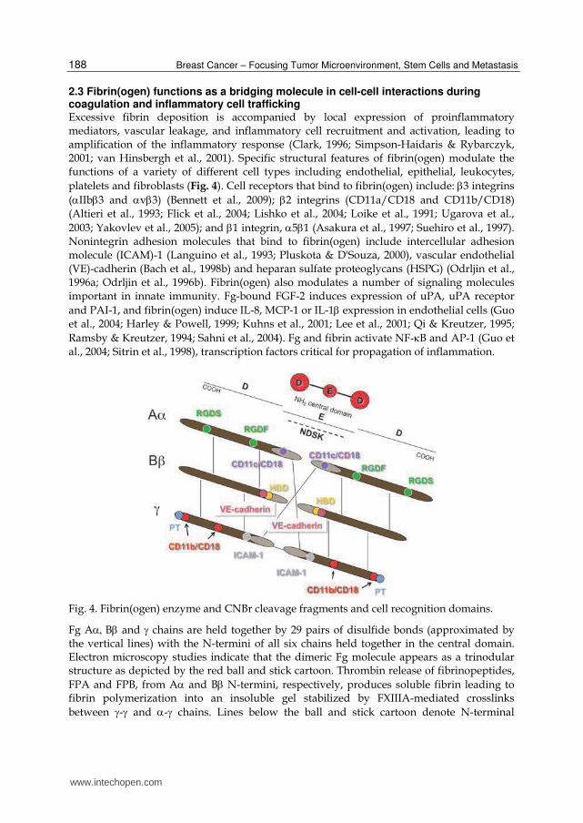
Fig. 4. Fibrin(ogen) enzyme and CNBr cleavage fragments and cell recognition domains.
Fg AB and chains are held together by 29 pairs of disulfide bonds (approximated by the vertical lines) with the N-termini of all six chains held together in the central domain. Electron microscopy studies indicate that the dimeric Fg molecule appears as a trinodular structure as depicted by the red ball and stick cartoon. Thrombin release of fibrinopeptides,
FPA and FPB, from A and B N-termini, respectively, produces soluble fibrin leading to fibrin polymerization into an insoluble gel stabilized by FXIIIA-mediated crosslinks
between - and - chains. Lines below the ball and stick cartoon denote N-terminal
www.intechopen.com

The Role of Fibrin(ogen) in Transendothelial Cell Migration During Breast Cancer Metastasis
189
plasmin cleavage fragment E and C-terminal fragments D. N-terminal disulfide knot (NDSK) (dashed line) is the minimal sequence of the central domain after CNBr cleavage and is structurally similar to plasmin E fragment. Residues on Fg for receptor-cell binding
domains are: CD11c/CD18, A17-19; integrin RGDF, A95-98 and RGDS, A572-575; ICAM-
1,117-133; CD11b/CD18, 190-202, 228-253 and 390-396; platelet (PT) binding, 400-411. The heparin
binding domain (HBD) at 15-42 overlaps the VE-cadherin binding site. The first fibrin
degradation products (FDPs) released by plasmin cleavage are the 15-42 domain and the C-
terminal 2/3rd of the A chain, termed C, which contain several cell binding domains.
2.4 Fibrin(ogen) in the stromal microenvironment in breast cancer The tumor microenvironment is a complex entity composed not only of extracellular matrix (ECM) constituents including: i) growth factors; ii) cytokines and chemokines; iii) proteases; and iv) matrix glycoproteins, glycosaminoglycans and proteoglycans—but also diverse cell populations that influence the behavior of cancer cells including: v) immune cells such as lymphocytes, NK cells, dendritic cells, macrophages and neutrophils; vi) stromal fibroblasts/myofibroblasts, adipocytes and stem cells; and vii) cells of the vasculature including endothelial cells, pericytes and smooth muscle cells (reviewed in (Andre et al., 2010; Anton & Glod, 2009; De Wever et al., 2008; Deryugina & Quigley, 2006; Tlsty & Coussens, 2006; Ulisse et al., 2009)). Although activated inflammatory cells in the tumor microenvironment play important roles in cancer initiation, progression, angiogenesis and metastasis, they are not the most numerous. Cancer-associated fibroblasts, similar to myofibroblasts of healing wounds, are the most abundant stromal cells in the tumor microenvironment (Tlsty & Coussens, 2006), and contribute significantly to chronic inflammation by production of chemokines, cytokines, and pro-angiogenic factors and deposition of matrix constituents that support new blood vessel formation required for tumor growth, cell migration and metastasis (De Wever et al., 2008). Solid tumors need to develop their own blood supply for nutrient delivery and removal of toxic waste. Angiogenesis, the formation of new blood vessels from existing vasculature, requires activation of proteases leading to degradation of the basement membrane, endothelial cell sprouting and pericyte attachment for vessel stabilization. Cancer-associated fibroblasts play an important role in synchronizing these events (De Wever et al., 2008). Furthermore, the topography of the ECM mediates vascular development and regulates the speed at which cells migrate during angiogenesis (Bauer et al., 2009). Vascular endothelial cells play a pivotal role in regulating leukocyte recruitment during inflammation (McGettrick et al., 2007). In most cases, cancers exploit pro-inflammatory mediators and recruited inflammatory cells to benefit their own survival (Lorusso & Ruegg, 2008) (also as reviewed in (Simpson-Haidaris et al., 2010)). Fg and fibrin deposition is found within the stroma of most solid tumors (Simpson-Haidaris
& Rybarczyk, 2001), and elevated levels of plasma Fg and fibrin degradation products
(FDPs) correlate positively with lymph node involvement and metastatic spread of
colorectal, ovarian, lung and breast cancers (Sahni et al., 2009; Varki, 2007). Fibrin deposition
at the tumor-normal host cell interface as well as in the stroma of primary tumors is well
documented, and is thought to protect tumors from infiltrating inflammatory cells by acting
as a barrier thereby preventing inflammatory reactions directed towards the tumor cells
(reviewed in (Simpson-Haidaris & Rybarczyk, 2001)). The presence of D-dimer, a fibrin
degradation product indicative of pathological fibrin formation and dissolution, correlates
www.intechopen.com

Breast Cancer – Focusing Tumor Microenvironment, Stem Cells and Metastasis
190
with poor prognosis in most solid tumors including colon, prostate, lung and breast
(Batschauer et al., 2010; Kilic et al., 2008; Knowlson et al., 2010). However, in some
malignancies, including breast, evidence demonstrating deposition of fibrin within the
primary tumor is lacking (reviewed in (Simpson-Haidaris & Rybarczyk, 2001)). Instead,
abundant Fg deposition occurs in breast tumor stroma in the absence of thrombin
generation (Costantini et al., 1991).
2.5 Cancer cells, including breast, synthesize and secrete fibrinogen The origin of tumor-associated fibrin(ogen) and fibrin(ogen) degradation products has historically been thought to be from exudation of plasma Fg due to the increased vascular permeability and subsequent procoagulant or fibrinolytic activity at the tumor site (Rybarczyk & Simpson-Haidaris, 2000). However, because Fg deposition in the stroma, but not fibrin formation, is considered a hallmark of breast cancer (Costantini et al., 1991), we hypothesized that breast cancer cells were capable of endogenous synthesis and secretion of Fg. We demonstrated that human MCF-7 cells are capable of synthesizing Fg chains,
although assembly of intact Fg is defective due to degradation of the B chain (Rybarczyk & Simpson-Haidaris, 2000). In addition, we have shown that lung, prostate and breast cancer epithelial cells synthesize and secrete Fg that enhances FGF-2-mediated cell proliferation, assembles into the ECM and binds to cancer cell surface receptors (Rybarczyk & Simpson-Haidaris, 2000; Sahni et al., 2008; Simpson-Haidaris, 1997; Simpson-Haidaris & Rybarczyk, 2001). Others have shown Fg production in cervical (Lee et al., 1996) and intestinal (Molmenti et al., 1993) cancer cell lines. Expression array profiling studies confirmed that Fg genes are expressed in breast (Pentecost et al., 2005) and lung carcinomas (Tan et al., 2005) from patients. Thus, Fg synthesized by cancer cells promotes growth of the primary tumor and supports tumor-associated angiogenesis characterized by localized VEGF production and leaky vessels (Dvorak, 2006). The importance of VEGF in promoting tumor vascular permeability, angiogenesis and leakage of plasma Fg into the perivascular space to induce tumor stroma desmoplasia is well known. However, whether tumor-associated fibrin(ogen) contributes to permeability of tumor vessels and breast cancer metastasis is unknown.
2.6 Fibrinogen is an extracellular matrix protein Although Fg is known for its hemostatic role, we showed that Fg, not fibrin, is a component of the insoluble fibrillar ECM of fibroblasts, alveolar epithelial cells, endothelial cells and breast epithelial cells (Guadiz et al., 1997; Pereira et al., 2002; Sahni et al., 2009; Simpson-Haidaris et al., 2010; Simpson-Haidaris & Sahni, 2010). Upon assembly into matrix fibrils, Fg
undergoes conformational changes exposing the cryptic 15-42 epitope in the absence of thrombin cleavage or covalent crosslinking (Guadiz et al., 1997; Simpson-Haidaris & Sahni, 2010). When Fg is pre-established in the ECM of adventitial fibroblasts prior to wounding, increased cell proliferation and migration enhance wound closure (Rybarczyk et al., 2003), which is dependent on de novo protein synthesis (Pereira & Simpson-Haidaris, 2001) but independent of added growth factors, PDGF and FGF-2 (Rybarczyk et al., 2003). However, assembly of Fg into mature matrix fibrils of breast epithelial cells appears to correlate negatively with the increasing invasive potential of the cell (Fig. 5). We also determined whether the cryptic HBD in soluble Fg (Odrljin et al., 1996b) was accessible in matrix Fg
using a specific MoAb (T2G1) (Kudryk et al., 1984). Whereas the T2G1 epitope (15-21) within
15-42 is not accessible for antibody binding in soluble Fg or Fg immobilized to a surface, the
www.intechopen.com

The Role of Fibrin(ogen) in Transendothelial Cell Migration During Breast Cancer Metastasis
191
results indicated that 15-21 is exposed on Fg assembled into matrix fibrils (Guadiz et al., 1997; Rybarczyk et al., 2003). Together these data suggest that matrix Fg possesses “fibrin-like” properties in the absence of fibrin polymerization and that Fg deposition rapidly changes the topology of the ECM to provide a surface for cell migration and matrix
remodeling during wound repair. However, the mechanisms by which 15-42 modulates cell-cell or cell-matrix adhesion are not well understood.
Fig. 5. Plasma fibrinogen assembles into mature matrix fibrils of nonmalinant cells (HFF and HBL-100) but poorly assembles in the matrix of malignant breast cancer cells (MCF-7 and MDA-MB-231). Primary human fibroblasts (HFF), a nonmalignant human breast cancer cell line (HBL-100) and two human breast cancer cell lines (MCF-7 and MDA-MB-231) were
grown on gelatin-coated glass coverslips and treated with Fg conjugated to Oregon Green
(30 g/ml) for 24 hr. The cells were washed, fixed, stained with anti-fibronectin (FN) polyclonal antibodies followed by rhodamine-goat anti-rabbit secondary antibodies, and visualized by epifluorescence microscopy. Green fluorescence is Fg-specific and red fluorescence denotes FN staining. Colocalization of Fg and FN results in yellow fluorescence. The loss of FN in the more invasive cell lines (MCF-7 and MDA-MB-231) is likely an explanation for purified plasma Fg binding to the surface of cells but failure to assembly into mature matrix fibrils, as we have shown that assembly of Fg into an elaborate fibrillar ECM depends on the assembly of FN fibrils as well (Pereira et al., 2002).
3. Role of Fibrin(ogen) in breast cancer metastasis
3.1 Importance of Fg peptide 15-42 in Fg-endothelial cell interactions
Fibrin(ogen) 15-42 sequences support a diverse array of biological functions mediated by fibrin(ogen). Although the primary structure of fibrinopeptide B (FPB) is poorly conserved
across species, the fibrin 15-42 domain is highly conserved, implying evolutionary
conservation of function (Courtney et al., 1994). The 15-42 region constitutes a cryptic domain in soluble Fg that is exposed in fibrin after thrombin cleavage (Odrljin et al., 1996b).
Both the HBD and overlapping binding site for VE-cadherin are localized to 15-42. VE-
www.intechopen.com

Breast Cancer – Focusing Tumor Microenvironment, Stem Cells and Metastasis
192
cadherin mediates homophilic cell-cell adhesion critical for the maintenance of barrier integrity of the endothelium. Disruption of VE-cadherin-mediated endothelial barrier function leads to altered vascular permeability found in a number of diseases including ischemia-reperfusion (IR) injury, inflammation, angiogenesis, and cancer growth and
metastasis (discussed in (Sahni et al., 2009)). Exposure of 15-42 and binding by VE-cadherin is also required for endothelial capillary tube formation in fibrin gels (Bach et al., 1998a; Chalupowicz et al., 1995); portions of the third extracellular domain (EC3) of VE-cadherin
constitute a fibrin 15-42 receptor (Bach et al., 1998b; Yakovlev & Medved, 2009). Newly
exposed chain residues, 15-GHRP-18, play a critical role in fibrin monomer aggregation during polymerization and clot formation during secondary hemostasis (Mosesson, 2005).
Furthermore, exposure of the 15-42 domain mediates heparin-dependent fibrin binding to endothelial cell surfaces (Odrljin et al., 1996a); promotes endothelial cell adhesion and spreading (Bunce et al., 1992); promotes the release of endothelial cell-specific markers of endothelial activation (Ribes et al., 1989); and stimulates proliferation of endothelial cells, fibroblasts and cancer cells (Rybarczyk et al., 2003; Sahni et al., 2008; Sporn et al., 1995).
3.2 Fibrin 15-42 protects the myocardium from Ischemic-Reperfusion (IR) injury
A synthetic peptide of fibrin residues 15-42 has been implicated as a potential therapeutic agent to reduce tissue damage and scarring after a heart attack (Hirschfield & Pepys, 2003; Petzelbauer et al., 2005b; Roesner et al., 2007; Zacharowski et al., 2006; Zacharowski et al.,
2007). Peptide 15-42 works by inhibiting leukocyte migration across the endothelium into
heart tissue, which prevents excessive inflammation and tissue damage. Peptide 15-42-mediated reduction of tissue injury depends on its ability to bind to VE-cadherin. Peptide
15-42 competes with FDP (e.g., the plasmin E domain of fibrin as depicted in Fig. 4) for binding to VE-cadherin to prevent transendothelial cell migration (TEM) of leukocytes during myocardial IR injury (Petzelbauer et al., 2005b; Roesner et al., 2007; Zacharowski et al., 2006; Zacharowski et al., 2007). These published reports demonstrate the physiologic
efficacy of fibrin 15-42 for treating IR injury. However, the molecular mechanisms induced by
fibrin(ogen) 15-42 binding to VE-cadherin to mediate enhanced paracellular permeability and whether
fibrinogen-induced cancer metastasis involves binding interactions with fibrin(ogen) 15-42 have not been previously studied.
3.3 Fibrin(ogen) 15-42 induces endothelial barrier permeability via VE-cadherin binding
interactions
In a recent report (Sahni et al., 2009), we sought to determine whether fibrin(ogen) 15-42
binding to VE-cadherin induced endothelial cell permeability, and whether fibrinogen-induced cancer metastasis involves binding interactions between VE-cadherin and
fibrin(ogen) 15-42. Using transwell insert culture systems, we showed that Fg 15-42 and VE-cadherin binding interactions promote endothelial cell barrier permeability (Sahni et al.,
2009) (Fig. 6). Peptides containing or missing residues 15-17 critical for 15-42 binding to VE-
cadherin (Gorlatov & Medved, 2002) and neutralizing antibodies that bind to Fg 15-21
(T2G1) and VE-cadherin (BV9) (Fig. 7A) were used to induce or inhibit permeability. Fg induced dose-dependent permeability of human umbilical vein endothelial cells (HUVEC) and microvascular endothelial cells (HMEC-1) (Fig. 6), but not epithelial cell barriers (as shown in Fig. 1 in ref (Sahni et al., 2009)), which could be inhibited by neutralizing
antibodies against 15-21 (T2G1) and VE-cadherin (BV9) and synthetic peptides (not shown).
www.intechopen.com

The Role of Fibrin(ogen) in Transendothelial Cell Migration During Breast Cancer Metastasis
193
However, the neutralizing antibodies (T2G1 and BV9) did not completely inhibit Fg-induced permeability (Fig. 7B), suggesting that additional cell recognition domains on Fg participate in fibrin(ogen)-induced vascular permeability.
Fig. 6. Fg-induced EC permeability involves Fg 15-42 and VE-cadherin. Cells were grown to confluency on Millicell™ 24-well cell culture inserts. Panel 6A, HUVEC were left untreated (control) or treated for 15 min with increasing concentrations of Fg or VEGF as indicated. Panel 6B, HUVEC were treated with 30 nM of Fg plus 1 mg/ml FITC-Dextran for the times indicated. The FITC-Dextran flux to the bottom chamber was measured by fluorometry and the data presented as the mean relative FITC-Dextran Flux ± SEM. Data points were derived from 3 or more independent experiments with the total number of replicates per condition ranging from 6-13. (Reprinted from (Sahni et al., 2009) with permission). P-values can be found in ref (Sahni et al., 2009).
Fig. 7. Fg-induced EC permeability involves Fg 15-42 sequences and VE-cadherin. Panel 7A,
schematics of the aminoterminus of the fibrin(ogen) B chain and the domain structure of VE-cadherin are depicted. The arrow denotes the thrombin cleavage site for release of FPB.
The 18C6 epitope maps to FPB, the T2G1 epitope maps to 15-21 and the VE-cadherin binding
site on fibrin maps to 15-42. The epitope of the VE-cadherin-specific monoclonal antibody
BV9 maps to the third and fourth extracellular domains (EC3-EC4). The fibrin 15-42 binding site on VE cadherin maps to EC3 near the EC3-EC4 junction. TM, transmembrane domain. Panel 7B, all monoclonal antibodies used are IgG1 isotype murine antibodies and
www.intechopen.com

Breast Cancer – Focusing Tumor Microenvironment, Stem Cells and Metastasis
194
nonimmune IgG1 was used for the control. Monoclonal antibodies were used at 3 nM in the absence of Fg, or with 0.3 nM or 30 nM Fg for 45 min. The data were plotted as the mean ± SEM of relative FITC-Dextran Flux and were obtained from three independent experiments with a total sample size of 6-9 per condition. (Reprinted from (Sahni et al., 2009) with permission). P-values can be found in ref (Sahni et al., 2009).
3.4 VE-cadherin binding domain of Fg (15-42) enhances transendothelial migration of
malignant breast epithelial cells
Because plasma Fg promotes metastasis of some types of cancer and Fg 15-42 sequences
promote endothelial cell permeability, we hypothesized Fg 15-42 sequences would play a role in promoting TEM of breast cancer cells. To test this hypothesis, breast cancer cells were labeled with a fluorescence cell-tracking dye (DiI) before they were mixed with increasing concentrations Fg. Breast cancer cells and Fg were allowed to pre-incubate for 15 minutes prior to addition to the upper chamber of a barrier monolayer of endothelial cells. After 45 minutes incubation, the relative number of breast cancer cells migrating to the underside of the transwell insert membrane were quantified by relative fluorescence and
Fig. 8. Fg enhances TEM of malignant breast epithelial cells (Panel A), induces gap formation between adjacent endothelial cells (Panel B, asterisks), promotes intracellular relocalization (Panel B, arrowheads) of VE-cadherin at membrane cell-cell junctions (Panel B, Control, arrow), assembles into ECM (Panel C, arrowhead), and shows punctate, cell surface receptor-like binding between adjacent endothelial cells (Panel C, arrows). Cells in Panels A and B were treated as described in Section 3.3. In Panel C, endothelial cells were treated for 24 hours with purified plasma Fg conjugated to Oregon Green. Cells were fixed, permeabilized and stained with anti-FGF-2 (red fluoresence). After staining, the coverslip was mounted upside down on a microscope slide so that the basolateral aspect (bottom of cells) and the subendothelial ECM appear as the “top” of the cells. Matrix Fg and receptor bound Fg are shown in green fluorescence. Cover Figure ref (Sahni et al., 2009).
www.intechopen.com

The Role of Fibrin(ogen) in Transendothelial Cell Migration During Breast Cancer Metastasis
195
visualized by microscopy. VEGF was used as a positive control to induce endothelial cell permeability and TEM of breast cancer cells. The results indicated that TEM of both MCF-7 and MDA-MB-231 cells was increased in a Fg-concentration-dependent manner (see Fig. 3a of ref (Sahni et al., 2009)) and as visualized by immunofluorescence microscopy showing MDA-MB-231 cells adhered to the bottom side of the transwell filter (Fig. 8A).
To determine whether VE-cadherin and/or Fg 15-42 were involved in Fg-enhanced TEM of MDA-MB-231 cells, the assay was repeated in the presence of the neutralizing and control antibodies (as shown in Fig. 3c of ref (Sahni et al., 2009)). To determine whether Fg promoted gap formation between cells, confluent HUVEC were treated with 150 or 480 nM
Fg or 100 Units/ml TNF-, a known inducer of endothelial permeability and gap formation, for 30 minutes then cells were fixed, permeabilized and immunostained with an anti-VE-cadherin. Fg treatment induced gap formation between adjacent endothelial cells, and such treatment promoted the subcellular relocalization of VE-cadherin from the cell periphery as
in control cells into the cytoplasm in Fg- and TNF--treated cells (Fig. 8B). Indirect evidence for Fg binding at endothelial cell-cell junctions was obtained by fluorescence microscopy. The data reveal that Fg binds to endothelial cell-cell junctions in a punctate pattern, consistent with cell surface receptor binding to the cell-cell adhesion receptor, VE-cadherin (Fig. 8C, arrows). Fg also assembles as part of the fibrillar subendothelial ECM (Fig. 8C, arrowhead). Taken together, the data in Fig. 6-8 demonstrate that the VE-cadherin binding
domain defined by residues 15-42 on the -chain of human Fg induces permeability of endothelial but not epithelial cell barriers and enhances TEM of malignant breast cancer cells by a VE-cadherin-dependent mechanism. In contrast, the basal level of TEM of nonmalignant breast epithelial cells was not enhanced by Fg treatment (Sahni et al., 2009).
3.5 Fibrinogen potentiates endothelial cell permeability at low doses of VEGF Both FGF-2 and VEGF bind to fibrin(ogen) at distinct sites with high affinity (Sahni & Francis, 2000; Sahni et al., 1998). Fg bound-FGF-2 potentiates endothelial cell proliferation over FGF-2 alone (Sahni et al., 2003; Sahni & Francis, 2004; Sahni et al., 2006; Sahni et al., 1999). Although Fg-bound VEGF remains active, it does not potentiate endothelial cell proliferation over VEGF alone (Sahni & Francis, 2000). Because Fg induces endothelial cell permeability through VE-cadherin binding interactions (Sahni et al., 2009) and VEGF binds to Fg (Sahni & Francis, 2000), we tested the hypothesis that Fg would potentiate VEGF-induced EC permeability (Fig. 9).
Fig. 9. Fg enhances permeability induced by low concentrations of VEGF.
www.intechopen.com

Breast Cancer – Focusing Tumor Microenvironment, Stem Cells and Metastasis
196
The data indicate that 10 g/ml (30 nM) Fg enhanced the flux of FITC-dextran to the bottom chamber of the transwell plate at low doses of VEGF (0.05 and 0.1 ng/ml); however, the additive effect on induction of endothelial cell permeability was lost at 0.5 ng/ml and higher concentrations of VEGF (Fig. 9). Fg-enhancement of VEGF-induced permeability is rapid and saturated within 5 min, whereas 5 ng/ml of VEGF is required to induce a similar amount of FITC-dextran flux as 30 nM Fg + 0.05 ng/ml, i.e., 100-fold less VEGF. Studies by others suggest that low-dose VEGF mediates inflammation to promote cell survival of vascular and nonvascular cells such as those of the CNS, prior to induction of angiogenesis (Abumiya et al., 2005; Croll et al., 2004). Furthermore, VEGF colocalizes with exuded Fg at sites of edema in renal cell carcinoma (Verheul et al., 2010). Together with the aforementioned published data, our results suggest that Fg may regulate vascular permeability induced by low doses of VEGF without inducing EC proliferation—such a response would be conducive to fibrinogen induction of breast cancer cell TEM.
4. Summary, therapeutic strategies and future research to elucidate fibrin(ogen)-mediated mechanisms of breast cancer metastasis
4.1 Summary and therapeutic strategy using free peptide 15-42 to inhibit breast cancer
metastasis as depicted in Fig. 10, Steps 1-11 Regardless of the subtype of breast cancer, once the primary tumor becomes established (Step 1), it needs to develop its own blood supply for nutrient delivery and removal of toxic waste (Step 2). Breast cancer cells produce VEGF, which initiates permeability of nearby blood vessels allowing plasma Fg to leak into the tumor stroma promoting desmoplasia and deposition of a provisional fibrin(ogen) matrix in the tumor microenvironment (Step 2). Alternatively, endogenous synthesis of Fg by breast cancer cells could induce cancer progression. Thus, the innate immune response is activated to defend the host against this neoplastic insult. Release of IL-6 systemically leads to increased production of plasma Fg
and fibrin formation resulting in exposure of 15-42 and binding to VE-cadherin, a step critical for angiogenesis (Bach et al., 1998b; Martinez et al., 2001). Furthermore, VEGF binds to Fg and fibrin with high affinity (Sahni & Francis, 2000), which may be necessary for Fg to enhance VEGF-mediated endothelial cell permeability without potentiating endothelial cell proliferation. In contrast, VE-cadherin and VEGF receptor-2 form a signaling complex to promote endothelial cell proliferation (Carmeliet et al., 1999; Dejana, 2004; Esser et al., 1998). Fibrin(ogen) potentiates FGF-2- but not VEGF-induced proliferation of endothelial cells, angiogenesis and cancer cell growth (Rybarczyk & Simpson-Haidaris, 2000; Sahni & Francis, 2000; Sahni et al., 2006; Sahni et al., 2008; Sahni et al., 1999; Simpson-Haidaris, 1997; Simpson-Haidaris & Rybarczyk, 2001). Furthermore, fibrin(ogen) enhances cell migration and cancer invasion through tumor stroma, and TEM, i.e., intravasation of breast cancer cells into the blood stream (Step 3) (Roche et al., 2003; Rybarczyk et al., 2003; Sahni et al., 2009). Fg and fibrin can bridge between cells of the same or different kinds (Kloczewiak et al., 1983; Languino et al., 1995; Languino et al., 1993; Saito et al., 2002; Sriramarao et al., 1996) and form aggregates or tumor emboli coated with fibrin(ogen) (Step 4). Because the host immune system does not recognize fibrin(ogen)-coated tumor emboli (Palumbo et al., 2005), immune-mediated destruction of tumor cells does not occur and these tumor emboli travel through the circulation to sites favorable for metastatic growth (Steps 5 & 6) such as lung. To establish metastatic growth, tumor emboli need to leave the circulation and enter lung tissue (Steps 7 and 8) where they find a receptive niche (Step 9) to begin the process again. Tumor
www.intechopen.com

The Role of Fibrin(ogen) in Transendothelial Cell Migration During Breast Cancer Metastasis
197
cell proliferation and angiogenesis (Step 10) in lung results in metastatic disease (Step 11).
We hypothesize that free peptide 15-42 will bind to VE-cadherin between endothelial cells to
block endothelial cell binding to 15-42 on intact fibrin(ogen) found in the tumor stroma or tumor vessels, thereby inhibiting tumor-associated angiogenesis (Step 2), intravasation (Step 3), extravasation (Step 8), and angiogenesis at metastatic tumor sites (Step 10) (as denoted by
the lightening bolts at these steps in Fig. 10).
Fig. 10. Schematic summarizing role of fibrin(ogen) 15-42 in breast cancer metastasis and
hypothesis development for employing free peptide 15-42 as a therapeutic strategy to treat metastatic breast cancers.
Successful demonstration of peptide 15-42 as an inhibitor of breast cancer metastasis and
tumor-associated inflammation and angiogenesis in vivo would significantly impact breast
cancer treatment in a timely manner. Peptide 15-42, an endogenous fragment of fibrin, is
already shown to be well tolerated in humans and effective in reducing damage to heart
muscle after a heart attack in preclinical models of IR injury. However, until now, no one
has proposed the use of peptide 15-42 as an inhibitor of breast cancer metastasis. A
precedent and pipeline for production of viable therapeutics based on peptide 15-42 exists
for treatment of damaged heart tissue, and Phase I and Phase II clinical trials are ongoing to
test the safety and efficacy, respectively, of free 15-42 peptide for IR injury (Hallen et al.,
2010; Petzelbauer et al., 2005a; Petzelbauer et al., 2005b; Roesner et al., 2007; Roesner et al.,
2009; Wiedemann et al., 2010; Zacharowski et al., 2006). Therefore, the timeline for
www.intechopen.com

Breast Cancer – Focusing Tumor Microenvironment, Stem Cells and Metastasis
198
successful translational to a therapeutic agent to treat metastatic disease in breast cancer
patients with different subtypes of the disease would be significantly shortened. Moreover,
even if the primary tumor develops its own blood supply before adjuvant therapy with
peptide 15-42 is begun, we predict that free peptide 15-42 will prevent subsequent steps
required for metastatic spread and growth of breast cancers. Another advantage to this
therapeutic strategy is that peptide 15-42 functions outside the cell, precluding the need to
deliver the peptide inside cells. Identifying molecular targets for therapeutic intervention of
breast cancer metastasis, recruitment of inflammatory cells and angiogenesis will increase
long-term disease-free survival and improve the quality of life for breast cancer patients.
4.2 Putative mechanisms whereby nonmalignant breast epithelial cells switch to a metastatic breast cancer cell phenotype responsive to fibrinogen induced TEM A class of molecules found in the ECM, inside cells and attached to cell surfaces, called heparan sulfate proteoglycans (HSPG), contribute to breast cancer progression by promoting cancer cell proliferation, TEM, and tumor-associated angiogenesis (Koo et al., 2008). The ability to affect any one of these functions would help to reduce breast cancer metastasis; however, if all three of the functions could be targeted with one therapeutic approach, the morbidity and mortality due to metastatic breast cancer could be significantly reduced. Heparin is widely used as an anticoagulant, but it also inhibits HSPG-dependent mechanisms of cancer metastasis (Levy-Adam et al., 2005). However, anti-metastatic heparins that also inhibit blood coagulation are, therefore, not good candidates for widespread use to treat metastatic breast cancer due to bleeding complications. Thus, another molecular target to inhibit the prometastatic effects of HSPG but not inhibit coagulation is greatly needed. Spontaneous blood-borne and lymphatic metastasis of tumor emboli requires fibrin(ogen) (Palumbo et al., 2002). In addition to binding to VE-cadherin
(Yakovlev et al., 2003), Fg 15-42 also binds to heparin and HSPG on endothelial cells with high affinity (Odrljin et al., 1996a; Odrljin et al., 1996b); however, a role for HSPG in Fg-mediated breast cancer metastasis has not been studied. Fg binding to heparin and HSPG
involves residues 15-42, and 15-42-dependent fibrin binding to EC surfaces can be inhibited
with heparin and heparan sulfate but not with chondroitin sulfate, indicating that Fg-15-42 represents a HBD (Odrljin et al., 1996a; Odrljin et al., 1996b). The Fg HBD was later mapped
to residues 15-57, which includes the 15-42 VE-cadherin binding domain (Yakovlev et al., 2003; Yakovlev & Medved, 2009). In our recent publication (Sahni et al., 2009), we unexpectedly discovered that Fg enhanced TEM of only malignant breast cancer cells (MCF-7 and MDA-MB-231) but not nonmalignant breast epithelial cells (MCF-10A), suggesting inherent differences in the ability of cancer vs. normal breast epithelial cells to interact with fibrin(ogen). Because TEM of nonmalignant epithelial cells (MCF-10A) could not be enhanced in the presence of Fg (Sahni et al., 2009), we hypothesize that loss of HSPG from the surface of premalignant breast epithelial cells serves as a molecular switch to induce a highly aggressive, metastatic breast cancer phenotype (Fig. 11A). We plan to investigate this hypothesis in future studies. Another mechanism to regulate Fg-enhanced TEM of malignant breast cancer cells is a gain
in function of cancer-associated Mucin-1 (MUC1), which is a membrane-associated mucin
expressed at low levels on the apical surface of normal polarized epithelial cells. MUC1 is a
tumor-associated glycoprotein aberrantly expressed in >90% of breast cancers (Singh &
Bandyopadhyay, 2007), promotes cancer cell proliferation and metastasis, and is associated
www.intechopen.com

The Role of Fibrin(ogen) in Transendothelial Cell Migration During Breast Cancer Metastasis
199
with poor survival (Hattrup & Gendler, 2006; Yuan et al., 2007). MUC1 is upregulated and
hypoglycosylated in breast cancers. The polarized expression of MUC1 is lost on cancer cells
such that it is expressed on the entire cell surface (Kondo et al., 1998; Moase et al., 2001;
Wesseling et al., 1996; Yang et al., 2007). The MUC1 extracellular domain protrudes ~200 nm
above the cell surface, whereas most cell surface receptors are ~35 nm long (Wesseling et al.,
1996). When MUC1 is interspersed between adhesion molecules, it nonspecifically reduces
cell-cell and cell-ECM interactions in vitro and in vivo, likely by steric hindrance caused by
the extreme length and high density of the MUC1 at the cell surface (Wesseling et al., 1996)
(Fig. 11B). MUC1 expression is found on MCF-7, MDA-MB-231, as well as other types of
breast cancer cells, particularly on those isolated from patients with a highly aggressive
subtype called inflammatory breast cancer (Alpaugh et al., 2002; Schroeder et al., 2003;
Walsh et al., 1999); elevated expression of MUC1 contributes to lymphovascular tumor
invasion of inflammatory breast cancer cells (Alpaugh et al., 2002).
Fig. 11. Putative mechanisms whereby nonmalignant breast epithelial cells switch to a metastatic breast cancer cell phenotype responsive to fibrinogen-induced TEM. Panel A, schematic depicting loss of function due to release of cell-surface HSPG. Panel B, schematic depicting gain of function by overexpression of MUC1 leading to loss of polarity and cell-cell adhesion in breast epithelial cells.
We predict that Fg could bind to normal breast cell surface HSPG through Fg 15-42, thus
preventing Fg 15-42 binding to VE-cadherin extracellular domain 3 (EC3) and inhibition of TEM. Enhanced heparanase expression and enzymatic digestion of HSPG in human tumors correlates with metastatic potential, tumor vascularity, and reduced postoperative survival of cancer patients (Vlodavsky et al., 2008). Heparanase-induced loss of breast epithelial cell surface HSPG during conversion of non- or pre-malignant to malignant breast cancers
would allow Fg 15-42 binding to VE-cadherin at cell-cell junctions to induce EC permeability. Fg would also bind to breast cancer cell integrins via binding sites on Fg C-terminal domains (see Fig. 4) then movement of VE-cadherin (induced by Fg binding to VE-cadherin as shown in Fig. 8B) in the endothelial cell membrane would induce paracellular transfer of Fg-bound breast cancer cells across the endothelial cell barrier to promote cancer metastasis. A precedent for this mechanism is already established; Fg binding to a counter adhesion molecule facilitates neutrophil TEM 20- to 30-fold (Languino et al., 1995). Overexpression of MUC1 could block accessibility of HSPG on breast cancer cells, which
www.intechopen.com

Breast Cancer – Focusing Tumor Microenvironment, Stem Cells and Metastasis
200
would also prevent Fg 15-42–HSPG binding interactions leaving Fg 15-42 available for binding to VE-cadherin. Alternatively, loss of cell surface HSPG and elevated expression of MUC1 may contribute to Fg-enhanced TEM of malignant compared to nonmalignant breast epithelial cells. These possibilities will be addressed by future experiments.
5. Acknowledgements
We thank present and past members of the Simpson-Haidaris lab and our collaborators for all their contributions to elucidate the role of fibrin(ogen) in wound repair, endothelial cell biology and cancer progression. Our work has been supported by numerous grants from the National Institutes of Health NHLBI and NIAID, the Breast Cancer Research Program sponsored by the Department of Defense, the Komen Race for the Cure Foundation, and the American Heart Association.
6. References
Abumiya, T., Yokota, C., Kuge, Y. & Minematsu, K. (2005). Aggravation of hemorrhagic transformation by early intraarterial infusion of low-dose vascular endothelial growth factor after transient focal cerebral ischemia in rats. Brain Res 1049(1):95-103.
Alpaugh, M.L., Tomlinson, J.S., Kasraeian, S. & Barsky, S.H. (2002). Cooperative role of E-cadherin and sialyl-Lewis X/A-deficient MUC1 in the passive dissemination of tumor emboli in inflammatory breast carcinoma. Oncogene 21(22):3631-43.
Altieri, D.C., Plescia, J. & Plow, E.F. (1993). The structural motif glycine 190-valine 202 of the fibrinogen gamma chain interacts with CD11b/CD18 integrin (alpha M beta 2, Mac-1) and promotes leukocyte adhesion. J Biol Chem 268(3):1847-53.
Andre, F., Berrada, N. & Desmedt, C. (2010). Implication of tumor microenvironment in the resistance to chemotherapy in breast cancer patients. Curr Opin Oncol 22(6):547-51.
Anton, K. & Glod, J. (2009). Targeting the tumor stroma in cancer therapy. Curr Pharm Biotechnol 10(2):185-91.
Asakura, S., Niwa, K., Tomozawa, T., Jin, Y., Madoiwa, S., Sakata, Y., Sakai, T., Funayama, H., Soe, G., Forgerty, F., Hirata, H. & Matsuda, M. (1997). Fibroblasts spread on immobilized fibrin monomer by mobilizing a beta1-class integrin, together with a vitronectin receptor alphavbeta3 on their surface. J Biol Chem 272(13):8824-9.
Bach, T.L., Barsigian, C., Chalupowicz, D.G., Busler, D., Yaen, C.H., Grant, D.S. & Martinez, J. (1998a). VE-Cadherin mediates endothelial cell capillary tube formation in fibrin and collagen gels. Exp Cell Res 238(2):324-34.
Bach, T.L., Barsigian, C., Yaen, C.H. & Martinez, J. (1998b). Endothelial cell VE-cadherin functions as a receptor for the β15-42 sequence of fibrin. J Biol Chem 273(46):30719-28.
Balkwill, F. & Mantovani, A. (2001). Inflammation and cancer: back to Virchow? Lancet 357(9255):539-45.
Barton, B.E. (2001). IL-6-like cytokines and cancer cachexia: consequences of chronic inflammation. Immunol Res 23(1):41-58.
Batschauer, A.P., Figueiredo, C.P., Bueno, E.C., Ribeiro, M.A., Dusse, L.M., Fernandes, A.P., Gomes, K.B. & Carvalho, M.G. (2010). D-dimer as a possible prognostic marker of operable hormone receptor-negative breast cancer. Ann Oncol 21(6):1267-72.
www.intechopen.com

The Role of Fibrin(ogen) in Transendothelial Cell Migration During Breast Cancer Metastasis
201
Bauer, A.L., Jackson, T.L. & Jiang, Y. (2009). Topography of extracellular matrix mediates vascular morphogenesis and migration speeds in angiogenesis. PLoS Comput Biol 5(7):e1000445.
Baumann, H. & Gauldie, J. (1994). The acute phase response. Immunol Today 15(2):74-80. Bennett, J.S., Berger, B.W. & Billings, P.C. (2009). The structure and function of platelet
integrins. J Thromb Haemost 7 Suppl 1200-5. Bunce, L.A., Sporn, L.A. & Francis, C.W. (1992). Endothelial cell spreading on fibrin requires
fibrinopeptide B cleavage and amino acid residues 15-42 of the β chain. J Clin Invest 89(3):842-50.
Caine, G.J., Lip, G.Y., Stonelake, P.S., Ryan, P. & Blann, A.D. (2004). Platelet activation, coagulation and angiogenesis in breast and prostate carcinoma. Thromb Haemost 92(1):185-90.
Carmeliet, P., Lampugnani, M.G., Moons, L., Breviario, F., Compernolle, V., Bono, F., Balconi, G., Spagnuolo, R., Oostuyse, B., Dewerchin, M., Zanetti, A., Angellilo, A., Mattot, V., Nuyens, D., Lutgens, E., Clotman, F., de Ruiter, M.C., Gittenberger-de Groot, A., Poelmann, R., Lupu, F., Herbert, J.M., Collen, D. & Dejana, E. (1999). Targeted deficiency or cytosolic truncation of the VE-cadherin gene in mice impairs VEGF-mediated endothelial survival and angiogenesis. Cell 98(2):147-57.
Chalupowicz, D.G., Chowdhury, Z.A., Bach, T.L., Barsigian, C. & Martinez, J. (1995). Fibrin II induces endothelial cell capillary tube formation. J Cell Biol 130(1):207-15.
Clark, R.A.F. The molecular and cellular biology of wound repair. New York: Plenum Press 1996.
Cohen, T., Nahari, D., Cerem, L.W., Neufeld, G. & Levi, B.Z. (1996). Interleukin 6 induces the expression of vascular endothelial growth factor. J Biol Chem 271(2):736-41.
Costantini, V., Zacharski, L.R., Memoli, V.A., Kisiel, W., Kudryk, B.J. & Rousseau, S.M. (1991). Fibrinogen deposition without thrombin generation in primary human breast cancer tissue. Cancer Res 51(1):349-53.
Courtney, M.A., Bunce, L.A., Neroni, L.A. & Simpson-Haidaris, P.J. (1994). Cloning of the complete coding sequence of rat fibrinogen Bβ chain cDNA: interspecies conservation of fibrin β15-42 primary structure. Blood Coagul Fibrinolysis 5(4):487-96.
Coussens, L.M. & Werb, Z. (2002). Inflammation and cancer. Nature 420(6917):860-7. Croll, S.D., Ransohoff, R.M., Cai, N., Zhang, Q., Martin, F.J., Wei, T., Kasselman, L.J.,
Kintner, J., Murphy, A.J., Yancopoulos, G.D. & Wiegand, S.J. (2004). VEGF-mediated inflammation precedes angiogenesis in adult brain. Exp Neurol 187(2):388-402.
De Wever, O., Demetter, P., Mareel, M. & Bracke, M. (2008). Stromal myofibroblasts are drivers of invasive cancer growth. Int J Cancer 123(10):2229-38.
Dejana, E. (2004). Endothelial cell-cell junctions: happy together. Nat Rev Mol Cell Biol 5(4):261-70.
Deryugina, E.I. & Quigley, J.P. (2006). Matrix metalloproteinases and tumor metastasis. Cancer Metastasis Rev 25(1):9-34.
Dirix, L.Y., Salgado, R., Weytjens, R., Colpaert, C., Benoy, I., Huget, P., van Dam, P., Prove, A., Lemmens, J. & Vermeulen, P. (2002). Plasma fibrin D-dimer levels correlate with tumour volume, progression rate and survival in patients with metastatic breast cancer. Br J Cancer 86(3):389-95.
Duan, H.O. & Simpson-Haidaris, P.J. (2003). Functional analysis of interleukin 6 response elements (IL-6REs) on the human gamma-fibrinogen promoter: binding of hepatic
www.intechopen.com

Breast Cancer – Focusing Tumor Microenvironment, Stem Cells and Metastasis
202
Stat3 correlates negatively with transactivation potential of type II IL-6REs. J Biol Chem 278(42):41270-81.
Dvorak, H.F. (1986). Tumors: wounds that do not heal. Similarities between tumor stroma generation and wound healing. N Engl J Med 315(26):1650-9.
Dvorak, H.F. (2006). Discovery of vascular permeability factor (VPF). Exp Cell Res Esser, S., Lampugnani, M.G., Corada, M., Dejana, E. & Risau, W. (1998). Vascular endothelial
growth factor induces VE-cadherin tyrosine phosphorylation in endothelial cells. J Cell Sci 111(Pt 13):1853-65.
Flick, M.J., Du, X., Witte, D.P., Jirouskova, M., Soloviev, D.A., Busuttil, S.J., Plow, E.F. & Degen, J.L. (2004). Leukocyte engagement of fibrin(ogen) via the integrin receptor aMb2/Mac-1 is critical for host inflammatory response in vivo. J Clin Invest 113(11):1596-606.
Gao, S.P., Mark, K.G., Leslie, K., Pao, W., Motoi, N., Gerald, W.L., Travis, W.D., Bornmann, W., Veach, D., Clarkson, B. & Bromberg, J.F. (2007). Mutations in the EGFR kinase domain mediate STAT3 activation via IL-6 production in human lung adenocarcinomas. J Clin Invest 117(12):3846-56.
Gorlatov, S. & Medved, L. (2002). Interaction of fibrin(ogen) with the endothelial cell receptor VE-cadherin: mapping of the receptor-binding site in the NH2-terminal portions of the fibrin β chains. Biochemistry 41(12):4107-16.
Guadiz, G., Sporn, L.A. & Simpson-Haidaris, P.J. (1997). Thrombin cleavage-independent deposition of fibrinogen in extracellular matrices. Blood 90(7):2644-53.
Guo, M., Sahni, S.K., Sahni, A. & Francis, C.W. (2004). Fibrinogen regulates the expression of inflammatory chemokines through NF-κB activation of endothelial cells. Thromb Haemost 92(4):858-66.
Hallen, J., Petzelbauer, P., Schwitter, J., Geudelin, B., Buser, P. & Atar, D. (2010). Impact of time to therapy and presence of collaterals on the efficacy of FX06 in acute ST elevation myocardial infarction: a substudy of the F.I.R.E., the Efficacy of FX06 in the prevention of myocardial reperfusion injury trial. EuroIntervention 5(8):946-52.
Harley, S.L. & Powell, J.T. (1999). Fibrinogen up-regulates the expression of monocyte chemoattractant protein 1 in human saphenous vein endothelial cells. Biochem J 341 (Pt 3):739-44.
Hartman, Z.C., Yang, X.Y., Glass, O., Lei, G., Osada, T., Dave, S.S., Morse, M.A., Clay, T.M. & Lyerly, H.K. (2011). HER2 overexpression elicits a proinflammatory IL-6 autocrine signaling loop that is critical for tumorigenesis. Cancer Res 71(13):4380-91.
Hattrup, C.L. & Gendler, S.J. (2006). MUC1 alters oncogenic events and transcription in human breast cancer cells. Breast Cancer Res 8(4):R37.
Hirschfield, G.M. & Pepys, M.B. (2003). C-reactive protein and cardiovascular disease: new insights from an old molecule. QJM 96(11):793-807.
Jones, J.M., McGonigle, N.C., McAnespie, M., Cran, G.W. & Graham, A.N. (2006). Plasma fibrinogen and serum C-reactive protein are associated with non-small cell lung cancer. Lung Cancer 53(1):97-101.
Kilic, M., Yoldas, O., Keskek, M., Ertan, T., Tez, M., Gocmen, E. & Koc, M. (2008). Prognostic value of plasma D-dimer levels in patients with colorectal cancer. Colorectal Dis 10(3):238-41.
Kloczewiak, M., Timmons, S. & Hawiger, J. (1983). Recognition site for the platelet receptor is present on the 15-residue carboxy-terminal fragment of the gamma chain of
www.intechopen.com

The Role of Fibrin(ogen) in Transendothelial Cell Migration During Breast Cancer Metastasis
203
human fibrinogen and is not involved in the fibrin polymerization reaction. Thromb Res 29(2):249-55.
Knowlson, L., Bacchu, S., Paneesha, S., McManus, A., Randall, K. & Rose, P. (2010). Elevated D-dimers are also a marker of underlying malignancy and increased mortality in the absence of venous thromboembolism. J Clin Pathol 63(9):818-22.
Knupfer, H. & Preiss, R. (2007). Significance of interleukin-6 (IL-6) in breast cancer. Breast Cancer Res Treat 102(2):129-35.
Kondo, K., Kohno, N., Yokoyama, A. & Hiwada, K. (1998). Decreased MUC1 expression induces E-cadherin-mediated cell adhesion of breast cancer cell lines. Cancer Res 58(9):2014-9.
Koo, C.Y., Sen, Y.P., Bay, B.H. & Yip, G.W. (2008). Targeting heparan sulfate proteoglycans in breast cancer treatment. Recent Patents Anticancer Drug Discov 3(3):151-8.
Kudryk, B., Rohoza, A., Ahadi, M., Chin, J. & Wiebe, M.E. (1984). Specificity of a monoclonal antibody for the NH2-terminal region of fibrin. Mol Immunol 21(1):89-94.
Kuhns, D.B., Nelson, E.L., Alvord, W.G. & Gallin, J.I. (2001). Fibrinogen induces IL-8 synthesis in human neutrophils stimulated with formyl-methionyl-leucyl-phenylalanine or leukotriene B(4). J Immunol 167(5):2869-78.
Langley, R.R. & Fidler, I.J. (2011). The seed and soil hypothesis revisited-The role of tumor-stroma interactions in metastasis to different organs. Int J Cancer 128(11):2527-35.
Languino, L.R., Duperray, A., Joganic, K.J., Fornaro, M., Thornton, G.B. & Altieri, D.C. (1995). Regulation of leukocyte-endothelium interaction and leukocyte transendothelial migration by intercellular adhesion molecule 1-fibrinogen recognition. Proc Natl Acad Sci U S A 92(5):1505-9.
Languino, L.R., Plescia, J., Duperray, A., Brian, A.A., Plow, E.F., Geltosky, J.E. & Altieri, D.C. (1993). Fibrinogen mediates leukocyte adhesion to vascular endothelium through an ICAM-1-dependent pathway. Cell 73(7):1423-34.
Lee, M.E., Kweon, S.M., Ha, K.S. & Nham, S.U. (2001). Fibrin stimulates microfilament reorganization and IL-1β production in human monocytic THP-1 cells. Mol Cells 11(1):13-20.
Lee, S.Y., Lee, K.P. & Lim, J.W. (1996). Identification and biosynthesis of fibrinogen in human uterine cervix carcinoma cells. Thromb Haemostas 75(3):466-70.
Levy-Adam, F., Abboud-Jarrous, G., Guerrini, M., Beccati, D., Vlodavsky, I. & Ilan, N. (2005). Identification and characterization of heparin/heparan sulfate binding domains of the endoglycosidase heparanase. J Biol Chem 280(21):20457-66.
Lin, W.W. & Karin, M. (2007). A cytokine-mediated link between innate immunity, inflammation, and cancer. J Clin Invest 117(5):1175-83.
Lishko, V.K., Podolnikova, N.P., Yakubenko, V.P., Yakovlev, S., Medved, L., Yadav, S.P. & Ugarova, T.P. (2004). Multiple binding sites in fibrinogen for integrin alphaMbeta2 (Mac-1). J Biol Chem 279(43):44897-906.
Loike, J.D., Sodeik, B., Cao, L., Leucona, S., Weitz, J.I., Detmers, P.A., Wright, S.D. & Silverstein, S.C. (1991). CD11c/CD18 on neutrophils recognizes a domain at the N terminus of the A alpha chain of fibrinogen. Proc Natl Acad Sci U S A 88(3):1044-8.
Lorusso, G. & Ruegg, C. (2008). The tumor microenvironment and its contribution to tumor evolution toward metastasis. Histochem Cell Biol 130(6):1091-103.
Martinez, J., Ferber, A., Bach, T.L. & Yaen, C.H. (2001). Interaction of fibrin with VE-cadherin. Ann N Y Acad Sci 936386-405.
www.intechopen.com

Breast Cancer – Focusing Tumor Microenvironment, Stem Cells and Metastasis
204
McGettrick, H.M., Filer, A., Rainger, G.E., Buckley, C.D. & Nash, G.B. (2007). Modulation of endothelial responses by the stromal microenvironment: effects on leucocyte recruitment. Biochem Soc Trans 35(Pt 5):1161-2.
Moase, E.H., Qi, W., Ishida, T., Gabos, Z., Longenecker, B.M., Zimmermann, G.L., Ding, L., Krantz, M. & Allen, T.M. (2001). Anti-MUC-1 immunoliposomal doxorubicin in the treatment of murine models of metastatic breast cancer. Biochim Biophys Acta 1510(1-2):43-55.
Molmenti, E.P., Ziambaras, T. & Perlmutter, D.H. (1993). Evidence for an acute phase response in human intestinal epithelial cells. J Biol Chem 268(19):14116-24.
Mosesson, M.W. (2005). Fibrinogen and fibrin structure and functions. J Thromb Haemost 3(8):1894-904.
Neurath, M.F. & Finotto, S. (2011). IL-6 signaling in autoimmunity, chronic inflammation and inflammation-associated cancer. Cytokine Growth Factor Rev 22(2):83-9.
Nierodzik, M.L. & Karpatkin, S. (2006). Thrombin induces tumor growth, metastasis, and angiogenesis: Evidence for a thrombin-regulated dormant tumor phenotype. Cancer Cell 10(5):355-62.
Odrljin, T.M., Francis, C.W., Sporn, L.A., Bunce, L.A., Marder, V.J. & Simpson-Haidaris, P.J. (1996a). Heparin-binding domain of fibrin mediates its binding to endothelial cells. Arterioscler Thromb Vasc Biol 16(12):1544-51.
Odrljin, T.M., Shainoff, J.R., Lawrence, S.O. & Simpson-Haidaris, P.J. (1996b). Thrombin cleavage enhances exposure of a heparin binding domain in the N-terminus of the fibrin beta chain. Blood 88(6):2050-61.
Paget, S. (1889). The distribution of secondary growths in cancer of the breast. Lancet 1571-3. Palumbo, J.S., Barney, K.A., Blevins, E.A., Shaw, M.A., Mishra, A., Flick, M.J., Kombrinck,
K.W., Talmage, K.E., Souri, M., Ichinose, A. & Degen, J.L. (2008). Factor XIII transglutaminase supports hematogenous tumor cell metastasis through a mechanism dependent on natural killer cell function. J Thromb Haemost 6(5):812-9.
Palumbo, J.S. & Degen, J.L. (2001). Fibrinogen and tumor cell metastasis. Haemostasis 31(Suppl 1):11-5.
Palumbo, J.S. & Degen, J.L. (2007). Mechanisms linking tumor cell-associated procoagulant function to tumor metastasis. Thromb Res 120 Suppl 2S22-8.
Palumbo, J.S., Kombrinck, K.W., Drew, A.F., Grimes, T.S., Kiser, J.H., Degen, J.L. & Bugge, T.H. (2000). Fibrinogen is an important determinant of the metastatic potential of circulating tumor cells. Blood 96(10):3302-9.
Palumbo, J.S., Potter, J.M., Kaplan, L.S., Talmage, K., Jackson, D.G. & Degen, J.L. (2002). Spontaneous hematogenous and lymphatic metastasis, but not primary tumor growth or angiogenesis, is diminished in fibrinogen-deficient mice. Cancer Res 62(23):6966-72.
Palumbo, J.S., Talmage, K.E., Massari, J.V., La Jeunesse, C.M., Flick, M.J., Kombrinck, K.W., Hu, Z., Barney, K.A. & Degen, J.L. (2007). Tumor cell-associated tissue factor and circulating hemostatic factors cooperate to increase metastatic potential through natural killer cell-dependent and-independent mechanisms. Blood 110(1):133-41.
Palumbo, J.S., Talmage, K.E., Massari, J.V., La Jeunesse, C.M., Flick, M.J., Kombrinck, K.W., Jirouskova, M. & Degen, J.L. (2005). Platelets and fibrin(ogen) increase metastatic potential by impeding natural killer cell-mediated elimination of tumor cells. Blood 105(1):178-85.
www.intechopen.com

The Role of Fibrin(ogen) in Transendothelial Cell Migration During Breast Cancer Metastasis
205
Pensa, S., Watson, C.J. & Poli, V. (2009). Stat3 and the inflammation/acute phase response in involution and breast cancer. J Mammary Gland Biol Neoplasia 14(2):121-9.
Pentecost, B.T., Bradley, L.M., Gierthy, J.F., Ding, Y. & Fasco, M.J. (2005). Gene regulation in an MCF-7 cell line that naturally expresses an estrogen receptor unable to directly bind DNA. Mol Cell Endocrinol 238(1-2):9-25.
Pereira, M., Rybarczyk, B.J., Odrljin, T.M., Hocking, D.C., Sottile, J. & Simpson-Haidaris, P.J. (2002). The incorporation of fibrinogen into extracellular matrix is dependent on active assembly of a fibronectin matrix. J Cell Sci 115(3):609-17.
Pereira, M. & Simpson-Haidaris, P.J. (2001). Fibrinogen modulates gene expression in wounded fibroblasts. Ann N Y Acad Sci 936(1):438-43.
Petzelbauer, P., Zacharowski, P.A., Miyazaki, Y., Friedl, P., Wickenhauser, G., Castellino, F.J., Groger, M., Wolff, K. & Zacharowski, K. (2005a). The fibrin-derived peptide Bbeta15-42 protects the myocardium against ischemia-reperfusion injury. Nat Med 11(3):298-304.
Petzelbauer, P., Zacharowski, P.A., Miyazaki, Y., Friedl, P., Wickenhauser, G., Castellino, F.J., Groger, M., Wolff, K. & Zacharowski, K. (2005b). The fibrin-derived peptide Bβ15-42 protects the myocardium against ischemia-reperfusion injury. Nat Med 11(3):298-304.
Pluskota, E. & D'Souza, S.E. (2000). Fibrinogen interactions with ICAM-1 (CD54) regulate endothelial cell survival. Eur J Biochem 267(15):4693-704.
Qi, J. & Kreutzer, D.L. (1995). Fibrin activation of vascular endothelial cells. Induction of IL-8 expression. J Immunol 155(2):867-76.
Ramsby, M.L. & Kreutzer, D.L. (1994). Fibrin induction of interleukin-8 expression in corneal endothelial cells in vitro. Invest Ophthalmol Vis Sci 35(12):3980-90.
Ravishankaran, P. & Karunanithi, R. (2011). Clinical significance of preoperative serum interleukin-6 and C-reactive protein level in breast cancer patients. World J Surg Oncol 9(1):18.
Ribes, J.A., Ni, F., Wagner, D.D. & Francis, C.W. (1989). Mediation of fibrin-induced release of von Willebrand factor from cultured endothelial cells by the fibrin β chain. J Clin Invest 84(2):435-42.
Roche, Y., Pasquier, D., Rambeaud, J.J., Seigneurin, D. & Duperray, A. (2003). Fibrinogen mediates bladder cancer cell migration in an ICAM-1-dependent pathway. Thromb Haemost 89(6):1089-97.
Roesner, J.P., Petzelbauer, P., Koch, A., Mersmann, J., Zacharowski, P.A., Boehm, O., Reingruber, S., Pasteiner, W., Mascher, H.D., Wolzt, M., Barthuber, C., Noldge-Schomburg, G.E., Scheeren, T.W. & Zacharowski, K. (2007). The fibrin-derived peptide Bβ15-42 is cardioprotective in a pig model of myocardial ischemia-reperfusion injury. Crit Care Med (7):1730-5.
Roesner, J.P., Petzelbauer, P., Koch, A., Tran, N., Iber, T., Vagts, D.A., Scheeren, T.W., Vollmar, B., Noldge-Schomburg, G.E. & Zacharowski, K. (2009). Bbeta15-42 (FX06) reduces pulmonary, myocardial, liver, and small intestine damage in a pig model of hemorrhagic shock and reperfusion. Crit Care Med 37(2):598-605.
Rybarczyk, B.J., Lawrence, S.O. & Simpson-Haidaris, P.J. (2003). Matrix-fibrinogen enhances wound closure by increasing both cell proliferation and migration. Blood 102(12):4035-43.
www.intechopen.com

Breast Cancer – Focusing Tumor Microenvironment, Stem Cells and Metastasis
206
Rybarczyk, B.J. & Simpson-Haidaris, P.J. (2000). Fibrinogen assembly, secretion, and deposition into extracellular matrix by MCF-7 human breast carcinoma cells. Cancer Res 60(7):2033-9.
Sahni, A., Altland, O.D. & Francis, C.W. (2003). FGF-2 but not FGF-1 binds fibrin and supports prolonged endothelial cell growth. J Thromb Haemost 1(6):1304-10.
Sahni, A., Arevalo, M.T., Sahni, S.K. & Simpson-Haidaris, P.J. (2009). The VE-cadherin binding domain of fibrinogen induces endothelial barrier permeability and enhances transendothelial migration of malignant breast epithelial cells. Int J Cancer 125(3):577-84.
Sahni, A. & Francis, C.W. (2000). Vascular endothelial growth factor binds to fibrinogen and fibrin and stimulates endothelial cell proliferation. Blood 96(12):3772-8.
Sahni, A. & Francis, C.W. (2004). Stimulation of endothelial cell proliferation by FGF-2 in the presence of fibrinogen requires αvβ3. Blood 1043635-41.
Sahni, A., Khorana, A.A., Baggs, R.B., Peng, H. & Francis, C.W. (2006). FGF-2 binding to fibrin(ogen) is required for augmented angiogenesis. Blood 107(1):126-31.
Sahni, A., Odrljin, T. & Francis, C.W. (1998). Binding of basic fibroblast growth factor to fibrinogen and fibrin. J Biol Chem 273(13):7554-9.
Sahni, A., Sahni, S.K., Simpson-Haidaris, P.J. & Francis, C.W. (2004). Fibrinogen binding potentiates FGF-2 but not VEGF induced expression of u-PA, u-PAR, and PAI-1 in endothelial cells. J Thromb Haemost 2(9):1629-36.
Sahni, A., Simpson-Haidaris, P.J., Sahni, S.K., Vaday, G.G. & Francis, C.W. (2008). Fibrinogen synthesized by cancer cells augments the proliferative effect of FGF-2. J Thromb Haemost 6176-83.
Sahni, A., Sporn, L.A. & Francis, C.W. (1999). Potentiation of endothelial cell proliferation by fibrin(ogen)-bound fibroblast growth factor-2. J Biol Chem 274(21):14936-41.
Saito, M., Shima, C., Takagi, M., Ogino, M., Katori, M. & Majima, M. (2002). Enhanced exudation of fibrinogen into the perivascular space in acute inflammation triggered by neutrophil migration. Inflamm Res 51(7):324-31.
Salgado, R., Junius, S., Benoy, I., Van Dam, P., Vermeulen, P., Van Marck, E., Huget, P. & Dirix, L.Y. (2003). Circulating interleukin-6 predicts survival in patients with metastatic breast cancer. Int J Cancer 103(5):642-6.
Schachtrup, C., Ryu, J.K., Helmrick, M.J., Vagena, E., Galanakis, D.K., Degen, J.L., Margolis, R.U. & Akassoglou, K. (2010). Fibrinogen triggers astrocyte scar formation by promoting the availability of active TGF-beta after vascular damage. J Neurosci 30(17):5843-54.
Schroeder, J.A., Adriance, M.C., Thompson, M.C., Camenisch, T.D. & Gendler, S.J. (2003). MUC1 alters beta-catenin-dependent tumor formation and promotes cellular invasion. Oncogene 22(9):1324-32.
Simpson-Haidaris, P.J. (1997). Induction of fibrinogen biosynthesis and secretion from cultured pulmonary epithelial cells. Blood 89(3):873-82.
Simpson-Haidaris, P.J., Pollock, S.J., Ramon, S., Guo, N., Woeller, C.F., Feldon, S.E. & Phipps, R.P. (2010). Anticancer role of PPARgamma agonists in hematological malignancies found in the vasculature, marrow, and eyes. PPAR Res 2010814609.
Simpson-Haidaris, P.J. & Rybarczyk, B. (2001). Tumors and fibrinogen. The role of fibrinogen as an extracellular matrix protein. Ann N Y Acad Sci 936(1):406-25.
www.intechopen.com

The Role of Fibrin(ogen) in Transendothelial Cell Migration During Breast Cancer Metastasis
207
Simpson-Haidaris, P.J. & Sahni, A. (2010). Fibrin-specific beta(15-42) domain is exposed in cell-associated intact fibrinogen: Response to Weijers et al comment on the study by A. Sahni et al. Int J Cancer 127(12):2982-6.
Singh, R. & Bandyopadhyay, D. (2007). MUC1: a target molecule for cancer therapy. Cancer Biol Ther 6(4):481-6.
Sitrin, R.G., Pan, P.M., Srikanth, S. & Todd, R.F., 3rd. (1998). Fibrinogen activates NF-kappa B transcription factors in mononuclear phagocytes. J Immunol 161(3):1462-70.
Sorensen, H.T., Mellemkjaer, L., Olsen, J.H. & Baron, J.A. (2000). Prognosis of cancers associated with venous thromboembolism. N Engl J Med 343(25):1846-50.
Sporn, L.A., Bunce, L.A. & Francis, C.W. (1995). Cell proliferation on fibrin: modulation by fibrinopeptide cleavage. Blood 86(5):1802-10.
Sriramarao, P., Languino, L.R. & Altieri, D.C. (1996). Fibrinogen mediates leukocyte-endothelium bridging in vivo at low shear forces. Blood 88(9):3416-23.
Starakis, I., Koutras, A. & Mazokopakis, E.E. (2010). Drug-induced thromboembolic events in patients with malignancy. Cardiovasc Hematol Disord Drug Targets 10(2):94-102.
Steinbrecher, K.A., Horowitz, N.A., Blevins, E.A., Barney, K.A., Shaw, M.A., Harmel-Laws, E., Finkelman, F.D., Flick, M.J., Pinkerton, M.D., Talmage, K.E., Kombrinck, K.W., Witte, D.P. & Palumbo, J.S. (2010). Colitis-associated cancer is dependent on the interplay between the hemostatic and inflammatory systems and supported by integrin alpha(M)beta(2) engagement of fibrinogen. Cancer Res 70(7):2634-43.
Suehiro, K., Gailit, J. & Plow, E.F. (1997). Fibrinogen is a ligand for integrin a5b1 on endothelial cells. J Biol Chem 272(8):5360-6.
Tan, Y.J., Tham, P.Y., Chan, D.Z., Chou, C.F., Shen, S., Fielding, B.C., Tan, T.H., Lim, S.G. & Hong, W. (2005). The severe acute respiratory syndrome coronavirus 3a protein up-regulates expression of fibrinogen in lung epithelial cells. J Virol 79(15):10083-7.
Tlsty, T.D. & Coussens, L.M. (2006). Tumor stroma and regulation of cancer development. Annu Rev Pathol 1119-50.
Trousseau, A. (1865). Phlegmasia alba dolens. In Clinique Medicale de l’Hotel-Dieu de Paris, vol. 3, pp. 654–712. JB Bailliere.
Ugarova, T.P., Lishko, V.K., Podolnikova, N.P., Okumura, N., Merkulov, S.M., Yakubenko, V.P., Yee, V.C., Lord, S.T. & Haas, T.A. (2003). Sequence gamma 377-395(P2), but not gamma 190-202(P1), is the binding site for the alpha MI-domain of integrin alpha M beta 2 in the gamma C-domain of fibrinogen. Biochemistry 42(31):9365-73.
Ugarova, T.P. & Yakubenko, V.P. (2001). Recognition of fibrinogen by leukocyte integrins. Ann N Y Acad Sci 936(1):368-85.
Ulisse, S., Baldini, E., Sorrenti, S. & D'Armiento, M. (2009). The urokinase plasminogen activator system: a target for anti-cancer therapy. Curr Cancer Drug Targets 9(1):32-71.
van Hinsbergh, V.W., Collen, A. & Koolwijk, P. (2001). Role of fibrin matrix in angiogenesis. Ann N Y Acad Sci 936426-37.
Varki, A. (2007). Trousseau's syndrome: multiple definitions and multiple mechanisms. Blood 110(6):1723-9.
Verheul, H.M., van Erp, K., Homs, M.Y., Yoon, G.S., van der Groep, P., Rogers, C., Hansel, D.E., Netto, G.J. & Pili, R. (2010). The relationship of vascular endothelial growth factor and coagulation factor (fibrin and fibrinogen) expression in clear cell renal cell carcinoma. Urology 75(3):608-14.
www.intechopen.com
Breast Cancer – Focusing Tumor Microenvironment, Stem Cells and Metastasis
208
Vlodavsky, I., Elkin, M., Abboud-Jarrous, G., Levi-Adam, F., Fuks, L., Shafat, I. & Ilan, N. (2008). Heparanase: one molecule with multiple functions in cancer progression. Connect Tissue Res 49(3):207-10.
Walsh, M.D., Luckie, S.M., Cummings, M.C., Antalis, T.M. & McGuckin, M.A. (1999). Heterogeneity of MUC1 expression by human breast carcinoma cell lines in vivo and in vitro. Breast Cancer Res Treat 58(3):255-66.
Walter, M., Liang, S., Ghosh, S., Hornsby, P.J. & Li, R. (2009). Interleukin 6 secreted from adipose stromal cells promotes migration and invasion of breast cancer cells. Oncogene 28(30):2745-55.
Wesseling, J., van der Valk, S.W. & Hilkens, J. (1996). A mechanism for inhibition of E-cadherin-mediated cell-cell adhesion by the membrane-associated mucin episialin/MUC1. Mol Biol Cell 7(4):565-77.
Wiedemann, D., Schneeberger, S., Friedl, P., Zacharowski, K., Wick, N., Boesch, F., Margreiter, R., Laufer, G., Petzelbauer, P. & Semsroth, S. (2010). The fibrin-derived peptide Bbeta(15-42) significantly attenuates ischemia-reperfusion injury in a cardiac transplant model. Transplantation 89(7):824-9.
Yakovlev, S., Gorlatov, S., Ingham, K. & Medved, L. (2003). Interaction of fibrin(ogen) with heparin: further characterization and localization of the heparin-binding site. Biochemistry 42(25):7709-16.
Yakovlev, S. & Medved, L. (2009). Interaction of fibrin(ogen) with the endothelial cell receptor VE-cadherin: Localization of the fibrin-binding site within the third extracellular VE-cadherin domain. Biochemistry 48(23):5171-9.
Yakovlev, S., Zhang, L., Ugarova, T. & Medved, L. (2005). Interaction of fibrin(ogen) with leukocyte receptor alpha M beta 2 (Mac-1): further characterization and identification of a novel binding region within the central domain of the fibrinogen gamma-module. Biochemistry 44(2):617-26.
Yamaguchi, T., Yamamoto, Y., Yokota, S., Nakagawa, M., Ito, M. & Ogura, T. (1998). Involvement of interleukin-6 in the elevation of plasma fibrinogen levels in lung cancer patients. Jpn J Clin Oncol 28(12):740-4.
Yang, E., Hu, X.F. & Xing, P.X. (2007). Advances of MUC1 as a target for breast cancer immunotherapy. Histol Histopathol 22(8):905-22.
Yigit, E., Gonullu, G., Yucel, I., Turgut, M., Erdem, D. & Cakar, B. (2008). Relation between hemostatic parameters and prognostic/predictive factors in breast cancer. Eur J Intern Med 19(8):602-7.
Yuan, Z., Wong, S., Borrelli, A. & Chung, M.A. (2007). Down-regulation of MUC1 in cancer cells inhibits cell migration by promoting E-cadherin/catenin complex formation. Biochem Biophys Res Commun 362(3):740-6.
Zacharowski, K., Zacharowski, P., Reingruber, S. & Petzelbauer, P. (2006). Fibrin(ogen) and its fragments in the pathophysiology and treatment of myocardial infarction. J Mol Med 84(6):469-77.
Zacharowski, K., Zacharowski, P.A., Friedl, P., Mastan, P., Koch, A., Boehm, O., Rother, R.P., Reingruber, S., Henning, R., Emeis, J.J. & Petzelbauer, P. (2007). The effects of the fibrin-derived peptide Bβ15-42 in acute and chronic rodent models of myocardial ischemia-reperfusion. Shock 27(6):631-7.
Zhang, G.J. & Adachi, I. (1999). Serum interleukin-6 levels correlate to tumor progression and prognosis in metastatic breast carcinoma. Anticancer Res 19(2B):1427-32.
www.intechopen.com
Breast Cancer - Focusing Tumor Microenvironment, Stem cells andMetastasisEdited by Prof. Mehmet Gunduz
ISBN 978-953-307-766-6Hard cover, 584 pagesPublisher InTechPublished online 14, December, 2011Published in print edition December, 2011
InTech EuropeUniversity Campus STeP Ri Slavka Krautzeka 83/A 51000 Rijeka, Croatia Phone: +385 (51) 770 447 Fax: +385 (51) 686 166www.intechopen.com
InTech ChinaUnit 405, Office Block, Hotel Equatorial Shanghai No.65, Yan An Road (West), Shanghai, 200040, China
Phone: +86-21-62489820 Fax: +86-21-62489821
Cancer is the leading cause of death in most countries and its consequences result in huge economic, socialand psychological burden. Breast cancer is the most frequently diagnosed cancer type and the leading causeof cancer death among females. In this book, we discussed characteristics of breast cancer cell, role ofmicroenvironment, stem cells and metastasis for this deadly cancer. We hope that this book will contribute tothe development of novel diagnostic as well as therapeutic approaches.
How to referenceIn order to correctly reference this scholarly work, feel free to copy and paste the following:
Patricia J. Simpson-Haidaris, Brian J. Rybarczyk and Abha Sahni (2011). The Role of Fibrin(ogen) inTransendothelial Cell Migration During Breast Cancer Metastasis, Breast Cancer - Focusing TumorMicroenvironment, Stem cells and Metastasis, Prof. Mehmet Gunduz (Ed.), ISBN: 978-953-307-766-6, InTech,Available from: http://www.intechopen.com/books/breast-cancer-focusing-tumor-microenvironment-stem-cells-and-metastasis/the-role-of-fibrin-ogen-in-transendothelial-cell-migration-during-breast-cancer-metastasis

© 2011 The Author(s). Licensee IntechOpen. This is an open access articledistributed under the terms of the Creative Commons Attribution 3.0License, which permits unrestricted use, distribution, and reproduction inany medium, provided the original work is properly cited.