the role of immunohistochemical markers for the diagnosis
TRANSCRIPT

Journal of
Personalized
Medicine
Article
The Role of Immunohistochemical Markers for the Diagnosisand Prognosis of Adrenocortical Neoplasms
Anna Angelousi 1,* , Georgios Kyriakopoulos 2, Fani Athanasouli 1, Anastasia Dimitriadi 3, Eva Kassi 4,Chrysanthi Aggeli 5, George Zografos 5 and Gregory Kaltsas 4
�����������������
Citation: Angelousi, A.;
Kyriakopoulos, G.; Athanasouli, F.;
Dimitriadi, A.; Kassi, E.; Aggeli, C.;
Zografos, G.; Kaltsas, G. The Role of
Immunohistochemical Markers for
the Diagnosis and Prognosis of
Adrenocortical Neoplasms. J. Pers.
Med. 2021, 11, 208. https://doi.org/
10.3390/jpm11030208
Academic Editors: Cristina L. Ronchi
and Barbara Altieri
Received: 5 February 2021
Accepted: 12 March 2021
Published: 15 March 2021
Publisher’s Note: MDPI stays neutral
with regard to jurisdictional claims in
published maps and institutional affil-
iations.
Copyright: © 2021 by the authors.
Licensee MDPI, Basel, Switzerland.
This article is an open access article
distributed under the terms and
conditions of the Creative Commons
Attribution (CC BY) license (https://
creativecommons.org/licenses/by/
4.0/).
1 Unit of Endocrinology, First Department of Internal Medicine, Laiko Hospital, National and KapodistrianUniversity of Athens, 11527 Athens, Greece; [email protected]
2 Department of Pathology, Evaggelismos Hospital, 11521 Athens, Greece; [email protected] Department of Surgical Pathology, General Hospital of Athens “G. Gennimatas”, 11527 Athens, Greece;
[email protected] First Department of Propaedeutic Internal Medicine, Laiko Hospital, National & Kapodistrian University of
Athens, 11527 Athens, Greece; [email protected] (E.K.); [email protected] (G.K.)5 Third Surgical Department of Surgery, General Hospital of Athens “G. Gennimatas”, 11527 Athens, Greece;
[email protected] (C.A.); [email protected] (G.Z.)* Correspondence: [email protected]; Tel.: +30-697-816-7876
Abstract: Adrenal cortical carcinoma (ACC) is a rare cancer with poor prognosis that needs to be dis-tinguished from adrenocortical adenomas (ACAs). Although, the recently developed transcriptomeanalysis seems to be a reliable tool for the differential diagnosis of adrenocortical neoplasms, it is notwidely available in clinical practice. We aim to evaluate histological and immunohistochemical mark-ers for the distinction of ACCs from ACAs along with assessing their prognostic role. Clinical datawere retrospectively analyzed from 37 patients; 24 archived, formalin-fixed, and paraffin-embeddedACC samples underwent histochemical analysis of reticulin and immunohistochemical analysisof p27, p53, Ki-67 markers and were compared with 13 ACA samples. Weiss and Helsinki scoreswere also considered. Kaplan−Meier and univariate Cox regression methods were implementedto identify prognostic effects. Altered reticulin pattern, Ki-67% labelling index and overexpressionof p53 protein were found to be useful histopathological markers for distinguishing ACAs fromACCs. Among the studied markers, only pathological p53 nuclear protein expression was found toreach statistically significant association with poor survival and development of metastases, althoughin a small series of patients. In conclusion, altered reticulin pattern and p53/Ki-67 expression areuseful markers for distinguishing ACCs from ACAs. Immunohistopathology alone cannot discrim-inate ACCs with different prognosis and it should be combined with morphological criteria andtranscriptome analysis.
Keywords: adrenocortical cancer; adrenal adenomas; adrenal tumors; p53; p27; ki-67; reticulin
1. Introduction
Adrenocortical carcinoma (ACC) is a highly aggressive malignancy with an estimatedworldwide prevalence of 4–12 cases per million adults and a five-year survival rate rangingfrom 16 to 38% [1,2]. Although several different scoring systems have been proposed toassess the malignant potential in adrenocortical neoplasms, the Weiss score remains themost utilized tool in distinguishing benign from malignant adrenocortical neoplasms [3].This score counts nine histopathologic criteria: eosinophilic (“dark”) cytoplasm in morethan 75% of tumor cells, a “patternless” diffuse architecture, necrosis, nuclear atypia,mitotic index above 5 per 50 high-power fields, atypical mitoses, sinusoidal, venous, andcapsular invasion [4]. An adrenocortical neoplasm is classified as malignant when itmeets three or more of these criteria [5]. However, the distinction of noninvasive low-grade ACC with a low Weiss score from adrenocortical adenoma (ACA) poses a diagnostic
J. Pers. Med. 2021, 11, 208. https://doi.org/10.3390/jpm11030208 https://www.mdpi.com/journal/jpm
J. Pers. Med. 2021, 11, 208 2 of 12
challenge especially in small-sized and purely localized lesions and in large tumors withoutinvasive features or cellular atypia in which well-differentiated cells resemble those seen inACAs [2,6].
In addition, intratumoral morphologic, proliferative, and molecular heterogeneityhave been recognized in these adrenocortical neoplasms. Microscopic regions with low-grade proliferative features can be encountered in high-grade ACCs, and low-grade ACCscan contain areas indistinguishable from ACAs [7,8]. Furthermore, recent observations alsosuggest the possibility of adenoma−carcinoma progression in some adenomas althoughthis needs to be confirmed [9–12]. In the last decade there has been enormous progressin our understanding of the molecular biology of adrenocortical neoplasms [13–15]. Thedevelopment of genomics has led to a new classification of ACC by two independentinternational cohorts; one from the European Network for the Study of Adrenal Tumors(ENSAT) network [14] in Europe and the other from the Cancer Genome Atlas [8] consor-tium in America, Europe and Australia, with two distinct molecular subgroups, C1A andC1B associated with poor (5-year survival rate of 20%) and good prognosis (5-year survivalrate of 91%), respectively [7,16,17]. The C1B group is characterized by low mutation rate,and a very low incidence of mutations of the main driver genes of ACC whereas the C1Agroup is characterized by high mutation rate and driver gene alterations. This group isfurther divided into a subgroup of aggressive tumors showing hypermethylation at thelevel of the CpG islands located in the promoter of genes (“CIMP phenotype”).
However, this transcriptome analysis is still not widely available making it necessaryto utilize currently readily available histochemical and immunohistochemical markers forthe distinction of adrenocortical neoplasms and their prognosis.
Several immunohistochemical markers have been proposed to improve the histo-logical recognition of malignancy and eventually obtain a more precise characterizationof these histologically characterized “grey zones” [16,18–20]. In order to provide prog-nostic biomarkers for the evaluation of surgical samples with adrenocortical neoplasms,we investigated the role of altered reticulin framework, a fast and cheap technique withhigh interobserver reproducibility, as well as of proteins involved in cell proliferation andmitotic spindle regulation such as Ki-67, p53, and p27 in a surgical series of benign andmalignant adrenocortical neoplasms. We also studied their association with the clinicalprognosis of patients with ACCs including progression-free survival (PFS) and overallsurvival (OS).
2. Materials and Methods2.1. Samples and Clinicopathologic Parameters
We identified 65 consecutively treated patients with histologically confirmed ACCs(n = 35) and ACAs (n = 30) from the Endocrine Unit of the Laiko General Hospital. Paraffin-embedded blocks were available for 37 of these patients (24 ACCs and 13 ACAs). Allsurgical samples were reviewed by two experienced pathologists blinded to clinical historyor outcome. The protocol of this study was approved by the institutional Research EthicsBoard of the National and Kapodistrian University of Athens.
In all cases investigated, three consecutive 4 µm thick tissue sections were obtainedfrom a representative neutral buffered formalin-fixed, paraffin-embedded sample. ACAsand ACCs were defined grossly and microscopically following the criteria and the nomen-clature system of pathological features proposed by Weiss et al. [3]. All primary malignantadrenal tumors reviewed as part of this study demonstrated three or more of the histopatho-logic criteria needed for the diagnosis of ACC [3].
Markers of adrenal cortical differentiation (Steroidogenic Factor 1 (SF)-1, Melan-A,calretinin, alpha-inhibin, and synaptophysin) were applied at the time of the diagnosticworkup of each neoplasm. All adrenal cortical neoplasms were classified according to theuniversal diagnostic criteria endorsed by the WHO classifications including the modifiedWeiss criteria as well as the Lin−Weiss−Bisceglia criteria [21,22]. The mitotic grade wasassessed based on mitotic count in 50 high power fields (HPF) from high mitotic density

J. Pers. Med. 2021, 11, 208 3 of 12
areas in all samples. ACCs displaying up to 20 mitotic figures per 50 HPF were classifiedas low-grade carcinomas, whereas those exceeding 20 mitotic figures per 50 HPF wererecorded as high-grade carcinomas [23,24]. Vascular invasion was defined by tumor cellsinvading through a vessel wall and/or intravascular tumor cells admixed with thrombuswas recorded in all ACCs.
The available follow-up clinical information was reviewed to determine the status ofdisease including relapse and mortality rate, distant metastasis, PFS and OS.
2.2. Histochemistry and Immunohistochemistry
Each section series was stained with different methods:Hematoxylin-Eosin (HE) to confirm the diagnosis of adrenal nodular lesions.Monoclonal antibodies against Ki-67 (clone MIB-1, DAKO), p53 (Mouse clone DO-7,
DAKO) and p27 (Mouse clone SX53G8, DAKO).Formalin-fixed paraffin-embedded tissue sections (4 µm) were dewaxed in 5 changes
of xylene and rehydrated through graded alcohols. Negative and positive control tissueswere selected based on manufacturer recommendations (p27) as well as previous pub-lications where these antibodies were applied (p53 and Ki-67%) [25]. Multiple controlexperiments were undertaken to optimize each antibody. Endogenous peroxidase wasblocked with 3% hydrogen peroxide. For the p27 (clone SX53G8, DAKO) and p53 (cloneDO-7, DAKO) immunohistochemistry the BOND Polymer Refine Detection System (LeicaBiosystems) was used which contains a peroxide block, postprimary, polymer reagent,DAB chromogen and hematoxylin counterstain all ready-to-use for the automated BONDsystem. The Ki-67 (clone MIB-1, DAKO) immunohistochemistry was performed in anautomated stainer (Ventana Benchmark).
Tissue microarray assays (TMA) blocks were subjected to Gordon-Sweet Silver his-tochemistry in order to reveal the reticulin framework in all tumors. The loss of reticulinnetwork was scored as follows: score 1: no loss of reticulin framework; score 2: minimalloss ( <25%) of reticulin framework; score 3: focal loss (25% to 50%) of reticulin framework;and score 4: obvious loss ( >50%) of reticulin framework. Qualitative pattern changes onthe reticulin framework were also documented [26].
For the evaluation of the Ki -67 proliferation index we calculated the percentage ofpositive cells by manual count of the hot spot area which always contained at least 500 cells.Weakly stained nuclei were also counted [27].
For the evaluation of p53 the recently suggested tripartite interpretation guide wasused where an “overexpressed or no expression (all or nothing)” nuclear staining patternwas highly predictive of an underlying TP53 mutation while a normal/wild type patternwas not. The distribution of nuclear staining in a “wild type” pattern ranged from afew positive cells to almost all (“high” wild type staining due to high proliferation) cellsstaining, but with variable intensity with a few nuclei stained strongly. Overexpressiondefined as nuclear staining in at least 50% of tumor cell nuclei while overexpression in atleast >80% was considered strongly associated with TP53 mutations [28,29].
For p27/kip-1 protein there was a qualitative and quantitative evaluation of the nu-clear expression of the protein (analyzed by the pathologists) in the tumor cells wherethe intensity of the expression was determined as 0 (no expression), 1 (weak expression),2 (moderate expression) and 3 (strong expression) and the percentage of tumor cells ex-pressed p27/kip-1 was scored as 0 (<5%), 1 (5–25%), 2 (26–50%), 3 (51–75%), 4 (76–100%).The percentage of positive nuclei of cells was calculated in more than 1000 cells of fivesuccessive and representative high-power fields (×400 magnification microscope). The im-munoreactive score (IRS) was applied to determine the final staining score by multiplicationof the intensity score and the distribution score [30].
2.3. Statistical Analysis
All the data are reported as median (range) for continuous parameters and proportionsfor categorical variables. Differences between patients with ACCs and ACAs were assessed

J. Pers. Med. 2021, 11, 208 4 of 12
using Mann−Whitney test for continuous variables and Fisher’s exact test for categoricalvariables. For correlation analysis we used Spearman’s rank correlation coefficient test. Thediagnostic accuracy of the markers was evaluated using the receiver operating characteristic(ROC) curve. The area under the ROC curve (AUC) was used to measure how well a markercan distinguish ACCs and ACAs. Based on the AUC, the test was considered excellentbetween 0.90 and 1.00; good between 0.80 and 0.90; fair between 0.70 and 0.80; and poorbetween 0.60 and 0.70, and the test was considered to have failed if the value was below0.60 [31]. The Kaplan–Meier method was used to evaluate the PFS and survival of ACCpatients. Univariate Cox proportional hazards models were used to examine the associationbetween histopathological and immunohistochemical characteristics and the main endpoints of our study (PFS and OS). No multivariate analyses were performed because ofthe small number of cases. Statistical significance was set at 0.05 and all computation weremade using PRISM 7.
3. Results3.1. Clinical and Morphological Features of ACC and ACA Patients
In this study, we retrospectively examined 24 ACCs (median size = 10 cm, range 1–24 cm)and 13 ACAs (median size = 5 cm, range 1.3–9 cm) samples obtained from the archivalfiles of 37 patients submitted to adrenal surgery (Table 1). The median age was 54.5 (21–76)and 63.5 (38–71) years for the 24 ACCs (14 females) and the 13 ACAs (9 females) patients,respectively. The 45.8% of the ACCs (3 cortisol-secreting, 2 aldosterone-secreting, 6 bothcortisol and androgen secreting) compared to 61.5% of the ACAs (7 cortisol-secreting,1 aldosterone-secreting) were functional.
Table 1. Comparisons of the clinical, histopathological and immunohistochemical characteristics ofthe adrenocortical neoplasms.
Characteristics ACCs (n = 24) ACAs (n = 13) p-Value
Clinical characteristics
Age (median(min−max)), ys 54.5(21–76) 63.5(38–71) 0.119Sex (Female, n(%)) 14(58.3) 9(69.2) 0.724
Size (median(min-max)), mm 10(1–24) 5(1.3–9) 0.002Functionality (n(%)) 11(45.8) 8(61.5) 0.495
Histopathological characteristics (median(min-max))
Weiss 6(4–9) 0(0–1) 0.001Helsinki 28(10–56) 3(1–5) <0.001
Reticulin score 4(3–4) 0(0–1) <0.001
Immunohistochemical characteristics
Ki-67% (median (min-max)) 23.5(15–45) 3(1–5) <0.001p27 (IRS) (median(min-max)) 12(4–12) 8(8–12) 0.121
p53 (pathological, n(%)) 15(88.2) 0(0) <0.001p53 (WT vs. overexpression20–49% vs. overexpression
≥50%, n(%))2(11.)/8(47.1)/7(41.2) All wild <0.001
Abbreviations: ACC: adrenal cortical carcinoma; ACA: adrenocortical adenoma; IRS: immunoreactive score; WT:wild type.
Nine ACCs were classified as ENSAT stage 1–2 and 15 ACC as ENSAT stage 3–4. Fiveout of 24 ACC patients (20.8%) exhibited in computed (CT) or magnetic resonance (MRI)imaging distant metastases at diagnosis (2 patients had pulmonary, one liver, one bothpulmonary and liver metastases and the last atrial thrombus) (Table 2).

J. Pers. Med. 2021, 11, 208 5 of 12
Table 2. Clinical and histopathological characteristics of ACC.
Clinical Characteristics
Stage (median (min-max)) 3 (1–4)1 or 2 (n(%)) 9 (37.5)3 or 4 (n (%)) 15 (62.5)
Metastatic at presentation (n (%)) 5 (20.8)Duration of mitotane treatment (median (min-max)),
months 9.8 (4.6–36.9)
PFS (median (min-max)), months 6.57 (1.93–19.7)Rate of PD (n (%)) 13 (54.2)
Mortality (median (min-max)), months 11.4 (5.63–27.5)Mortality rate (n (%)) 8 (33.3)
Follow-up (median (min-max)), months 18.4 (2.13–101.9)
Histopathological Characteristics
Capsular invasion (n (%)) 19 (79.2)Vascular invasion (n (%)) 11 (45.8)
Necrosis (n (%)) 23 (95.8)Mitoses >20 per 50 HPF (n (%)) 11 (45.8)
Atypical mitoses (n (%)) 21 (87.5)Abbreviations: ACC: adrenal cortical carcinoma; PFS: progression free survival; PD: progression disease; HPF:high-power fields.
Eight out of 24 ACC patients died (mortality rate 33.3%) at a median time of 18.4(2.13–101.9) months from diagnosis. Thirteen (54.2%) patients relapsed or developeddisease progression with a median PFS of 6.57(1.93–19.7) months during a median follow-up of 18.4 months (Table 2). Functionality in ACC patients was significantly associatedwith relapse (p = 0.001) and increased mortality (p = 0.043) (Table 3). All patients weretreated with mitotane that was initiated either immediate after surgery for ENSAT stages3,4 (n = 13) and for stages 1,2 (n = 5) or during relapse (for stages 3,4, (n = 3) and forstages 1,2, ( n = 3). The Kaplan−Meier survival curve for PFS and OS of all patientswith ACCs is shown in (Figures S1 and S2). Univariate Cox regression analysis showeda significant association between PFS and functionality (p = 0.003), and PFS and ENSATStage (p = 0.047).
Table 3. Association of clinical, histopathological and immunohistopathological markers with prognostic factors.
Characteristics No Relapse Relapse p-Value No Death Death p-Value
ClinicalSize 9(3.5–15.2) 10(1–24) 0.417 9.5(3.5–24) 11(1–16) 0.759
Functionality(n (%)) 1(9.09) 10(76.9) 0.001 5(31.3) 6(75) 0.043
HistopathologicalWeiss (median
(min-max)) 6(4–8) 6(4–9) 0.434 6(4–8) 6.5(6–9) 0.481
Helsinki (median(min-max)) 30(10–56) 28(20–53) 0.619 28(10–56) 33(23–53) 0.426
Capsular invasion(n (%)) 7(63.6) 12(92.3) 0.142 12(75) 7(87.5) 0.631
Vascular invasion(n (%)) 4(36.4) 7(53.9) 0.444 6(37.5) 5(62.5) 0.390
Nuclear atypia(n (%)) 10(90.9) 13(100) 0.458 15(93.8) 8(100) 0.999
Mitoses >20 per50HPF (n (%)) 3(27.3) 8(61.5) 0.123 7(43.8) 4(50) 0.999
Reticulin(score 4, n (%)) 5(55.6) 4(44.4) 0.999 6(50) 3(50) 0.999
J. Pers. Med. 2021, 11, 208 6 of 12
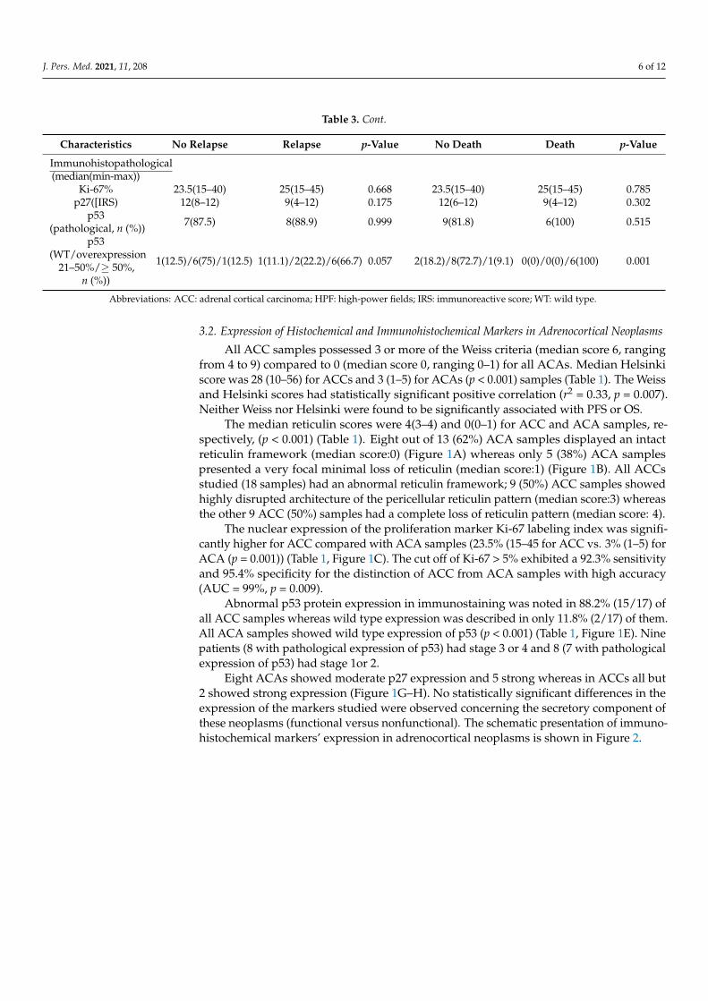
Table 3. Cont.
Characteristics No Relapse Relapse p-Value No Death Death p-Value
Immunohistopathological(median(min-max))
Ki-67% 23.5(15–40) 25(15–45) 0.668 23.5(15–40) 25(15–45) 0.785p27([IRS) 12(8–12) 9(4–12) 0.175 12(6–12) 9(4–12) 0.302
p53(pathological, n (%)) 7(87.5) 8(88.9) 0.999 9(81.8) 6(100) 0.515
p53(WT/overexpression
21–50%/≥ 50%,n (%))
1(12.5)/6(75)/1(12.5) 1(11.1)/2(22.2)/6(66.7) 0.057 2(18.2)/8(72.7)/1(9.1) 0(0)/0(0)/6(100) 0.001
Abbreviations: ACC: adrenal cortical carcinoma; HPF: high-power fields; IRS: immunoreactive score; WT: wild type.
3.2. Expression of Histochemical and Immunohistochemical Markers in Adrenocortical Neoplasms
All ACC samples possessed 3 or more of the Weiss criteria (median score 6, rangingfrom 4 to 9) compared to 0 (median score 0, ranging 0–1) for all ACAs. Median Helsinkiscore was 28 (10–56) for ACCs and 3 (1–5) for ACAs (p < 0.001) samples (Table 1). The Weissand Helsinki scores had statistically significant positive correlation (r2 = 0.33, p = 0.007).Neither Weiss nor Helsinki were found to be significantly associated with PFS or OS.
The median reticulin scores were 4(3–4) and 0(0–1) for ACC and ACA samples, re-spectively, (p < 0.001) (Table 1). Eight out of 13 (62%) ACA samples displayed an intactreticulin framework (median score:0) (Figure 1A) whereas only 5 (38%) ACA samplespresented a very focal minimal loss of reticulin (median score:1) (Figure 1B). All ACCsstudied (18 samples) had an abnormal reticulin framework; 9 (50%) ACC samples showedhighly disrupted architecture of the pericellular reticulin pattern (median score:3) whereasthe other 9 ACC (50%) samples had a complete loss of reticulin pattern (median score: 4).
The nuclear expression of the proliferation marker Ki-67 labeling index was signifi-cantly higher for ACC compared with ACA samples (23.5% (15–45 for ACC vs. 3% (1–5) forACA (p = 0.001)) (Table 1, Figure 1C). The cut off of Ki-67 > 5% exhibited a 92.3% sensitivityand 95.4% specificity for the distinction of ACC from ACA samples with high accuracy(AUC = 99%, p = 0.009).
Abnormal p53 protein expression in immunostaining was noted in 88.2% (15/17) ofall ACC samples whereas wild type expression was described in only 11.8% (2/17) of them.All ACA samples showed wild type expression of p53 (p < 0.001) (Table 1, Figure 1E). Ninepatients (8 with pathological expression of p53) had stage 3 or 4 and 8 (7 with pathologicalexpression of p53) had stage 1or 2.
Eight ACAs showed moderate p27 expression and 5 strong whereas in ACCs all but2 showed strong expression (Figure 1G–H). No statistically significant differences in theexpression of the markers studied were observed concerning the secretory component ofthese neoplasms (functional versus nonfunctional). The schematic presentation of immuno-histochemical markers’ expression in adrenocortical neoplasms is shown in Figure 2.

J. Pers. Med. 2021, 11, 208 7 of 12
J. Pers. Med. 2021, 11, x FOR PEER REVIEW 7 of 13
Figure 1. Histochemical and immunohistochemical expression of reticulin, Ki-67%, p53 and p27 in adrenocortical neoplasms. Intact reticulin framework with the characteristic acinar pattern in adre-nocortical adenoma (ACA) (×200) (A). Highly disrupted architecture and loss of the reticulin framework in adrenocortical carcinoma ( ACC )(×400) (B). Increase of positive (brown stained) nuclei in Ki-67 immunostaining in ACA (C) compared to ACC (D). Strong nuclear expression (brown stained nuclei) of p53 protein in a few tumor cells in ACA (wild type pattern) (×400) (E), Strong nuclear expression of p53 protein in >80% of tumor cells in ACC (overexpression) (×400) (F). Moderate nuclear staining of p27 protein in ACA (×400) (G). Strong nuclear staining of p27 protein in the majority of cells in ACC (×400) (H).
Abnormal p53 protein expression in immunostaining was noted in 88.2% (15/17) of all ACC samples whereas wild type expression was described in only 11.8% (2/17) of them. All ACA samples showed wild type expression of p53 (p < 0.001) (Table 1, Figure 1E). Nine patients (8 with pathological expression of p53) had stage 3 or 4 and 8 (7 with pathological expression of p53) had stage 1or 2.
Eight ACAs showed moderate p27 expression and 5 strong whereas in ACCs all but 2 showed strong expression (Figure 1G–H). No statistically significant differences in the expression of the markers studied were observed concerning the secretory component of these neoplasms (functional versus nonfunctional). The schematic presentation of im-munohistochemical markers’ expression in adrenocortical neoplasms is shown in Figure 2.
Figure 1. Histochemical and immunohistochemical expression of reticulin, Ki-67%, p53 and p27 in adrenocortical neoplasms.Intact reticulin framework with the characteristic acinar pattern in adrenocortical adenoma (ACA) (×200) (A). Highlydisrupted architecture and loss of the reticulin framework in adrenocortical carcinoma ( ACC )(×400) (B). Increase ofpositive (brown stained) nuclei in Ki-67 immunostaining in ACA (C) compared to ACC (D). Strong nuclear expression(brown stained nuclei) of p53 protein in a few tumor cells in ACA (wild type pattern) (×400) (E), Strong nuclear expressionof p53 protein in >80% of tumor cells in ACC (overexpression) (×400) (F). Moderate nuclear staining of p27 protein in ACA(×400) (G). Strong nuclear staining of p27 protein in the majority of cells in ACC (×400) (H).
J. Pers. Med. 2021, 11, x FOR PEER REVIEW 8 of 13
Figure 2. Expression of the Ki-67%, p53, p27 immunohistochemical nuclear markers as well as reticulin expression in adrenocortical neoplasms. All ACA cases exhibited Ki-67% labeling index ≤ 5% (median: 3%) and expressed the wild type p53 expression. No altered reticulin pattern was observed and median IRIS of p27% staining was 8. In all ACC cases Weiss was higher than 3, with altered reticulin pattern and similar IRS of p27 staining. However, those with relatively lower Weiss 3–5, exhibited lower median Ki-67% labeling index and p53 staining compared with ACC cases with Weiss scores greater than 6. Abbreviations: ACA: adrenocortical adenoma; ACC: adrenal cortical carcinoma; IRS: immunoreactive score; WT: wild type
3.3. Prognostic Role of the Histopathological Markers in ACC Patients All ACC samples with altered reticulin pattern had concomitant necrosis and/or cap-
sular/vascular invasion and/or increased mitotic activity (>5/50 HPF). Thus, using the re-ticulin algorithm all ACCs would be diagnosed as malignant (significant positive correla-tion with Weiss score, r2 = 0.72 p < 0.0001). Ki-67 labeling index was positively associated with a mitotic count >5/50 HPF (r2 = 0.16, p = 0.049) and a Weiss score (r2 = 0.31, p = 0.007) but not with vascular or capsular invasion. The p53 expression was also significantly cor-related with mitotic count (>5/50 HPF) (p = 0.02), but not with capsular or vascular inva-sion. No statistically significant difference was noted between high-grade and low-grade ACCs in respect to reticulin pattern, p27 staining (p = 0.366), p53 staining (p = 0.485) and Ki-67 labeling index (p= 0.074) although in the last case it reached statistical significance and high grade ACCs had higher median (min-max) Ki-67 labeling index (30% (15–45)) compared with low-grade ACCs (16% (15–45)).
Neither Ki-67 labeling index, p27 expression nor reticulin pattern score were found to be significantly associated with PFS or OS (Table S2). However, p53 abnormal expres-sion (≥ 50% of positive cells) was statistically significantly higher in metastatic ACC sam-ples compared to nonmetastatic ACC samples (p = 0.035) as well as in nonsurvivors com-pared to survivors (p < 0.001) (Table S1, Table 3). A cut off of 23% of the percentage of p53-immunostaining positive cells was significantly associated with PFS (AUC 82%, p = 0.0013, sensitivity = 71%, specificity 100%).
Figure 2. Expression of the Ki-67%, p53, p27 immunohistochemical nuclear markers as well as reticulin expression inadrenocortical neoplasms. All ACA cases exhibited Ki-67% labeling index ≤ 5% (median: 3%) and expressed the wild typep53 expression. No altered reticulin pattern was observed and median IRIS of p27% staining was 8. In all ACC cases Weisswas higher than 3, with altered reticulin pattern and similar IRS of p27 staining. However, those with relatively lower Weiss3–5, exhibited lower median Ki-67% labeling index and p53 staining compared with ACC cases with Weiss scores greaterthan 6. Abbreviations: ACA: adrenocortical adenoma; ACC: adrenal cortical carcinoma; IRS: immunoreactive score; WT:wild type

J. Pers. Med. 2021, 11, 208 8 of 12
3.3. Prognostic Role of the Histopathological Markers in ACC Patients
All ACC samples with altered reticulin pattern had concomitant necrosis and/orcapsular/vascular invasion and/or increased mitotic activity (>5/50 HPF). Thus, usingthe reticulin algorithm all ACCs would be diagnosed as malignant (significant positivecorrelation with Weiss score, r2 = 0.72 p < 0.0001). Ki-67 labeling index was positivelyassociated with a mitotic count >5/50 HPF (r2 = 0.16, p = 0.049) and a Weiss score (r2 = 0.31,p = 0.007) but not with vascular or capsular invasion. The p53 expression was also sig-nificantly correlated with mitotic count (>5/50 HPF) (p = 0.02), but not with capsular orvascular invasion. No statistically significant difference was noted between high-gradeand low-grade ACCs in respect to reticulin pattern, p27 staining (p = 0.366), p53 staining(p = 0.485) and Ki-67 labeling index (p= 0.074) although in the last case it reached statisticalsignificance and high grade ACCs had higher median (min-max) Ki-67 labeling index (30%(15–45)) compared with low-grade ACCs (16% (15–45)).
Neither Ki-67 labeling index, p27 expression nor reticulin pattern score were found tobe significantly associated with PFS or OS (Table S2). However, p53 abnormal expression(≥ 50% of positive cells) was statistically significantly higher in metastatic ACC samplescompared to nonmetastatic ACC samples (p = 0.035) as well as in nonsurvivors comparedto survivors (p < 0.001) (Table S1, Table 3). A cut off of 23% of the percentage of p53-immunostaining positive cells was significantly associated with PFS (AUC 82%, p = 0.0013,sensitivity = 71%, specificity 100%).
4. Discussion
In the current study, among the studied markers, altered reticulin pattern, Ki-67%labeling index value and abnormal nuclear expression of p53 protein were found to bestatistically significant histopathological and molecular markers for distinguishing malig-nant from benign adrenal neoplasms although in a small series of patients. In contrast,p27 expression was not found to be a significant marker for the distinction of benignfrom malignant adrenal cortical neoplasms. However, only the pathological p53 nuclearprotein expression was found to have a prognostic role since it was significantly cor-related with mortality rate, PFS and metastatic status. The reticulin algorithm definesmalignancy through an altered reticulin framework associated with one of the three fol-lowing parameters: necrosis, high mitotic rate, and vascular invasion for the diagnosis ofACCs [18,19,32,33]. The “reticulin” diagnostic algorithm has been proposed, based on theobservation that the tumoral reticulin framework (highlighted by reticulin silver-basedhistochemical staining) is consistently disrupted in malignant cases but only in a smallsubset of benign cases. Several case series on ACCs have confirmed the usefulness of thereticulin algorithm for the distinction of ACCs from ACAs [2,19,32–34], although withoutshowing any correlation with prognosis in these patients [6,35]. Our series confirmedthe quantitative loss of reticulin framework as a significant finding, distinguishing ACCsand ACAs, showing also a significant positive correlation with Weiss score without beingsignificantly associated with prognosis.
Adrenal cortical malignancy is a proliferation-driven malignancy and this studydemonstrated significantly increased expression levels of markers related to cell prolifer-ation such as Ki-67 and p53 in ACCs compared with ACAs. Nuclear expression of theproliferative marker Ki-67 was also significantly correlated with mitotic activity and Weissscore. We have also demonstrated with high accuracy that a Ki-67 labeling index cut-offvalue > 5% (92.3% sensitivity and 95.4% specificity) could discriminate ACCs from ACAs,confirming the existing data of the literature [9,10,36]. However, this series failed to confirmthe current literature and showed no statistically significant association of Ki-67% with PFSand OS, probably due to the small number of samples and the short follow-up period. Inline with these data in the literature, there are also some studies that have not confirmed asignificant association of Ki-67% with either OS [37] or disease-free survival [38] althoughin the last study Ki-67% was found to be an independent prognostic factor for OS. More-over, although the practical utility of Ki-67% staining was indisputable and confirmed in

J. Pers. Med. 2021, 11, 208 9 of 12
many studies, there are also studies supporting the idea that it is hard to set a diagnosticthreshold that is mainly attributed to possible interobserver variations [39].
The cell cycle regulation molecular marker p53 encoding a protein that promotes DNArepair, was present in almost all ACC (15/17) and absent in ACA samples. The half-life andexpression of p53 protein is low and therefore undetectable by immunohistochemistry [6,40,41].Aberrant nuclear immunohistochemical staining for p53 in ACC samples varies in theliterature from 5% to 60% [42,43]. In adult sporadic ACCs, about one quarter of tumorsharbor somatic TP53 mutations [43–45], and more than a half harbor loss of heterozygosityat the TP53 locus [13,46]. Previous studies, have shown that ACAs had significantly lowerlevels of immunohistochemical p53 nuclear expression than ACCs [6], whereas others havefailed to show such a difference [47]. Moreover, it has also been reported that high-gradeACCs exhibit higher p53 expression than low-grade ones, a finding consistent with theenrichment of TP53 mutations in high grade carcinomas [9,48].
Transcriptome studies have led to further understanding of the role of p53 in sporadicACCs. Indeed, TP53 mutated tumors are enriched in a subgroup of ACCs identified byunsupervised clustering of the tumors [17]. Finally, genes positively regulated by p53 suchas RRM2B, TP53INP1 and MDM2, were found to be downregulated in this subgroup. Thepresent series confirmed the aberrant nuclear immunohistochemical expression of p53 inACCs compared to ACAs, although 11.8% of the ACCs showed wild type expression ofp53 as all ACAs. Abnormal expression of p53 was the only marker in our series of adrenalneoplasms that showed to have a prognostic role since it was associated with increasedmortality rate and the presence of metastases, whereas a cut off of 23% of the percentageof p53-immunostaining positive cells was significantly associated with PFS. Although inmolecular analysis the prognostic role of the abnormal p53 expression in ACC is clear [17],immunohistochemical data on the prognostic role of p53 are rather conflicting. Severalstudies have failed to show a prognostic role of p53 protein [6,42,49,50], whereas othershave shown that patients with abnormal p53 staining tended to have higher grade andstage ACCs tumors, increased relapse rates and poorer disease-free survival [46,51].
The protein encoded by p27 (CDKN1B) is another cell cycle regulator marker, thatwhen it is upregulated, results in cell cycle arrest and apoptosis [52]. Our study found nostatistical difference of p27 expression between ACCs and ACAs. Accordingly, two previ-ous studies [50,53] have failed to show that p27 could be used as an immunohistochemicalmarkers for distinguishing ACCs from ACAs. However, a more recent study showed thatp27 staining was significantly higher in ACCs compared with ACAs, using an automatedmethod of analysis with a high diagnostic accuracy of 7.23% as the best cut-off value [47].This study had the novelty to use an automated method of analysis in contrast to previousstudies which were carried out by direct observation by the researchers [50,53].
All ACC samples exhibited positive staining for Ki-67, p53 and p27 except for twoACC samples that were p53 negative and p27 and Ki-67 positive. In contrast to p53 stainingwhich was found normal in all ACAs (wild type), Ki-67 and p27 were positive in all ACAs.The overexpression of p27 in ACC samples is somewhat contradictory. Several explanationshave been proposed, either that adrenal cancer cells develop a tolerance to this inhibitor ofcell cycle progression, suggesting that p27 could be present but inactive to arrest the cellcycle, or that p27 gene is mutated resulting in a modified p27 protein [47,54].
Our study has several limitations, that are mainly related to its retrospective natureand the number of samples analyzed. The relatively limited number of samples didnot allow a more fruitful statistical analysis that, along with the short follow-up period,may have affected the identification of the prognostic role of the immunohistochemicalbiomarkers in these tumors. We anticipate that the inclusion of more patients may providethe additional power to reach meaningful clinical findings.
In conclusion, Ki-67, p53 as well as abnormal reticulin pattern, but not p27 expression,could be used to define malignancy in adrenocortical neoplasms and differentiate ACCsfrom ACAs. Furthermore, p53 expression was significantly associated with increasedmortality, metastatic status and lower PFS. However, the small number of patients did

J. Pers. Med. 2021, 11, 208 10 of 12
not allow a more robust conclusion; perhaps if confirmed in larger studies it may offer adiagnostic/prognostic tool available in everyday clinical practice. Immunohistopathologyalone cannot fully discriminate ACCs with poor prognosis from those with good prognosisand it should be combined with further morphological criteria and recently developedtranscriptome analysis which have shown clear differences between adenomas and high-or low-grade carcinomas.
Supplementary Materials: The following are available online at https://www.mdpi.com/2075-4426/11/3/208/s1, Table S1: Association of clinical, histopathological and immunohistopathologicalmarkers with prognostic factors, Table S2: Univariate Cox regression analysis for risk factors associ-ated with PFS and OS in 24 patients with ACC, Figure S1. Kaplan−Meier curve for PFS of patientswith ACC, Figure S2. Kaplan−Meier curve for OS of patients with ACC.
Author Contributions: Conceptualization, G.K. (Gregory Kaltsas); methodology, G.K. (GeorgiosKyriakopoulos), A.A., A.D. and F.A.; software, F.A., A.A.; validation, G.K. (Gregory Kaltsas) andE.K.; formal analysis, F.A., A.A.; investigation, A.A., F.A., G.K. (Georgios Kyriakopoulos); resources,A.A., A.D., G.Z., C.A.; data curation, A.A., F.A., G.K. (Gregory Kaltsas); writing—original draftpreparation, A.A., G.K. (Georgios Kyriakopoulos), G.K. (Gregory Kaltsas); writing—review andediting, G.K. (Gregory Kaltsas), E.K.; visualization, G.K. (Gregory Kaltsas), E.K.; supervision, G.K.(Gregory Kaltsas). All authors have read and agreed to the published version of the manuscript.
Funding: This research received no external funding.
Institutional Review Board Statement: The study was conducted according to the guidelines of theDeclaration of Helsinki, and approved by the Institutional Review Board (or Ethics Committee) ofLaiko Hospital (protocol code 115/04.01.2021).
Informed Consent Statement: Informed consent was obtained from all subjects involved in the study.
Data Availability Statement: The data presented in this study are available on request from thecorresponding author. The data are not publicly available due to privacy restrictions.
Conflicts of Interest: The authors declare no conflict of interest.
References1. Allolio, B.; Fassnacht, M. Adrenocortical Carcinoma: Clinical Update. J. Clin. Endocrinol. Metab. 2006, 91, 2027–2037. [CrossRef]2. Ciaramella, P.D.; Vertemati, M.; Petrella, D.; Bonacina, E.; Grossrubatscher, E.; Duregon, E.; Volante, M.; Papotti, M.; Loli, P.
Analysis of histological and immunohistochemical patterns of benign and malignant adrenocortical tumors by computerizedmorphometry. Pathol. Res. Pr. 2017, 213, 815–823. [CrossRef]
3. Weiss, L.M. Comparative histologic study of 43 metastasizing and nonmetastasizing adrenocortical tumors. Am. J. Surg. Pathol.1984, 8, 163–170. [CrossRef] [PubMed]
4. Lau, S.K.; Weiss, L.M. The Weiss system for evaluating adrenocortical neoplasms: 25 years later. Hum. Pathol. 2009, 40, 757–768.[CrossRef] [PubMed]
5. Aubert, S.; Wacrenier, A.; Leroy, X.; Devos, P.; Carnaille, B.; Proye, C.; Wemeau, J.L.; Lecomte-Houcke, M.; Leteurtre, E. Weisssystem revisited: A clinicopathologic and immunohistochemical study of 49 adrenocortical tumors. Am. J. Surg. Pathol. 2002, 26,1612–1619. [CrossRef] [PubMed]
6. Mete, O.; Asa, S.L.; Giordano, T.J.; Papotti, M.; Sasano, H.; Volante, M. Immunohistochemical Biomarkers of Adrenal CorticalNeoplasms. Endocr. Pathol. 2018, 29, 137–149. [CrossRef] [PubMed]
7. de Reyniès, A.; Assié, G.; Rickman, D.S.; Tissier, F.; Groussin, L.; René-Corail, F.; Dousset, B.; Bertagna, X.; Clauser, E.; Bertherat, J.Gene expression profiling reveals a new classification of adrenocortical tumors and identifies molecular predictors of ma-lignancyand survival. J. Clin. Oncol. 2009, 27, 1108–1115. [CrossRef]
8. Zheng, S.; Cherniack, A.D.; Dewal, N.; Moffitt, R.A.; Danilova, L.; Murray, B.A.; Lerario, A.M.; Else, T.; Knijnenburg, T.A.; Ciriello,G.; et al. Comprehensive Pan-Genomic characterization of adrenocortical carcinoma. In Proceedings of the AACR 106th AnnualMeeting, Philadelphia, PA, USA, 18–22 April 2015.
9. Else, T.; Kim, A.C.; Sabolch, A.; Raymond, V.M.; Kandathil, A.; Caoili, E.M.; Jolly, S.; Miller, B.S.; Giordano, T.J.; Hammer, G.D.Adrenocortical Carcinoma. Endocr. Rev. 2014, 35, 282–326. [CrossRef] [PubMed]
10. Duan, K.; Giordano, T.; Mete, O. Adrenal cortical proliferations. In Endocrine Pathology; Mete, O., Asa, S.L., Eds.; CambridgeUniversity Press: Cambridge, UK, 2016; pp. 602–627.
11. Mete, O.; Asa, S.L. Precursor lesions of endocrine system neoplasms. Pathology 2013, 45, 316–330. [CrossRef]

J. Pers. Med. 2021, 11, 208 11 of 12
12. Heaton, J.H.; Wood, M.A.; Kim, A.C.; Lima, L.O.; Barlaskar, F.M.; Almeida, M.Q.; Fragoso, M.C.B.V.; Kuick, R.; Lerario, A.M.;Simon, D.P.; et al. Progression to Adrenocortical Tumorigenesis in Mice and Humans through Insulin-Like Growth Factor 2 andβ-Catenin. Am. J. Pathol. 2012, 181, 1017–1033. [CrossRef] [PubMed]
13. Assie, G.; Giordano, T.; Bertherat, J. Gene expression profiling in adrenocortical neoplasia. Mol. Cell. Endocrinol. 2012, 351,111–117. [CrossRef]
14. Assié, G.; Letouzé, E.; Fassnacht, M.; Jouinot, A.; Luscap, W.; Barreau, O.; Omeiri, H.; Rodriguez, S.; Perlemoine, K.; René-Corail,F.; et al. Integrated genomic characterization of adrenocortical carcinoma. Nat. Genet. 2014, 46, 607–612. [CrossRef]
15. Espiard, S.; Bertherat, J. The Genetics of Adrenocortical Tumors. Endocrinol. Metab. Clin. N. Am. 2015, 44, 311–334. [CrossRef][PubMed]
16. Giordano, T.J. Adrenocortical tumors: An integrated clinical, pathologic, and molecular approach at the University of Michi-gan.Arch. Path. Lab. Med. 2010, 134, 1440–1443. [CrossRef] [PubMed]
17. Ragazzon, B.; Libé, R.; Gaujoux, S.; Assié, G.; Fratticci, A.; Launay, P.; Clauser, E.; Bertagna, X.; Tissier, F.; De Reyniès, A.; et al.Transcriptome Analysis Reveals that p53 and β-Catenin Alterations Occur in a Group of Aggressive Adrenocortical Cancers.Cancer Res. 2010, 70, 8276–8281. [CrossRef] [PubMed]
18. Papotti, M.; Volante, M.; Duregon, E.; Delsedime, L.; Terzolo, M.; Berruti, A.; Rosai, J. Adrenocortical Tumors With MyxoidFeatures: A Distinct Morphologic and Phenotypical Variant Exhibiting Malignant Behavior. Am. J. Surg. Pathol. 2010, 34, 973–983.[CrossRef] [PubMed]
19. Duregon, E.; Cappellesso, R.; Maffeis, V.; Zaggia, B.; Ventura, L.; Berruti, A.; Terzolo, M.; Fassina, A.; Volante, M.; Papotti, M.Validation of the prognostic role of the "Helsinki Score” in 225 cases of adrenocortical carcinoma. Hum. Pathol. 2017, 62, 1–7.[CrossRef] [PubMed]
20. Mete, O.; Asa, S.L. Pathological definition and clinical significance of vascular invasion in thyroid carcinomas of follicularepithelial derivation. Mod. Pathol. 2011, 24, 1545–1552. [CrossRef]
21. Bisceglia, M.; Ludovico, O.; Di Mattia, A.; Ben-Dor, D.; Sandbank, J.; Pasquinelli, G.; Lau, S.K.; Weiss, L.M. AdrenocorticalOncocytic Tumors: Report of 10 Cases and Review of the Literature. Int. J. Surg. Pathol. 2004, 12, 231–243. [CrossRef]
22. Wong, D.D.; Spagnolo, D.V.; Bisceglia, M.; Havlat, M.; McCallum, D.; Platten, M.A. Oncocytic adrenocortical neoplasms: Aclinicopathologic study of 13 new cases emphasizing the importance of their recognition. Hum. Pathol. 2011, 42, 489–499.[CrossRef]
23. Miller, B.S.; Gauger, P.G.; Hammer, G.D.; Giordano, T.J.; Doherty, G.M. Proposal for modification of the ENSAT staging system foradrenocortical carcinoma using tumor grade. Langenbeck’s Arch. Surg. 2010, 395, 955–961. [CrossRef] [PubMed]
24. Assié, G.; Antoni, G.; Tissier, F.; Caillou, B.; Abiven, G.; Gicquel, C.; Leboulleux, S.; Travagli, J.-P.; Dromain, C.; Bertagna, X.; et al.Prognostic Parameters of Metastatic Adrenocortical Carcinoma. J. Clin. Endocrinol. Metab. 2007, 92, 148–154. [CrossRef] [PubMed]
25. Wanis, K.N.; Kanthan, R. Diagnostic and prognostic features in adrenocortical carcinoma: A single institution case series andreview of the literature. World J. Surg. Oncol. 2015, 13, 1–13. [CrossRef] [PubMed]
26. Mete, O.; Gucer, H.; Kefeli, M.; Asa, S.L. Diagnostic and Prognostic Biomarkers of Adrenal Cortical Carcinoma. Am. J. Surg.Pathol. 2018, 42, 201–213. [CrossRef] [PubMed]
27. Klöppel, G.; La Rosa, S. Ki67 labeling index: Assessment and prognostic role in gastroenteropancreatic neuroendocrine neoplasms.Virchows Arch. Pathol. Anat. Histol. 2017, 472, 341–349. [CrossRef] [PubMed]
28. Wang, Y.C.; Lin, R.K.; Tan, Y.H.; Chen, J.T.; Chen, C.Y.; Wang, Y.C. Wild-Type p53 Overexpression and Its Correlation With MDM2and p14ARF Alterations: An Alternative Pathway to Non–Small-Cell Lung Cancer. J. Clin. Oncol. 2005, 23, 154–164. [CrossRef][PubMed]
29. Köbel, M.; Ronnett, B.M.; Singh, N.; Soslow, R.A.; Gilks, C.B.; McCluggage, W.G. Interpretation of P53 Immunohistochemistry inEndometrial Carcinomas. Int. J. Gynecol. Pathol. 2019, 38, S123–S131. [CrossRef] [PubMed]
30. Xiong, D.D.; He, R.Q.; Lan, A.H.; Chen, W.J.; Luo, Y.H.; Ye, Z.H.; Ma, J.; Chen, G.; Dang, Y.W. Clinical significances of p27 indigestive tract cancers: A comprehensive analysis on immunohistochemistry staining, published literatures, microarray andRNA-seq data. Oncotarget 2018, 9, 12284–12303. [CrossRef]
31. Fan, J.; Upadhye, S.; Worster, A. Understanding receiver operating characteristic (ROC) curves. CJEM 2006, 8, 19–20. [CrossRef]32. Fonseca, D.; Murthy, S.S.; Tagore, K.R.; Rao, B.V.; Thamminedi, S.R.; Raju, K.; Sharma, R.; Challa, S. Diagnosis of adrenocortical
tumors by reticulin algorithm. Indian J. Endocrinol. Metab. 2017, 21, 734. [CrossRef]33. Volante, M.; Bollito, E.; Sperone, P.; Tavaglione, V.; Daffara, F.; Porpiglia, F.; Terzolo, M.; Berruti, A.; Papotti, M. Clinico-
pathological study of a series of 92 adrenocortical carcinomas: From a proposal of simplified diagnostic algorithm to prognosticstratification. Histopathology 2009, 55, 535–543. [CrossRef] [PubMed]
34. Duregon, E.; Fassina, A.; Volante, M.; Nesi, G.; Santi, R.; Gatti, G.; Cappellesso, R.; Ciaramella, P.D.; Ventura, L.; Gambacorta, M.The reticulin algorithm for adrenocortical tumor diagnosis: A multicentric validation study on 245 unpublished cases. Am. J.Surg. Pathol. 2013, 37, 1433–1440. [CrossRef] [PubMed]
35. Sung, T.Y.; Choi, Y.M.; Kim, W.B.; Lee, Y.M.; Kim, T.Y.; Shong, Y.K.; Song, D.E. Myxoid and Sarcomatoid Variants of AdrenocorticalCarcinoma: Analysis of Rare Variants in Single Tertiary Care Center. J. Korean Med Sci. 2017, 32, 764–771. [CrossRef]
36. Morimoto, R.; Satoh, F.; Murakami, O.; Suzuki, T.; Abe, T.; Tanemoto, M.; Abe, M.; Uruno, A.; Ishidoya, S.; Arai, Y. Immuno-histochemistry of a proliferation marker Ki67/MIB1 in adrenocortical carcinomas: Ki67/MIB1 labeling index is a predictor forrecurrence of adrenocortical carcinomas. Endocr. J. 2008, 55, 49–55. [CrossRef]
J. Pers. Med. 2021, 11, 208 12 of 12
37. Babinska, A.; Peksa, R.; Wisniewski, P.; Swiatkowska-Stodulska, R.; Sworczak, K. Diagnostic and prognostic role of SF1, IGF2,Ki67, p53, adiponectin, and leptin receptors in human adrenal cortical tumors. J. Surg. Oncol. 2017, 116, 427–433. [CrossRef][PubMed]
38. Jouinot, A.; Assie, G.; Libe, R.; Fassnacht, M.; Papathomas, T.; Barreau, O.; De La Villeon, B.; Faillot, S.; Hamzaoui, N.; Neou, M.;et al. DNA methylation is an independent prognostic marker of survival in adrenocortical cancer. J. Clin. Endocrinol. Metab. 2016,102, 923–932. [CrossRef] [PubMed]
39. Papathomas, T.G.; Pucci, E.; Giordano, T.J.; Lu, H.; Duregon, E.; Volante, M.; Papotti, M.; Lloyd, R.V.; Tischler, A.S.; VanNederveen, F.H.; et al. An International Ki67 Reproducibility Study in Adrenal Cortical Carcinoma. Am. J. Surg. Pathol. 2016, 40,569–576. [CrossRef]
40. Manfredi, J.J. The Mdm2-p53 relationship evolves: Mdm2 swings both ways as an oncogene and a tumor suppressor. Genes Dev.2010, 24, 1580–1589. [CrossRef]
41. Moll, U.M.; Petrenko, O. The MDM2-p53 interaction. Mol. Cancer Res. 2003, 1, 1001–1008.42. Stojadinovic, A.; Ghossein, R.A.; Hoos, A.; Nissan, A.; Marshall, D.; Dudas, M.; Cordon-Cardo, C.; Jaques, D.P.; Brennan, M.F.
Adrenocortical Carcinoma: Clinical, Morphologic, and Molecular Characterization. J. Clin. Oncol. 2002, 20, 941–950. [CrossRef]43. Reincke, M.; Karl, M.; Travis, W.H.; Mastorakos, G.; Allolio, B.; Linehan, H.M.; Chrousos, G.P. p53 mutations in human
adrenocortical neoplasms: Immunohistochemical and molecular studies. J. Clin. Endocrinol. Metab. 1994, 78, 790–794. [CrossRef][PubMed]
44. Barzon, L.; Chilosi, M.; Fallo, F.; Martignoni, G.; Montagna, L.; Palù, G.; Boscaro, M. Molecular analysis of CDKN1C and TP53 insporadic adrenal tumors. Eur. J. Endocrinol. 2001, 145, 207–212. [CrossRef] [PubMed]
45. Libè, R.; Fratticci, A.; Bertherat, J. Adrenocortical cancer: Pathophysiology and clinical management. Endocr. Relat. Cancer 2007,14, 13–28. [CrossRef] [PubMed]
46. Gicquel, C.; Bertagna, X.; Gaston, V.; Coste, J.; Louvel, A.; Baudin, E.; Bertherat, J.; Chapuis, Y.; Duclos, J.M.; Schlumberger, M.;et al. Molecular markers and long-term recurrences in a large cohort of patients with sporadic adrenocortical tumors. Cancer Res.2001, 61, 6762–6767. [PubMed]
47. Pereira, S.S.; Morais, T.; Costa, M.M.; Monteiro, M.P.; Pignatelli, D. The emerging role of the molecular marker p27 in thedifferential diagnosis of adrenocortical tumors. Endocr. Connect. 2013, 2, 137–145. [CrossRef]
48. Wasserman, J.D.; Zambetti, G.P.; Malkin, D. Towards an understanding of the role of p53 in adrenocortical carcinogenesis. Mol.Cell. Endocrinol. 2012, 351, 101–110. [CrossRef]
49. Edgren, M.; Eriksson, B.; Wilander, E.; Westlin, J.E.; Nilsson, S.; Oberg, K. Biological characteristics of adrenocortical carcinoma: Astudy of p53, IGF, EGF-r, Ki-67 and PCNA in 17 adrenocortical carcinomas. Anticancer. Res. 1997, 17, 1303–1309.
50. Stojadinovic, A.; Brennan, M.F.; Hoos, A.; Omeroglu, A.; Leung, D.H.Y.; Dudas, M.E.; Nissan, A.; Cordon-Cardo, C.; Ghossein,R.A. Adrenocortical Adenoma and Carcinoma: Histopathological and Molecular Comparative Analysis. Mod. Pathol. 2003, 16,742–751. [CrossRef] [PubMed]
51. Waldmann, J.; Patsalis, N.; Fendrich, V.; Langer, P.; Saeger, W.; Chaloupka, B.; Ramaswamy, A.; Fassnacht, M.; Bartsch, D.K.; Slater,E.P. Clinical impact of TP53 alterations in adrenocortical carcinomas. Langenbeck’s Arch. Surg. 2011, 397, 209–216. [CrossRef]
52. Lee, J.; Kim, S.S. The function of p27KIP1during tumor development. Exp. Mol. Med. 2009, 41, 765–771. [CrossRef] [PubMed]53. Nakazumi, H.; Sasano, H.; Iino, K.; Ohashi, Y.; Orikasa, S. Expression of cell cycle inhibitor p27 and Ki-67 in human adre-nocortical
neoplasms. Mod. Pathol. 1998, 11, 1165–1170. [PubMed]54. Nickeleit, I.; Zender, S.; Kossatz, U.; Malek, N.P. p27kip1: A target for tumor therapies? Cell Div. 2007, 2, 13. [CrossRef] [PubMed]