the role of intraluminal sodium in glucose absorption in vivo
TRANSCRIPT

The Role of Intraluminal Sodium
in Glucose Absorption In Vivo
DAvI A. SALrzMAN, FLOYD C. RECroR, JR., and JoEH S. FowmnAuNFrom the Department of Internal Medicine, The University of TexasSouthwestern Medical School at Dallas, Dallas, Texas 75235
A B S T R A C T Active glucose absorption is thought todepend on a gradient of sodium ion concentration acrossthe brush border xnembrane of intestinal epithelial cells.This concept is generally accepted, although its validityhas never been adequately evaluated in the human smallintestine in vivo. According to this hypothesis, the rateof glucose absorption should decrease markedly if theluminal sodium concentration is markedly reduced, andglucose absorption against a concentration gradientshould cease entirely if luminal sodium is lower than in-tracellular sodium concentration. In the present seriesof experiments we were not able to show an importantrole of intraluminal sodium concentration in the activeabsorption of glucose from the human, rat, and dog ileumin vivo. Specifically, glucose absorption was minimallyreduced or not reduced at all when intraluminal sodiumconcentration was reduced from 140 to as low as 2.5mEq/liter. The discrepancy between our results andthose of previous workers whose data suggest that re-moval of intraluminal sodium should markedly inhibitactive glucose absorption is not entirely clear, but thereare a number of differences in experimental designbetween most previous studies and our own. Althoughour data show that active glucose absorption proceedsat a near normal rate even when lumen sodium concen-tration is reduced below 3 mEq/liter, our results do notdisprove the sodium gradient theory because of thetheoretic possibility that the microclimate adjacent tothe brush border has a high concentration of sodium evenwhen luminal sodium concentration is markedly reduced.The validity of the sodium gradient hypothesis wouldappear to be critically dependent on such a microclimate.
Dr. Saltzman was supported by U. S. Public HealthService Training Grant 5 TOl AM 05490 from the NationalInstitute of Arthritis and Metabolic Diseases.Received for publication 7 July 1971 and in revised form
1 November 1971.
INTRODUCTIONRecent studies have suggested that glucose absorptioncannot proceed unless sodium is present in the gut lu-men (1), and that inhibition of the sodium pump (e.g.with cardiac glycosides) results in inhibition of glucosetransport (2). Based mainly on these observations, thesodium gradient hypothesis for active glucose absorptionwas proposed by Crane (3, 4). Crane's experimentshave been carried out primarily in the hamster intestine,and there are two main features of his model: first, theaffinity of a mobile carrier for glucose, located in thebrush border membrane of the epithelial cells, is di-rectly proportional to sodium concentration; second, thesodium concentration within the absorbing cells is main-tained at a low level by an outwardly directed sodiumpump. Because the sodium concentration in the gutlumen is higher than that inside the cell, the affinity ofthe carrier for glucose at the luminal surface of the brushborder membrane is higher than that at the intracellularsurface. This differential affinity results in net movementof glucose from gut lumen into the cell. Once insidethe cell, glucose diffuses passively across the serosalmembrane to complete the absorptive process.
Extensive studies in the rabbit ileum have providedstrong support for this hypothesis, although differencesin kinetic detail between the rabbit and hamster havebeen observed, and the original hypothesis has beenmodified slightly (5). In the revised (rabbit) model,sodium and sugar combine with a mucosal transport site,which may or may not be a mobile carrier; in the ab-sence of sodium in the mucosal solution, this ternarycomplex cannot be formed, and sugar transfer does notoccur (5). In both models active sugar transport isdriven by a sodium concentration difference across thebrush border membrane, and this is the essence of thesodium gradient hypothesis.
876 The Journal of Clinical Investigation Volume 51 1972
According to the sodium gradient hypothesis (3-7),a lower concentration of sodium inside the cell than inthe lumnen is essential for the active transport of glu-cose. If the sodium pump mechanism were poisoned bycardiac glycosides, the intracellular sodium concentra-tion would increase and active glucose absorption wouldcease. Likewise, according to this hypothesis, the rate ofglucose absorption should decrease markedly or cease en-tirely if the luminal sodium concentration is markedlyreduced.
This concept is now widely accepted (4) and is alsoapplied to the absorption of other substrates, such asamino acids (8, 9), and to transport in many tissuesother than the intestine (4, 10). However, the hypothesishas not, in our opinion, been adequately evaluated in thehuman small intestine or in any other in vivo system.In the present experiments, we have examined the ef-fects of luminal sodium concentration on glucose absorp-tion in the intact perfused ileum of humans, rats, anddogs.
METHODS
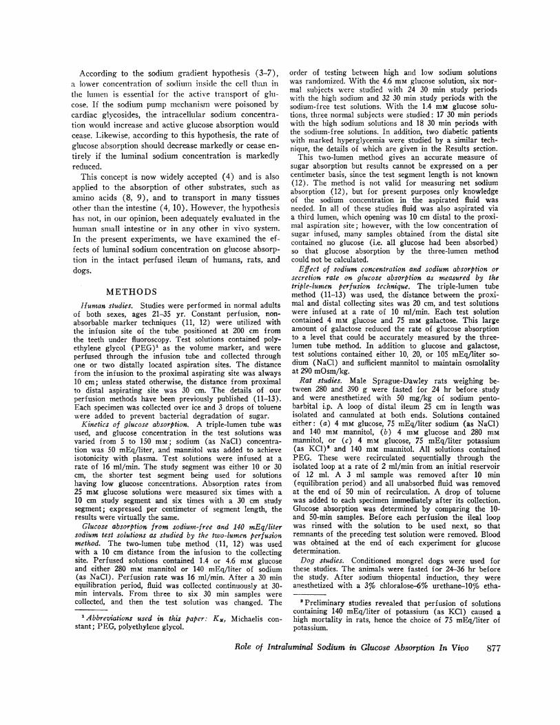
Human studies. Studies were performed in normal adultsof both sexes, ages 21-35 yr. Constant perfusion, non-absorbable marker techniques (11, 12) were utilized withthe infusion site of the tube positioned at 200 cm fromthe teeth under fluoroscopy. Test solutions contained poly-ethylene glycol (PEG)' as the volume marker, and wereperfused through the infusion tube and collected throughone or two distally located aspiration sites. The distancefrom the infusion to the proximal aspirating site was always10 cm; unless stated otherwise, the distance from proximalto distal aspirating site was 30 cm. The details of ourperfusion methods have been previously published (11-13).Each specimen was collected over ice and 3 drops of toluenewere added to prevent bacterial degradation of sugar.
Kinetics of glucose absorption. A triple-lumen tube wasused, and glucose concentration in the test solutions wasvaried from 5 to 150 mM; sodium (as NaCl) concentra-tion was 50 mEq/liter, and mannitol was added to achieveisotonicity with plasma. Test solutions were infused at arate of 16 ml/min. The study segment was either 10 or 30cm, the shorter test segment being used for solutionshaving low glucose concentrations. Absorption rates from25 mm glucose solutions were measured six times with a10 cm study segment and six times with a 30 cm studysegment; expressed per centimeter of segment length, theresults were virtually the same.
Glucose absorption from sodium-free and 140 mEq/litersodium test solutions as studied by the two-lumen perfusionmethod. The two-lumen tube method (11, 12) was usedwith a 10 cm distance from the infusion to the collectingsite. Perfused solutions contained 1.4 or 4.6 mm glucoseand either 280 mm mannitol or 140 mEq/liter of sodium(as NaCl). Perfusion rate was 16 ml/min. After a 30 minequilibration period, fluid was collected continuously at 30-min intervals. From three to six 30 min samples werecollected, and then the test solution was changed. The
'Abbreviations used in this paper: Km, Michaelis con-stant; PEG, polyethylene glycol.
order of testing between high and low sodium solutionswas randomized. With the 4.6 mm glucose solution, six nor-mal subjects were studied with 24 30 min study periodswith the high sodium and 32 30 min study periods with thesodium-free test solutions. With the 1.4 mm glucose solu-tions, three normal subjects were studied: 17 30 min periodswith the high sodium solutions and 18 30 min periods withthe sodium-free solutions. In addition, two diabetic patientswith marked hyperglycemia were studied by a similar tech-nique, the details of which are given in the Results section.
This two-lumen method gives an accurate measure ofsugar absorption but results cannot be expressed on a percentimeter basis, since the test segment length is not known(12). The method is not valid for measuring net sodiumabsorption (12), but for present purposes only knowledgeof the sodium concentration in the aspirated fluid wasneeded. In all of these studies fluid was also aspirated viaa third lumen, which opening was 10 cm distal to the proxi-mal aspiration site; however, with the low concentration ofsugar infused, many samples obtained from the distal sitecontained no glucose (i.e. all glucose had been absorbed)so that glucose absorption by the three-lumen methodcould not be calculated.
Effect of sodium concentration and sodium absorption orsecretion rate on glucose absorption as measured by thetriple-lumen perfusion technique. The triple-lumen tubemethod (11-13) was used, the distance between the proxi-mal and distal collecting sites was 20 cm, and test solutionswere infused at a rate of 10 ml/min. Each test solutioncontained 4 mm glucose and 75 mm galactose. This largeamount of galactose reduced the rate of glucose absorptionto a level that could be accurately measured by the three-lumen tube method. In addition to glucose and galactose,test solutions contained either 10, 20, or 105 mEq/liter so-dium (NaCl) and sufficient mannitol to maintain osmolalityat 290 mOsm/kg.Rat studies. Male Sprague-Dawley rats weighing be-
tween 280 and 390 g were fasted for 24 hr before -studyand were anesthetized with 50 mg/kg of sodium pento-barbital i.p. A loop of distal ileum 25 cm in length wasisolated and cannulated at both ends. Solutions containedeither: (a) 4 mm glucose, 75 mEq/liter sodium (as NaCl)and 140 mm mannitol, (b) 4 mm glucose and 280 mMmannitol, or (c) 4 mm glucose, 75 mEq/liter potassium(as KCl)' and 140 mm mannitol. All solutions containedPEG. These were recirculated sequentially through theisolated loop at a rate of 2 ml/min from an initial reservoirof 12 ml. A 3 ml sample was removed after 10 min(equilibration period) and all unabsorbed fluid was removedat the end of 50 min of recirculation. A drop of toluenewas added to each specimen immediately after its collection.Glucose absorption was determined by comparing the 10-and 50-min samples. Before each perfusion the ileal loopwas rinsed with the solution to be used next, so thatremnants of the preceding test solution were removed. Bloodwas obtained at the end of each experiment for glucosedetermination.Dog studies. Conditioned mongrel dogs were used for
these studies. The animals were fasted for 24-36 hr beforethe study. After sodium thiopental induction, they wereanesthetized with a 3% chloralose-6% urethane-10% etha-
'Preliminary studies revealed that perfusion of solutionscontaining 140 mEq/liter of potassium (as KCl) caused ahigh mortality in rats, hence the choice of 75 mEq/liter ofpotassium.
Role of Intraluminal Sodium in Glucose Absorption In Vivo 877

TABLE IEffect of Incubating a Glucose Solution with Stool Suspensions
0.1 ml 50 mM 0.1 ml 5O mMglucose glucose 0.1 ml 50 mM+0.9 ml +0.9 ml glucosethin thick +0.9 ml
Time suspension suspension saline
mM glucose0 5.03 4.77 4.890 + 1 drop toluene 5.05 4.771 hr + 1 drop toluene 4.85 4.022 hr + 1 drop toluene 4.98 2.52
nol solution. A distal ileal loop of 20 cm was isolated, can-nulated at both ends and perfused with solutions containingPEG, glucose 5 mm, and either mannitol 280 mm or sodium140 mEq/liter (as NaCl); these were recirculated sequen-tially through the isolated loop at 2 ml/min from a 30 mlreservoir. These experiments were otherwise performed asin the rat studies.
Bacteriological studies. To determine the effect of bac-teria in lowering glucose concentration in aspirated samples,one thick and one thin stool suspension (from a normalindividual) in saline were prepared. Known amounts ofglucose were added, and the suspension incubated at 37'C.At zero time, and at 1 and 2 hr, bacterial counts were madeon blood agar medium, after which 1 drop of toluene wasadded to stop bacterial metabolism, and glucose recoverydetermined. The results are shown in Tables I and II.These studies reveal that > 600,000 colonies/ml and 1 hror greater incubation are necessary for a significant lower-ing of the glucose content.During nine human studies on the effect of sodium con-
centration on glucose absorption, bacterial counts on aspi-rated fluid were measured and shown to contain from 0to 150,000 colonies/ml. These relatively low colony counts,plus the fact that toluene was always added as soon assamples were removed from the intestine, rule out bacterialcontamination as a factitious cause of glucose "absorption."
Analytical methods. Samples were analyzed for PEGand electrolytes by methods described previously (13), forglucose by glucose oxidase (Glucostat, Worthington Bio-chemical Corp., Freehold, N. J.) and for galactose by theSomogyi method. Absorption rates were calculated fromthe perfusion rate, the change in concentration of thenonabsorbable marker, and the change in concentration of
TABLE I IColony Count on Stool Suspensions from 0
to 2 Hr after Addition of Glucose
No. bacterial coloniesper ml
Thin sus- Thick sus-Time pension pension
hr0 110,000 >600,0001 114,000 >600,0002 125,000 >600,000
1.0 F-_
0.8 F
GLUCOSE u-0ABSORPTION
mmoles/cm per hr0A
0.2
//!II
0 50 100 150GLUCOSE CONCENTRATION
mM
FIGURE 1 Rate of human ileal glucose absorption as afunction of glucose concentration at the proximal aspirationsite of a three-lumen perfusion tube. From 7 to 23 studieswere performed at each point.
the test substance (11-13). Results are expressed as themean +1 SE.
RESULTS
Human studiesKinetics of glucose absorption. As shown in Fig. 1,
glucose absorption increased rapidly with slight increasesin luminal glucose concentrations between 0 and 85 mM/liter. Above a concentration of 85 mm, the rate of glu-cose absorption did not increase. The maximum rate ofglucose absorption was approximately 0.84 mmoles/cmper hr. One-half maximal transport was achieved at aconcentration of approximately 15 mm. Blood fromthese subjects had a glucose concentration of 4.5 ±0.3mm. Since glucose was absorbed from luminal contentshaving concentrations as low as 1.3 ±0.2 mm, absorptionagainst a concentration gradient has been demonstrated.
Glucose absorption from sodium-free and 140 mEq/liter sodium test solutions. Data comparing the effect
TABLE IIIInfluence of Sodium Concentration on 4.6 mM
Glucose Absorption in the Human Ileum
Concentration ataspiration site
AbsorptionSolution perfused Sodium Glucose of glucose
mEqiliter mM mmoles/hrGlucose 4.60 40.16 mM 139 41.1 1.64 40.08 2.83 40.10NaCl 140 mMNo. = 24
Glucose 4.58 40.23 mM 10.4 ±1.2 2.02 40.10 2.44 ±0.13NaCl 0Mannitol 280 mMNo. = 32
Average blood glucose in all studies = 4.8 ±0.3 mM.
878 D. A. Saltzman, F. C. Rector, Jr., and J. S. Fordtran
AC, Lr
10

4
GLUCOSEABSORPTIONmmoles/ hour
3
;.5 .
1.0 :!. .;0.0 %'
f 0* i.5 .
0
0 20 40 140SODIUM CONCENTRATION ATASPIRATION SITE- mEq/1iter
FIGURE 2 Rate of human ileal glucose absorption from a4.6 mm glucose test solution at low and high intraluminalsodium concentration. The sodium concentration in aspiratedileal fluid is shown.
of luminal sodium concentration on glucose absorptionfrom a 4.6 mm glucose solution are shown in Table IIIand Fig. 2. The average glucose concentration at theaspiration site for these studies was 1.6 ±0.1 mm forthe solution containing 140 mm NaCl and 2.0 ±0.1 mmfor the solution with no NaCl, while the mean serumglucose concentration was 4.8 ±0.3 mm. Thus, sugarabsorption from these solutions was occurring againsta concentration gradient. Mean values for glucose ab-sorption were 2.8 mmoles/hr with the high sodium solu-tion and 2.4 mmoles/hr with a no-sodium solution. Asshown in Fig. 2, glucose absorption continued to occureven when the luminal sodium concentration was as lowas 3 mEq/liter.An additional 18 study periods in three subjects were
done with test solutions containing only 1.4 mm glucose,and the mean results are shown in Table IV. Glucoseabsorption rate when sodium was 141 mEq/liter was0.72 mmoles/hr, compared to an absorption rate of0.58 mmoles/hr when the solution containing no sodiumwas perfused. As shown in Fig. 3, glucose absorptionoccurred even when the luminal sodium concentrationwas as low at 3.5 mEq/liter.
In order to study glucose absorption against a lumento plasma concentration gradient when lumen glucoseconcentration was higher than normal plasma concentra-tion of 4.5 mm, experiments in two insulin-dependentdiabetic subjects were done. These patients had plasmasugar concentrations of 20-30 mm, and their lower ileumwas perfused with 15 mm glucose solutions, with and with-out the addition of 135 mEq/liter NaCl. As shown in Fig.4, glucose absorption in these subjects was against asteep lumen-to-plasma concentration gradient, and therate of glucose absorption was 10-fold higher than when1.4 mm glucose was perfused in normal subjects. Never-theless, the rate of glucose absorption was not signifi-cantly reduced when sodium was omitted from the testsolution and when lumen sodium fell from 135 to as lowas 2.5 mnEq/liter.
TABLE IVInfluence of Sodium Concentration on 1.4 mM
Giucose Absorption in the Human Ileum
Concentration ataspiration site
AbsorptionSolution perfused Sodium Glucose of glucose
mEq/liter mM mmoles/hrGlucose 1.40 40.01 mM 141 40.6 0.625 A:0.06 0.719 =10.06NaCl 140 mm
No. = 17
Glucose 1.38 1:0.01 mM 10.1 :1:1.8 0.732 :40.06 0.583 40.07NaCl 0Mannitol 280 mm
No. = 18
Average blood glucose in all studies = 4.5 10.2 mM.
Effect of sodium concentration and sodium absorptionor secretion rate on glucose absorption as measured bythe triple-lumen perfusion technique. In order to measurenet sodium absorption or secretion at varying luminalsodium concentrations, and to assess the effect of sodiumabsorption or secretion (as well as sodium concentration)on sugar absorption, the triple-lumen method was em-ployed, and galactose was added in relatively high con-centrations so as to reduce the rate of glucose absorptionto a level that could be measured by this technique. Theresults in Table V demonstrate that glucose and galac-tose absorption were the same whether sodium wassecreted (with the 10 mEq/liter Na solution), absorbedat a slow rate (with the 20 mEq/liter Na solution) orabsorbed rapidly (with the 105 mEq/liter Na solution).Water was absorbed rapidly with each test solution;this was mainly secondary to galactose absorption withthe low Na solutions, and due to both galactose and so-dium absorption with the 105 xnEq/liter Na solution.
1.5
GLUCOSEABSORPTION 1.0m moles/ hour
0.5
0
0 0
0
*0.9 *
0 20 40" 140SODIUM CONCENTRATION ATASPIRATION SITE-mEq/liter
FIGURE 3 Rate of human ileal glucose absorption froma 1.4 mm glucose test solution at low and high intraluminalsodium concentration. The sodium concentration in aspiratedileal fluid is shown. Results for three individual subjectswith high and low sodium concentrations are indicated bythe different symbols.
Role of Intraluminal Sodium in Glucose Absorption In Vivo
l,
879

40
GLUCOSECONCENTRATION
mM 20
01
140
130
10
LUMEN [No]mEq/l iter
GLUCOSEABSORPTI ON
mmoles/hr
10
5
BLOOD
- °-J-.-,-O.OH,--.-OOoLUMEN
0-0
I~~~~~~~% ~~~~~~I\ III- I
I I I I I I ...I ..I
BLOOD
of-O~ LUMEN
140
I rP-.-. 130
\ I
% ,_ III/I
II
10
5
TIME (20-minute intervals)
FIGURE 4 Effect of luminal sodium concentration on ileal glucose absorptionrate in two insulin-dependent diabetic patients. Each subject had markedhyperglycemia. The ileum was perfused with 15 mm glucose solution. Glucoseabsorption, against steep lumen to plasma concentration gradients, was notaffected when lumen sodium concentration (substituted by mannitol) wasmarkedly reduced.
Rat studies
Fig. 5 depicts the relationship of intraluminal sodiumconcentration and glucose absorption in the rat. Theaverage glucose absorption rate from the solution con-
taining 75 mEq/liter sodium was 0.85 ±0.03 umoles/cm per 40 min as compared with 0.88 ±0.01 Amoles/cmper 40 min when the perfusing solution contained man-
nitol and no NaCl. The mean sodium concentrationachieved in the lumen during perfusion of this latter
solution was 10.3 ±0.5 mEq/liter. With a solution con-
taining 75 mEq/liter potassium, glucose absorptionwas 0.85 ±0.05 umoles/cm per 40 min, with a mean
intraluminal sodium concentration of 15.5 ±1.1 mEq/liter. Mean blood glucose concentration was 5.1 mm.
Dog studies
As shown in Table VI, glucose absorption rate was
2.2 ,moles/cm per 40 min when intraluminal sodium
TABLE VEffect of Sodium Concentration and Sodium Absorption Rate on Glucose and
Galactose Absorption in the Human Ileum*
Mean concentration intest segment Absorption or secretion
Test solution: Na Glucose Galactose H20 Na Glucose Galactose
mEq! liter mM mM pLiter/ AEq/ pmoles/ 5Lmoleslhr/cm hr/cm hr/cm hr/cm
[Na] = 10 15.0 2.46 49.7 -2676 + 48 -56 -550[Na] = 20 24.2 2.75 54.4 -2506 - 19 -47 -530[Na] = 105 106.8 3.52 59.5 -4646 -420 -46 -500
* Five studies were done with each test solution; the order of perfusion was randomized. -, absorption;+, secretion.$ Each test solution had 75 mM galactose, 4 mm glucose, and a chloride concentration equal to thatfor sodium. Mannitol was added in amounts necessary to make each solution have an osmolality of290 mOsm/kg.
880 D. A. Saltzman, F. C. Rector, Jr., and J. S. Fordtran
] 40
_ 20
.a0

concentration was 137 mEq/liter, and 1.8 umoles/cmper 40 min when intraluminal sodium concentration wasreduced to 10 mEq/liter.
DISCUSSIONThe present studies were designed to determine therole of intraluminal sodium concentration on glucoseabsorption rate in humans, rats, and dogs in vivo. Theileum was chosen for study because, in contrast to thejejunum, its passive permeability to NaCl is negligible,and it can therefore maintain steep concentration gradi-ents of sodium between blood plasma and intestinallumen.
Before testing the effect of luminal sodium concen-tration on active glucose absorption, it was necessaryto determine the Michaelis constant (Ki) for glucose,since previous in vitro studies have suggested an im-portant role of intraluminal sodium when sugar con-centration is near or below the Ki, but not when thesugar concentration greatly exceeds the Km (4). Asshown in Fig. 1, glucose was clearly absorbed againsta concentration gradient, indicating an active process.In addition, the transport process exhibited saturationkinetics with a V.a. of approximately 0.84 mmoles/cmper hr, and a Km of approximately 15 mM.
In the present human studies the effect of luminalsodium concentration on glucose absorption was studiedby perfusing either 4.6 or 1.4 mm glucose solutions,with and without the addition of NaCl, into the ileum.These experiments revealed that lowering the sodiumconcentration (substituting with mannitol) of ilealfluid from approximately 140 to as low as 3 mEq/literhad only a minimal effect (<20% inhibition) on therate of glucose absorption, even though the glucoseconcentration was far below the glucose Km. Similar
1.6 - Mannitol
1.2F
GLUCOSEABSORPTION 0.8
ji moles /cm per 40 min
I.1_
NaCI KCI
- ..
I
0.4
0 j 1 11O 10 20 70 20 10 0MEAN SODIUM CONCENTRATION
mEq/fl ter
FIGURE 5 Rate of glucose absorption from 4 mm glucosesolutions in the rat ileum at varying luminal concentrationsof sodium. The average of initial and final sodium concen-tration is plotted. Either mannitol or KCl was substitutedfor NaCl.
TABLE VIInfluence of Sodium Concentration on Glucose
Absorption in the Dog Ileum*
Mean concentrationt Absorp-tion of
Solution perfused Sodium Glucose glucose
mEqiliter mM jsmoles/cmper 40 min
Glucose 5 mm 137 ±0.3 4.4 40.1 2.2 40.3NaCl 140 mM
Glucose 5 mM 10 ±t2.4 4.4 ±0.1 1.8 40.3NaCl 0Mannitol 280 mM
* No. = 7. Average blood glucose in all studies= 5.2 40.7 mM.t Arithmetic average of initial and final concentrations.
results were noted in the dog ileum. In the rat, lower-ing luminal sodium concentrations from 75 to as lowas 7 mEq/liter had no inhibitory effect at all onglucose absorption.Although these experiments were done with glucose
concentrations lower than that in plasma, it cannot bestated with certainty that glucose was being absorbedagainst a concentration gradient since it is theoreticallypossible that glucose absorbed from such low concen-trations might be metabolized by the gut mucosal cellsrather than be delivered into the bloodstream. In otherwords, if the rate of glucose utilization approachedthe rate of glucose absorption, glucose absorption mightnot be against a concentration gradient even thoughlumen glucose concentration is lower than that inplasma. In order to examine this possibility, studieswere performed in insulin-dependent diabetic subjectsin whom blood glucose was much higher than normaland in whom ileal absorption of glucose against con-centration gradients could be studied when lumen glu-cose was higher than 4.5 mm. As shown in Fig. 4,perfusion of 15 mm glucose solution resulted in a 10-fold higher rate of glucose absorption than with 1.4mM glucose solutions (from about 0.6 to about 7mmoles/hr). At these high rates of glucose absorptionthe relative effect of tissue metabolism would be mini-mized, and it is reasonable to conclude that glucoseabsorption under these conditions is active, i.e., againsta concentration gradient. Nevertheless, the rate ofglucose absorption in these diabetic subjects was notsignificantly reduced when luminal sodium concentra-tion was lowered from 135 to as low as 2.5 mEq/liter.These results are surprising in view of the large
body of evidence (14) which suggests that removalof sodium from the lumen markedly reduces or com-pletely inhibits active sugar and amino acid absorp-tion. To illustrate the discrepancy, the present results
Role of Intraluminal Sodium in Glucose Absorption In Vivo 881

can be compared with those of Crane, Forstner, andEichholz (15), who noted an eightfold reduction in invitro 6-deoxyglucose transport when the sodium con-centration was reduced from 145 to 24 mEq/liter (esti-mated from their Fig. 1, using a sugar concentrationof 3.3 mM). We, on the other hand, noted no reductionin in vivo sugar absorption when luminal sodium con-centration was reduced from 140 to 15 mEq/liter(Table IV), and even when lumen sodium concentra-tion was reduced to as low as 2-3 mEq/liter, activeglucose absorption continued to occur at greater than80% of the control rate (Figs. 2-4). The present re-sults as well as previous studies must be carefullyexamined in an attempt to explain this discrepancy.
Previous in vivo studies on the effect of sodiumremoval on glucose absorption are few in number.Csaky and Zollicoffer (16) perfused an isolated loopof rat upper jejunum with solutions containing variouscations and a glucose concentration of 2.8 mm, sothat absorption of glucose was against a concentrationgradient. They noted marked inhibition of glucosetransport in the solutions containing ionic substitutesas compared to sodium solutions. In three rats, theyshowed 91% inhibition of glucose uptake if the animalswere perfused with a lithium solution, 86% inhibitionwith potassium, and a 75% inhibition with magnesium.In a later paper, these investigators found only 40%inhibition of glucose absorption with mannitol substi-tuting for sodium (17). Annegers (18) measured glu-cose absorption from 14 mm glucose solutions in thepresence of different ionic substances in the ileum andjejunum of the dog. Although he noted a 60% inhibi-tion of glucose absorption in the ileum perfused withKCl as compared with NaCl chloride, there was onlya 16% inhibition with substitution of ammonium forsodium. Thus, the absence of sodium was not con-
sistently associated with a marked diminution in therate of glucose absorption. Olsen and Ingelfinger (19),perfusing the ileum and jejunum of human subjects,noted a slight inhibition of glucose absorption whensodium was omitted from their test solutions (re-placed by mannitol or Tris) and when glucose con-
centration was 3.4 mm or less. Absence of sodium fromtheir test solutions had no effect when sugar concen-
tration was 6 mm or greater. Although sodium-freesolutions were infused, the concentration of sodium inthe lumen rose to as high as 25 mEq/liter in the ileumand as high as 55 mEq/liter in the jejunum. There-fore, very low levels of intraluminal sodium concen-
tration were not consistently achieved, and the authorswere not able to decide whether or not their studiesindicated a difference between in vivo and in vitrodependence of glucose absorption on luminal sodiumconcentration. Unfortunately, glucose absorption as a
function of intraluminal sodium concentration for in-dividual studies was not reported.These previous in vivo studies suggest that removal
of sodium inhibits glucose absorption, but that the de-gree of inhibition depends to a large extent on thenature of the osmotic substitute. Substitution withlithium and potassium caused a marked inhibition ofglucose absorption, while substitution with ammonium,mannitol, and Tris resulted in much less inhibition. Theonly serious discrepancy between our results and pre-vious in vivo data concerns the rat studies. Csakyfound a marked inhibition of glucose absorption in therat when sodium was replaced by potassium and a 42%inhibition when sodium was replaced by mannitol,whereas we found no reduction in glucose absorptionwhen sodium was replaced by these two solutes. How-ever, Csaky's studies were carried out in the jejunum,while ours were in the ileum.
In vitro studies favoring an important role for intra-luminal sodium concentration on sugar absorption areextensive, but there are a number of differences in theexperimental design of our experiments and previousin vitro experiments. First, in most in vitro studiessodium was replaced on both the mucosal and serosalsurfaces, whereas we replaced sodium only on the mu-cosal surface. Removal of sodium from both surfacesmay have a profound effect on all cellular metabolicprocesses, and such studies cannot, in our opinion, beused as evidence for the sodium gradient theory. Sec-ond, in most of the in vitro studies analogues of glu-cose, such as 3-methyl glucose, galactose, or 6-deoxy-glucose, rather than glucose, were studied. It is possiblethat sodium removal may reduce absorption of suchsugars to a greater degree than it reduces glucose trans-port. Third, we used mannitol as an osmotic substi-tute for sodium (except in our rats, where potassiumwas also used), while others have used mainly ionicsubstitutes, especially potassium and lithium. Finally,the stirring and mixing rate in in vitro experimentshas probably been greater in most instances than in ourin vivo perfusions.
Differences in the degree of mixing might theoreti-cally explain the discrepancy, since poor mixing in vivomight allow the concentration of sodium immediatelyadjacent to the brush border membrane to be muchhigher than in the bulk luminal fluid due to an "un-stirred layer" effect. Since in perfusion experimentswith zero sodium in the initial test solution some so-
dium diffuses into the lumen, the presence of an un-
stirred layer of fluid adjacent to the mucosal cells mightimpede sodium diffusion away from the membrane, andresult in local concentrations higher than in the core
luminal fluid, which we sampled. Poor stirring wouldemphasize the importance of the unstirred layer. How-
882 D. A. Saltzman, F. C. Rector, Jr., and J. S. Fordtran

ever, as shown in the Appendix, calculations based onthe rate of sodium entry into the perfused solutions, thediffusion constants for NaCl and glucose in water, andthe glucose transport data, suggest that the unstirredlayer would cause local concentration gradients of so-dium between brush border membrane and luminalfluid of a maximum of 3-4 mEq/liter. Such small gradi-ents could not account for our failure to find an im-portant role for luminal sodium concentration on glucosetransport. In addition to these calculations, the data inTable V place a limit, by direct experiments, on theextent to which an unstirred layer and poor mixingin vivo could cause a discrepancy between measured in-traluminal sodium concentration and the sodium con-centrations immediately adjacent to the brush bordermembrane. As shown in Table V, reducing luminalsodium concentration from 105 to 24 mEq/liter had noeffect on glucose absorption. Since sodium was absorbedfrom the 24 mEq/liter sodium solution, the concentrationof sodium at the mucosal surface must have been lessthan 24 mEq/liter, if the unstirred layer impedes so-dium diffusion. By extrapolation of the experimentswith 10 and 20 mEq/liter sodium concentrations shownin Table V, net sodium movement was zero at approxi-mately 20 mEq/liter luminal sodium concentration, andunder conditions of zero net movement, the sodium con-centration in the lumen must equal that at the mucosalborder, regardless of the unstirred layer.
Although we consider these observations to militateagainst inadequate mixing as an explanation for thediscrepancy between our in vivo results and the dataof others in vitro, there is a second possible mechanismwhereby sodium concentrations adjacent to the brushborder might be high in spite of a very low luminalsodium concentration. It has been suggested that thebrush border or fuzzy coat might have a high density offixed negative charges and that these maintain a highconcentration of hydrogen ions in the microclimate ofthe luminal surface of the brush border (20). It istheoretically possible that such a mechanism mightmaintain high local concentrations of sodium, evenwhen lumen sodium concentration is very low. How-ever, it is not clear whether or not sodium ions main-tained in the vicinity of fixed negative charges wouldbe available for reaction with the glucose carrier, or,if available, would have the necessary electrochemicalpotential to maintain the near normal rates of glucosetransport we observed when lumen sodium concentra-tion approaches zero. Nevertheless, if membrane-boundnegative charges did maintain a high sodium concen-tration in the microclimate, this might explain the dis-crepancy between our data and some of the previouswork in vitro, since we used mainly mannitol andothers used mainly ionic substitutes as sodium is re-
moved. Ionic substitutes might react with the fixednegative charges, and thus limit their ability to main-tain a microclimate high in sodium, whereas mannitolwould not have this property.
It is also possible that some of the discrepancy is dueto the fact that our experiments were carried out invivo while most of the data which support the hy-pothesis are from in vitro studies. However, to ourknowledge, no in vitro studies have been done wheremannitol was the replacement solute, where glucosewas the test sugar, and where sodium was removedfrom only the mucosal surface of the epithelial cells;therefore, direct comparison of our in vivo studieswith similar in vitro experiments is not possible.Our interpretation of the data presented in this
paper is that luminal sodium can be lowered drasticallywith only a trivial reduction of active glucose trans-port, and that glucose absorption is the same whensodium is being secreted into the ileal lumen as whensodium is being absorbed.With regard to the sodium gradient hypothesis, our
data show that the first evidence which suggested thisconcept (i.e. that removal of sodium ions from thelumen markedly inhibited active glucose absorption)does not apply to the in vivo absorption of glucose.However, it is theoretically possible that the luminalsodium concentration immediately adjacent to the brushborder could be high, even when lumen sodium is zero,if the brush border membrane or fuzzy coat is elec-trically charged in such a way as to trap sodium ionson the luminal surface. Although there is no con-vincing evidence to suggest that the membrane does,in fact, trap sodium ions, the possibility exists, andfor this reason the present data do not disprove thesodium gradient theory. It is our opinion, however,that the validity of this hypothesis is now criticallydependent on the existence of some mechanism formaintaining a microclimate of high sodium concentra-tion adjacent to the brush border membrane.
APPENDIXRole of the unstirred layer. When sodium-free solutions
were perfused into the ileum, fluid collected 10 cm distallyhad a sodium concentration of from 2.5 to 25 mEq/liter.This sodium has two possible origins: first, the infusedtest solution may be mixed with a variable amount of en-dogenous fluids which come from above the infusion siteand which are rich in sodium; second, sodium may diffuseacross the ileal mucosa, even though previous studies haveshown the ileum to be relatively impermeable to sodiumdiffusion (compared to the upper small intestine) (13, 21).The contribution of this second source, which is importantin interpreting the present results (see below), was esti-mated by the use of a third collection, situated 10 cmbeyond the first collecting tube. Any increase in sodiumnconcentration between the proximal and distal collectingsites must be due to sodium diffusion across the ileal mu-
Role of Intraluminal Sodium in Glucose Absorption In Vivo 883

cosa. In the experiments shown in Figs. 2 and 3, the risein sodium concentration over this 10 cm ileal segment aver-aged 4.1 +0.6 mEq/liter.
It is theoretically possible that the diffusion of thisamount of sodium might create a locally high sodium con-centration at the outer border of the brush border mem-brane, provided a poorly mixed, unstirred layer of fluidcoating the membrane is postulated. Thus, while the aspi-rated luminal contents might have a sodium concentrationas low as 2.5 mEq/liter, the sodium concentration adjacentto the mucosal membrane might be much higher. The degreeto which such a sodium gradient between the outer borderof the microvillous membrane and the core solution couldbe maintained is dependent on the thickness of the unstirredlayer. Fortunately, the effect of this unstirred layer onthe local concentration of sodium can be estimated fromthe glucose absorption data.
If the unstirred layer constitutes a diffusion barrier forsodium entering the luminal fluid from plasma, and thiscauses high local concentrations of sodium which couldinfluence glucose transport, the location of the barrier mustnecessarily be such that it would also constitute a barrierto diffusion of glucose from lumen to mucosal transportsites.In steady-state perfusion experiments, the following as-
sumptions are made: (a) diffusion of sodium (NaCl) acrossunstirred layer must equal diffusion of sodium (NaCl)from plasma to lumen; (b) diffusion of glucose acrossunstirred layer must equal rate of glucose absorption.The following equations may then be written:
ADGACGTG = JG = AX (1)
where TG is the glucose transport rate (Amoles cm' secD),Jo the rate of movement across the unstirred layer, A thesurface area per centimeter length of perfused intestine, AXthe thickness of the unstirred layer, ACG the glucose con-centration gradient across the unstirred layer, and Da isthe diffusion coefficient of glucose in free solution. Thevalue for DG is 6.7 X 10' cm2 sec' (International CriticalTables).
ADNaCIACNac2JNaC1 = AX (2)
where JN.o1 is equal to diffusion of NaCl across the un-stirred layer (and also the diffusion of NaCl from plasmato lumen), ACCN.c1 the NaCl concentration gradient acrossthe unstirred layer and DNacI is 1.84 X 10' cm' sec' (In-ternational Critical Tables).Although the absolute values for A and AX in the per-
fused intestinal segments are not known they are the samein equations 1 and 2. It is possible, therefore, by simultane-ous solution of these two equations to obtain an estimateof the sodium concentration gradient across the unstirredlayer.
JNaC1 DGACNaC1 ACG. (3)
TG DNaC1
From Fig. 1, the rate of glucose absorption when the lu-minal concentration was 5 mm was 0.21 mmoles cmf' hr'or 58 X 10- omoles cm' sec-'. At this concentration, theglucose concentration gradient across the unstirred layercould be only 5 mm as a maximal limiting value. Since
absorption of glucose was still taking place, the glucoseconcentration at the transport site must have been greaterthan zero, and the concentration gradient across the un-stirred layer must have been less than this maximal valueof 5 mm. However, for purposes of present calculations,5 mM is used for the value of ACG in equation 3.
In the steady state, JNacl must equal the inward diffusionof NaCl from plasma to lumen. Since the sodium concen-tration rose an average of 4 mEq/liter over a 10 cm lengthof ileum at a perfusion rate of 16 ml/min, the net influxwas 6.4 ,.tEq/min per cm length of ileum, or 107 X 10';zEq cm1 sec.1
Substituting these values into equation 3 gives:
107 X 103, Eq cm-' sectA\CNaC1 = 58 X 10-3 Amoles cm-l sec 1
6.7X 10⁻⁶ cm2 seC-X 18.4 X 10⁻⁶ cm2 secl X 5 ,moles ml-',
ACNaC1 = 3.4,Eq ml-l or 3.4 mEq/liter.
Thus, the concentration of sodium at the glucose trans-port site would be 3.4 mEq/liter higher than the concen-tration of sodium in the luminal fluid. This is a maximumvalue, since the calculation was made on the basis of amaximum value for AC for glucose.
ACKNOWLEDGMENTSThe authors express thanks to William L. Moore, who con-ducted the dog experiments, and to Jack A. Barnett forassistance with the bacteriologic studies. We are also grate-ful to Stephen Morawski, Martha Irvin, and Barbara Tay-lor, who gave expert technical assistance, and to CarolynWickwire and Jean Harber for preparing the manuscript.This work was supported by U. S. Public Health Service
Research Grant 5 RO1 AM 06506 from the National In-stitute of Arthritis and Metabolic Diseases.
REFERENCES
1. Riklis, E., and J. H. Quastel. 1958. Effects of cationson sugar absorption by isolated surviving guinea pigintestine. Can. J. Biochem. Physiol. 36: 347.
2. CsAky, T. Z., H. G. Hartzog III, and G. W. Fernald.1961. Effect of digitalis on active intestinal sugar trans-port. Amer. J. Physiol. 200: 459.
3. Crane, R. K. 1962. Hypothesis for mechanism of in-testinal active transport of sugar. Fed. Proc. 21: 891.
4. Crane, R. K. 1968. Absorption of sugars. Handb. Phys-iol. 3(Sec. 6): 1323.
5. Goldner, A. M., S. G. Schultz, and P. F. Curran. 1969.Sodium and sugar fluxes across the mucosal border ofrabbit ileum. J. Gen. Physiol. 53: 362.
6. Curran, P. F. 1968. Coupling between transport pro-cesses in intestine. Physiologist. 11: 3.
7. Schultz, S. G., and P. F. Curran. 1969. The role ofsodium in non-electrolyte transport across animal cellmembranes. Physiologist. 12: 437.
8. Hajjar, J. J., A. S. Lamont, and P. F. Curran. 1970.The sodium-alanine interaction in rabbit ileum. Effectof sodium on alanine fluxes. J. Gen. Physiol. 55: 277.
9. Curran, P. F., J. J. Hajjar, and I. M. Glynn. 1970.The sodium-alanine interaction in rabbit ileum. Ef-fect of alanine on sodium fluxes. J. Gen. Physiol. 55:297.
884 D. A. Saltzman, F. C. Rector, Jr., and J. S. Fordtran

10. Whittam, R., and K. P. Wheeler. 1970. Transportacross cell membranes. Ann. Rev. Physiol. 32: 21.
11. Fordtran, J. S. 1966. Marker perfusion techniques formeasuring intestinal absorption in man. Gastroenterol-ogy. 51: 1089.
12. Fordtran, J. S. 1969. Segmental perfusion techniques.Gastroenterology. 56: 987.
13. Fordtran, J. S., F. C. Rector, Jr., M. F. Ewton, N.Soter, and J. Kinney. 1965. Permeability characteristicsof the human small intestine. J. Clin. Invest. 44: 1935.
14. Schultz, S. G., and P. F. Curran. 1970. Coupled trans-port of sodium and organic solutes. Physiol. Rev. SO:637.
15. Crane, R. K., G. Forstner, and A. Eichholz. 1965.Studies on the mechanism of intestinal absorption ofsugars. X. An effect of Na+ concentration on the ap-parent Michaelis constants for intestinal sugar trans-port in vitro. Biochim. Biophys. Acta. 109: 467.
16. Csaky, T. Z., and L. Zollicoffer. 1960. Ionic effect onintestinal transport of glucose in the rat. Amer. J.Physiol. 198: 1056.
17. Cgaky, T. Z. 1963. A possible link between active trans-port of electrolytes and non-electrolytes. Fed. Proc. 22:3.
18. Annegers, J. H. 1964. Some effects of cations and ofwater absorption on intestinal hexose, glycine and cationabsorption. Proc. Soc. Exp. Biol. Med. 116: 933.
19. Olsen, W. A., and F. J. Ingelfinger. 1968. The role ofsodium in intestinal glucose absorption in man. J. Clin.Invest. 47: 1133.
20. Hogben, C. A. M., D. J. Tocco, B. B. Brodie, and L.S. Schanker. 1959. On the mechanism of intestinal ab-sorption of drugs. J. Pharmacol. Exp. Ther. 125: 275.
21. Fordtran, J. S., F. C. Rector, Jr., and N. W. Carter.1968. The mechanisms of sodium absorption in the hu-man small intestine. J. Clin. Invest. 47: 884.
Role of Intraluminal Sodium in Glucose Absorption In Vivo 885