the role of mirnas 34a, 146a, 320a and 542 in the synergistic ... · microtubule targeting agent...
TRANSCRIPT
Submitted 13 March 2018Accepted 14 August 2018Published 17 September 2018
Corresponding authorMohd Rais Mustafa, [email protected]
Academic editorMelissa Davis
Additional Information andDeclarations can be found onpage 20
DOI 10.7717/peerj.5577
Copyright2018 Hasanpourghadi et al.
Distributed underCreative Commons CC-BY 4.0
OPEN ACCESS
The role of miRNAs 34a, 146a, 320a and542 in the synergistic anticancer effects ofmethyl 2-(5-fluoro-2-hydroxyphenyl)-1H-benzo[d]imidazole-5-carboxylate (MBIC)with doxorubicin in breast cancer cellsMohadeseh Hasanpourghadi1, Nazia Abdul Majid2 and Mohd Rais Mustafa1
1Department of Pharmacology, Faculty of Medicine, University of Malaya, Kuala Lumpur, Malaysia2 Institute of Biological Sciences, Faculty of Science, University of Malaya, Kuala Lumpur, Malaysia
ABSTRACTCombination Index (CI) analysis suggested that MBIC and doxorubicin synergisticallyinhibited up to 97% of cell proliferation in ER+/PR+MCF-7 and triple negative MDA-MB-231 breast cancer cell lines. Moreover, treatment of the breast cancer cells withthe combined drugs resulted in lower IC50 values in contrast to the individual drugtreatment. Small noncoding microRNAs (miRNA) may function as non-mutationalgene regulators at post-transcriptional level of protein synthesis. In the present study,the effect of the combined treatment of MBIC and doxorubicin on the expressionlevel of several miRNAs including miR-34a, miR-146a, miR-320a and miR-542 wereevaluated in MCF-7 and MDA-MB-231 breast cancer cell lines. These miRNAshave the potential to alter the protein level of survivin, the anti-apoptotic proteinand reduce the metastatic activity in human breast cancer cell lines by interferingwith the nuclear accumulation of NF-κB. Our results demonstrated the several foldchanges in expression of miRNAs, which is drug and cell line dependent. This findingdemonstrated a functional synergistic network between miR-34a, miR-320a and miR-542 that are negatively involved in post-transcriptional regulation of survivin in MCF-7 cells. While in MDA-MB-231 cells, changes in expression level of miR-146a wascorrelated with inhibition of the nuclear translocation of NF-κB. The overall resultsuggested that alteration in protein level and location of survivin and NF-κB bymiR-34a, miR-320a, miR-146a and miR-542, remarkably influenced the synergisticenhancement of combined MBIC and doxorubicin in treatment of aggressive and lessaggressive human breast cancer cell lines.
Subjects Cell Biology, OncologyKeywords Breast cancer, microRNA, Survivin, Synergism, NF-κB
INTRODUCTIONEvaluation of drug-drug synergistic interactions are important in medicine (Zhao, Au&Wientjes, 2010). Synergism is one of the nature of interaction, when the overall effectof two drugs in combination is higher than the effect of each individual drug alone.Synergism is attributed to multiplicity of intracellular targets of the individual drugs and
How to cite this article Hasanpourghadi et al. (2018), The role of miRNAs 34a, 146a, 320a and 542 in the synergistic anticancer effects ofmethyl 2-(5-fluoro-2-hydroxyphenyl)-1H- benzo[d]imidazole-5-carboxylate (MBIC) with doxorubicin in breast cancer cells. PeerJ 6:e5577;DOI 10.7717/peerj.5577
their interactions (Chou, 2006). We recently reported that doxorubicin, a well-knownDNA-damaging agent (DDA) (Gavet & Pines, 2010) and MBIC, a recently introducedMicrotubule Targeting Agent (MTA) (Hasanpourghadi, Pandurangan & Mustafa, 2017) incombination caused a further reduction of 39.5% and 56.8% of tumor volume comparedto doxorubicin or MBIC monotherapy respectively (Hasanpourghadi et al., 2017).
Recently accumulating evidence indicate the alteration of particular miRNAs areinvolved in the initiation and development of carcinogenesis. Similarly, the expressionprofiling of selective miRNAs is associated with the sensitivity of the cancer cells to theanticancer drugs. More specifically, miRNAs are modulating the sensitivity of the cancercells to anticancer drugs (Blower et al., 2008). miRNAs are small noncoding RNAs thatcontrol the formation, stability and function of messenger RNA post-transcriptionally(Shi et al., 2014). Growing evidence reveals that miRNAs contribute to the efficacy ofdrugs by altering the expression of proteins that are targeted by those particular drugs(Rodrigues et al., 2011; Pogribny & Beland, 2013; Choudhuri, Cui & Klaassen, 2010). Hence,expanding the knowledge about those miRNAs involved in the expression of proteins thatare targets of respective drugs is becoming increasingly important (Zheng et al., 2010).miRNA-based processes offer a valuable tool for understanding the synergistic outcome ofdrug combinations (Richner et al., 2015). Previously, the importance of the bi-functionalrole of survivin protein was reported, in order to increase the benefits that a patientwith breast cancer may receive from anticancer effect of MBIC (Hasanpourghadi et al.,2017). Therefore, in the present study we examined the association of several miRNAsthat are reported to be involved in the expression of survivin protein following treatmentwith anticancer drugs. miR-34a, miR-320a and miR-542 are reported to target mRNAtranscripts of the anti-apoptotic protein survivin (Cao et al., 2013; Diakos et al., 2010; Yoonet al., 2010).
MCF-7 and MDA-MB-231 are metastatic breast cancer cells with different level ofaggression. This common characteristic of two cell lines motivated us to add a miRNA tothe study, which is known to correlate with themetastasismechanism.miR-146a is reportedto reduce the metastatic activity of MDA-MB-231 cells (Bhaumik et al., 2008) throughinhibition of the activity of NF-κB (Bhaumik et al., 2008). In this study, the cytotoxiceffects of MBIC in the presence of several chemotherapeutic drugs was examined. Thecombination of doxorubicin with MBIC demonstrated the greatest synergistic inductionof cell death in breast cancer cell lines. Secondly, the effect of the drug combinationon the expression level of miR-34a, miR-146a, miR-320a and miR-542 was evaluated.Moreover, the potential contribution of miRNAs in the protein level and activation of twotarget-proteins, survivin and NF-κB were evaluated.
MATERIAL & METHODSCulture conditionAggressive and highly metastatic human breast cancer cell line, MDA-MB-231 and lessaggressive human breast cancer cell line, MCF-7 were obtained from American TypeCulture Collection (ATCC, Manassas, VA, USA). Cells were maintained in RPMI-1640
Hasanpourghadi et al. (2018), PeerJ, DOI 10.7717/peerj.5577 2/23
medium and supplemented with 1% penicillin/streptomycin and 10% Fetal Bovine Serum(FBS) obtained from Gibco, Thermo Fisher Scientific (Rockville, MD, USA).
Drug combination treatmentTo assess the possible synergistic effect of MBIC with conventional drugs, cells were seededand treated, then the half-maximal inhibitory concentration (IC50) of drugs were calculatedin single-drug or multiple-drug treatments, and finally calculated the CombinationIndex (CI) value. Cells were acquired in 1 × 10⁴ cells/well and were incubated at 37 ◦Covernight. Thereafter, drugs were applied into the cells. First, the IC50 value of MBIC andselected conventional anticancer drugs—colchicine, paclitaxel, nocodazole, tamoxifen,5-fluorouracil (5-FU) and doxorubicin were determined in single-drug treatment. TheIC50 values of MBIC in both breast cancer cell lines, were reported in our previous study(Hasanpourghadi et al., 2017). IC50 value represents the inhibitory concentration of thedrug against the cell, and is used to evaluate the performance of the drug in terms of thebest efficacy. There are millions of compounds introduced as anticancer drug, which firstmust pass the IC50 value test in order to be classified as drugs with most likely desiredqualities. Further, the best lead compounds are tested in different concentrations to beevaluated for different properties in different level of toxicities, such as highest toxicity withlowest possible concentration and/or best effectiveness and advantages with a balanceddosage (Sebaugh, 2011).
Treatments of combined drugs were then evaluated in three types of combinationsetting. In the first setting, MCF-7 and MDA-MB-231 cells were incubated with variousconcentrations of MBIC and various concentrations of the second drug (one conventionaldrug per plate). Unlike the first setting, in second and third settings, only the concentrationof one of two drugs are variable. The second combination setting was designed to determinethe combination effects of each conventional drug at various concentrations, while theconcentration of MBIC remained fixed at IC50 concentration. Third combination settingwas designed to investigate combination effects of MBIC at various concentrations, whilethe concentration of each conventional drug remained fixed at their IC50 concentration(according to the previously reported method (Tsakalozou, Eckman & Bae, 2012)). Thevariable concentrations of MBIC used in this study were between 0.048 µM to 100 µM.
Moreover, in combination study, obtaining the synergism, additivity and antagonismare required by calculating the CI value. MTT assay was done 24 h after treatment andabsorbance was quantified to determine the IC50 values. CI value was determined usingthe following equation:
CI (50%)= [MBIC conc]/[MBIC conc X]+[2nd drug conc]/[2nd drug conc X]+
[MBIC conc]×[2nd drug conc]/[MBIC conc X]×[2nd drug conc X].
‘‘X’’ is concentration of MBIC or second drug (one of conventional anticancer drugs)alone wherein they caused 50% inhibition. The concentration of MBIC or second drugwithout (X), is concentration of each in combination, wherein together they caused50% inhibition (Tsakalozou, Eckman & Bae, 2012). Moreover, 75%, 90%, 95% and 97%inhibition of cell proliferation were obtained as well.
Hasanpourghadi et al. (2018), PeerJ, DOI 10.7717/peerj.5577 3/23
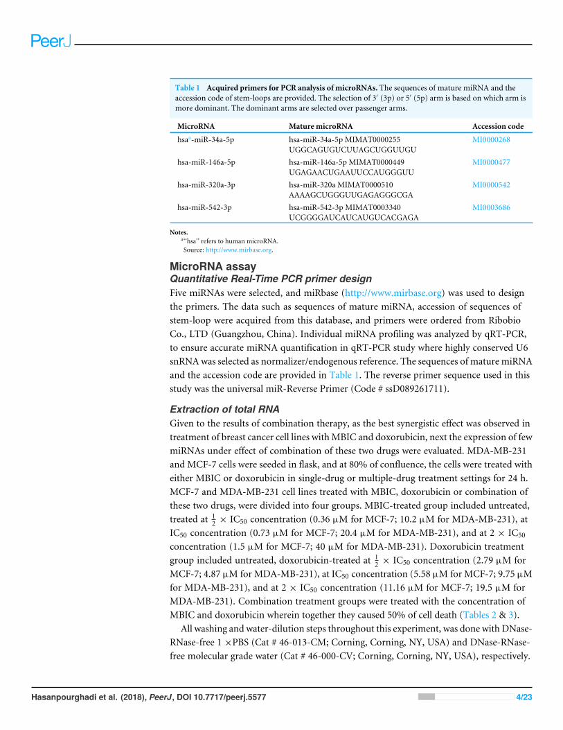
Table 1 Acquired primers for PCR analysis of microRNAs. The sequences of mature miRNA and theaccession code of stem-loops are provided. The selection of 3′ (3p) or 5′ (5p) arm is based on which arm ismore dominant. The dominant arms are selected over passenger arms.
MicroRNA Mature microRNA Accession code
hsaa-miR-34a-5p hsa-miR-34a-5p MIMAT0000255UGGCAGUGUCUUAGCUGGUUGU
MI0000268
hsa-miR-146a-5p hsa-miR-146a-5p MIMAT0000449UGAGAACUGAAUUCCAUGGGUU
MI0000477
hsa-miR-320a-3p hsa-miR-320a MIMAT0000510AAAAGCUGGGUUGAGAGGGCGA
MI0000542
hsa-miR-542-3p hsa-miR-542-3p MIMAT0003340UCGGGGAUCAUCAUGUCACGAGA
MI0003686
Notes.a‘‘hsa’’ refers to human microRNA.Source: http://www.mirbase.org.
MicroRNA assayQuantitative Real-Time PCR primer designFive miRNAs were selected, and miRbase (http://www.mirbase.org) was used to designthe primers. The data such as sequences of mature miRNA, accession of sequences ofstem-loop were acquired from this database, and primers were ordered from RibobioCo., LTD (Guangzhou, China). Individual miRNA profiling was analyzed by qRT-PCR,to ensure accurate miRNA quantification in qRT-PCR study where highly conserved U6snRNAwas selected as normalizer/endogenous reference. The sequences of mature miRNAand the accession code are provided in Table 1. The reverse primer sequence used in thisstudy was the universal miR-Reverse Primer (Code # ssD089261711).
Extraction of total RNAGiven to the results of combination therapy, as the best synergistic effect was observed intreatment of breast cancer cell lines withMBIC and doxorubicin, next the expression of fewmiRNAs under effect of combination of these two drugs were evaluated. MDA-MB-231and MCF-7 cells were seeded in flask, and at 80% of confluence, the cells were treated witheither MBIC or doxorubicin in single-drug or multiple-drug treatment settings for 24 h.MCF-7 and MDA-MB-231 cell lines treated with MBIC, doxorubicin or combination ofthese two drugs, were divided into four groups. MBIC-treated group included untreated,treated at 1
2 × IC50 concentration (0.36 µM for MCF-7; 10.2 µM for MDA-MB-231), atIC50 concentration (0.73 µM for MCF-7; 20.4 µM for MDA-MB-231), and at 2 × IC50
concentration (1.5 µM for MCF-7; 40 µM for MDA-MB-231). Doxorubicin treatmentgroup included untreated, doxorubicin-treated at 1
2 × IC50 concentration (2.79 µM forMCF-7; 4.87 µM for MDA-MB-231), at IC50 concentration (5.58 µM for MCF-7; 9.75 µMfor MDA-MB-231), and at 2 × IC50 concentration (11.16 µM for MCF-7; 19.5 µM forMDA-MB-231). Combination treatment groups were treated with the concentration ofMBIC and doxorubicin wherein together they caused 50% of cell death (Tables 2 & 3).
All washing and water-dilution steps throughout this experiment, was done with DNase-RNase-free 1 ×PBS (Cat # 46-013-CM; Corning, Corning, NY, USA) and DNase-RNase-free molecular grade water (Cat # 46-000-CV; Corning, Corning, NY, USA), respectively.
Hasanpourghadi et al. (2018), PeerJ, DOI 10.7717/peerj.5577 4/23
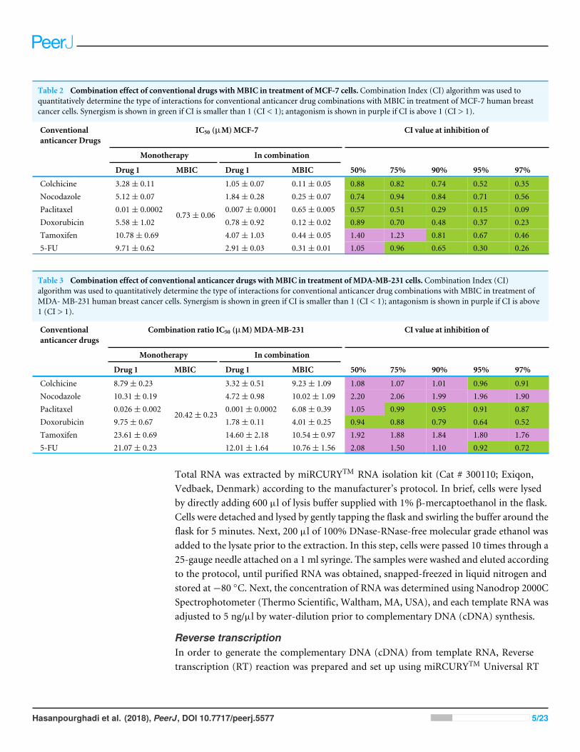
Table 2 Combination effect of conventional drugs withMBIC in treatment of MCF-7 cells. Combination Index (CI) algorithm was used toquantitatively determine the type of interactions for conventional anticancer drug combinations with MBIC in treatment of MCF-7 human breastcancer cells. Synergism is shown in green if CI is smaller than 1 (CI < 1); antagonism is shown in purple if CI is above 1 (CI > 1).
Conventionalanticancer Drugs
IC50 (µM)MCF-7 CI value at inhibition of
Monotherapy In combination
Drug 1 MBIC Drug 1 MBIC 50% 75% 90% 95% 97%
Colchicine 3.28± 0.11 1.05± 0.07 0.11± 0.05 0.88 0.82 0.74 0.52 0.35Nocodazole 5.12± 0.07 1.84± 0.28 0.25± 0.07 0.74 0.94 0.84 0.71 0.56Paclitaxel 0.01± 0.0002 0.007± 0.0001 0.65± 0.005 0.57 0.51 0.29 0.15 0.09Doxorubicin 5.58± 1.02 0.78± 0.92 0.12± 0.02 0.89 0.70 0.48 0.37 0.23Tamoxifen 10.78± 0.69 4.07± 1.03 0.44± 0.05 1.40 1.23 0.81 0.67 0.465-FU 9.71± 0.62
0.73± 0.06
2.91± 0.03 0.31± 0.01 1.05 0.96 0.65 0.30 0.26
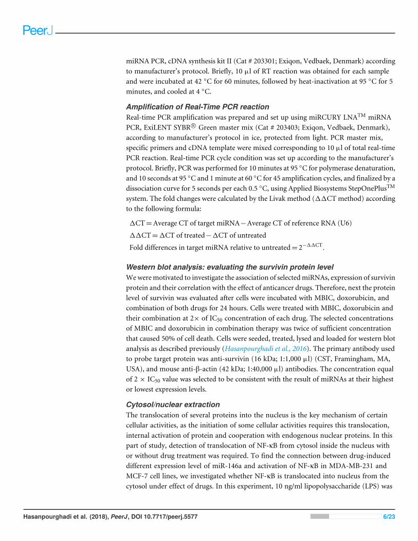
Table 3 Combination effect of conventional anticancer drugs withMBIC in treatment of MDA-MB-231 cells. Combination Index (CI)algorithm was used to quantitatively determine the type of interactions for conventional anticancer drug combinations with MBIC in treatment ofMDA- MB-231 human breast cancer cells. Synergism is shown in green if CI is smaller than 1 (CI < 1); antagonism is shown in purple if CI is above1 (CI > 1).
Conventionalanticancer drugs
Combination ratio IC50 (µM)MDA-MB-231 CI value at inhibition of
Monotherapy In combination
Drug 1 MBIC Drug 1 MBIC 50% 75% 90% 95% 97%
Colchicine 8.79± 0.23 3.32± 0.51 9.23± 1.09 1.08 1.07 1.01 0.96 0.91Nocodazole 10.31± 0.19 4.72± 0.98 10.02± 1.09 2.20 2.06 1.99 1.96 1.90Paclitaxel 0.026± 0.002 0.001± 0.0002 6.08± 0.39 1.05 0.99 0.95 0.91 0.87Doxorubicin 9.75± 0.67 1.78± 0.11 4.01± 0.25 0.94 0.88 0.79 0.64 0.52Tamoxifen 23.61± 0.69 14.60± 2.18 10.54± 0.97 1.92 1.88 1.84 1.80 1.765-FU 21.07± 0.23
20.42± 0.23
12.01± 1.64 10.76± 1.56 2.08 1.50 1.10 0.92 0.72
Total RNA was extracted by miRCURYTM RNA isolation kit (Cat # 300110; Exiqon,Vedbaek, Denmark) according to the manufacturer’s protocol. In brief, cells were lysedby directly adding 600 µl of lysis buffer supplied with 1% β-mercaptoethanol in the flask.Cells were detached and lysed by gently tapping the flask and swirling the buffer around theflask for 5 minutes. Next, 200 µl of 100% DNase-RNase-free molecular grade ethanol wasadded to the lysate prior to the extraction. In this step, cells were passed 10 times through a25-gauge needle attached on a 1 ml syringe. The samples were washed and eluted accordingto the protocol, until purified RNA was obtained, snapped-freezed in liquid nitrogen andstored at−80 ◦C. Next, the concentration of RNA was determined using Nanodrop 2000CSpectrophotometer (Thermo Scientific, Waltham, MA, USA), and each template RNA wasadjusted to 5 ng/µl by water-dilution prior to complementary DNA (cDNA) synthesis.
Reverse transcriptionIn order to generate the complementary DNA (cDNA) from template RNA, Reversetranscription (RT) reaction was prepared and set up using miRCURYTM Universal RT
Hasanpourghadi et al. (2018), PeerJ, DOI 10.7717/peerj.5577 5/23
miRNA PCR, cDNA synthesis kit II (Cat # 203301; Exiqon, Vedbaek, Denmark) accordingto manufacturer’s protocol. Briefly, 10 µl of RT reaction was obtained for each sampleand were incubated at 42 ◦C for 60 minutes, followed by heat-inactivation at 95 ◦C for 5minutes, and cooled at 4 ◦C.
Amplification of Real-Time PCR reactionReal-time PCR amplification was prepared and set up using miRCURY LNATM miRNAPCR, ExiLENT SYBR R© Green master mix (Cat # 203403; Exiqon, Vedbaek, Denmark),according to manufacturer’s protocol in ice, protected from light. PCR master mix,specific primers and cDNA template were mixed corresponding to 10 µl of total real-timePCR reaction. Real-time PCR cycle condition was set up according to the manufacturer’sprotocol. Briefly, PCR was performed for 10 minutes at 95 ◦C for polymerase denaturation,and 10 seconds at 95 ◦C and 1minute at 60 ◦C for 45 amplification cycles, and finalized by adissociation curve for 5 seconds per each 0.5 ◦C, using Applied Biosystems StepOnePlusTM
system. The fold changes were calculated by the Livak method (11CT method) accordingto the following formula:
1CT=Average CT of target miRNA−Average CT of reference RNA (U6)
11CT=1CT of treated−1CT of untreated
Fold differences in target miRNA relative to untreated= 2−11CT.
Western blot analysis: evaluating the survivin protein levelWeweremotivated to investigate the association of selectedmiRNAs, expression of survivinprotein and their correlation with the effect of anticancer drugs. Therefore, next the proteinlevel of survivin was evaluated after cells were incubated with MBIC, doxorubicin, andcombination of both drugs for 24 hours. Cells were treated with MBIC, doxorubicin andtheir combination at 2× of IC50 concentration of each drug. The selected concentrationsof MBIC and doxorubicin in combination therapy was twice of sufficient concentrationthat caused 50% of cell death. Cells were seeded, treated, lysed and loaded for western blotanalysis as described previously (Hasanpourghadi et al., 2016). The primary antibody usedto probe target protein was anti-survivin (16 kDa; 1:1,000 µl) (CST, Framingham, MA,USA), and mouse anti-β-actin (42 kDa; 1:40,000 µl) antibodies. The concentration equalof 2 × IC50 value was selected to be consistent with the result of miRNAs at their highestor lowest expression levels.
Cytosol/nuclear extractionThe translocation of several proteins into the nucleus is the key mechanism of certaincellular activities, as the initiation of some cellular activities requires this translocation,internal activation of protein and cooperation with endogenous nuclear proteins. In thispart of study, detection of translocation of NF-κB from cytosol inside the nucleus withor without drug treatment was required. To find the connection between drug-induceddifferent expression level of miR-146a and activation of NF-κB in MDA-MB-231 andMCF-7 cell lines, we investigated whether NF-κB is translocated into nucleus from thecytosol under effect of drugs. In this experiment, 10 ng/ml lipopolysaccharide (LPS) was
Hasanpourghadi et al. (2018), PeerJ, DOI 10.7717/peerj.5577 6/23
used in positive control group. Each cell lines were divided into five groups of treatment,including untreated, LPS-treated, MBIC-treated, doxorubicin-treated and combination-treated groups.
To prepare the cytosol/nuclear extract, 5×10⁶ MDA-MB-231 and MCF-7 cell line wereseeded and harvested 24 hours after treatment. Cells were washed with 1× PBS and wereprocessed for cytoplasmic and nuclear protein fractions using NE-PER nuclear/cytoplasmicextraction reagent kit (Cat # 78833; Thermo Scientific, Waltham, MA, USA) accordingto the manufacturer. In brief, harvested cells were centrifuged at 500× g for 5 minutes.Cell pellets were treated with 500 µl of Cytoplasmic Extraction Reagent 1 (CERI), andwere vortexed for 15 s on the highest setting to totally suspend the cell pellet. Tubes wereincubated on ice for 10 minutes and then 27.5 µl of CERII was added to each tube. Tubeswere vortexed for 5 seconds on the highest setting and followed by incubation in the icefor 1 minute. Tubes were centrifuged at 16,000× g for 5 minutes. After centrifugation, thesupernatant that is the cytoplasmic extract, was transferred into a pre-chilled tube. Theinsoluble part of fraction contains nuclei proteins, were treated with 250 µl of NuclearExtraction Reagent (NER). Tubes were kept on ice and were vortexed for 15 seconds every10 minutes for total of 40 minutes. Tubes were centrifuged at 16,000× g for 10 minutesand the supernatant which contains nuclear extract fraction, was immediately transferredto a pre-chilled tube.
Western blot analysis: evaluating activation of NF-κBThe extracted nuclear and cytoplasmic proteins were used for western blot analysis. GAPDHand Lamin B1 proteins were used as markers of cytoplasm and nucleus respectively. Thesetwo endogenous markers were also probed as negative controls for the opposing fractions.GAPDH was used as negative control for nuclear fraction, while Lamin B1 was used asnegative control for the cytosolic fraction. The primary antibody used to probe targetproteins were anti-NF-κB (65 kDa; 1:1,000 µl) (CST, Framingham, MA, USA), anti-GAPDH (35 kDa; 1:20,000 µl) (Santa Cruz Biotechnology, Santa Cruz, CA , USA), andmouse anti-Lamin B1 (66 kDa; 1:20,000 µl) (CST, Framingham, MA, USA) antibodies.
Statistical analysisA one-way analysis of variance (ANOVA) at statistically significance levels that wereexpressed as P value ≤ 0.05 shown as ‘‘*’’; P value ≤ 0.01 shown as ‘‘**’’; P value ≤ 0.001shown as ‘‘***’’; P value ≤ 0.0001 shown as ‘‘****’’ were conducted. P value > 0.05 wasconsidered not significant and was shown as ‘‘ns’’. The Bonferroni pos t -test was usedto test the statistical differences between control and treated groups. Statistical analysiswas performed using GraphPad Prism version 7.00 (Graph Pad Software, San Diego, CA,USA). The intensities of western blot’s protein bands were quantified by imageJ version1.51j8 (NIH, Bethesda, MD, USA), by basic intensity quantification. Data were expressedas mean ± SD of three independent experiments.
Hasanpourghadi et al. (2018), PeerJ, DOI 10.7717/peerj.5577 7/23
RESULTSMBIC displayed a synergistic effect with doxorubicin in MCF-7and MDA-MB-231 cell linesTo maximize the cytotoxic effect of MBIC, breast cancer cells were sequentially treatedwith different known anticancer drugs and IC50s were determined. In Tables 2 and 3,a Combination Index (CI) algorithm was used to quantitatively determine the type ofinteractions for each drug combination as follows, synergism if CI is smaller than 1 (CI< 1), additivity if CI is equal 1 (CI = 1), and antagonism if CI is above 1 (CI > 1).Tables 2 and 3 showed the results following combination of MBIC with each of the sixconventional anticancer drugs in MCF-7 and MDA-MB-231 cell lines. The synergisticeffects of combination of two drugs are shown in green. This color represented twodrugs that in combination have higher effect than the effect of each individual drug.The antagonistic effect where two drugs in combination that have less effect comparedto each individual drug, was shown in purple in Tables 2 and 3. Besides, the synergisticand antagonistic effects were classified based on the percentage of cells killed by thecombined drugs (50% to 97% of cell death). Doxorubicin exhibited synergistic effect withMBIC at throughout the entire range of 50% to 97% of inhibition in both MCF-7 andMDA-MB-231 cell lines. Another interesting point was that the concentration of eitherMBIC or doxorubicin in combination that is required for killing 50% of the cells, decreasedsignificantly, especially in MCF-7 cells. Similarly, colchicine, nocodazole and paclitaxelexhibited synergistic effects with MBIC at the full range of 50% to 97% in MCF-7 butnot in MDA-MB-231 cell line (Tables 2 & 3). Nocodazole and tamoxifen demonstratedadditive effects for the entire scopes of CI value (50% to 97%) in MDA-MB-231 cells.However, colchicine, paclitaxel and 5-FU in combination with MBIC, indicated selectivesynergistic effect ranging between 50% to 97% of inhibition, in both breast cancer celllines. In Tables 2 and 3 synergism is displayed in green, while antagonism is exhibited inpurple.
MicroRNA profilingAs the greatest synergistic anticancer effect was observed following the combined treatmentof MBIC with doxorubicin in MDA-MB-231 and MCF-7 cell lines, further the effect ofboth drugs on the expression of several miRNAs including miR-34a, miR-146a, miR-320aand miR-542 were determined (Figs. 1A–1D, 2A–2D, 3A–3D & 4A–4D; Tables 4 & 5).MBIC treatment at 2× IC50 concentration caused 9.5-fold elevated expression level ofmiR-34a in MCF-7 cells, but only 1.7-fold increase in MDA-MB-231 cells. The elevatedlevel of expression of miR-34a was 1.5-fold in doxorubicin-treated MCF-7, while the resultin doxorubicin treatment of MDA-MB-231 was reduced 0.8-fold. Following the combineddrug treatment, a marked increase of 32.3-fold in expression level of miR-34a in MCF-7,and 13.9-fold in expression level of miR-34a in MDA-MB-231 cells were observed.
Individual treatment with MBIC and doxorubicin at 2× IC50 concentration in MCF-7cells caused a 0.5-fold and 0.8-fold decrease expression of miR-146a, respectively. Incontrast, combined treatment with the two drugs reduced the expression of miR-146ato 0.2-fold in MCF-7 cells. In MDA-MB-231 cells, individual treatment with MBIC and
Hasanpourghadi et al. (2018), PeerJ, DOI 10.7717/peerj.5577 8/23
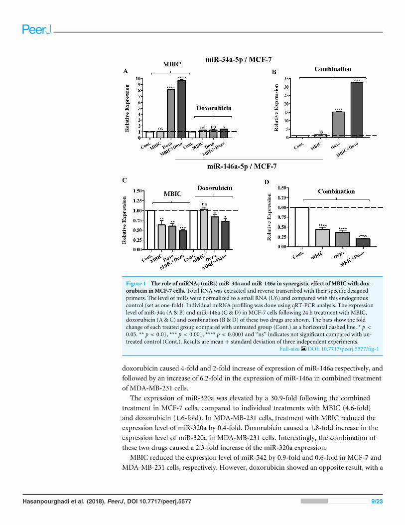
Figure 1 The role of miRNAs (miRs) miR-34a andmiR-146a in synergistic effect of MBIC with dox-orubicin inMCF-7 cells. Total RNA was extracted and reverse transcribed with their specific designedprimers. The level of miRs were normalized to a small RNA (U6) and compared with this endogenouscontrol (set as one-fold). Individual miRNA profiling was done using qRT-PCR analysis. The expressionlevel of miR-34a (A & B) and miR-146a (C & D) in MCF-7 cells following 24 h treatment with MBIC,doxorubicin (A & C) and combination (B & D) of these two drugs are shown. The bars show the foldchange of each treated group compared with untreated group (Cont.) as a horizontal dashed line. * p <
0.05. ** p < 0.01, *** p < 0.001, **** p < 0.0001 and ‘‘ns’’ indicates not significant compared with un-treated control (Cont.). Results are mean+ standard deviation of three independent experiments.
Full-size DOI: 10.7717/peerj.5577/fig-1
doxorubicin caused 4-fold and 2-fold increase of expression of miR-146a respectively, andfollowed by an increase of 6.2-fold in the expression of miR-146a in combined treatmentof MDA-MB-231 cells.
The expression of miR-320a was elevated by a 30.9-fold following the combinedtreatment in MCF-7 cells, compared to individual treatments with MBIC (4.6-fold)and doxorubicin (1.6-fold). In MDA-MB-231 cells, treatment with MBIC reduced theexpression level of miR-320a by 0.4-fold. Doxorubicin caused a 1.8-fold increase in theexpression level of miR-320a in MDA-MB-231 cells. Interestingly, the combination ofthese two drugs caused a 2.3-fold increase of the miR-320a expression.
MBIC reduced the expression level of miR-542 by 0.9-fold and 0.6-fold in MCF-7 andMDA-MB-231 cells, respectively. However, doxorubicin showed an opposite result, with a
Hasanpourghadi et al. (2018), PeerJ, DOI 10.7717/peerj.5577 9/23

Figure 2 The role of miRNAs (miRs) miR-320a andmiR-542 in synergistic effect of MBIC with dox-orubicin inMCF-7 cells. Total RNA was extracted and reverse transcribed with their specific designedprimers. The level of miRs were normalized to a small RNA (U6) and compared with this endogenouscontrol (set as one-fold). Individual miRNA profiling was done using qRT-PCR analysis. The expressionlevel of miR-320a (A & B) and miR-542a (C & D) in MCF-7 cells following 24 h treatment with MBIC,doxorubicin (A & C) and combination (B & D) of these two drugs are shown. The bars show the foldchange of each treated group compared with untreated group (Cont.) as a horizontal dashed line. * p <
0.05. ** p < 0.01, **** p < 0.0001 and ‘‘ns’’ indicates not significant compared with untreated control(Cont.). Results are mean + standard deviation of three independent experiments.
Full-size DOI: 10.7717/peerj.5577/fig-2
1.5-fold increase of expression in MCF-7 and no change of modification in MDA-MB-231cells. On the other hand, the seemingly synergistic effect of two drugs caused a 3.5-fold and2.1-fold elevated level in expression of miR-542 at 2× IC50 concentration in MCF-7 andMDA-MB-231 cells, respectively.
Tables 4 and 5 showed fold differences for the expression of fourmiRNAs in three groupsof treatment compared to untreated (2−11CT )MCF-7 andMDA-MB-231 cells respectively.In Figs. 1A–1D, 2A–2D, 3A–3D & 4A–4D the bar graphs represented fold-changes in thelevel of expression of miRNAs. Figure 5 is an illustration of elevation or reduction ofexpression of miRNAs in breast cancer cell lines.
Hasanpourghadi et al. (2018), PeerJ, DOI 10.7717/peerj.5577 10/23

Figure 3 The role of miRNAs (miRs) miR-34a andmiR-146a in synergistic effect of MBIC with dox-orubicin inMDA-MB-231 cells. Total RNA was extracted and reverse transcribed with their specific de-signed primers. The level of miRs were normalized to a small RNA (U6) and compared with this endoge-nous control (set as one-fold). Individual miRNA profiling was done using qRT-PCR analysis. The expres-sion level of miR-34a (A & B) and miR-146a (C & D) in MDA-MB-231 cells following 24 h treatment withMBIC, doxorubicin (A & C) and combination (B & D) of these two drugs are shown. The bars show thefold change of each treated group compared with untreated group (Cont.) as a horizontal dashed line. *p < 0.05. ** p < 0.01, *** p < 0.001, **** p < 0.0001 and ‘‘ns’’ indicates not significant compared withuntreated control (Cont.). Results are mean+ standard deviation of three independent experiments.
Full-size DOI: 10.7717/peerj.5577/fig-3
Survivin may negatively correlate with miR-34a, miR-320a andmiR-542 expression in MCF-7 cell lineThe protein level of survivin was evaluated in four groups of untreated, MBIC-treated,doxorubicin-treated and combination-treated groups of MDA-MB-231 and MCF-7 celllines by western blot analysis (Figs. 6A–6D). Comparison between expression level of threemiRNAs, miR-34a, miR-320a, miR-542 (Table 4), and the protein level of survivin in thedifferent groups of treatments in the MCF-7 cell line (Figs. 6A & 6C), indicated that thereis a negative correlation between the protein level of survivin and these three miRNAs. Theobserved negative correlation was highlighted in combination treatment of MCF-7 cells,wherein the expression level of miR-34a, miR-320a and miR-542 were elevated 32.3-fold,30.9-fold and 3.5-fold respectively. Thereby, the three miRNAs at 2× IC50 concentration
Hasanpourghadi et al. (2018), PeerJ, DOI 10.7717/peerj.5577 11/23
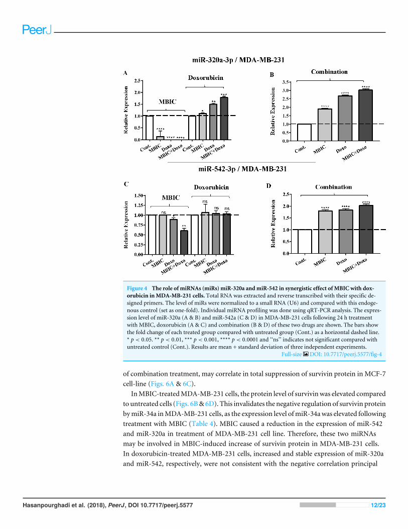
Figure 4 The role of miRNAs (miRs) miR-320a andmiR-542 in synergistic effect of MBIC with dox-orubicin inMDA-MB-231 cells. Total RNA was extracted and reverse transcribed with their specific de-signed primers. The level of miRs were normalized to a small RNA (U6) and compared with this endoge-nous control (set as one-fold). Individual miRNA profiling was done using qRT-PCR analysis. The expres-sion level of miR-320a (A & B) and miR-542a (C & D) in MDA-MB-231 cells following 24 h treatmentwith MBIC, doxorubicin (A & C) and combination (B & D) of these two drugs are shown. The bars showthe fold change of each treated group compared with untreated group (Cont.) as a horizontal dashed line.* p < 0.05. ** p < 0.01, *** p < 0.001, **** p < 0.0001 and ‘‘ns’’ indicates not significant compared withuntreated control (Cont.). Results are mean + standard deviation of three independent experiments.
Full-size DOI: 10.7717/peerj.5577/fig-4
of combination treatment, may correlate in total suppression of survivin protein in MCF-7cell-line (Figs. 6A & 6C).
InMBIC-treatedMDA-MB-231 cells, the protein level of survivinwas elevated comparedto untreated cells (Figs. 6B&6D). This invalidates the negative regulation of survivin proteinbymiR-34a inMDA-MB-231 cells, as the expression level ofmiR-34awas elevated followingtreatment with MBIC (Table 4). MBIC caused a reduction in the expression of miR-542and miR-320a in treatment of MDA-MB-231 cell line. Therefore, these two miRNAsmay be involved in MBIC-induced increase of survivin protein in MDA-MB-231 cells.In doxorubicin-treated MDA-MB-231 cells, increased and stable expression of miR-320aand miR-542, respectively, were not consistent with the negative correlation principal
Hasanpourghadi et al. (2018), PeerJ, DOI 10.7717/peerj.5577 12/23
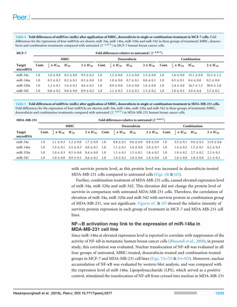
Table 4 Fold differences of miRNAs (miRs) after application of MBIC, doxorubicin in single or combination treatment inMCF-7 cells. Folddifferences for the expression of four miRNAs are shown; miR-34a, miR-146a, miR-320a and miR-542 in three groups of treatment; MBIC, doxoru-bicin and combination treatments compared with untreated (2−11CT) in MCF-7 human breast cancer cells.
MCF-7 Fold differences relative to untreated ( 2−11CT)
MBIC Doxorubicin Combination
TargetmicroRNA
Cont. 12 × IC50 IC50 2× IC50 Cont. 1
2 × IC50 IC50 2× IC50 Cont. 12 × IC50 IC50 2× IC50
miR-34a 1.0 1.0± 0.0 8.2± 0.0 9.5± 0.2 1.0 1.2± 0.0 1.3± 0.0 1.5± 0.0 1.0 1.6± 0.0 15.1± 0.9 32.3± 1.2miR-146a 1.0 0.3± 0.2 0.2± 0.1 0.5± 0.0 1.0 1.0± 0.0 0.7± 0.1 0.8± 0.1 1.0 0.5± 0.1 0.4± 0.0 0.2± 0.0miR-320a 1.0 1.2± 0.1 5.4± 0.3 4.6± 0.3 1.0 0.9± 0.0 1.0± 0.0 1.6± 0.0 1.0 2.4± 0.0 16.7± 1.3 30.9± 2.8miR-542 1.0 0.8± 0.2 0.8± 0.0 0.9± 0.2 1.0 1.1± 0.3 1.5± 0.1 1.5± 0.2 1.0 1.0± 0.3 3.0± 0.4 3.5± 0.2
Table 5 Fold differences of miRNAs (miRs) after application of MBIC, doxorubicin in single or combination treatment inMDA-MB-231 cells.Fold differences for the expression of four miRNAs are shown; miR-34a, miR-146a, miR-320a and miR-542 in three groups of treatment; MBIC,doxorubicin and combination treatments compared with untreated (2−11CT) in MDA-MB-231 human breast cancer cells.
MDA-MB-231 Fold differences relative to untreated (2−11CT)
MBIC Doxorubicin Combination
TargetmicroRNA
Cont. 12 × IC50 IC50 2× IC50 Cont. 1
2 × IC50 IC50 2× IC50 Cont. 12 × IC50 IC50 2× IC50
miR-34a 1.0 1.1± 0.3 1.2± 0.0 1.7± 0.0 1.0 0.8± 0.1 0.6± 0.0 0.8± 0.0 1.0 5.5± 0.1 9.6± 0.3 13.9± 0.6miR-146a 1.0 1.9± 0.1 3.5± 0.3 4.0± 0.2 1.0 1.1± 0.1 1.6± 0.0 2.0± 0.3 1.0 1.4± 0.2 1.5± 0.1 6.2± 0.4miR-320a 1.0 1.0± 0.3 0.5± 0.0 0.4± 0.0 1.0 1.1± 0.1 1.5± 0.1 1.8± 0.2 1.0 1.9± 0.2 2.7± 0.2 2.3± 0.3miR-542 1.0 1.0± 0.0 0.9± 0.1 0.6± 0.2 1.0 1.0± 0.2 1.0± 0.0 1.0± 0.0 1.0 1.8± 0.0 1.8± 0.0 2.1± 0.1
with survivin protein level, as this protein level was increased in doxorubicin-treatedMDA-MB-231 cells compared to untreated cells (Figs. 6B & 6D).
Further, combination treatment of MDA-MB-231 cells, caused elevated expression levelof miR-34a, miR-320a and miR-542. This elevation did not change the protein level ofsurvivin in comparison with untreated MDA-MB-231 cells. Therefore, the correlation ofelevation of miR-34a, miR-320a and miR-542 with survivin protein in combination groupof MDA-MB-231, was not significant. Figures 6C & 6D showed the relative intensity ofsurvivin protein expression in each group of treatment in MCF-7 and MDA-MB-231 celllines.
NF-κB activation may link to the expression of miR-146a inMDA-MB-231 cell lineSince miR-146a at elevated expression level is reported to correlate with suppression of theactivity of NF-κB in metastatic human breast cancer cells (Bhaumik et al., 2008), in presentstudy, this correlation was evaluated. Nuclear translocation of NF-κB was evaluated in allfour groups of untreated, MBIC-treated, doxorubicin-treated and combination-treatedgroups in MCF-7 and MDA-MB-231 cell lines (Figs. 7A–7D & 8A–8D). Moreover, nuclearaccumulation of NF-κB was evaluated by western blot analysis, and was compared withthe expression level of miR-146a. Lipopolysaccharide (LPS), which served as a positivecontrol, stimulated the translocation of NF-κB from cytosol into nucleus in MDA-MB-231
Hasanpourghadi et al. (2018), PeerJ, DOI 10.7717/peerj.5577 13/23
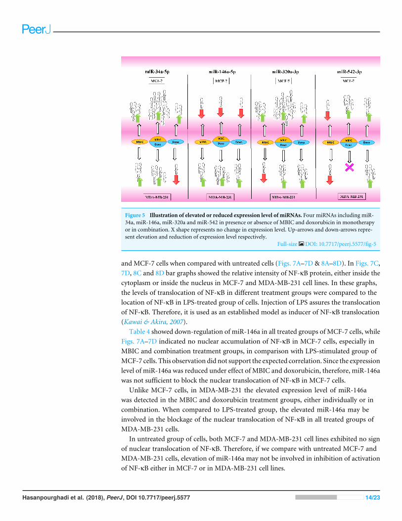
Figure 5 Illustration of elevated or reduced expression level of miRNAs. Four miRNAs including miR-34a, miR-146a, miR-320a and miR-542 in presence or absence of MBIC and doxorubicin in monotherapyor in combination. X shape represents no change in expression level. Up-arrows and down-arrows repre-sent elevation and reduction of expression level respectively.
Full-size DOI: 10.7717/peerj.5577/fig-5
and MCF-7 cells when compared with untreated cells (Figs. 7A–7D & 8A–8D). In Figs. 7C,7D, 8C and 8D bar graphs showed the relative intensity of NF-κB protein, either inside thecytoplasm or inside the nucleus in MCF-7 and MDA-MB-231 cell lines. In these graphs,the levels of translocation of NF-κB in different treatment groups were compared to thelocation of NF-κB in LPS-treated group of cells. Injection of LPS assures the translocationof NF-κB. Therefore, it is used as an established model as inducer of NF-κB translocation(Kawai & Akira, 2007).
Table 4 showed down-regulation of miR-146a in all treated groups of MCF-7 cells, whileFigs. 7A–7D indicated no nuclear accumulation of NF-κB in MCF-7 cells, especially inMBIC and combination treatment groups, in comparison with LPS-stimulated group ofMCF-7 cells. This observation did not support the expected correlation. Since the expressionlevel of miR-146a was reduced under effect of MBIC and doxorubicin, therefore, miR-146awas not sufficient to block the nuclear translocation of NF-κB in MCF-7 cells.
Unlike MCF-7 cells, in MDA-MB-231 the elevated expression level of miR-146awas detected in the MBIC and doxorubicin treatment groups, either individually or incombination. When compared to LPS-treated group, the elevated miR-146a may beinvolved in the blockage of the nuclear translocation of NF-κB in all treated groups ofMDA-MB-231 cells.
In untreated group of cells, both MCF-7 and MDA-MB-231 cell lines exhibited no signof nuclear translocation of NF-κB. Therefore, if we compare with untreated MCF-7 andMDA-MB-231 cells, elevation of miR-146a may not be involved in inhibition of activationof NF-κB either in MCF-7 or in MDA-MB-231 cell lines.
Hasanpourghadi et al. (2018), PeerJ, DOI 10.7717/peerj.5577 14/23
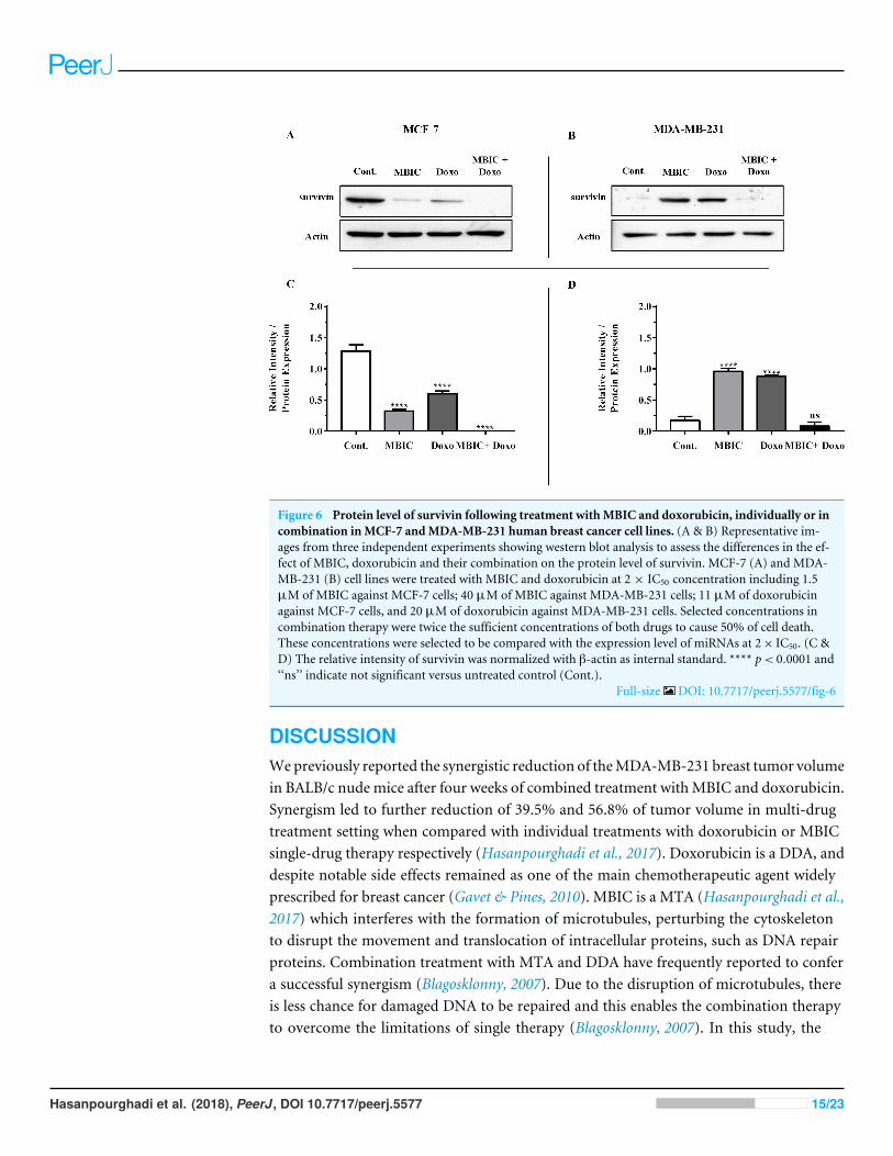
Figure 6 Protein level of survivin following treatment withMBIC and doxorubicin, individually or incombination inMCF-7 andMDA-MB-231 human breast cancer cell lines. (A & B) Representative im-ages from three independent experiments showing western blot analysis to assess the differences in the ef-fect of MBIC, doxorubicin and their combination on the protein level of survivin. MCF-7 (A) and MDA-MB-231 (B) cell lines were treated with MBIC and doxorubicin at 2 × IC50 concentration including 1.5µM of MBIC against MCF-7 cells; 40 µM of MBIC against MDA-MB-231 cells; 11 µM of doxorubicinagainst MCF-7 cells, and 20 µM of doxorubicin against MDA-MB-231 cells. Selected concentrations incombination therapy were twice the sufficient concentrations of both drugs to cause 50% of cell death.These concentrations were selected to be compared with the expression level of miRNAs at 2× IC50. (C &D) The relative intensity of survivin was normalized with β-actin as internal standard. **** p< 0.0001 and‘‘ns’’ indicate not significant versus untreated control (Cont.).
Full-size DOI: 10.7717/peerj.5577/fig-6
DISCUSSIONWepreviously reported the synergistic reduction of theMDA-MB-231 breast tumor volumein BALB/c nude mice after four weeks of combined treatment withMBIC and doxorubicin.Synergism led to further reduction of 39.5% and 56.8% of tumor volume in multi-drugtreatment setting when compared with individual treatments with doxorubicin or MBICsingle-drug therapy respectively (Hasanpourghadi et al., 2017). Doxorubicin is a DDA, anddespite notable side effects remained as one of the main chemotherapeutic agent widelyprescribed for breast cancer (Gavet & Pines, 2010). MBIC is a MTA (Hasanpourghadi et al.,2017) which interferes with the formation of microtubules, perturbing the cytoskeletonto disrupt the movement and translocation of intracellular proteins, such as DNA repairproteins. Combination treatment with MTA and DDA have frequently reported to confera successful synergism (Blagosklonny, 2007). Due to the disruption of microtubules, thereis less chance for damaged DNA to be repaired and this enables the combination therapyto overcome the limitations of single therapy (Blagosklonny, 2007). In this study, the
Hasanpourghadi et al. (2018), PeerJ, DOI 10.7717/peerj.5577 15/23
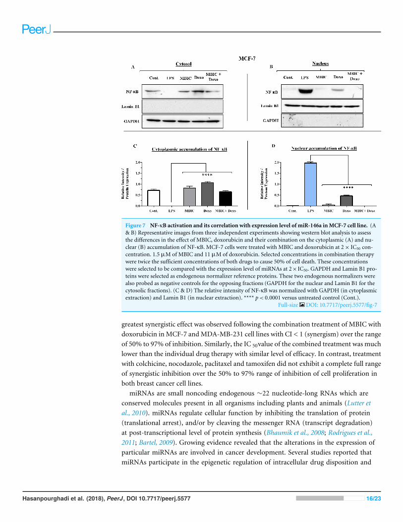
Figure 7 NF-κB activation and its correlation with expression level of miR-146a inMCF-7 cell line. (A& B) Representative images from three independent experiments showing western blot analysis to assessthe differences in the effect of MBIC, doxorubicin and their combination on the cytoplasmic (A) and nu-clear (B) accumulation of NF-κB. MCF-7 cells were treated with MBIC and doxorubicin at 2× IC50 con-centration. 1.5 µM of MBIC and 11 µM of doxorubicin. Selected concentrations in combination therapywere twice the sufficient concentrations of both drugs to cause 50% of cell death. These concentrationswere selected to be compared with the expression level of miRNAs at 2× IC50. GAPDH and Lamin B1 pro-teins were selected as endogenous normalizer reference proteins. These two endogenous normalizers werealso probed as negative controls for the opposing fractions (GAPDH for the nuclear and Lamin B1 for thecytosolic fractions). (C & D) The relative intensity of NF-κB was normalized with GAPDH (in cytoplasmicextraction) and Lamin B1 (in nuclear extraction). **** p< 0.0001 versus untreated control (Cont.).
Full-size DOI: 10.7717/peerj.5577/fig-7
greatest synergistic effect was observed following the combination treatment of MBIC withdoxorubicin in MCF-7 andMDA-MB-231 cell lines with CI < 1 (synergism) over the rangeof 50% to 97% of inhibition. Similarly, the IC 50value of the combined treatment was muchlower than the individual drug therapy with similar level of efficacy. In contrast, treatmentwith colchicine, nocodazole, paclitaxel and tamoxifen did not exhibit a complete full rangeof synergistic inhibition over the 50% to 97% range of inhibition of cell proliferation inboth breast cancer cell lines.
miRNAs are small noncoding endogenous ∼22 nucleotide-long RNAs which areconserved molecules present in all organisms including plants and animals (Lutter etal., 2010). miRNAs regulate cellular function by inhibiting the translation of protein(translational arrest), and/or by cleaving the messenger RNA (transcript degradation)at post-transcriptional level of protein synthesis (Bhaumik et al., 2008; Rodrigues et al.,2011; Bartel, 2009). Growing evidence revealed that the alterations in the expression ofparticular miRNAs are involved in cancer development. Several studies reported thatmiRNAs participate in the epigenetic regulation of intracellular drug disposition and
Hasanpourghadi et al. (2018), PeerJ, DOI 10.7717/peerj.5577 16/23
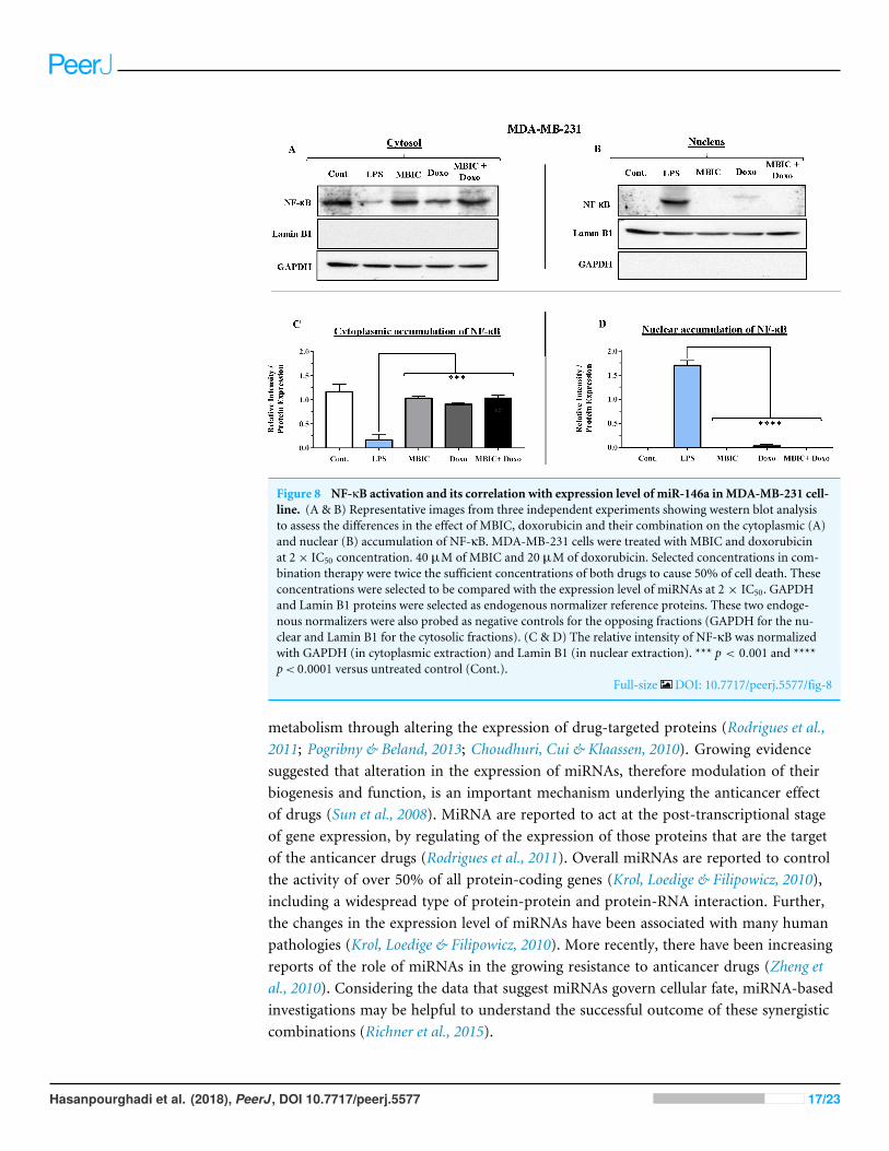
Figure 8 NF-κB activation and its correlation with expression level of miR-146a inMDA-MB-231 cell-line. (A & B) Representative images from three independent experiments showing western blot analysisto assess the differences in the effect of MBIC, doxorubicin and their combination on the cytoplasmic (A)and nuclear (B) accumulation of NF-κB. MDA-MB-231 cells were treated with MBIC and doxorubicinat 2× IC50 concentration. 40 µM of MBIC and 20 µM of doxorubicin. Selected concentrations in com-bination therapy were twice the sufficient concentrations of both drugs to cause 50% of cell death. Theseconcentrations were selected to be compared with the expression level of miRNAs at 2 × IC50. GAPDHand Lamin B1 proteins were selected as endogenous normalizer reference proteins. These two endoge-nous normalizers were also probed as negative controls for the opposing fractions (GAPDH for the nu-clear and Lamin B1 for the cytosolic fractions). (C & D) The relative intensity of NF-κB was normalizedwith GAPDH (in cytoplasmic extraction) and Lamin B1 (in nuclear extraction). *** p < 0.001 and ****p< 0.0001 versus untreated control (Cont.).
Full-size DOI: 10.7717/peerj.5577/fig-8
metabolism through altering the expression of drug-targeted proteins (Rodrigues et al.,2011; Pogribny & Beland, 2013; Choudhuri, Cui & Klaassen, 2010). Growing evidencesuggested that alteration in the expression of miRNAs, therefore modulation of theirbiogenesis and function, is an important mechanism underlying the anticancer effectof drugs (Sun et al., 2008). MiRNA are reported to act at the post-transcriptional stageof gene expression, by regulating of the expression of those proteins that are the targetof the anticancer drugs (Rodrigues et al., 2011). Overall miRNAs are reported to controlthe activity of over 50% of all protein-coding genes (Krol, Loedige & Filipowicz, 2010),including a widespread type of protein-protein and protein-RNA interaction. Further,the changes in the expression level of miRNAs have been associated with many humanpathologies (Krol, Loedige & Filipowicz, 2010). More recently, there have been increasingreports of the role of miRNAs in the growing resistance to anticancer drugs (Zheng etal., 2010). Considering the data that suggest miRNAs govern cellular fate, miRNA-basedinvestigations may be helpful to understand the successful outcome of these synergisticcombinations (Richner et al., 2015).
Hasanpourghadi et al. (2018), PeerJ, DOI 10.7717/peerj.5577 17/23

In this study, we investigated the role of several miRNAs including miR-34a, miR-146a,miR-320a and miR-542 in the synergistic anticancer actions of MBIC and doxorubicindrugs on breast tumors. Fig. 5 illustrates the regulation of miR-34a, miR-320a, miR-146aand miR-542 in MCF-7 and MDA-MB-231 cell lines following MBIC and doxorubicinsingle and multi-therapies. The fold changes of the expression of these four miRNAswere evaluated at the highest concentration (2× IC50) of MBIC and doxorubicin either asmonotherapy or in combination therapy. miR-34a is one of the most prominent miRNAswhich generally functions as a tumor suppressor (Zhang et al., 2014). miR-34a is down-regulated in a majority of cancer types and alteration of the expression of miR-34a inhibitscellular proliferation and induces apoptosis in cancer cells (Bader, Brown &Winkler, 2010;Li, Ren & Tang, 2014). miR-34a has been reported to be associated with resistance of aMTA, docetaxel in MCF-7 and MDA-MB-231 cell lines (Kastl, Brown & Schofield, 2012).miR-34a also is known to target the post-transcriptional regulation of survivin proteinin gastric cancer cells (Cao et al., 2013). Survivin is a member of inhibitor of apoptosisfamily (Mckenzie & Grossman, 2012). Increase expression of survivin promotes cancer cellproliferation by suppressing the apoptosis of the cancer cells (Church & Talbot, 2012). In thepresent study, combined treatment of MBIC with doxorubicin increased the expression ofmiR-34a in both MCF-7 (32.3 fold) and MDA-MB-231 cell lines (13.9 fold). As miR-34a isa well-known target of p53 (Navarro & Lieberman, 2015; Okada et al., 2014; Raver-Shapiraet al., 2007), it is tempting to speculate that this may occur due to enhanced p53 activationas a consequences of DNA damage.
In contrast, increased expression of miR-320a is reported to be linked to suppressionof survivin and induction of apoptosis (Diakos et al., 2010). In the present study, thecombination therapy of MBIC with doxorubicin resulted in a 30.9-fold and 2.3-foldincrease of expression of miR-320a in MCF-7 and MDA-MB-231 cells, respectively.
Several studies reported the increased expression of miR-542 inhibited the expressionof survivin in cancer cells (Yoon et al., 2010). The combined treatment of MBIC withdoxorubicin treatment was associated with less than four-fold increase in the expressionof miR-542 in both cancer cell lines. miR-34a, miR-320a and miR-542 are reported tobe inversely correlated with the protein and gene expression levels of survivin in severalcancer types (Cao et al., 2013; Diakos et al., 2010; Yoon et al., 2010). Treatment of MCF-7cells with MBIC, doxorubicin or the drug combination induced up-regulation of miR-34a,miR-320a and miR-542 and this may in turn leads to reduction in the protein level ofsurvivin especially in MCF-7 cells.
The present finding suggests the DDA andMTA combination may exert a synergisticallygreater effect on the miRNAs, especially in MCF-7 cells. In MDA-MB-231 cell line, reducedexpression of miR-542 following treatment with MBIC was consistent with increase ofsurvivin protein compared to untreated group of MDA-MB-231 cells. There was noassociation between changes in expression level of miR-34a, miR-320a and miR-542 withprotein level of survivin in combination treatment of MDA-MB-231 cells. It is noteworthythat combined treatment of MBIC with doxorubicin succeeded to suppress the survivinprotein in MDA-MB-231 cells, in comparison with monotherapy of MBIC or doxorubicinindividually.
Hasanpourghadi et al. (2018), PeerJ, DOI 10.7717/peerj.5577 18/23
A wide range of stimuli causes nuclear accumulation of the transcription factor, NF-κB(Wan & Lenardo, 2010). Nuclear activation of NF-κB is reported to be associated withincreased metastatic potentials (Bhaumik et al., 2008). Highly expressed miR-146a isreported to correlate with suppression of the activity of NF-κB in metastatic human breastcancer cells (Bhaumik et al., 2008). miR-146a in highly expressed condition (by lentivirus),is reported to reduce the metastatic activity of MDA-MB-231 cells (Bhaumik et al., 2008).In this study, the association of nuclear accumulation of NF-κB with the expressionlevel of miR-146a was investigated. MBIC and doxorubicin either as monotherapy orin combination, reduced the expression of miR-146a in MCF-7 cells. However the drugtreatment led to blockage of the nuclear accumulation of NF-κB in MCF-7 cells. Thisfinding indicated that miR-146a may not be involved in the inhibitory effect of MBICand doxorubicin on the translocation of NF-κB in MCF-7 cells. This suggests that thesuppression of NF-κB activity could be due to other signaling pathways in MCF-7 cells.
On the contrary, the combination treatment elevated the expression of miR-146a upto 6.2-fold in MDA-MB-231 cells. The elevated expression of miR-146a in combinationtreatment is higher than elevated level of this miRNA following the individual treatmentwith MBIC and doxorubicin. The results obtained from MBIC, doxorubicin andcombination treatment of MDA-MB-231 cells, demonstrated that elevated expression levelof miR-146a is consistent with inhibition of NF-κB nuclear translocation in comparisonwith LPS-treated MDA-MB-231 cells, the positive control group. miR-146a may beassociated with blockage of the nuclear translocation of NF-κB in highly metastaticMDA-MB-231 cell line. Interference with the nuclear translocation curtails the activity ofNF-κB and it is reported to cause the loss of invasion and metastatic properties (Bhaumiket al., 2008).
The mechanistic cellular details of miRNAs involvement in depletion of survivin andinhibition of nuclear translocation of NF-κB are yet to be fully characterized. Resultsof the present study provided further insights into the role of several miRNAs that areemployed by the cancer cells. Our results suggestes that several miRNAs that not onlymay function as tumor suppressors under effect of MBIC and doxorubicin (based on theirpossible correlation with depletion of survivin protein), but may also act synergistically onmiR-34a, miR-320a and miR-542. These preliminary findings may provide the impetus forfurther studies to gradually characterize the miRNAs network and crosstalk between them.
CONCLUSIONCollectively, our observation revealed that miR-34a, miR-320a and miR-542 expressionelevated markedly in breast cancer cell lines following treatment with doxorubicin andMBIC in combination. This elevated expression, in a synergistically regulatory network,may be involved in the depletion of an anti-apoptotic protein survivin in less aggressivehuman breast cancer cells, MCF-7. The synergistic effect of MBIC and doxorubicin inthe more aggressive MDA-MB-231 cells could be correlated with elevated expression ofmiR-146a. Once miR-146a is sharply expressed, the nuclear translocation of NF-κB issubsequently inhibited in MDA-MB-231 cells compared to positive control group of thiscell line.
Hasanpourghadi et al. (2018), PeerJ, DOI 10.7717/peerj.5577 19/23
ADDITIONAL INFORMATION AND DECLARATIONS
FundingThe authors received financial support from a PPP grant, project number: PG048-2015A.The funders had no role in study design, data collection and analysis, decision to publish,or preparation of the manuscript.
Grant DisclosuresThe following grant information was disclosed by the authors:PPP grant: project number: PG048-2015A.
Competing InterestsThe authors declare there are no competing interests.
Author Contributions• Mohadeseh Hasanpourghadi conceived and designed the experiments, performed theexperiments, analyzed the data, prepared figures and/or tables, authored or revieweddrafts of the paper, approved the final draft.• Nazia Abdul Majid authored or reviewed drafts of the paper, approved the final draft.• Mohd Rais Mustafa conceived and designed the experiments, contributedreagents/materials/analysis tools, prepared figures and/or tables, authored or revieweddrafts of the paper, approved the final draft.
Data AvailabilityThe following information was supplied regarding data availability:
The raw data are provided in the Supplemental Files.
Supplemental InformationSupplemental information for this article can be found online at http://dx.doi.org/10.7717/peerj.5577#supplemental-information.
REFERENCESBader AG, Brown D,Winkler M. 2010. The promise of microRNA replacement therapy.
Cancer Research 70(18):7027–7030 DOI 10.1158/0008-5472.CAN-10-2010.Bartel DP. 2009.MicroRNAs: target recognition and regulatory functions. Cell
136(2):215–233 DOI 10.1016/j.cell.2009.01.002.Bhaumik D, Scott G, Schokrpur S, Patil C, Campisi J, Benz C. 2008. Expression of
microRNA-146 suppresses NF-κB activity with reduction of metastatic potential inbreast cancer cells. Oncogene 27(42):5643–5647 DOI 10.1038/onc.2008.171.
BlagosklonnyMV. 2007.Mitotic arrest and cell fate: why and how mitotic inhibi-tion of transcription drives mutually exclusive events. Cell Cycle 6(1):70–74DOI 10.4161/cc.6.1.3682.
Hasanpourghadi et al. (2018), PeerJ, DOI 10.7717/peerj.5577 20/23
Blower PE, Chung JH, Verducci JS, Lin S, Park JK, Dai Z, Liu CG, Schmittgen TD,ReinholdWC, Croce CM. 2008.MicroRNAs modulate the chemosensitivity oftumor cells.Molecular Cancer Therapeutics 7(1):1–9.
CaoW, Fan R,Wang L, Cheng S, Li H, Jiang J, GengM, Jin Y,Wu Y. 2013. Expressionand regulatory function of miRNA-34a in targeting survivin in gastric cancer cells.Tumor Biology 34(2):963–971 DOI 10.1007/s13277-012-0632-8.
Chou TC. 2006. Theoretical basis, experimental design, and computerized simulation ofsynergism and antagonism in drug combination studies. Pharmacological Reviews58(3):621–681 DOI 10.1124/pr.58.3.10.
Choudhuri S, Cui Y, Klaassen CD. 2010.Molecular targets of epigenetic regulationand effectors of environmental influences. Toxicology and Applied Pharmacology245(3):378–393 DOI 10.1016/j.taap.2010.03.022.
Church DN, Talbot DC. 2012. Survivin in solid tumors: rationale for development of in-hibitors. Current Oncology Reports 14(2):120–128 DOI 10.1007/s11912-012-0215-2.
Diakos C, Zhong S, Xiao Y, ZhouM, Vasconcelos GM, Krapf G, Yeh RF, Zheng S, KangM,Wiencke JK. 2010. TEL-AML1 regulation of survivin and apoptosis via miRNA-494 and miRNA-320a. Blood 116(23):4885–4893DOI 10.1182/blood-2009-02-206706.
Gavet O, Pines J. 2010. Progressive activation of CyclinB1-Cdk1 coordinates entry tomitosis. Developmental Cell 18(4):533–543 DOI 10.1016/j.devcel.2010.02.013.
Hasanpourghadi M, Karthikeyan C, Pandurangan AK, Looi CY, Trivedi P, KobayashiK, Tanaka K,WongWF, Mustafa MR. 2016. Targeting of tubulin polymerizationand induction of mitotic blockage by Methyl 2-(5-fluoro-2-hydroxyphenyl)-1 H-benzo [d] imidazole-5-carboxylate (MBIC) in human cervical cancerHeLa cell. Journal of Experimental & Clinical Cancer Research 35(1):58–71DOI 10.1186/s13046-016-0332-0.
Hasanpourghadi M, Pandurangan AK, Karthikeyan C, Trivedi P, Mustafa MR. 2017.Mechanisms of the anti-tumor activity of Methyl 2-(-5-fluoro-2-hydroxyphenyl)-1 H-benzo [d] imidazole-5-carboxylate against breast cancer in vitro and in vivo.Oncotarget 8(17):28840–28853 DOI 10.18632/oncotarget.16263.
Hasanpourghadi M, Pandurangan AK, Mustafa MR. 2017. Microtubule targetingagents in cancer therapy: elucidating the underlying molecular mechanisms. In:Molecular oncology: underlying mechanisms and translational advancements. Berlin:Springer, 15–65.
Kastl L, Brown I, Schofield AC. 2012.miRNA-34a is associated with docetaxel resistancein human breast cancer cells. Breast Cancer Research and Treatment 131(2):445–454DOI 10.1007/s10549-011-1424-3.
Kawai T, Akira S. 2007. Signaling to NF-κB by Toll-like receptors. Trends in MolecularMedicine 13(11):460–469 DOI 10.1016/j.molmed.2007.09.002.
Krol J, Loedige I, FilipowiczW. 2010. The widespread regulation of microRNA biogene-sis, function and decay. Nature Reviews Genetics 11(9):597–610DOI 10.1038/nrg2843.
Hasanpourghadi et al. (2018), PeerJ, DOI 10.7717/peerj.5577 21/23
Li X, Ren Z, Tang J. 2014.MicroRNA-34a: a potential therapeutic target in humancancer. Cell Death & Disease 5(7):e1327 DOI 10.1038/cddis.2014.270.
Lutter D, Marr C, Krumsiek J, Lang EW, Theis FJ. 2010. Intronic microRNAs supporttheir host genes by mediating synergistic and antagonistic regulatory effects. BMCGenomics 11(1):224 DOI 10.1186/1471-2164-11-224.
Mckenzie JA, Grossman D. 2012. Role of the apoptotic and mitotic regulator survivin inmelanoma. Anticancer Research 32(2):397–404.
Navarro F, Lieberman J. 2015.miR-34 and p53: new insights into a complex functionalrelationship. PLOS ONE 10(7):e0132767 DOI 10.1371/journal.pone.0132767.
Okada N, Lin CP, Ribeiro MC, Biton A, Lai G, He X, Bu P, Vogel H, Jablons DM, KellerAC. 2014. A positive feedback between p53 and miR-34 miRNAs mediates tumorsuppression. Genes & Development 28(5):438–450 DOI 10.1101/gad.233585.113.
Pogribny IP, Beland FA. 2013. Role of microRNAs in the regulation of drug metabolismand disposition genes in diabetes and liver disease. Expert Opinion on DrugMetabolism & Toxicology 9(6):713–724 DOI 10.1517/17425255.2013.783817.
Raver-Shapira N, Marciano E, Meiri E, Spector Y, Rosenfeld N, Moskovits N, BentwichZ, OrenM. 2007. Transcriptional activation of miR-34a contributes to p53-mediatedapoptosis.Molecular Cell 26(5):731–743 DOI 10.1016/j.molcel.2007.05.017.
Richner M, Victor MB, Liu Y, Abernathy D, Yoo AS. 2015.MicroRNA-based conver-sion of human fibroblasts into striatal medium spiny neurons. Nature Protocols10(10):1543–1555 DOI 10.1038/nprot.2015.102.
Rodrigues AC, Li X, Radecki L, Pan YZ,Winter JC, HuangM, Yu AM. 2011.MicroRNAexpression is differentially altered by xenobiotic drugs in different human cell lines.Biopharmaceutics & Drug Disposition 32(6):355–367 DOI 10.1002/bdd.764.
Sebaugh J. 2011. Guidelines for accurate EC50/IC50 estimation. Pharmaceutical Statistics10:128–134 DOI 10.1002/pst.426.
Shi S, Han L, Deng L, Zhang Y, Shen H, Gong T, Zhang Z, Sun X. 2014. Dual drugs(microRNA-34a and paclitaxel)-loaded functional solid lipid nanoparticles forsynergistic cancer cell suppression. Journal of Controlled Release 194:228–237DOI 10.1016/j.jconrel.2014.09.005.
SunM, Estrov Z, Ji Y, Coombes KR, Harris DH, Kurzrock R. 2008. Curcumin (difer-uloylmethane) alters the expression profiles of microRNAs in human pancreaticcancer cells.Molecular Cancer Therapeutics 7:464–473DOI 10.1158/1535-7163.MCT-07-2272.
Tsakalozou E, Eckman AM, Bae Y. 2012. Combination effects of docetaxel and doxoru-bicin in hormone-refractory prostate cancer cells. Biochemistry Research International2012:832059–832069 DOI 10.1155/2012/832059.
Wan F, LenardoMJ. 2010. The nuclear signaling of NF-κB: current knowledge, new in-sights, and future perspectives. Cell Research 20(1):24–33 DOI 10.1038/cr.2009.137.
Yoon S, Choi YC, Lee S, Jeong Y, Yoon J, Baek K. 2010. Induction of growth ar-rest by miR-542-3p that targets survivin. FEBS Letters 584(18):4048–4052DOI 10.1016/j.febslet.2010.08.025.
Hasanpourghadi et al. (2018), PeerJ, DOI 10.7717/peerj.5577 22/23

Zhang C, Mo R, Yin B, Zhou L, Liu Y, Fan J. 2014. Tumor suppressor microRNA-34ainhibits cell proliferation by targeting Notch1 in renal cell carcinoma. OncologyLetters 7(5):1689–1694 DOI 10.3892/ol.2014.1931.
Zhao L, Au JLS, Wientjes MG. 2010. Comparison of methods for evaluating drug-druginteraction. Frontiers in Bioscience 2:241–249.
Zheng T,Wang J, Chen X, Liu L. 2010. Role of microRNA in anticancer drug resistance.International Journal of Cancer 126(1):2–10 DOI 10.1002/ijc.24782.
Hasanpourghadi et al. (2018), PeerJ, DOI 10.7717/peerj.5577 23/23