the role of p38 mitogen-activated protein kinase...
TRANSCRIPT

The Role of p38� Mitogen-Activated Protein KinaseActivation in Renal Fibrosis
COSIMO STAMBE,*† ROBERT C. ATKINS,*† GREG H. TESCH,*† TAKAO MASAKI,*GEORGE F. SCHREINER,‡ and DAVID J. NIKOLIC-PATERSON*†
*Department of Nephrology and †Monash University Department of Medicine, Monash Medical Centre,Clayton, Victoria, Australia; and ‡Scios Inc., San Francisco, California.
Abstract. The p38 mitogen-activated protein kinase (MAPK)pathway transduces external stress stimuli and is important inextracellular matrix synthesis in cell types in vitro; however, itsrole in renal fibrosis is not known. Explored was the role thep38 MAPK pathway in rat unilateral ureteric obstruction(UUO), a model of renal fibrosis induced by a noninflamma-tory surgical insult. In a time-course study, a marked increasein phosphorylation (activation) of p38 in both interstitial myo-fibroblasts and tubules was shown. Rats were then treated dailywith a specific inhibitor of p38�, NPC 31169, from the time ofUUO surgery until being killed 7 d later. Compared withvehicle, NPC 31169–treated rats had a significant reduction in
renal fibrosis assessed by interstitial volume, collagen IV dep-osition, and mRNA levels. This was primarily due to a reduc-tion in the accumulation of interstitial myofibroblasts, asshown by a reduction in the area of immunostaining for alpha–smooth muscle actin and heat shock protein 47. The increase inrenal TGF-�1 mRNA and protein levels in UUO was unalteredwith NPC 31169 treatment; however, connective tissue growthfactor mRNA was reduced. These results demonstrate thatp38� MAPK plays an important role in renal fibrosis, actingdownstream of TGF-�1. Blockade of p38 MAPK reducesextracellular matrix production and may be considered a po-tential therapeutic option in the treatment of renal fibrosis.
Irrespective of the nature of the initial insult, renal fibrosis isconsidered to be the common final pathway by which kidneydisease progresses to end-stage renal failure. The p38 mitogen-activated protein kinase (MAPK) pathway is an importantintracellular signal transduction pathway involved in the pro-duction of proinflammatory and profibrotic mediators. How-ever, its role in the promotion of fibrosis in vivo, independentof its proinflammatory function, has not been described.
The p38 MAPK pathway is activated by multiple stimuli,including IL-1, TNF-�, lipopolysaccharide, ultraviolet light,and TGF-�1. Activation results in sequential phosphorylationand activation of a series of upstream kinases, resulting inphosphorylation and activation of p38 MAPK (1–3). Currently,four isoforms of p38 are described, �, �, � and �, which sharea degree of amino acid sequence homology but differ in celland tissue distribution (4–7). The �, �, and � isoforms arepredominant in the kidney. Furthermore, the � and � isoformsare present in macrophages, T cells (8), and myofibroblasts (9),cell types that infiltrate the kidney in disease. Phosphorylationof p38� results in its translocation to the nucleus and theactivation of transcription factors involved in production of
proinflammatory mediators (10,11) and extracellular matrixproteins (12–14).
TGF-�1 is an important profibrotic mediator critical forcollagen and matrix deposition (15), and TGF-�1 has beenshown in vitro to be associated with the activation of p38�(16,17). Recent evidence has suggested that TGF-�1 activatesthe p38� and � isoform of the p38 MAPK pathway via MKK3,and that TGF-�1–stimulated mesangial cells from MKK3-deficient mice had a reduction in pro–alpha I collagen expres-sion compared with wild-type mice (18). In addition, p38MAPK activation has been associated with production andsecretion of TGF-�1 and extracellular matrix proteins (19). Itis likely, therefore, that p38 MAPK activation has an importantrole in in vivo fibrosis independent of its proinflammatoryeffects, as both a downstream target of TGF-�1 signaling andan inducer of TGF-�1. In addition, connective tissue growthfactor (CTGF), induced by TGF-�1 signaling, is an importantmediator of collagen and tissue matrix protein production (20),but its relationship to p38 MAPK activation has not previouslybeen studied.
p38 MAPK activation has been demonstrated in the lung ofpatients with pulmonary fibrosis (21). Blockade of p38 hasbeen demonstrated to inhibit TGF-�1–induced collagen ex-pression in fibroblasts, hepatic cells, and mesangial cells invitro (22–24). In animal models of toxic inflammatory lunginjury, blockade of p38 MAPK has been shown to reducefibrosis (25,26). Fibrosis in these animal models is in part as aconsequence of an initial inflammatory insult, and inhibition oftissue inflammation by p38 blockade may have contributed toa reduction in tissue fibrosis. Thus, direct evidence of a role forp38 MAPK activation in fibrosis in vivo is lacking.
Received June 20, 2003. Accepted October 31, 2003.Correspondence to: Dr. Cosimo Stambe, Department of Nephrology, MonashMedical Centre, 246 Clayton Road, Clayton, Melbourne, Australia, 3168.Phone: 61-3-95943568; Fax: 61-3-9594-3650; E-mail: [email protected]
1046-6673/1502-0370Journal of the American Society of NephrologyCopyright © 2004 by the American Society of Nephrology
DOI: 10.1097/01.ASN.0000109669.23650.56
J Am Soc Nephrol 15: 370–379, 2004

We thus examined activation of p38 MAPK in a model ofrenal fibrosis induced by a noninflammatory surgical insult,unilateral ureteric obstruction (UUO) and localized activationof the pathway to cells within obstructed kidney. With the useof a specific inhibitor of p38�, NPC 31169, we examined thecontribution of p38� activation to the development of renalfibrosis.
Materials and MethodsRat UUO
Renal fibrosis was induced by ligation of left ureter, UUO, ininbred female Sprague-Dawley rats (140 to 180 g, Monash AnimalServices, Melbourne, Australia). Briefly, rats were anesthetized, lap-arotomy performed, and the left ureter identified and ligated at twopoints along the ureter 1 cm apart. Groups of six animals were killedat 6 h, 1 d, and 7 d after UUO. When killed, the kidneys were fixedfor 4 h in 4% formalin for histopathology analysis. Animal experi-ments were approved by the Monash Medical Centre Animal EthicsCommittee.
AntibodiesThe following mouse monoclonal antibodies were used in this
study: anti-phospho p38 (p-p38, Sigma-Aldrich, St. Louis, MO) raisedagainst the dual phosphorylated tyrosine and threonine residues of thep38 peptide and recognizing all of the phosphorylated p38 isoforms;anti-p38� (anti-SAPK2a, Upstate, New York, NY), recognizing thenonphosphorylated and phosphorylated p38� isoform; ED1, anti-CD68 recognizing rat macrophages (Serotec, Oxford, UK); OX-1,anti-CD45 supernatant recognizing rat leukocytes; R73, recognizingrat lymphocytes; anti–�1-tubulin (Sigma-Aldrich); 1A4, anti–alphasmooth muscle actin (�-sma, Sigma-Aldrich); anti-collagen IV (Dako,Glostrup, Denmark); and anti–heat shock protein 47 (Hsp47) recog-nizing rat collagen chaperone protein (Stressgene Biotechnologies,Victoria, Canada). The following rabbit polyclonal antibody wasused: anti-TGF-�1 (Santa Cruz, Santa Cruz, CA). Horseradish per-oxidase and alkaline phosphatase–conjugated goat anti-mouse IgG,and peroxidase-conjugated mouse anti-peroxidase complexes (PAP)were purchased from Dako.
Western Blot AnalysisAt the time the animals were killed, the obstructed kidney from
each animal was crushed and suspended in 1.0 ml of lysis buffercontaining 10 mM Tris-HCl pH 7.4, 100 mmol NaCl, 1 mM EDTA,1 mM EGTA, 1 mM NaF, 2 mM Na3VO4, 0.1% SDS, 1% TritonX-100, 0.5% deoxycholate, 1 mM phenylmethylsulfonyl fluoride, and10% proteinase inhibitor (Sigma-Aldrich) and left on ice for 15 min,vortexing every 2 min. The samples were centrifuged at 14,000 rpmfor 10 min and the supernatant stored at �80°C. Protein estimationswere performed with a Bradford assay (Pierce, Rockford, IL). Proteinwas loaded at 100 �g per well and separated by SDS-PAGE on a12.5% acrylamide gel. Gels were electroblotted onto a PVDF mem-brane, and the blots were incubated for 2 h in 20 ml of blocking buffer(50 mM Tris-HCl, 350 mM NaCl, 0.5% Tween20 with 5% skimmilk). Blots were then washed three times in wash buffer (50 mMTris-HCl, 350 mM NaCl, 0.02% Tween20 pH 7.5) and incubated witheither anti-p38� (1 �g/ml) or anti–p-p38 (2 �g/ml) in 5% BSA inwash buffer overnight at 4°C. Blots were washed three times, thenincubated with horseradish peroxidase conjugated goat anti-mouseIgG for 2 h at room temperature, washed three times, and the mem-
brane-bound antibody detected by Supersignal West Pico chemilumi-nescent substrate (Pierce) and captured on x-ray film.
To determine the equivalence of protein loading, membranes werestripped with �1 stripping buffer (Chemicon International, Temecula,CA), blocked with 20 ml of blocking buffer for 2 h, and then probedwith anti–�1-tubulin (1:2000) in 5% BSA in wash buffer overnight.Membranes were washed three times and incubated with horseradishperoxidase conjugated goat anti-mouse IgG, washed in wash bufferthree times, and developed with chemiluminescence (Pierce) andcaptured on x-ray film. Densitometry analysis was performed by a GelPro analyser program (Media Cybernetics, Silver Spring, MD).
ImmunohistochemistrySlices of the obstructed kidney were fixed in 4% buffered formalin
(for p-p38, CD68, 1A4, Hsp47, fibronectin, and TGF-�1 immuno-staining) and embedded in paraffin. Four-micron sections were cut,dewaxed in histosol, and rehydrated. Alternatively, tissue was fixed in2% paraformaldehyde-lysine-periodate for 3.5 h, snap-frozen in OCT,and 4-�m cryostat sections cut for p38�, CD45, T cell, and collagenIV immunostaining. Immunohistochemical staining was performed asdescribed previously (27). Briefly, the sections were microwave ov-en–heated in a Dako microwave buffer for 10 min and allowed tocool. All sections were washed in PBS, blocked with 10% normalsheep serum plus 10% FCS in PBS for 30 min at room temperature,and incubated overnight at 4°C with anti–p-p38, anti-p38�, anti-CD68, anti-CD45, R73, 1A4, anti-collagen IV, anti-Hsp47, or anti–TGF-�1 in 10% normal rat serum, 1% BSA in PBS. Sections weresubsequently washed once in PBS, endogenous peroxidase inactivatedin 1% H2O2 in methanol for 20 min, incubated with horseradishperoxidase–conjugated goat anti-mouse (or rabbit) IgG followed bymouse (or rabbit) PAP, and developed with 3,3-diamenobenzidine toproduce a brown color. We have previously demonstrated specificityof p-p38 immunostaining by using absorption of the p-p38 antibodywith p-p38 or irrelevant phosphopeptides (28).
When double labeling, the sections were immunostained by meansof the PAP method described above and then were microwave oven–heated, blocked with 10% normal sheep serum and 10% FCS in PBS,and incubated with 1A4 (1:2000) overnight at 4°C in 10% normal ratserum and 1% BSA, incubated with horseradish peroxidase–conju-gated goat anti-mouse IgG followed by mouse PAP, and developedwith Vector SG (Vector Laboratories, Burlingame, CA) to produce agray color.
NPC 31169 Kinase AssaysNPC 31169 was developed by Scios Inc. (San Francisco, CA) and
inhibits the active (phosphorylated) form of p38� MAPK. Specificityfor p38� inhibition was determined in kinase assays. Individual ki-nases were isolated from cell lysates by immunoprecipitation. Kinaseassays were performed as described previously (29). Briefly, kinaseswere incubated with their specific target substrates together with 0.1mM [g32P]ATP for 10 min at 30°C in the presence of increasingconcentrations of NPC 31169. Protein was precipitated, washed in 50mM phosphoric acid to remove ATP, and radioactivity counted. Theconcentration of NPC 31169 required to inhibit kinase activity by50% was recorded as a 50% inhibitory concentration value (Table 1).
p38� MAPK Blockade in Rat UUOGroups of eight animals were gavaged with NPC 31169 at 40
mg/kg in polyethylene glycol 400 or with vehicle alone 2 h before theligation of the left ureter, and treatment continued as twice-dailygavages of NPC 31169 (40 mg/kg) or vehicle alone for 7 d. Animals
J Am Soc Nephrol 15: 370–379, 2004 p38� in Renal Fibrosis 371

were killed and blood and tissue collected. Serum creatinine measure-ments were performed by the Department of Biochemistry, MonashMedical Centre, with a Dupont ARL analyzer. Full blood counts wereperformed on a Cell-Dyn 3700 automated cell counter (Abbott Lab-oratories, Abbot Park, IL). Obstructed kidney was fixed for immuno-staining, or total RNA extracted for Northern blot test by tissue lysiswith TriZol (Life Technologies, Grand Island, NY) according to themanufacturer’s instruction and stored at �80°C.
Quantification of Interstitial Volume, Leukocyte andMyofibroblast Accumulation, and ExtracellularMatrix Deposition
All analyses were performed on blinded slides. Interstitial volumewas assessed on 3-�m periodic acid–Schiff–stained formalin-fixedsections at �250 magnification by counting the number of intersect-ing points that fall between tubules on a defined 100-point grid.Glomeruli and large vessels were excluded. A total of 10 fields peranimal were counted and the results expressed as a percentage of thetotal number of grid points counted. Interstitial T cells and macro-phages were scored by counting the number of immunostained cellsper high-power field (�400) on 20 fields per animal and expressed ascells per square millimeter. �-sma, collagen IV, and TGF-�1 stainingwere assessed by image analysis of 20 fields (�250 magnification),excluding glomeruli, per animal (Image-Pro Plus Software, MediaCybernetics) and the results expressed as the percentage of the area ofthe cortex stained. Interstitial Hsp47 staining was scored by countingthe number of intersecting points of a grid falling on positively stainedinterstitial cells on 4-�m formalin-fixed sections at �250 magnifica-tion. A total of 20 fields per animal were counted and the resultsexpressed as a percentage of the number of Hsp47-positive points per1000 points counted. Glomeruli and large vessels were excluded.
Northern Blot AnalysisProbes used for Northern blot analysis were as follows. Rat �-sma,
TGF-�1, and glyceraldehyde phosphate dehydrogenase (GAPDH)cDNA were cloned into pMOSBlueT-vector (Amersham, Bucking-hamshire, England). Mouse collagen �1 (IV) and rat CTGF werecloned into pCRII-TOPO vector (InVitrogen, San Diego, CA). Except
for GAPDH, anti-sense cRNA probes were labeled with digoxigenin(DIG)–UTP (Roche, Mannheim, Germany) by using either T7 RNApolymerase for pMOS-Blue T-vector or SP6 RNA polymerase for thecDNA inserts cloned into pCRII-TOPO vectors. The antisenseGAPDH cRNA probe was labeled with fluorescein (FITC)–UTP(Roche) by using the T7 RNA polymerase.
Total RNA from normal and obstructed kidney was extracted withTriZol (InVitrogen) according to the manufacturer’s instructions. Atotal of 15 �g of RNA was denatured with glyoxal and DMSO andsize-fractionated on 1.2% agarose gels and capillary-blotted ontonylon membranes (Amersham). Membranes were hybridized in DIGEasy Hybridization Buffer (Roche) with either DIG-labeled RNAprobes or FITC-labeled cRNA probes at 68°C. After hybridization,membranes were finally washed in 0.1� SSC/0.1% SDS at 68°C.Bound probes were detected by means of alkaline phosphatase con-jugated sheep anti-DIG antibody (Fab) or sheep anti-FITC antibodyand then incubated with CDP-Star (Roche) reagent and chemilumi-nescence captured on Kodak BMR film. Densitometry analysis wasperformed by the Gel Pro analyzer program (Media Cybernetics).
Statistical AnalysesData are presented as mean �1 SD. Comparisons were made
between groups of animals by ANOVA by the Bonferroni correctionfor multiple comparisons (GraphPad Software, San Diego, CA).
Resultsp38 MAPK Activation in Normal andObstructed Kidney
In normal kidney, the tubules are tightly packed with fewinterstitial cells (Figure 1a). By Western blot test, p38� andp-p38 were present in whole kidney lysates of normal animals(Figure 2, a and b). Immunostaining demonstrated that p38�was localized to the luminal surface of many tubules in normalkidney (Figure 3a). In most tubules with luminal staining,nuclear staining for p38� was not seen, suggesting that p38�was not activated in these cells. Conversely, nuclear, but notluminal, p38� staining was seen in a few tubules. Takentogether, these data suggest tubular activation and nucleartranslocation of cytoplasmic p38� in a few tubules of normalrat kidney (Figure 3a). p-p38 was localized to the nuclei ofsome tubular cells in normal kidney, with variation in theintensity of staining (Figure 3e).
Unilateral ureteric ligation caused tubular dilation in theobstructed kidney, leading to the loss of tubular epithelial cellsand an increase in interstitial volume and cellularity by day 7(Figure 1b). Western blot test showed that the level of p38�protein was increased within 6 h of ureteric ligation comparedwith normal, and continued to increase up to day 7 of obstruc-tion (Figure 2a). At 6 h UUO, there was a loss of p38� luminalstaining within dilated tubules, with an increase in nuclearstaining in these tubules (Figure 3b). This immunostainingpattern was more pronounced at day 7 UUO, in which luminalstaining was absent in many tubules with increased nuclearstaining (Figure 3c)
By Western blot test, there was an increase in p-p38 seen inthe obstructed kidney compared with normal, being elevated at6 h after UUO and increasing to 13-fold above normal by day7 (Figure 2b). The number of p-p38–positive nuclei was in-
Table 1. Specificity of p38 inhibition by NPC 31169 in vitro
Kinase IC50 (�M)a
p38� 0.0032p38� 0.12p38� �50ERK2 �50JNK1 �50TGF�-R1 �50TGF�-R2 �50PKC �50PKA �50cdc2 �50EGFR �50PKD �50MAPKAPK2 �50
a Concentrations of NPC 31169 (�M) resulting in a 50%reduction in the activity of kinase enzymes (IC50) as determined byin vitro phosphorylation of their substrates.
372 Journal of the American Society of Nephrology J Am Soc Nephrol 15: 370–379, 2004
creased in dilated tubules at 6 h and day 7 UUO (Figure 3, fand g).
The obstructed kidney is characterized by an interstitialinfiltrate of myofibroblasts, macrophages, and lymphocytes.Myofibroblasts detected by immunostaining for a cytoplasmicmarker, �-sma, were positive for both p38� and p-p38 in anuclear staining pattern (Figure 3, d and h). �-sma staining wasrestricted to interstitial cells and blood vessels (Figure 3, d andh). Serum creatinine estimations at the time of death werenormal in drug- and vehicle-treated animals, suggesting thatsignificant NPC 31169 nephrotoxicity in the normal nonob-structed contralateral kidney does not occur (51.5 � 6.3�mol/L in drug-treated animals versus 53.2 � 7.1 �mol/L invehicle-treated animals; P � 0.23). No toxicity in terms ofbody weight, condition of the animal, hemoglobin, white bloodcell count, and platelet count was seen (data not shown).
p38� Blockade Reduces Interstitial MyofibroblastAccumulation and Renal Fibrosis
To assess the functional contribution of p38 MAPK activa-tion to the development of interstitial fibrosis in UUO, wetreated animals with a specific inhibitor of p38�, NPC 31169(Table 1). Compared with vehicle-treated animals, NPC 31169treatment reduced interstitial volume by 56% in rats at day 7after UUO (Figure 4a). In addition, there was a 51% reductionin collagen IV mRNA expression in whole kidney with NPC31169 treatment (Figure 5a) and a 29% (percentage of areastained) reduction in matrix collagen IV immunostaining com-pared with vehicle treatment (Figure 6, a–c). This reductionwas not attributed to a reduction in infiltrating leukocytesbecause the number of interstitial macrophages and lympho-cytes were not different in NPC 31169–treated animals com-pared with vehicle-treated animals (Figure 4b).
Infiltrating myofibroblasts at day 7 after ureteric ligationwas reduced NPC 31169 treatment, as shown by a 61% reduc-tion in �-sma mRNA expression in whole kidney by Northernblot test (Figure 5b) and a 44% reduction in interstitial �-smaimmunostaining as determined by the percentage of area ofcortex stained (Figure 6, d–f) compared with vehicle-treatedanimals. Collagen production and secretion by both myofibro-blasts and tubular cells are associated with Hsp47 production.Interstitial Hsp47 may therefore be a useful marker of thenumber of myofibroblasts present. In animals treated with NPC31169, interstitial Hsp47 immunostaining was reduced by 45%compared with vehicle-treated animals (point counting, Figure6, g–i).
Upregulation of TGF-�1 Production in UUO Is NotAffected by p38� MAPK Blockade
To determine whether the reduction in renal fibrosis withp38� blockade was associated with an alteration in TGF-�1,we examined TGF-�1 protein production by immunostainingand TGF-�1 mRNA by Northern blot test. Immunohistochem-istry of normal kidney shows weak TGF-�1 staining in a fewcortical tubules (data not shown). In obstructed kidney, therewas strong immunostaining of TGF-�1 in dilated and injured
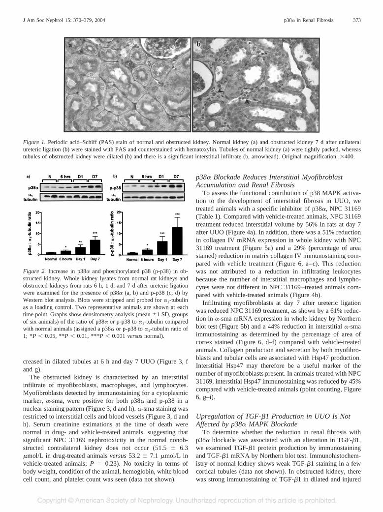
Figure 1. Periodic acid–Schiff (PAS) stain of normal and obstructed kidney. Normal kidney (a) and obstructed kidney 7 d after unilateralureteric ligation (b) were stained with PAS and counterstained with hematoxylin. Tubules of normal kidney (a) were tightly packed, whereastubules of obstructed kidney were dilated (b) and there is a significant interstitial infiltrate (b, arrowhead). Original magnification, �400.
Figure 2. Increase in p38� and phosphorylated p38 (p-p38) in ob-structed kidney. Whole kidney lysates from normal rat kidneys andobstructed kidneys from rats 6 h, 1 d, and 7 d after ureteric ligationwere examined for the presence of p38� (a, b) and p-p38 (c, d) byWestern blot analysis. Blots were stripped and probed for �1-tubulinas a loading control. Two representative animals are shown at eachtime point. Graphs show densitometry analysis (mean �1 SD, groupsof six animals) of the ratio of p38� or p-p38 to �1-tubulin comparedwith normal animals (assigned a p38� or p-p38 to �1-tubulin ratio of1; *P � 0.05, **P � 0.01, ***P � 0.001 versus normal).
J Am Soc Nephrol 15: 370–379, 2004 p38� in Renal Fibrosis 373

Figure 3. Immunolocalization of p38� and phosphorylated p38 (p-p38) in normal and obstructed kidney. In normal rat kidney, p38� (a, brown,arrows) was localized to the luminal surface of most tubules. Many of these tubules do not exhibit nuclear p38� localization. In some tubules,however, p38� was localized to the nucleus with no cytoplasmic staining evident (arrowhead). At 6 h after unilateral ureteric obstruction (UUO)p38� (b) was localized to the nucleus with a loss of luminal staining (arrowheads) in dilated and injured tubules. At day 7 after ureteric ligation,there was a further loss of p38� cytoplasmic staining with an increase in nuclear staining (c, arrowheads). At day 7 UUO, p38� (d, brown)was also localized to the nuclei of myofibroblasts (arrowheads), detected by immunostaining for a cytoplasmic marker of myofibroblasts,alpha–smooth muscle actin (�-sma, gray). In normal rat kidney, p-p38 (e, brown, arrowheads), was localized to the nuclei of some tubules.Nuclear p-p38 was increased in dilated tubules at 6 h (f), with a further increase at 7 d after ureteric ligation (g, arrowhead). At day 7 UUO,p-p38 (h, brown) was also localized to the nuclei of myofibroblasts (arrowheads), detected by immunostaining for alpha smooth muscle actin(�-sma; gray). Original magnification, �800.
374 Journal of the American Society of Nephrology J Am Soc Nephrol 15: 370–379, 2004

tubules, and to a much smaller degree the interstitium. Com-pared with vehicle-treated animals, TGF-�1 immunostaining,as determined by the percentage of area of cortex stained, isunaltered with NPC 31169 treatment at day 7 obstruction(Figure 7, a–c). Northern blot analysis demonstrated an in-crease in TGF-�1 mRNA at day 7 obstruction, which wasunaffected by NPC 31169 treatment (Figure 7d). In addition,we examined CTGF, a downstream target of TGF-�1 involvedin extracellular matrix production. Compared with vehicle-treated animals, CTGF mRNA expression was reduced by 62%in NPC 31169–treated animals at day 7 UUO (Figure 7e).
DiscussionIn the study presented here, we examined the role of p38
MAPK activation in renal fibrosis in a model of surgicallyinduced tissue injury, rat UUO. We first established an increasein both p38� and activated p38 (p-p38) in obstructed kidneythrough a time course of UUO. By immunolocalization, wedemonstrated a loss of luminal p38� with a concomitant in-crease in nuclear p38� and p-p38 within injured and dilatedtubules. This staining pattern suggests that cytoplasmic p38� isactivated by phosphorylation and translocates to the nucleus ofthe injured tubular cell during obstruction. In addition, welocalized both p38� and p-p38 to nuclei of interstitial myofi-broblasts—cell types central to the development of interstitialfibrosis in the obstructed kidney. With the use of a specificinhibitor of p38�, we demonstrated a functional role for p38�MAPK pathway activation in renal fibrosis, independent ofTGF-�1 protein and mRNA levels, but with a reduction inCTGF mRNA expression.
The reduction in extracellular matrix deposition with p38MAPK blockade in UUO was proportional to the reduction ininterstitial myofibroblast accumulation, as demonstrated by areduction in �-sma staining and mRNA expression. However,recent evidence has suggested that p38 may be involved inTGF-�1–induced �-sma production, and therefore, �-sma maynot necessarily reflect the amount of tissue myofibroblast in-filtration (30,31). To confirm our findings, we assessed inter-stitial Hsp47, a collagen chaperone protein produced by myo-fibroblasts during collagen production and independent of p38activation. The amount of interstitial Hsp47 protein may there-fore more accurately represent the number of interstitial myo-fibroblasts present. Compared with vehicle-treated animals,treatment with NPC 31169 showed a reduction in interstitialHsp47 staining, consistent with the degree of reduction ininterstitial �-sma–positive cell accumulation.
This reduction in interstitial myofibroblast accumulationmay be as a result of inhibition of myofibroblast migration ora decrease in myofibroblast transdifferentiation of tubular andsmooth muscle cells. Previous in vitro reports have demon-strated the importance of p38 MAPK activation to PDGF-BBmediated migration of hepatic myofibroblasts (32). Further-more, TGF-�1 has been demonstrated to induce myofibroblasttransdifferentiation of tubular cells both in vitro (33) and inUUO (34) and TGF-�1–induced mammary epithelial cell mi-gration and subsequent fibroblast transdifferentiation is depen-dent on p38 MAPK activation (35). It is therefore possible thatp38 MAPK inhibition in this study resulted in a reduction ininterstitial myofibroblast accumulation by interfering with bothTGF-�1 mediated myofibroblast and epithelial migration, andmyofibroblast transdifferention of tubular cells and smoothmuscle cells.
p38 MAPK activation occurred within tubules and infiltrat-ing myofibroblasts of the obstructed kidney, and p38� MAPKblockade may have inhibited collagen production in this studyvia an effect on either or both of these cell types. In vitrostudies have demonstrated TGF-�1–induced collagen expres-sion in myoblasts via p38 MAPK activation (36). TGF-�1–
Figure 4. p38� mitogen-activated protein kinase (MAPK) blockadereduces interstitial volume in obstructed kidneys but does not reducethe number of infiltrating interstitial leukocytes. (a) Compared withnormal kidneys (checkered bar), there was a dramatic increase ininterstitial volume by point counting 7 d after ureteric ligation inanimals treated with vehicle (open bar). This was reduced by p38�blockade with NPC 31169 treatment (solid bar). (b) NPC 31169treatment (solid bar) did not reduce the number of interstitial macro-phages, T cells, and total leukocytes in day 7 obstructed kidneyscompared with vehicle treatment (open bar). Data are represented asthe mean (�1 SD) of groups of eight animals.
Figure 5. p38� mitogen-activated protein kinase (MAPK) blockadereduces collagen IV and alpha–smooth muscle actin (�-sma) mRNAexpression in obstructed kidney. Compared with normal kidneys(checkered bar), there was a dramatic increase in (a) collagen IV and(b) �-sma mRNA expression by Northern blot test in obstructedkidneys of vehicle-treated (open bar) animals 7 d after ureteric liga-tion. p38� blockade with NPC 31169 treatment (solid bar) reducedcollagen IV and �-sma mRNA expression in obstructed kidneys. Fourrepresentative animals are shown for each group. Graphs show den-sitometry analysis (mean �1 SD, groups of eight animals) of the ratioof collagen IV and �-sma to glyceraldehyde phosphate dehydrogenase(GAPDH) compared with normal animals (assigned an �-sma orcollagen IV to GAPDH ratio of 1).
J Am Soc Nephrol 15: 370–379, 2004 p38� in Renal Fibrosis 375

induced extracellular matrix production by neonatal rat pri-mary cardiac fibroblasts is inhibited by SB203055, an inhibitorof the p38 pathway (37), and activated mutants of MKK3 andMKK6, both upstream kinases of the p38 pathway, is associ-ated with an increase in cardiac fibrosis (38). Proximal renaltubular cells are a known source of TGF-�1 and may promoteperitubular myofibroblast collagen production and interstitialfibrosis (39). In addition, these cells may act as effectors forcollagen IV production and deposition, an effect mediated byTGF-�1 (40).
Despite recent in vitro evidence of a role for p38 activationin TGF-�1 production (19), we were unable to demonstratethis in vivo. In our study, TGF-�1 mRNA expression andprotein is increased in obstructed kidney and unaffected by p38MAPK blockade. TGF-�1 was localized predominantly to thedilated and injured tubules of obstructed kidney. The inabilityto suppress TGF-�1 production by p38 blockade is consistentwith recent studies demonstrating that angiotensin II–inducedTGF-�1 production in vascular smooth muscle cells is medi-ated via the extracellular signal-related kinase MAPK andprotein kinase C pathways (41,42).
CTGF is a downstream effector of TGF-�1 signaling and isimportant in the production of extracellular matrix proteins. Itis localized to both tubular epithelial cells and the interstitium
in UUO, and TGF-�1–induced fibronectin production is re-duced with CTGF oligonucleotide antisense transfection ofcultured renal fibroblasts (43). In this study, CTGF mRNAexpression in vivo is reduced with p38� blockade and parallelsthe reduction in myofibroblast accumulation. From this study,it is not possible to discern whether the reduction in CTGFmRNA expression is as a consequence of the reduction in thenumber of interstitial myofibroblasts or whether there is adirect inhibitory effect on myofibroblast CTGF productionwith p38 blockade.
In UUO and other models of renal fibrosis, angiotensin IIcontributes to the development and progression of renal fibro-sis by promoting the synthesis of growth factors and cytokines,including TGF-�1 (44) and TNF-� (45–47). TNF-� receptorI–deficient mice and pharmacologic inhibition of TNF-� re-ceptor I partially reduces renal fibrosis in UUO (48). Further-more, TNF-� is both a stimulus for p38 activation and isproduced and secreted in response to p38 activation. The p38MAPK is a common downstream target of TGF-�1 and TNF-�signaling in UUO, and blockade of p38 may therefore reduceinterstitial fibrosis by inhibition of TGF-�1– and TNF-�–dependent extracellular matrix production.
UUO results in severe renal fibrosis as a consequence of aconstant surgical insult. In this model, p38� blockade was
Figure 6. p38� mitogen-activated protein kinase (MAPK) blockade reduces collagen IV, alpha–smooth muscle actin (�-sma) and heat shockprotein 47 (Hsp47) immunostaining in obstructed kidneys. Compared with vehicle treatment (open bar), obstructed kidneys of animals treatedwith NPC 31169 (solid bar) had a reduction in collagen IV (a–c, brown), interstitial myofibroblast accumulation (as determined by �-smastaining; d–f, brown), and Hsp47 (g–i, brown) at day 7 after ureteric ligation. Graphs show the analysis of the percentage of area of cortexstained for collagen IV and �-sma staining. For Hsp47 staining, the graph shows the number of Hsp47-positive interstitial points counted per1000 points (expressed as a percentage). The data are represented as a mean (�1 SD) for groups of eight animals; normal kidney, checkeredbar; vehicle-treated animals, open bar; NPC 31169–treated animals, solid bar. Original magnification, �400.
376 Journal of the American Society of Nephrology J Am Soc Nephrol 15: 370–379, 2004
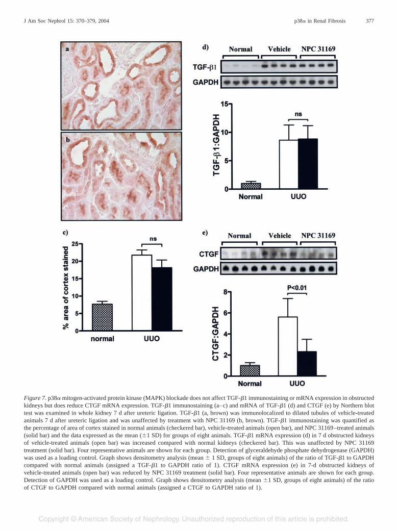
Figure 7. p38� mitogen-activated protein kinase (MAPK) blockade does not affect TGF-�1 immunostaining or mRNA expression in obstructedkidneys but does reduce CTGF mRNA expression. TGF-�1 immunostaining (a–c) and mRNA of TGF-�1 (d) and CTGF (e) by Northern blottest was examined in whole kidney 7 d after ureteric ligation. TGF-�1 (a, brown) was immunolocalized to dilated tubules of vehicle-treatedanimals 7 d after ureteric ligation and was unaffected by treatment with NPC 31169 (b, brown). TGF-�1 immunostaining was quantified asthe percentage of area of cortex stained in normal animals (checkered bar), vehicle-treated animals (open bar), and NPC 31169–treated animals(solid bar) and the data expressed as the mean (�1 SD) for groups of eight animals. TGF-�1 mRNA expression (d) in 7 d obstructed kidneysof vehicle-treated animals (open bar) was increased compared with normal kidneys (checkered bar). This was unaffected by NPC 31169treatment (solid bar). Four representative animals are shown for each group. Detection of glyceraldehyde phosphate dehydrogenase (GAPDH)was used as a loading control. Graph shows densitometry analysis (mean � 1 SD, groups of eight animals) of the ratio of TGF-�1 to GAPDHcompared with normal animals (assigned a TGF-�1 to GAPDH ratio of 1). CTGF mRNA expression (e) in 7-d obstructed kidneys ofvehicle-treated animals (open bar) was reduced by NPC 31169 treatment (solid bar). Four representative animals are shown for each group.Detection of GAPDH was used as a loading control. Graph shows densitometry analysis (mean �1 SD, groups of eight animals) of the ratioof CTGF to GAPDH compared with normal animals (assigned a CTGF to GAPDH ratio of 1).
J Am Soc Nephrol 15: 370–379, 2004 p38� in Renal Fibrosis 377

associated with a significant reduction in fibrosis despite thepresence of persistent stimulus for extracellular matrix produc-tion, ureteric ligation. Furthermore, increased TGF-�1 mRNAexpression or protein production in UUO was not reduced afterp38 blockade, suggesting that p38 activation mediates fibrosisdownstream or independent of TGF-�1.
Two recent studies have demonstrated a reduction in bleo-mycin-induced pulmonary fibrosis with p38 inhibitors (25,26).Fibrosis after bleomycin is dependent on leukocyte mediatedpulmonary injury (49,50). Thus, these studies cannot determinewhether p38 blockade inhibited lung fibrosis through an actionon leukocytes or through direct action on the myofibroblasts.We have demonstrated a reduction in fibrosis in a model ofsurgically mediated renal injury, consistent with a recent reportdemonstrating a reduction in cardiac fibrosis by using a trans-genic approach to inhibit the p38� MAPK pathway in a non-inflammatory, pressure-overload model of cardiac hypertrophyand fibrosis (51).
In summary, we have identified a direct functional role forp38 MAPK activation in progressive renal fibrosis. p38� andp-p38 were increased in obstructed kidney, and we localizedp38 MAPK activation to dilated and injured tubules, andinfiltrating myofibroblasts. With the use of a novel and selec-tive inhibitor of p38�, we were able to inhibit interstitialmyofibroblast accumulation, reduce collagen IV mRNA expres-sion and protein deposition, and reduce CTGF mRNA expressionin spite of a continued increase in TGF-�1 mRNA expression andprotein production. Therefore, p38� MAPK blockade may be atherapeutic option for the treatment of renal fibrosis.
AcknowledgmentThis research was supported by grants from the National Health
and Medical Research Council of Australia.
References1. Lee JC, Laydon JT, McDonnell PC, Gallagher TF, Kumar S,
Green D, McNulty D, Blumenthal MJ, Heys JR, Landvatter SW,Strickler JE, McLaughlin MM, Siemens IR, Fisher SM, Livi GP,White JR, Adams JL, Young PR: A protein kinase involved inthe regulation of inflammatory cytokine biosynthesis. Nature372: 739–746, 1994
2. Raingeaud J, Gupta S, Rogers JS, Dickens M, Han J, UlevitchRJ, Davis RJ: Pro-inflammatory cytokines and environmentalstress cause p38 mitogen-activated protein kinase activation bydual phosphorylation on tyrosine and threonine. J Biol Chem270: 7420–7426, 1995
3. Ono K, Han J: The p38 signal transduction pathway: Activationand function. Cell Signal 12: 1–13, 2000
4. Wang XS, Diener K, Manthey CL, Wang S, Rosenzweig B, BrayJ, Delaney J, Cole CN, Chan-Hui PY, Mantlo N, Lichenstein HS,Zukowski M, Yao Z: Molecular cloning and characterization ofa novel p38 mitogen-activated protein kinase. J Biol Chem 272:23668–23674, 1997
5. Jiang Y, Chen C, Li Z, Guo W, Gegner JA, Lin S, Han J:Characterization of the structure and function of a new mitogen-activated protein kinase (p38beta). J Biol Chem 271: 17920–17926, 1996
6. Jiang Y, Gram H, Zhao M, New L, Gu J, Feng L, Di Padova F,Ulevitch RJ, Han J: Characterization of the structure and function
of the fourth member of p38 group mitogen-activated proteinkinases, p38delta. J Biol Chem 272: 30122–30128, 1997
7. Li Z, Jiang Y, Ulevitch RJ, Han J: The primary structure of p38gamma: A new member of p38 group of MAP kinases. BiochemBiophys Res Commun 228: 334–340, 1996
8. Hale KK, Trollinger D, Rihanek M, Manthey CL: Differentialexpression and activation of p38 mitogen-activated protein ki-nase alpha, beta, gamma, and delta in inflammatory cell lineages.J Immunol 162: 4246–4252, 1999
9. Wang YZ, Zhang P, Rice AB, Bonner JC: Regulation of inter-leukin-1beta–induced platelet-derived growth factor receptor-al-pha expression in rat pulmonary myofibroblasts by p38 mitogen-activated protein kinase. J Biol Chem 275: 22550–22557, 2000
10. Waas WF, Lo HH, Dalby KN: The kinetic mechanism of the dualphosphorylation of the ATF2 transcription factor by p38 mito-gen-activated protein (MAP) kinase alpha: Implications for sig-nal/response profiles of MAP kinase pathways. J Biol Chem 276:5676–5684, 2001
11. Rawadi G, Ramez V, Lemercier B, Roman-Roman S: Activation ofmitogen-activated protein kinase pathways by Mycoplasma fermen-tans membrane lipoproteins in murine macrophages: Involvementin cytokine synthesis. J Immunol 160: 1330–1339, 1998
12. Ivaska J, Reunanen H, Westermarck J, Koivisto L, Kahari VM,Heino J: Integrin alpha2beta1 mediates isoform-specific activa-tion of p38 and upregulation of collagen gene transcription by amechanism involving the alpha2 cytoplasmic tail. J Cell Biol147: 401–416, 1999
13. Reddy MA, Adler SG, Kim YS, Lanting L, Rossi J, Kang SW,Nadler JL, Shahed A, Natarajan R: Interaction of MAPK and12-lipoxygenase pathways in growth and matrix protein expres-sion in mesangial cells. Am J Physiol Renal Physiol 283: F985–F994, 2002
14. Kucich U, Rosenbloom JC, Shen G, Abrams WR, Hamilton AD,Sebti SM, Rosenbloom J: TGF-beta1 stimulation of fibronectintranscription in cultured human lung fibroblasts requires activegeranylgeranyl transferase I, phosphatidylcholine-specific phos-pholipase C, protein kinase C-delta, and p38, but not erk1/erk2.Arch Biochem Biophys 374: 313–324, 2000
15. Sime PJ, O’Reilly KM: Fibrosis of the lung and other tissues:New concepts in pathogenesis and treatment. Clin Immunol 99:308–319, 2001
16. Ge B, Gram H, Di Padova F, Huang B, New L, Ulevitch RJ, LuoY, Han J: MAPKK-independent activation of p38alpha mediatedby TAB1-dependent autophosphorylation of p38alpha. Science295: 1291–1294, 2002
17. Hanafusa H, Ninomiya-Tsuji J, Masuyama N, Nishita M, Fuji-sawa J, Shibuya H, Matsumoto K, Nishida E: Involvement of thep38 mitogen-activated protein kinase pathway in transforminggrowth factor-beta–induced gene expression. J Biol Chem 274:27161–27167, 1999
18. Wang L, Ma R, Flavell RA and Choi ME: Requirement ofmitogen actived protein kinase kinase 3 (MKK3) for activationof p38alpha and p38delta MAPK isoforms by TGF-beta 1 inmurine mesangial cells. J Biol Chem, 277: 47257–47262, 2002
19. Gruden G, Zonca S, Hayward A, Thomas S, Maestrini S, GnudiL, Viberti GC: Mechanical stretch-induced fibronectin and trans-forming growth factor-beta1 production in human mesangialcells is p38 mitogen-activated protein kinase-dependent. Diabe-tes 49: 655–661, 2000
20. Leask A, Holmes A, Abraham DJ: Connective tissue growthfactor: A new and important player in the pathogenesis of fibro-sis. Curr Rheumatol Rep 4: 136–142, 2002
378 Journal of the American Society of Nephrology J Am Soc Nephrol 15: 370–379, 2004

21. Yoshida K, Kuwano K, Hagimoto N, Watanabe K, Matsuba T,Fujita M, Inoshima I, Hara N: MAP kinase activation and apo-ptosis in lung tissues from patients with idiopathic pulmonaryfibrosis. J Pathol 198: 388–396, 2002
22. Varela-Rey M, Montiel-Duarte C, Oses-Prieto JA, Lopez-Za-balza MJ, Jaffrezou JP, Rojkind M, Iraburu MJ: p38 MAPKmediates the regulation of alpha1(I) procollagen mRNA levelsby TNF-alpha and TGF-beta in a cell line of rat hepatic stellatecells(1). FEBS Lett 528: 133–138, 2002
23. Sato M, Shegogue D, Gore EA, Smith EA, McDermott PJ, Tro-janowska M: Role of p38 MAPK in transforming growth factor betastimulation of collagen production by scleroderma and healthydermal fibroblasts. J Invest Dermatol 118: 704–711, 2002
24. Chin BY, Mohsenin A, Li SX, Choi AM, Choi ME: Stimulationof pro-alpha(1)(I) collagen by TGF-beta(1) in mesangial cells:Role of the p38 MAPK pathway. Am J Physiol Renal Physiol280: F495–504, 2001
25. Underwood DC, Osborn RR, Bochnowicz S, Webb EF, RiemanDJ, Lee JC, Romanic AM, Adams JL, Hay DW, Griswold DE:SB 239063, a p38 MAPK inhibitor, reduces neutrophilia, inflam-matory cytokines, MMP-9, and fibrosis in lung. Am J PhysiolLung Cell Mol Physiol 279: L895–L902, 2000
26. Matsuoka H, Arai T, Mori M, Goya S, Kida H, Morishita H,Fujiwara H, Tachibana I, Osaki T, Hayashi S: A p38 MAPKinhibitor, FR-167653, ameliorates murine bleomycin-inducedpulmonary fibrosis. Am J Physiol Lung Cell Mol Physiol 283:L103–L112, 2002
27. Lan HY, Mu W, Nikolic-Paterson DJ, Atkins RC: A novel,simple, reliable, and sensitive method for multiple immunoen-zyme staining: Use of microwave oven heating to block antibodycrossreactivity and retrieve antigens. J Histochem Cytochem 43:97–102, 1995
28. Stambe C, Atkins RC, Tesch GH, Kapoun AM, Hill PA, Schre-iner GF, Nikolic-Paterson DJ: Blockade of p38alpha MAPKameliorates acute inflammatory renal injury in rat anti-GBMglomerulonephritis. J Am Soc Nephrol 14: 338–351, 2003
29. Davies SP, Reddy H, Caivano M, Cohen P: Specificity andmechanism of action of some commonly used protein kinaseinhibitors. Biochem J 351: 95–105, 2000
30. Masamune A, Satoh M, Kikuta K, Sakai Y, Satoh A, Shimose-gawa T: Inhibition of p38 mitogen-activated protein kinaseblocks activation of rat pancreatic stellate cells. J Pharmacol ExpTher 304: 8–14, 2003
31. Reeves HL, Dack CL, Peak M, Burt AD, Day CP: Stress-activated protein kinases in the activation of rat hepatic stellatecells in culture. J Hepatol 32: 465–472, 2000
32. Tangkijvanich P, Santiskulvong C, Melton AC, Rozengurt E,Yee HF Jr: p38 MAP kinase mediates platelet-derived growthfactor-stimulated migration of hepatic myofibroblasts. J CellPhysiol 191: 351–361, 2002
33. Fan JM, Ng YY, Hill PA, Nikolic-Paterson DJ, Mu W, AtkinsRC, Lan HY: Transforming growth factor-beta regulates tubularepithelial-myofibroblast transdifferentiation in vitro. Kidney Int56: 1455–1467, 1999
34. Iwano M, Plieth D, Danoff TM, Xue C, Okada H, Neilson EG:Evidence that fibroblasts derive from epithelium during tissuefibrosis. J Clin Invest 110: 341–350, 2002
35. Bakin AV, Rinehart C, Tomlinson AK, Arteaga CL: p38 mito-gen-activated protein kinase is required for TGFbeta-mediatedfibroblastic transdifferentiation and cell migration. J Cell Sci115: 3193–3206, 2002
36. Rodriguez-Barbero A, Obreo J, Yuste L, Montero JC, Rodri-guez-Pena A, Pandiella A, Bernabeu C, Lopez-Novoa JM: Trans-forming growth factor-beta1 induces collagen synthesis and ac-cumulation via p38 mitogen-activated protein kinase (MAPK)pathway in cultured L(6)E(9) myoblasts. FEBS Lett 513: 282–288, 2002
37. Akiyama-Uchida Y, Ashizawa N, Ohtsuru A, Seto S, TsukazakiT, Kikuchi H, Yamashita S, Yano K: Norepinephrine enhancesfibrosis mediated by TGF-beta in cardiac fibroblasts. Hyperten-sion 40: 148–154, 2002
38. Liao P, Georgakopoulos D, Kovacs A, Zheng M, Lerner D, Pu H,Saffitz J, Chien K, Xiao RP, Kass DA, Wang Y: The in vivo roleof p38 MAP kinases in cardiac remodeling and restrictive car-diomyopathy. Proc Natl Acad Sci U S A 98: 12283–12288, 2001
39. Abbate M, Zoja C, Rottoli D, Corna D, Tomasoni S, Remuzzi G:Proximal tubular cells promote fibrogenesis by TGF-beta1–me-diated induction of peritubular myofibroblasts. Kidney Int 61:2066–2077, 2002
40. Grande JP, Warner GM, Walker HJ, Yusufi AN, Cheng J, GrayCE, Kopp JB, Nath KA: TGF-beta1 is an autocrine mediator ofrenal tubular epithelial cell growth and collagen IV production.Exp Biol Med (Maywood) 227: 171–181, 2002
41. Hamaguchi A, Kim S, Izumi Y, Zhan Y, Yamanaka S, Iwao H:Contribution of extracellular signal-regulated kinase to angioten-sin II–induced transforming growth factor-beta1 expression invascular smooth muscle cells. Hypertension 34: 126–131, 1999
42. Gibbons GH, Pratt RE, Dzau VJ: Vascular smooth muscle cellhypertrophy vs. hyperplasia: Autocrine transforming growth fac-tor-beta 1 expression determines growth response to angiotensinII. J Clin Invest 90: 456–461, 1992
43. Yokoi H, Mukoyama M, Sugawara A, Mori K, Nagae T, MakinoH, Suganami T, Yahata K, Fujinaga Y, Tanaka I, Nakao K: Roleof connective tissue growth factor in fibronectin expression andtubulointerstitial fibrosis. Am J Physiol Renal Physiol 282:F933–F942, 2002
44. Kaneto H, Morrissey J, Klahr S: Increased expression of TGF-beta 1 mRNA in the obstructed kidney of rats with unilateralureteral ligation. Kidney Int 44: 313–321, 1993
45. Klahr S, Morrissey JJ: The role of vasoactive compounds,growth factors and cytokines in the progression of renal disease.Kidney Int Suppl 75: S7–S14, 2000
46. Hisada Y, Sugaya T, Yamanouchi M, Uchida H, Fujimura H,Sakurai H, Fukamizu A, Murakami K: Angiotensin II plays apathogenic role in immune-mediated renal injury in mice. J ClinInvest 103: 627–635, 1999
47. Klahr S, Morrissey J: Angiotensin II and gene expression in thekidney. Am J Kidney Dis 31: 171–176, 1998
48. Guo G, Morrissey J, McCracken R, Tolley T, Liapis H, Klahr S:Contributions of angiotensin II and tumor necrosis factor-alphato the development of renal fibrosis. Am J Physiol Renal Physiol280: F777–F785, 2001
49. Izbicki G, Segel MJ, Christensen TG, Conner MW, Breuer R:Time course of bleomycin-induced lung fibrosis. Int J ExpPathol 83: 111–119, 2002
50. Okazaki T, Nakao A, Nakano H, Takahashi F, Takahashi K,Shimozato O, Takeda K, Yagita H, Okumura K: Impairment ofbleomycin-induced lung fibrosis in CD28-deficient mice. J Im-munol 167: 1977–1981, 2001
51. Zhang S, Weinheimer C, Courtois M, Kovacs A, Zhang CE,Cheng AM, Wang Y, Muslin AJ: The role of the Grb2-p38MAPK signaling pathway in cardiac hypertrophy and fibrosis.J Clin Invest 111: 833–841, 2003
J Am Soc Nephrol 15: 370–379, 2004 p38� in Renal Fibrosis 379