the roles of radiologic imaging in sturge-weber...
TRANSCRIPT
The Roles of Radiologic Imaging in Sturge-Weber Syndrome
Audrey S. Wang, HMS-IIIBIDMC Department of Radiology
May 19, 2008
Overview
• Patient TR• The Phakomatoses• Sturge-Weber Syndrome
– Clinical Features– Epidemiology– Pathogenesis
• Patient TR: Imaging • Companion Patient RW• Summary
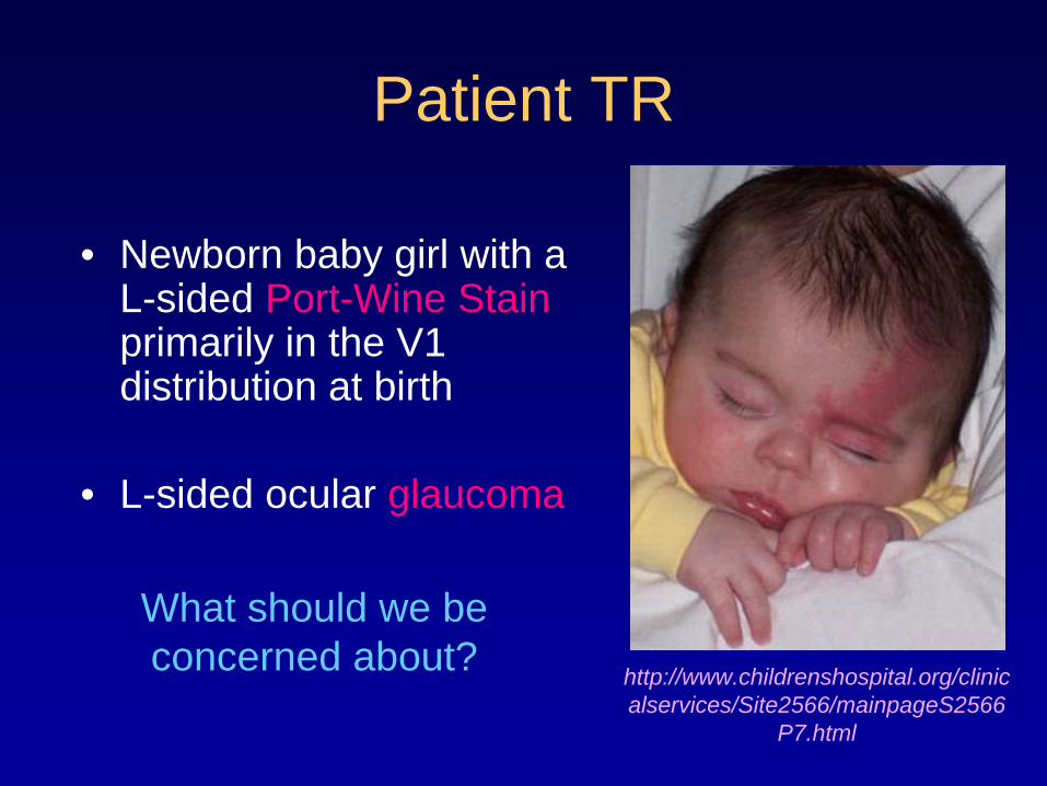
Patient TR
• Newborn baby girl with a L-sided Port-Wine Stain primarily in the V1 distribution at birth
• L-sided ocular glaucoma
What should we be concerned about? http://www.childrenshospital.org/clinic
alservices/Site2566/mainpageS2566 P7.html
Patient TR: Sturge-Weber Syndrome?
She has classic features of Sturge-Weber Syndrome, a phakomatosis.
The Phakomatoses
• “Phakos” (Gr.) = birth mark, spot, mole• Neurocutaneous syndromes/Congenital
neuroectodermal dysplasias:– Neurofibromatosis– Tuberous Sclerosis– Von Hippel Lindau– Sturge-Weber Syndrome (SWS)
• the only phakomatosis that is NOT associated with intracranial neoplasms (Di Rocco and Tamburrini, 2006)
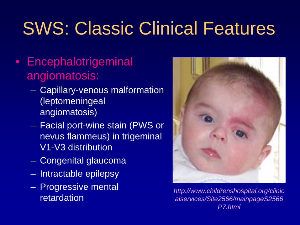
SWS: Classic Clinical Features
• Encephalotrigeminal angiomatosis:– Capillary-venous malformation
(leptomeningeal angiomatosis)
– Facial port-wine stain (PWS or nevus flammeus) in trigeminal V1-V3 distribution
– Congenital glaucoma– Intractable epilepsy– Progressive mental
retardationhttp://www.childrenshospital.org/clinic alservices/Site2566/mainpageS2566
P7.html
SWS: Epidemiology
• Rare (estimated 1/50,000 live births)• Sporadic• Affects males and females with equal
frequency• No racial bias
(Di Rocco and Tamburrini, 2006)
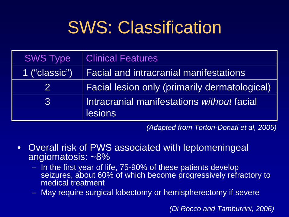
SWS: Classification
• Overall risk of PWS associated with leptomeningeal angiomatosis: ~8%– In the first year of life, 75-90% of these patients develop
seizures, about 60% of which become progressively refractory to medical treatment
– May require surgical lobectomy or hemispherectomy if severe
SWS Type Clinical Features1 (“classic”) Facial and intracranial manifestations
2 Facial lesion only (primarily dermatological)3 Intracranial manifestations without facial
lesions
(Di Rocco and Tamburrini, 2006)
(Adapted from Tortori-Donati et al, 2005)
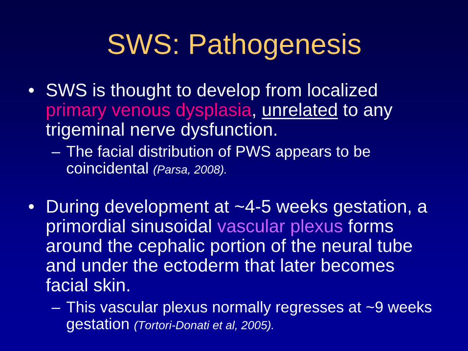
SWS: Pathogenesis• SWS is thought to develop from localized
primary venous dysplasia, unrelated to any trigeminal nerve dysfunction.– The facial distribution of PWS appears to be
coincidental (Parsa, 2008).
• During development at ~4-5 weeks gestation, a primordial sinusoidal vascular plexus forms around the cephalic portion of the neural tube and under the ectoderm that later becomes facial skin. – This vascular plexus normally regresses at ~9 weeks
gestation (Tortori-Donati et al, 2005).
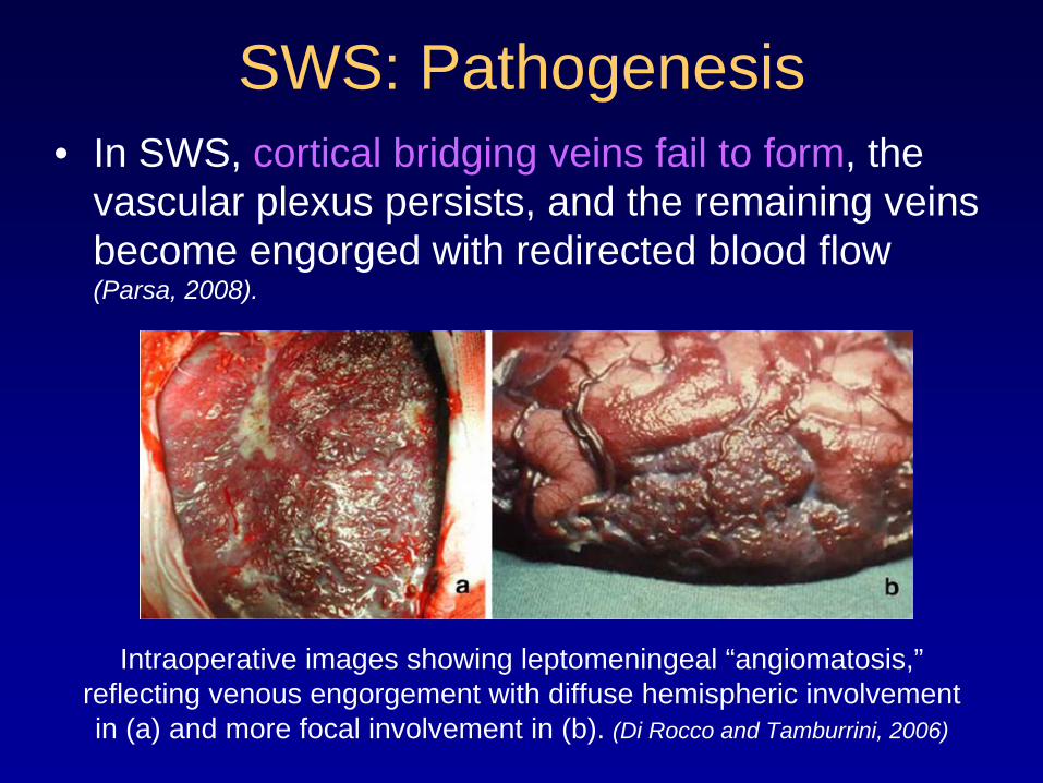
• In SWS, cortical bridging veins fail to form, the vascular plexus persists, and the remaining veins become engorged with redirected blood flow (Parsa, 2008).
SWS: Pathogenesis
Intraoperative images showing leptomeningeal “angiomatosis,” reflecting venous engorgement with diffuse hemispheric involvement in (a) and more focal involvement in (b). (Di Rocco and Tamburrini, 2006)
Cerebral Veins are Emissary Veins
Let’s review the cerebral venous system to better understand the key features of SWS.
* Key point: Because all emissary veins lack valves, they allow for bidirectional flow.
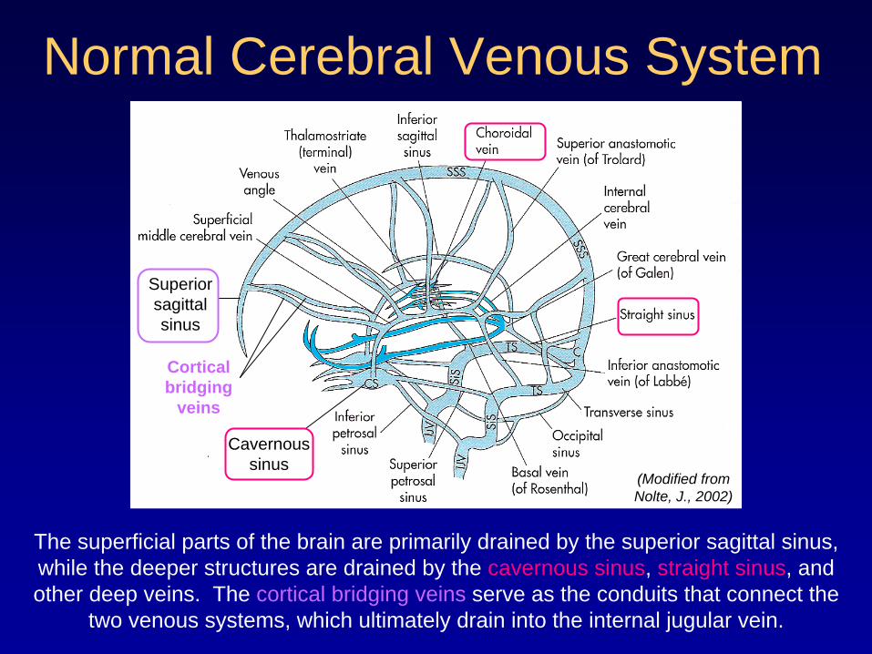
Normal Cerebral Venous System
(Modified from Nolte, J., 2002)
Cavernous sinus
Cortical bridging
veins
Superior sagittal sinus
The superficial parts of the brain are primarily drained by the superior sagittal sinus, while the deeper structures are drained by the cavernous sinus, straight sinus, and other deep veins. The cortical bridging veins serve as the conduits that connect the
two venous systems, which ultimately drain into the internal jugular vein.
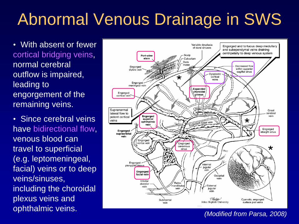
Abnormal Venous Drainage in SWS
(Modified from Parsa, 2008)
• With absent or fewer cortical bridging veins, normal cerebral outflow is impaired, leading to engorgement of the remaining veins.
• Since cerebral veins have bidirectional flow, venous blood can travel to superficial (e.g. leptomeningeal, facial) veins or to deep veins/sinuses, including the choroidal plexus veins and ophthalmic veins.
Neurologic Deterioration in SWS
• Processes (including normal brain development and seizures) that increase the oxygen and glucose demand of brain tissue lead to increased cerebral blood flow. • These changes exacerbate the pre-existing venous
engorgement and further elevate venous pressures, resulting in more severe cerebral ischemia and tissue damage (Parsa, 2008).
• With this in mind, let’s return to Patient TR…
Back to Patient TR
• Newborn baby girl with a L-sided Port-Wine Stain in primarily the V1 distribution at birth
• L-sided ocular glaucoma
• We are concerned about intracranial involvement of SWS.
Which imaging modality should we use?
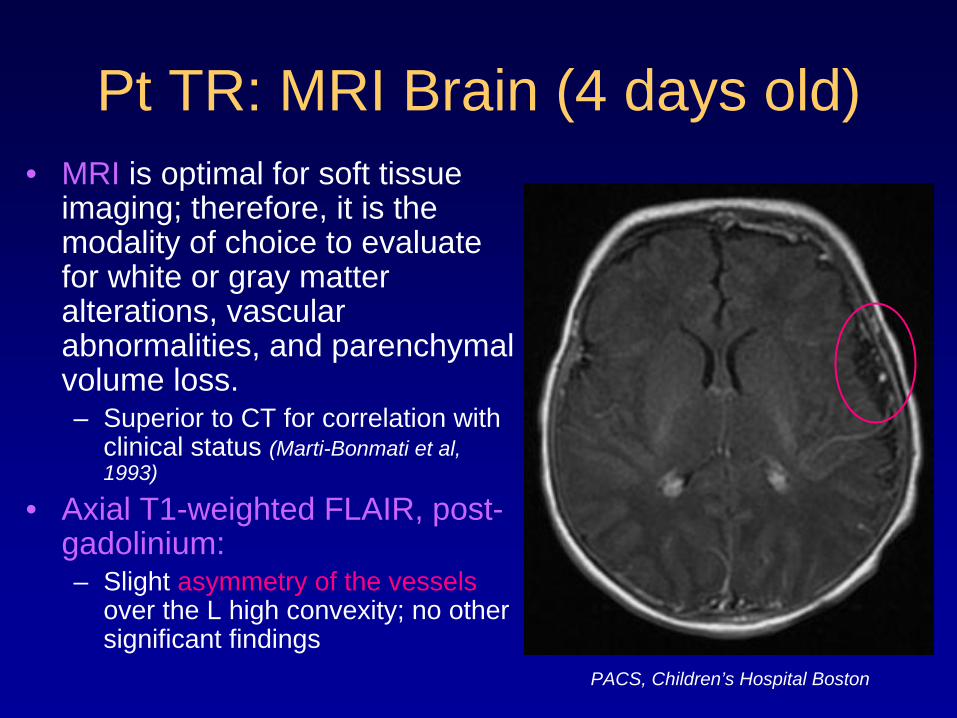
Pt TR: MRI Brain (4 days old)• MRI is optimal for soft tissue
imaging; therefore, it is the modality of choice to evaluate for white or gray matter alterations, vascular abnormalities, and parenchymal volume loss. – Superior to CT for correlation with
clinical status (Marti-Bonmati et al, 1993)
• Axial T1-weighted FLAIR, post- gadolinium:– Slight asymmetry of the vessels
over the L high convexity; no other significant findings
PACS, Children’s Hospital Boston
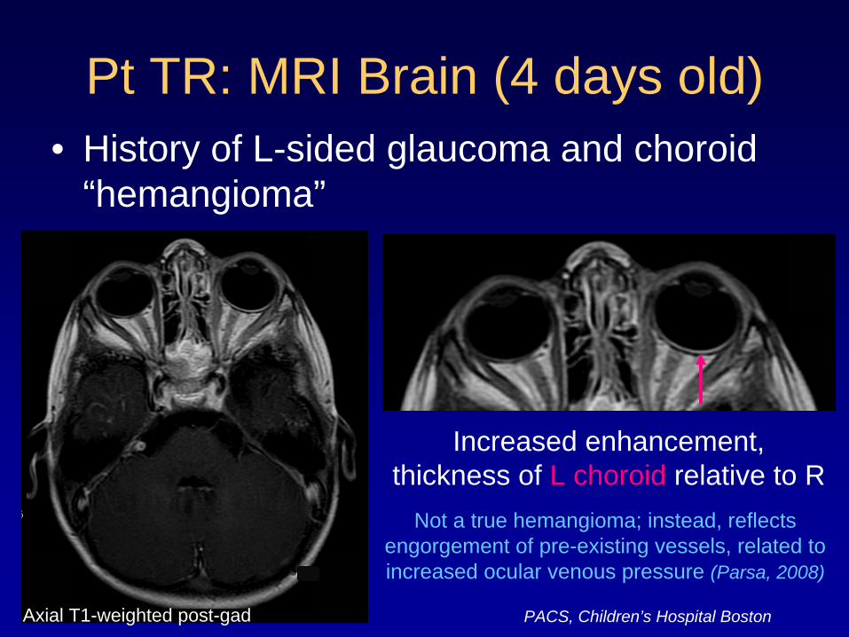
Pt TR: MRI Brain (4 days old)• History of L-sided glaucoma and choroid
“hemangioma”
Increased enhancement, thickness of L choroid relative to R
Not a true hemangioma; instead, reflects engorgement of pre-existing vessels, related to increased ocular venous pressure (Parsa, 2008)
PACS, Children’s Hospital Boston Axial T1-weighted post-gad
Pt TR: At 8 Months of Age• Patient TR presented to the ED with new-onset
seizures/status epilepticus, R facial droop, and little spontaneous movement of her R arm.
• No head trauma• No family history of seizures• Afebrile, vital signs within normal limits
What kind of imaging would you do to R/O possible causes of her seizures?
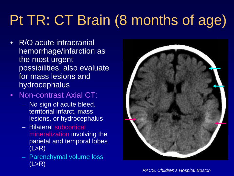
Pt TR: CT Brain (8 months of age)• R/O acute intracranial
hemorrhage/infarction as the most urgent possibilities, also evaluate for mass lesions and hydrocephalus
• Non-contrast Axial CT:– No sign of acute bleed,
territorial infarct, mass lesions, or hydrocephalus
– Bilateral subcortical mineralization involving the parietal and temporal lobes (L>R)
– Parenchymal volume loss (L>R)
PACS, Children’s Hospital Boston
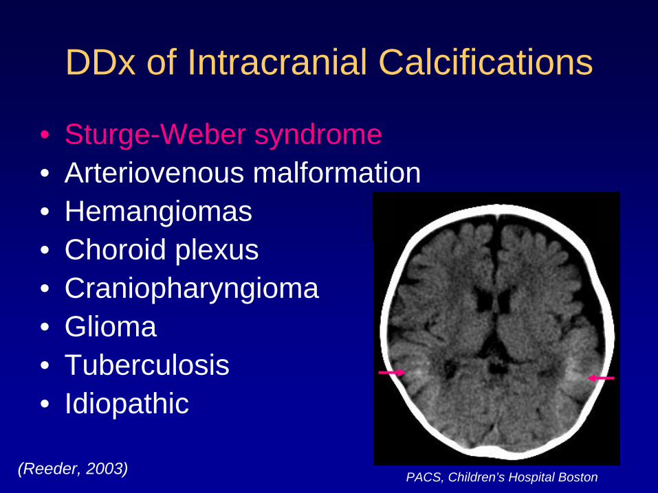
DDx of Intracranial Calcifications
• Sturge-Weber syndrome• Arteriovenous malformation• Hemangiomas• Choroid plexus• Craniopharyngioma• Glioma• Tuberculosis• Idiopathic
PACS, Children’s Hospital Boston (Reeder, 2003)
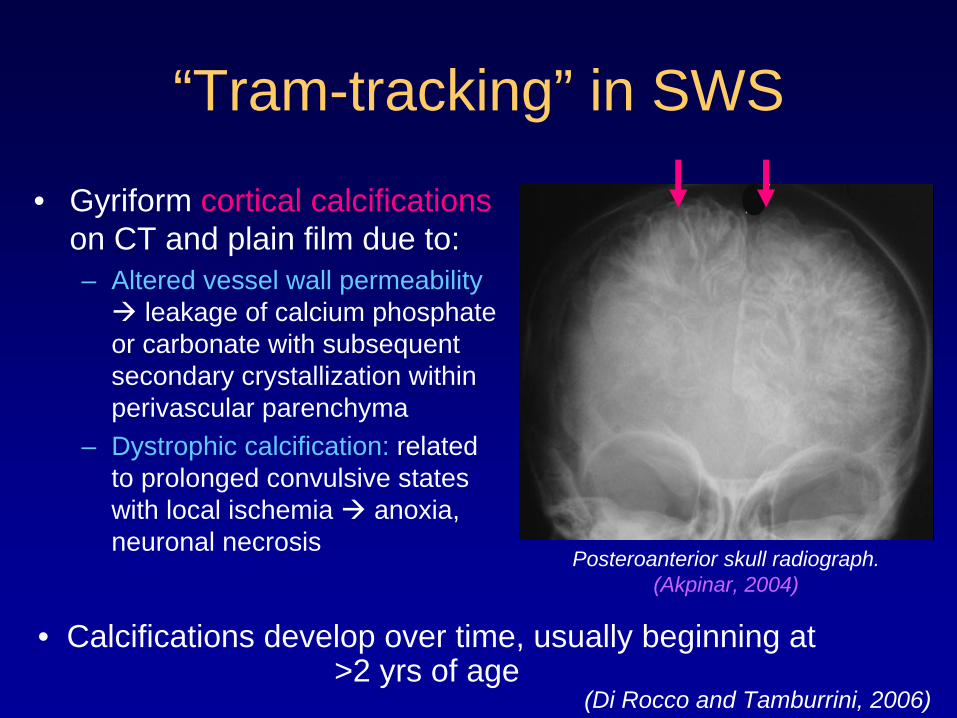
“Tram-tracking” in SWS
• Gyriform cortical calcifications on CT and plain film due to:– Altered vessel wall permeability
leakage of calcium phosphate or carbonate with subsequent secondary crystallization within perivascular parenchyma
– Dystrophic calcification: related to prolonged convulsive states with local ischemia anoxia, neuronal necrosis
Posteroanterior skull radiograph. (Akpinar, 2004)
• Calcifications develop over time, usually beginning at >2 yrs of age
(Di Rocco and Tamburrini, 2006)
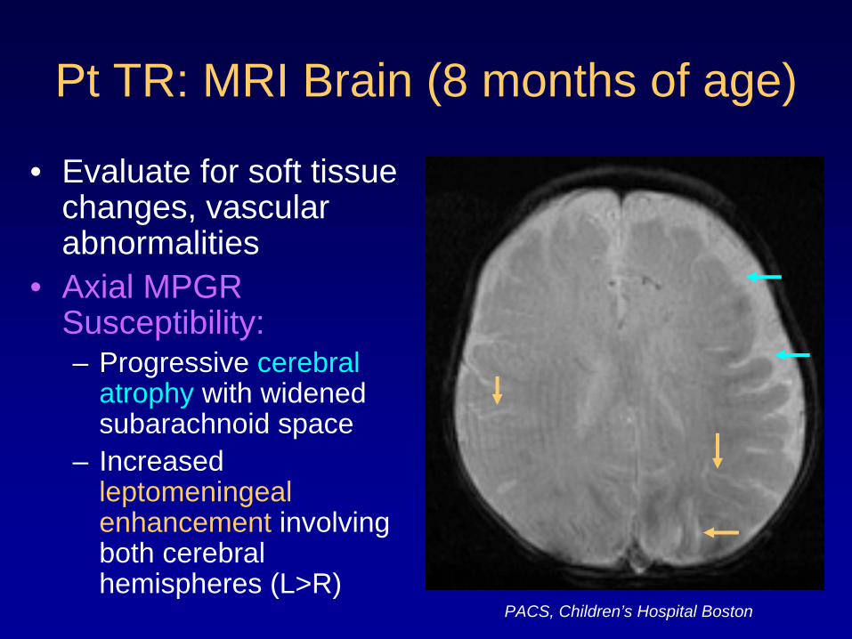
Pt TR: MRI Brain (8 months of age)
• Evaluate for soft tissue changes, vascular abnormalities
• Axial MPGR Susceptibility:– Progressive cerebral
atrophy with widened subarachnoid space
– Increased leptomeningeal enhancement involving both cerebral hemispheres (L>R)
PACS, Children’s Hospital Boston
Pt TR: At 2 Years of Age
• Patient TR presented to the ED with an increasing frequency of seizures and a change from her baseline seizure characteristics
• Developmental delay was noted on exam
What kind of imaging would you do?
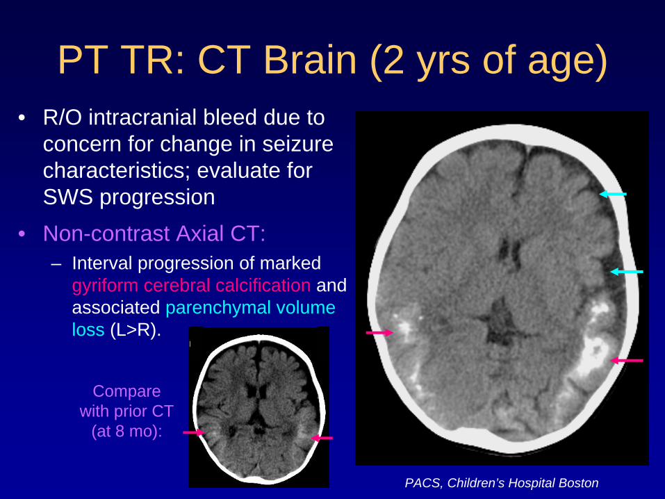
PT TR: CT Brain (2 yrs of age)• R/O intracranial bleed due to
concern for change in seizure characteristics; evaluate for SWS progression
• Non-contrast Axial CT:– Interval progression of marked
gyriform cerebral calcification and associated parenchymal volume loss (L>R).
Compare with prior CT
(at 8 mo):
PACS, Children’s Hospital Boston
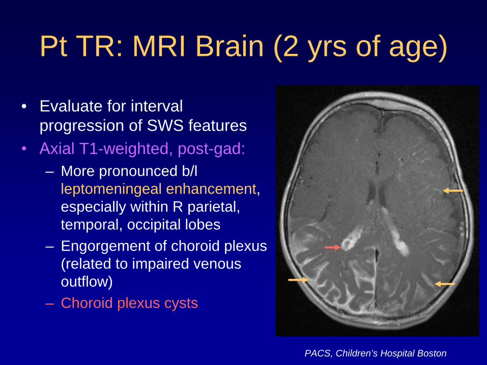
Pt TR: MRI Brain (2 yrs of age)
• Evaluate for interval progression of SWS features
•
Axial T1-weighted, post-gad:– More pronounced b/l
leptomeningeal enhancement, especially within R parietal, temporal, occipital lobes
– Engorgement of choroid plexus (related to impaired venous outflow)
– Choroid plexus cysts
PACS, Children’s Hospital Boston
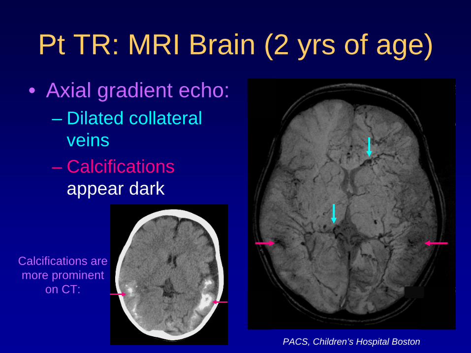
Pt TR: MRI Brain (2 yrs of age)• Axial gradient echo:
– Dilated collateral veins
– Calcifications appear dark
Calcifications are more prominent
on CT:
PACS, Children’s Hospital Boston
Patient TR: Management
• Patient TR’s seizures responded to oxcarbazepine and valproic acid.
• She was discharged home on these anticonvulsants with instructions to follow up with the neurology service.
Companion Patient RW: History
• Boy with Sturge-Weber Syndrome with associated R-sided facial port-wine stain in V1 distribution
• Diagnosed at 5 weeks of age with SWS based on MRI findings
• First seizures presented at 6 months of age
Let’s look at some imaging.
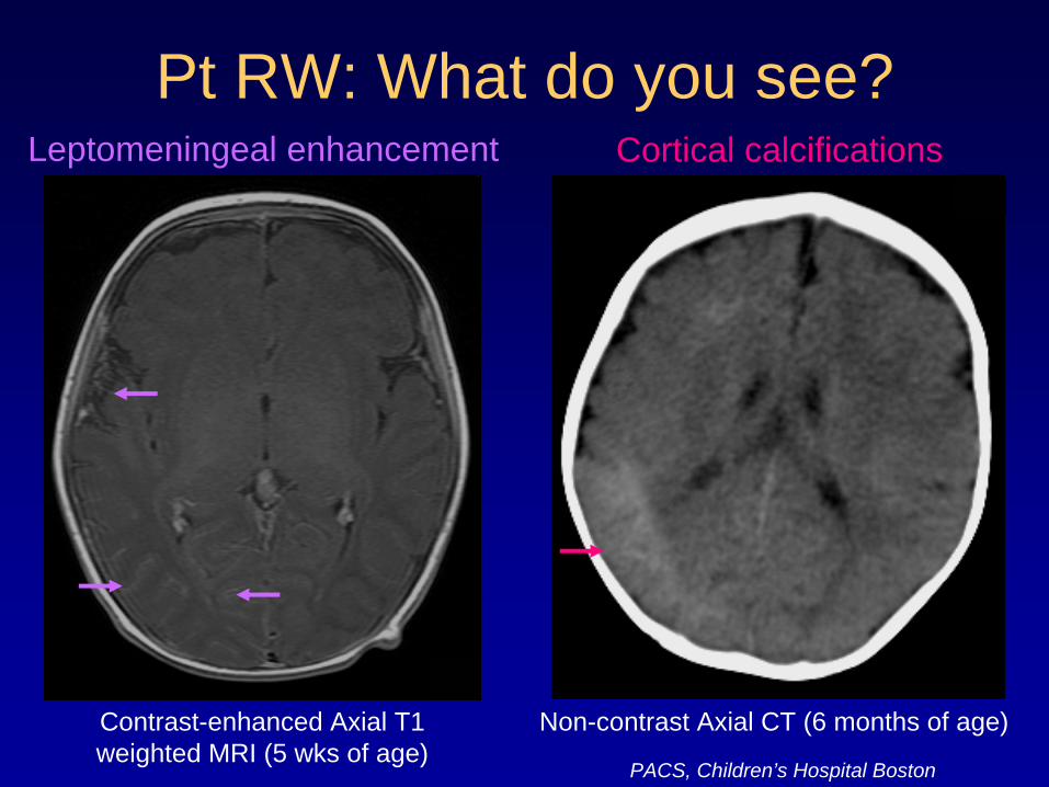
Pt RW: What do you see?
Contrast-enhanced Axial T1 weighted MRI (5 wks of age)
Non-contrast Axial CT (6 months of age)
Leptomeningeal enhancement Cortical calcifications
PACS, Children’s Hospital Boston
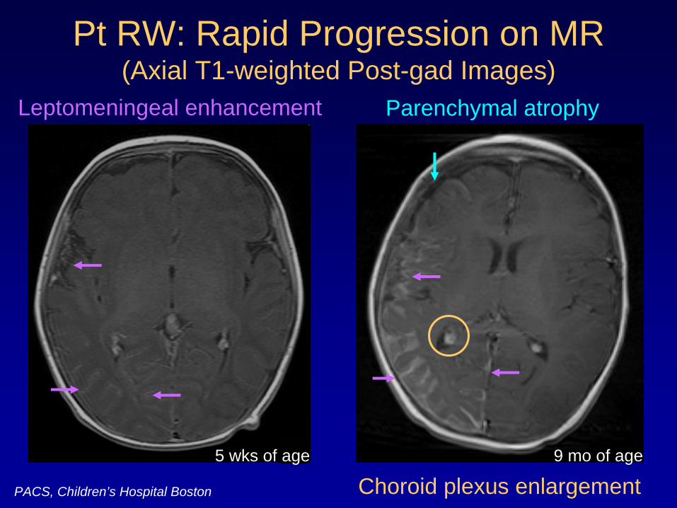
Pt RW: Rapid Progression on MR (Axial T1-weighted Post-gad Images)
5 wks of age
Leptomeningeal enhancement Parenchymal atrophy
Choroid plexus enlargement9 mo of age
PACS, Children’s Hospital Boston
Pt RW: Progression to Intractable Seizures
• Patient RW continued to have seizures, which became refractory to medical therapy.
• Surgical options were considered to remove the epileptogenic focus and protect the normal brain from excitotoxic injury secondary to the seizures.
How can we functionally determine the extent of brain involvement?
Nuclear Imaging: PET• Positron Emission Tomography
• F-18-FDG (Fluorodeoxyglucose)– Measure F-18-FDG metabolism in brain tissue
to determine its metabolic function
• More recently used for prognostic evaluation and to aid surgical planning– Extent and severity of FDG hypometabolism
have been shown to correlate with seizure severity and cognitive decline (Lee et al, 2001)
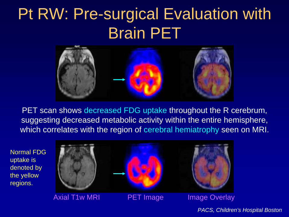
Pt RW: Pre-surgical Evaluation with Brain PET
PET scan shows decreased FDG uptake throughout the R cerebrum, suggesting decreased metabolic activity within the entire hemisphere, which correlates with the region of cerebral hemiatrophy seen on MRI.
PACS, Children’s Hospital Boston
Axial T1w MRI PET Image Image Overlay
Normal FDG uptake is denoted by the yellow regions.
Pt RW: Management
PET scanning demonstrated diffuse reduction in metabolic activity throughout the entire R cerebrum.
Therefore, the decision was made for Patient RW to undergo R cerebral hemispherectomy.
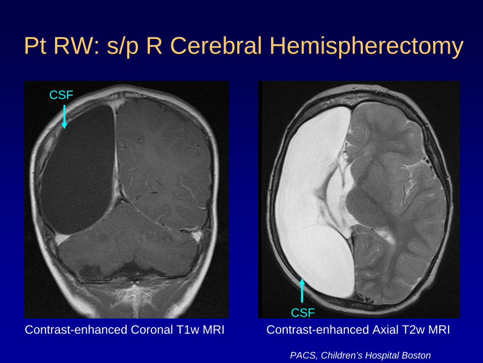
Pt RW: s/p R Cerebral Hemispherectomy
Contrast-enhanced Axial T2w MRIContrast-enhanced Coronal T1w MRICSF
CSF
PACS, Children’s Hospital Boston
Let’s Review• You have been introduced to two different
patients with Sturge-Weber Syndrome.• Both demonstrated classic radiologic
findings on MRI and CT but clinically progressed at different rates.
• One patient’s seizures were managed medically, but the other patient’s became intractable, requiring surgical intervention.
We can use what we have learned from these two patients to construct a simple algorithm for the diagnosis and management of
SWS patients in general.
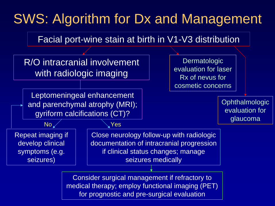
SWS: Algorithm for Dx and ManagementFacial port-wine stain at birth in V1-V3 distribution
R/O intracranial involvement with radiologic imaging
Leptomeningeal enhancement and parenchymal atrophy (MRI);
gyriform calcifications (CT)?
Close neurology follow-up with radiologic documentation of intracranial progression
if clinical status changes; manage seizures medically
Ophthalmologic evaluation for
glaucoma
Dermatologic evaluation for laser
Rx of nevus for cosmetic concerns
Repeat imaging if develop clinical symptoms (e.g.
seizures)
Consider surgical management if refractory to medical therapy; employ functional imaging (PET)
for prognostic and pre-surgical evaluation
No Yes
Summary
• Sturge-Weber Syndrome is a rare, sporadic condition due to primary venous dysplasia causing impaired venous outflow and subsequent cerebral ischemia.
• Certain types can progress to intractable epilepsy and may necessitate radical surgical intervention.
• Radiologic imaging plays several key roles in the management of SWS patients.
Summary - 2• MRI correlates better than CT with clinical
progression.– Confirms the diagnosis of intracranial
involvement– Helps document the extent of involvement
(e.g. R/O bilateral SWS)
• CT is more sensitive than MRI for detecting cortical calcifications.
Summary - 3
• PET offers functional (metabolic activity) data to determine the full extent of involvement of the brain parenchyma.– Complements MRI/CT findings– Provides prognostic information that can help
guide management, including surgical planning
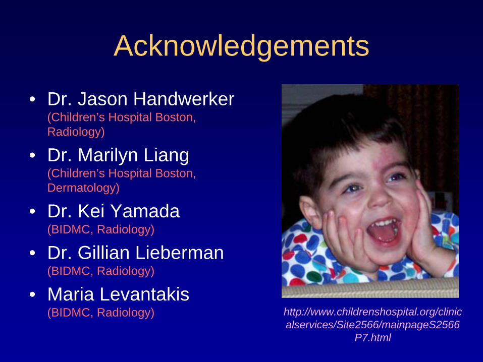
Acknowledgements
• Dr. Jason Handwerker (Children’s Hospital Boston, Radiology)
• Dr. Marilyn Liang (Children’s Hospital Boston, Dermatology)
• Dr. Kei Yamada (BIDMC, Radiology)
• Dr. Gillian Lieberman (BIDMC, Radiology)
• Maria Levantakis (BIDMC, Radiology) http://www.childrenshospital.org/clinic
alservices/Site2566/mainpageS2566 P7.html
References• Akpinar E. The tram-track sign: cortical calcifications. Radiology. 2004;231:515-6. • Di Rocco C, Tamburrini G. Sturge-Weber syndrome. Childs Nerv Syst. 2006;22:909-21.• Lee JS, Asano E, Muzik O, Chugani DC, Juhász C, Pfund Z, Philip S, Behen M, Chugani
HT. Sturge-Weber syndrome: correlation between clinical course and FDG PET findings. Neurology. 2001;57:189-95.
• Martí-Bonmatí L, Menor F, Mulas F. The Sturge-Weber syndrome: correlation between the clinical status and radiological CT and MRI findings. Childs Nerv Syst. 1993;9:107-9.
• Nolte, J. The Human Brain: An Introduction to Its Functional Anatomy, 5th ed. St. Louis: Mosby, 2002.
• Parsa CF. Sturge-Weber Syndrome: A Unified Pathophysiologic Mechanism. Curr Treat Options Neurol. 2008;10:47-54.
• Reeder MM. Reeder and Felson’s Gamuts in Radiology, 4th ed. New York: Springer, 2003.
• Tortori-Donati P, Rossi A, Biancheri R, Andreula CF. Pediatric Neuroradiology: Brain. Berlin: Springer, 2005.
• Children’s Hospital Boston - Sturge-Weber Syndrome Clinic. http://www.childrenshospital.org/clinicalservices/Site2566/mainpageS2566P7.html Accessed 5/18/2008.