the sages manual a practical guide to bariatric surgery (springer, 2008)
TRANSCRIPT

The SAGES Manual

The SAGES ManualA Practical Guide to Bariatric Surgery
Ninh T. Nguyen, MDAssociate Professor of Surgery, Chief, Division of Gastrointestinal and Bariatric Surgery, University of California, Irvine Medical Center, Orange, CA, USA
Editor-in-chief
Eric J. DeMaria, MDProfessor and Vice Chairman, Network General Surgery, Director, Bariatric and EndoSurgery,Duke University, Durham, NC, USA
Sayeed Ikramuddin, MDAssociate Professor of Surgery, Co-Director, Minimally Invasive Surgery, Director, Gastrointestinal Surgery, Katherine and Robert Goodale Chair in Minimally Invasive Surgery, University of Minnesota, Minneapolis, MN, USA
Matthew M. Hutter, MD, MPHDirector, Codman Center of Clinical Effectiveness in Surgery, Department of Surgery, Massachusetts General Hospital, Boston, MA, USA
Editors

Ninh T. Nguyen, MDAssociate Professor of Surgery Chief, Division of Gastrointestinal and Bariatric Surgery University of California Irvine Medical CenterOrange, CAUSA
Eric J. DeMaria, MDProfessor and Vice ChairmanNetwork General Surgery Director, Bariatric and EndoSurgeryDuke University, Durham, NCUSA
Sayeed Ikramuddin, MDAssociate Professor of SurgeryCo-Director Minimally Invasive Surgery Director, Gastrointestinal Surgery Katherine and Robert Goodale Chair in Minimally Invasive SurgeryUniversity of Minnesota Minneapolis, MN USA
Matthew M, Hutter, MD, MPHDirector, Codman Center of Clinical Effectiveness in Surgery Department of Surgery Massachusetts General HospitalBoston, MAUSA
ISBN: 978-0-387-69170-1 e-ISBN: 978-0-387-69171-8
© 2008 Springer Science+Business Media, LLCAll rights reserved. This work may not be translated or copied in whole or in part without the written permission of the publisher (Springer Science + Business Media, LLC, 233 Spring Street, New York, NY 10013, USA), except for brief excerpts in connection with reviews or scholarly analysis. Use in connection with any form of information storage and retrieval, electronic adaptation, computer software, or by similar or dissimilar meth-odology now known or hereafter developed is forbidden.The use in this publication of trade names, trademarks, service marks, and similar terms, even if they are not identifi ed as such, is not to be taken as an expression of opinion as to whether or not they are subject to proprietary rights. While the advice and information in this book are believed to be true and accurate at the date of going to press, neither the authors nor the editors nor the publisher can accept any legal responsibility for any errors or omissions that may be made. The publisher makes no warranty, express or implied, with respect to the material contained herein.
Printed on acid-free paper
springer.com
Library of Congress Control Number: 2008936875

Preface
The fi eld of bariatric surgery has grown at an exponential rate over the past decade. The number of bariatric operations has increased from less than 10,000 operations in 1998 to nearly 200,000 operations in 2008. Bariatric surgery has become an integral part of gastrointestinal surgery and is now an important part of education for surgical residents and fellows in minimally invasive surgery. Edu-cational initiatives in bariatric surgery take many different forms and currently many textbooks on the topic of bariatric surgery are commercially available. However, there is a need for a quick-reference manual that provides up-to-date, easy access to information about this new and complex surgical specialty.
The SAGES Manual: A Practical Guide to Bariatric Surgery began as a project developed within the SAGES Bariatric Liaison group. The name of the book previews its primary purpose, as it is meant to provide practical information within a small pocket-sized book that can serve as a portable resource anywhere, including the ward and clinic. This manual provides a concise, practical guide to promote high-quality, safe care for patients undergoing weight loss surgery. It is intended to be used by any members of the multidisciplinary team, includ-ing surgeons, surgeons-in-training, medical physicians, medical students, nurses, nurse practitioners, physician’s assistants, nutritionists, and psychologists.
We would like to thank members of the SAGES Bariatric Liaison group who have contributed to the manual, the SAGES leadership whose support and encouragement were critical for its development, and Springer for helping to make this project a reality. Our goal is that knowledge gained from using this manual will help to promote safe and effective care for patients undergoing bariatric surgery.
Ninh T. Nguyen, MDEric J. DeMaria, MD
Matthew M. Hutter, MD, MPHSayeed Ikramuddin, MD

Contents
Preface ............................................................................................ v
Contributors ................................................................................... xi
I. Essentials of Bariatric Surgery
1. The Rationale for Bariatric Surgery ..................................... 3Xingxiang Li, Orit Kaidar-Person, and Raul J. Rosenthal
2. Overview of Bariatric Operations ........................................ 9Daniel E. Swartz and Edward L. Felix
3. Identifi cation of Comorbidities and Their Management ...... 23Benjamin E. Schneider
4. Defi nition of Obesity and Indications for Surgery ............... 27Jamie D. Adair and Mark A. Pleatman
5. Preoperative Nutritional Assessment and Postoperative Dietary Guidelines .................................. 31Andrew B. Lederman
6. Essentials of a Bariatric Program ......................................... 37Troy A. Markel and Samer G. Mattar
7. Psychological Assessment .................................................... 43Eldo E. Frezza, Mitchell S. Wachtel, and Rolf Gordhamer
8. Preoperative Check List ....................................................... 51Rami R. Zanoun and Giselle G. Hamad
9. Postoperative Care Pathway ................................................. 59Daniel M. Herron and Murali N. Naidu
10. Long-term Follow-up Protocol of Bariatric Patients ............ 67Steven Teich and Marc P. Michalsky
11. An Economic Approach to Opening a Bariatric Practice..... 75
Bradley T. Ewing and Eldo E. Frezza

II. Techniques
12. Roux-en-Y Gastric Bypass ................................................... 87Robin P. Blackstone
13. Lap-Band® and Adjustment Schedule .................................. 101Dean J. Mikami
14. Laparoscopic Duodenal Switch ............................................ 109Manish Parikh, Michel Gagner, and Alfons Pomp
15. Laparoscopic Sleeve Gastrectomy: A Staged Procedure for Super-Super Obese Patients ........................... 131Kuldeep Singh
16. Laparoscopic Staged Roux-en-Y: A Staged Procedure for Super-Super Obese Patients ............................................ 137Ninh T. Nguyen and Marcelo W. Hinojosa
III. Outcomes
17. Outcomes of Laparoscopic Gastric Bypass .......................... 145Samuel Szomstein and Olga N. Tucker
18. Outcomes of Laparoscopic Adjustable Gastric Banding ..... 153David A. Provost
19. Outcomes of Duodenal Switch and Other Malabsorptive Procedures .................................................... 157Peter F. Crookes
20. Outcomes of Bariatric Surgery in Adolescents .................... 167Go Miyano and Thomas H. Inge
21. Which Operation Is Best?..................................................... 177Sayeed Ikramuddin and Gonzalo Torres-Villalobos
IV. Complications
22. Anastomotic Leaks after Laparoscopic Gastric Bypass ....... 193Alexander Perez and Eric J. DeMaria
23. Gastric Bypass: Gastrointestinal Bleeding ........................... 199Ross L. McMahon
viii Contents

24. Intestinal Obstruction after Laparoscopic Gastric Bypass ... 205Alexander Perez and Eric J. DeMaria
25. Roux-en-Y Gastric Bypass: Stomal Stenosis ....................... 211Janey S.A. Pratt
26. Gastric Bypass: Marginal Ulceration ................................... 213Bradley J. Needleman
27. Laparoscopic Adjustable Gastric Banding: Infection, Slippage, and Hiatal Hernia ................................. 219Marina Kurian
28. Gastric Erosion Following Adjustable Gastric Banding ...... 227Helmuth T. Billy
V. Guidelines and Accreditation in Bariatric Surgery
29. Society of American Gastrointestinal and Endoscopic Surgeons (SAGES) Bariatric Credentialing Guideline ....................................................... 239Shawn Tsuda and Daniel B. Jones
30. Reporting of Bariatric Surgery Outcomes ............................ 243Gavitt A. Woodard and John M. Morton
31. The Betsy Lehman Center Guidelines for Weight Loss Surgery ............................................................ 253Matthew M. Hutter
32. American Society for Metabolic and Bariatric Surgery (ASMBS) Centers of Excellence Program ............. 257Stacy A. Brethauer, Bipan Chand, and Philip R. Schauer
33. The American College of Surgeons (ACS) Bariatric Surgery Center Network ........................................ 261Bruce Schirmer
Index .............................................................................................. 275
Contents ix

Contributors
Jamie D. Adair, MD Department of Surgery, St. Joseph Mercy Oakland Hospital, Bloomfi eld Hills, MI
Helmuth T. Billy, MDDirector of Bariatric Surgery, St. John’s Regional Medical Center, Ventura Advanced Surgical Associates, Ventura, CA
Robin P. Blackstone, MD, FACSClinical Associate Professor of Surgery, University of Arizona School of Medicine-Phoenix, Medical Director, Scottsdale Bariatric Center, Scottsdale, AZ
Stacy A. Brethauer, MDStaff Surgeon, Bariatric and Metabolic Institute, Cleveland Clinic, Cleveland, OH
Bipan Chand, MDDirector, Surgical Endoscopy, Bariatric and Metabolic Institute, Cleveland Clinic, Cleveland, OH
Peter F. Crookes, MDAssociate Professor of Surgery, University of Southern California, Department of Surgery, University Hospital, Los Angeles, CA
Eric J. DeMaria, MDProfessor and Vice Chairman, Network General Surgery, Director, Bariatric and EndoSurgery, Duke University, Durham, NC
Bradley T. Ewing, PhDRawls Endowed Professor of Operations Management, Texas Tech University, Rawls College of Business, Lubbock, TX
Edward L. Felix, MDAdvanced Bariatric Center, Assistant Clinical Professor, UCSF-Fresno, Director of Bariatric Surgery, Clovis Community Medical Center, Fresno, CA

Eldo E. Frezza, MD, MBA, FACSProfessor, Texas Tech University Health Sciences Center, Chief of General Surgery, University Medical Center, Lubbock, TX
Michel Gagner, MD, FACS, FRCSCChairman, Department of Surgery, Mount Sinai Medical Center, Miami Beach, FL
Rolf Gordhamer, PhDPsychologist and Consultant, Texas Tech University, Lubbock, TX
Giselle G. Hamad, MD, FACSAssistant Professor of Surgery, University of Pittsburgh School of Medicine, Department of Surgery, Magee-Women’s Hospital of UPMC, Pittsburgh, PA
Daniel M. Herron, MDAssociate Professor of Surgery, Mount Sinai School of Medicine, Chief, Section of Bariatric Surgery, Mount Sinai Medical Center, New York, NY
Marcelo W. Hinojosa, MDResident Physician, Department of Surgery, University of California, Irvine Medical Center, Orange, CA
Matthew M. Hutter, MD, MPHDirector, Codman Center for Clinical Effectiveness in Surgery, Department of Surgery, Massachusetts General Hospital, Boston, MA
Sayeed Ikramuddin, MDAssociate Professor of Surgery, Co-Director, Minimally Invasive Surgery, Director, Gastrointestinal Surgery, Katherine and Robert Goodale Chair in Minimally Invasive Surgery, University of Minnesota, Minneapolis, MN
Thomas H. Inge, MD, PhDAssociate Professor of Pediatrics and Surgery, University of Cincinnati, Department of Pediatric General & Thoracic Surgery, Cincinnati Children’s Hospital Medical Center, Cincinnati, OH
xii Contributors

Daniel B. Jones, MDAssociate Professor, Chief, Section of Minimally Invasive Surgery, Harvard Medical School, Director, Bariatric Program, Beth Israel Deaconess Medical Center, Boston, MA
Orit Kaidar-Person, MDThe Bariatric and Metabolic Institute, Cleveland Clinic Florida, Weston, FL
Marina Kurian, MDAssistant Professor of Surgery, New York University School of Medicine, Department of Surgery, New York University Medical Center, New York, NY
Andrew B. Lederman, MD, FACSMedical Director of Bariatric Surgery, Berkshire Medical Center, Assistant Professor of Surgery, University of Massachusetts Medical School, Pittsfi eld, MA
Xingxiang Li, MDThe Bariatric and Metabolic Institute, Cleveland Clinic Florida, Weston, FL, Department of Minimally Invasive Surgery, Shanghai Changhai Hospital, Shanghai, China
Troy A. Markel, MDGeneral Surgery Resident, Indiana University School of Medicine, Indianapolis, IN
Samer G. Mattar, MD, FRCS, FACSAssociate Professor of Surgery, Department of Surgery, Indiana Universi-ty School of Medicine, Clarian North Medical Center, Indianapolis, IN
Ross L. McMahon, MD, FRCSC, FACSMedical Director, Department of Bariatric Surgery, Swedish Medical Center, Seattle, WA
Marc P. Michalsky, MD, FACS, FAAPAssistant Professor of Clinical Surgery, Ohio State University, Department of Pediatric Surgery, Nationwide Children’s Hospital, Columbus, OH
Contributors xiii

Dean J. Mikami, MDAssistant Professor of Surgery, The Ohio State University, Departmentof Minimally Invasive Surgery, Ohio State University Hospital, Columbus, OH
Go Miyano, MDFellow, Department of Pediatric General & Thoracic Surgery, Cincinnati Children’s Hospital, Cincinnati, OH
John M. Morton, MD, MPH, FACSDirector of Bariatric Surgery, Stanford University Medical Center, Stanford, CA
Murali N. Naidu, MDAssociate Physician, The Permanente Medical Group, Antioch, CA
Bradley J. Needleman, MD, FACSAssistant Professor of Surgery, The Ohio State University, Director, Bariatric Surgery, The Ohio State University Medical Center, Columbus, OH
Ninh T. Nguyen, MDAssociate Professor of Surgery, Division of Gastrointestinal and Bariatric Surgery, University of California, Irvine Medical Center, Orange, CA
Manish Parikh, MDClinical Fellow in Laparoscopic and Bariatric Surgery, Weill Medical College of Cornell University, Department of Surgery, New York Presbyterian Hospital, New York, NY
Alexander Perez, MDMinimally Invasive Surgery Fellow, Duke University Medical Center, Durham, NC
Mark A. Pleatman, MDAttending Physician, St. Joseph Mercy Oakland, Pontiac, MI
Alfons Pomp, MD, FRCSC, FACSLeon C. Hirsch Professor of Surgery, Chief, Section of Laparoscopic and Bariatric Surgery, Weill Medical College of Cornell University, New York Presbyterian Hospital, New York, NY
xiv Contributors

Janey S.A. Pratt, MD, FACSInstructor of Surgery, Harvard University School of Medicine, Department of Surgery, Massachusetts General Hospital, Boston, MA
David A. Provost, MDAssociate Professor, Division of Gastrointestinal Endocrine Surgery, University of Texas Southwestern Medical Center, Dallas, TX
Raul J. Rosenthal, MD, FACSDirector, Bariatric Institute, Department of General and Minimally Invasive Surgery, Cleveland Clinic Florida, Weston, FL
Philip R. Schauer, MDProfessor of Surgery, Cleveland Clinic Lerner College of Medicine, Cleveland, OH
Bruce Schirmer, MDStephen H. Watts Professor of Surgery, University of Virginia, Charlottesville, VA
Benjamin E. Schneider, MDHarvard Medical School, Department of Surgery, Beth Israel Deaconess Medical Center, Boston, MA
Kuldeep Singh, MBBS, MBA, FACSDepartment of Surgery, St. Agnes Hospital, Highland, MD
Daniel E. Swartz, MDDepartment of Surgery, Bariatric Program, Saint Agnes Medical Center, Fresno, CA
Samuel Szomstein, MD, FACSClinical Assistant Professor of Surgery, NOVA, Southeastern University, Associate Director, The Bariatric and Metabolic Institute, Division of Minimally Invasive Surgery, Cleveland Clinic Florida, Weston, FL
Contributors xv

Steven Teich, MDClinical Assistant Professor of Surgery, The Ohio State University, Department of Pediatric Surgery, Nationwide Children’s Hospital, Columbus, OH
Gonzalo Torres-Villalobos, MDAdvanced Laparoscopic Surgery Fellow, University of Minnesota, Department of Surgery, University of Minnesota Medical Center, Minneapolis, MN
Shawn Tsuda, MDDepartment of Surgery, Beth Israel Deaconess Medical Center, Boston MA
Olga N. Tucker, MD, FRSCIDepartment of Minimally Invasive Surgery, Cleveland Clinic Florida, Weston, FL
Mitchell S. Wachtel, MDAssociate Professor, Texas Tech University Health Sciences Center, Lubbock, TX
Gavitt A. Woodard, BSStanford University School of Medicine, Stanford, CA
Rami R. Zanoun, BSMS-II, University of Pittsburgh, Pittsburgh, PA
xvi Contributors

A. Introduction
The aim of these guidelines is to systematically review the clinical effectiveness of the various bariatric surgical procedures and support bariatric surgeons and allied physicians in the provision of high-quality care for morbidly obese patients.
Obesity is a serious worldwide health problem. It has been shown to predis-pose to various diseases, particularly cardiovascular disease, diabetes mellitus, sleep apnea, and osteoarthritis. Studies have shown that obesity is an important independent risk factor for morbidity and mortality from coronary disease; conse-quently, the American Heart Association continues to emphasize the importance of obesity as a major modifi able risk factor in the treatment of coronary artery disease. In the United States, the mortality rate from obesity exceeds 400,000 patients a year, and obesity is considered to be the second cause of preventable death after cigarette smoking. The long-term implications of obesity are detri-mental to patients’ health and are costly. It is estimated that the annual cost spent on the treatment of obesity and obesity-related health problems exceeds $100 billion. Despite various pharmacological treatments, diets, exercise, and behav-ioral therapy, most patients regain all lost weight within a period of 2 years.
Obesity is a disease in which the natural energy reserve, stored as fat, is increased to a point where it compromises the patient’s state of being. The etiol-ogy of obesity is multifactorial, and is related to genotypic and environmental factors. Environmental factors such as social and cultural aspects, in association with genotypic factors, cause the abnormal physiology, metabolism, and behav-ioral and psychological pathways that result in the obesity phenotype.
The defi nition and classifi cation of obesity is primarily based on the body mass index (BMI), calculated as weight divided by the square of height, by means of kilograms per square meter as the unit of measurement. Body mass index pro-vides a reliable indicator of the level of fat in the body for most people (but not athletes), and is used to screen for weight categories that may lead to health prob-lems. For example, Caucasians with a BMI of 30–35 kg/m2 is considered as class 1 obesity, 35–40 kg/m2 as class 2, and > 40 kg/m2 as class 3. Morbid obesity is usually defi ned as a BMI ≥ 40 kg/m2 or a BMI ≥ 35 kg/m2 in patients with comorbidities. In addition, in some cases, patients are defi ned as suffering from super- and mega-obesity, if their BMI > 50 or 70 kg/m2, respectively. Alternatively, absolute or relative increase in body weight may be used to defi ne obesity.
Morbid obesity is a debilitating disease; it imposes physiological–psychological stress and is often associated with social isolation, depression, and other psycho-logical and somatic comorbidities. These include metabolic complications
1. The Rationale for Bariatric Surgery
Xingxiang Li, Orit Kaidar-Person, and Raul J. Rosenthal

4 X Li et al.
(type II diabetes, fatty liver, cholelithiasis and hyperlipidemia), hypertension, ischemic heart disease, arthritis and respiratory system complications (obesity-hypoventilation syndrome and sleep apnea syndrome). Other common comorbidities include joint degeneration, endocrine disorders including sex hormone secretion disorders, vein congestion, and deep vein thrombosis. Disturbingly, obesity has been ignored for decades, although there is considerable evidence that suggests that obesity plays an important role in cancer pathogenesis. Obesity has been clearly associated with increased risks for kidney cancer in both genders, and in endometrial cancer and postmenopausal breast cancer in women. Studies suggest that obesity and overweight also are related to increase risk of colorectal cancer and gall bladder cancer. Obesity and overweight are often associated with gastric-refl ux disease; thus, obesity may play an important role in the increasing incidence of esophageal cancer. Obesity as a predisposing factor for thyroid cancer and prostate cancer is still under evaluation.
In a recent study, the association between different grades of obesity and the number of life-years lost indicated that life expectancy is up to 20 years shorter in severe obesity. The World Health Organization (WHO) considers obesity to be the fi fth major unhealthful dangerous factor because it brings inestimably potential health problems. Therefore, awareness and aggressive intervention are imperative in order to improve the patients’ well-being.
B. Treatment Selection and Indications for Surgery
Weight reduction should be an integral part of any treatment regimen. Studies have confi rmed that obesity is far more complex than overindulgence. These patients usually suffer from a complex disorder with genetic, metabolic, hormonal, psychosocial, and perhaps central nervous system disturbances. What is more troubling is that the pathogenesis of this disease is poorly understood and varies from patient to patient, making conventional treatment options more complicated and often unsuccessful. Weight loss can be achieved by various measures, such as nutritional modifi cation, exercise, drugs, and bariatric surgery. Bariatric surgery has been found in numerous studies to be the most effi cacious long-term treatment option for weight reduction, resulting in improvement or complete remission of comorbidities.
Surgical therapy should be considered for individuals with a BMI > 40 kg/m2 or a BMI ≥35 kg/m2 and signifi cant comorbidities, in accordance with the National Institute of Health (NIH) consensus criteria for morbid obesity updated by the American Society for Metabolic and Bariatric Surgery (ASBS) in 2002.
C. Surgical Treatment: Benefi t and Risk
The number of procedures continues to increase exponentially. This dramatic growth resulted from increased patient acceptance, which can be attributed in part to the introduction of laparoscopic surgery, as well as major progress achieved

in other vital areas, such as anesthesia, critical care, and parenteral nutrition. Performing major surgery such as Roux-en-Y gastric bypass by laparoscopy has offered patients signifi cant advantages, such as less pain, fewer wound complica-tions, and early recovery with relatively low complication rates.
There are a variety of surgical options, which can be classifi ed into the following three categories: restrictive procedures, malabsorptive procedures, and combined restrictive/malabsorptive procedures.
Restrictive procedures limit the patient’s ability to take in food, but do not directly interfere with the normal digestive process. In contrast, malabsorptive procedures promote weight loss by interrupting the digestive process, causing food to be poorly digested and absorbed. Some purely malabsorptive operations are no longer recommended due to their potential to cause nutritional defi cien-cies. Reduced energy intake is a common goal of all procedures. Bariatric pro-cedures can be done either by the open or laparoscopic method. Each type of bariatric procedure has associated benefi ts, drawbacks, and risks. The possible benefi t and risk of each procedure should be carefully considered to accommo-date individual patient needs and preferences. Consultation with other specialists regarding surgical options and potential risks of surgery may be appropriate. The surgeon should be quite familiar with all past and present procedures as well.
Bariatric surgery, as any other surgical procedure, carries the potential for serious morbidity and mortality. Obese patients are considered at high risk for complications in part due to the presence of signifi cant comorbidities. Any surgi-cal procedure performed on this population is diffi cult, and is often associated with technical problems related to their unusual anatomy, resulting in peculiar situations when administering drugs, positioning, and more. General anesthesia also imposes a great risk for these patients—especially patients with obstructive sleep apnea or those with symptomatic gastroesophageal refl ux and other predis-posing conditions—due to the increased risk for both pulmonary gastric aspiration and diffi cult airways. Thus, severely obese patients necessitate a multidisciplinary evaluation prior to surgery.
Complications may be classifi ed in relation to the operative procedure (intra-operative, early, and late postoperative). The most common causes of postop-erative morality include unrecognized anastomotic leak, deep vein thrombosis (DVT) with secondary pulmonary embolism (PE), and cardiac and pulmonary complications.
Early postoperative complications (< 30 days) include bleeding, anastomotic leak, infection secondary to leak, strictures, anastomotic obstruction, and small bowel obstruction. Late complications (≥ 30 days) include ulcers, stricture, obstruction, nutritional defi ciency, internal/incisional hernia, redundant skin, failure of weight loss or regain, and psychological complications.
Psychological side effects include increased depression and disruption of social relationships, and may result from unrealistic expectation from surgery and exacerba-tion of preoperative physiological pathology. Thus, meticulous physiological screening and informative preoperative consultation are imperative for successful outcomes.
Relative and absolute contraindications for weight loss surgery include but are not limited to high risk for cardiac complications, poor myocardial reserve, signifi cant chronic obstructive airways disease or respiratory dysfunction, noncompliance with medical treatment, signifi cant psychological disorders, or signifi cant eating disorders.
1. The Rationale for Bariatric Surgery 5

6 X Li et al.
Several studies have attempted to identify risk factors associated with postoperative bariatric surgery mortality. These studies have generally found preoperative weight, male gender, age, and surgeon experience to predict increased mortality risk. Comorbidities, including diabetes mellitus and hypertension, have also been identifi ed as preoperative predictors of increased postoperative risk. The risk–benefi t ratio for the aforementioned group is com-plicated, as patients with these pathologies often have the greatest potential to benefi t from weight loss.
As Centers for Medicare and Medicaid Services (CMS) consensus regarding Medicare coverage for new bariatric surgical interventions continues to evolve, further studies may be necessary to reach a conclusion about the risks and ben-efi ts of bariatric surgery in obese patients with BMIs between 28 and 35 kg/m2.
The increase in adult morbid obesity is becoming a major cause of death and disability in the United States and coincides with an increase in adolescent morbid obesity and the development of adult-like comorbidities. Studies show that 50–77% of obese children and adolescents carry their obesity into adulthood, with an increase in risk to 80% if there is at least one obese parent. Currently available literature provides limited data regarding the pharmaceutical and surgi-cal treatment of obesity in adolescent and pediatric patients. The existing data on adults may be inapplicable based on the unique needs and selection criteria of the adolescent patient population. Nevertheless, behavior and lifestyle interventions for adolescent obesity have limited success as in adults, and it is unreasonable to expect adolescents with severe obesity to become normal-weight adults. In addition, obese teens experience related comorbidities with high frequency and severity. Thus, recommendations regarding bariatric surgery for adolescents have been proposed by multidisciplinary teams and published. A recent report of a multicenter study of Roux-en-Y gastric bypass outcomes at 1 year in 30 morbidly obese adolescents demonstrated excellent weight loss and resolution of comor-bidities, as in adults. The frequency of complications was similar to that seen in adults. The small sample, however, precluded clear delineation of the frequency of complications. Further studies are necessary to confi rm this initial favorable experience in the adolescent population.
D. Global Credentialing Requirements
To meet the global credentialing requirements in bariatric surgery, the appli-cant should have credentials at an accredited facility to perform gastrointestinal and bariatric surgery.
Documentation that the surgeon is working within an integrated program for the care of the morbidly obese patient that provides ancillary services such as specialized nursing care, dietary instruction, counseling, support groups, exercise training, and psychological assistance is needed. Experience in diagnosing, man-aging, monitoring and treating short- and long-term complications is essential for successful outcomes. The trainee should participate in follow-up visits and should either be directly supervised by the bariatric surgeon of record or other health care professionals who are appropriately trained in perioperative manage-ment of bariatric patients and part of an integrated program. Although applicants

1. The Rationale for Bariatric Surgery 7
cannot guarantee patient compliance with follow-up recommendations, they should demonstrate evidence of adequate patient education regarding the impor-tance of follow-up as well as adequate access to follow-up.
E. Experience in Bariatric Surgery Required to Train Applicants
For the purposes of this document, experienced bariatric surgeons serving as trainers for applicants should meet global credentialing requirements and have experience with at least 200 bariatric procedures in the appropriate category of pro-cedure in which the applicant is seeking privileges prior to training the applicant.
F. Summary
Morbid obesity is a signifi cant health concern. Medical management usually fails to achieve sustained weight loss, and medical management of obesity-related morbidities remains expensive and largely ineffective. Currently, bari-atric surgical procedures are the most effective means to achieve signifi cant, sustained weight loss, and thereby provide effective and durable treatment of obesity-associated morbidities. Experience and training in weight loss surgery, advanced surgical skills, and a commitment to long-term patient care are required for successful treatment of these patients.
G. Selected References
American Society for Bariatric Surgery. Guidelines for granting privileges in bariatric surgery. Obes Surg 2003;13:238–240.
Charuzi I, Ovnat A, Peiser J, et al. The effect of surgical weight reduction on sleep quality in obesity-related sleep apnea syndrome. Surgery 1985;97(5):535–538.
Cottam DR, Mattar S, Lord J, et al. Training and credentialing for the performance of laparoscopic bariatric surgery. Laparosc SLS Rept 2003;2(1):15–21.
Fernandez AZ Jr, DeMaria EJ, Tichansky DS, et al. Multivariate analysis of risk factors for death following gastric bypass for treatment of morbid obesity. Ann Surg 2004;239(5):698–702.
Fernandez AZ Jr, DeMaria EJ, Tichansky DS, et al. Experience with over 3,000 open and laparoscopic bariatric procedures: multivariate analysis of factors related to leak and resultant mortality. Surg Endosc 2004;18(2):193–197.
Flum DR, Dellinger EP. Impact of gastric bypass operation on survival: a population-based analysis. J Am Coll Surg 2004;199(4):543–551.
Flum DR, Salem L, Elrod JA, et al. Early mortality among Medicare beneficiaries under-going bariatric surgical procedures. JAMA 2005;294(15):1903–1908.
8 X Li et al.
Gastrointestinal surgery for severe obesity: National Institutes of Health Consensus Development Conference Statement. Am J Clin Nutr 1992;55:615S–619S.
Herrera MF, Deitel M. Cardiac function in massively obese patients and the effect of weight loss. Can J Surg 1991;34:431–434.
Hu FB, Manson JE, Stampfer MJ, et al. Diet, lifestyle, and the risk of type 2 diabetes mel-litus in women. N Engl J Med 2001;345:790–797.
Livingston EH, Huerta S, Arthur D, et al. Male gender is a predictor of morbidity and age a predictor of mortality for patients undergoing gastric bypass surgery. Ann Surg 2002;236(5):576–582.
McGoey BV, Deitel M, Saplys RJ, et al. Effect of weight loss on musculoskeletal pain in the morbidly obese. J Bone Joint Surg Br 1990;72(2):322–323.
Mun EC, Blackburn GL, Mathews JB. Current status of medical and surgical therapy for obesity. Gastroenterology 2001;120:669–681.
Nguyen NT, Ho HS, Palmer LS, et al. A comparison study of laparoscopic versus open gastric bypass for morbid obesity. J Am Coll Surg 2000;191(2):149–155.
Oliak D, Ballantyne GH, Weber P, et al. Laparoscopic Roux-en-Y gastric bypass: defining the learning curve. Surg Endosc 2003;17(3):405–408.
Schauer P, Ikramuddin S, Gourash W, et al. Outcomes after Laparoscopic Roux-en-Y gastric bypass. Ann Surg 2000;232:515–529.
Wittgrove AC, Clark GW. Laparoscopic gastric bypass, Roux-en-Y- 500 patients: tech-nique and results, with 3–60 month follow-up. Obes Surg 2000;10(3):233–239.
Wolfe BM, Morton JM. Weighing in on bariatric surgery: procedure use, readmission rates, and mortality. JAMA 2005;19;294(15):1960–1963.

2. Overview of Bariatric Operations
Daniel E. Swartz and Edward L. Felix
A. Overview of Bariatric Surgery
Overweight, obesity, and morbid obesity, defi ned as body mass indices greater than or equal to 25, 30, and 40 kg/m2, respectively, constitute a burgeon-ing global epidemic. Approximately 30% of Americans are obese, of whom over 5 million suffer from morbid obesity. For the latter cohort, bariatric surgery is the only effective means to achieve signifi cant weight loss with improvement or resolution of comorbid diseases. The fi eld of bariatric surgery began over 50 years ago and has grown steadily and, over the last decade, explosively, with over 100,000 procedures performed annually in the United States.
The purpose of this chapter is to present the reader with a framework for understanding the numerous described bariatric surgical procedures along with their historical development. The evolution of these operations has not been a linear process, as previously abandoned procedures have been modifi ed and re-introduced. As newer technologies emerge, this framework will permit the reader to compare their function, advantages, and limits of use to existing procedures.
Bariatric operations are classifi ed as purely malabsorptive, purely restric-tive, or combined malabsorptive-restrictive (Fig. 2.1). An additional category, entitled “miscellaneous,” contains the procedures that do not fi t into the three standard classes. Note that no distinction between “laparoscopic” or “open” pro-cedures is made, since these are merely approaches to perform a given proce-dure. The advantages of a laparoscopic approach (less pain, faster recovery, and fewer wound-related complications) are well established and require no further discussion here. The bariatric surgeon requires a thorough understanding of the recognized operations and, based on his or her ability, may perform them utiliz-ing a laparoscope or a laparotomy.
B. Purely malabsorptive procedures
Purely malabsorptive procedures were initially popular in the 1960s and 1970s. Because of the risk of vitamin and protein defi ciencies as well as diarrheal issues, these procedures are no longer performed as primary bariatric surgery in the United States.

10 D E Swartz and E L Felix
1. Jejunoileal bypass
a. Development. The fi rst surgical procedure performed on a large scale to treat obesity was the jejunoileal bypass (JIB). Early animal studies began at the University of Minnesota in 1953 and led to the fi rst pub-lished clinical series by Kremen in 1954, who performed an end-to-end jejunoileostomy with drainage of the bypassed bowel into the colon. Severe complications and early failures led to the development of the classic 14-4 end-to-side jejunoileostomy.
b. Technique. The proximal jejunum is divided 14 inches (35.5 cm) from the ligament of Treitz and anastomosed to the terminal ileum 4 inches (10 cm) proximal to the ileocecal valve (Fig. 2.2).
c. Outcome. Approximately 25,000 patients have undergone a JIB. Patients achieved roughly 50% of excess body weight loss (EBWL). Malabsorptive side effects were signifi cant, with severe electrolyte, nutrient and vitamin defi ciencies; protein-energy malnutrition with alopecia and liver failure; renal oxalate urolithiasis from intestinal binding of dietary calcium by fatty acids; polyarthropathy by circulating immune complexes from bacterial proliferation and absorption in the bypassed limb; and socially impairing profuse and foul-smelling diarrhea from malabsorption of fat.
d. Current status. This operation has been abandoned since the early 1980s and most of the patients are thought to have been reversed or revised to other procedures. Our knowledge of intestinal malabsorption and, in particular, bypass enteritis has been signifi cantly advanced from this procedure. Today, all bariatric procedures have intestinal limbs through which pass either food or bile so as to avoid the blind loop.
Figure 2.1. Venn diagram of the recognized bariatric operations.

2. Overview of Bariatric Operations 11
C. Combined Restrictive–Malabsorptive Procedures
1. Biliopancreatic diversion
a. Development. Scopinaro fi rst described this procedure in 1979, which was designed to enhance the benefi ts of a malabsorptive procedure while minimizing the profi le of side effects. Although the procedure involves a hemigastrectomy, leaving a 250- to 500-ml pouch, the re-striction of this procedure is limited as the stomach stretches, and the long-term weight loss and comorbidity resolution is attributed to the
Figure 2.2. Jejunoileal bypass.
12 D E Swartz and E L Felix
signifi cant malabsorption. Distal gastrectomy is essential so as not to leave an intact antrum leading to uninhibited gastrin secretion with marginal ulcer formation, otherwise known as the “retained antrum syndrome.” Adequate pouch size is similarly essential in order to coun-teract protein and macronutrient malabsorption by increasing intake. Scopinaro hypothesized that direct contact of undigested food with the ileal mucosa is thought to cause early satiety and, in the initial postop-erative period, mild discomfort and vomiting; a state referred to as the “post-cibal syndrome.”
b. Technique. Distal gastrectomy including the pylorus is performed, leaving a 250- to 500-ml proximal gastric pouch. The ileum is divided 250 cm proximal to the ileocecal valve and the distal stump is anastomosed to the gastric pouch. The proximal stump (biliopancreatic limb) is anastomosed to the distal ileum 50 cm from the ileocecal valve (Fig. 2.3).
c. Outcome. Two large series of patients with 15-year follow-up demonstrat-ed approximately 71% EBWL regardless of preoperative BMI and co-morbidity resolution that was equal or superior to results following gastric bypass. Morbidity occurs in 30%, including protein-energy malnutrition in 12.6%, ulcers in 8.3%, and a perioperative mortality of 1.3%.
d. Current status. The BPD achieves excellent weight loss and comorbidity resolution even in the superobese; however, mortality and long-term morbidity rates that exceed other bariatric procedures have tempered the enthusiasm for this procedure in North America. Most surgeons who advocated a preference for the BPD have migrated in favor of the duodenal switch (see the following).
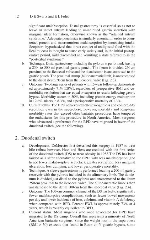
2. Duodenal switch
a. Development. DeMeester fi rst described this surgery in 1987 to treat bile refl ux; however, Hess and Hess are credited with the fi rst series of the duodenal switch (DS) to treat obesity in 1988.The DS has been lauded as a safer alternative to the BPD, with less malabsorption (and hence fewer malabsorptive sequelae), greater restriction, less marginal ulceration, less dumping, and lower perioperative mortality.
b. Technique. A sleeve gastrectomy is performed leaving a 200-ml gastric reservoir with the pylorus included in the alimentary limb. The duode-num is divided just distal to the pylorus and anastomosed to the ileum 250 cm proximal to the ileocecal valve. The biliopancreatic limb is then anastamosed to the ileum 100 cm from the ileocecal valve (Fig. 2.4).
c. Outcome. The 100-cm common channel of the DS has led to signifi cantly fewer malabsorptive complications, such as fewer bowel movements per day and lower incidence of iron, calcium, and vitamin A defi ciency when compared with BPD. Percent EWL is approximately 73% at 4 years, which is roughly equivalent to BPD.
d. Current status. Most surgeons who once advocated for BPD have migrated to the DS camp. Overall this represents a minority of North American bariatric surgeons. Since the weight loss in the superobese (BMI > 50) exceeds that found in Roux-en-Y gastric bypass, some

2. Overview of Bariatric Operations 13
Figure 2.3. Biliopancreatic diversion.
surgeons have advocated for this technique in this group of patients either as a single- or two-staged procedure. Others have performed DS as a secondary procedure following other failed bariatric operations.
3. Gastric bypass
a. Development. Mason and Ito are credited with the fi rst gastric bypass (GBP) for morbid obesity in 1966. Their operation included a hori-zontal gastric pouch with a 100- to 150-ml reservoir anastomosed to a loop of jejunum. This operation has evolved over the last four decades into what is considered the gold standard bariatric procedure to which all other procedures are compared. The fundamental modifi cations
14 D E Swartz and E L Felix
included a Roux-en-Y drainage, vertical pouch based on the less-disten-sible lesser curvature, isolated gastric pouch (divided from the gastric remnant) with less than 30-ml volume and a 10- to 15-mm anastomosis. Brolin randomized superobese patients (BMI > 50) to 75 vs. 150 cm alimentary (Roux) limb lengths and found signifi cantly improved excess weight loss at 2 years (50% vs. 64%, respectively).
b. Technique. The gastric pouch is created by creating a 15- to 30-ml pouch based on the lesser curve by stapling either “free-hand” or around a 32–34 French gastric lavage tube or Baker balloon. Care is taken to avoid injury to the left gastric artery, which supplies the pouch, and to exclude the fundus by not dividing the stomach to the left of the angle of His. The proximal jejunum is divided and the distal stump (alimentary
Figure 2.4. Duodenal switch.

2. Overview of Bariatric Operations 15
limb) is brought antecolic, retrocolic antegastric, or retrocolic retrogastric and anastomosed to the gastric pouch to create a 10- to 12-mm diam-eter stoma. The proximal stump of jejunum (biliopancreatic limb) is anastamosed to the alimentary limb either 75 to 100 cm distal to the gastrojejunostomy (BMI < 50) or 150 cm (BMI ≥ 50) (Fig. 2.5).
c. Outcome. Similar to the BPD and DS, the GBP results in dramatic metabolic and weight changes but with fewer malabsorptive sequelae. Excess body weight loss varies from 60% to 75% for 10 years and 50% at 14 years. Reported rates for comorbidity resolution are diabe-tes (80%), hypertension (70%), hypercholesterolemia (65%), gastro-esophageal refl ux disease (75%), and obstructive sleep apnea syndrome (75%). Thirty-day perioperative mortality is 0.5%. Potential vitamin and mineral defi ciencies from malabsorption requiring lifelong moni-toring include iron, calcium, folic acid, and vitamin B
12. The most se-
vere complications include leaks (0–3%), internal herniation with or without strangulated bowel obstruction (2–5%), and perforated margin-al ulcer (1%). Less severe complications include anastomotic stenosis (5–10%). Perioperative (30-day) mortality rates are 0.2% to 1% in most recent published series; however, larger regional surveys have reported up to 2%.
d. Current status. The GBP is the most commonly performed bariatric surgery, accounting for 85% of procedures in the United States and 65% worldwide. This is due to its excellent and durable results with low morbidity and mortality rates.
D. Purely Restrictive Procedures
1. Gastroplasty
a. Development. The gastroplasty procedures were an attempt to create a safer more physiologic procedure without intestinal anastomoses where leaks may occur. The stapled gastroplasties in which a partial partition was made by either horizontally or vertically placed staples to create a restrictive gastric pouch. However, the staple lines tended to break down with complete loss of restriction. Various modifi cations were described without success until Mason’s series on vertical banded gastroplasties (VBGs) in 1982. This procedure utilized a restrictive pouch based on the lesser curvature with multiple staple lines and a stoma reinforced with prosthetic mesh.
b. Technique. A 32-French bougie is placed via the mouth and advanced along the lesser curve. An EEA stapler anvil is passed full thickness through the stomach from the lesser sac approximately 5 cm distal to the gastroesophageal junction. Several applications of a TA-90 or simi-lar stapler are fi red vertically to the left of the bougie across the angle of His. The stoma is then reinforced with a band of prosthetic material (Fig. 2.6).

16 D E Swartz and E L Felix
c. Outcome. Morbidity and perioperative mortality rates were low (10% and 0.25%) and patients achieved 35% to 60% EBWL during the fi rst year, but many patients regain signifi cant weight over the long term. Staple-line dehiscences with marginal ulcerations as well as stomal stenoses with refl ux were commonly encountered.
d. Current status. The gastroplasty procedures have been largely aban-doned given their long-term failures and high rates of requiring revisional procedures.
Figure 2.5. Roux-en-Y gastric bypass.

2. Overview of Bariatric Operations 17
2. Gastric banding
a. Development. Gastric banding procedures are the least invasive, have the lowest propensity for vitamin and nutrient defi ciencies, and have the lowest morbidity and mortality among bariatric operations. With the advent of the adjustable band using a subcutaneous port, this procedure has become the most commonly performed bariatric operation in Aus-tralia and parts of Europe. Nonadjustable prosthetic material wrapped around the proximal stomach over a Nissen fundoplication was fi rst described by Wilkinson in 1981, and 2 years later Bo described the fi rst placement of a gastric band. Kuzmak introduced the adjustable gastric band (AGB) connected to a Port-A-Cath–type self-sealing res-ervoir placed in the subcutaneum in 1990. Currently performed on an outpatient basis or 24-hour stay, these bands induce satiety by exerting
Figure 2.6. Vertical banded gastroplasty.

18 D E Swartz and E L Felix
a constant, gentle pressure on the proximal gastric wall that leads to a dramatic reduction in appetite and food intake. In order to be effective, the procedure requires regular outpatient adjustments and a patient who is highly disciplined in avoiding energy-dense liquids. Initial rates of complications such as posterior gastric prolapses and erosions re-ported with the perigastric technique through the lesser sac have been markedly reduced using the pars fl accida technique.
b. Technique. Minimal dissection is the key as the gastrophrenic ligament is dissected suffi ciently to safely pass a blunt instrument posterior to the fundus. The pars fl accida of the gastrohepatic ligament is divided to expose the right crus. A small window through the phrenoesophageal ligament along the right crus is made to pass a blunt instrument through the retrogastric tissue to create a tunnel just large enough to pass the band. The tubing is passed through the buckle, where it is fastened and anterior gastrogastric sutures are placed to create an anterior tunnel to prevent anterior prolapses. The tubing is externalized where it is con-nected to the subcutaneously placed port (Fig. 2.7).
Figure 2.7. Gastric band—adjustable.
2. Overview of Bariatric Operations 19
c. Outcome. Weight loss following AGB is gradual, reaching 50–60% EWL by 3 years, which remains stable over periods up to 7 years. The US experience, however, has been more variable, with higher failure rates and band explantations. Resolution of medical comorbidi-ties is good but generally does not attain the superior results of the GBP, BPD, or DS. The most severe complications include gastric prolapse, or “band slippage,” in up to 5%, and band erosion in 0% to 1%. However, tube breakages, leaks, and port problems requiring surgical correction occur in 10% to 15%. Perioperative mortality is 0.05%.
d. Current status. The AGB is the preferred procedure in Australia and parts of Europe, accounting for 30% of bariatric surgeries worldwide and 15% in the United States. Long-term data demonstrate that this procedure is effective and durable. The AGB is a good option for the motivated patient willing to comply with a postoperative adjustment schedule of every 4 to 6 weeks in the initial year and who understands that the weight loss is gradual over 3 years.
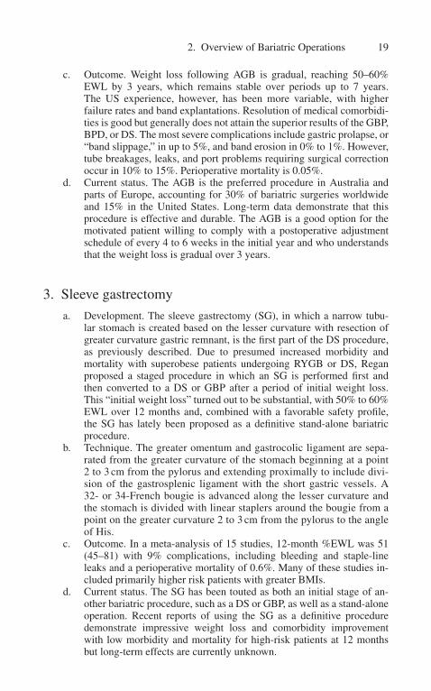
3. Sleeve gastrectomy
a. Development. The sleeve gastrectomy (SG), in which a narrow tubu-lar stomach is created based on the lesser curvature with resection of greater curvature gastric remnant, is the fi rst part of the DS procedure, as previously described. Due to presumed increased morbidity and mortality with superobese patients undergoing RYGB or DS, Regan proposed a staged procedure in which an SG is performed fi rst and then converted to a DS or GBP after a period of initial weight loss. This “initial weight loss” turned out to be substantial, with 50% to 60% EWL over 12 months and, combined with a favorable safety profi le, the SG has lately been proposed as a defi nitive stand-alone bariatric procedure.
b. Technique. The greater omentum and gastrocolic ligament are sepa-rated from the greater curvature of the stomach beginning at a point 2 to 3 cm from the pylorus and extending proximally to include divi-sion of the gastrosplenic ligament with the short gastric vessels. A 32- or 34-French bougie is advanced along the lesser curvature and the stomach is divided with linear staplers around the bougie from a point on the greater curvature 2 to 3 cm from the pylorus to the angle of His.
c. Outcome. In a meta-analysis of 15 studies, 12-month %EWL was 51 (45–81) with 9% complications, including bleeding and staple-line leaks and a perioperative mortality of 0.6%. Many of these studies in-cluded primarily higher risk patients with greater BMIs.
d. Current status. The SG has been touted as both an initial stage of an-other bariatric procedure, such as a DS or GBP, as well as a stand-alone operation. Recent reports of using the SG as a defi nitive procedure demonstrate impressive weight loss and comorbidity improvement with low morbidity and mortality for high-risk patients at 12 months but long-term effects are currently unknown.

20 D E Swartz and E L Felix
E. Miscellaneous Procedures
1. Jaw wiring
Maxillomandibular fi xation (MMF) is a temporary method to prevent over-feeding using orthodontic devices with wires. Although this procedure was more popular in the last century, it is still offered by some practitioners. The wires need to be removed for several days every 4 to 6 weeks to prevent stiffness and they are rarely left in place beyond 6 months. They have been shown to induce a moder-ate degree of weight loss in some patients the weight usually returns once it is removed. Wire cutters need to be carried at all times in case of emergencies such as vomiting or choking. With the established safety, effectiveness, and durability of other bariatric procedures for even the larger, higher-risk patient, there is little benefi t to be obtained by MMF.
2. Intragastric balloon
Endoscopic placement of an intragastric balloon fi lled with 400 to 700 ml of fl uid has seen resurgence in popularity in recent years. Like MMF, it is a temporary procedure with a strict recommendation to remove it within 6 months. Weight loss during this period has been reported up to 33% EWL, with complete weight regain following defl ation if a defi nitive bariatric procedure does not en-sue. Patients at high risk for a defi nitive surgery may improve their risk profi le with an initial substantial weight loss, but complications such as obstruction, gastric perforation, and death have been reported. The intragastric balloon is at present only investigational in the United States.
3. Implanted gastric pacemaker
Electrical impulses to induce gastroparesis and anorexia serve as the impetus for the implanted gastric pacemaker (IGP). Cigaina reported these results in ani-mal studies in which an implanted pacemaker that stimulated the lesser curvature of the stomach between the gastroesophageal junction and pylorus. Clinical trials are few but the largest experience comes from Europe, where morbidly obese patients underwent placement of the IGP. At 1- and 2-year follow-up, % EWL was 20 and 25, respectively, with minimal morbidity. At this time, the IGP is solely experimental.
F. Summary
After half a century of growth and development, bariatric surgery is still an array of procedures in evolution. The application of the laparoscope along with improvements in safety and a dramatic reduction in morbidity and mortality has made these procedures more acceptable to patients. Despite their popularity, the large volume of bariatric operations performed has not kept pace with the epidemic rise in obesity rates worldwide.

2. Overview of Bariatric Operations 21
The armamentarium of procedures to treat obesity attests to the lack of a single ideal surgical remedy. As further refi nements are made and new technolo-gies become available, we will undoubtedly see even greater and more durable weight loss, better outcomes for comorbidities, and enhanced safety profi les. This brief overview of existing procedures will hopefully provide the reader a framework in which to evaluate current treatments and integrate future ones.
G. Selected References
Belachew M, Legrand M, Vincent V, et al. Laparoscopic adjustable gastric banding. World J Surg 1998;22:955–963.
Bo O, Modalsli O. Gastric banding, a surgical method of treating morbid obesity: prelimi-nary report. In J Obes 1983;7:493–499.
Brolin RE, Kenler HA, Gorman JG, et al. Long-limb gastric bypass in the superobese: a prospective randomized study. Ann Surg 1992;215:387–395.
Brolin RE, Kowalski C. Operations for morbid obesity. In: Yeo CJ, Dempsey DT, Klein AS, et al. editors. Shackleford’s surgery of the alimentary tract, 6th ed. Philadelphia: Saunders/Elsevier, 2007:928–939.
Cigaina V, Pinato G, Rigo V, et al. Gastric peristalsis control by mono situ electrical stimu-lation: a preliminary study. Obes Surg 1996;6:247–249.
De Csepel J, Quinn T, Pomp A, et al. Conversion to a laparoscopic biliopancreatic diver-sion with a duodenal switch for failed laparoscopic adjustable silicone gastric band-ing. J Laparoendosc Adv Surg Tech A 220;12:237–240.
DeMeester TR, Fuchs KH, Ball CS, et al. Experimental and clinical results with proximal end-to-end duodenojejunostomy for pathologic duodenogastric reflux. Ann Surg 1987;205:414–426.
Doherty C. Vertical banded gastroplasty. Surg Clin N Am 2001;81:1097–1112.Griffen WO, Young VL, Stevenson CC. A prospective comparison of gastric and jeju-
noileal bypass operation for morbid obesity. Ann Surg 1977;186:500–507.Gumbs AA, Gagner M, Dakin G, et al. Sleeve gastrectomy for morbid obesity. Obes Surg
2007;17:962–969.Hess DS, Hess DW. Biliopancreatic diversion with duodenal switch. Obes Surg
1988;8:267–282.Keshishian A, Zahriya K, Hartoonian T, et al. Duodenal switch is a safe operation for
patients who have failed other bariatric operations. Obes Surg 2004;14:1187–1192.Kremen AJ, Linner LH, Nelson CH. An experimental evaluation of the nutritional impor-
tance of proximal and distal small intestine. Ann Surg 1954;140:439–448.Kuzmak LI. Surgery for morbid obesity. Using an inflatable gastric band. AORN J
1990;51:1307–1324.MacLean LD, Rhode BM, Sampalis J, et al. Results of the surgical treatment of obesity.
Am J Surg 1993;165:155–162.Marceau P, Hould FS, Simard S, et al. Biliopancreatic diversion with duodenal switch.
World J Surg 1998;22:947–954.Marinari GM, Murelli F, Camarini G, et al. A 15-year evaluation of biliopancreatic diver-
sion according to the Bariatric Analysis Reporting Outcome System (BAROS). Obes Surg 2004;14:325–328.
Mason EE. Vertical banded gastroplasty for obesity. Arch Surg 1982;117:701–706.

22 D E Swartz and E L Felix
Mason EE. Why the operation I prefer is the vertical banded gastroplasty 5.0. Obes Surg 1991;1:181–183.
Mason EE, Ito C. Gastric bypass in obesity. Surg Clin N Am 1967;47:1345–1351.O’Brien PE, Dixon JB. Laparoscopic adjustable gastric banding in the treatment of morbid
obesity. Arch Surg 2003;138:376–382.Poiries WJ, Swanson MS, MacDonald KG, et al. Who would have thought it? An opera-
tion proves to be the most effective therapy for adult-onset diabetes mellitus. Ann Surg 1995;222:339–350.
Regan JP, Inabnet WB, Gagner M, et al. Early experience with two-stage laparoscopic Roux-en-Y gastric bypass as an alternative in the super-super obese patient. Obes Surg 2003;13:861–864.
Schauer P. Gastric bypass for severe obesity: approaches and outcomes. Surg Obes Rel Dis 2005;1:297–300.
Scopinaro N, Adami GF, Mariari GM, et al. Biliopancreatic diversion. World J Surg 1998;22:936–946.
Scopinaro N, Gianetta E, Civalleri D. Biliopancreatic bypass for obesity: II. Initial experi-ence in man. Br J Surg 1979;66:618–620.
Slater GH, Fielding GA. Combining laparoscopic adjustable gastric banding and biliopan-creatic diversion after failed bariatric surgery. Obes Surg 2004;14:677–682.
Torres JC, Oca CF, Garrison RN. Gastric bypass: Roux-en-Y gastrojejunostomy from the lesser curvature. South Med J 1983;76:1217–1221.
Wilkinson LH, Peoloso OA. Gastric (reservoir) reduction for morbid obesity. Arch Surg 1981;116:602–605.

A. Introduction
Weight loss surgery (WLS) has become accepted as a treatment for obesity and many of its comorbidities. Current indications for WLS require that patients with a body mass index between 35 and 40 kg/m2 suffer from severe comorbid disease in order to attain insurance coverage for surgery. Obesity associated diseases including sleep apnea, diabetes, hypertension, cholelithiasis, thrombophlebitis, and pulmonary embolus may all affect surgical morbidity and mortality. Preoperative identifi cation of existing comorbidities may be necessary in order to secure access to surgical therapy and enable care givers to intervene in order to optimize outcomes.
B. Obstructive Sleep Apnea
The incidence of sleep apnea in patients undergoing WLS may be 35% to 77%. Patients undergoing WLS frequently exhibit characteristics concerning for obstructive sleep apnea (OSA) such as BMI greater than 35 kg/m2, increased neck circumference, nasal obstruction, micrognathia, macroglossia, snoring, frequent nocturnal arousal from sleep, and somnolence.
OSA has been associated with pulmonary hypertension, stroke, hypertension, cardiac ischemia, dysrhythmia, and sudden death. Preoperative diagnosis and treat-ment of OSA may reduce hypoxia/hypercarbia, reduce airway mucosal edema, reduce pulmonary shunting, and potentially improve surgical outcomes. Patients undergoing WLS should be evaluated with overnight polysomnography if suspected of moderate to severe sleep apnea. In patients with diagnosed sleep apnea, either CPAP or BiPAP should be instituted preoperatively. Ideally, patients will bring their own CPAP/BiPAP equipment with them on the day of surgery as this is generally better tolerated following anesthesia. The perioperative anesthetic approach to patients with OSA includes judicious use of narcotic and benzodiazepines, elevation of the head during intubation and extubation, and extubation when the patient is fully awake. Postoperatively, patients should be monitored for airway obstruction and hypoxia.
C. Cardiovascular Disease
It is predictable that some measure of cardiovascular dysfunction is rela-tively common among patients undergoing WLS given the incidence of meta-bolic syndrome, hyperlipidemia, hypertension, diabetes, peripheral vascular
3. Identifi cation of Comorbidities and Their Management
Benjamin E. Schneider

24 B E Schneider
disease, and sleep apnea in the severely obese. Generally patients undergoing WLS are stratifi ed by American Heart Association guidelines as intermediate risk. All patients should undergo an electrocardiogram and have a review of a recent lipid panel. The clinical history and exam should help delineate those patients in whom poor functional status, cardiac risk predictors, or symptoms necessitate further cardiac testing. Subsequent evaluation with either a dobutamine stress echo or nuclear stress testing may then help identify patients requiring a referral to a cardiologist. A previous history of rheumatic fever or use of diet pills (e.g., phentermine-fenfl uramine) should undergo an echocardiogram to assess for valvular disease. Although lacking in high level evidence and recently more controversial, consideration should be given to the use of β-blockers.
D. Deep Vein Thrombosis
Among the medical conditions associated with morbid obesity are venous insuffi ciency and thrombosis. Obesity is associated with derangements in coagu-lation, which may lead to hypercoagulability. Preoperative assessment should focus upon patient characteristics, including history of thromboembolic events, venous stasis/insuffi ciency, smoking, oral contraceptive use, sleep apnea, age, and hypercoagulable states. Generally we advise patients to refrain from smoking and oral contraception for 8 weeks prior to surgery. Unless otherwise contraindicated mechanical as well as either unfractionated or low-molecular-weight heparin should be employed perioperatively. In patients at highest risk consideration may be given to preoperative placement of an inferior vena cava fi lter.
E. Type 2 Diabetes
Among comorbidities, type 2 diabetes has the strongest link with obesity; 80% of type 2 diabetics are obese. Screening of patients prior to WLS is important as half of type 2 diabetics are previously undiagnosed. Routine screening should include fasting plasma glucose and glycosylated hemoglobin (HbA1C). Patients diagnosed with diabetes should have their blood glucose controlled prior to surgery. HgA1C refl ects the degree of glycemic control over the erythrocyte life span (120 days) generally should be < 9%.
F. Liver Disease
In addition to diabetes and central obesity, nonalcoholic fatty liver disease (NAFLD) is an established consequence of metabolic syndrome. As the global epidemic of obesity has grown, the incidence of NAFLD has risen to estimates as high as 25%. The clinical spectrum of the disease ranges from relatively benign steatosis to nonalcoholic steatohepatitis (NASH), the latter of which may progress to cirrhosis. NASH may progress to fi brosis in 25% to 30% of cases.

Preoperatively patients should be screened with liver function testing, platelet count, coagulation panel, and serum albumen. Serum aminotransferase levels and bilirubin are not completely sensitive, as values may be normal in the set-ting of severe disease. Advanced fi brosis and infl ammation should be suspected in patients with an AST/ALT ratio of greater than 1. A liver ultrasound may demonstrate a bright-hyperechogenic parenchyma, hepatomegaly. Doppler may be helpful in assessing hepatic and portal vein fl ow. Preoperative weight loss may improve ALT levels and liver size. Intraoperative liver biopsy should be considered in patients with hepatic dysfunction, metabolic syndrome, or gross liver disease.
G. Cholelithiasis
The incidence of cholelithiasis among morbidly obese patients may be 45%, as compared with 10% to 20% in the general population. Furthermore, an increase in the incidence of gallstones following weight loss is well described. This led surgeons to advocate for cholecystectomy at the time of WLS, particularly in the case of malabsorptive procedures in which rapid weight loss is achieved. Sugerman (1995) described the routine use of prophylactic ursodiol in order to reduce the risk of stone formation following gastric bypass surgery. While uniform standards do not exist for routine screening with ultrasound either preoperatively or intraoperatively, it is our practice to attempt to identify patients with gallstones. Patients may then be counseled as to appropriate management.
H. Selected References
Dixon JB. Surgical treatment for obesity and its impact on NASH. Clin Liver Dis 2007;11:86–101.
Gross JB et al. Practice guidelines for the perioperative management of patient with obstructive sleep apnea. Anesthesiology 2006;104:1081–1093.
Saltzman E, et al. Criteria for patient selection and multidisciplinary evaluation and treat-ment of the weight loss surgery patient. Obes Res 2005;13:234–243.
3. Identifi cation of Comorbidities and Their Management 25

A. Indications
Obesity is an excess of body fat that frequently results in a signifi cant impairment of health. It is a chronic, lifelong, genetically related, life-threatening disease of excessive fat storage. Obesity results when the size or number of fat cells in a person’s body increases. A normal-sized person has 30 to 35 billion fat cells. When a person gains weight, these fat cells fi rst increase in size and later in number. One pound of body fat represents about 3,500 calories. The prevalence of overweight and obesity in the United States make obesity a leading public health problem that can have medical, social, psychological, and economic consequences. Obesity is growing at an exponential rate: It is estimated that there are more than 150,000 bariatric operations performed each year in the United States.
Although “overweight” technically refers to an excess of body weight and “obesity” to an excess of fat, these two words can be defi ned operationally in terms of body mass index. The body mass index (BMI) is the most practical way to evaluate the degree of obesity, although it does not take into account the different ratios of adipose to lean tissue. Visceral fat (or central obesity) has a much stronger correlation with certain diseases, such as cardiovascular disease, than the BMI alone. The absolute waist circumference (>102 cm in men and >88 cm in women) or waist–hip ratio (> 0.9 for men and > 0.85 for women) are both used as measures of central obesity.
Another way to determine obesity is to assess the percent of body fat but this can be a little challenging and often requires specialized equipment. The most accurate measures are to weigh a person underwater or to use an X-ray test called dual energy X-ray absorptiometry (DEXA). It is generally agreed that men with more than 25% body fat and women with more than 30% body fat are considered obese.
Bariatric surgeons use the BMI when evaluating candidates for potential bariatric surgery. It is calculated from the height and weight as follows:
BMI = body weight (in kg) ÷ square of stature (height, in meters)
For example, a man who is 5¢ 10² (1.78 meters) tall and weighs 285 lbs (135 kg) would have a BMI of 130/1.78 × 1.78 = 41.
Overweight is defi ned as a BMI between 25 and 30 kg/m2 and obesity as a BMI greater than 30 kg/m2. The current defi nitions commonly in use establish the following values, agreed in 1997 and published by the WHO in 2000 are summarized in the following:
4. Defi nition of Obesity and Indications for Surgery
Jamie D. Adair and Mark A. Pleatman

28 J D Adair and M A Pleatman
● A BMI less than 18.5 is underweight● A BMI of 18.5–24.9 is normal weight● A BMI of 25–29.9 is overweight● A BMI of 30–39.9 is obese● A BMI of equal to or greater than 40 is severely (or morbidly) obese
The latest estimates are that around 30% of adults in the United States are obese and 5% are considered morbidly obese. Morbid obesity (or clinically severe obesity) is recognized as a major public health risk throughout the world, and has been clearly shown to reduce life expectancy. Certain comorbid conditions associated with obesity are largely responsible for the mortality and morbidity of this disease. Cardiac disease, diabetes mellitus type II, obstructive sleep apnea, hypertension, dyslipidemia, gastroesophageal refl ux disease, stress urinary incontinence, arthritis of the weight-bearing joints, infertility, and some cancers have all been linked to obesity (Table 4.1). Bariatric surgery has proven to be an effective means to aid in the management of these comorbidities. The previous chapter deals with the iden-tifi cation of these comorbid conditions and their management.
B. Indications for Operation
Medical management alone has a high failure rate to sustain greater than 10% weight loss in obese patients; and management of comorbidities is often expensive and insuffi cient. The surgical treatment of morbid obesity has been well established as being safe and effective. In addition, the short-term and long-term improvement in comorbidities has been well documented. Surgery should be only one part of a long-term multidisciplinary approach that should include monitoring for nutritional and metabolic complications and dietary counseling to prevent weight gain. Psychological and behavioral factors as well as an assess-ment of perioperative risk and complications must be considered before bariatric surgery. For these reasons, the NIH in 1991 issued a consensus statement and acknowledged that “alone, objective clinical features is not suffi cient to make a decision regarding surgery.” Although each potential surgical case should be assessed for risks and benefi ts, the consensus statement offered the following guidelines for patient selection:
● Patients should have a low likelihood of responding to traditional, non-surgical therapy. Often, these patients have previously tried medically sound weight loss programs without success.
Table 4.1. Comorbidities associated with obesity.
Hypertension Certain carcinomas Venous stasisCardiovascular dysfunction Sexual hormone dysfunction Degenerative arthritisRespiratory insuffi ciency Infectious complications Pseudotumor cerebriGERD Hyperlipidemia Psychosocial impairmentDiabetes Heart disease Chronic lower back painSkin disorders Gout Sleep apenaAsthma Urinary stress incontinence

● Patients must be well informed and motivated and accept the operative risks. They also need to be able to participate in and comply with treat-ment and follow-up.
● Patients should have a BMI in excess of 40 kg/m2 or body weight greater than 100 lbs above ideal body weight.
● Patients are candidates if they have a BMI between 35 and 40 kg/m2 along with more than one high-risk comorbid condition or body weight greater than 80 lbs above ideal body weight with a comorbidity.
● An important conclusion of the 1991 National Institutes Consensus Development Conference Statement on the surgical treatment of obesity was that “patients judged by experienced clinicians to have a low probability of success with non-surgical measures, as demonstrated, for example, by failure in established weight control programs or reluctance by the patient to enter such a program, may be considered for surgical treatment.”
The Society of American Gastrointestinal Endoscopic Surgeons (SAGES) there-fore recommends that surgical therapy should be considered for individuals who:
Have a body mass index (BMI) of greater than 40 kg/m2
ORhave a BMI greater than 35 kg/m2 with signifi cant comorbiditiesAND
can show that dietary attempts at weight control have been ineffective
The indications for laparoscopic treatment of obesity are the same as for open surgery.
Guidelines in the past also recommended that patients should be over the age of 18 or under 60 years of age. There is not a lot of literature with regard to these patients undergoing bariatric surgery, but a recent study indicates that it may be safe for some of these individuals.
There are certain contraindications for bariatric surgery, including: no medical management attempted, life-threatening diseases, lack of social support, inability to follow up, substance abuse, and patients who have psychiatric disorders that have been evaluated by a psychiatrist.
C. Selected References
Cowan GSM Jr, Hiler ML, Buffington CK. Criteria for selection of patients for bariatric surgery. In: Deitel M, Cowan GSM Jr, eds. Update: surgery for the morbidly obese patient. Toronto: FD Communications, 2000:161–170.
Gastrointestinal surgery for severe obesity. Proceedings of a National Institutes of Health Consensus Development Conference. March 25–27, 1991, Bethesda, MD. Am J Clin Nutr 1992;55:487S–619S.
Hazzan D, Chin EH, Steinhagen E, et al. Laparoscopic bariatric surgery can be safe for treatment of morbid obesity in patients older than 60 years. Surg Obes Relat Dis 2006;2:613–616.
Health Implications of Obesity. NIH Consensus Development Conference Statement. Ann Intern Med 1985;103:1073–1077.
4. Defi nition of Obesity and Indications for Surgery 29

30 J D Adair and M A Pleatman
Kellum JM, DeMaria EJ, Sugarman H. The surgical treatment of morbid obesity. Curr Probl Surg 1998;35:796–851.
Mun EC, Blackburn GL, Mathews JB. Current status of medical and surgical therapy for obesity. Gastroenterology 2001;120:669–681.
Ogden CL, Carroll MD, Curtin LR, et al. Prevalence of overweight and obesity in the United States, 1999–2004. JAMA 2006;295:1549–1555.
Peeters A, Barendregt JJ, Willekens F, et al. Obesity in adulthood and its consequences for life expectancy: a life-table analysis. Ann Intern Med 2003;138:24–32.
World Health Organization. WHO technical report series 894: Obesity: preventing and managing the global epidemic. A Report of a WHO Consultation. Geneva, 2000.

A. Preoperative Nutritional Assessment
Nutritional assessment is an essential part of the patient evaluation prior to weight loss surgery. Morbidly obese patients often have clinical or subclinical nutri-tional defi ciencies. Although the morbidly obese patient has an excess store of fat, there may be defi ciencies in protein or micronutrients. This may be due to baseline eating habits or poorly managed efforts at weight loss. Preoperative defi ciencies, if left untreated, may worsen after weight loss surgery, and subclinical defi ciencies may manifest into signifi cant illness with potentially devastating consequences.
1. Common preoperative defi ciencies
Common preoperative defi ciencies include low levels of protein, vitamin D (25-OH), thiamine, iron, and folate.
a. Protein defi ciencies preoperatively are usually related to diet and may raise the risk of complications with surgery. Defi ciency may worsen after surgery due to dietary restriction and/or malabsorption.
b. Vitamin D defi ciency may commonly be found preoperatively. Since increased bone turnover may be a concern after RYGB, vitamin D levels should be corrected if low prior to surgery. Calcium defi ciency can be asso-ciated with malabsorptive procedures, and may exacerbate osteopenia.
c. Body stores of thiamine last a relatively short 6 to 8 weeks, so preop-erative defi ciencies may quickly develop after surgery, with potentially devastating neurologic consequences.
d. Iron defi ciency, and related anemia, is common, especially in women of childbearing age. Since iron is predominantly absorbed in the duodenum, defi ciencies may worsen after surgery.
e. B12 and folate are less often a problem before surgery, but postopera-tive diet restriction and malabsorption may lead to decreased levels and subsequent anemia. Assessment of preoperative levels may be useful for following levels after surgery.
f. Vitamin A defi ciency has been associated with obesity, although not as commonly as other micronutrients. Postoperative defi ciency has been seen with malabsorptive procedures. Both reversible and irreversible sequelae have been seen with postoperative vitamin A defi ciency.
5. Preoperative Nutritional Assessment and Postoperative Dietary Guidelines
Andrew B. Lederman

32 A B Lederman
g. Other micronutrients or vitamins, such as copper, zinc, selenium, niacin, biotin, vitamin B6, and fat-soluble vitamins E and K are rarely defi cient preoperatively, but may become problematic after surgery. Since defi -ciencies are less common, serum levels are not routinely measured prior to surgery unless a baseline is desired for postoperative comparison.
2. Role of the dietitian
Consultation with a registered dietitian or nutritionist prior to surgery is required of all patients. This encounter should include an assessment of current eating habits, including behaviors and food choices, the calculation of postop-erative nutritional goals, and the education of the patient on how to reach these goals. The dietitian may also help with preoperative weight loss should it be needed.
3. History and physical exam
A complete history and physical exam may identify signs of nutritional defi -ciency. Eating habits, food preferences, and diet history are part of a complete history. Physical fi ndings of chronic rash, poor wound healing, or hair loss may all be signs of pre-existing defi ciency.
4. Laboratory evaluation
Serum levels are the best way to assess the different elements of nutrition, and are an important part of the preoperative evaluation. Protein nutrition should be evaluated by prealbumin and albumin. Thiamine, folate, 25-OH vitamin D, iron, and ferritin should all be checked. Calcium and parathyroid hormone should be checked if vitamin D levels are low or the patient is known to have osteopenia. Prothrombin time should be measured if vitamin K is a concern. Levels of vitamin B
12, vitamin A, zinc, selenium, niacin, biotin, and copper may
be useful preoperatively as a baseline for postoperative follow-up.Preoperative defi ciencies should be corrected with supplementation.
B. Postoperative Nutritional Guidelines
Nutritional requirements after weight loss surgery will vary with the type of procedure, the degree of malabsorption, and the patient. Specifi c diets and the progression from one to the next may vary among programs. However, some general guidelines can be followed.
1. Caloric goals
Most postoperative diets focus on achieving appropriate levels of protein and limiting empty sources of calories. The restriction provided by weight loss procedures such as gastric bypass, gastric banding, and sleeve gastrectomy limits

5. Preoperative Nutritional Assessment 33
the volume that can be eaten, but this effect may wane over time, and a caloric goal may be useful to help patients limit their intake. For most patients, caloric intake should range from 1,000 to 1,400 kcal/day.
2. Protein
Dietary protein is essential to safe and effective weight loss. Most postoperative diets aim for a daily protein intake of 40 to 60 g/day. Patients with BPD may require 60 to 90 g/day due to the greater malabsorption of that operation.
Early after surgery, many patients use high-protein supplements to reach their protein goal. Many high-protein supplements are also high in calories, and these should be avoided. Protein shakes should be consumed as meals, not sipped throughout the day, since that may lead to excessive intake of either calories or protein.
As patients progress toward a more regularly textured diet, they should strive to achieve their protein goal via dietary sources. Appropriate sources of protein include low-fat dairy products, beans, poultry, eggs, fi sh, and soy. Lean meats are also acceptable. A typical day might include four servings of dairy (approxi-mately 1/2 cup) and four servings of another protein source each day to reach the protein goal.
The early satiety associated with restrictive procedures can limit protein intake. Patients should be encouraged to eat protein fi rst with each meal and avoid beverages immediately prior or during meals. This prevents them from reaching satiety prior to consuming their protein.
3. Carbohydrates and simple sugars
Carbohydrates and simple sugars should make up only a small portion of the diet. Patients should be limited to one to two small servings of carbohydrates each day, with 1/4 cup of mashed potatoes considered to be one serving. Diets should provide about 50 g of carbohydrates each day, with milk, fruit, vegetables, and starches as appropriate sources of carbohydrates.
Patients on an extremely low carbohydrate diet may develop ketosis. This is common in the early postoperative period when patients are on a full liquid or puréed diet and it is harder to achieve 50–100 g/day of carbohydrates.
Simple sugars should be avoided as empty calories and as stimuli for dump-ing syndrome. In general, a portion needs to have less than 10 g of simple sugars to avoid dumping syndrome. Some sugars, such as fructose, glucose, and galac-tose, tend to be better tolerated than sucrose or high-fructose corn syrup.
Sugar substitutes, such as aspartame, saccharin, or sucralose, may be used. Sugar alcohols such as sorbitol may not be well tolerated.
4. Fats
Fats should be taken sparingly, but are essential. Most recommendations are for 25 g/day or less. Fatty acids are required for absorption of fat-soluble vita-mins, and essential fatty acids such as arachidonic acid can only be obtained from dietary sources. Fats should preferably be omega-3 fatty acids found in fi sh,

34 A B Lederman
canola oil, and fl ax seed. Polyunsaturated fats from vegetable oils and saturated fats from animal fats and coconut oil should be avoided.
5. Vitamins, minerals, and supplements
Patients will all require vitamin supplementation after surgery. A daily mul-tivitamin is recommended for all weight loss surgery patients. This will provide maintenance levels of vitamins A, D, E, and K, thiamine, folate, niacin, ribofl a-vin, vitamin B6, vitamin C, folate, and vitamin B
12.
Additional supplementation is based upon postoperative serum levels and other risk factors. Menstruating women may require iron supplementation in the form of ferrous sulfate at 45 to 60 mg/day. Postmenopausal women are at a high risk of osteoporosis with weight loss, and should be on calcium citrate with vita-min D at 500 mg three times each day.
All patients with malabsorptive procedures should have vitamin B12
supplemen-tation, in the form of 1,000 mcg/month intramuscularly, 100 to 1,000 mg/day orally, 500 to 1,000 mg weekly sublingually, or 500 mg each week as a nasal spray.
Combination vitamins that include variable amounts of vitamin B12
, intrinsic factor, iron, vitamin C, and folate (i.e., Conison, Foltrin, Trinsicon, and Fergon Plus) are available and are both effective and convenient for treating several defi -ciencies.
Hair loss in the setting of adequate protein intake may indicate defi ciencies of zinc, selenium, or biotin. Zinc sulfate 200 mg/day or zinc gluconate 30 to 50 mg/day is acceptable. Selenium supplementation is usually 50 to 100 mcg/day. Biotin is available as part of a good B-complex vitamin or can be taken 300 to 600 mg/day if needed.
Folate, thiamine, niacin, vitamins A, E, K, C, B6, and ribofl avin are usu-
ally adequate with a daily multivitamin. These micronutrients and trace elements such as copper should be followed with serum levels. In the setting of a specifi c defi ciency each can be supplemented appropriately.
6. Postoperative diet progression
Most programs use a progression of diets after surgery. These diets are usu-ally designed to be low in calories, high in protein, and low in carbohydrates and fats. The diets restrict the texture of food to allow the stomach and any anasto-mosis to heal.
a. Full liquid diet uses liquid meals that are separate and distinct from any beverages consumed during the day to maintain hydration.
b. Pureéd or blended diet allows a thicker consistency, but no large chunks of food. Food should be the consistency of applesauce or thinner.
c. Soft foods are fork-tender, and need to be well chewed before swallowed. Raw vegetables, red meat, or other tough foods are excluded. Bread or other soft foods that form a fi rm bolus when chewed and swallowed should also be avoided.

5. Preoperative Nutritional Assessment 35
d. The last stage incorporates regular textured foods. This allows foods of any consistency, as long as they are low fat, low calorie, low carbohy-drate, and high protein. The exception may be fi brous or sticky foods that may cause diffi culty with the stoma of a LAGB.
7. Eating behaviors
Modifi cation of preoperative eating habits is critical to postoperative success and avoiding complications. Patients need to be taught and encouraged to follow some simple rules:
a. Eat only at regularly scheduled times. This usually means three small meals each day, with two small snacks during the day.
b. Eat slowly and chew food well. Food should be chewed to the con-sistency of a liquid.
c. Patients should eat protein fi rst so that early satiety does not limit protein intake.
d. Avoid drinking during meals and for 30 minutes before and after meals. Beverages near meal times can cause enough satiety that it can be diffi cult to get adequate amounts of protein.
e. Measure portions to avoid overeating. Using a small salad plate rather than a dinner plate helps maintain small portions. Patients have a tendency over time to slowly increase their portion size, which may lead to weight regain.
f. Stop when satiety is reached.
g. Avoid sticky or fi brous foods. This can be particularly important for LAGB patients. Foods such as asparagus or celery may block the stoma of an adjustable gastric band. Sticky foods such as reheated rice or pasta can cause similar problems.
C. Selected References
Flancbaum L, Belsley S, Drake V, et al. Preoperative nutritional status of patients undergoing Roux-en-Y gastric bypass for morbid obesity. J Gastrointest Surg 2006;10:1033–1037.
Kuruba R, Koche LS, Murr MM. Preoperative assessment and perioperative care of patients undergoing bariatric surgery. Med Clin North Am 2007;91:339–351.
Parkes E. Nutritional management of patients after bariatric surgery. Am J Med Sci 2006; 331(4):207–213.
Tucker ON, Szomstein S, Rosenthal RJ. Nutrition consequences of weight loss surgery. Med Clin North Am 2007;91:499–514.

A. Introduction
The obesity epidemic continues to grow at an alarming rate. As the number of weight loss operations grows exponentially, so does scrutiny on the outcomes of these complex procedures. Such oversight has led to the recognition that surgical outcomes in morbidly obese patients are optimized through a detailed evaluation of the patient, and the ability to provide a wide range of services at all stages of management. Such thorough and holistic management can only be delivered through a comprehensive weight loss program that includes a variety of components that synergistically address all aspects of the care of the morbidly obese patient.
This chapter briefl y discusses the components of a comprehensive bariatric program, including essential staff, support programs, material infrastructure, and education. The successful integration of these components is essential in making the results of weight loss surgery both safe and durable.
B. Facilities
Bariatric offi ces should be attractive and appealing. Patient acquisition and retention will be enhanced by providing furnishings and room plans that not only accommodate every patient, but also positively signal that these patients are welcome and strongly desired. This philosophy should commence at the initial surgeon’s practice, and continue throughout all aspects of patient care, including within other consultant offi ces, as well as within hospital facilities.
Programs specializing in bariatric care should have oversized chairs in wait-ing areas and patient exam rooms. In addition, specialized examination tables with hydraulic lift capabilities are essential for assisting patients in appropriately positioning them for a physical examination. Scales for weighing patients, as well as all additional medical equipment, including blood pressure cuffs, should be of an adequate size to provide appropriate measurements. Patient gowns and other clothing articles should be large and accommodating, and readily available in all examination rooms.
In the hospital, patient beds should have the weight capacity to safely bear morbidly obese patients. In addition, they should be appropriately constructed to prevent bed sores or injury. Operating rooms should be spacious, and all surgical equipment must be compatible with larger patients. It is imperative that operating
6. Essentials of a Bariatric Program
Troy A. Markel and Samer G. Mattar

38 T A Markel and S G Mattar
room staff be familiar with the limitations of all equipment, and be able to follow alternative arrangements if patients exceed those limitations. Adequate restroom facilities, including wide stalls and shower facilities, as well as commodes that are affi xed to the fl oor as opposed to the wall, are an essential accommodation in any bariatric program. Finally, all doors and wheelchairs should be wide enough to allow for easy patient access and transport.
C. Personnel
1. Nurse coordinator and bariatric nurse
A nurse, who may also occupy the position of program coordinator, is an essential member of any bariatric program. It is important that patients be evalu-ated by a skilled professional who is familiar with the physical and psychologi-cal characteristics of the morbidly obese patient. Many postoperative patients experience a variety of symptoms that may be effectively managed by a skilled nurse. The bariatric nurse should occupy a central position in any program, and should play a pivotal role in facilitating interactions between patients and other staff members, and communicate with hospital staff regarding specifi c patient requirements.
2. Dietician/nutritional consultants
There is a growing recognition that bariatric surgeons are essentially metabolic surgeons. Bariatric surgeons are assuming this role as a result of the profound metabolic consequences of restrictive or malabsorptive weight loss procedures. Although the majority of these effects are benefi cial, there is potential for negative outcomes due to nutritional defi ciencies, some of which may be lethal. For these reasons, as well as many others, it is imperative that skilled nutritionists be available who have a deep understanding of the special feeding characteristics of morbidly obese patients and the nutritional challenges they face.
At the fi rst encounter, nutritionists evaluate the patients’ eating patterns and meal proportions, and carefully analyze their relationship with food. This evaluation often infl uences the development of special preoperative dietary plans that prepare the patient for the expected changes and challenges of weight loss surgery. In the postoperative period, many patients often have no appetite, sometimes for the fi rst 6 months or more. Most bariatric postsurgical patients have to adhere to a schedule for eating since they will not be prompted by hunger.
Nutritionists and dieticians also counsel postoperative patients on the appro-priate food items and quantity of foodstuffs to consume. Proteins are the most important food source as they allow for appropriate maintenance of muscle tissue. Nutritionists advise patients on food sources with high protein content, and on appropriate chewing and swallowing techniques. Patients and their families also need to be made aware of the dumping syndrome, which often accompanies operative procedures that bypass the duodenum. Patients should be instructed to
avoid high carbohydrate foods, particularly those that also contain a high proportion of fats, such as chocolate and pastries.
The daily administration of vitamin and nutrient supplementation is criti-cal after any weight loss surgery, and it is the incumbent responsibility of the surgeon to ensure that patients maintain adequate vitamin and nutrient balance. The most frequent nutrient aberrations involve iron, B
12, calcium, and vitamin
D. Calcium losses and low vitamin D levels are common after bariatric proce-dures, and active supplementation is required to prevent the sequelae of second-ary hyperparathyroidism.
3. Psychologists
Although many bariatric programs outsource their psychological evalu-ations, active counselors are an integral part of any successful weight loss program. Psychologists offer invaluable support in assessing patients’ mental conditions, and frequently counsel patients to withstand the lifelong changes associated with surgical weight loss procedures. Counselors aid patients in allaying the apprehensions of weight loss, and guide them in obtaining healthy perspectives of self-image. It is important that patients have a realistic expecta-tion of the rate of weight loss, as many patients may feel that weight loss will occur immediately after surgery. In addition, although outcome predictors are not completely understood, it is well recognized that patient knowledge, psycho-social adaptation, and motivational factors are important in achieving successful weight loss after bariatric surgery.
4. Surgeon
Physicians who perform surgical weight loss procedures should have in-depth training within the fi eld of general surgery, as well as comprehensive training in bariatric and minimally invasive procedures. The learning curve is steep; therefore, some form of advanced training is recommended. Although a bariatric fellowship is not required for clinical privileges, many surgeons who anticipate having a high volume of bariatric procedures may choose to undergo a 1-year fellowship that specializes in bariatric and minimally invasive surgery. However, many practicing surgeons are unable to interrupt their practices to dedicate an entire year to specialized training. Our studies have indicated that providing carefully designed mini-fellowship programs to practicing general surgeons in the form of a 6-week minimally invasive bariatric fellowship provides adequate exposure for these physicians to competently perform surgical weight loss procedures.
Ultimately the surgeon is responsible for the entire comprehensive bariatric care service, and he or she should be involved in the immediate and long-term postoperative care of the bariatric patient. A structured follow-up plan is essential to ensure the continued effectiveness of the weight loss procedure. With time, the medical supervision of bariatric patients may be transferred to other physicians, such as bariatricians, or perhaps the patients’ primary physi-cians, provided they adhere to the recommended follow-up outline as set up by the bariatric program.
6. Essentials of a Bariatric Program 39

40 T A Markel and S G Mattar
5. Anesthesiologist
Morbidly obese patients often have multiple medical comorbidities, and may suffer substantial and severe limitations in physiologic reserve. Anatomi-cal limitations due to thick necks and crowded oral cavities may pose additional anesthetic challenges. Such circumstances raise the anesthetic risk and require the presence of skilled and experienced anesthesiologists. Special anesthetic equipment and ventilators capable of generating high pressures are required to overcome the mass effect of body habitus. The volume of distribution of some medications may also be altered based on the proportion of fat tissue. All these factors dictate the need for anesthesiologists who are familiar with the safe peri-operative management of this high-risk population.
D. Education and Support
1. Educational seminars
Patients considering bariatric surgery will undoubtedly benefi t from preoperative educational seminars. Patients gain an acquaintance to the over-all scheme of the program, including the staff, the pathway they will follow in the preoperative evaluation, the characteristics of the various bariatric procedures, the expected postoperative course, and the dietary changes that are required should they participate in a surgical weight loss procedure. Emphasis is placed on the need for lifelong follow-up and periodic assessment of nutri-tional parameters.
The educational seminars are usually 1 to 2 hours in duration, and preferably involve presentations by the surgeon and other program staff, supplemented with appropriate audiovisual aids and printed materials for future review. Patients are encouraged to bring their family members to these sessions, and both patients and their family should be given ample opportunities to ask questions in order to understand the surgical options and follow-up plans.
2. Support group meetings
Support group sessions are designed to promote discussion among patients regarding appropriate expectations for weight loss and progression toward healthy living. Discussions should be moderated by a health care professional with familiarity of the challenges and changes that bariatric patients face. These sessions are primarily intended for postoperative patients and their families, but should also be attended by preoperative patients who may be considering weight loss surgery. Topics for discussion should be broad and free-spirited, but always controlled and monitored for realistic and appropriate content. An actual schedule of future topics adds an educational fl avor to these meetings, and occasional lay presentations by clinical experts are enthusiastically received by patients.

6. Essentials of a Bariatric Program 41
3. Social programs
Many comprehensive bariatric programs promote fun and educational activities for their patients. Postoperative activities may include exercise programs and social outings. In addition, cooking classes are often held onsite to show patients how to prepare healthy foods. These classes are often combined with educational workshops by the nutritionists, and serve to promote healthy eating habits. In addi-tion, a variety of exercise programs that involve weight training and cardiovascular activities promote physical activity in the long-term postoperative period.
4. Electronic resources
Due to the proliferation of the internet, potential bariatric patients are able to research weight loss surgery from numerous sources. This concept may be benefi cial, but with the plethora of information available, patients also have access to erroneous information. Therefore, it is advised that comprehensive bariatric programs invest in creating a dedicated electronic resource. Such a web site should contain an explanation of the problems associated with obesity, as well as the potential medical and surgical solutions to these problems. All infor-mation should be presented in lay terms, and should describe the organization of the program, as well as the preoperative and postoperative pathways that patients will follow. Additionally, patients may submit a basic application form electroni-cally via the web site, so that staff could prescreen patients prior to attending the educational seminar. Chat rooms for patients, as well as an area where patients could directly contact the members of the care team, may provide a means of communication that is more effective and effi cient than traditional telephone communication.
It is also imperative for bariatric centers to maintain a database of informa-tion that includes patient demographics and comorbidities, as well as operative procedures, postoperative complications, and follow-up results. A comprehen-sive database that links all bariatric centers will provide valuable information for both clinical and research purposes. In addition, a well-maintained database will allow the care team to analyze past performances, complication rates, and other parameters that will allow them to improve the care provided to their patients.
E. Conclusion
As the rate of obesity continues to rise, and the benefi cial outcomes of weight loss surgery continue to be recognized, there will be increasing demand on surgeons to safely and effectively treat patients. Since obesity is a disease that affects patients globally, it makes intuitive sense to treat these patients through a comprehensive bariatric care team. Led by the surgeon, this team should be able to effectively evaluate prospective patients and prepare them mentally and physically for the profound behavioral changes associated with surgery. Such a comprehensive team needs talented and dedicated personnel, a strong infra-structure, and most importantly, a pervasive philosophy that morbid obesity is

42 T A Markel and S G Mattar
a chronic disease process that can be effectively treated within the confi nes of a comprehensive bariatric surgical program.
F. Selected References
Benotti P, Wood GC, Still C, et al. Obesity disease burden and surgical risk. Surg Obes Relat Dis 2006;2(6):600–606.
Cottam D, Holover S, Mattar SG, et al. The mini-fellowship concept: a six-week focused training program for minimally invasive bariatric surgery. Surg Endosc 2007;21(12):2237–2239.
Markel TA, Mattar SG. Management of gastrointestinal disorders in the bariatric patient. Med Clin North Am 2007;91(3):443–450.
Mattar SG, Rogula T. Essentials of a Bariatric Surgery Program. In: Sugarman HJ, Nguyen N, eds. Management of morbid obesity. New York: Taylor & Francis, 2006:61–73.
Poitou Bernert C, Ciangura C, Coupaye M, et al. Nutritional deficiency after gastric bypass: diagnosis, prevention and treatment. Diabetes Metab 2007;33(1):13–24.
Pratt GM, McLees B, Pories WJ. The ASBS Bariatric Surgery Centers of Excellence pro-gram: a blueprint for quality improvement. Surg Obes Relat Dis 2006;2(5):497–503, discussion.
Tucker ON, Szomstein S, Rosenthal RJ. Nutritional consequences of weight loss surgery. Med Clin North Am 2007;91(3):499–514, xii.

A. Introduction
Although it would seem logical for the “eating public” to grasp the reality of the obesity epidemic and to appropriately diet, emotional obstacles to successful weight loss are monumental. Eating is a stress reliever, a friend, a pacifi er, and a companion on lonely nights. Eating until stuffed, for many, numbs fl oating anxiety, helping sufferers to cope with the general existential angst of modern life. Before undergoing bariatric surgery, emotional elements must be assessed and treated to ensure a successful outcome. This chapter outlines our approach to determining whether a patient is psychologically appropriate for weight loss surgery.
B. Checklists
1. Symptom Checklist 90
An array of tools is available to psychologists. The Symptom Checklist 90 (SCL-90), reported in Table 7.1, may or may not satisfy a particular psycholo-gist’s needs, but one can add or delete items as needed. SCL-90 has 90 items divided into nine parts each with ten items. In our experience, some reorganiza-tion proved useful to better clarify issues requiring the most emphasis.
While women check off far more areas of concern, men sometimes simply check two or three of the 90 items. When queried about any other items they might consider noting, such men usually say nothing else is relevant. This con-trasts with the clinical interview, which generally reveals a traumatic history or a frustrated life.
As might be expected, the most commonly checked items relate to physical complaints, depression, and personal sensitivity. Obese persons are often in physical distress, are very conscious and insecure in public, and often are depressed. Compulsive behavior items are commonly checked. Often, two or more phobias are indicated, especially “afraid of tight places” and “feeling uneasy in crowds.” In terms of paranoia, patients often indicate that they agree that they have the “feeling that you are being watched or talked about,” consistent with their being hypersensitive to the glances and looks by people in crowds.
It is critical to explore any items checked that indicate psychotic thinking. Men sometimes check the item “having thoughts about sex that bother you a lot”; this generally relates to feeling rejected by women due to their weight, which can
7. Psychological Assessment
Eldo E. Frezza, Mitchell S. Wachtel, and Rolf Gordhamer

44 E E Frezza et al.
yield hostile sexual fantasies. Occasionally checked is “the idea that something is wrong with your mind,” which sometimes serves as an explanation for obesity.
C. Wahler Physical Symptoms Inventory
The Wahler Physical Symptoms Inventory (WPSI) asks about 42 physical symptoms and is quite useful as a means of assessing the range and extent of physical discomfort. This tool is very useful for evaluating a patient for hyper-chondriasis. One needs to ascertain if there are more complaints than expected or beyond a typical response. Such persons can have a diffi cult recovery after sur-gery, are prone to excessively call the physician with physical concerns and post-surgical complaints, and sometimes have unrealistic expectations of the degree of relief of physical symptoms as they lose weight.
The patient reviews each of the 42 physical symptoms, rating their occur-rence as: almost never (0), once a year (1), once a month (2), once a week (3), twice a week (4), or nearly every day (5). The scores are summed and divided by 42 to yield an average, which is converted into a percentage. The test is standard-ized, with male and female norms derived from tests of the adult population in general. The most commonly reported symptoms are back aches, numbness of body parts, muscular tension, headaches, diffi culty sleeping, feeling tired, dif-fi culty breathing, pain in the hands feet, arms, and legs, and bowel trouble. One should compare these results with those of the health problems section of the Personality Assessment Screen.
D. Personality Assessment Screen
The Personality Assessment Screen (PAS), a brief form of the Personality Assessment Inventory (PAI), which has 344 items and takes 40 to 50 minutes to complete, has 22 items in 10 personality categories, as detailed in Table 7.2. Each question is scored false (0), slightly true (1), mainly true (2), or very true (3); totals are classifi ed as low (0–12), normal (13–15), mild (16–18), moderate (19–23), marked (24–44), and extreme (45–66). Care must be exercised if the “health problems” section totals 4 or more. Social withdrawal and alienation are frequently high. The two questions that compose the subcategory “psychotic features” are “some people do things to make me look bad” and “some people try to keep me from getting ahead”; although the PAS sees these statements as an
Table 7.1. Main categories of the SCL 90.
1. Negative effect 6. Social withdrawal2. Health problems 7. Hostility control3. Psychotic features 8. Suicidal thinking4. Acting out 9. Alienation5. Anger control 10. Alcohol problems

7. Psychological Assessment 45
indication of a psychotic nature, in today’s competitive offi ce environment, they can simply refl ect reality. The questions can generate hostile responses by the morbidly obese. All assessment devices have weaknesses; psychologists must be cognizant of each tool’s shortcomings and take them into consideration when reaching conclusions about a client.
E. Other Assessment Tools
We have used a modifi ed SF-36 Questionnaire (Table 7.3); our paper showed that women improved in physical functioning and mental health after bariatric surgery.
F. Formal Interview
Before interviewing the patient, the results of the checklists (SCL-90, WPSI, and PAS) are reviewed; a set of guided question is then prepared. The purposes of the interview are to: (1) ensure that the patient is free from psychological factors that may interfere with good long-term results and (2) assess the need for presurgical counseling. Patients, who usually have never seen a psychologist apart from Dr. Phil or some actor on television portraying a psychologist, enter the offi ce with apprehension; a gentle approach is mandatory.
After reviewing insurance information, the spelling of their name, and their address, the fi rst question is always, “Why the extra weight?” Some respond with, “What?” and a confused look, as if was the least probable question they would have been asked. Some say, “I have no idea why I’m fat. I just am,” which sug-gests a potentially long interview to sort out the causes or factors for the obesity. Others quickly lay out a history of their life, detailing every trauma ever experienced,
Table 7.2. Main categories of the PAS test.
1. Physical concerns 5. Anxiety2. Compulsive behavior 6. Phobias3. Personal sensitivity 7. Hostility4. Depression 8. Psychotic thinking 9. Paranoia
Table 7.3. Main categories of the SF-36 test.
1. Physical function2. Physical role limitations3. Emotional role limitations4. Bodily pain5. Mental health6. General health

46 E E Frezza et al.
with an explanation of how it impacted their eating behavior. Direction is essential: One is not interested in the loss of a dog at 6 years of age or the scholastic fail-ures of patient’s children. Sometimes it is necessary to interrupt and move on to more relevant data. Another common response to, “Why the extra weight?” is, “It’s genetic,” which is a way of saying, “Don’t blame me for the obesity.” Others responses include, “I just love food,” trauma (e.g., divorce, death of a child, job loss), childhood abuse, and in the case of women, repeated pregnancies.
A patient’s weight history is always important. One asks about weight gain from childhood in 5- to 10-year increments, using such prompts as, “What was your weight when leaving high school?” to stimulate specifi c memory. Most patients are able to recall specifi c weights. Also important is learning if the patient ever lost what he or she considered a signifi cant amount of weight, why this occurred, and why it was not permanent.
One then asks if eating results for stress or boredom, which almost always yields a positive response, including such things as, “I’m an emotional eater; any emotion will trigger my eating.” Patients often reveal a physical injury involving their back, feet, or hip that slowed or stopped physical activity. “I used to jog or play softball but I can’t anymore” is commonly expressed.
G. Family Weight
One next delves into the childhood eating milieus. How did the family deal with food? What were their attitudes toward meals? Was it a sit-down meal every night with balanced food items or run out for fast food? A vital question is, “Is anyone in your immediate family, mother, father, sisters, or brothers 80 to 100 lbs overweight?” This explores the infl uences at play in the family system. Usually, one hears that multiple relatives, especially siblings, are morbidly obese. Often, parents or grandparents are dying or have died of obesity-related diseases. Some-times, one side of the family comprises obese persons. Some will even say such things as, “I want this surgery because I watched my aunt waste away from….” Families are often completely out of touch with their bodies, with a limited connection between what they put in their mouths and the state of their health.
H. Daily Food Intake
The next question is, “What is a typical day’s food consumption for you?” Breakfast can be variable. Often patients can overeat so much in the evening that the morning lacks hunger. A common answer begins, “I stop for one or two breakfast burritos on the way to work.” A few patients will tell about a cup of tea and piece of toast or cup of yogurt. Most patients who work face the same limited lunch options that everyone else does; lunch is often fast food on the run. A few have sit-down lunches with friends where they can special order and eat low-calorie dishes. The great dangers are: (1) the snack room and (2) the various celebrations, with caloric treats, undertaken by fellow employees to celebrate a birthday or simply to charm others with sweets. Dinner for most single

7. Psychological Assessment 47
patients is fast food. Those who are married with children enjoy a meal of meat, potatoes, and vegetables. When asked, most patients report eating until they are totally full. Commonly, patients have “something sweet” before bed. Some wake at 2:00 am to snack. Others continuously snack during the day, often drinking caloric soft drinks at the same time. This information is vital to assess the barri-ers patients will face after surgery. A strategy is necessary; patients need to know temptation prowls and how to distract themselves and avoid situations that will defeat the purpose of bariatric surgery.
I. Obesity and the Body
The next question is, “How has the obesity affected your physical body?” Commonly reported are back pain, swelling of limbs, hypertension, exhaustion, foot pain, joint pain, diabetes, and sleep problems. When bariatric surgery is considered, patients are usually at the tail end of a battle against their bodies. Watching someone struggle with cancer or crutches, hauling 350–400 lbs into your offi ce chair can be diffi cult. On some occasions, the surgical assistance is sought at the fi rst sign of physical deterioration. Most, however, seem to be at death’s door before they seek help; the patient realizes that “if I don’t do some-thing radical I will be totally incapacitated or dead.” It is this thought that most often prompts a decision to seek weight loss surgery.
J. Commercial Weight Loss Systems
The next question is, “Have you tried other weight loss systems?” Almost all patients have tried a variety of the many commercial approaches available in our society. A few say, “I have tried my own system once or twice.” Most people, especially women, are able to rattle off a staggering list of commercial systems they have tried, including Nutri-System, Weight Watchers, Slim-Fast, Tops, Herba Life, Opti Fast, and Dr. Atkins diet. Patients generally report some initial success but then they level off. They stray from the diet and in turn gain more weight on top of where they started.
K. Emotional Ideation: Suicidal Thinking
The next question is, “Do you have any thoughts of harming yourself?” Most patients deny this, but the psychologist must listen and observe the patient care-fully, especially for a pause, which usually indicates a defensive, evasive, or dis-honest response. The patient may clear their throat, or look in another direction or shift uncomfortably in the chair. In such cases, one might follow up with, “I think perhaps there is more to tell about this.” Some recall past thoughts or attempts during teen years or after a traumatic event; when this occurs, one should ask for details and check how close they are to being back at that point on a scale of

48 E E Frezza et al.
1 to 10. One rarely hears about present thoughts of self-harm, but indications that these are present include that the patient was thinking about such things a month or less ago. Sometimes, thoughts of self-harm center about a specifi c event from which they are recovering. More worrisome are periodic suicidal thoughts without apparent trigger or event. In such cases, it is vital to learn if the patient sought pro-fessional help and suggest the acquisition of professional assistance (from a psy-chiatrist, psychologist, licensed professional counselor, or clinical social worker with a master’s degree) if this has not been done. Some patients consider their youth counselor at the church or pastor to be adequate as a therapist; the patient must in such cases be directed to seek the advice of a licensed professional.
L. Depression
One next asks about symptoms and diagnoses made by others of depres-sion. Many patients arrive with their depression medicines in hand for you to examine. There is emotional pain innate in weighing over 100 lbs above your recommended weight, which usually leads to spouses’ avoiding intimate rela-tions, children being embarrassed by their obese parents, and being stared at and commented upon by strangers in malls and grocery stores. Obese women particularly sense the disapproval of the general population. The obese person generally thinks poorly of themselves having let their condition get to a point of endangering their health. They are very critical of not having the will power or self-discipline to lose the extra pounds. They have experienced a long history of failure in weight loss. Most, if not all, obese persons suffer some degree of depression. When asked if the depression is weight-related or due to other circumstances, most respond by saying the depression is related to the obesity.
M. Substance Abuse
Patients in our experience usually deny alcohol, although they may report alcohol or drug use in the remote past. This may be due to our patient population, which comprises mostly middle-class persons from New Mexico and Texas with relatively strict standards of appropriate behavior; in other locales, such as large urban areas, this might be a more important issue. Our patients, when asked, also usually deny laxative abuse and bulimia. Notwithstanding this, we also routinely ask if patients have sought counseling or therapy before, the issues related to such professional care, and also if they were ever hospitalized for emotional or substance abuse issues.
N. Physical/Sexual Abuse
Childhood experiences of sexual abuse, molestation, rape, or physical abuse are vital; some women who have faced such trauma gain weight to avoid the sex-ual attention of men. It is common for young girls to have experienced inappropriate

7. Psychological Assessment 49
sexual attention from older male neighbors and relatives, often grandfathers. Not uncommon is molestation by another child. The patient often denies that the incident was signifi cant or states that some insuffi cient response, such as telling their mother or attending one or two professional sessions, permitted them to “move on” from the experience.
Recently, a patient reported having been repeatedly raped by her father as a child. She neither told anyone about nor sought therapy for these incidents. The patient felt much of her obesity was related to this trauma. Because we were unsure how the patient would react to having the protective weight removed, she was referred for extensive therapy before bariatric surgery. Some female patients report physical abuse by a spouse or signifi cant other; although the abuse was usually from an ex-husband or ex-boyfriend, it is always important to ascertain if abuse is occurring at present and take appropriate action to ensure that it stops. Although not related to abuse itself, it is always important to know if the spouse or signifi cant other also has weight problems.
O. Supportive People
The next question is, “Who is emotionally supportive to you in this proc-ess?” It is important to uncover the attitude of the spouse, usually the husband, toward bariatric surgery. Although the usual response is, “he wants me to do what makes me happy,” what is being sought for is the spouse affl icted by jealousy or insecurity. Such men can interpret bariatric surgery as an attempt to look bet-ter and fi nd another man for herself; this can result in attempts to sabotage the results, either blatant or subtle. Of cultural interest, Hispanic men sometimes desire their wives to be obese, perhaps as an indication of commitment to the marriage. Whenever such issues are uncovered, discussions with the marriage partner are vital to ensure that the surgery will be successful. Such discussions are always of some use because the spouse, even when not insecure, will be able to learn of the adjustments that may occur in the life of the family, including din-ing habits, leisure habits, and socialization patterns.
Hearing that the patient’s family and friends are involved is a very positive sign. Patients should be told that the end of obesity sometimes means a change in social systems. Some obese friends may no longer feel comfortable with the new improved body of the patient. The patient may be spending their time at the gym or recreation center instead of snacking in someone’s living room. Sometimes, the patient will know of another who underwent bariatric surgery; such persons can be vital sources of support in the postsurgical period. Another support system is of quite modern origin: There exist numerous online support groups, one of which may be of great use for the patient. Assuring the presence of a support system before surgery is a good means of ensuring long-term success.
P. Obstacles After Surgery
The interview usually ends with an exploration of potential obstacles after the surgery, many of which may have been suggested by the prior discussion. One must take into account the potential of spouses, family, work site, and community
50 E E Frezza et al.
to create stress or postsurgical psychological barriers. These should be carefully analyzed, together with a discussion of what one should do to counter any obsta-cles that might develop.
Mostly often, patients will say, “I don’t see any obstacles, everyone is behind me.” They should then be told that surprises are not uncommon and should not be cause for undue dismay. If the patient has been hospitalized, this should be explored; although most have had positive experiences, this is not always true. In such cases, therapy may be important before the surgery to sort out any residual emotional issues.
Q. Summary
Some patients with morbid obesity have serious underlying psychological issues, which, if present, require professional attention prior to bariatric surgery. By combining standardized assessment tools with a structured interview, a psy-chologist can ensure that such problems are not present and better enable bariat-ric surgery to be successful.
R. Selected References
Chau WY, Schmidt HJ, Kouli W, et al. Patient characteristics impacting excess weight loss following laparoscopic adjustable gastric banding. Obes Surg 2005;15(3):346–350.
Choban PS, Onyejekwe J, Burge JC, et al. A health status assessment of the impact of weight loss following Roux-en-Y gastric bypass for clinically severe obesity. J Am Coll Surg 1999;188(5):491–497.
Frezza EE, Shebani KO, Wachtel MS. Laparoscopic gastric bypass for morbid obesity decreases bodily pain, improves physical functioning, and mental and general health in women. JLAST 2007;17(4):440–444.
Karl JG. Morbid obesity and related health risks. Ann Intern Med 1985;103:1043–1047.Klesges RC, Klem M, Hansson CL. The effects of applicant’s health status and qualifications
on simulated hiring decisions. Int J Obesity 1990;14:527–535.Kushner RF. Body weight and mortality. Nutr Rev 1993;51:127–136.Naslund II, Agren G. Social and economic effects of bariatric surgery. Obes Surg 1991;1:
137–140.Pagano M, Gauvreau K. Principles of biostatistics, 2nd ed. Pacific Grove, CA: Duxbury
Thomson Learning, 2000.Tarlov AR, Ware JE, Greenfield S, et al. An application of methods for monitoring the
results of medical care. JAMA 1989;262:925–930.Wadden TA, Stukard AJ. Social and psychological consequences of obesity. Ann Int Med
1985;103:1002–1012.Ware JE, Sherbourne CD. The MOS 36-SF health Survey (SF 36). Concept framework and
item selection. Med Care 1992;30:473–483.www.parinc.comwww.wpspublish.com
A. Introduction
The primary objectives of bariatric surgery are to improve obesity-related comorbidities and quality of life. The success of the bariatric surgical procedure relies not only on the technical expertise of the surgeon, but also on the proper preop-erative evaluation and management of the patient’s comorbid conditions.
In the preoperative setting, the bariatric surgeon should utilize a multi-disciplinary approach for assessment and optimization of the various weight-related comorbidities of the bariatric patient. Once diagnostic studies have been reviewed, the surgeon can properly determine the patient’s candidacy for surgery, assess the patient’s surgical risk, and take preventive measures to minimize morbidity and mortality.
In addition to patient education, nutritional assessment, and evaluation of routine preoperative studies, a bariatric surgeon may recommend psychiatric and medical subspecialty consultation. Although the preoperative checklist serves as a reference guide for bariatric surgeons, each patient’s case is unique and may require additional diagnostic studies.
The process of patient selection for bariatric surgery is unique for each surgeon because of the variation in a surgeon’s bariatric surgical experience, personnel, hospital resources, and support.
B. Preoperative Testing
The goals of the preoperative testing are to assess the bariatric patient’s oper-ative risk and diagnose previously undiagnosed conditions. Table 8.1 lists routine laboratory tests and their corresponding indications. Additional testing may be indicated based on the patient’s history and physical fi ndings.
C. Preoperative Evaluation
1. Contraindications for bariatric surgery
Many clinicians regard the following conditions as absolute contraindications for performing bariatric surgery:
8. Preoperative Check List
Rami R. Zanoun and Giselle G. Hamad

52 R R Zanoun and G G Hamad
Table 8.1. Recommended preoperative testing.
Test Indication
Complete blood count and platelet count Routine preoperative bariatric laboratory evaluation
Basic metabolic panel, serum glucoseComprehensive metabolic panel, liver
function testsHemoglobin A1CAlbuminTSHFasting lipid panel/cholesterolVitamin B
12, folate, thiamine levels Exclude vitamin defi ciency
Iron level Exclude iron defi ciencyHypercoagulability testing (lupus antico-
agulant assay, prothrombin gene variant, antithrombin III activity, Protein C and S levels and activity, activated protein C resistance, MTHFR homocysteine level, Factor V Leiden
Personal or family history of venous thromboembolism or hypercoagulable disorder
Upper endoscopy Symptoms or diagnosis of gastroesophageal refl ux disease or peptic ulcer disease
Colonoscopy Patients ≥50 years or ≥40 years with fi rst-degree family member with colon cancer, or 10 yrs younger than affected relative, whichever is earlier
Electrocardiogram Men ≥40 years, women ≥50 years, history of arrhythmia, or younger patients with known coronary artery disease, hypertension, hypercholesterolemia, or diabetes mellitus
Echocardiogram Heart murmur, valvular heart disease, or history of fenfl uramine use
Chest radiograph Age ≥50 years, known or suspected cardiac or pulmonary disease
Cardiac stress test or dobutamine echocardiogram
Refer to American Heart Association guidelines
Polysomnography Snoring or Epworth score ≥10Urine pregnancy test Women of childbearing agePap smear Exclude cervical cancerMammogram Women ≥40 yearsPSA Men ≥50 years
Abbreviations: MTHFR, methylenetetrahydrofolate reductase; PSA, prostate specifi c antigen; TSH, thyroid-stimulating hormone.

8. Preoperative Check List 53
a. mental impairment that interferes with a patient’s ability to weigh the risks and benefi ts of surgery
b. active neoplastic disease c. cirrhosis with portal hypertension d. unstable or incurable pre-existing comorbidities, including unstable
coronary artery disease (CAD), uncontrolled severe obstructive sleep apnea (OSA) with pulmonary hypertension, AIDS, or an uncontrolled psychiatric condition
e. pregnancy f. immobility (e.g., wheelchair- or scooter-dependence) g. inability or unwillingness to comply with postoperative regimens,
including vitamin and mineral supplementation, dietary changes, and follow-up
h. active substance abuse
Several studies have shown that age alone is not a contraindication for bariatric surgery. However, when advanced age is combined with other severe medical comorbidities, the risk of surgery may become prohibitive.
2. Risk assessment
The mortality risk of bariatric surgery varies from 0.24% to 1.2% in the literature. The Obesity Surgery Mortality Risk Score, developed by DeMaria et al., utilizes fi ve patient characteristics to stratify perioperative mortality for bariatric surgery:
a. male genderb. age greater than or equal to 45 yearsc. BMI greater than or equal to 50 kg/m2
d. hypertensione. risk of pulmonary embolism
Each variable is scored as 1 point. In a multicenter study involving more than 4,000 patients, having zero to one, two to three, or four to fi ve risk factors corresponded to mortality rates of 0.37%, 1.21%, and 2.4%, respectively.
D. Preoperative Checklist
1. Patient education
Encourage patient attendance at a bariatric surgical information session that explains the details of the procedure(s), alternatives, risks, benefi ts, insurance requirements, postoperative regimen, and recovery.
2. Nutritional counseling
Meeting with a registered dietitian can facilitate preoperative weight loss and promote a patient’s ability to comply with postoperative dietary modifi cations and

54 R R Zanoun and G G Hamad
vitamin supplementation. Furthermore, involving the patient’s family members can assist the patient in committing to these changes in lifestyle.
3. Psychological/psychiatric consultation
Many centers for bariatric surgery require psychological/psychiatric clear-ance. The aims are to determine whether the patient has the cognitive capacity to weigh the risks and benefi ts of surgery, diagnose occult mental illnesses that might require intervention, and ascertain whether the patient’s psychiatric condi-tions pose undue risk to patient compliance with the postoperative regimen.
4. Informed consent
Discuss the course and risks of bariatric surgery.
a. Early complications: conversion to laparotomy, bleeding, infection, venous thromboembolism, anastomotic leak, intestinal obstruction, possible need for reoperation or readmission, and death.
b. Late complications: intestinal obstruction, marginal ulceration, inter-nal hernia, stomal stenosis, cholelithiasis, band slippage and erosion, port site complications, failure of weight loss, and weight regain.
c. Discuss hospital course, including ambulation, incentive spirometry, pain control, drains and catheters, radiographic studies, length of hospital stay, and recovery time.
d. Discuss postoperative regimen, including dietary requirements, fl uid intake, vitamin and mineral supplementation, exercise, and follow-up visits.
e. Discuss outcomes after bariatric surgery, including health benefi ts, improvement in quality of life, reduction in mortality, keys to success, and causes of failure of weight loss or weight regain.
5. Management of comorbidities
In order to reduce postoperative morbidity, proper measures must be under-taken to stabilize comorbid conditions.
a. Cardiac: High risk with coronary artery disease will require postopera-tive cardiac monitoring and may benefi t from β-blockers administered perioperatively and postoperatively
b. Pulmonary: Patients with obstructive sleep apnea (OSA) should be encouraged to use their prescribed continuous positive airway pres-sure (CPAP) or bilevel positive airway pressure (BiPAP) treatments during sleep prior to the bariatric procedures [1, 3].
c. Thromboprophylaxis: For patients at high risk for venothromboembolic events (VTEs) (e.g., prior VTE, hypercoagulable disorder, venous stasis disease, obesity hypoventilation syndrome, BMI ≥60), consider inferior vena cava fi lter placement and/or extended anticoagulation.
d. Endocrine: Because of the elevated risk of infection, diabetics should be counseled to control hyperglycemia aggressively. Endocrinologic consultation may be necessary.

8. Preoperative Check List 55
e. Gastrointestinal: Patients with gastroesophageal refl ux disease should undergo upper endoscopy and may require proton pump inhibitor therapy and/or treatment of Helicobacter pylori.
f. Cholelithiasis: Ursodiol therapy has been shown to reduce the inci-dence of cholelithiasis after bariatric surgery. Some surgeons advo-cate routine prophylactic cholecystectomy during the bariatric proce-dure, whereas others perform cholecystectomy selectively in patients with symptomatic cholelithiasis.
g. Substance abuse: Smoking cessation and abstinence from alcohol or drug abuse is critical for reducing postoperative morbidity and promoting compliance.
6. Medication management
a. Discuss medications to discontinue prior to surgery (nonsteroidal anti-infl ammatory drugs and aspirin, oral contraceptives, insulin, diuretics, and oral hypoglycemics).
b. For patients on Coumadin, discontinue Coumadin and order preopera-tive heparin or low-molecular-weight heparin.
c. Steroid-dependent patients will need perioperative stress-dose steroids.
E. Intraoperative Considerations
1. Ensure facility readiness
a. Armless chairs for patients in the waiting areas and patient rooms provide convenience and comfort. The wheelchairs, gurneys, operating room ta-ble, patient beds, and toilets must be able to accommodate the weight of bariatric patient comfortably. A bariatric bed may be required for pa-tients whose weight exceeds the capacity of the standard hospital bed.
b. Air mattress transfer devices may be useful for transferring the patient to and from the operating table. They may be placed on the gurney in the holding area prior to surgery.
c. The operating table must be able to handle frequent changes in positioning, especially reverse Trendelenburg. A padded footboard is placed under the patient’s plantar surfaces and the ankles are taped so that they do not rotate when the patient is in reverse Trendelenburg.
2. Prepare operating room equipment
a. Trocars b. Insuffl ator c. Light source d. Camera e. Video monitors f. Angled laparoscope g. Liver retractor

56 R R Zanoun and G G Hamad
h. Laparoscopic graspers i. Laparoscopic scissors j. Suction and irrigation device k. Clip appliers l. Electrocautery m. Ultrasonic shears n. Linear and/or circular laparoscopic staplers and cartridges o. Laparoscopic suturing devices or needle drivers p. Upper endoscope
3. Prior to surgery
a. Identify patient upon arrival to the operating room. b. The patient should be secured to the table because of frequent changes
in table positioning during bariatric surgery. Pressure points should be well padded to prevent potential neuropathies.
c. Administer preoperative intravenous antibiotic prophylaxis. d. Administer preoperative thromboprophylaxis (sequential compression
devices activated prior to the induction of anesthesia and/or pharma-cologic agents).
e. Perform Foley catheterization.
F. Conclusion
A successful outcome after bariatric surgery depends on optimization of medical conditions, patient education, postoperative compliance, and support from the bariatric surgical team, family, friends, and coworkers. A multidisci-plinary approach is of critical importance and thorough preoperative preparation is mandatory.
G. Selected References
Collazo-Clavell ML, Clark MM, McAlpine DE, et al. Assessment and preparation of patients for bariatric surgery. Mayo Clin Proc 2006;81(10 Suppl):S11–17.
DeMaria EJ, Carmody BJ. Perioperative management of special populations: obesity. Surg Clin North Am 2005; 85(6):1283–1289, xii.
DeMaria EJ, Portenier D, Wolfe L. Obesity surgery mortality risk score: proposal for a clinically useful score to predict mortality risk in patients undergoing gastric bypass. Surg Obes Relat Dis 2007;3(2):134–140.
Flancbaum L, Belsley S. Factors affecting morbidity and mortality of Roux-en-Y gastric bypass for clinically severe obesity: an analysis of 1,000 consecutive open cases by a single surgeon. J Gastrointest Surg 2007;11:500–507.

8. Preoperative Check List 57
Hamad GG, Bergqvist D. Venous thromboembolism in bariatric surgery patients: an update of risk and prevention. Surg Obes Relat Dis 2007;3(1):97–102.
Kuruba R, Koche LS, Murr MM. Preoperative assessment and perioperative care of patients undergoing bariatric surgery. Med Clin North Am 2007;91(3):339–351, ix.
Mason EE, Renquist KE, Huang YH, et al. Causes of 30-day bariatric surgery mortality: with emphasis on bypass obstruction. Obes Surg 2007;17(1):9–14.
McGlinch BP, Que FG, Nelson JL, et al. Perioperative care of patients undergoing bariatric surgery. Mayo Clin Proc 2006;81(10 Suppl):S25–33.
Schirmer B, Erenoglu C, Miller A. Flexible endoscopy in the management of patients undergoing Roux-en-Y gastric bypass. Obes Surg 2002;12:634–638.
Sugerman HJ, Brewer WH, Shiffman ML, et al. A multicenter, placebo-controlled, rand-omized, double-blind, prospective trial of prophylactic ursodiol for the prevention of gallstone formation following gastric-bypass-induced rapid weight loss. Am J Surg 1995;169(1):91–96; discussion 96–97.

A. Rationale for Care Pathways
Bariatric patients, both because of their size and comorbid conditions, present many unique issues in their postoperative care. The utilization of postoperative care pathways can help to address these issues, provide a more uniformly high level of care, and minimize the risk of in-hospital postoperative complications. Furthermore, care pathways can reduce the variability in care that occurs due to changes in resident and nursing staff. Suggested postoperative orders for laparo-scopic gastric bypass and laparoscopic adjustable gastric band patients are provided at the end of this chapter.
B. General Guidelines for Postoperative Hospitalization and Monitoring
1. Length of stay
The length of stay for bariatric patients is generally quite short. With this narrow window to identify potentially life-threatening postoperative complica-tions, care guidelines regarding nursing care and patient monitoring are extremely helpful in identifying complications early.
a. The typical laparoscopic gastric bypass patient spends two nights in the hospital. Some centers have reduced the stay to a single night.
b. Laparoscopic adjustable gastric band patients are admitted for a single night, or may even be treated as outpatients.
c. Duodenal switch patients follow a postoperative course similar to gastric bypass patients. However, they are substantially more likely to require additional days due to postoperative pain and nausea.
d. Sleeve gastrectomy patients follow a postoperative course similar to gastric bypass patients.
2. Nursing care guidelines
a. Bariatric patients with minimal comorbidities may be safely trans-ferred from the recovery room directly to the fl oor.
b. Patients with cardiac or pulmonary issues, particularly sleep apnea, may benefi t from a monitored setting such as a stepdown unit. Routine use of the ICU is unnecessary, unless it is the only monitored setting available.
9. Postoperative Care Pathway
Daniel M. Herron and Murali N. Naidu

60 D M Herron and M N Naidu
c. Vital signs should be checked at least every 4 hours and more frequently if possible.
d. Fever greater than 38.5 °C, tachycardia above 110, oliguria, a patient who “doesn’t look right” or who describes a sense of “impending doom” should trigger a call to a physician for further evaluation. These fi ndings may be the fi rst indication that a leak is present and should not be ignored. The threshold for leak or sepsis workup must be very low.
e. It is critically important that all caregivers understand that physical exam of the abdomen in a morbidly obese patient is unreliable. The obese abdomen may appear benign on exam, even in the presence of fl orid peritonitis; this must be clear to all personnel involved in post-operative care so that an expedited workup for postoperative leak or sepsis may be initiated if needed.
C. Postoperative Tubes and Drains
1. Nasogastric tubes
Most surgeons performing laparoscopic bariatric procedures do not routinely leave a nasogastric tube in after surgery. While these tubes may be inserted during the operation to decompress the stomach or check for leaks, they are typically removed in the operating room. If a nasogastric tube is left after an open bariatric procedure, it may usually be removed on the fi rst postoperative day. Nasogastric tubes generally should not be reinserted after removal.
2. Intra-abdominal drains
Data regarding the routine use of intra-abdominal drains are controversial, but most surgeons feel that their use does not improve outcome. If drains are used, they should be left in place until the time of greatest risk of leakage has passed, generally 2 to 7 days after surgery.
3. Foley catheters
A urinary catheter may be placed during surgery and left in postoperatively. In addition to providing an accurate measurement of the patient’s urine output, it may contribute to the patient’s comfort.
D. Deep Vein Thrombosis Prophylaxis
Since deep vein thrombosis with pulmonary embolism is one of the most feared and potentially lethal complications of bariatric surgery, all bariatric patients should receive at least one form of prophylaxis. At present, there is no general consensus regarding the best protocol.

9. Postoperative Care Pathway 61
1. Sequential compression devices
Most institutions use sequential compression devices starting before the induc-tion of anesthesia and continuing until the patient is fully ambulatory or discharged.
2. Heparin therapy
Bariatric patients are generally given nonfractionated heparin 5,000 units subcutaneously every 8 hours.
a. Note the more frequent dosing of nonfractionated heparin in the morbidly obese population compared with the twice-daily dosage used in the nonobese.
b. Heparin therapy may be started before the surgical procedure or immediately thereafter. Some surgeons feel that its use before surgery may increase the risk of bleeding in the early postoperative period, although this has not been confi rmed.
c. Fractionated heparin may also be used.
E. Use of Bariatric Beds
Hospital beds are gradually being manufactured in larger sizes as our population continues to grow heavier. Typical hospital beds will accommodate patients up to 350 lbs or 28 in. wide without diffi culty. Heavier or wider patients require specially designed bariatric beds. In addition to stronger construction, these beds often provide the ability to take on a chair confi guration and assist the patient in getting OOB to a standing position.
F. Postoperative Diet
1. General dietary guidelines
Most bariatric programs utilize special dietary regimens for their patients; these are often given special names such as “Stage I,” “Stage II,” etc. to differen-tiate them from nonbariatric diets.
a. In general, bariatric diets consist of small portions of low-calorie, low-sugar, low-fat items.
b. The use of small “shot-glass” size cups will remind patients to drink small amounts.
c. Carbonated beverages and caffeine are avoided in the early postop-erative phase. Some surgeons tell their patients to permanently avoid carbonation on the theory that it may stretch the gastric pouch.
d. Concentrated sweets should be avoided as these may trigger dumping syndrome in the gastric bypass patient. Standard general surgical postoperative diets such as the traditional “clear liquid diet” may contain substantial amounts of carbohydrate and should be avoided.

62 D M Herron and M N Naidu
2. Postoperative diet progression
Every program has a unique approach to dietary advancement. It is important that patients are educated in advance regarding their particular program’s approach. We have found that it is helpful to provide patients with a handout delineating the dietary progression well in advance of their operation. This allows the patient to purchase foods and drinks before admission so that they are available after discharge. Nurses, nutritional staff, and ancillary personnel should be educated regarding the special dietary needs of the postbariatric patient.
a. Liquids (Stage I) The fi rst diet consists of sips of low-calorie, noncarbonated liquids. Non-
caloric sweeteners may be used. This diet may be given to laparoscopic gastric bypass patients on their fi rst postoperative day, or to adjustable gastric band patients immediately following surgery.
b. Pureed (Stage II) Some programs maintain patients on a liquid diet during their entire
inpatient stay. Others advance them to a pureed diet as soon as the second postoperative day. The Stage II diet is also low-calorie, low-fat, and without carbonated beverages.
c. Solid (Stage III) Most surgeons wait until the stomach is fully healed (3–4 weeks)
before fi nally advancing their patients to solid food.
2. Vitamins, minerals, and medications
a. Most surgeons start patients on supplementation postoperatively. Multivitamins or calcium should be chewable or liquid during the early postoperative phase.
b. Iron supplementation should be avoided in the early postoperative phase, as it may exacerbate constipation while the patient is still taking narcotic pain medication.
3. Oral medications
Most patients tolerate oral medication adequately, even during the immediate postoperative phase, although very large pills may need to be cut in pieces or crushed, if possible.
G. Postoperative Imaging
1. Routine upper GI series a. Gastric bypass Routine postoperative imaging has not been shown to reduce morbidity
or mortality after laparoscopic gastric bypass surgery. However, many surgeons believe that obtaining such imaging in the early stages of a new bariatric program will generate expertise among the radiology

9. Postoperative Care Pathway 63
and surgery staff by providing knowledge of normal postoperative anatomy.
b. Adjustable gastric band Routine upper GI series is useful after laparoscopic adjustable gastric
band placement. It provides a baseline indication of band positioning that will prove valuable in the future if there is concern about prolapse or slippage. It also confi rms patency of the band, which guides diet advancement.
2. Selective imaging workup
a. Upper GI series This study is very useful in evaluating the febrile, tachycardic, or
oliguric patient, or even the patient who “just doesn’t look right.” The upper GI series (UGI) is considered to be the gold standard for demonstrating leaks from the gastric pouch or proximal anastomo-sis, but is ineffective in evaluating the distal anastomosis. If a leak is suspected distal to the gastrojejunostomy, immediate return to the operating room should be considered.
b. Computed tomography scan Computed tomography (CT) is useful in evaluating the gastric pouch,
proximal anastomosis and distal anastomosis. It should be remembered that CO
2 insuffl ation is generally resorbed by 24 hours and that free intra-
abdominal gas seen after that time should raise the suspicion of a leak. c. The negative study UGI and CT are not 100% sensitive! A negative imaging study should
never preclude a return to the operating room if there is suspicion of an intra-abdominal leak.
3. Choice of contrast
Water-soluble contrast such as Gastrografi n should be used in the early post-operative period, although barium is also safe if the possibility of a leak has been ruled out.
H. Pain Management
In general, patients have less postoperative pain after a laparoscopic procedure than an open one. Nonetheless, it remains important to ensure that even laparoscopic patients have access to adequate postoperative pain control. Some bariatric programs use a combination of scheduled parenteral nonsteroi-dal anti-infl ammatory drugs (NSAIDs; e.g., ketorolac) in conjunction with an injectable narcotic as needed. Others fi nd that patient-controlled analgesia (PCA) plays a valuable role in postoperative pain control. Aggressive control of postop-erative pain aids in early ambulation, which improves pulmonary mechanics and reduces the risk of DVT/PE. Use of NSAIDs should be immediately discontinued if there is suspicion of postoperative bleeding.
64 D M Herron and M N Naidu
I. Postoperative Antibiotics
We routinely provide a weight-adjusted dose of fi rst-generation cepha-losporin 30 minutes prior to incision. As with other clean-contaminated cases, there is no Level I evidence supporting prophylactic postoperative antibiotics.
J. Consolidation of Postoperative Ordersets in Bariatric Postoperative Ordersets
Every effort should be made to formalize the bariatric management concepts described here into a written care protocol that includes printed or computerized ordersets and formal nursing care pathways. Without such formal organization, it is inevitable that important components such as DVT prophylaxis will be sporadically missed. While postoperative care protocols do not guarantee a good outcome, their use increases compliance and optimizes care. Sample ordersets for laparoscopic gastric bypass and laparoscopic adjustable gastric banding are given in the following.
Appendix I. Sample Postlaparoscopic Gastric Bypass Orderset
(Please note that these sample ordersets serve as a framework only; they should not be copied word-for-word and must be individualized for each patient and hospital.)
Postoperative orders
● Admit patient to recovery; transfer to stepdown unit if sleep apnea present; otherwise transfer to fl oor when stable
● Use bariatric bed if patient weighs over 350 lbs● Diagnosis: S/P laparoscopic gastric bypass● Condition: Stable● Vital signs q 2 hours for 12 hours, then q 4 hours● OOB with assistance starting today● Sequential compression device to both legs while in bed● Record strict I/Os● Foley catheter to gravity bag● Incentive spirometry per routine● Notify MD for T > 38.4 °C, HR > 110, Systolic BP < 100● NPO except ice chips and medications with a sip of water● IV: D5LR @ 150 cc/hour● Heparin 5,000 Units SubQ q8 hours● Ketorolac 30 mg IV q6 hours

9. Postoperative Care Pathway 65
● Hydromorphone 2–4 mg IV prn breakthrough pain● Ondansetron 4 mg IV q6 hours PRN nausea/vomiting● Labs: CBC in am tomorrow● (Remember to resume cardiac medications and CPAP if required, and
monitor glucose appropriately for diabetic patients.)
Postoperative day #1
● If T > 38.4 °C or HR > 110, Upper GI series with Gastrografi n to rule out leak
● Notify MD immediately if study is positive● If study was not needed or is negative:
○ Remove Foley catheter○ Advance diet to Bariatric Stage I (low calorie, noncarbonated liquids,
small portions)○ Heplock IV when tolerating diet○ Out of bed and ambulate with assistance at least TID
Postoperative day #2
● Advance diet to Bariatric Stage II (low-calorie pureed, small portion)● Discontinue hydromorphone● Acetaminophen/oxycodone 325/5 mg 1–2 tabs po q 4 hrs PRN pain;
take crushed with yogurt or applesauce● Review all discharge teaching with patient; give printed instructions● Discharge home when tolerating Stage II diet and pain well controlled
Appendix II. Sample postlaparoscopic adjustable gastric band orderset
(Please note that these sample ordersets serve as a framework only; they should not be copied word-for-word and must be individualized for each patient and hospital.)
Postoperative orders
● Admit patient to recovery; transfer to stepdown unit if sleep apnea present; otherwise, transfer to fl oor when stable
● Use bariatric bed if patient weighs over 350 lbs● Diagnosis: S/P laparoscopic adjustable gastric band● Condition: Stable● Vital signs q 2 hours for 12 hours, then q 4 hours● OOB with assistance starting today● Sequential compression device to both legs while in bed● Record strict I/Os● Foley catheter to gravity bag
66 D M Herron and M N Naidu
● Incentive spirometry per routine● Notify MD for T > 38.4 °C, HR > 110, Systolic BP < 100● Diet: Bariatric Stage I (low calorie, noncarbonated liquids, small
portions)● IV: D5LR @ 150 cc/hour● Heparin 5,000 Units SC q8 hours● Ketorolac 30 mg IV q6 hours● Hydromorphone 2–4 mg IV prn breakthrough pain● Ondansetron 4 mg IV q6 hours PRN nausea/vomiting● Labs: CBC in am tomorrow● Schedule Gastrografi n swallow for tomorrow am● (Remember to resume cardiac medications and CPAP if required, and
monitor glucose appropriately for diabetic patients.)
Postoperative day #1
● IF UGI shows leak or malpositioned band, contact MD immediately.● If UGI study is negative for leak and band is in good position:
○ Remove Foley catheter.○ Advance diet to Bariatric Stage II (low calorie pureed, small por-
tions).○ Heplock IV when tolerating diet.○ Out of bed and ambulate with assistance at least TID.○ Discontinue hydromorphone.○ Acetaminophen/Hydromorphone 325/5 mg 1–2 tabs po q 4 hrs PRN
pain; take crushed with yogurt or applesauce.○ Review all discharge teaching with patient; give printed instructions.○ Discharge home when tolerating Stage II diet and pain well controlled.
Selected References
Cooney RN, Bryant P, Haluck R, et al. The impact of a clinical pathway for gastric bypass surgery on resource utilization. J Surg Res 2001;98:97–101.
Herron DM. The surgical management of severe obesity. Mt Sinai J Med 2004;71:63–71.Herron DM. Establishing and organizing a bariatric surgery program. In: Inabnet WB,
DeMaria EJ, Ikramuddin S, eds. Laparoscopic bariatric surgery. Philadelphia: Lippincott Williams & Wilkins, 2005.
Huerta S, Heber D, Sawicki MP, et al. Reduced length of stay by implementation of a clinical pathway for bariatric surgery in an academic health care center. Am Surg 2001;67:1128–1135.
Pieracci FM, Barie PS, Pomp A. Critical care of the bariatric patient. Crit Care Med 2006;34:1796–1804.
Yeats M, Wedergren S, Fox N, et al. The use and modification of clinical pathways to achieve specific outcomes in bariatric surgery. Am Surg 2005;71:152–154.

A. Introduction
Bariatric procedures induce weight loss using a combination of restric-tive and/or malabsorptive surgical techniques. The metabolic and nutritional complications of bariatric surgery are, in part, directly related to the surgically created anatomical changes in the gastrointestinal tract. They also occur due to patient noncompliance with nutritional supplementation and dietary altera-tions following surgery. Despite having huge stores of energy in the form of excess fat, morbidly obese patients (BMI ≥35 kg/m2) may have clinical or sub-clinical nutritional defi ciencies preoperatively as a consequence of poor diet over a prolonged period of time. Therefore, the severity of the postoperative nutritional defi cit is dependent on several factors, including the patient’s preop-erative nutritional status, the specifi c bariatric procedure undertaken, postop-erative complications, patient-related nutritional compliance, and routine fol-low-up with his or her surgeon and primary care provider.
At the end of this chapter we have included Tables 10.1 to 10.3, which describe the site of absorption of nutrients, mechanism of action of various bari-atric surgical procedures, and common nutritional and metabolic complications of bariatric surgery. Table 10.4 contains suggested mineral and vitamin supple-mentation following various bariatric surgical procedures, and Table 10.5 provides recommended screening tests prior to bariatric surgery and postoperatively.
B. Vitamin Defi ciencies
1. Vitamin B12
and folate
Vitamin B12
defi ciency is common after bariatric surgery when restrictive procedures such as Roux-en-Y gastric bypass (RYGB) are utilized. The parietal cells that secrete acid and intrinsic factor and the chief cells that secrete pepsino-gen are mainly located in the fundus and body of the stomach. The small residual pouch created with the RYGB has minimal acid production and pepsin-related digestion of protein-bound cobalamins in food. This coupled with decreased production of intrinsic factor means that vitamin B
12 cannot be separated from
foods and crystalline B12
cannot be absorbed in the terminal ileum. The prevalence of B
12 defi ciency after RYGB is 12% to 33%, but clinical symptoms are less
prevalent. The body storage of vitamin B12
is large compared to daily needs;
10. Long-term Follow-up Protocol of Bariatric Patients
Steven Teich and Marc P. Michalsky

68 S Teich and M P Michalsky
the normal human reserve is 2 to 5 years. Therefore, vitamin B12
defi ciency can appear years after surgery; so patients undergoing RYGB need to be monitored long-term and treated with vitamin B
12 once they demonstrate low blood lev-
els. The most common clinical manifestations of B12
defi ciency are macrocytic anemia and neurologic symptoms (i.e., peripheral neuropathy, paresthesias, and demyelination of the corticospinal and dorsal columns).
Folate defi ciency, while less frequent than B12
defi ciency, occurs mainly due to reduced dietary intake but also occurs secondary to reduced gastric acid and lack of exposure to the upper small intestine. Folate is absorbed primarily from the proximal third of the small intestine, but with adaptation it can be absorbed along the entire length of the small bowel. Folate defi ciency occurs in 9-35% of bypass patients and presents as megaloblastic anemia. It is promptly corrected with multivitamin supplementation (1 mg folate/day).
2. Thiamine
Thiamine is normally absorbed in the acid environment of the duodenum. Thiamine defi ciency occurs through reduced acid production by the gastric pouch, reduced dietary intake, and frequent vomiting. Severe thiamine defi ciency leads to Wernicke encephalopathy, which consists of ocular changes (nystagmus and ocular nerve palsies), ataxia, and apathetic mental confusion. Acute thia-mine defi ciency requires parental thiamine 50 to 200 mg per day until symptoms resolve, then 50 to 100 mg daily by mouth. However, thiamine defi ciency can usually be prevented with a daily multivitamin.
3. Calcium and vitamin D
Calcium defi ciency after RYGB and biliopancreatic diversion (BPD) is common due to the surgically induced changes. Reduced calcium absorption occurs second-ary to exclusion of the duodenum and proximal jejunum, where maximal calcium absorption occurs. Vitamin D is preferentially absorbed in the jejunum and ileum. Calcium malabsorption is aggravated by the defective absorption of fat and fat-soluble vitamins, including vitamin D. The lack of calcium results in increased production of parathormone (PTH), which then causes increased production of 1,25 dihydroxy-vitamin D and increased release of calcium from bone. Slater et al. found that over the fi rst 4 years post-BPD the incidence of hypocalcemia increased from 15% to 48%, with a corresponding increase in PTH levels in 69% of patients.
Monitoring for calcium defi ciency must include serum calcium, ionized cal-cium, serum phosphorus, alkaline phosphatase, PTH, and 25(OH) vitamin D levels. All patients undergoing RYGB and BPD require a high intake of calcium (2 g/day) along with calcium supplementation (1,200–1,500 mg elemental calcium/day) and vitamin D (400 IU/day). Calcium citrate rather than calcium carbonate should be prescribed because it is more soluble in the absence of stomach acid.
4. Vitamin A
Vitamin A, along with the other fat-soluble vitamins D, E, and K, diffuse across the brush border plasma membrane of the intestinal epithelial cell inside

10. Long-term Follow-up Protocol 69
micelles formed by bile salts and lipids. The majority of fat-soluble vitamin absorption occurs in the proximal two thirds of the jejunum. Defi ciencies occur following malabsorptive procedures due to bypassing the duodenum and a vari-able length of jejunum with inadequate mixing of food with biliary and pancreatic secretions, causing steatorrhea, bile salt wasting, and malabsorption of fat soluble vitamins. Vitamin A defi ciency is associated with eye disease: night blindness, xerophthalmia, Bitot’s spots, and keratomalacia. Skin manifestations of vitamin A defi ciency include xerosis and petechiae. The current recommendation for vitamin A replacement therapy is 25,000 IU daily.
C. Mineral Defi ciencies
1. Iron
Iron defi ciency and anemia occur frequently in preoperative as well as post-operative bariatric patients. The postoperative causes of iron defi ciency include decreased absorption of iron due to bypass of the duodenum, decreased gastric acid (which is needed to reduce iron to its more absorbable ferrous form), and reduced iron intake from avoidance of certain foods. Symptoms of iron defi ciency include fatigue, glossitis, stomatitis, and complaints of feeling cold due to impaired temperature regulation. The incidence of preoperative iron defi ciency is reported to be between 14% and 44%. Postoperative iron defi ciency occurs in 20% to 74% of patients undergoing RYGB, with the greatest risk in menstruating women. All patients undergoing bariatric surgery should receive supplementation, but noncom-pliance is a problem due to gastrointestinal intolerance and constipation.
There are many iron supplementation regimens with various forms of iron therapy. Some examples include iron gluconate or sulfate 40 to 65 mg three times per day and ferrous sulfate 320 mg twice per day. Adding vitamin C to iron therapy is benefi cial because it promotes iron absorption. The amount of iron supplementation should be increased if iron defi ciency anemia is present or if the hemoglobin level falls below 11 g/dl.
2. Zinc
Zinc defi ciency causes hair loss, dermatitis, impaired immunity, and delayed wound healing. In the postoperative bariatric patient it can be due to bypass of the primary site of zinc absorption in the small intestine, reduced intake of foods that provide zinc, and increased stool losses due to steatorrhea. In patients who have normal zinc levels preoperatively, zinc supplementation should be initiated within 2 months after bariatric surgery. Zinc intake as well as serum and urinary levels should be followed after surgery.
3. Copper
Copper is essential to the structure and function of the nervous system. In post-operative bariatric patients copper defi ciency can present as a neuropathy of the

70 S Teich and M P Michalsky
lower extremities. Copper is likely absorbed in the stomach and duodenum. When prescribing high-dose zinc supplementation it is important to recognize that copper absorption is inhibited due to increasing levels of copper-binding protein.
D. Selected References
Brolin RE. Gastric bypass. Surg Clin North Am 2001;81:1077–1095.Brolin RE, Gorman JH, Gorman RC, et al. Prophylactic iron supplementation after Roux-
en-Y gastric bypass: a prospective, double-blind, randomized study. Arch Surg 1998;133:740–744.
Brolin RE, Leung M. Survey of vitamin and mineral supplementation after gastric bypass and biliopancreatic diversion for morbid obesity. Obes Surg 1999;9:150–154.
Carrodeguas L, Kaidar-Person O, Szomstein S, et al. Preoperative thiamine deficiency in obese population undergoing laparoscopic bariatric surgery. Surg Obes Relat Dis 2005;1:517–522.
Cominetti C, Garrido AB Jr, Cozzolino SM. Zinc nutritional status of morbidly obese patients before and after Roux-en-Y gastric bypass: a preliminary report. Obes Surg 2006;16:448–453.
Flancbaum L, Belsley S, Drake V, et al. Preoperative nutritional status of patients undergoing Roux-en-Y gastric bypass for morbid obesity. J Gastrointest Surg 2006;10:1033–1037.
Goldenberg L. Nutritional deficiencies following bariatric surgery. Obesity Care Special Edition. 2007;Summer 65–72.
Kalfarentzos F, Kechagias I, Soulikia K, et al. Weight loss following vertical banded gas-troplasty: intermediate results of a prospective study. Obes Surg 2001;11:265–270.
Kumar N, Ahlskog JE, Gross JB. Acquired hypocupremia after gastric surgery. Clin Gastroenterol Hepatol 204;2:1074–1079.
Kushner R. Managing micronutrient deficiencies in the bariatric surgical patient. Obesity Mgmt 2005;203–206.
Madan AK, Orth WS, Tichansky DS, et al. Vitamin and trace mineral levels after laparo-scopic gastric bypass. Obes Surg 2006;16:603–606.
Malinowski SS. Nutritional and metabolic complications of bariatric surgery. Am J Med Sci 2006;4:219–225.
Rhode BM, Shustik C, Christou NV, et al. Iron absorption and therapy after gastric bypass. Obes Surg 1999;9:17–21.
Sheu WH, Wu HS, Wang CV, et al. Elevated plasma homocysteine concentrations six months after gastroplasty in morbidly obese subjects. Intern Med 2001;40:584–588.
Skroubis G, Sakellaropoulos G, Pouggouras K, et al. Comparison of nutritional deficien-cies after Roux-en-Y gastric bypass. Obes Surg 2002;12:551–558.
Slater GH, Ren CJ, Siegel N, et al. Serum fat-soluble vitamin deficiency and abnormal calcium metabolism after malabsorptive bariatric surgery. J Gastrointest Surg 2004;8:48–55.
Tucker ON, Szomstein S, Rosenthal RJ. Nutritional consequences of weight-loss surgery. Med Clin North Am 2007;499–514.

10. Long-term Follow-up Protocol 71
Table 10.1. Site of absorption of nutrients.
Intestinal site of absorption Nutrients
Duodenum Calcium Thiamine Iron Protein CarbohydrateJejunum/proximal ileum Fat Vitamins A, D, E, K Water-soluble vitamins Folate Essential minerals Copper Zinc Phosphorus MagnesiumTerminal ileum Vitamin B
12 Bile salts
Table 10.2. Mechanism of action of surgical procedures.
Surgical procedure Mechanism of action
Roux-en-Y gastric bypass 1ary restriction/2ary malabsorptionAdjustable gastric band Purely restrictionSleeve gastrectomy Purely restrictionBiliopancreatic diversion 1ary malabsorption/2ary restriction
Table 10.3. Common nutritional and metabolic complications of bariatric procedures.
Roux-en-Y gastric bypass Biliopancreatic diversion
Vitamin B12
defi ciency Vitamin B12
defi ciencyIron defi ciency Iron defi ciencyThiamine defi ciency Folate defi ciencyMetabolic bone disease Thiamine defi ciencyCholelithiasis Fat-soluble vitamin defi ciency Calcium defi ciency Metabolic bone disease Cholelithiasis Fat malabsorption Severe malnutrition
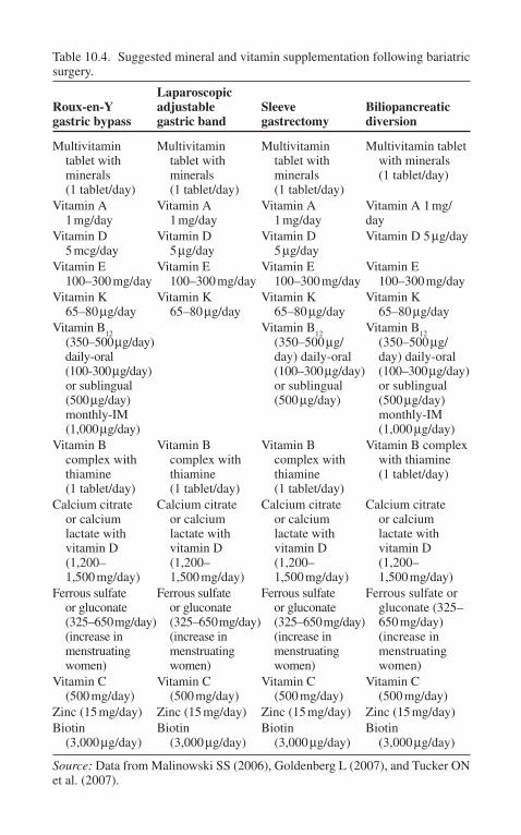
Table 10.4. Suggested mineral and vitamin supplementation following bariatric surgery.
Roux-en-Y gastric bypass
Laparoscopic adjustable gastric band
Sleeve gastrectomy
Biliopancreatic diversion
Multivitamin tablet with minerals (1 tablet/day)
Multivitamin tablet with minerals (1 tablet/day)
Multivitamin tablet with minerals (1 tablet/day)
Multivitamin tablet with minerals (1 tablet/day)
Vitamin A 1 mg/day
Vitamin A 1 mg/day
Vitamin A 1 mg/day
Vitamin A 1 mg/day
Vitamin D 5 mcg/day
Vitamin D 5 µg/day
Vitamin D 5 µg/day
Vitamin D 5 µg/day
Vitamin E 100–300 mg/day
Vitamin E 100–300 mg/day
Vitamin E 100–300 mg/day
Vitamin E 100–300 mg/day
Vitamin K 65–80 µg/day
Vitamin K 65–80 µg/day
Vitamin K 65–80 µg/day
Vitamin K 65–80 µg/day
Vitamin B12
(350–500 µg/day) daily-oral (100-300 µg/day) or sublingual (500 µg/day) monthly-IM (1,000 µg/day)
Vitamin B12
(350–500 µg/ day) daily-oral (100–300 µg/day) or sublingual (500 µg/day)
Vitamin B12
(350–500 µg/ day) daily-oral (100–300 µg/day) or sublingual (500 µg/day) monthly-IM (1,000 µg/day)
Vitamin B complex with thiamine (1 tablet/day)
Vitamin B complex with thiamine (1 tablet/day)
Vitamin B complex with thiamine (1 tablet/day)
Vitamin B complex with thiamine (1 tablet/day)
Calcium citrate or calcium lactate with vitamin D (1,200– 1,500 mg/day)
Calcium citrate or calcium lactate with vitamin D (1,200– 1,500 mg/day)
Calcium citrate or calcium lactate with vitamin D (1,200– 1,500 mg/day)
Calcium citrate or calcium lactate with vitamin D (1,200– 1,500 mg/day)
Ferrous sulfate or gluconate (325–650 mg/day) (increase in menstruating women)
Ferrous sulfate or gluconate (325–650 mg/day) (increase in menstruating women)
Ferrous sulfate or gluconate (325–650 mg/day) (increase in menstruating women)
Ferrous sulfate or gluconate (325– 650 mg/day) (increase in menstruating women)
Vitamin C (500 mg/day)
Vitamin C (500 mg/day)
Vitamin C (500 mg/day)
Vitamin C (500 mg/day)
Zinc (15 mg/day) Zinc (15 mg/day) Zinc (15 mg/day) Zinc (15 mg/day)Biotin (3,000 µg/day)
Biotin (3,000 µg/day)
Biotin (3,000 µg/day)
Biotin (3,000 µg/day)
Source: Data from Malinowski SS (2006), Goldenberg L (2007), and Tucker ON et al. (2007).

10. Long-term Follow-up Protocol 73
Table 10.5. Recommended screening tests prior to bariatric surgery and postop-eratively (3 months, 6 months, 1 year, then annually).
CBC, PT/PTTLipid profi leHg A
1cIron studies: Serum iron, TIBC, ferritin, transferrinSerum folate and vitamin B
12Thiamine (vitamin B
1)
Serum albumin and prealbuminSerum chemistries: including calcium, magnesium, and phosphorusVitamin AVitamin D
25—total
Selenium, zinc, ceruloplasmin (copper)PTH
Source: Data from Goldenberg L (2007) and Tucker ON et al. (2007).

A. Introduction
Nearly one fourth of all Americans are obese or morbidly obese. In fact, the number of morbidly obese persons has been increasing over time and this trend is expected to continue. Not surprisingly, many physicians are considering specializing in bariatrics and opening new practices.
From a business standpoint, the decision to open a bariatric practice should be based on whether or not the enterprise can achieve an acceptable rate of profi t. The application of economic tools in conjunction with techniques of operations manage-ment can increase the likelihood that this decision will be optimal. Clearly, the cen-tral question of whether to take on a project such as opening a specialized practice is: Do the benefi ts outweigh the costs? The guidelines presented here focus on iden-tifying factors that lead to value enhancement; the bariatric physician should con-sider these when making decisions about the business side of his or her practice.
B. Outline the Business
Prior to opening a practice, it is necessary for the bariatric physician to outline the business and operations strategy, mission statement, core competencies, and service and process design for the bariatric services to be provided. This overall plan has several purposes. First, this plan is geared at providing a market forecast of the service provision (i.e., production) requirements for the future. Secondly, this plan deals with analyzing and selecting a service location or locations, and planning the capacity of the practice facility. Additionally, this plan is designed to provide information on what the practice facility will look like and how it will function. Our approach to the business model of opening a bariatric practice relies on sound economic principles, knowledge of operations management and busi-ness strategy, as well as a fundamental understanding of bariatric medicine.
C. Mission Statement
As is true of any successful business venture, a company must know who or what it is. In order to position itself for the future, that is, to be able to make profi t in both good times and bad, a strong sense of mission is required. Thus, the fi rst
11. An Economic Approach to Opening a Bariatric Practice
Bradley T. Ewing and Eldo E. Frezza

76 B T Ewing and E E Frezza
step in our model is to create a mission statement. The mission statement clearly and concisely answers three questions:
1. What business is our practice really in?2. Who will be the customers?3. How will the practice’s basic beliefs defi ne the business?
Key components of mission and vision statements include brief descriptions of the business, customers, and core competencies. For example, will the prac-tice be in the business of providing healthy and sustainable lifestyle changes or simply weight loss solutions? Notice how the former suggests ongoing care for patients and allows for future technologies and advances in medicine to defi ne the specifi c procedures that the practice may provide. The latter, weight loss solutions, is narrowly defi ned and may not fully capture plans for follow-up care, recurrence, and other issues such as preventive medical services.
The customers of the practice may be the patients themselves (narrow view) or extend to family members of those affected by bariatric patients (broader view) to society in general (broadest view). The choice of customer market focus is impor-tant and will guide the type of services, doctors and nurses, procedures, location, and even purchases of equipment that your practice will acquire and utilize.
Core competencies are those things that a company does better than everyone else. They are those activities or abilities that should be focused on no matter what is currently happening in the industry or economy. For example, if a practice provides personal attention better than anything else, then it will be optimal for decisions to be made with this in mind. The practice will do best by asking, “Is this activity (purchase, new service, etc.) in line with our core competencies?” If it is not, then it is often the case that the activity under question will be a drag on resources. A rule of thumb is that the core competencies will align well with the values and belief systems of the organization. A mission statement that is clear from the onset is capable of guiding decision making far into the future and thus defi ning the economic scope and scale of the practice for years to come.
D. Competitive Advantages and Priorities
How does a practice gain a competitive advantage in the market place? The answer to this question comes from the practice’s core competencies. In order to take full advantage of core competencies, it is necessary to determine one’s competitive priorities. There are four broad categories of competitive priorities: cost, quality, time (speed), and fl exibility. It is crucial to determine on which of these the new practice will concentrate most. It is here where the practice can differentiate itself from others. Firms that compete on:
• Cost are the low-cost provider.• Quality offer the “best” service.• Time (speed) offer services in the most timely manner.• Flexibility are able to change or alter their mix of services quickly.
Each of these priorities is an admirable goal and many fi rms excel by focusing on all of them to some extent. However, economic trade-offs are often involved

11. An Economic Approach to Opening a Bariatric Practice 77
in choosing one priority over another; therefore, a balance must be struck. For example, it is diffi cult to offer the highest quality service (since it is costly for the provider) and simultaneously offer the services at the lowest cost to patients. Successful businesses tend to choose one priority that will be the basis for com-peting in the marketplace while maintaining a desired level or standard for the other priorities. Thus, a practice may choose a quality level (e.g., private rooms or semiprivate rooms) and then, for that level of quality, provide the services at the lowest cost to patients.
Overall, the business strategy of the practice comes from these competitive priorities. The key to success and the optimal operation of the practice is to know how they will support the corporate strategy or mission of the practice.
E. Environmental Scanning
Understanding the economic environment in which the practice will operate is critical for making good decisions. The focus of environmental scanning is on identifying market trends, opportunities, existing strengths, and threats or weak-nesses. A standard tool for this portion of the business planning model is the strengths, weaknesses, opportunities, and threats (SWOT) analysis. A general framework for the SWOT analysis is as follows:
1. Identify generic issues or problem areas: ethics, leadership, facilities, planning, political, technology, legal, etc.
2. Develop a list of facts pertaining to the issues or problem areas.3. Determine whether each fact is internal (i.e., practice has the power to
change or infl uence this issue or problem) or external to the practice.4. Value the factoids from on a scale from weak (–10) to strong (+10),
with 0 being neutral.5. Limit the compiled list to those items valued at either –10 or +10.
The results of the SWOT analysis can be interpreted based on the following:
• Strengths: facts with high, positive, internal values• Weaknesses: facts with high, negative, internal values• Opportunities: facts with high, positive, external values• Threats: facts with high, negative, external values
Knowledge gained from a SWOT analysis allows the fi rm to develop strate-gies to overcome potential problems as well as how to exploit existing strengths and opportunities.
Other methods of assessing the economic environment include economic impact analysis (EIA) at the regional, state and national levels. Obesity and its associated illnesses are known to lower productivity and wages and increase health care costs, a combination that has led to proposals for governmental interven-tion. A recent study focused on the economic impact of obesity on the statewide economy of New Mexico. It was determined that total labor income impacts are nearly $200 million, $1,660 of output income per household, and $245 of labor income per household. Obesity cost New Mexico over 7,300 jobs and cut state and local tax revenues by over $48 million. For New Mexico, impacts total over

78 B T Ewing and E E Frezza
$1.3 billion. It is expected that the impacts in different markets, such as rural and border areas, might differ substantially from that of more populated and affl uent regions. Given the differences in patient and population demographics that exist between areas even within the same state, it is imperative to conduct focused studies based on specifi c regions. Appropriate public health policy and health care provision decisions will depend on accurate assessment of the economic consequences of obesity in those regions.
Furthermore, when analyzing trends in patient base, revenues, market size, etc., a thorough industry level analysis may include time series econometric models to determine underlying trends and interactions among many relevant factors. Econometric models provide insight as to the responsiveness of one variable to a change in another (e.g., how market demand responds to a change in per-capita personal income). These models may also allow the practice to simulate their performance under a number of different economic scenarios. It is likely that this type of analysis would be outsourced to a consulting economist who deals with larger-scale econometric models on a routine basis. However, the benefi t-to-value ratio might be quite high, for example, especially if the practice avoids locating in less profi table area.
F. The Service Package and Service Process
A detailed description of your service(s) needs to be established. The service package is a grouping of physical, sensual, and psychological benefi ts that are purchased together as a part of the service. The type of service process that will be utilized should be determined. In particular, the choice of service design will impact the effi ciency and thus costs and profi ts. There are three general categories of service design. The fi rst focuses on substituting technology for people. This design process is akin to how McDonald’s uses technology ( fryers, ovens, conveyor belts, etc.) to make fairly standard products available to the masses. The second design process focuses on getting the customer involved. This process is seen in self-checkouts at retailers such as Home Depot and also in salad bars, etc. The third service design process involves high customer atten-tion. This process attempts to make the experience unique to the customer and is highly customized, such as shopping at Nordstrom. Each of these designs, or a combination of them, may be utilized in the bariatric practice. Routine follow-up care (e.g., monitoring weight, blood tests, etc.) may be standardized. Having the patient conduct tests at home and send the results to the lab/facility would be an example of getting the customer involved. Physician–patient meetings to discuss particular treatment options, etc., would likely be highly customized.
In choosing an optimal mix of services, the practice must determine the range of services to be provided. Typically, the steps involved include:
• Idea development for the service package• Service screening (including a break-even analysis)• Preliminary design and testing (surveys, limited market tests, etc.)• Final design (description of resources necessary to provide your service(s) )

11. An Economic Approach to Opening a Bariatric Practice 79
G. Economic Benefi ts to All
Studies have suggested that the economic benefi ts of bariatric surgery outweigh the costs; thus, more practices might be socially optimal. However, the costs of bariatric surgery are a concern to some. Effective cost reduction strategies are vital if payers are to be convinced of the benefi ts of surgery.
Hospital or health center margins are calculated as the difference between price (i.e., revenue received for service) and cost. Economists often advocate the practice of markup pricing in order to obtain optimal earnings levels. Cost thus plays an important role in any type of markup (or cost-plus) strategy, and iden-tifying differences in cost determinants for related procedures is the fi rst step in increasing margins and profi tability.
The practice should be designed so as to facilitate effective cost reduction strategies. These strategies require cost analyses of each individual procedure as results for one procedure cannot necessarily be generalized to another procedure even if overall costs do not differ. Contributions of subtypes of charges do not necessarily bear a simple relationship to the overall cost of a procedure and, in fact, each procedure requires its own cost analysis. Knowledge of these charge–cost relationships has important implications for improving hospital margins. When a single price for a procedure is charged (e.g., when hospital and surgeon fees are lumped together), the organization must focus on reducing those cost components that will add most signifi cantly to margin.
H. Quantitative Issues: Forecasting Demand and Determining Capacity
Identifying patient demographics and demand for bariatric surgery and related services is necessary before investing time and resources into starting a practice. This aspect of the business model is aimed at identifying the market forces from the demand side of health care and determining who are likely to be potential patients or “customers” of bariatric surgery and treatment. In conjunction with data on surgery (or service) costs, estimates of the economic demand for bariatric surgery may be obtained for individuals in a particular regional market. An example of a demand study is provided by Frezza and Ewing (2006). The basic steps involved in forecasting demand for bariatric services are:
• Decide what to forecast (e.g., patient visits, etc.).• Select appropriate data.• Select the forecasting model (e.g., moving average or ARMA).• Generate forecasts.
Another quantitative aspect of the business model involves determining the necessary capacity of the facility. This component should be measured in terms of number of patients that can be served based on demand forecasts and determine appropriate input and output measures for productivity measurement.

80 B T Ewing and E E Frezza
I. Location and Supply Chain Issues
The choice of where to locate the practice should be identifi ed based on a number of relevant factors. While much of the location choice is subjective in nature, business models have identifi ed several dominant location factors that should be considered in the service location decision. These factors are:
• Proximity to sources of supply (or ease in which supplies may be delivered)• Proximity to customers• Proximity to sources of employees• Community considerations• Site considerations• Quality of life issues
Experts in operations management often utilize a factor rating model to compare alternative sites. The usefulness of these models relies on accurately weight-ing the location factors. The facility location decision should complement the practice’s supply chain strategy in order to take advantage of cost savings and information sharing between the practice and its suppliers, outsource partners, and other strategic alliances.
J. Offi ce Business
With the change in the way in which we practice and the corresponding increase in insurance-related issues, surgeons need to be aware of overhead analysis when opening a bariatric practice. There are multiple publications and books to assist surgeons in learning about business models from a surgical point of view. However, in order to implement successful business models in their practices, surgeons must increase their business knowledge.
Today, a surgeon is considered the manager of his or her practice and there-fore must learn to act like a manager, just as if he or she were the manager of typical company. The fi rst thing to start in a bariatric practice is a method to improve offi ce collections. Co-payments can be quite high, and some patients pay over $300 for a surgical weight management evaluation. Patients need to know that they have to pay the co-payment just like they have to pay a dental co-payment, for example. Unfortunately, patients are not accustomed to paying for physician time and labor; therefore, they often ignore this aspect in their care and treatment plan. If the co-payment or payment for the visit is not col-lected prior to the visit, most of the time the payment gets lost and the surgeon loses revenue.
Additionally, whenever the surgeon sees a patient for the fi rst time for a high grade visit, such as in the bariatric clinic, he or she can charge for a consultation for a new patient, which is CPT code 99245. The surgeon can also charge for an additional hour for an additional 30 minutes accordingly (CPT code 99355 for 1/2 hour or 99354 for 1 hour).
Recently, more surgeons are charging a fee when the surgery is scheduled. For instance, charging a nominal fee such as $250 to $500 would decrease the

11. An Economic Approach to Opening a Bariatric Practice 81
payment after surgery. Scheduling payments in this manner would help alleviate potential cold feet before the surgery so that the surgeon does not lose an opening in his or her daily activity. Front-loading payment, whenever feasible, is always a good business practice.
In today’s offi ce environment, every practice needs to be aware of embezzlement. Little has been reported or done to avoid embezzlement. Physi-cians do not usually implement proper internal controls. It is very important that jobs and duties in the offi ce are divided among various employees. No one person needs to be in charge of collections, deposits, and recalculated with the number of visits and number of co-payments. It is very important to give vacation to employ-ees as this time can be used to check if income increases in their absence. More can be found in recent publications. It is very important that the surgeon knows his or her bariatric practice. One of the most important things to know is what type of surgery is going to be performed, why the surgery is better than the alternatives, and the practices position in the community. All of the requirements for insurance need to be known and fi led accordingly with a very well-written letter sent to the insurance company.
Given that so many people are having bariatric surgery today, surgeons need to know their leverage and create leverage within the community or within their practice. Of course, after creating leverage, surgeons need to know how to fast track approval. The fi rst thing is to know what type of requirements the insur-ance company has and analyze the approval. Table 11.1 shows the importance of knowing the steps to fast track insurance approval. Table 11.2 reports our preoperative evaluation needed before surgery but this will not matter unless the offi ce has a multidisciplinary team with a dietitian, a physiatrist, and other pro-fessionals. The 6-month diet is important. Certain insurance companies require a 1- or 5-year follow-up on patients before approving them for surgery. Medical evaluation is important to make sure that nothing is overlooked or missed. Flex-ibility is important to know when to change the procedure but it is very important to be consistent in procedures, as well.
Cost-effectiveness and analyzing the environment for the patient is important to point out how well this operation can be performed. It can also be cited how much the effect of bariatric surgery has impacted the state economy.
Table 11.1. Fast track insurance: Important points.
A multidisciplinary team6-month dietComprehensive medical evaluationFlexibility and consistencyAnalyze each patient’s indications and their cost-effectivenessLong-term monitoring and benefi tsAppeal letter
Source: Frezza EE. Six steps to Fast Track Insurance Approval for Bariatric Surgery. Obesity Surgery 2006; 16:651–663.

82 B T Ewing and E E Frezza
Table 11.2. Example of preoperative order to clear the patient medically before scheduling surgery.
Pre-op Surgery Order for Bariatric Surgery
Patient Name:_____
The above named patient will be undergoing Laparoscopic Roux-en-Y Gastric Bypass or Laparoscopic Adjustable Gastric Banding surgery. The tests below are required prior to the patient undergoing surgery. Please fax the following test results to my offi ce ASAP. Thank you for your prompt attention to this matter.Psychologist evaluation (psychiatrist)EGD (adjustable band only)Manometry (adjustable band only)
Sleep apnea test/sleep study test
H-Pylori (lab)Complete metabolic panel/AIC levelCBC w/diffChest x-rayEKGEchocardiogramPulmonary function testsGallbladder ultrasound (if applicable)Ultrasound of the leg to rule out deep venous thrombosisCardiologist clearance (performed by a Specialist only)Pulmonary clearance (performed by a Specialist only)Nutritionist evaluation (performed on initial offi ce visit)
Source: Frezza EE. Six steps to Fast Track Insurance Approval for Bariatric Sur-gery. Obesity Surgery 2006; 16:659–663.
A good appeal letter with appropriate references also needs to be reported to help the offi ce in case there is a denial (Table 11.3). In the offi ce, some patients want to pay out-of-pocket but he or she does not have the money; therefore, it is important to have the appropriate paperwork to drive the patient toward either home equity or a loan. This has been discussed in the literature and it is benefi cial to have the material on hand.
K. Conclusion
Running a medical practice is a business and running a bariatric practice is even more diffi cult. Physicians should be the managers of their practice and understand what it takes to make their practice fi nancially sound and clinically successful.
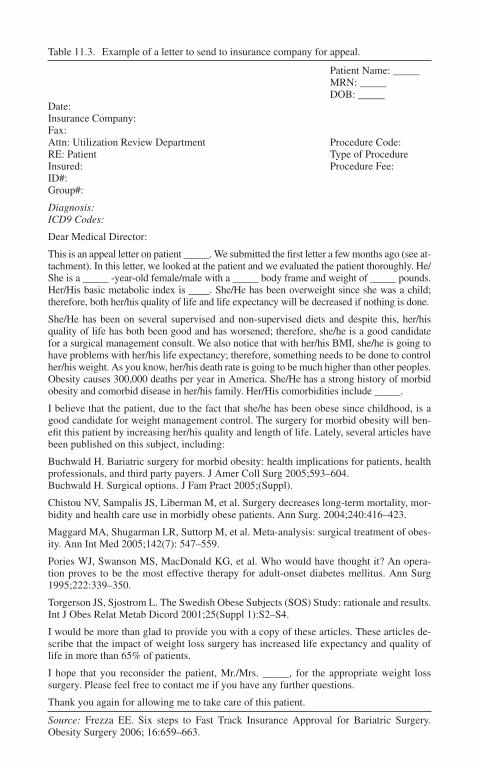
Table 11.3. Example of a letter to send to insurance company for appeal.
Patient Name: _____ MRN: _____ DOB: _____Date: Insurance Company: Fax: Attn: Utilization Review Department Procedure Code:RE: Patient Type of ProcedureInsured: Procedure Fee:ID#: Group#:
Diagnosis: ICD9 Codes:
Dear Medical Director:
This is an appeal letter on patient _____. We submitted the fi rst letter a few months ago (see at-tachment). In this letter, we looked at the patient and we evaluated the patient thoroughly. He/She is a _____ -year-old female/male with a _____ body frame and weight of _____ pounds. Her/His basic metabolic index is ____. She/He has been overweight since she was a child; therefore, both her/his quality of life and life expectancy will be decreased if nothing is done.
She/He has been on several supervised and non-supervised diets and despite this, her/his quality of life has both been good and has worsened; therefore, she/he is a good candidate for a surgical management consult. We also notice that with her/his BMI, she/he is going to have problems with her/his life expectancy; therefore, something needs to be done to control her/his weight. As you know, her/his death rate is going to be much higher than other peoples. Obesity causes 300,000 deaths per year in America. She/He has a strong history of morbid obesity and comorbid disease in her/his family. Her/His comorbidities include _____.
I believe that the patient, due to the fact that she/he has been obese since childhood, is a good candidate for weight management control. The surgery for morbid obesity will ben-efi t this patient by increasing her/his quality and length of life. Lately, several articles have been published on this subject, including:
Buchwald H. Bariatric surgery for morbid obesity: health implications for patients, health professionals, and third party payers. J Amer Coll Surg 2005;593–604. Buchwald H. Surgical options. J Fam Pract 2005;(Suppl).
Chistou NV, Sampalis JS, Liberman M, et al. Surgery decreases long-term mortality, mor-bidity and health care use in morbidly obese patients. Ann Surg. 2004;240:416–423.
Maggard MA, Shugarman LR, Suttorp M, et al. Meta-analysis: surgical treatment of obes-ity. Ann Int Med 2005;142(7): 547–559.
Pories WJ, Swanson MS, MacDonald KG, et al. Who would have thought it? An opera-tion proves to be the most effective therapy for adult-onset diabetes mellitus. Ann Surg 1995;222:339–350.
Torgerson JS, Sjostrom L. The Swedish Obese Subjects (SOS) Study: rationale and results. Int J Obes Relat Metab Dicord 2001;25(Suppl 1):S2–S4.
I would be more than glad to provide you with a copy of these articles. These articles de-scribe that the impact of weight loss surgery has increased life expectancy and quality of life in more than 65% of patients.
I hope that you reconsider the patient, Mr./Mrs. _____, for the appropriate weight loss surgery. Please feel free to contact me if you have any further questions.
Thank you again for allowing me to take care of this patient.
Source: Frezza EE. Six steps to Fast Track Insurance Approval for Bariatric Surgery. Obesity Surgery 2006; 16:659–663.
84 B T Ewing and E E Frezza
L. Selected References
Baum C, Ford W. The wage effects of obesity: a longitudinal study. Health Econ 2004;13:885–899.
Frezza EE. Six steps to fast track insurance approval for bariatric surgery. Obesity Surg 2006;16(5):659–663.
Frezza EE. A plan to establish a private-pay bariatric surgery in the U.S.A. Obesity Surg 2006;16(1):110–111.
Frezza EE. Overhead analysis in a surgical bariatric practice. JLAST 2006;16(4):390–393.Frezza EE. Avoiding embezzlement in the bariatric practice office. Obesity Surg
2006;16(10):1397–1398.Frezza EE. Knowing your bariatric practice. What’s your insurance fee? What’s your
leverage? Obesity Surg 2006;16(7):942–944.Frezza EE. The business of surgery. Norwalk, CT: Cine-Med Publishing, 2007.Frezza EE. How to improve front desk and office collection in a bariatric practice. Brief
communication. Obesity Surg 2005;15(9):1352–1354.Frezza EE, Ewing BT. Who seeks bariatric surgery? A comparison of patient demograph-
ics. Society of American Gastrointestinal Endoscopic Surgeons, Poster Session, Dallas, TX, April 2006.
Frezza EE, Wachtel MS. Practicing medicine in a new environment: what can we learn from the business managers? J Med Pract Mgmt 2006;22(1):52–54.
Frezza EE, Wachtel M, Ewing BT. The economic impact of morbid obesity on the state economy: an initial evaluation. Surg Obesity Rel Dis 2006;2:504–508.
Frezza EE, Wachtel MS, Ewing BT. Bariatric surgery costs and implications for hospital margins: comparing laparoscopic gastric bypass and laparoscopic gastric banding. Surg Laparoscop Endoscop Percut Tech. In press.
Must A, Spadano J, Coakley EH, et al. The disease burden associated with overweight and obesity. JAMA 1999;282:1523–1529.
Reid RD, Sanders NR. Operations management: an integrated approach. New York: John Wiley & Sons, 2005.
Statistical Abstract of the United States, 2007. Washington, DC: U.S. Census Bureau.Wolf AM. What is the economic case for treating obesity? Obes Res 1998;6(Suppl 1):
2S–7S.
Acknowledgment: Bradley T. Ewing acknowledges fi nancial support from the Center for Healthcare Innovation, Education and Research.

Medicine is a most diffi cult art to acquire.—Sir William Osler
A. Indications
The Roux-en-Y gastric bypass (RYGBP) is based on the principles of restric-tive and malabsorptive bariatric operations. First performed by Mason and Ito in 1966, it has evolved into the centerpiece bariatric operation for the resolution of comorbid disease and excess weight in the United States. Two critical changes to the operation have contributed to its current reputation: Griffen modifi ed the loop gastrojejunostomy to a Roux-en-Y in 1977 and Wittgrove and colleagues reported the laparoscopic approach in 1994. Essential steps for successful com-pletion of this operation involve:
1. Creating a small pouch out of the larger stomach2. Attaching a length of jejunum for egress of food out of the restrictive
pouch3. Creating an opening through which digestive effl uent may combine
with food for digestion to occur
B. Approach: Open versus Laparoscopic
Public perception is that laparoscopic surgery is less invasive, but there are additional advantages for a surgeon evolving from an open to a laparoscopic approach. Pfuzziferi in a randomized, prospective trial with 3-year outcomes showed an advantage for laparoscopic surgery related to length of hospital stay, incisional pain, return to work, and a markedly decreased rate of incisional hernia between open cases (39%) vs. laparoscopic (5%, p < 0.01). An additional comparison by Nguyen between open (n = 6,065) and laparoscopic (n = 16,357) Roux-en-Y gastric bypass procedures performed in a university setting demon-strated that major complications such as an anastomotic leak, venous thrombosis, and pneumonia were increased in patients with an open approach. Seventy-three percent of the cases in this setting were laparoscopic, which refl ects the growth
12. Roux-en-Y Gastric Bypass
Robin P. Blackstone

88 R P Blackstone
of this type of access. Both studies show no difference between the open and laparoscopic approach in regard to resolution of comorbid disease, quality of life, or excess weight loss.
C. Patient Position and Room Setup
The patient is prepared in the preoperative area with intravenous fl uid rehydration, intravenous antibiotics, and subcutaneous anticoagulation. The operating room table should be able to sustain the weight of the patient being operated, capable of being manipulated into reverse Trendelenburg, and covered with gel foam padding to reduce the incidence of cutaneous pressure neuropathies. Antithrombolic stockings are started prior to the institution of anesthesia. Patients can be positioned with legs in a supine or lithotomy position (Fig. 12.1).
Figure 12.1. Operating room set-up.

12. Roux-en-Y Gastric Bypass 89
D. Operative Procedure
This procedure is one of the most complex performed laparoscopically, and is associated with a signifi cant learning curve. Initial cases should be proctored and focused on patients with lower BMI and less complex comorbid disease. Effi cient operating room times usually run between 90 and 120 minutes.
1. Establish the pneumoperitoneum
Most commonly a Veress needle is placed through the abdominal wall just below the left rib cage. Insuffl ation to 15 mmHg will usually be adequate to achieve visualization. Once the shape of the abdomen has been established with an initial insuffl ation of 4 L, trochars can be placed under direct vision.
2. Placement of trochars
A local anesthetic is used to infi ltrate the skin of the abdomen prior to incision. The fi rst trochar (12 or 5 mm) should be placed with a zero degree camera under direct visualization one handbreadth below the xiphoid/manubrial junction and slightly to the left of midline to avoid the ligamentum teres. Using the xiphoid as the starting point will usually result in the midline trochar being too low, especially in male patients with deep diaphragms. In patients with a previous midline or right upper quadrant incision, the fi rst trochar should be placed in the left upper quadrant in the mid-clavicular line after fi rst establishing where the point of the V will be in the midline. For the remainder of the operation an angled (30- or 45-degree) scope is used. Subsequent trochars are placed in the mid-clavicular and anterior axillary line on both sides in the shape of a V. The steepness of the V will be determined by the body shape and confi guration once insuffl ation is complete. An additional 5-mm trochar is placed just to the left of the xiphoid where the liver retractor can be placed. The use of blunt trochars should be used to decrease the incidence of incisional hernia. Examination of the abdomen should be carried out (Fig. 12.2).
3. Retraction of the liver
The liver is often very large and is an impediment to visualization of the esopha-geal hiatus. Use of a Nathanson-type retractor through an epigastric port (usually 5 mm) may be necessary for extremely large or heavy livers; otherwise a toothed grasper attached to the diaphragm usually will be suffi cient. Over time the liver “fatigues” and tends to fold over the retractor. The liver should be retracted so that both the Angle of His and the pars fl accida can be seen in the fi eld.
4. Creation of gastric pouch
Opening the peritoneum over the diaphragmatic muscle at the Angle of His allows the surgeon to visualize the angle of dissection of the pouch. A thin band of connective tissue in front of the fi rst short gastric protects the superior and

90 R P Blackstone
medial edge of the spleen during the dissection. Dissection of the gastrohepatic ligament directly on the stomach wall, decreases blood loss, and preserves the function of the vagus nerve. Using an ultrasonic dissector may be preferred. A small 2- to 3-cm opening along the lesser curve below the anterior fat pad and above the fi rst vessel is the correct place to access the lesser sac. If a balloon has been placed into the stomach to size the pouch, the linear stapler should be closed just below the bottom of the balloon but the linear stapler should not be fi red until the balloon is removed. Sequential division of the stomach using the anterior fat pad as the anterior guide to the creation of the pouch and angling the stapler up to the angle of His should result in a 15- to 20-cc pouch. It is critical to change direction of the stapler from left (fi rst fi ring) to right (subsequent fi rings), angling directly to the angle of His to avoid injury to the spleen and avoid including the fundus of the stomach in the pouch (Figs. 12.3–12.5).
5. Gastrojejunostomy
Three established methods of creating the gastrojejunostomy have been described, and many variations exist. Trade-offs exist between the rate of stricture and
Figure 12.2. Laparoscopic port placement.

12. Roux-en-Y Gastric Bypass 91
Figure 12.3. Gastric pouch construction.
Figure 12.4. Gastric pouch construction.
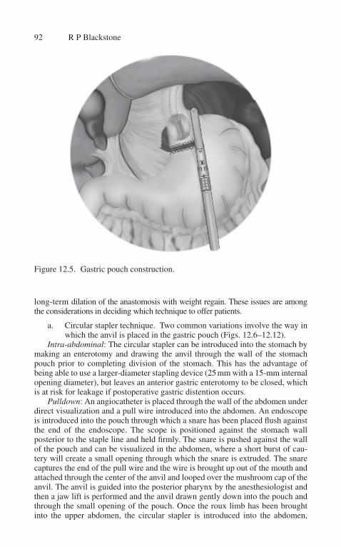
92 R P Blackstone
Figure 12.5. Gastric pouch construction.
long-term dilation of the anastomosis with weight regain. These issues are among the considerations in deciding which technique to offer patients.
a. Circular stapler technique. Two common variations involve the way in which the anvil is placed in the gastric pouch (Figs. 12.6–12.12).
Intra-abdominal: The circular stapler can be introduced into the stomach by making an enterotomy and drawing the anvil through the wall of the stomach pouch prior to completing division of the stomach. This has the advantage of being able to use a larger-diameter stapling device (25 mm with a 15-mm internal opening diameter), but leaves an anterior gastric enterotomy to be closed, which is at risk for leakage if postoperative gastric distention occurs.
Pulldown: An angiocatheter is placed through the wall of the abdomen under direct visualization and a pull wire introduced into the abdomen. An endoscope is introduced into the pouch through which a snare has been placed fl ush against the end of the endoscope. The scope is positioned against the stomach wall posterior to the staple line and held fi rmly. The snare is pushed against the wall of the pouch and can be visualized in the abdomen, where a short burst of cau-tery will create a small opening through which the snare is extruded. The snare captures the end of the pull wire and the wire is brought up out of the mouth and attached through the center of the anvil and looped over the mushroom cap of the anvil. The anvil is guided into the posterior pharynx by the anesthesiologist and then a jaw lift is performed and the anvil drawn gently down into the pouch and through the small opening of the pouch. Once the roux limb has been brought into the upper abdomen, the circular stapler is introduced into the abdomen,
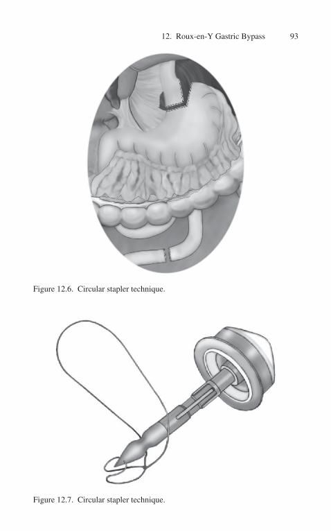
12. Roux-en-Y Gastric Bypass 93
Figure 12.6. Circular stapler technique.
Figure 12.7. Circular stapler technique.

94 R P Blackstone
Figure 12.8. Circular stapler technique.
Figure 12.9. Circular stapler technique.

12. Roux-en-Y Gastric Bypass 95
Figure 12.10. Circular stapler technique.
Figure 12.11. Circular stapler technique.

96 R P Blackstone
Figure 12.12. Circular stapler technique.
usually through the left mid-clavicular port site after dilation of the port site from 12 to 25 mm using Hegar dilators. The circular stapler is placed into the lumen of the jejunum and the needle of the stapler is extruded through the wall of the jejunum, approximately 4 cm from the cut end. The needle is docked to the anvil under direct vision checking to assure the tissue below the stapler is not included and rotating the roux limb to lie in a “candy cane” confi guration. The stapler is fi red and removed by pulling backward and turning the stapler in a half circle. The end of the roux limb is closed with a linear stapler. The integrity of the “donuts” of resected tissue can be inspected, but the ultimate judge of adequate closure is the leak test performed at the end of the procedure. The bent end of the candy cane should not be very long, and undesired excess jejunum can be excised while closing the end of the roux limb. At the conclusion of the procedure the left mid-clavicular port site is irrigated with antibiotic irrigation and the fascial stitches are tied.
Technical Note: Occasionally the anvil will seem to be diffi cult to pull down, letting go of the pull wire and allowing the anvil to “bounce” up the esophagus and then gently pulling again slowly will usually solve this problem. Most of the time the anvil is caught at the cricopharyngeal junction and sometimes taking down the balloon of the tracheostomy tube will be necessary to facilitate passage of the sta-pler. The liver retractor can also be a barrier to passage of the anvil (see Fig. 12.4).
b. Hand-sewn technique. The hand-sewn anastomosis requires advanced laparoscopic suturing technique. It is the least expensive option and has the added benefi t of decreasing the infection rate in the port site through

12. Roux-en-Y Gastric Bypass 97
which the circular stapler is placed. The anastomosis is usually created anterior on the gastric pouch. A posterior, running layer is created between the staple line of the gastric pouch and antimesenteric side of the roux limb using 3-0 polyglactin suture. Enterotomies are made in the gastric pouch and jejunum and a second posterior running, full thickness layer is made and continued anteriorly. Prior to completion of the anastomosis a 34 F bougie is passed carefully through the anastomosis and the anterior inner (full thickness) and outer (seromuscular) layers are completed.
c. Linear stapler technique. After the roux limb is brought into the upper abdomen a small enterotomy is made in both the roux limb and pouch. The linear cutter is placed and fi red for a length of about 1.5 cm. The enterotomy is closed from either end using an absorbable Vicryl suture (3-0 polyglactin) until the last stitch is ready to be tied down. The 30 F endoscope is passed through the anastomosis and the stitch is tied down using the endoscope as a “stent.” A second layer of absorbable suture is required to complete the anastomosis.
d. Checking for anastomotic leak. Prior to completing the operation all anastomosis, regardless of technique, are checked by obstructing the roux limb with an endo-glassman clamp. The endoscope is positioned under direct vision just above the gastrojejunal anastomosis. Air is insuffl ated through the endoscope into the gastrojejunal anastomosis with air until tense insuffl ation of the roux limb above the clamp occurs. Flooding the intra-abdominal fi eld with water will reveal any air leak, which signals a leak of the anastomosis. Additional stitches can be taken until the test yields a satisfactory result. Early in a surgeon’s experience opening to correct the leak may be the best option. False tests can result if tense insuffl ation is not achieved.
6. Jejunojejunostomy
The jejunojejunostomy is constructed using a toothed grasper or a stitch to stabilize both the stapled end of the antimesenteric surface of the biliopancreatic limb and the roux limb at the desired length. Small enterotomies are made with an ultrasonic dissector (cautery may result in a larger defect) and an anasto-mosis of 45–70 mm is created between the two limbs. Firing in two opposite directions may decrease the incidence of obstruction. The enterotomy is closed with an additional horizontal fi ring of the linear stapler or by hand sewing the defect closed. All staple lines should be visually inspected to assure closure and evaluate the opening size. Careful attention should be paid to the way the roux limb lies, and if “kinking” is observed a Bolin stitch can be taken to allow the roux limb to lie more naturally (Fig. 12.13).
7. Roux limb: antecolic vs. retrocolic
a. Retrocolic. The lesser sac is opened through the pars lucida and a 1-cm white Penrose drain placed behind the stomach. The colon is refl ected cephalad and the ligament of Treitz is identifi ed. A 2- to 4-cm opening is made above the pancreas in the mesocolon and the Penrose drain is

98 R P Blackstone
retrieved. The jejunum is divided approximately 15 cm from the ligament of Treitz using a linear stapler and the mesentery is “notched” on the side of the roux limb. A 100-cm (Routine) or 150-cm (long limb) is measured, and then the jejunojejunostomy is created. Once the jejunojejunostomy has been completed, the end of the roux limb is attached to the Penrose drain and placed carefully (pulling only on the Penrose drain) through the mesocolon and behind the stomach. Replacing the colon in its usual position in the abdomen the Penrose drain is gently retracted back from the lesser sac drawing the roux limb gently into the upper abdomen where it should lie without slipping back under the stomach. The roux limb is brought up through the lesser sac and out through the gastrohepatic liga-ment, allowing the gastrojejunal anastomosis to be created where the back wall of the anastomosis lies on the soft tissue of the ligament, creating a “patch” of soft tissue that heals quickly.
b. Antecolic. Division of the omentum down to the anterior colon wall using an ultrasonic dissector is made. The colon is then refl ected into the upper abdomen and the ligament of Treitz identifi ed. A 30- to 35-cm biliopancreatic limb is created to facilitate bringing the 65- to 70-cm roux limb anterior to the colon. The roux limb is brought into the upper abdo-men through the notched defect in the omentum. The total length of the bypassed jejunum is approximately equal to the retrocolic approach.
Figure 12.13. Jejunojejunostomy.
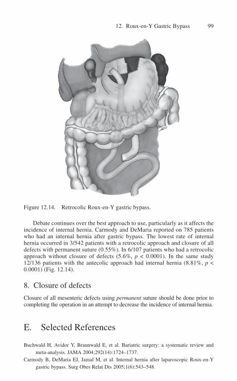
12. Roux-en-Y Gastric Bypass 99
Debate continues over the best approach to use, particularly as it affects the incidence of internal hernia. Carmody and DeMaria reported on 785 patients who had an internal hernia after gastric bypass. The lowest rate of internal hernia occurred in 3/542 patients with a retrocolic approach and closure of all defects with permanent suture (0.55%). In 6/107 patients who had a retrocolic approach without closure of defects (5.6%, p < 0.0001). In the same study 12/136 patients with the antecolic approach had internal hernia (8.81%, p < 0.0001) (Fig. 12.14).
8. Closure of defects
Closure of all mesenteric defects using permanent suture should be done prior to completing the operation in an attempt to decrease the incidence of internal hernia.
E. Selected References
Buchwald H, Avidor Y, Braunwald E, et al. Bariatric surgery: a systematic review and meta-analysis. JAMA 2004;292(14):1724–1737.
Carmody B, DeMaria EJ, Jamal M, et al. Internal hernia after laparoscopic Roux-en-Y gastric bypass. Surg Obes Relat Dis 2005;1(6):543–548.
Figure 12.14. Retrocolic Roux-en-Y gastric bypass.
100 R P Blackstone
Gagner M, Garcia-Ruiz A, Arca MJ, et al. Laparoscopic isolated gastric bypass for morbid obesity. Surg Endosc 1999;13:S6.
Griffen WO, Young VL, Stevenson CC. A prospective comparison of gastric and jeju-noileal bypass procedures for morbid obesity. Ann Surg 1977;186:500–507.
Higa KD, Boone KB, Ho T, et al. Laparoscopic Roux-en-Y gastric bypass for morbid obesity: technique and preliminary results of our first 400 patients. Arch Surg 2000;9:1029–1033.
Mason EE, Ito C. Gastric bypass in obesity. Surg Clin North Am 1967;47:1345–1352.Murr MM, Gallagher SF. Technical considerations for transabdominal loading of the
circular stapler in laparoscopic Roux-en-Y gastric bypass. Am J Surg 2003;185(6):585–588.
Nguyen NT, Hinojosa M, Fayad C, et al. Use and outcomes of laparoscopic versus open gastric bypass at academic medical centers. J Am Coll Surg 2007;205(2):248–255.
Puzziferri N, Austrheim-Smith IT, Wolfe BM, et al. Three-year follow-up of a prospec-tive randomized trial comparing laparoscopic versus open gastric bypass. Ann Surg 2006;243(2):181–188.
Schauer PR, Ikramuddin S, Gourash W, et al. Outcomes after laparoscopic roux-en-Y gastric bypass for morbid obesity. Ann Surg 2000;232(4):515–529.
Schauer PR, Ikramuddin SI, Hamad G, et al. The learning curve for laparoscopic Roux-en-Y gastric bypass is 100 cases. Surg Endosc 2003;17:212–215.
Wittgrove AC, Clark GW, Tremblay LF. Laparoscopic gastric bypass, Roux-en-Y: prelimi-nary report of five cases. Obes Surg 1994;4:353–357.
Acknowledgments:All fi gures in this chapter are reprinted with permission, ©1999–2007 Ethicon Endo-Surgery, Inc. All rights reserved.
Disclosure:Dr. Blackstone holds an appointment as associate clinical professor of surgery at the University of Arizona College of Medicine-Phoenix. She is a consultant for Ethicon Endosurgery and Allergan Medical. Scottsdale Bariatric Center par-ticipates in FDA clinical trials for Enteromedics and Allergan Medical and in protocols with the National Institutes of Health. She is a member of the board of directors for the Surgical Review Corporation and has served on the Bariatric Surgical Review Committee of the Surgical Review Corporation for 4 years. She was elected to the Executive Committee of the American Society for Metabolic and Bariatric Surgery in 2007. She is a founding member of the Board of Direc-tors of the Obesity Action Coalition.
A. Indication and Description
In June 2001, the Lap-Band® System (Allergan, Inc., Irvine, CA) became the fi rst FDA-approved laparoscopic adjustable gastric band (LAGB) for patients 18 years and older in the United States. Indications for LAGB place-ment are the same for any bariatric procedure (BMI > 35 with one or more obesity-related comorbidity or BMI > 40). LAGB is a restrictive procedure in which an infl atable silicone band is placed around the upper part of the stomach. The band is placed just below the gastroesophageal junction and secured with sutures. The adjustment port is then tunneled through the abdominal wall and secured to the anterior abdominal wall fascia. The creation of a smaller stomach pouch results in restriction of the amount of food that can be consumed at one time. LAGB increases the time it takes for the stomach to empty. Weight loss is achieved by limiting food intake, reducing appetite, and slowing digestion. There are currently three Lap-Band sizes (Fig. 13.1) available in the United States: 9.75/10, VG, and the AP.
B. Patient Position and Room Setup1. Position the patient supine with both arms out. The patient should be secured to the OR table to prevent the patient
from sliding downward during reverse Trendelenburg positioning. We use a split leg OR table with the operating surgeon standing between the patient’s legs.
2. The video monitors are placed at the patient’s left and right shoulder.
3. Sequential compression devices should be placed on before anesthetic induction.
4. An appropriate antibiotic should be given prior to incision.
13. Lap-Band® and Adjustment Schedule
Dean J. Mikami

102 D J Mikami
Figure 13.1. a Lap-Band VG, 10.0 and 9.75. Source: Reprinted with kind per-mission from Allergan, Inc. b Lap-Band AP. Source: Reprinted with kind per-mission from Allergan, Inc.

13. Lap-Band® and Adjustment Schedule 103
C. Trocar Position and Choice of Laparoscope
1. The surgeon stands between the patient’s legs and the assistant surgeon stands on the patient’s left side.
2. A Veress needle is then inserted at the left mid-clavicular position, below the left costal margin. The fi rst trocar (15-mm) is inserted at this site (Fig. 13.2, scarlet dot).
3. A 30-degree, 5- or 10-mm laparoscope is then place above the umbilicus (Fig. 13.2, gray dot).
4. Two additional 5-mm trocars are placed (Fig. 13.2, white dots).5. A Nathanson liver retractor is placed at the epigastrium (Fig. 13.2,
green dot).
Figure 13.2. Trocar position for laparoscopic adjustable band placement.
104 D J Mikami
D. Operative Technique
1. Pars fl accida technique (preferred).2. Dissection of the angle of His, freeing the gastrophrenic peritoneal
attachments to expose the left crus.3. Creation of a 15–20 cc pouch.4. Posterior dissection just below the crura.5. Anterior fi xation of the fundus and anterior gastric wall over the band
(Fig. 13.3).6. Complete defl ation of the band at time of placement.7. The end of the band is brought through a port site.8. The access port is placed and sutured to the anterior rectus sheet with
permanent suture.
E. Advantages
1. Low mortality rate2. No division of stomach or small bowel3. Adjustable4. Reversible5. Low postoperative complication rate6. Low malnutrition rate7. Shorter hospital stay
F. Disadvantages
1. Slower weight loss2. Less weight loss as compared with combined restrictive/malabsorptive
operations (gastric bypass, duodenal switch)3. Requires an implantable medical device4. Band slippage, erosion, and esophageal dilatation5. Access port malfunctions6. Requires ongoing patient monitoring and adjustments for optimal
weight loss
G. Complications
1. Mortality rate: 0.05%2. Total complication rate: 11%3. Major complications
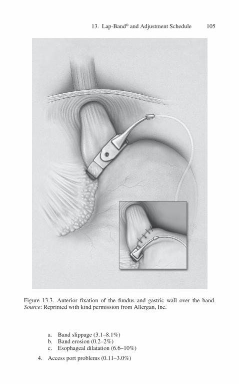
13. Lap-Band® and Adjustment Schedule 105
a. Band slippage (3.1–8.1%)b. Band erosion (0.2–2%)c. Esophageal dilatation (6.6–10%)
4. Access port problems (0.11–3.0%)
Figure 13.3. Anterior fi xation of the fundus and gastric wall over the band. Source: Reprinted with kind permission from Allergan, Inc.

106 D J Mikami
H. Results: US Data
1. 12-month Excess body weight loss percentage (EBWL%): 33–502. 24-month EBWL%: 45–633. 36-month EBWL%: 44–654. 48-month EBWL%: 58–625. 60-month EBWL%: 50–65
I. Lap-Band Adjustments
1. Typically starts 6 weeks after surgery 2. Weight loss goal is 0.5 to 1 kg per week 3. Can be done in the offi ce or with fl uoroscopy depending on the depth
of the access port 4. The adjustment goal is to achieve a sensation of prolonged satiety 5. Adjustment can be done every 4 to 6 weeks or as needed 6. 9.75- and 10-cm band (max volume: 4 cc)
a. 0.5–1 cc added at fi rst adjustmentb. 0.3–0.5 cc added or removed at subsequent adjustments
7. VG and AP Standard (APS) (max volume: 10 cc), AP Large (APL) (max volume: 14 cc)
a. 3–4 cc added at fi rst adjustmentb. 1–1.5 cc added or removed at subsequent adjustments
8. Consider adding saline
a. Inadequate weight lossb. Rapid loss of satiety after mealsc. Hunger between meals
9. Adjustment not required
a. Adequate rate of weight loss (>0.5 kg and <1 kg per week)b. Eating a reasonable range of foodc. No negative symptoms
10. Consider removing saline
a. Persistent nausea and emesisb. Wheezing, coughing spells, and chokingc. Diffi culty with a broad range of foods

13. Lap-Band® and Adjustment Schedule 107
J. Selected References
Allen JW. Laparscopic gastric band complications. Med Clin North Am 2007;91.DeMaria EJ, Jamal MK. Laparoscopic adjustable gastric banding: evolving clinical
experience. Surg Clin North Am 2005;85:773–787.Favretti F, O’Brien PE, Dixon JB. Patient management after LAP-BAND placement. Am
J Surg 2002;184:38s–41s.Halloway JA, Forney FA, Gould DE. The Lap-Band is an effective tool for weight loss
even in the United States. Am J Surg 2004;188:659–662.Ponce J, Paynter S, Fromm R. Laparoscopic adjustable gastric banding: 1014 consecutive
cases. J Am Coll Surg 2005;05:529–535.Provost DA. Laparoscopic adjustable gastric banding: an attractive option. Surg Clin North
Am 2005;85:789–805.
A. Introduction
Laparoscopic biliopancreatic diversion with duodenal switch (BPD-DS) is one of the most effective weight loss procedures currently available. Both short- and long-term weight loss exceed that of any other bariatric operation. BPD-DS involves a 150- to 200-cc sleeve or vertical gastrectomy, a duodenoileal anas-tomosis, and a long Roux-en-Y with a 150-cm alimentary limb and a 100-cm common channel (Fig. 14.1). The key features of this operation are that the lesser curvature, antrum, pylorus, fi rst portion of the duodenum, and vagal innervation are all spared, while parietal cell mass is reduced, thus allowing a better digestive behavior while decreasing the likelihood of the dumping syndrome and marginal ulceration. Furthermore, by placing the ileoileal anastomosis 100 cm proximal to the ileocecal junction (instead of 50 cm, as in the classic Scopinaro BPD), metabolic disturbances and the number of surgical revisions for malnutrition or diarrhea are considerably less.
B. Indications/Special Preoperative Preparation
1. All patients should meet NIH criteria for obesity surgery (namely, BMI >40 kg/m2 or >35 kg/m2 with obesity-related comorbidities). Particularly the superobese (BMI >50 kg/m2) and super-superobese (BMI >60 kg/m2) are excellent candidates for this procedure, as BPD-DS provides superior weight loss outcomes in these patients compared with gastric bypass. Other indications and contraindications are listed in Table 14.1.
2. EGD to exclude gastric or duodenal pathology, including Helicobacter pylori infection.
3. If H. pylori is present, patients are treated prior to surgery.4. Upper GI series if the patient has had a prior bariatric procedure.5. Medical clearance.6. Psychiatric evaluation and instruction by registered dietitian/ nutritionist.7. Screening colonoscopy if the patient is above age 50.8. Evaluation for obstructive sleep apnea (if applicable).9. Clear liquid diet the day prior to surgery.
14. Laparoscopic Duodenal Switch
Manish Parikh, Michel Gagner, and Alfons Pomp

C. Patient Position and Room Setup (Fig. 14.2)
1. Alpha-Max table (Maquet; Rastatt, Germany) with foot-plate attachments
2. French, split-leg position with the legs abducted, but not fl exed, and properly secured
Figure 14.1. Lap BPD/DS: Sleeve gastrectomy, duodenoileostomy and a long
110 M Parikh et al.
Roux-en-Y with a 150 cm alimentary limb and 100 cm Common channel.

14. Laparoscopic Duodenal Switch 111
Table 14.1. Indications and contraindications for Lap BPD-DS.
Indications• BMI >40 kg/m2 or >35 kg/m2 with obesity-related comorbidities• Superobese (BMI >50 kg/m2) and super-superobese (BMI >60 kg/m2)• Patients who want to avoid the dietary/volume restrictions of the
gastric bypass• Patients considering gastric bypass but who require high-dose anti-
infl ammatory medications (e.g., for severe arthritis)• Patients who require periodic surveillance of their stomach• Patients who have failed vertical-banded gastroplasty, gastric banding,
or gastric bypassContraindications• Patients who are bedridden• Patients who are vegetarian (patients need to ingest at least 80–100 g
protein daily)• Patients with prior colon resection (may be prone to developing
diarrhea)• Patients with severe gastroesophageal refl ux (may be better served
with gastric bypass)• Patients with infl ammatory bowel disease• Patients with prior gastrectomy
3. The surgeon stands between the patient’s legs, the fi rst assistant ( liver retractor and camera holder) stands on the patient’s right, and the second assistant stands on the patient’s left.
D. Trocar Placement
1. Seven trocars total (Fig. 14.3)
a. 10-mm trocar at umbilicus (we prefer the open technique to enter the peritoneal cavity)—diagnostic laparoscopy is then performed with the 10-mm 30-degree laparoscope
b. Two 10-mm trocars—left epigastric paramedian position (optics) and right subcostal position in the mid-clavicular line (liver retraction)
c. Two 5- to 15-mm Versaports (Tyco Healthcare, Norwalk, CT)—right mid-clavicular line just superior to umbilicus and left mid-clavicular line, four fi ngerbreadths inferior to the costal margin
d. 5- to 12-mm Versaport in the subxiphoid positione. 5-mm port in left anterior axillary line lateral to the Versaport
2. A second insuffl ator is attached to optimize pneumoperitoneum (15 mmHg carbon dioxide)
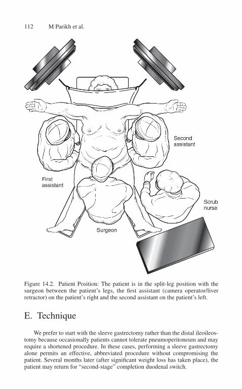
112 M Parikh et al.
E. Technique
We prefer to start with the sleeve gastrectomy rather than the distal ileoileos-tomy because occasionally patients cannot tolerate pneumoperitoneum and may require a shortened procedure. In these cases, performing a sleeve gastrectomy alone permits an effective, abbreviated procedure without compromising the patient. Several months later (after signifi cant weight loss has taken place), the patient may return for “second-stage” completion duodenal switch.
Figure 14.2. Patient Position: The patient is in the split-leg position with the surgeon between the patient’s legs, the fi rst assistant (camera operator/liver retractor) on the patient’s right and the second assistant on the patient’s left.

14. Laparoscopic Duodenal Switch 113
1. Sleeve gastrectomy
a. The patient is placed in steep reverse Trendelenburg position and the table is tilted right side down to optimize visualization of the gastro-esophageal junction.
b. The laparoscope is placed through the 10-mm left epigastric paramedi-an trocar. If the stomach is distended the anesthesiologist should place an orogastric tube to decompress the stomach; this tube must be re-moved as soon as the stomach is decompressed to avoid any problems during subsequent stapling.
c. A 10-mm liver retractor (fan-type retractor) is placed through the right subcostal port to retract the liver anteriorly and expose the entire stom-ach and gastroesophageal junction. The surgeon’s working ports are the subxiphoid Versaport and the left subcostal Versaport. The second assistant retracts the omentum laterally with a bowel grasper through the 5-mm left lateral trocar.
Figure 14.3. Trocar Positions.

114 M Parikh et al.
d. Dissection begins along the distal greater curvature by dividing the branches of the gastroepiploic artery near the gastric wall with the ultrasonic scalpel. The greater curvature is devascularized in this man-ner proximally to the level of the left crus (including division of the short gastric vessels). The second assistant’s grasper is frequently repositioned superiorly to maximize retraction. Exposure while divid-ing the short gastric vessels and dissecting near the left crus is often diffi cult. Helpful maneuvers include:
• Place the second assistant’s grasper on the lateral fold of the omentum (in the mid-gastrosplenic ligament) and retract this laterally toward the spleen.
• Temporarily increase the pneumoperitoneum to 20 mmHg.• Place the patient in maximal reverse Trendelenburg position.• Tilt the patient more toward the right side.• Ask the anesthetist to give an additional dose of paralytics.• Position the second assistant on the posterior fundus and retract
this toward the patient’s right side.• Occasionally an additional 5-mm trocar is required to retract the
perigastric fat and adequately expose the gastroesophageal junction.
e. All posterior attachments to the pancreas must be freed, taking care not to injure the splenic artery. Placing the second assistant’s graspers on the posterior fundus and retracting this toward the patient’s right shoulder exposes these attachments. It is important to divide these at-tachments prior to stapling because these attachments can tear and cre-ate signifi cant bleeding. However, one must not be too aggressive near the lesser curvature because the blood supply to the sleeve will come from the lesser curvature vasculature only.
f. Now the left crus can be visualized by lifting the stomach anteriorly. The left crus and gastroesophageal junction must be completely ex-posed. The ligament attaching the stomach and diaphragm must be divided. The anterior perigastric fat just to the left of the gastroesopha-geal junction must be cleared to minimize tissue thickness during sub-sequent stapling. However, avoid dissecting to the right of the gastro-esophageal junction because of risk of injury to the vagus nerve. If the patient has a signifi cant hiatal hernia, this should be reduced because failure to recognize/repair herniated fundus may lead to weight loss failure and refl ux after sleeve gastrectomy.
g. Next the remainder of the greater curvature is liberated distally to 2 cm beyond the pylorus. The second assistant retracts the greater curvature anteriorly toward the patient’s right shoulder. The surgeon’s left hand grasps the fat of the gastrocolic ligament (via the right mid-clavicular Versaport) and retracts it caudad. The surgeon’s right hand manipu-lates the ultrasonic scalpel. The remainder of the gastrocolic ligament between the antrum and gastroepiploic arcade is divided with the ultra-sonic scalpel.
h. Instrument palpation is used to confi rm the anatomic position of the pylorus. Approximately 6 to 8 cm proximal to the pylorus (at the level of the “crow’s foot,” just distal to the incisura) the sleeve gastrectomy

14. Laparoscopic Duodenal Switch 115
is begun along the greater curvature (Fig. 14.4). Initiating the sleeve less than 6 cm proximal to the pylorus may compromise the antrum and could lead to gastric emptying problems.
i. The surgeon’s left hand holds the 4.8-mm linear stapler (60 mm in length) through the right mid-clavicular Versaport. The second assist-ant retracts the body of the stomach toward the patient’s left side. The stapler should be positioned such that at least 2 cm of anterior stomach serosa is visible between the stapler and lesser curvature.
j. The fi rst two fi rings of the stapler are performed, aiming approximate-ly 2 cm away from the lesser curvature. These fi rings should be done slowly because the stomach is thickest in this area. Additional sutures may be required if the tissue is too thick for the stapler. We routinely use buttress material (Bioabsorbable Seamguard; Gore, Flagstaff, AZ) similar to the Maxon suture (United States Surgical, Norwalk, CT), which is sandwiched between, over, and below the anterior and pos-terior gastric wall. This bioabsorbable material reduces staple-line hemorrhage and possibly the leakage rate.
k. The anesthesiologist inserts a 60 Fr orogastric bougie and, under lapar-oscopic vision, the bougie is aligned medially along the lesser curva-ture into the duodenum. Two bowel graspers can be used to help direct the bougie posteriorly into the sleeve toward the pylorus. Inserting the
Figure 14.4. Mobilization of the branches of the gastroepiploic artery and short gastric vessels in preparation for the sleeve gastrectomy.

Figure 14.5. Posterior and antero-lateral retraction around the bougie.
bougie after the fi rst two stapler fi rings helps align the bougie along the lesser curvature (Fig. 14.5). For all duodenal switch cases, we cur-rently use the 60 Fr bougie to ensure enough volume of the stomach to permit adequate protein intake. (For primary sleeve gastrectomy we use a smaller bougie, 32–40 Fr.)
l. The remainder of the sleeve gastrectomy is completed by sequential fi rings of the 4.8-mm linear stapler along the bougie toward the angle of His (Fig. 14.6). Although we have used the 3.5-mm linear stapler in the past, we believe that it is safest to use the 4.8-mm linear stapler for the entire sleeve gastrectomy due to the thick stomach in these morbidly obese patients. The differences in hemostasis between the two staplers are no longer seen with the routine use of the buttress-ing Seamguard material. The surgeon’s right hand holds the stapler via the left mid-clavicular Versaport and aims toward the left crus; the surgeon’s left hand (via the subxiphoid Versaport) grasps the anterior wall of the stomach (or the perigastric fat) and retracts this toward the patient’s right side. The second assistant holds the posterior wall of the stomach and retracts this toward the patient’s left side. A total of fi ve to six staple fi rings are typically required to complete the sleeve. The anesthesiologist must pay careful attention that the bougie does not retract during stapling to prevent the tip of the bougie from being incorporated into the staple line.
m. Next, the anesthesiologist removes the bougie. We prefer to place fi g-ure-of-eight 3-0 Maxon (monofi lament absorbable) sutures at the apex of the sleeve gastrectomy (the area most prone to developing leak), at the intersections of the staple lines (also prone to suboptimal healing) and at the most distal end of the staple line (thickest part of stomach). The second assistant can retract the stomach toward the patient’s right side to help expose the apex of the sleeve.
n. If there is any doubt about the integrity of the staple line, a methylene blue test should be performed prior to proceeding to the next stage. The
116 M Parikh et al.

14. Laparoscopic Duodenal Switch 117
anesthesiologist inserts an 18 Fr orogastric tube. The surgeon clamps near the pylorus and the anesthesiologist injects methylene blue mixed with saline through the tube. Approximately 120 cc are required to dis-tend the sleeve. Another option is to insert a gastroscope and check for leak (and intraluminal bleeding) via air insuffl ation; this latter option is used less often because of the tendency of air to pass through the pylorus and distend the small bowel.
o. The right mid-clavicular trocar site is enlarged, the abdominal wall is dilated with an atraumatic clamp (the circular stapler will be intro-duced through this site later), a large impermeable bag is introduced and the specimen is extracted. Grasping the end of the sleeve and pull-ing it out progressively may make extraction easier and require less abdominal wall dilation.
2. Duodenal transection and preparation for duodenoileostomy
a. The pylorus and fi rst portion of duodenum are palpated. Any remaining branches between the gastroepiploic arcade and the antrum/pylorus are divided with the ultrasonic scalpel, proceeding toward the inferior aspect of the fi rst portion of the duodenum. Typically, the dissection
Figure 14.6. The laparoscopic sleeve gastrectomy is created by sequential fi rings of the 4.8-mm stapler along a 60-Fr bougie towards the angle of HIS.

extends to just beyond the vascular complex inferior to the pylorus. It is important to avoid hemostatic clips in this area (especially on the duo-denal side) to prevent having clips interfere with the staple line. Any duode-nal adhesions from prior cholecystectomy must be divided at this time.
b. The retroduodenal and supraduodenal tissue are dissected free with the ultrasonic scalpel. The second assistant retracts the stomach laterally and anteriorly so the surgeon can see both the greater curvature and the posterior stomach for the retroduodenal dissection. The gastroduode-nal artery, which lies posteriorly between the fi rst and second portion of duodenum, marks the distal aspect of dissection.
c. Using a 10-mm right-angle dissector, a 1-cm window (enough to accommodate a linear stapler) is made posterior and superior to the duodenum, medial to the common bile duct (Fig. 14.7). Ideally, the supraduodenal window is between the serosa of the duodenum and the pyloric branches of the right gastric artery, thus maximizing blood supply to the subsequent anastomosis.
d. The duodenum is transected with a 3.5-mm Endo-GIA linear stapler (60 mm in length; Tyco Healthcare, Norwalk, CT) buttressed with Bio-absorbable Seamguard leaving a 2- to 5-cm duodenal cuff (typically via the left mid-clavicular Versaport; Fig. 14.8). Sometimes, the right mid-clavicular Versaport provides a better angle for transection. The second assistant retracts the antrum toward the patient’s left side to facilitate this. The Seamguard buttressing material obviates the need to oversew the duodenal stump.
Figure 14.7. Peridoudenal dissection.
118 M Parikh et al.

14. Laparoscopic Duodenal Switch 119
Figure 14.8. Duodenal transection with 3.5-mm linear stapler (60 mm in length).
e. If the surgeon is faced with a scenario where he or she is unable to complete the supraduodenal window, an alternate method is to transect the inferior two thirds of the duodenum with the linear stapler, com-plete the supraduodenal window, and then transect the remaining duo-denum with another fi ring of the stapler.
f. We prefer to use a circular stapler for the duodenoileostomy, specifi -cally the CEEA 21 (Tyco Healthcare, Norwalk, CT). The CEEA 25 is too large for the ileum and frequently tears the ileum during insertion. The 21 anvil is delivered transabdominally through the right mid-cla-vicular trocar site. The 21 anvil is problematic to deliver through the proximal duodenal stump using the modifi ed nasogastric tube-anvil apparatus commonly used for gastric bypass because it does not fl ex and traverses the pylorus only with some diffi culty.
g. The ultrasonic scalpel is used to remove 1 to 2 cm of the proximal duodenal staple line and the base of the 21 anvil is placed into the duo-denal lumen. Once the anvil is in place, it is secured with a 3-0 Prolene pursestring suture (Fig. 14.9).
h. Next the surgeon must assess the ability of the ileum to be brought toward the duodenum antecolic and in a tension-free manner. Particu-larly in patients with bulky omentum, one must divide the omentum

120 M Parikh et al.
Figure 14.9. 21-mm anvil inserted into proximal duodenum and secured with 3–0 Prolene purse-string sulture.
along its right lateral third to facilitate subsequent passage of the ileum toward the duodenum. It is important that the right side of the omen-tum (and not the left side, as commonly done in gastric bypass) is divided because an oblique line runs from the ileocecal valve toward the pylorus.
3. Small bowel measurement
a. The surgeon and fi rst assistant move to the patient’s left side. The pa-tient is placed in the Trendelenburg position and then tilted left side down to aid in exposure of the ileocecal region.
b. With the laparoscope in the left epigastric paramedian port and the surgeon’s working hands in the subxiphoid and umbilical ports, the distal small intestine is measured from the ileocecal valve using a 50-cm umbilical tape. Flat 5-mm forceps (Dorsey, Karl Storz; Tutlingen, Germany) are used to avoid serosal tears during measurements. Any adhesions that prevent proper measurement of the small intestine (e.g., prior lower abdominal or pelvic surgery) must be divided. The um-bilical tape should be kept on the antimesenteric side during measure-ments. We prefer umbilical tape for these measurements because it is a more reliable method of measuring small intestine vs. estimating distances with a bowel grasper.
c. At 100 cm proximal to the ileocecal valve, clips are placed on the ileal mesentery (to mark the site of anastomosis of the future common

14. Laparoscopic Duodenal Switch 121
channel). Another 150 cm is measured proximally from this point ( future alimentary limb). Here, a window is made in the ileal mesen-tery with a 10-mm right-angle clamp. It is best to not dissect directly on the bowel wall, as the mesentery advances over the intestinal wall in the morbidly obese. Instead, make the window in the mesentery approximately 1 cm away from the bowel wall.
d. The ileum is transected with a 2.5-mm linear stapler (45 mm in length is adequate) buttressed with Bioabsorbable Seamguard (via the left mid-clavicular Versaport). The ultrasonic scalpel is used to divide an-other 1 to 2 cm of mesentery between the two ends of bowel. Dividing more than 1 to 2 cm of mesentery is unnecessary and places the subse-quent anastomosis at risk of becoming ischemic.
e. The grasper remains on the distal bowel (with the umbilical tape) to maintain proper orientation. The staple line and buttress material are completely excised from the distal ileum to permit entry of the 21-mm circular stapler. The umbilical tape is then removed. Care is taken to make sure there is no twisting of the mesentery or misidentifi cation of bowel limbs by running the bowel again from the ileocecal valve proximally to the transected (and opened) ileum.
4. Duodenoileal anastomosis
a. A suture is attached to the spike of the CEEA 21 for easy retrieval. The CEEA 21 is secured to a plastic camera drape (for wound protection) with a Steri-Strip and is introduced via the right mid-clavicular trocar site.
b. Three graspers are used to triangulate the opened end of ileum: two are placed at the 2:00 and 10:00 positions and a third grasper is placed at the 6:00 position adjacent to the mesentery.
c. The stapler is gently maneuvered into the distal ileal enterotomy and advanced cephalad in a clockwise manner toward the duodenal cuff containing the anvil. The fi rst two graspers are removed. The grasper at 6:00 remains in place to prevent the stapler from coming out of the bowel. It is critical that the ileum is brought toward the duodenum (and not vice versa) under minimal or no tension. The omentum needs to be divided to facilitate this, as described earlier.
d. The fi rst assistant moves between the patient’s legs and the surgeon remains on the patient’s left (second assistant remains on the patient’s right to retracting the liver anteriorly).
e. The white plastic perforator is advanced through the antimesenteric wall of ileum approximately 6 to 7 cm distal to the opened ileum. The spike is removed by grasping the suture attached to the spike. The an-vil (in the duodenum) is then united with the stapler (Fig. 14.10). It is important that there is no tissue between the ileum and duodenum and that there is no pinching of the bowel wall (which can create an obstruction later).
f. The stapler is fi red and an end-to-side anastomosis is created. The CEEA 21 is not a fl ipped-top; therefore, two to three rotations of the stapler are required in conjunction with counter-traction on the antrum in order to pull the CEEA through the anastomosis.

122 M Parikh et al.
g. Next, the Steri-Strip holding the wound protector is removed and the CEEA is removed from the trocar site while advancing the wound pro-tector over the tip of the stapler in order to prevent the stapler from contacting the wound.
h. The surgeon returns between the legs and the fi rst assistant returns to the patient’s right side. The right mid-clavicular Versaport is re-inserted.
i. The open ileal limb is inspected for bleeding—any oozing emanating from the enterotomy may indicate bleeding from the duodenoileos-tomy and must be further evaluated. If there is no bleeding from the open limb, a 2.5-mm linear stapler (45 mm in length) buttressed with Bioabsorbable Seamguard (via the subxiphoid or left mid-clavicular trocar) is used to transect the blind limb. The tips of the stapler must be in the ileal mesentery to ensure that the opened ileum has been completely closed (otherwise a leak can occur into the mesentery). The specimen is extracted from the right mid-clavicular trocar site.
j. The duodenoileostomy staple line (including the upper and lower cor-ners) is reinforced with a running 3-0 Maxon suture. We prefer mono-fi lament absorbable sutures for this because permanent sutures (e.g., Silk sutures) have been associated with marginal ulcers and strictures. One helpful maneuver to provide adequate exposure of the superior corner (where leaks are most prone to occur) is to place the second
Figure 14.10. The anvil of the CEEA 21 and the stapler are united.

14. Laparoscopic Duodenal Switch 123
assistant’s graspers on the antrum and retract this toward the patient’s left lower quadrant in order to pull the anastomosis toward the midline. To facilitate exposure of the posterior aspect of the staple line, the sec-ond assistant should grasp the posterior antrum and gently retract this toward the patient’s right shoulder.
k. Next, the anesthesiologist inserts an 18 Fr orogastric tube just proximal to the anastomosis and then instills approximately 120 cc methylene blue mixed with saline with a Tumi syringe. Simultaneously, the sur-geon clamps the ileum distal to the duodenoileostomy. The area around the anastomosis is irrigated with saline to help identify any methylene blue. Once the test has been completed and no leak has been identi-fi ed, the gastric sleeve is completely aspirated and the orogastric tube is removed.
5. Distal ileoileostomy
a. The surgeon and fi rst assistant return to the patient’s left side and the laparoscope is placed through the left epigastric paramedian trocar. The table is tilted left side down and the patient is placed in slight Trendelenburg position.
phoid and umbilical port to run the alimentary limb from the duode-noileostomy distally to the level of the previously placed clips on the ileal mesentery (at 100 cm proximal to the ileocecal valve).
c. We prefer the “M” triple-staple technique, which is a completely stapled anastomosis that provides a large patent anastomosis while avoiding the risk of narrowing the bowel lumen during closure of the enterotomy (Fig. 14.11). The clips on the ileal mesentery are removed.
d. An enterotomy is made with the ultrasonic scalpel on the anti- mesenteric side of the marked ileum. Another enterotomy is made approximately 1 to 2 cm away from the stapled end of the proximal ileum. The stapled proximal ileum (biliopancreatic limb) should be on the patient’s left and the alimentary limb should be on the patient’s right side. Again, one must take care that there has been no twisting of the mesentery and that both staples are fi red on the antimesenteric border to avoid ischemia.
e. The 2.5-mm linear stapler (60 mm in length) is introduced through the subxiphoid Versaport, aiming toward the pelvis. It is best to insert the larger jaw of the stapler into the proximal ileum (larger diameter) and the smaller jaw into the distal ileum (narrower). A standard side-to-side anastomosis is created between the biliopancreatic limb and the last 100 cm of distal ileum.
f. Through the same enterotomy, the linear stapler (2.5, 60 mm in length) is then fi red between the alimentary limb and the common channel. A third fi ring of the 2.5-mm linear stapler (via the left mid-clavicular Ver-saport) closes the enterotomy transversely. The specimen is removed without contaminating the wound. An alternate option for enterotomy closure is to close the enterotomy with a running 2-0 silk suture, either in one or two layers.
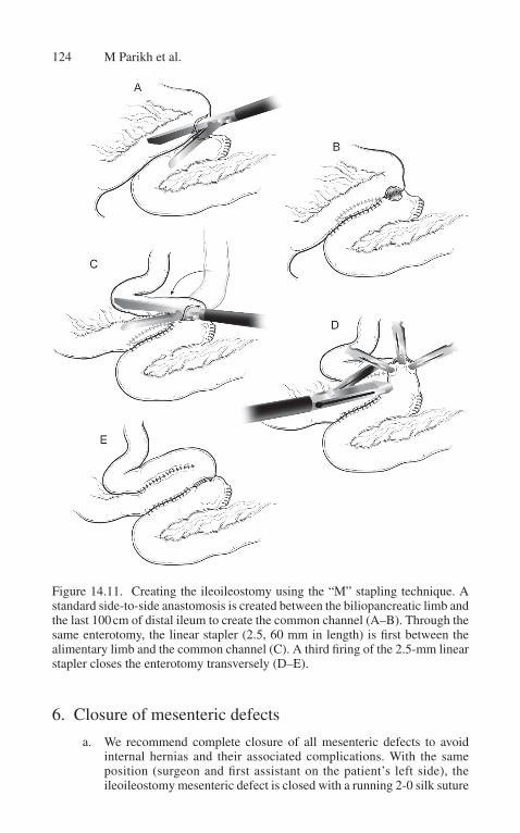
124 M Parikh et al.
6. Closure of mesenteric defects
a. We recommend complete closure of all mesenteric defects to avoid internal hernias and their associated complications. With the same position (surgeon and fi rst assistant on the patient’s left side), the ileoileostomy mesenteric defect is closed with a running 2-0 silk suture
Figure 14.11. Creating the ileoileostomy using the “M” stapling technique. A standard side-to-side anastomosis is created between the biliopancreatic limb and the last 100 cm of distal ileum to create the common channel (A–B). Through the same enterotomy, the linear stapler (2.5, 60 mm in length) is fi rst between the alimentary limb and the common channel (C). A third fi ring of the 2.5-mm linear stapler closes the enterotomy transversely (D–E).

14. Laparoscopic Duodenal Switch 125
(24 cm in length). We prefer to incorporate the serosa of the ileum in the last stitch of this closure.
b. The surgeon then returns between the legs in order to close Petersen’s defect (Fig. 14.12). The patient is placed in slight reverse Trendelenburg position. Sometimes, it is necessary to insert an additional 5-mm trocar in the left lower quadrant to optimize suturing angles.
c. The omentum is retracted superiorly to the transverse colon. The fi rst assistant grasps the epiploic appendage of the transverse colon and retracts this cephalad. Now this space between the transverse colon mesentery and the ileal mesentery should be completely exposed, and this defect is closed with a running 2-0 silk suture.
Figure 14.12. Closure of Petersen’s defect.
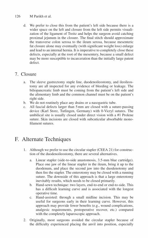
126 M Parikh et al.
d. We prefer to close this from the patient’s left side because there is a wider space on the left and closure from the left side permits visuali-zation of the ligament of Treitz and helps the surgeon avoid catching proximal jejunum in the closure. The fi nal stitch should approximate the transverse colon serosa to the ileum serosa, because mesenteric fat closure alone may eventually (with signifi cant weight loss) enlarge and lead to an internal hernia. It is imperative to completely close these defects, especially at the root of the mesentery, because a small defect may be more susceptible to incarceration than the initially large patent defect.
7. Closure
a. The sleeve gastrectomy staple line, duodenoileostomy, and ileoileos-tomy are all inspected for any evidence of bleeding or leakage. The biliopancreatic limb must be coming from the patient’s left side and the alimentary limb and the common channel must be on the patient’s right side.
b. We do not routinely place any drains or a nasogastric tube.c. All fascial defects larger than 5 mm are closed with a suture-passing
device (Karl Storz, Tutlingen, Germany) with 0-Vicryl sutures. The umbilical site is usually closed under direct vision with a #1 Prolene suture. Skin incisions are closed with subcuticular absorbable mono-fi lament sutures.
F. Alternate Techniques
1. Although we prefer to use the circular stapler (CEEA 21) for construc-tion of the duodenoileostomy, there are several alternatives.
a. Linear stapler (side-to-side anastomosis, 3.5-mm blue cartridge). Place one jaw of the linear stapler in the ileum, bring it up to the duodenum, and place the second jaw into the duodenotomy and then fi re the stapler. The enterotomy may be closed with a running suture. The downside of this approach is that a large enterotomy inevitably results, which needs to be closed primarily.
b. Hand-sewn technique: two layers, end-to-end or end-to-side. This has a diffi cult learning curve and is associated with the longest operative time.
c. Hand-assisted: through a small midline incision. This may be useful for surgeons early in their learning curve. However, this approach may provide fewer benefi ts (e.g., wound complications, analgesic requirements, postoperative recover, etc.) compared with the completely laparoscopic approach.
2. Originally, most surgeons avoided the circular stapler because of the diffi culty experienced placing the anvil into position, especially

14. Laparoscopic Duodenal Switch 127
when trying to pass the anvil through the pylorus (using the modi-fi ed nasogastric tube apparatus). We have adapted our technique to position the anvil via duodenotomy (followed by Prolene pursestring suture to secure the anvil in place), and this has signifi cantly reduced our operative time.
3. Alternate techniques for the ileoileostomy include hand-sewn (asso-ciated with longer operating times) and the double-stapled technique (biliopancreatic limb to common channel and closure of enterotomy). Our concern with the latter technique is that this may narrow the com-mon channel during closure of the enterotomy, particularly due to the smaller-diameter ileum.
4. The most critical factor that results in a leak at the duodenoileostomy is exaggerated tension at the anastomosis. Maneuvers to eliminate ten-sion at the anastomosis include:
a. Division of the omentum needs along its right lateral aspect.b. Mobilization of the right colon.c. Division of blood vessels superior to the pylorus: one must be cautious
because this may compromise the blood supply to the anastomosis.d. We have not found retrocolic duodenoileostomy to be necessary;
the preceding maneuvers should ensure a tension-free anastomo-sis. Moreover, we generally avoid retrocolic construction due its association with increased incidence of internal hernias.
5. The surgeon should always consider the two-stage approach in the high-risk superobese or in the patient who cannot tolerate prolonged pneumoperitoneum, has extensive intra-abdominal adhesions, or lacks adequate working space despite higher pneumoperitoneum pressures. A two-stage approach (laparoscopic sleeve gastrectomy followed by completion duodenal switch 6–12 months later) has the advantages of technical facility, shorter operative times, and interval weight loss between stages, which in turn results in decreased morbidity in this high-risk group.
6. It is advisable to keep the total operative time to less than 4 hours to avoid the attendant risks (pulmonary, thromboembolic, and rhabdomy-olysis) of prolonged general anesthesia, especially in the high-risk bariatric patient.
G. Postoperative Care
1. Patients are closely monitored for at least 6 hours.2. Patients are placed on continuous positive airway pressure (CPAP) if
sleep apnea is present or suspected.3. Maintenance intravenous fl uids are continued to ensure urine output of
at least 0.5 to 1 cc/kg/hour.4. Early ambulation is critical. The majority of our patients ambulate the
very evening of surgery.5. We use postoperative upper gastrointestinal contrast studies selectively:

128 M Parikh et al.
a. In cases of any intraoperative technical diffi culties (e.g., positive methylene blue test requiring additional sutures)
b. For patients who manifest signs and symptoms concerning for leak (e.g., fever greater than 38.5 °C, tachycardia, tachypnea, som-nolence and failure to thrive, etc.)
6. Patients usually require an intravenous patient-controlled analgesic pump for the fi rst 2 days.
7. They receive clear liquids on the fi rst postoperative day. If they do well, their Foley catheters are removed and they are weaned off the intravenous fl uids.
8. Patients are advanced to a pureed diet the next day. 9. Our nutritionist sees all patients postoperatively, and their dietary rec-
ommendations are reviewed.10. Patients are usually discharged home by the third postoperative day
on a pureed diet and oral analgesics.11. Patients are seen 4 weeks after discharge. They receive follow-up nu-
tritional counseling for a protein-enriched diet, and are given twice-daily multivitamins, oral calcium supplements, iron, and fat-soluble vitamins.
12. Patients with intact gall bladders are prescribed ursodiol 300 mg twice daily for 6 months for gallstone prophylaxis.
13. At 3 months postoperatively, a thorough nutritional evaluation is performed, including serum levels of iron, ferritin, vitamin B
12,
folate, albumin, PTH, calcium, phosphorus, alkaline phosphatase, zinc, selenium, lipids, vitamins A and D, total protein, and hematol-ogy panels.
H. Selected References
Baltasar A, Bou R, Miro J, et al. Laparoscopic biliopancreatic diversion with duodenal switch: technique and initial experience. Obes Surg 2002;12:245–248.
Buchwald H, Avidor Y, Braunwald E, et al. Bariatric surgery. A systematic review and meta-analysis. JAMA 2004;292:1724–1737.
Comeau E, Gagner M, Inabnet W, et al. Symptomatic internal hernias after laparoscopic bariatric surgery. Surg Endosc 2005;19:34–39.
Consten E, Gagner M, Pomp A, et al. Decreased bleeding after laparoscopic sleeve gas-trectomy with or without duodenal switch for morbid obesity using a stapled but-tressed absorbable polymer membrane. Obes Surg 2004;14:1360–1366.
Feng J, Gagner M, Pomp A, et al. Effect of standard vs. extended Roux limb length on weight loss outcomes after laparoscopic Roux-en-Y gastric bypass. Surg Endosc 2003;17:1055–1060.
Gagner M, Inabnet W, Pomp A. Laparoscopic sleeve gastrectomy with second stage biliopancreatic diversion and duodenal switch in the superobese. In: Inabnet W, DeMaria E, Ikramuddin S. Laparoscopic bariatric surgery. Philadelphia: Lippincott Williams & Wilkins, 2005:143–150.

14. Laparoscopic Duodenal Switch 129
Gagner M, Matteotti R. Laparoscopic biliopancreatic diversion with duodenal switch. Surg Clin North Am 2005;85:141–149.
Gastrointestinal Surgery for Severe Obesity. NIH Consensus Statement, March 25–27, 1991;9:1–20.
Marceau P, Hould F, Simard S, et al. Biliopancreatic diversion with duodenal switch. World J Surg 1998;22:947–954.
Prachand V, Davee R, Alverdy J. Duodenal switch provides superior weight loss in the super-obese (BMI >50 kg/m2) compared with gastric bypass. Ann Surg 2006;255:611–619.
Rabkin R. The duodenal switch as an increasing and highly effective operation for morbid obesity. Obes Surg 2004;14:861–865.
Rabkin R, Rabkin J, Metcalf B, et al. Laparoscopic technique for performing duodenal switch with gastric reduction. Obes Surg 2003;13;263–268.
Regan J, Inabnet W, Gagner M, et al. Early experience with two-stage laparoscopic Roux-en-Y gastric bypass as an alternative in the super-super obese patient. Obes Surg 2003;13:861–864.
A. Indications and Rationale
Although laparoscopic bariatric surgery has evolved into a very safe and effective procedure, the super-super obese patients (BMI >60) continue to pose signifi cant technical challenges. This translates to higher complications and poor outcomes in this group of patients, irrespective of the nature of the surgery. There seems to be a linear correlation between the outcomes and the risk of the surgery. The riskier the operation is—laparoscopic bilio-pancreatic diversion with duodenal switch (BPD-DS), laparoscopic Roux-en-Y gastric bypass (LGBP)—the more effective weight loss appears to be.
The challenges in super-superobese patients (BMI >60) are both functional as well as technical. The functional challenges include poor cardiopulmonary reserve, exercise intolerance, restrictive as well as obstructive lung disorders (translates into inability to tolerate anesthesia) and inability to ambulate.
The technical challenges are:
• Diffi cult access to abdominal cavity, higher pneumoperitoneum pressures• Large and stiff abdominal wall, decreasing the maneuverability of the
instruments• Large fatty left lobe of the liver making diffi cult the exposure of the
gastroesophageal junction and making prone to injury to organs such as liver, spleen, esophagus, and stomach
• Thick and foreshortened small bowel mesentery from fatty infi ltration creating tension at anastomosis and vascular compromise of the intes-tine by either stretching or by attempts to lengthen by the mesenteric division
Sleeve gastrectomy was originally conceived by Marceau in 1993 as modifi cation to partial gastrectomy in BPD. BPD-DS is a long and complex operation with signifi cant morbidity and mortality. In order to reduce to operating times, sim-plify the operation into two stages and let patient lose some weight and thus decrease the complication rate, Gagner and colleagues suggested BPD-DS as a staged procedure where LSG is the fi rst stage. Subsequent follow-up of these patients has suggested that sometimes LSG may be the only operation needed in some patients.
15. Laparoscopic Sleeve Gastrectomy: A Staged Procedure for Super-Super Obese Patients
Kuldeep Singh

132 K Singh
LSG obviates the need for infra-colic intestinal reconstruction, simplifi es and shortens the procedure, and effectively reduces the comorbidities/obesity. This makes the second (malabsorptive) stage of the BPD easier and faster to accom-plish, if needed. The Sleeve also makes gastric pouch reconstruction relatively easy to perform in LGBP. Its simplicity and effectiveness has led to its gaining acceptance as a staged procedure (Fig. 15.1).
B. Patient Positioning and Room Setup
1. The patient is placed supine with arms on the sides on the arm boards. The feet rest on a foot board fi xed to the bed. There is a folded blanket under the knees, the legs and thighs are strapped to the bed. A Foley catheter is placed in aseptic fashion.
2. The video monitors are on either side of the patient’s shoulders.3. SQ Heparin given preop and sequential compression devices are placed
on the legs.4. Preoperative antibiotics are given.5. Orogastric tube is placed and stomach decompressed. The tube is then
replaced by size 34 Maloney Bougie. It is ensured laparoscopically that the tip of the bougie is beyond the pylorus.
C. Port Position
1. Access is obtained by using 12-mm Visiport (Covidien, Mansfi eld, MA) using 10-mm zero degree scope, traversing the layers of abdomi-nal wall under vision. The ideal position for entry if left paramedian where layers are very well defi ned.
Figure 15.1. Laparoscopic sleeve gastrectomy: a staged procedure.

15. Laparoscopic Sleeve Gastrectomy 133
2. Pneumoperitoneum of 17 mm of mercury is obtained and general inspection of the abdomen is done noting the size of the liver, any injuries of entrance and other incidental fi ndings.
3. The port position is shown in Fig. 15.2. One additional 15-mm trocar is placed in right paramedian position, one in left subcostal margin and one 5-mm trocar are inserted in the right subcostal margins. One 5-mm subxiphoid obturator is inserted and a medium Nathanson retractor (Cook Medical, Bloomington, IN) is inserted to retract the left lobe of the liver. This is held by Iron Intern retractor system (Automated Medical Product Corp, Edison, NJ) attached to left side of the side rails.
4. A 10-mm 45-degree scope is used for the surgery.
Figure 15.2. Laparoscopic sleeve gastrectomy: port placement.

134 K Singh
D. Stage-1. Laparoscopic Sleeve Gastrectomy
1. The anatomical landmarks such as Crow’s foot and gastroesophageal junction are identifi ed. Any dimpling of the hiatus is explored by dividing the fat pad over the EG junction and hiatal hernia repaired if present.
2. Gastrocolic and gastrosplenic omentum is divided with Ligasure ( Covidien, Mansfi eld, MA) at the greater curvature of the stomach and the stomach completely mobilized and rotated medially. Exposure of the left crus of the diaphragm ensures that the dissection is complete and pouch remains small.
3. The Crow’s feet (the terminal branches of nerves of Laterjet) on less-er curvature is identifi ed and a point diagonally opposite on greater curvature is identifi ed as starting point for division. This is approxi-mately 6 cm proximal to the pylorus.
4. The fi rst stapler is fi red with Endo GIA stapler of green thickness (4.8 mm) 60 mm in length, introduced through the right-sided 15-mm trocar. This thickness is needed to divide the thick pylorus and an-tral muscle fi bers. The remaining stapler loads are fi red parallel to the bougie as shown in Fig. 15.3.
Figure 15.3. Laparoscopic sleeve gastrectomy: creation of sleeve.

15. Laparoscopic Sleeve Gastrectomy 135
5. The remainders of the fi rings are done with either green or blue thick-ness stapler load reinforced with staple-line reinforcement products.
6. Great care is taken to divide close to the bougie as adhesions to the retroperitoneum or fatty infi ltration of the lesser curvature mislead to a larger sleeve.
7. The Angle of His clearly identifi ed and stomach is divided just lateral to the left diaphragmatic crus.
8. The areas of staple line overlap or transition is oversewn with Zero Surgidac suture using Endostich instrument.
9. The pylorus is clamped and gastroscope is used to insuffl ate the stomach under water and areas of air leak and lamberted.
10. A size 10 fl at JP is inserted and specimen removed through an en-larged 15-mm trocar site.
11. All 12- and 15-mm trocars are closed with zero Polysorb suture.
E. Stage-2. Laparoscopic Roux-en-Y Gastric Bypass or Laparoscopic Biliopancreatic Diversion—Duodenal Switch
1. Port placements are done same as one stage procedure.2. In GBP, the stomach is transected 5 cm distal to GE junction by 60 mm
long, 4.8-mm thick stapler. The Roux-en-Y reconstruction is done in usual fashion (retrocolic-retogastric linear staple technique).
3. In BPD-DS, the technique is described by M. Gagner and colleagues. Dissection and division of fi rst portion of the duodenum is done fol-lowed by creation of duodeno-ileostomy by 25-mm EEA stapler (transoral technique). Ileoileostomy is then created by linear stapling technique creating 150 cm of alimentary limb and 100 cm of common channel. All the mesenteric defects are closed in running fashion.
F. Selected References
Gagner M, Inabnet WB, Pomp A. Laparoscopic sleeve gastrectomy with second stage biliopancreatic diversion and duodenal switch in the superobese. In: Inabnet WB, DeMaria EJ, Ikramuddin S, eds. Laparoscopic bariatric surgery, 2nd ed. Philadelphia: Lippincott Williams &Wilkins, 2005:145–150.
Gumbs A, Gagner M, Dakin G, et al. Sleeve gastrectomy for morbid obesity. Obes Surg 2007;17:962–969.
Marceau P, Biron S, Bourque RA, et al. Biliopancreatic diversion with a new type of gastrectomy. Obes Surg 1993;3:29–35.

A. Indications
Laparoscopic Roux-en-Y gastric bypass in the super-super obese (BMI >60 kg/m2) population can be challenging. Factors that contribute to the technical diffi culty include male gender, android body habitus, and high BMI. Men often have an android body habitus with high content of visceral fat, which increases intra-abdominal pressure and reduces intraoperative laparoscopic visualiza-tion. In addition, android body habitus and high BMI are often associated with fatty liver disease and enlarged liver lobes that may obscure visualization of the gastroesophageal junction. All of these factors can contribute to the degree of intraoperative technical diffi culty and should be weighed in the selection of appropriate patients to undergo laparoscopic gastric bypass.
Although a high BMI is not in itself a contraindication to surgery, laparo-scopic gastric bypass in the super-super obese can be associated with a higher morbidity and mortality. In an effort to minimize perioperative morbidity, the concept of a two-stage operation was developed. The initial two-stage procedure consists of a sleeve gastrectomy (fi rst stage) followed by an interval Roux-en-Y gastric bypass or duodenal switch. However, the sleeve gastrectomy can still be challenging in the super-super obese, particularly in patients with an enlarged liver from nonalcoholic steatohepatitis or liver cirrhosis obscuring visualization of the gastroesophageal junction near the angle of His. The alternative two-stage procedure is the staged Roux-en-Y, which consists of a modifi ed Roux-en-Y operation with construction of a larger gastric pouch and a low gastrojejunal anastomosis. Construction of a large gastric pouch avoids the diffi cult dissec-tion of the angle of His. The construction of a low gastrojejunal anastomosis minimizes tension on the anastomosis and hence reduces the chance for leaks. In the second stage, 6 to 12 months later, the volume of the gastric pouch is reduced by performing a sleeve gastrectomy of the gastric fundus.
B. Patient Position and Room Setup
1. Position the patient supine with both arms to the side. A foot board should be placed. The patient’s legs should be secured to prevent the patient from sliding during reverse Trendelenburg position.
16. Laparoscopic Staged Roux-en-Y: A Staged Procedure for Super-Super Obese Patients
Ninh T. Nguyen and Marcelo W. Hinojosa

138 N T Nguyen and M W Hinojosa
2. Place the video monitors at the patient’s left and right shoulder.3. Place a Foley catheter.4. The sequential compression device should be active during anesthetic
induction.5. Antibiotics should be given.
C. Trocar Position and Choice of Laparoscope
1. The surgeon stands on the patient’s right side. The assistant surgeon stands on the patient’s left side.
2. Place the Veress needle at the left mid-clavicular position above the umbilicus.
3. The fi rst trocar (11-mm) is inserted at this site.4. Four additional abdominal trocars are placed (one 12-mm and three
5-mm trocars), as shown in Fig. 16.1.5. A 45-degree angle laparoscope should be used.
D. Laparoscopic Staged Roux-en-Y (First Stage)
1. The operation begins with construction of the jejunojejunostomy.2. Identify the ligament of Treitz and divide the jejunum 40-cm distally
with a linear stapler (blue load). The jejunum mesentery is divided with a linear stapler (gray load).
3. The Roux limb is measured at 150-cm. The base of the Roux limb is fashioned to the jejunal biliopancreatic limb. A side-to-side jejunojejunostomy is constructed using a 60-mm linear stapler (white load). The remaining jejunal defect is closed with a running, two-layer closure. The small bowel mesenteric defect is closed with a running suture.
4. The greater omentum is divided to provide an antecolic path for the Roux limb.
5. The patient is switched to a reverse Trendelenburg position.6. The hepatogastric ligament, close to the lesser curvature of the stom-
ach, is divided using a linear stapler (gray load). An anterior gastrot-omy is made low on the lesser curvature. The 25-mm anvil is inserted transabdominally and placed through the gastrotomy and positioned on the anterior gastric wall approximately 4 or 5 cm distal to the gas-troesophageal junction. The stomach is divided starting on the lesser curvature and completed on the greater curvature (Fig. 16.2a).
7. Pass the end of the Roux limb, on an antecolic path, into proximity to the gastric pouch, taking care to avoid twisting of the mesentery.
8. The gastrojejunal anastomosis is constructed with a 25-mm circu-lar stapler. The circular stapler is inserted transabdominally and then positioned through the end of the Roux limb. The surgeon con-nects the anvil with the stapler to construct the gastrojejunostomy. The end of the Roux limb is closed with a linear stapler (blue load).

16. Laparoscopic Staged Roux-en-Y 139
9. The small bowel is clamped. An endoscopy is performed to test the integrity of the anastomosis with air insuffl ation while the anasto-mosis is submerged under water.
10. Close port sites larger than 5 mm at the fascial level.
E. Laparoscopic Staged Roux-en-Y (Second Stage)
1. The surgeon stands on the patient’s right side. The assistant surgeon stands on the patient’s left side.
2. An upper endoscopy is performed to examine for marginal ulcera-tion. Place the endoscope through the gastrojejunal anastomosis.
Figure 16.1. Trocar Position for laparoscopic staged Roux-en-Y.

140 N T Nguyen and M W Hinojosa
3. A laparoscopic exploration is performed with fi ve abdominal trocars (see Fig. 16.1).
4. The gastrojejunal anastomosis is identifi ed with the endoscope in place.
5. The gastric pouch is mobilized along the greater curvature using the ultrasonic shear.
6. A sleeve gastrectomy is performed (see Fig. 16.3), using multiple applications of the linear staplers (green load), starting at the level of the gastrojejunal anastomosis and completed at the Angle of His.
7. The endoscope is withdrawn and insuffl ated to check the integrity of the gastric pouch staple lines.
8. The sleeve gastrectomy specimen is removed through an enlarged trocar.
Figure 16.2. Laparoscopic staged Roux-en-Y (1st stage): Laparoscopic construction of a modifi ed Roux-en-Y gastric bypass with a large gastric pouch and a low gastrojejunal anastomosis to minimize tension.

16. Laparoscopic Staged Roux-en-Y 141
F. Selected References
Nguyen NT, Longoria M, Gelfand DV, et al. Staged laparoscopic Roux-en-Y: a novel two-stage bariatric operation as an alternative in the super-obese with massively enlarged liver. Obes Surg 2005;15:1077–1081.
Oliak D, Ballantyne GH, Davies RJ, et al. Short-term results of laparoscopic gastric bypass in patients with BMI > or = 60. Obes Surg 2002;12:643–647.
Regan JP, Inabnet WB, Gagner M, et al. Early experience with two-stage laparoscopic Roux-en-Y gastric bypass as an alternative in the super-super obese patient. Obes Surg 2003;13:861–864.
Figure 16.3. Laparoscopic staged Roux-en-Y (2nd stage): a sleeve gastrectomy is per-formed to reduce the volume of the gastric pouch.

A. Introduction
In the morbidly obese, surgery is the most effective option to achieve weight loss with resolution of comorbidity. Since its fi rst description in 1967, the gastric bypass (RYGB) has continued to gain popularity. The introduction of the lapar-oscopic approach by Wittgrove in 1994 further increased patient acceptance, and it is now the most common bariatric procedure performed worldwide. The advantages of the laparoscopic approach include reduced mean operative time, operative blood loss, length of intensive care stay, postoperative pain, and in-hos-pital stay, with a faster recovery compared with the open technique with similar perioperative morbidity and mortality rates. The laparoscopic RYGB (LRYGB) procedure achieves weight loss by a combination of moderate restriction by reduction in gastric volume to 15 to 30 ml with a narrow outlet, the dumping syndrome in response to ingestion of food with a high sugar or fat content, and micronutrient malabsorption.
Short-term outcome after LRYGB is determined by surgical technique, surgeon volume, hospital volume, and surgical reintervention rate. Early outcome measures include perioperative rates of morbidity and mortal-ity, surgical reintervention rate, and readmission rate. Early complications are classifi ed as those occurring in the fi rst 30 days after surgery, and late complications as those observed after 30 days. Major complications are life- threatening complications and/or those that lead to early reoperation. Long-term outcome is determined by the avoidance of nutritional defi ciencies, early detection and treatment of complications, and close follow-up in a multidis-ciplinary care program.
B. Short-Term Outcome
1. Perioperative mortality
Operative mortality (<30 days) (0.2–1.9%).Risk factors:
• Body mass index• Male gender
17. Outcomes of Laparoscopic Gastric Bypass
Samuel Szomstein and Olga N. Tucker

146 S Szomstein and O N Tucker
• Age• Comorbidities
Cause:
• Anastomotic leak with sepsis• Pulmonary embolus (0.2%)
2. Perioperative morbidity
Anastomotic leak:
• Incidence: 0.9–4.4%• Staple line• Gastrojejunal• Jejunojejunal
Hemorrhage:
• Incidence: 1.1–3.8%• Gastric pouch• Gastric remnant• Gastrojejunostomy• Jejunojejunostomy• Other source
Small bowel obstruction:
• Incidence: 0.6–3.8%• Trocar site hernia• Anastomotic stenosis or edema
˚ Gastrojejunal
˚ Jejunojejunal
• Intussusception• Hematoma
˚ Intramural
˚ Intraluminal
˚ Mesenteric
• Internal hernia
˚ Incidence: 0.2–0.7%
˚ Petersen’s space
˚ Mesocolic defect
˚ Jejunojejunostomy defect
Acute gastric remnant distention:Deep venous thrombosisPulmonary embolusAbdominal compartment syndrome

17. Outcomes of Laparoscopic Gastric Bypass 147
Wound infection:
• Incidence: 0.1–8.7%• Dehiscence
Rhabdomyolysis + acute renal failure
C. Long-Term Outcome
An appropriate follow-up program after LRYGB should detect:
• Weight loss• Weight regain/failure to lose weight• Comorbidity resolution• Surgical complications• Metabolic/nutritional complications• Psychological stability• Mortality
1. Weight loss
Mean percentage of excess weight loss (EWL%) 62% at 2 years (range 57–66%)
2. Weight regain/failure to lose weight
Weight regain5% regain weight at 12–13 months15% regain weight at 18 months to 2 yearsCause:
• Errors in technique: large pouch, large stoma• Binge eating• Low energy expenditure
Failure to lose weight15% failure of weight loss at 3 years
3. Comorbidity resolution
Resolution of comorbid conditions:
• Diabetes: resolution in 77%• Hyperlipidemia: improvement in 70%• Hypertension: resolution in 62%• Sleep apnea: resolution in 85%

148 S Szomstein and O N Tucker
4. Surgical complications
Chronic early:
• Marginal ulceration, hemorrhage, abdominal pain• Small bowel obstruction: internal hernias, Petersen’s defect, mesocolic
defect, biliopancreatic limb• Pouch dilatation• Anastomotic (stomal) dilation (gastrojejunostomy) (Fig. 17.1)• Anastomotic strictures: gastrojejunal (incidence: 1.6–11.4%), jejuno
jejunal• Biliary tract pathology: symptomatic gallstones, acute cholecystitis,
choledocholithiasis• Gastropathic fi stulae (Fig. 17.2)
Chronic late:
• Weight regain• Failure of weight loss• Malnutrition
5. Metabolic/nutritional complications
Restriction of oral intake and malabsorption following LRYGB can result in nutritional defi ciencies with metabolic consequences. Following LRYGB ade-quate oral intake with daily vitamin and mineral supplementation is essential with regular assessment by the dietician. Following LRYGB, pregnancy should be not considered for the fi rst 18 months after surgery.
The following defi ciencies may arise after LRYGB:
• Fat-soluble vitamins: A, D, E, K• Water-soluble vitamins: B
1 thiamine, B
2 ribofl avin, B
3 niacin, biotin,
pantothenic acid, B6, B
12, folic acid, C
• Minerals: iron, calcium, zinc
Figure 17.1. Upper gastrointestinal endoscopy after laparoscopic Roux-en-Y gastric bypass. a. Normal gastrojejunostomy. b. Stenosis of gastrojejunostomy.
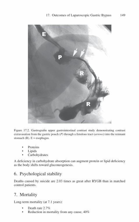
17. Outcomes of Laparoscopic Gastric Bypass 149
• Proteins• Lipids• Carbohydrates
A defi ciency in carbohydrate absorption can augment protein or lipid defi ciency as the body shifts toward gluconeogenesis.
6. Psychological stability
Deaths caused by suicide are 2.03 times as great after RYGB than in matched control patients.
7. Mortality
Long-term mortality (at 7.1 years):
• Death rate 2.7%• Reduction in mortality from any cause, 40%
Figure 17.2. Gastrografi n upper gastrointestinal contrast study demonstrating contrast extravasation from the gastric pouch (P) through a fi stulous tract (arrows) into the remnant stomach (R). E = esophagus.

150 S Szomstein and O N Tucker
• Reduction in cause-specifi c mortality: coronary artery disease 56%; diabetes 92%; cancer 60%
• Estimated number of lives saved 136/100,000 gastric bypasses
D. Revisional Surgery
1. Revisional surgery for complicationsIndications:
• Acute: technical problems/complications
˚ Anastomotic leak
˚ Bleeding
˚ Gastric dilatation
˚ Small bowel obstruction
• Chronic: early (technical problems)
˚ Anastomotic stricture
˚ Anastomotic ulceration
˚ Gastropathic fi stula
˚ Internal hernia
˚ Slippage (banded gastroplasty)
• Late: remedial/metabolic problems
˚ Malnutrition
˚ Weight regain
˚ Failure of weight loss
2. Revisional surgery for debilitating morbidity3. Revisional surgery for failed gastric bypass
• Inadequate weight loss• Weight regain• Non resolution of comorbidity• Error in technique: Large pouch, large stoma
E. Selected References
Adams TD, Gress RE, Smith SC, et al. Long-term mortality after gastric bypass surgery. N Engl J Med 2007;357(8):753–761.
Buchwald H, Avidor Y, Braunwald E, et al. Bariatric surgery: a systematic review and meta-analysis. JAMA 2004;292(14):1724–1737.
Fernandez AZ, Jr., DeMaria EJ, Tichansky DS, et al. Multivariate analysis of risk fac-tors for death following gastric bypass for treatment of morbid obesity. Ann Surg 2004;239(5):698–702.

17. Outcomes of Laparoscopic Gastric Bypass 151
Hall JC, Watts JM, O’Brien PE, et al. Gastric surgery for morbid obesity. The Adelaide Study. Ann Surg 1990;211(4):419–427.
Rosenthal RJ, Szomstein S, Kennedy CI, et al. Laparoscopic surgery for morbid obes-ity: 1,001 consecutive bariatric operations performed at The Bariatric Institute, Cleveland Clinic, Florida. Obes Surg 2006;16(2):119–124.
Wittgrove AC, Clark GW. Laparoscopic gastric bypass, Roux-en-Y- 500 patients: tech-nique and results, with 3–60 month follow-up. Obes Surg 2000;10(3):233–239.

A. Introduction
Laparoscopic adjustable gastric banding (LAGB) is a proven effective procedure for the treatment of morbid obesity. The most common bariatric procedure outside of the United States, LAGB has increased rapidly in the United States since its introduction in 2001. Touted advantages of LAGB over Roux- en-Y gastric bypass (RYGB) include its technically simpler laparoscopic approach, reduced perioperative morbidity, and mortality, adjustability, and reversibility. Numerous studies from Europe and Australia had documented signifi cant and durable weight loss with improvement or resolution of associated comorbidities following LAGB placement with follow-up beyond 5 years, and contemporary reports from the United States parallel international results.
Despite favorable outcomes with LAGB in most recent publications, disap-pointing results with LAGB continue to be reported in some series, with high rates of reoperation, band loss, and poor weight loss. Two factors account for these confl icting results. The replacement of perigastric dissection with the pars fl accida technique for band placement has dramatically decreased the rate of complications and band extirpations. Weight loss is dependent upon post operative follow-up and appropriate band adjustments. Frequent, clinically based adjustments are necessary to maintain the proper degree of restriction and successful weight loss. LAGB placement using the pars fl accida technique, com-bined with appropriate adjustment schedules and follow-up are mandatory for optimal outcomes.
B. Perioperative Outcomes
A major advantage of the LAGB is its safety. An evidence-based review by the Australian Safety and Effi cacy Register of New Interventional Procedures-Surgical ASERNIP-S) found that LAGB was associated with a median overall morbidity rate of 11.3% with a mean short-term mortality of 0.05%. This com-pares with 23.6% morbidity and 0.5% mortality for RYGB. In 14 series from the United States utilizing the pars fl accida technique, overall operative mortality was 0.08%. Gastric or esophageal perforation during band placement is reported in 0% to 0.4%, venous thromboembolism in 0.1% to 0.2%, and wound infections in 1%. Open conversion is required in less than 1% of cases.
18. Outcomes of Laparoscopic Adjustable Gastric Banding
David A. Provost

154 D A Provost
Early obstruction, a problem reported in 1% to 8% of early series using the pars fl accida technique, has virtually been eliminated with the introduction of larger-diameter bands and recognition of the importance of excising excessive perigastric fat when the band appears tight.
C. Late Complications
Late band-related complications plagued early series of LAGB, with pro-lapse rates of 15% to 40% and extirpation rates as high as 60% when using the perigastric technique. Port and tubing complications requiring reoperation occurred in 10% to 20%, until the introduction of a redesigned port and refi ned placement techniques.
Studies utilizing the perigastric technique are of historical interest only. Contemporary results with the pars fl accida technique yield prolapse rates less than 5%, erosions in fewer than 1%, port related complications in 2% to 6%, and band extirpations in fewer than 3% of patients undergoing LAGB. Esophageal dilatation and esophageal refl ux occur only with over-tightening of the band, responding to fl uid removal, or with prolapse, and is reported infrequently. Symptoms of refl ux following LAGB should lead to prompt loosening of the band and investigation of band position. When care is taken, esophageal dilation is an avoidable complication.
D. Weight Loss
Weight loss following LAGB is clearly dependent upon proper band adjustments and a frequent adjustment schedule during the fi rst 1 to 2 years postoperatively. The need for frequent adjustments results from the loss of per-igastric fat within the band with weight loss, loosening the band, and permit-ting increased intake. Weight loss following LAGB is signifi cantly slower than following RYGB, so comparisons between the operations demonstrate superior weight loss for RYGB when early results are reported. Weight loss contin-ues beyond 3 to 4 years in most series, and late regain has been infrequently reported. Potential benefi ts of the gradual weight loss observed with LAGB include a reduced incidence of nutritional defi ciencies, hair loss, and chole-lithiasis. With follow-up of 4 to 9 years, percent excess weight loss exceeds 50% in the majority of series, comparing favorably with the long-term weight loss reported after RYGB.
The need for appropriate follow-up and adjustment with LAGB cannot be overemphasized. Shen et al. demonstrated that weight loss with LAGB is related to the frequency of follow-up, in contrast to RYGB. Regularly scheduled visits, at 4- to 6-week intervals for the fi rst year or two postoperatively, with adjust-ments as needed, will produce optimal results. Patients who fail to return for follow-up visits are more likely to have poorer results.

18. Outcomes of Laparoscopic Adjustable Gastric Banding 155
E. Comorbidity Resolution
Weight loss is a benefi t of bariatric surgery, but improvements in comorbid illness and quality of life are the primary goals. Type 2 diabetes mellitus is a major contributor to the long-term morbidity associated with morbid obesity. While the immediate impact on insulin resistance observed following RYGB is not seen with LAGB, with weight loss improvement or resolution of diabetes in 65% to 80% of patients is observed after LAGB. Hypertension resolves or improves in 60% to 85%. LAGB has also been demonstrated to improve lipid profi les, asthma, sleep apnea, menstrual irregularities and infertility, depression, joint and back pain, stress incontinence, self-esteem, and overall quality of life.
Early reports of an increased rate of gastroesophageal refl ux (GERD) follow-ing LAGB and an increased incidence of prolapse in patients with hiatal hernia have led some to question LAGB placement in the presence of a hiatal hernia. Refl ux following LAGB is usually an indication of an overly tightened band or a sign of prolapse or pouch dilatation, a complication greatly reduced by the adop-tion of the pars fl accida approach. O’Brien reported total resolution of GERD in 89% of LAGB patients, improvement in 5%, no change in 2.5%, and aggravation of symptoms in 2.5%.
F. Conclusions
LAGB provides a safer option for morbidly obese patients seeking weight loss, with an incidence of complications one half of the rate following gastric bypass, and one tenth the mortality. Complications, unlike anastamotic leaks or intestinal obstruction, are rarely life threatening but must be balanced against the risk of erosion and increased need for revision. Most importantly, weight loss has proved durable. Laparoscopic adjustable gastric banding is a safe and effective option for the morbidly obese patient seeking weight loss surgery.
G. Selected References
Chapman AE, Kiroff G, Game P, et al. Laparoscopic adjustable gastric banding in the treatment of obesity: a systematic literature review. Surgery 2004;135(3):326–351.
O’Brien PE, Dixon JD, et al. A prospective randomized trial of placement of the laparo-scopic adjustable gastric band: comparison of the perigastric and pars flaccida path-ways. Obes Surg 2005;15:820–826.
Ponce J, Paynter S, Fromm R. Laparoscopic adjustable gastric banding: 1,014 consecutive cases. J Am Coll Surg 2005;201:529–535.
Shen R, Dugay G, Rajaram K, et al. Impact of patient follow-up on weight loss after bari-atric surgery. Obes Surg 2004;14:514–519.

A. Background
The fi rst bariatric operations were entirely malabsorptive and left the stomach untouched. They came to be known as jejunoileal bypass (JIB). In the most popular version of JIB, the jejunum was divided 14 inches beyond the ligament of Treitz and the proximal end anastomosed to the terminal ileum 4 inches from the ileocecal valve (the 14-4 operation). The stimulus to their development was derived from recognition of the short gut syndrome, in which it was clear that patients with massive intestinal resection lost weight despite a high oral intake. The fi rst formal program to follow a series of obese patients after intestinal bypass was developed by a private practice surgeon (J. Howard Payne) and an endocrinologist (Loren DeWind) in Los Angeles, beginning in 1957. Their care-ful reports identifi ed a number of serious complications that could make their appearance many years after the surgery. These complications included pro-tein-calorie malnutrition and vitamin defi ciencies, electrolyte imbalance, renal calculi, and local perianal problems, as a consequence of the extreme malabsorp-tion and the diarrhea it produced. Further, arthropathy and progressive liver fail-ure occurred in a signifi cant percentage of patients, evidently the result of bacte-rial overgrowth in the lengthy blind loop of intestine (all but 45 cm). Despite the benefi cial effects of weight loss and resolution of major comorbidities such as diabetes mellitus, hyperlipidemia, and obstructive sleep apnea, the frequency and potential severity of these problems, often requiring reversal of the bypass, led to the abandonment of intestinal bypass in favor of purely restrictive procedures or a Roux-en-Y reconstruction to a small gastric pouch.
These disadvantages prompted the Italian surgeon Nicola Scopinaro to develop a malabsorptive procedure in which the degree of malabsorption was more controlled and the blind limb of proximal intestine was exposed to bile and pancreatic juice, thus avoiding the problems of bacterial overgrowth in the defunc-tionalized small bowel. This operation, termed biliopancreatic diversion (BPD), thus avoided most of the complications of the original JIB. Scopinaro has the long-est follow-up of any bariatric surgeon and recently reported results with a mean of 18-year follow-up. The characteristics of the BPD are illustrated in Fig. 19.1. In brief, a distal gastrectomy involving all the antrum and some of the body of the stomach is performed, leaving a pouch estimated at 200 to 500 ml capacity. The small bowel is divided 250 cm from the ileocecal valve, and the proximal end
19. Outcomes of Duodenal Switch and Other Malabsorptive Procedures
Peter F. Crookes

158 P F Crookes
( biliopancreatic limb) anastomosed to the ileum 50 cm from the ileocecal valve. The distal end of the transected bowel (alimentary limb, or Roux limb) is anasto-mosed to the gastric pouch. The logic behind the operation is that the restriction of the stomach is minimal, allowing essentially unrestricted eating: carbohydrate and some protein absorption is possible in the alimentary limb, but fat absorption is restricted to the short common channel since it requires the presence of bile and pancreatic enzymes. This accounts for the relatively selective fat malabsorption induced by the BPD, as well as the benefi cial effect on lipid and cholesterol metabolism.
The BPD appears to be less popular outside Italy, because of the perceived risk of various types of malnutrition. This led the Spanish surgeon Alvaro Larrad-Jimenez to propose a modifi cation in which the biliopancreatic limb is limited to 50 cm, the common channel to 50 cm, and the alimentary limb the remainder. This operation, termed the 50-50 BPD, is claimed to produce comparable weight loss but with less severe nutritional complications.
However, the modifi cation of the BPD most widely adopted has been the procedure commonly termed the duodenal switch (DS), developed by Marceau et al. in Quebec and Hess et al. in the United States. It differs from the BPD in that the stomach remnant is not a proximal stomach pouch, but a narrow gastric
Figure 19.1. Schematic representation of biliopancreatic diversion procedure.
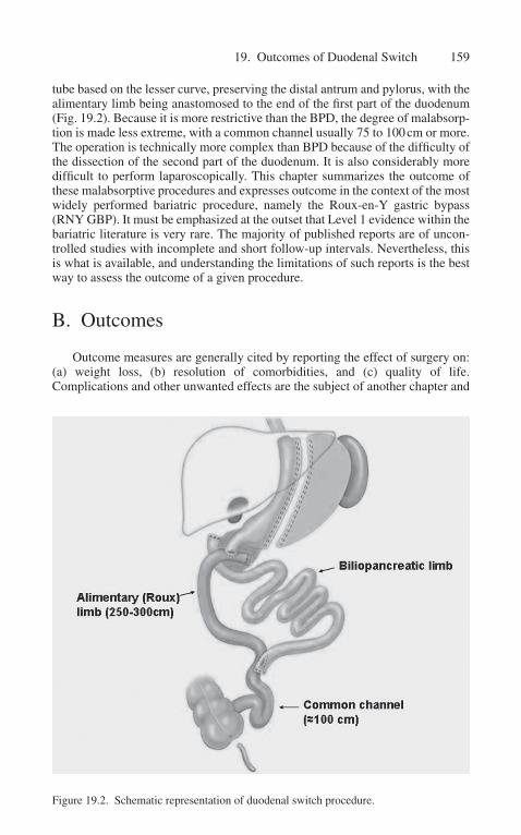
19. Outcomes of Duodenal Switch 159
tube based on the lesser curve, preserving the distal antrum and pylorus, with the alimentary limb being anastomosed to the end of the fi rst part of the duodenum (Fig. 19.2). Because it is more restrictive than the BPD, the degree of malabsorp-tion is made less extreme, with a common channel usually 75 to 100 cm or more. The operation is technically more complex than BPD because of the diffi culty of the dissection of the second part of the duodenum. It is also considerably more diffi cult to perform laparoscopically. This chapter summarizes the outcome of these malabsorptive procedures and expresses outcome in the context of the most widely performed bariatric procedure, namely the Roux-en-Y gastric bypass (RNY GBP). It must be emphasized at the outset that Level 1 evidence within the bariatric literature is very rare. The majority of published reports are of uncon-trolled studies with incomplete and short follow-up intervals. Nevertheless, this is what is available, and understanding the limitations of such reports is the best way to assess the outcome of a given procedure.
B. Outcomes
Outcome measures are generally cited by reporting the effect of surgery on: (a) weight loss, (b) resolution of comorbidities, and (c) quality of life. Complications and other unwanted effects are the subject of another chapter and
Figure 19.2. Schematic representation of duodenal switch procedure.

160 P F Crookes
will be briefl y summarized here. All outcome measures are diffi cult to compare because of differences in the duration and completeness of follow-up in indi-vidual reports. The most comprehensive published summary is the meta-analysis of Buchwald et al. (2004) where outcomes of a total of 22,094 patients were categorized. From this paper it is clear that BPD with or without DS has compa-rable weight loss to gastric bypass.
1. Weight loss
a. Pathophysiology of weight loss. It is often asserted that DS is a purely malabsorptive operation. This is incorrect: In contrast to the BPD, in which the reservoir portion of the stomach is essentially untouched, the DS procedure involves a substantial gastric resection leaving the stomach as a fairly narrow tube based on the lesser curve. The anatomy of the lesser curve is characterized by tough horizontal “clasp” fi bers, and the amount of stomach that can respond to food ingestion by re-ceptive relaxation is very limited. Therefore, it combines a substantial amount of restriction. Many surgeons who perform sleeve gastrectomy as a stand-alone procedure make the stomach narrower (using a smaller indwelling bougie) than if it is being performed as part of a DS. How-ever, in the early weeks after DS, food capacity is not much more than that experienced by GBP patients. We studied the patients’ perception of their food intake after weight stabilization and reported that they can eat an estimated 30% to 50% of their preoperative intake.
Loss of appetite and early satiety are reported by patients after DS. The underlying pathophysiologic mechanisms are still obscure, but removal of the fundus and reduction of ghrelin secretion may play a part.
b. Degree of weight loss after BPD and DS. Within the bariatric commu-nity, and especially among patients who express their views on websites, there is a strong belief that weight loss after DS is both greater and more durable than after RNY GBP procedures. There are few rigor-ous studies in the published literature to support this belief. Two recent studies from respected centers in Portland and Chicago each report-ed head-to-head non-randomized comparisons of RNY and DS: One found no difference in weight loss, whereas the other reported superior weight loss in superobese patients (BMI >50) after DS compared with RNY GBP. In both studies, there was a tendency to perform DS for the heavier patients with more severe metabolic abnormalities. The study from Portland followed patients for 2 years, and found that DS had a slightly longer length of hospital stay, a slightly higher leak rate, but mortality was similar (0.8% for GBP vs. 0.9% for DS), and EBWL was 67% for GBP compared with 63% for DS after 2 years. In the Chicago study length of stay was also slightly longer for DS vs. GBP, mortal-ity was low in both groups (0.05% for DS, zero for GBP), but patients with DS tended to have greater EBWL (72%) than patients after GBP (60%) after 2 years, with both groups gaining small amounts of weight by 3 years. The reasons for the disparity between these two studies are unclear, but may be a function of incomplete follow-up. In the Portland study only 93 of the original 350 patients were available for follow-up 2

19. Outcomes of Duodenal Switch 161
years later, and in the Chicago study, also with 350 patients, only 65 had 3-year follow-up. In light of this incomplete evidence, it would appear that DS or BPD does not have a reliable superiority over RNY GBP except possibly in the superobese.
2. Relief of comorbidities
Some of the typical comorbidities are primarily a function of physically increased weight, and only improve as weight loss occurs. Degenerative joint disease in the spine and weight-bearing joints, urinary incontinence, obstructive sleep apnea, and pulmonary impairment are in this category. Other problems, such as diabetes, dyslipidemia, and possibly hypertension are related to altered metabolism and may improve even before any weight loss occurs. Different procedures thus have different effects on these metabolically -induced comor-bidities. Purely restrictive procedures, although partially effective in resolving diabetes and dyslipidemia, are less effective than bypass procedures such as RNY, GBP, and BPD/DS.
a. Type 2 diabetes. Relief of diabetes after BPD and DS appears to be greater than with any other operation. In the meta-analysis of Buchwald, 17 stud-ies involving 4,096 patients were analyzed, with improvement or resolu-tion noted in 98%, higher than after any other procedure analyzed in this study. The recent work of Rubino and colleagues has identifi ed two major mechanisms whereby glucose tolerance is improved after bariatric surgery, which may be summarized as a proximal gut factor and a distal gut fac-tor. The proximal factor is related to the diversion of food away from the duodenum, through mechanisms as yet unidentifi ed, appears to improve glucose control. This proximal mechanism is operative in BPD, DS, and RNYGBP, but not after purely restrictive procedures such as the lap-band or sleeve gastrectomy. The distal factor appears to result from undigested food reaching the ileum, and is believed to be mediated by the release of GLP-1. GLP-1 delays gastric emptying and enhances insulin secretion and stimulates the formation of more β-cells in the pancreatic islets. This distal mechanism is more pronounced in malabsorptive operations such as BPD and DS and results in superior resolution of diabetes and impaired glucose tolerance compared with any other bariatric procedure.
c. Hyperlipidemia. For similar reasons, the distal fat-malabsorbing effect of BPD and DS cause almost universal reduction of cholesterol and trig-lycerides, in the few studies in which these measurements were made.
d. Obstructive sleep apnea. Unlike diabetes and dyslipidemias, improve-ment in sleep apnea is dependent on weight loss, and in published reports averages 80% to 90%, which is not signifi cantly different from the degree of improvement in other contemporary operations.
e. Weight-related comorbidities. Many of these conditions such as degenerative joint disease and urinary stress incontinence are not eas-ily quantifi ed. Many morbidly obese patients who would otherwise be good candidates for joint replacement surgery are recommended to lose weight before such surgery will be considered. We recently reported that the long-term outcome of knee replacement was not affected by
162 P F Crookes
moderate degrees of obesity, offering some reassurance that patients whose weight is reduced to a BMI in the region of 30 kg/m2 can undergo joint replacement without sabotaging the operation.
Improvement in musculoskeletal conditions, especially involving the spine, foot, and ankle, were recently reported for gastric bypass, in which the percentage of patients reporting such symptoms dropped from 100% to 37% after surgery. However, comparable studies have not been reported for BPD or DS.
f. What is the cost? Long-term side effects and problems after BPD and DS. Despite the benefi cial effects achieved by these procedures, there remains worry that long-term problems of malnutrition may detract from the early benefi ts.
Almost all patients who achieve suffi cient weight loss report increased malodorous fl atulence and more frequent bowel movements. These symptoms tend to be related to fat intake, and may be associated with crampy abdominal pain. As patients learn how the reconstructed in-testinal tract responds to different foods, some adaptation takes place. Dumping in response to sweet intake tends to be rare. Although intui-tively it was suggested that the preservation of the pylorus in the DS led to improved symptomatic outcome compared with BPD, the only study to compare the symptomatic outcome of the two procedures detected no differences between them.
g. Long-term defi ciencies. All patients after any kind of bypass, whether GBP or DS, have a problem with Iron and calcium absorption, since both are normally absorbed in the bypassed portion of the GI tract. Con-sequently, microcytic anemia is common, especially in younger women who are still menstruating. Even before the serum iron is abnormal, defective iron stores are detectable when serum ferritin drops and the total iron-binding capacity increases. Oral supplementation with fer-rous sulfate is the cheapest and most readily available method, but fer-rous gluconate or fumarate may be more easily absorbed. If iron levels are persistently subnormal despite oral supplements, a loading dose of intravenous iron may be given, usually under the supervision of a hematologist.
Macrocytic anemia is rarely seen after BPD and DS, since it is not com-mon for B
12 and folate levels to be low unless patients fail to comply
with the prescribed vitamin supplementation regimen. As a class, DS patients tend to be knowledgeable and motivated; hence, this is rarely a problem.
Calcium defi ciency may not be immediately apparent, because serum calcium tends to remain in the low normal range. Indirect evidence of defective calcium absorption may be sought by measuring parathyroid hormone (PTH). We recently reported that elevated PTH was more com-mon in patients after DS with 75 cm common channel compared with those in whom the common channel was 100 cm long. Vitamin D levels tend to be lower after DS because vitamin D is a fat-soluble vitamin and is poorly absorbed. The best estimate of vitamin D stores is 25 (OH) D. In contrast, levels of 1,25 (OH)2 D are increased by PTH in the presence of vitamin D defi ciency. Vitamin D is important in promoting absorption

19. Outcomes of Duodenal Switch 163
of calcium from the gut, but it also has independent effects on muscle strength and patients with vitamin D defi ciency frequently report weak-ness, tiredness, and fatigability, which is then labeled with diagnoses such as fi bromyalgia and chronic fatigue syndrome. Even after aggressive oral supplementation of vitamin D, absorption may still be inadequate. The easiest way to enhance vitamin D is solar exposure, especially in white individuals. When this is not possible, twice weekly intramuscular injec-tions of vitamin D may be given for a period of several weeks until stores are repleted.
Clinical vitamin A defi ciency is rarely reported in isolation, but it has been reported in the ophthalmology literature. Since it is a fat soluble vitamin it may be malabsorbed in the same way as vitamin D. The ma-jor effects of vitamin A defi ciency are ocular.
Protein-calorie malnutrition is characterized by muscle loss, excessive weight loss, hypoalbuminemia, and frequently edema. Unlike the mal-nutrition associated with poor intake, it is often associated with diarrhea, and hypokalemia.
All of the preceding defi ciencies were more frequent when short com-mon channels (50 cm) were employed. In our experience and that of oth-ers, a common channel of 100 cm is associated with a reduced incidence of such problems. The incidence of malnutrition of such severity as to require partial revision of the operation varies from 2% to 5%. The most common scenario is a combination of excessive weight loss with edema and diarrhea. The simplest way to correct the degree of malabsorption is to perform a side-to-side anastomosis between the biliopancreatic and alimentary limbs (the “kissing X anastomosis”). Weight regain is sur-prisingly limited after this procedure, averaging about 15 lbs, and there is good correction of albumin, hemoglobin, and potassium.
h. Other late outcomes BPD and DS, like other bypass procedures, have an intrinsic proclivity to lead to small bowel obstruction, often many years afterward. This may be due to internal herniation, simple adhe-sions, or kinking at the jejunojejunostomy. A unique form of obstruc-tion after BPD and DS can be caused by obstruction of the biliopancre-atic limb. Small case series of this important complication have been reported, but the denominator is not known and therefore assessment of the risk is imprecise. The biliopancreatic limb in BPD and DS is long, approximately 300 cm, and it ends blindly at the second portion of the duodenum. Therefore, it acts like other forms of closed loop obstruc-tion, and the development of ischemia and gangrene is rapid. It cannot be decompressed by NG suction, it is inaccessible to endoscopy, and since the alimentary limb often remains open, the other typical features of small bowel obstruction are not present. All patients presenting with severe crampy abdominal pain persisting for more than a few hours should seek urgent medical attention and undergo CT scanning, since this is the only way to make the diagnosis quickly (Fig. 19.3). Since the patient may present to another hospital where the DS is not performed, the general surgeon on call may be unfamiliar with the anatomical alterations of BPD or DS procedures, and may waste valuable time in attempting to transfer the patient to the original operating surgeon.

164 P F Crookes
Patients with the BPD and DS should be given a card with a diagram or other depiction of their reconstruction, and should be warned to seek help for persistent (>6 hours) abdominal pain.
i. Special situations. Pregnancy after BPD and DS is generally uneventful. However, there is more concern over vitamin defi ciencies, and pa-tients of childbearing age must be especially counseled to maintain compliance with vitamin and mineral supplements. Isolated cases of microphthalmia, caused by vitamin A defi ciency at the critical stage of organogenesis, have been reported, but there is no systematic study of pregnancy after BPD or DS.
C. Selected References
Anthone GJ, Lord RV, DeMeester TR, et al. The duodenal switch operation for the treat-ment of morbid obesity. Ann Surg 2003;238:618–628.
Buchwald H, Avidor Y, Braunwald E, et al. Bariatric surgery: a systematic review and meta-analysis. JAMA 2004;292(14):1724–1737.
Cossu ML, Meloni GB, Alagna S, et al. Emergency surgical conditions after biliopancre-atic diversion. Obes Surg 2007;17(5):637–641.
Figure 19.3. Abdominal CT Scan showing biliopancreatic obstruction. Note the distended loops without contrast.

19. Outcomes of Duodenal Switch 165
Deveney CW, MacCabee D, Marlink K, et al. Roux-en-Y divided gastric bypass results in the same weight loss as duodenal switch for morbid obesity. Am J Surg 2004;187:655–659.
Dolan K, Hatzifotis M, Newbury L, et al. A clinical and nutritional comparison of bilio-pancreatic diversion with and without duodenal switch. Ann Surg 2004;240:51–56.
Hamoui N, Cantor S, Anthone GJ, et al. Effect of obesity on long term outcome of total knee arthroplasty. Obes Surg 2006;16:35–38.
Hamoui N, Chock B, Anthone GJ, et al. Revision of the duodenal switch: technique, indi-cations, and outcome. J Am Coll Surg 2007;204(4):603–608.
Hamoui N, Kim K, Anthone G, et al. The significance of elevated levels of parathyroid hormone in patients with morbid obesity before and after bariatric surgery. Arch Surg 2003;138:891–897.
Hess DS, Hess DW, Oakley RS. The biliopancreatic diversion with the duodenal switch: results beyond 10 years. Obes Surg 2005;15:408–416.
Hooper MM, Stellato TA, Hallowell PT, et al. Musculoskeletal findings in obese sub-jects before and after weight loss following bariatric surgery. Int J Obes (Lond) 2007;31(1):114–1120.
Larrad-Jimenez A, Diaz-Guerra CS, de Cuadros Borrajo P, et al. Short-, mid- and long-term results of Larrad biliopancreatic diversion. Obes Surg 2007;17:202–210.
Marceau P, Biron S, Bourque RA, et al. Biliopancreatic diversion with a new type of gas-trectomy. Obes Surg 1993;3:29–35.
Marceau P, Hould FS, Simard S, et al. Biliopancreatic diversion with duodenal switch. World J Surg 1998;22:947–954.
Polizzi A, Schenone M, Sacca SC, et al. Role of impression cytology during hypovitami-nosis A. Br J Ophthalmol 1998;82(3):303–305.
Prachand VN, Davee RT, Alverdy JC. Duodenal switch provides superior weight loss in the super-obese (BMI > or = 50 kg/m2) compared with gastric bypass. Ann Surg 2006;244(4):611–619.
Rubino F, Forgione A, Cummings DE, et al. The mechanism of diabetes control after gastrointestinal bypass surgery reveals a role of the proximal small intestine in the pathophysiology of type 2 diabetes. Ann Surg 2006;244(5):741–749.
Scopinaro N. Biliopancreatic diversion: mechanisms of action and long-term results. Obes Surg 2006;16(6):683–689.
Scopinaro N, Adami GF, Marinari GM, et al. Biliopancreatic diversion. World J Surg 1998;22(9):936–946.
Smets KJ, Barlow T, Vanhaesebrouck P. Maternal vitamin A deficiency and neona-tal microphthalmia: complications of biliopancreatic diversion? Eur J Pediatr 2006;165(7):502–504.
A. Epidemiologic Trends and Behavioral Weight Management
The adult obesity epidemic has grown in severity over the past several decades, and an equally worrisome rise in obesity prevalence in children ominously portends a future worsening of this obesity epidemic in all ages. The prevalence of adolescent obesity has tripled over the last three decades and approximately 4% of US children are currently affected with extreme obesity (BMI for age > 99th percentile). The consequences of pediatric and adolescent obesity are becoming clearer, and are worrisome.
Although it is widely held that behavioral and dietary treatment approaches have greater effi cacy for pediatric obesity than for adult obesity, extreme pediatric obesity is usually not amenable to either conventional dietary and medication regimens, with only a 2% to 3% decrease in BMI expected. Thus, it is unlikely that dietary interventions alone will effect durable long-term weight reduction and comorbidity resolution for most adolescents.
B. Surgical Weight Loss in Adolescents
Although numerous studies have now reported surgical outcomes for adolescents undergoing bariatric procedures, most report short-term outcomes with very limited follow-up, and all are retrospective in design. We examined the data from 13 recently published studies, seven of which reported outcomes after laparoscopic adjustable gastric banding (LAGB), and six of which reported outcomes after Roux-en-Y gastric bypass (RYGB).
1. BMI change after LAGB
Seven LAGB studies reported weight and BMI change, with length of follow-up ranging from 1.7 to 3.3 years. Study characteristics and weight and BMI data appear in Table 20.1 and Fig. 20.1. Among fi ve larger case series, a total of 262 adolescent patients aged 9 to 19 were included, with an average mean preopera-tive BMI of 42 to 48 kg/m2. The patients lost 37% to 70% of excess body weight during 6-month to 7-year follow-up. Long-term weight loss outcomes for the LAGB and standardized descriptions of comorbidity change are still lacking.
20. Outcomes of Bariatric Surgery in Adolescents
Go Miyano and Thomas H. Inge

168 G Miyano and T H Inge
Table 20.1. Weight loss outcomes for LAGB and RYGB.
n agef/u (year) BMI % EWL
pre-op post-op
LAGBNadler (2007) 53 15.9 47.6 37.5–62.7Yitzhak (2006) 60 16 3.3 43 30Silberhumer (2006) 50 17.1 2.9 45.2 32.6 61.4Angrisani (2005) 58 18 3 46.1 37.8 39.7–55.6Fielding (2005) 41 15.6 1.7 42.4 30.2 70Horgan (2005) 4 17.8 50.5 15.0–87.0Abu-Abeid (2003) 11 15.7 1.9 46.6 32.5
RYGBCollins (2007) 11 16.5 1.8 50.5 28 60.8Lawson (2006) 35 17.6 1 56.5 35.8Barnett (2005) 14 15.7 51 64Sugerman (2003) 33 16 5 52 33Strauss (2001) 10 16.2 6.3 53.6 35.2 65Rand (1994) 34 17 6 47 32 66
EWL: excess weight loss.
Figure 20.1. BMI before and after LAGB.

20. Outcomes of Bariatric Surgery in Adolescents 169
However, Yitzhak et al. reported that at least 80% of adolescents had sustained weight loss 5 years after AGB, and Fielding et al. reported 30% weight reduction 3 years postoperatively in 18 patients.
2. BMI change in the RYGB
Six studies reported weight and BMI data following RYGB with length of fol-low-up ranging from 1.0 to 6.3 years. Study characteristics and weight and BMI data appear in Table 20.1 and Fig. 20.2.
Lawson’s study from Cincinnati Children’s Hospital was a cohort-controlled multicenter study in which laparoscopic RYGB was compared with outcomes of extremely obese adolescents who participated for 1 year in a pediatric behavioral treatment program. Laparoscopic RYGB resulted in a change in mean BMI from 56.5 to 35.8 kg/m2, as compared with no signifi cant change in BMI in the compari-son group with behavioral intervention. Strauss et al., evaluating results of gastric bypass in 10 adolescents, reported that mean preoperative BMI was 52.4 and maxi-mal weight loss was achieved at 12 to 15 months postoperatively. Sugerman et al. have provided the largest retrospective study of adolescent bariatric surgery with the longest follow-up to date. Over a 21-year period, mean BMI measured preop-eratively and at 1, 5, 10, and 14 years after surgery was 52, 36, 33, 34, and 38 kg/m2, respectively. Five patients (15%) regained all or most of their lost weight at 5 to 10
Figure 20.2. BMI before and after RYGB.

170 G Miyano and T H Inge
years after surgery. The precise frequency of weight regain after adolescent RYGB is not clear, but is estimated to range from 10% to 20%. The postoperative BMI tran-sition in both LAGB and RYGB is displayed graphically in Figs. 20.3 and 20.4.
C. Comorbidity
Five of seven LAGB studies reported comorbidity change, and the resolution rates were: 89% (8/9) for type 2 diabetes mellitus (T2DM), 100% (11/11) for obstructive sleep apnea syndrome (OSAS), and 65% (11/17) for hypertension (HTN). Four of six RYGB studies reported comorbidity data, the resolution rates were: 63% (5/8) for DM, 90% (18/20) for OSAS, and 75% (15/20) for HTN. A summary of the data appears in Table 20.1 and Fig. 20.5. These studies also in general reported favorable change in dyslipidemia, nonalcoholic steatohepatitis, gastroesophageal refl ux disease, weight-related arthropathies, and pseudotumor cerebri, although it is important to consider the following limitation: These data are derived from very few published studies, often without clear documentation of how comorbidities were assessed preoperatively and postoperatively. These facts signifi cantly limit our ability to accurately interpret or generalize from the fi ndings.
Figure 20.3. Kinetics of BMI change over time following LAGB.

20. Outcomes of Bariatric Surgery in Adolescents 171
Figure 20.4. Kinetics of BMI change over time following RYGB.
Figure 20.5. Comorbidity change after adolescent bariatric surgery.
172 G Miyano and T H Inge
1. Type 2 diabetes mellitus
The most compelling reasons for performing bariatric procedures in adoles-cence are to reduce or resolve obesity-related comorbidity, and some of the most severe consequences of obesity seen in adolescents include T2DM and OSAS. A steep rise in prevalence of type 2 diabetes is occurring worldwide in parallel with an increasing rate of obesity in children and adolescents. When considering all of the as yet limited surgical outcome data (LAGB and RYGB studies), T2DM appears to resolve in 76% (13/17). In the adult surgical literature, duration of dia-betes seems to predict completeness of resolution postoperatively. This may be an important factor when considering the timing of bariatric surgery for younger diabetic patients.
2. Obstructive sleep apnea syndrome
Marcus et al. reported abnormal polysomnograms in 36% of obese children and adolescents, and also showed a positive correlation between the degree of obesity and the severity of OSAS. When considering all available pediatric outcome data, OSAS resolution following LAGB and RYGB was 94% (29/31). Kalra et al., utilizing pre- and postoperative polysomnography, reported that OSAS was diagnosed in 55% of 34 patients studied preoperatively. Ten of these patients returned for follow-up PSG, and OSA signifi cantly improved or resolved after RYGB in all the ten subjects, with nine of ten returning to a normal frequency of OSAS events.
3. Quality of life
Adverse psychosocial and educational consequences of extreme adolescent obesity are a topic of considerable concern, and being an overweight young adult has a lasting impact on life satisfaction and aspirations. Some have noted an increase in employment and educational status in those adolescents who lost the most weight postoperatively. Several recent studies also showed signifi cant improvement in post-operative QOL after LAGB and RYGB in adolescents using formal measures.
D. Mortality and Postoperative Adverse Events
1. Mortality
No perioperative death was reported in both the LAGB and the RYGB. However, four late deaths not linked to the surgery or weight loss have been reported by Lawson, Barnett, and Sugarman.
2. The adverse events on the LAGB
All LAGB studies reported adverse events (summarized in Table 20.2). Twenty-six reoperations were performed to correct various complications in
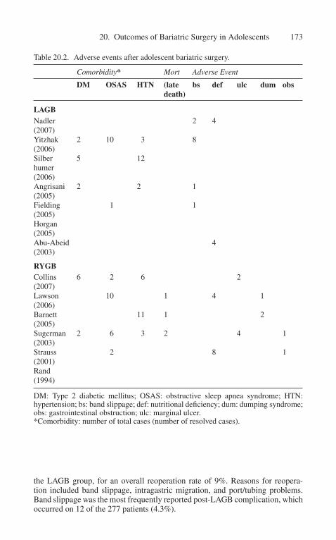
20. Outcomes of Bariatric Surgery in Adolescents 173
Table 20.2. Adverse events after adolescent bariatric surgery.
Comorbidity* Mort Adverse Event
DM OSAS HTN (late death)
bs def ulc dum obs
LAGBNadler(2007)
2 4
Yitzhak(2006)
2 10 3 8
Silberhumer (2006)
5 12
Angrisani(2005)
2 2 1
Fielding(2005)
1 1
Horgan(2005)Abu-Abeid(2003)
4
RYGBCollins(2007)
6 2 6 2
Lawson(2006)
10 1 4 1
Barnett(2005)
11 1 2
Sugerman(2003)
2 6 3 2 4 1
Strauss(2001)
2 8 1
Rand(1994)
DM: Type 2 diabetic mellitus; OSAS: obstructive sleep apnea syndrome; HTN: hy pertension; bs: band slippage; def: nutritional defi ciency; dum: dumping syndrome; obs: gastrointestinal obstruction; ulc: marginal ulcer.*Comorbidity: number of total cases (number of resolved cases).
the LAGB group, for an overall reoperation rate of 9%. Reasons for reopera-tion included band slippage, intragastric migration, and port/tubing problems. Band slippage was the most frequently reported post-LAGB complication, which occurred on 12 of the 277 patients (4.3%).
174 G Miyano and T H Inge
3. The adverse events on the RYGB
All six RYGB studies also reported adverse events (summarized in Table 20.2). Early complications of RYGB in adolescents included pulmonary embolism, post-operative bleeding, staple line leak, wound infection, stomal stenosis, and marginal ulceration. Late complications included small bowel obstruction, incisional hernias, severe malnutrition, symptomatic cholelithiasis, and late weight regain. Problems related to protein calorie malnutrition and micronutrient defi ciency, such as iron, vitamin B
12, vitamin D, thiamine, and calcium, were the most frequently reported
complications after RYGB. Since it is not known to what degree risks may increase over time, prospective long-term studies are vital to determine the true prevalence and clinical signifi cance of complications, including mechanical/technical prob-lems and nutritional defi ciencies following bariatric surgery in adolescents.
E. Which Operation Is Best for Adolescents?
When gastric bypass and adjustable gastric banding have been compared, reports have cited greater weight loss and comorbidity resolution with gastric bypass at early time intervals (1–3 years), but similar weight loss effi cacy at later time intervals (5 year). The appeal of adjustable gastric banding for adolescents lies in the reversibility, low incidence of morbidity and mortality, and potential avoidance of severe nutritional risks associated with malabsorptive procedures; however, potential worrisome later complications, including band obstruction, gastric prolapse, tubing problems, frequency of band explanation, will be critical for clinical decision making for adolescents who have perhaps fi ve or six decades to live with the device. Although this device holds a great deal of promise for treatment of adolescent with extreme obesity, more information about long-term effi cacy and durability is needed.
While biliopancreatic diversion (BPD) has been reported in adolescents, most believe that the risks of BPD and even duodenal switch likely outweigh their potential benefi t of greater weight loss as compared with RYGB and AGB. For this reason, these operations are not recommended for most adolescents.
The laparoscopic sleeve gastrectomy (LSG) is a new operation that produces signifi cant initial weight loss with low operative risk in adult studies. The LSG may be a safe option for adolescents with fewer nutritional and long-term device-related risks than other operations. However, further studies are needed to deter-mine objectively the risk–benefi t ratio for adolescents.
F. Selected References
Abu-Abeid S, Gavert N, Klausner JM, et al. Bariatric surgery in adolescence. J Pediatr Surg 2003;38:1379–1382.
Aggarwal S, Kini SU, Herron DM. Laparoscopic sleeve gastrectomy for morbid obesity: a review. Surg Obes Relat Dis 2007;3:189–194.

20. Outcomes of Bariatric Surgery in Adolescents 175
Angrisani L, Favretti F, Furbetta F, et al. Obese teenagers treated Lap-Band system: the Italian experience. Surgery 2005;138:877–881.
Ball K, Crawford D, Kenardy J, et al. Longitudinal relationships among overweight, life satisfaction, and aspirations in young women. Obes Res 2004;12:1019–1030.
Barnett SJ, Grimshaw JM, Wells GA, et al. Long-term follow-up and the roll of surgery in adolescents with morbid obesity. Surg Obes Relat Dis 2005;1:394–398.
Chapman AE, Kiroff G, Game P, et al. Laparoscopic adjustable gastric banding in the treatment of obesity: a systematic literature review. Surgery 2004;135:326–351.
Collins J, Mattar S, Qureshi F, et al. Initial outcomes of laparoscopic Roux-en-Y gastric bypass in morbidly obese adolescents. Surg Obes Relat Dis 2007;3:147–152.
Fielding GA, Duncombe JE. Laparoscopic adjustable gastric banding in severely adoles-cents. Surg Obes Relat Dis 2005;1:399–405.
Freedman DS, Mei Z, Sriniavasan SR, et al. Cardiovascular risk factors and excess adipos-ity among overweight children and adolescents: the Bogalusa heart study. J Pediatr 2007;150:12–17.
Greenstein RJ, Rabner JG. Is adolescent gastric-restrictive antiobesity surgery warranted? Obes Surg 1995;5:138–144.
Horgan S, Holterman MJ, Jacobsen GR, et al. Laparoscopic adjustable gastric banding for the treatment of adolescent mobid obesity in the United States: a safe alternative to gastric bypass. J Pediatr Surg. 2005;40:86–90.
Kalra M, Inge T, Garcia V, et al. Obstructive sleep apnea in extremely overweight adoles-cents undergoing bariatric surgery. Obes Res 2005;13:1175–1179.
Lawson ML, Kirk S, Mitchell T, et al. One-year outcomes of Roux-en-Y gastric bypass for morbidly obese adolescents: a multicenter study from the Pediatric Bariatric Study Group. J Pediatr Surg 2006;41:137–143.
Levine MD, Ringham RM, Kalarchian MA, et al. Is family-based behavioral weight con-trol appropriate for severe pediatric obesity? J Eat Disord 2001;30:318–328.
Marcus CL, Curtis S, Koerner CB, et al. Evaluation of pulmonary function and polysom-nography in obese children and adolescents. Pediatr Pulmonol 1996;21:176–183.
Mokdad AH, Bowman BA, Ford ES, et al. The continuing epidemics of obesity and dia-betes in the United States. JAMA 2002;282:1195–1200.
Nadler EP, Youn HA, Ginsburg HB, et al. Short-term results in 53 US obese pediat-ric patients treated with laparoscopic adjustable gastric banding. J Pediatr Surg 2007;42:137–141.
Ogden CL, Flegal KM, Carroll MD, et al. Prevalence and trends in overweight among US children and adolescents. JAMA 2002;288:1728–1732.
Papadia FS, Adami GF, Marinari GM, et al. Bariatric surgery in adolescents: a long-term follow-up study. Surg Obes Relat Dis 2007;3:465–468.
Rand CS, Macgregor AM. Adolescents having obesity surgery: a 6-year follow-up.South Med J 1994;87:1208–1213.Schauer PR, Burguera B, Ikramuddin S, et al. Effect of laparoscopic Roux-en-Y gastric
bypass on type 2 diabetes mellitus. Ann Surg 2003;238:467–485.Silberhumer GR, Miller K, Kriwanek S, et al. Laparoscopic adjustable gastric banding in
adolescents: the Austrian experience. Obes Surg 2006;16:1062–1067.

176 G Miyano and T H Inge
Strauss RS, Bradley LJ, Brolin RE. Gastric bypass surgery in adolescents with morbid obesity. J Pediatr 2001;138:499–504.
Sugerman HJ, Sugerman EL, DeMaria EJ, et al. Bariatric surgery for severely obese ado-lescents. J Gastrointest Surg 2003;7:102–108.
Yitzhak A, Mizrahi S, Avinoach E. Laparoscopic gastric banding in adolescents. Obes Surg 2006;16:1318–1322.

A. Introduction
There is no clear consensus as to which is the best operation in bariatric surgery. Perhaps a better question is, “Which is the right operation for the given patient?” No operation is uniformly accepted as the best one. There are too many confounding factors to make this kind of decision making possible. Primarily, the lack of long-term follow-up, established defi nitions of success, and paucity of randomized data make this unfeasible. In addition, patients may have different goals or biases that determine which operation they choose. Importantly, not all bariatric surgeons perform all bariatric procedures. The preceding sections have reported the outcomes of laparoscopic adjustable banding (LAGB), roux-en-Y gastric bypass (RYGB), sleeve gastrectomy (SG) and the biliopancreatic diversion/duodenal switch (DS) operation. Specifi c considerations have also been covered for adolescent patients. This chapter attempts to put these data into perspective and offer recommendations that are suitable for a particular patient or clinical circumstance. The appropriate choice of operation begins with a full assessment of the patient’s reasons for choos-ing as well as expectations of weight loss surgery. Information can then be gathered from the history and physical examination, laboratory data, imaging and endoscopic studies, and prior operative notes. Arbitrarily, choice of pro-cedure can be determined by weight, presence of comorbid illness, age, plans of conception, or relevant previous surgery. Collectively, some of these factors can be used to determine a patient’s perioperative risk, which might represent an independent way to choose an operation. For example, what would be the best operation for a 26-year-old superobese man with a history of a previous Nissen fundoplication? Certainly even the most singularly aligned surgeon to a particular operation might give pause to the fact that there needs to be particular consideration in this case and that his or her “best” operation, or any operation for that matter, may not be appropriate.
The four most commonly performed procedures for morbid obesity at this time are RYGB, LAGB, DS, and SG. Each procedure has advantages and disad-vantages; accordingly, it is important to consider multiple factors when recom-mending the type of surgery. Few randomized trials exist comparing the different procedures. Algorithms using review of the literature have been developed to match a given patient to a given operation. This algorithm has not yet been tested in a clinical setting and cannot be absolute.
21. Which Operation Is Best?
Sayeed Ikramuddin and Gonzalo Torres-Villalobos

178 S Ikramuddin and G Torres-Villalobos
B. Risk
Some patients have severe obesity-related comorbidities that expose them to a high risk of perioperative complications. Many of these patients seek weight loss surgery as they have exhausted medical management. Preoperatively, the strategy for these patients is to reduce this risk. The approach for these high-risk patients includes rigorous goals aiming for weight loss, physical therapy, optimi-zation of medical disorders (diabetes, hypertension, reversible heart disease, etc.) and psychological counseling.
For any high-risk surgical candidate, less extensive, less invasive, and shorter procedures are likely to have lower perioperative morbidity and mortality. Patients with marked comorbidity might be considered for an SG or LAGB because they are technically less complicated and there are no gastrointestinal anastomoses. Morbidly obese individuals with marked comorbidity are considered for sleeve gastrectomy as the fi rst part of a two-stage procedure. Substantial weight loss generated by this restrictive procedure signifi cantly reduces risk associated with the second-stage procedure. A second-stage procedure consisting of an RYGB or DS is performed 6 to 12 months later for long-term weight reduction. The SG has been performed as the sole weight reduction procedure in high-risk patients.
The LAGB can be a good option for patients with marked comorbidities. The procedure is technically simple. Morbidity and mortality are very low, and the length of the procedure is signifi cantly less than other bariatric operations. In addi-tion, LAGB is easier to perform; the absence of an intestinal anastomosis removes the disastrous complication of leak or staple line breakdown in the high-risk mor-bidly obese patient. A randomized study that compares the outcomes of the SG and LAGB demonstrates that weight loss and loss of feeling of hunger after 1 and 3 years were better after SG than LAGB. GERD was more frequent at 1 year after SG and at 3 years after LAGB. The number of reoperations was important in both groups, but the severity of complications was higher in SG. It is important to remember that there is a fi nite risk of leakage and bleeding in the SG that far exceeds that seen in the LAGB. Conversely, issues such as slippage do arise long term in this patient population. Outcomes in the sleeve gastrectomy for high-risk patients are limited. In small series outcome was excellent with good resolution of comorbid illness. Stud-ies directly examining the band in patients who are at high risk do not exist.
C. Age
The extremes of age (younger that 18, older than 60 years) remain an area of controversy. With growing awareness of the benefi ts of bariatric surgery, sur-geons are seeing more patients in the extremes of age. The literature dealing with bariatric surgery in the elderly, as in the adolescent population, is sparse. NIH guidelines do not address adolescents.
It is estimated that 25% of US children are obese or overweight, an incidence that has doubled in the last 30 years. In 2004, eligibility criteria for weight loss surgery in this population were suggested (Table 21.1).
Results of both RYGB and LAGB have been retrospectively reviewed in small series of adolescents. The LAGB is attractive because of its low perioperative

21. Which Operation Is Best? 179
risk, but long-term data are lacking and there are concerns of the longevity of the device itself. The risk of slippage and erosion appear to increase over time, although changes in the design of the band and refi nements in placement tech-niques may reduce the likelihood of these complications. The RYGB imposes a higher perioperative risk, but it has a longer record of success in weight loss and comorbid resolution. In addition there is some concern in the possibility of long-term metabolic complications. There is a price to pay in terms of iron defi ciency and postoperative hypoglycemia (a variant of the dumping syndrome) that must be taken into account. Admittedly it is far easier to manage failed weight loss from a band than that of a gastric bypass. However, some would argue that the band is more likely to fail in this setting. What is clear now is that adolescents should undergo evaluation and treatment at large centers where there is vast experience in bariatric surgery. Adolescents with limited comor-bidities may benefi t from a multidisciplinary approach incorporating LAGB with behavioral modifi cation techniques. In addition if the LAGB is unsuccessful in these younger patients, later conversion to another procedure can be performed. There is a growing movement to support the use of the SG in this population. Arguments include the elimination of a bypassed stomach and no signifi cant malabsorption; it still remains true that there is a risk of leakage from the staple line that is fi nite. The claims that this procedure does not have a malabsorp-tive component still need to be substantiated given the extensive resection of the stomach that is necessary with concomitant removal of intrinsic factor–secreting cell mass. Further, there is still no standardization on how to perform the SG. Many surgeons include the antrum of the stomach in the resection. The antrum plays an important role in reducing the size of food particles. Destruction of the antrum theoretically might lead to increased postoperative nausea. It has been our experience that the follow-up of adolescents is a signifi cant challenge that should not be overlooked. Many factors are responsible for this; younger adoles-cents need to depend on transportation to be brought in to clinic appointments. Older adolescents struggle because they tend to be highly mobile or attending school, which makes it a struggle to keep appointments.
Likewise, the indication for bariatric surgery for those above age 60 years should be considered on an individual basis. Proof of a favorable risk–benefi t ratio
Table 21.1. Bariatric surgery for severely overweight adolescents: recommendations.
Presence of severe obesity (BMI >40 kg/m2)Presence of comorbiditiesSkeletal/physiological maturityFailure of ≥6 months of structured attempts at weight lossCommitment to evaluation before and after surgeryCommitment and ability to follow nutritional guidelines after surgeryCommitment to avoid pregnancy for ≥1 year postoperativelyInformed consentDecision-making capacity
Source: Reprinted from Inge TH, Garcia V, Daniels S, et al. A multidisciplinary approach to the adolescent bariatric patient. J Pediatr Surg 2004;39:442–447, with permission from Elsevier.

180 S Ikramuddin and G Torres-Villalobos
must be demonstrated in elderly or ill patients before surgery is contemplated. In elderly patients, the primary objective of surgery is to improve quality of life, even though surgery may be unlikely to increase longevity. Morbidity and mortality for surgery increase with advancing age. In a recent article patients older than 65 years were at a signifi cantly higher risk of death (both early and late) after bari-atric surgery. However, it has been demonstrated in many recent studies that both gastric bypass and the LAGB are safe and effi cacious in elderly patients. What is clear now is that elder patients should undergo evaluation and treatment at large centers where there is vast experience in bariatric surgery.
Our older patients appear to carry more of a metabolic burden. In this case they would benefi t more from RYGB, as it clearly has superior results in terms of remis-sion of type 2 diabetes mellitus (T2DM) and dyslipidemia when cohorts of differ-ent studies are compared. For patients with established metabolic disease and who are at very high risk, the role of the SG or LAGB comes into question. In many of these patients anticoagulation must be continued around the time of surgery. This makes procedures such as the SG less attractive. At this time the SG is not covered by Medicare. The LAGB remains an attractive option for these patients.
D. Superobesity
Superobese patients are defi ned as those with a BMI ≥50 kg/m2. Tradi-tionally complications from bariatric surgery are much higher in this group of patients. The expected average weight loss and weight maintenance increases with the following procedures: LAGB, RYGB, and DS. The mean excess weight losses for these procedures are 48.6%, 68.6%, and 68.8%, respectively. On the contrary, the surgical complexity and potential surgical and long-term metabolic risks of procedures decrease inversely. Subgroup analysis demonstrates that for patients undergoing an RYGB the percent of excess weight loss is lower in heavier patients. The argument remains as to the objective of the surgery—to reduce weight, improve comorbid illness, or both. Admittedly patient satisfaction is a very important consideration. Superobese patients are better candidates for DS or RYGB procedures. The DS is associated with greater weight loss compared with the RYGB and might be a better option for superobese patients. The mal-absorptive complications in these patients cannot be ignored. Patients typically have at least three bowel movements per day. The incidence of fl atulence and odor is a problem in many of these patients. Also the incidence of revision for malabsorption is 4%. There are few data to support the use of the LABG in the United States for this population.
SG can be done as the fi rst part of a two-stage procedure for long-term weight loss in super-superobese patients, those with a BMI >60 kg/m2. Com-plications associated with laparoscopic DS in super-superobese patients are high. Morbidity has been reported as high as 23% and the mortality as high as 6.5%. Neither open nor laparoscopic one-stage DS has been satisfactory in this group of patients. A second procedure, either RYGB or DS, is performed in the second stage 6 to 12 months after the SG. Important weight loss generated by this restrictive procedure signifi cantly reduces risks associated with the second-stage procedure. It is worrisome that the fi rst-stage operation is susceptible to substantial complications, such as bleeding or leakage.

21. Which Operation Is Best? 181
E. Previous Surgery
1. Nissen fundoplication
Some obese patients present for weight loss surgery having previously undergone antirefl ux procedures. Several authors have reported their experience with gastric bypass following antirefl ux procedures and have found that although technically more challenging and carrying a higher risk of morbidity, RYGB is a feasible method for treating obesity and recurrent symptoms of refl ux. Some technical considerations must be taken into account, such as the use of pledgets at the fi rst operation, which can make the procedure far more diffi cult. Some have advocated performing a gastric bypass in these patients by stapling under-neath the wrap. Although from a technical point of view this is attractive it results in a grossly enlarged pouch and perhaps a higher theoretical risk of gastric stasis, marginal ulcer, and therefore impaired quality of life in these patients. A modifi ed DS has been proposed, but has compromised weight loss in patients who do not undergo reduction of gastric volume at the same time. A biliopancreatic diversion (BPD) is an option, but this operation has not received acceptance in the United States secondary to the large volume in consumed calories and the multiple number of bowel movements per day. Likely the best course of action for these patients is referral to a center with considerable experience in performing this type of procedure and converting these patients to an RYGB.
2. Colon or small bowel resection
Surgical history can signifi cantly affect surgical morbidity and mortality in a bariatric procedure. Prior abdominal surgery may lead to dense intra-abdominal adhesions that create prohibitive risk for competition of intestinal bariatric proce-dures. In adhesions, previous intestinal resections, including the ileocecal valve, small bowel, or pylorus, may prevent good evolution of the bariatric procedure or expose the patient to intractable diarrhea or dumping. Accordingly, LAGB may be the safest bariatric procedure in morbidly obese patients with a history of intestinal or colonic resection.
F. Comorbidities
1. Diabetes
T2DM is a common disease associated with morbid obesity. Obesity is the single most important predictor of T2DM. The risk of developing T2DM increases exponentially related to the BMI. Approximately 40% of morbidly obese patients have either impaired fasting glucose or impaired glucose tolerance and nearly 20% have T2DM. Mortality is in the range of 5% per year in morbidly obese diabetics.
The majority of obese patients who undergo bariatric surgery experience a dramatic and usually rapid resolution of T2DM. In the Swedish Obese Subjects study, the largest prospective series reported to date, there was a dramatic decrease in the

182 S Ikramuddin and G Torres-Villalobos
incidence of T2DM in surgical patients, with 72% at 2 years (odds ratio, 0.14%; 95% confi dence interval [CI], 0.08–0.24) and 36% at 10 years (odds ratio, 0.25; 95% CI, 0.17–0.38), confi rming that bariatric surgery could prevent new-onset T2DM as well as ameliorate pre-existing disease. The rate of T2DM resolution seemed to vary depending on the type of surgery. Resolution occurs most com-monly and rapidly after DS (98%) and RYGB (84%) compared with LAGB (48%). RYGB and DS should be the procedures of choice in patients with T2DM. DS can be considered for higher BMI (superobese patients).
2. Gastroesophageal refl ux disease
Gastroesophageal refl ux disease (GERD) is extremely common in Western society. Fifteen to twenty percent of adults in the United States experience epi-sodes of GERD. The incidence of GERD in the obese has been reported as high as 72%. A relationship between GERD and increasing BMI has been suggested in females. Some authors have shown a relationship between obesity and estrogen as risk factor for GERD symptoms; there also appears to be an augmentation of this relationship with the addition of postmenopausal hormones. In males, a dose-dependent observation between an increase in BMI and refl ux symptoms was observed. Obesity may also increase the severity of GERD. A BMI greater than 35 kg/m2 is a signifi cant risk factor for developing GERD and esophagitis. Other studies have shown increase in GERD and GERD symptoms in relation to increase in BMI.
There are differences in the outcome of GERD depending on the bariatric procedure. Results of the LAGB are mixed, slippage is known to occur up to 1.5% of the time, and may predispose to GERD. Many studies have shown worsening refl ux symptoms after LAGB. Conversely, there are reports of improvement in GERD symptoms after LAGB. RYGB has been deemed to be a benefi cial surgical treatment for GERD in the obese population. In theory, a small gastric pouch (15–20 ml) produces little in the way of acid to refl ux. By virtue of gastric exclusion, duodenogastroesophageal refl ux is rare, making RYGB an ideal oper-ation to correct acid and biliary refl ux in the obese population. Fewer data exist for the DS procedure. Certainly bile refl ux will be markedly diminished. The effect of DS in GERD remains unclear. Resolution of GERD has been reported after DS.
Despite early resolution of symptoms after LAGB, most patients had a return of refl ux symptoms by 6 months, indicating that RYGB may be the procedure of choice for bariatric patients with GERD.
RYGB is a better option in patients with large hiatal hernia and those with paraesophageal hernia or shortened esophagus. Patients with a history of peptic ulcer disease might be better served with LAGB or the DS.
3. Ventral hernia
Many patients who require surgical treatment for morbid obesity also present with abdominal wall hernias. Morbidly obese patients are predisposed to develop ventral hernias, and the risk is even greater than in patients with chronic steroid

21. Which Operation Is Best? 183
use. The great majority of ventral hernias are associated with previous abdominal surgeries. Surgeries that require an extensive bowel manipulation, such as RYGB and DS, may need to be done open. For this reason LAGB or SG seem to be attractive options since they do not involve extensive bowel handling.
There is no consensus regarding the optimal treatment of ventral hernias in patients who present for weight loss surgery. Regardless of the prevalence of this challenging clinical problem there is a paucity of literature on this sub-ject. Repair techniques consist of deferred treatment of the ventral hernia until adequate weight loss, or concomitant hernia repair either primarily, or with biodegradable mesh. The issue of simultaneous repair of anterior abdominal wall hernia with mesh during minimally invasive bariatric surgery remains to be answered. Two studies showed that the deferred treatment is dangerous, as it resulted in more than one third of patients developing small bowel obstruc-tion, which required urgent operative management. The reluctance for simul-taneous repair is mainly because of the high recurrence rates after primary repair, with a recurrence rate as high as 67%. This is especially true in obese patients. In addition there is contamination related with divided bowel, thus precluding the use of synthetic mesh. Even small hernias (3–4 cm), closed with primarily transfi xion transabdominal sutures similar to the technique used for closure of 12-mm trocar sites, are associated with a recurrence rate as high as 22%.
The development of tension-free hernia repair has clearly improved the out-come of ventral hernia repair, by reducing the recurrence rate to a range of 8% to 17% with the use of prosthetic material. Randomized controlled trials have confi rmed the superiority of mesh repair over primary repair. The adoption of laparoscopic principles to the Stoppa-Rives method has further improved the treatment of ventral hernias by reducing the recurrence rate to 0% to 3%.
Some articles support the use of biomaterials to concomitant repair of ventral hernias in the patient undergoing laparoscopic RYGB with a very low recurrence rate. The problem of mesh contamination is less for the LAGB because there is no contact with bowel contents. Outcome is less certain.
G. Transplant
Morbid obesity is associated with an increased risk of complications and death in transplant patients. Postoperative weight gain following organ transplan-tation may in part be explained by a direct corticosteroid effect. Bariatric surgery has been performed after organ transplantation to aid weight loss, improving medical comorbidities, and consequently graft survival. There are a few reports of bariatric surgery in morbidly obese patients, candidates for transplant, and post-transplant patients.
1. Renal transplant
End-stage renal failure is most commonly caused by obesity-related dis-eases, diabetes mellitus, and essential hypertension, and is best treated with
184 S Ikramuddin and G Torres-Villalobos
renal transplantation. Also, because operative risks, complications, and death are greater in morbidly obese patients, transplantation may be denied. The major reason for poor outcome in morbidly obese patients after transplantation relates to an increased risk of cardiovascular disease, which is now the leading cause of death in kidney-transplant patients after the fi rst year following transplant. It is well established that diabetes, hypertension, and hyperlipidemia all contribute to the risk of cardiovascular disease and that these complications are exceptionally high both after transplantation and in association with morbid obesity. BMI is a strong independent risk factor for both patient mortality and death-censored graft failure after kidney transplantation.
The potential benefi t of RYGB in morbidly obese dialysis and transplant patients has been reported. RYGB leads to a reduction in comorbid conditions that are associated with an increased risk for cardiovascular deaths. In addition RYGB can make previously nontransplantable patients acceptable for transplant. Some series suggest that the indications for RYGB in renal failure and transplant patients should be the same as for the general population.
LAGB can be a good option for patients who are candidates for renal trans-plant and post-transplant patients. The procedure is technically simple. The mor-bidity and mortality of the operation are very low, and the length of the procedure is signifi cantly less than other bariatric operations. In addition, LAGB is easier to perform; the absence of intestinal anastomosis removes a disastrous complica-tion from the risk for the high-risk morbidly obese patient. This may be a less attractive option in patients with T2DM.
2. Liver transplant
Obesity is common before and after liver transplantation and has been associated with signifi cant morbidity and mortality. Obesity may cause graft dysfunction through the development of recurrent nonalcoholic steatohepatitis. Liver transplantation has been successfully performed in the morbidly obese. However, wound infection, prolonged ventilatory support, multiorgan failure, and death have been described in the immediate postoperative period. Patients often develop a dramatic increase in weight after liver transplantation.
The feasibility of bariatric surgery in liver transplant recipients is practically unknown. There are some case reports of RYGB in morbidly obese liver trans-plant recipients with adequate reduction of weight and normalization of liver laboratory tests, lipid profi le, diabetes, and hypertension. However, RYGB after liver transplantation can be extremely technically demanding because of adhe-sions, mostly between the liver and stomach. LAGB in liver transplant patients has not been studied.
H. Other
Patients who crave sweet carbohydrate-rich foods probably benefi t from procedures that give a negative biofeedback, such as RYGB. Although supe-riority of the bypass over the band has not been clearly demonstrated in this population, this is an important consideration. Conversely, for those patients who are compulsive sweet eaters, a minority can develop post–gastric bypass
21. Which Operation Is Best? 185
hypoglycemia. Although this can be controlled by diet, it can be a vexing prob-lem to manage.
In patients who need endoscopic surveillance of the stomach, RYGB is con-traindicated. The presence of gastric pathology, such as gastric ulcers, polyps, or gastritis with metaplasia, is a relative contraindication for RYGB. The gastric remnant cannot be accessed directly using standard endoscopic techniques in this population. These patients might be better suited for LAGB or DS. This may be an important consideration for those patients who live in areas such as Japan or Chile with increased prevalence of gastric cancer.
Patients who require chronic anti-infl ammatory drugs probably benefi t from the DS, which is usually nonulcerogenic or LAGB.
Nonalcoholic fatty liver disease occurs in 30% to 100% of obese adults. Non-alcoholic fatty liver disease can lead to cirrhosis and liver failure. Even with this high prevalence, few patients (1.4%) undergoing bariatric surgery have had clini-cally signifi cant cirrhosis. For any cirrhotic patients undergoing general anesthe-sia independently of the surgery (except liver transplantation), the overall 30-day mortality has been reported to be as great as 11.6%. In addition, more than 30% of this patient population experienced at least one postoperative complication. The benefi ts of weight loss surgery must be evaluated considering the potential opera-tive risk in this subset of patients. Surgical weight loss has been associated with both improvement and worsening of liver disease. The malnutrition caused by the rapid weight loss in the early postoperative period can acutely worsen liver function and occasionally might be associated with mortality. Given our lack of understand-ing and data on the subject and our resultant inability to make informed decisions, it seems prudent to intervene with an operation that would cause the least harm. Shorter and less invasive procedures such as LAGB and SG are likely to result in less risk of worsening liver function in the short and long term. These surgeries are not malabsorptive and have less risk of liver failure compared with the DS. It is unknown how much weight loss is required to achieve optimal improvement in liver function among patients with nonalcoholic fatty liver disease as the etiology of their cirrhosis. Some authors advocate the use of more aggressive procedures such as DS as the fi rst treatment in cirrhotic patients because substantial weight loss after bariatric surgery can lead to considerable improvement in the histologic features of fatty liver disease, reversal of nonalcoholic steatohepatitis, and even true reversal of fi brosis (albeit rare). In many patients the hepatic pathology is related to T2DM. In these patients without synthetic dysfunction the best course of therapy might be to consider RYGB. Of the spectrum of procedures available this has potent resolution of diabetes without the risk of substantial malabsorp-tion. The benefi ts of bypass for patients with T2DM must be balanced with the risk of variceal bleeding from the gastric remnant, which can be problematic.
I. Conclusion
There are many good operations available to the bariatric surgeon. No opera-tion is universally applicable. A thorough assessment of a patient medical status is paramount in choosing a procedure and should be the basis for operative choice. Those surgeons not performing the “best” match may consider referral to another center.

186 S Ikramuddin and G Torres-Villalobos
J. Selected References
Ablassmaier B, Klaua S, Jacobi CA, et al. Laparoscopic gastric banding after heart trans-plantation. Obes Surg 2002;12(3):412–415.
Abu-Abeid S, Gavert N, Klausner JM, et al. Bariatric surgery in adolescence. J Pediatr Surg 2003;38(9):1379–1382.
Alexander JW, Goodman H. Gastric bypass in chronic renal failure and renal transplant. Nutr Clin Pract 2007;22(1):16–21.
Alexander JW, Goodman HR, Gersin K, et al. Gastric bypass in morbidly obese patients with chronic renal failure and kidney transplant. Transplantation 2004;78(3):469–474.
Almogy G, Crookes PF, Anthone GJ. Longitudinal gastrectomy as a treatment for the high-risk super-obese patient. Obes Surg 2004;14(4):492–497.
Anthony T, Bergen PC, Kim LT, et al. Factors affecting recurrence following incisional herniorrhaphy. World J Surg 2000;24(1):95–100;discussion 1.
Baltasar A, Bou R, Bengochea M, et al. Duodenal switch: an effective therapy for morbid obesity—intermediate results. Obes Surg 2001;11(1):54–58.
Birgisson G, Park AE, Mastrangelo MJ Jr, et al. Obesity and laparoscopic repair of ventral hernias. Surg Endosc 2001;15(12):1419–1422.
Brolin RE, Bradley LJ, Taliwal RV. Unsuspected cirrhosis discovered during elective obes-ity operations. Arch Surg 1998;133(1):84–88.
Buchwald H. A bariatric surgery algorithm. Obes Surg 2002;12(6):733–746; discussion 47–50.
Buchwald H, Avidor Y, Braunwald E, et al. Bariatric surgery: a systematic review and meta-analysis. JAMA 2004;292(14):1724–1737.
Burger JW, Luijendijk RW, Hop WC, et al. Long-term follow-up of a randomized control-led trial of suture versus mesh repair of incisional hernia. Ann Surg 2004;240(4):578–583; discussion 83–85.
Capella JF, Capella RF. Bariatric surgery in adolescence. is this the best age to operate? Obes Surg 2003;13(6):826–832.
Chu CA GM, Quinn T, et al. Two-stage laparoscopic biliopancreatic diversion with duo-denal switch: an alternative approach to super-super morbid obesity. Surg Endosc 2002;16(Abs):S069.
Costanza MJ, Heniford BT, Arca MJ, et al. Laparoscopic repair of recurrent ventral her-nias. Am Surg 1998;64(12):1121–1125; discussion 6–7.
Cottam D, Qureshi FG, Mattar SG, et al. Laparoscopic sleeve gastrectomy as an initial weight-loss procedure for high-risk patients with morbid obesity. Surg Endosc 2006;20(6):859–863.
Csendes A, Burgos AM, Smok G, et al. Effect of gastric bypass on Barrett’s esophagus and intestinal metaplasia of the cardia in patients with morbid obesity. J Gastrointest Surg 2006;10(2):259–264.
de Jong JR, van Ramshorst B, Timmer R, et al. Effect of laparoscopic gastric banding on esophageal motility. Obes Surg 2006;16(1):52–58.

21. Which Operation Is Best? 187
DeMaria EJ, Sugerman HJ, Kellum JM, et al. Results of 281 consecutive total laparoscopic Roux-en-Y gastric bypasses to treat morbid obesity. Ann Surg 2002;235(5):640–645; discussion 5–7.
Dixon JB, Dixon AF, O’Brien PE. Improvements in insulin sensitivity and beta-cell func-tion (HOMA) with weight loss in the severely obese. Homeostatic model assessment. Diabet Med 2003;20(2):127–134.
Dixon JB, O’Brien PE. Gastroesophageal reflux in obesity: the effect of lap-band place-ment. Obes Surg 1999;9(6):527–531.
Dolan K, Creighton L, Hopkins G, et al. Laparoscopic gastric banding in morbidly obese adolescents. Obes Surg 2003;13(1):101–104.
Duchini A, Brunson ME. Roux-en-Y gastric bypass for recurrent nonalcoholic steatohepatitis in liver transplant recipients with morbid obesity. Transplantation 2001;72(1):156–159.
Eid GM, Mattar SG, Hamad G, et al. Repair of ventral hernias in morbidly obese patients undergoing laparoscopic gastric bypass should not be deferred. Surg Endosc 2004;18(2):207–210.
El-Serag HB, Johanson JF. Risk factors for the severity of erosive esophagitis in Helicobacter pylori–negative patients with gastroesophageal reflux disease. Scand J Gastroenterol 2002;37(8):899–904.
Fielding GA. Laparoscopic adjustable gastric banding for massive superobesity (>60 body mass index kg/m2). Surg Endosc 2003;17(10):1541–1545.
Fielding GA, Duncombe JE. Laparoscopic adjustable gastric banding in severely obese adolescents. Surg Obes Relat Dis 2005;1(4):399–405; discussion 7.
Fisher BL, Pennathur A, Mutnick JL, et al. Obesity correlates with gastroesophageal reflux. Dig Dis Sci 1999;44(11):2290–2294.
Flanagan L Jr. Gastric bypass after cardiac transplantation. Obes Surg 1995;5(2):183–185.Flum DR, Salem L, Elrod JA, et al. Early mortality among Medicare beneficiaries under-
going bariatric surgical procedures. JAMA 2005;294(15):1903–1908.Forsell P, Hallerback B, Glise H, et al. Complications following Swedish adjustable gastric
banding: a long-term follow-up. Obes Surg 1999;9(1):11–16.Frezza EE, Ikramuddin S, Gourash W, et al. Symptomatic improvement in gastroesopha-
geal reflux disease (GERD) following laparoscopic Roux-en-Y gastric bypass. Surg Endosc 2002;16(7):1027–1031.
Gentileschi P, Kini S, Catarci M, et al. Evidence-based medicine: open and laparoscopic bariatric surgery. Surg Endosc 2002;16(5):736–744.
Gianetta E, Civalleri D, Bonalumi U, et al. [2 cases of death caused by hepatic insufficiency after jejuno-ileal bypass for obesity]. Minerva Chir 1979;34(15–16):1087–1096.
Gomez Escudero O, Herrera Hernandez MF, Valdovinos Diaz MA. [Obesity and gastro-esophageal reflux disease]. Rev Invest Clin 2002;54(4):320–327.
Himpens J, Dapri G, Cadiere GB. A prospective randomized study between laparoscopic gastric banding and laparoscopic isolated sleeve gastrectomy: results after 1 and 3 years. Obes Surg 2006;16(11):1450–1456.
Howard RJ, Patton PR, Reed AI, et al. The changing causes of graft loss and death after kidney transplantation. Transplantation 2002;73(12):1923–1928.

188 S Ikramuddin and G Torres-Villalobos
Hu FB, Manson JE, Stampfer MJ, et al. Diet, lifestyle, and the risk of type 2 diabetes mel-litus in women. NEJM 2001;345(11):790–797.
Inge TH, Garcia V, Daniels S, et al. A multidisciplinary approach to the adolescent bariat-ric surgical patient. J Pediatr Surg 2004;39(3):442–447; discussion 6–7.
Keeffe EB, Gettys C, Esquivel CO. Liver transplantation in patients with severe obesity. Transplantation 1994;57(2):309–311.
Kellogg TA, Andrade R, Maddaus M, et al. Anatomic findings and outcomes after antire-flux procedures in morbidly obese patients undergoing laparoscopic conversion to Roux-en-Y gastric bypass. Surg Obes Relat Dis 2007;3(1):52–57; discussion 8–9.
Kim WW, Gagner M, Kini S, et al. Laparoscopic vs. open biliopancreatic diversion with duodenal switch: a comparative study. J Gastrointest Surg 2003;7(4):552–557.
Lawson ML, Kirk S, Mitchell T, et al. One-year outcomes of Roux-en-Y gastric bypass for morbidly obese adolescents: a multicenter study from the Pediatric Bariatric Study Group. J Pediatr Surg 2006;41(1):137–143; discussion 43.
Leslie D, Kellogg TA, Ikramuddin S. Bariatric surgery primer for the internist: keys to the surgical consultation. Med Clin North Am 2007;91(3):353–381, x.
Liakakos T, Karanikas I, Panagiotidis H, et al. Use of Marlex mesh in the repair of recur-rent incisional hernia. Br J Surg 1994;81(2):248–249.
Locke GR 3rd, Talley NJ, Fett SL, et al. Risk factors associated with symptoms of gastro-esophageal reflux. Am J Med 1999;106(6):642–649.
Luyckx FH, Desaive C, Thiry A, et al. Liver abnormalities in severely obese subjects: effect of drastic weight loss after gastroplasty. Int J Obes Relat Metab Disord 1998;22(3):222–226.
Mattar SG, Velcu LM, Rabinovitz M, et al. Surgically-induced weight loss significantly improves nonalcoholic fatty liver disease and the metabolic syndrome. Ann Surg 2005;242(4):610–617; discussion 8–20.
Meier1 Kriesche HU, Arndorfer JA, Kaplan B. The impact of body mass index on renal transplant outcomes: a significant independent risk factor for graft failure and patient death. Transplantation 2002;73(1):70–74.
Mognol P, Chosidow D, Marmuse JP. Laparoscopic sleeve gastrectomy as an initial bariatric operation for high-risk patients: initial results in 10 patients. Obes Surg 2005;15(7):1030–1033.
Nair S, Verma S, Thuluvath PJ. Obesity and its effect on survival in patients undergoing orthotopic liver transplantation in the United States. Hepatology 2002;35(1):105–109.
Nilsson M, Johnsen R, Ye W, et al. Obesity and estrogen as risk factors for gastroesopha-geal reflux symptoms. JAMA 2003;290(1):66–72.
Patterson EJ, Davis DG, Khajanchee Y, et al. Comparison of objective outcomes follow-ing laparoscopic Nissen fundoplication versus laparoscopic gastric bypass in the morbidly obese with heartburn. Surg Endosc 2003;17(10):1561–1565.
Pontiroli AE, Folli F, Paganelli M, et al. Laparoscopic gastric banding prevents type 2 diabetes and arterial hypertension and induces their remission in morbid obesity: a 4-year case-controlled study. Diabetes Care 2005;28(11):2703–2709.
Pories WJ, Swanson MS, MacDonald KG, et al. Who would have thought it? An operation proves to be the most effective therapy for adult-onset diabetes mellitus. Ann Surg 1995;222(3):339–350; discussion 50–52.
21. Which Operation Is Best? 189
Prachand VN, Davee RT, Alverdy JC. Duodenal switch provides superior weight loss in the super-obese (BMI > or =50 kg/m2) compared with gastric bypass. Ann Surg 2006;244(4):611–619.
Raftopoulos I, Awais O, Courcoulas AP, et al. Laparoscopic gastric bypass after antireflux surgery for the treatment of gastroesophageal reflux in morbidly obese patients: initial experience. Obes Surg 2004;14(10):1373–1380.
Regan JP, Inabnet WB, Gagner M, et al. Early experience with two-stage laparoscopic Roux-en-Y gastric bypass as an alternative in the super-super obese patient. Obes Surg 2003;13(6):861–864.
Reich D, Rothstein K, Manzarbeitia C, et al. Common medical diseases after liver trans-plantation. Semin Gastrointest Dis 1998;9(3):110–125.
Rios A, Rodriguez JM, Munitiz V, et al. Factors that affect recurrence after incisional herniorrhaphy with prosthetic material. Eur J Surg 2001;167(11):855–859.
Sarr MG. Is a bariatric procedure appropriate in patients with portal hypertension second-ary to cirrhosis? Surg Obes Relat Dis 2006;2(3):405–406, discussion 6–7.
Schauer PR, Burguera B, Ikramuddin S, et al. Effect of laparoscopic Roux-en-Y gastric bypass on type 2 diabetes mellitus. Ann Surg 2003;238(4):467–484; discussion 84–85.
Schauer PR, Ikramuddin S. Laparoscopic surgery for morbid obesity. Surg Clin North Am 2001;81(5):1145–1179.
Schuster R, Curet MJ, Alami RS, et al. Concurrent gastric bypass and repair of anterior abdominal wall hernias. Obes Surg 2006;16(9):1205–1208.
Sjostrom L, Lindroos AK, Peltonen M, et al. Lifestyle, diabetes, and cardiovascular risk factors 10 years after bariatric surgery. NEJM 2004;351(26):2683–2693.
Spivak H, Hewitt MF, Onn A, et al. Weight loss and improvement of obesity-related illness in 500 U.S. patients following laparoscopic adjustable gastric banding procedure. Am J Surg 2005;189(1):27–32.
Strauss RS, Bradley LJ, Brolin RE. Gastric bypass surgery in adolescents with morbid obesity. J Pediatr 2001;138(4):499–504.
Strignano P, Collard JM, Michel JM, et al. Duodenal switch operation for pathologic transpyloric duodenogastric reflux. Ann Surg 2007;245(2):247–253.
Sugerman HJ, DeMaria EJ, Kellum JM, et al. Effects of bariatric surgery in older patients. Ann Surg 2004;240(2):243–247.
Sugerman HJ, Kellum JM Jr, Reines HD, et al. Greater risk of incisional hernia with morbidly obese than steroid-dependent patients and low recurrence with prefascial polypropylene mesh. Am J Surg 1996;171(1):80–84.
Suter M, Dorta G, Giusti V, et al. Gastric banding interferes with esophageal motility and gastroesophageal reflux. Arch Surg 2005;140(7):639–643.
Tichansky DS, Madan AK. Laparoscopic Roux-en-Y gastric bypass is safe and feasible after orthotopic liver transplantation. Obes Surg 2005;15(10):1481–1486.
Varela JE, Wilson SE, Nguyen NT. Outcomes of bariatric surgery in the elderly. Am Surg 2006;72(10):865–869.
Wang Y. Cross-national comparison of childhood obesity: the epidemic and the relationship between obesity and socioeconomic status. Int J Epidemiol 2001;30(5):1129–1136.
Weiss H, Nehoda H, Labeck B, et al. Organ transplantation and obesity: evaluation, risks and benefits of therapeutic strategies. Obes Surg 2000;10(5):465–469.

190 S Ikramuddin and G Torres-Villalobos
Willett WC, Dietz WH, Colditz GA. Guidelines for healthy weight. NEJM 1999;341(6):427–434.
Wilson LJ, Ma W, Hirschowitz BI. Association of obesity with hiatal hernia and esophagi-tis. Am J Gastroenterol 1999;94(10):2840–2844.
Zimmet P, Alberti KG, Shaw J. Global and societal implications of the diabetes epidemic. Nature 2001;414(6865):782–787.
Ziser A, Plevak DJ, Wiesner RH, et al. Morbidity and mortality in cirrhotic patients under-going anesthesia and surgery. Anesthesiology 1999;90(1):42–53.
A. Background
Anastomotic leak is a rare early complication of laparoscopic gastric bypass surgery and is an independent risk factor for mortality.1
B. Incidence
Leak rates range from 0% to 5.2% in contemporary series (Table 22.1).
1. Factors associated with increased risk of anastomotic leak
Older, heavier male patients with multiple comorbid conditions, limited sur-gical experience, open approach, antecolic approach, stapled closure, single-layer closure, and revisional surgery.
2. Factors associated with a decreased risk of anastomotic leak
Increased surgical experience, laparoscopic approach, retrocolic approach, hand-sewn closure, double-layer closure, use of reinforcing the anastomosis with peristripsor fi brin sealant, and use of Robot-assisted approach.
C. Diagnosis
Intraoperative endoscopic pneumatic testing facilitates early identifi cation of leaks.
The majority of patients will present within the fi rst 3 days after surgery with tachycardia, fever, and/or abdominal pain. These nonspecifi c symptoms may be delayed or more subtle compared with nonobese patients. Laboratory fi ndings are also nonspecifi c and usually consist of leukocytosis.
Persistent tachycardia and tachypnea may occur before a temperature elevation or impressive leukocytosis and the differential may include pulmonary
22. Anastomotic Leaks after Laparoscopic Gastric Bypass
Alexander Perez and Eric J. DeMaria

194 A Perez and E J DeMaria
embolism. The patient’s weight limits the diagnostic modalities available. Many institutions use an upper GI series (UGI) in search of extravasation of contrast and subsequently computed tomography (CT) to determine feasibility of draining a fl uid collection or after an inconclusive UGI study in the face of symptoms suggestive of an anastomotic leak. Use of UGI and CT to detect leaks has been well documented in this setting. UGI found leaks 30%, CT found leaks 56%, and combined found leaks 70% of the time in one published report. When a leak is identifi ed it is mostly from a single source and predominantly at the gastrojeju-nostomy, and less likely from the remnant stomach or jejunojejunostomy. These leaks may be contained or drain freely into the left upper abdominal quadrant. Initial UGI did not detect 9/10 jejunojejunostomy leaks in one series and the median detection time was longer in the jejunojejunostomy leak group than the gastrojejunostomy leak group. Patients with normal studies may harbor leaks, especially at the jejunojejunostomy or excluded stomach. Normal study fi ndings should not delay therapy if clinical signs suggest a leak.
D. Management
Many patients with leaks may be managed successfully nonoperatively with nothing by mouth, antibiotics, CT-guided drainage, and TPN.
Table 22.1. Incidence of leaks after laparoscopic gastric bypass.a
Author Institution Year n Incidence
DeMaria EJ Virginia Commonwealth University 2002 281 4.6%Fernandez AZ Jr Virginia Commonwealth University 2004 554 4.2%Csendes A University Hospital Santiago Chile 2005 557 2.1%Schweitzer MA Johns Hopkins Medical Institute 2006 251 0%Sekhar N Vanderbilt University 2006 340 0%Kligman M University of Maryland 2007 257 0.8%Edwards MA Medical College of Georgia 2007 353 4.8%Lee S Virginia Commonwealth University 2007 1,080 5.2%Gonzalez R Multi-institutionb 2007 1,539 2.1%Nguyen NT University Health-System Consortiumc 2007 1,6357 1.4%
a Series with more than 100 patients to avoid learning curve effect.b University of South Florida Health Sciences Center, Mayo Clinic, Rochester, Emory University, Cleveland Clinic Florida.c UHC is an alliance of 97 academic medical centers and 153 of their affi liated hospi-tals, representing 90% of the nation’s nonprofi t academic medical centers.

22. Anastomotic Leaks after Laparoscopic Gastric Bypass 195
Others may require surgery with either open or laparoscopic repair of leak, anastomotic revision or resection, drainage, and placement of a gastrostomy tube in the remnant stomach for postoperative nutrition.
Several of these of patients require intubation for respiratory insuffi ciency and transfer to the ICU to manage multisystem organ failure.
E. Outcome
Long-term morbidity and mortality associated with leaks include wound infection, gastrogastric fi stula, and death (6% to 22%). The average length of stay is 18 to 28 days.
At 18 months after surgery percent excess body weight loss was 71%. Mortality rate from gastrojejunostomy leaks was 2.3%. Jejunojejunostomy leak was asso-ciated with a 40% mortality rate.
F. Conclusion
Anastomotic leaks after laparoscopic gastric bypass are rare but potentially lethal. Meticulous attention to surgical technique including a linear-stapled, double-layered hand-sewn gastrojejunostomy with oversewing of the gastric remnant, intraoperative endoscopic insuffl ation, and methodical postoperative approach to search for leaks with routine UGI with selective CT and high clinical suspicion, will reduce the incidence of leaks and the associated mor-bidity and mortality.
G. Selected References
Blachar A, Federle MP, Pealer KM, et al. Gastrointestinal complications of laparoscopic Roux-en-Y gastric bypass surgery: clinical and imaging findings. Radiology 2002;223(3):625–632.
Buckwalter JA, Herbst CA. Complications of gastric bypass for morbid obesity. Am J Surg 1980;139(1):55–60.
Carucci LR, Turner MA, Conklin RC, et al. Roux-en-Y gastric bypass surgery for morbid obesity: evaluation of postoperative extraluminal leaks with upper gastrointestinal series. Radiology 2006;238(1):119–127.
Csendes A, Burdiles P, Burgos AM, et al. Conservative management of anastomotic leaks after 557 open gastric bypasses. Obes Surg 2005;15(9):1252–1256.
DeMaria EJ, Sugerman HJ, Kellum JM, et al. Results of 281 consecutive total laparoscopic Roux-en-Y gastric bypasses to treat morbid obesity. Ann Surg 2002;235:640–647.
Edwards MA, Jones DB, Ellsmere J, et al. Anastomotic leak following antecolic versus retrocolic laparoscopic Roux-en-Y gastric bypass for morbid obesity. Obes Surg 2007;7(3):292–297.

196 A Perez and E J DeMaria
Fernandez AZ, DeMaria EJ, Tichansky DS, et al. Experience with over 3,000 open and laparoscopic bariatric procedures: multivariate analysis of factors related to leak and resultant mortality. Surg Endosc 2004;18(2):193–197.
Fernandez AZ, DeMaria EJ, Tichansky DS, et al. Multivariate analysis of risk factors for death following gastric bypass for treatment of morbid obesity. Ann Surg 2004;239(5):698–703.
Gonzalez R, Haines K, Gallagher SF, et al. Does experience preclude leaks in laparoscopic gastric bypass? Surg Endosc 2006;20(11):1687–1692.
Gonzalez R, Sarr MG, Smith CD, et al. Diagnosis and contemporary management of anas-tomotic leaks after gastric bypass for obesity. J Am Coll Surg 2007;204(1):47–55.
Gorecki P, Wise L, Brolin RE, et al. Complications of combined gastric restrictive and malabsorptive procedures: Part 1. Curr Surg 2003;60(2):138–144.
Jamal MK, DeMaria EJ, Johnson JM, et al. Impact of major co-morbidities on mortality and complications after gastric bypass. Surg Obes Relat Dis 2005;1(6):511–516.
Kligman MD. Intraoperative endoscopic pneumatic testing for gastrojejunal anas-tomotic integrity during laparoscopic Roux-en-Y gastric bypass. Surg Endosc 2007;21(8):1403–1405.
Lee S, Carmody B, Wolfe L, et al. Effect of location and speed of diagnosis on anastomotic leak outcomes in 3828 gastric bypass cases. J Gastrointest Surg 2007;11(6):708–713.
Madan AK, Lanier B, Tichansky DS. Laparoscopic repair of gastrointestinal leaks after laparoscopic gastric bypass. Am Surg 2006;72(7):586–591.
Marshall JS, Srivastava A, Gupta SK, et al. Roux-en-Y gastric bypass leak complications. Arch Surg 2003;138(5):520–524.
Mason EE, Printen KJ, Hartford CE, et al. Optimizing results of gastric bypass. Ann Surg 1975;182(4):405–414.
Matthews BD, Sing RF, DeLegge MH, et al. Initial results with a stapled gastrojeju-nostomy for the laparoscopic isolated roux-en-Y gastric bypass. Am J Surg 2000;179(6):476–481.
Nguyen NT, Hinojosa M, Fayad C, et al. Use and outcomes of laparoscopic versus open gastric bypass at academic medical centers. J Am Coll Surg 2007;205(2):248–255.
Schweitzer MA, Lidor A, Magnuson TH. A zero leak rate in 251 consecutive laparo-scopic gastric bypass operations using a two-layer gastrojejunostomy technique. J Laparoendosc Adv Surg Tech A 2006;16(2):83–87.
See C, Carter PL, Elliott D, Mullenix P, Eggebroten W, Porter C, Watts D. An institutional experience with laparoscopic gastric bypass complications seen in the first year compared with open gastric bypass complications during the same period. Am J Surg 2002;183(5):533–538.
Sekhar N, Torquati A, Lutfi R, et al. Endoscopic evaluation of the gastrojejunostomy in laparoscopic gastric bypass. A series of 340 patients without postoperative leak. Surg Endosc 2006;20(2):199–201.
Shauer P, Ikramuddin S, Hamad G, et al. The learning curve for laparoscopic Roux-en-Y gastric bypass is 100 cases. Surg Endosc 2003;17:212–215.
Silecchia G, Boru CE, Mouiel J, et al. Clinical evaluation of fibrin glue in the prevention of anastomotic leak and internal hernia after laparoscopic gastric bypass: preliminary results of a prospective, randomized multicenter trial. Obes Surg 2006;16(2):125–131.

22. Anastomotic Leaks after Laparoscopic Gastric Bypass 197
Yu J, Turner MA, Cho SR, et al. Normal anatomy and complications after gastric bypass surgery: helical CT findings. Radiology 2004;231(3):753–760.
Yu SC, Clapp BL, Lee MJ, et al. Robotic assistance provides excellent outcomes during the learning curve for laparoscopic Roux-en-Y gastric bypass: results from 100 robotic-assisted gastric bypasses. Am J Surg 2006;192(6):746–749.

A. Introduction
Gastrointestinal bleeding can be a complication of any gastrointestinal surgery. However, in bariatric surgery, and Roux-en-Y gastric bypass surgery in particular, gastrointestinal bleeding can cause symptoms in the postoperative period that are similar to other common postoperative complications (pulmonary embolism, leak/sepsis). The incidence of postoperative GIH after laparoscopic GBP ranges from 0.8% to 9.4%. In a recent series of 155 patients who underwent laparoscopic Roux-en-Y gastric bypass, the incidence of GIH was 3.2%.
Prompt recognition of bleeding, as well as prompt identifi cation of site, allows the bariatric surgeon to tailor the management of the patient and avoid unnecessary tests and procedures.
B. Etiology
Postoperative gastrointestinal bleeding after Roux-en-Y gastric bypass can be categorized by the time frames in which it occurs as acute, subacute, or chronic. Acute bleeding generally occurs within 72 hours of surgery. Subacute bleeding can occur beyond 72 hours, but should occur within 30 days of surgery. Chronic bleeding generally occurs after 30 days.
Classifi cation of postoperative bleeding according to the time frame of its presentation is helpful because it points to the possible etiology of the hem-orrhage. It should be noted that cases of bleeding in the chronic postoperative phase (beyond 30 days) may also be “acute” in the sense that serious, life-threatening hemorrhage may occur. Again, the classifi cation scheme of acute, subacute, and chronic is useful primarily in assessment of possible diagnoses and treatment strategies for postoperative GI bleeding. The patient’s condition, including hemodynamics and serial blood counts, should guide assessment of the severity and “acuteness” of the hemorrhage itself.
1. Acute bleeding
Bleeding within the fi rst 72 hours after surgery is usually the result of bleeding from the staple lines or suture lines; vessels within the cut tissue are not compressed adequately by staple lines or suture to overcome the blood pressure within them. In the Roux-en-Y gastric bypass, there are three prime locations for
23. Gastric Bypass: Gastrointestinal Bleeding
Ross L. McMahon

200 R L McMahon
staple line/suture line bleeding: the gastric pouch-jejunostomy site, jejunostomy site, and excluded stomach.
Conditions that make a staple line leak more likely include: anticoagulation medications (warfarin, heparin, low-molecular-weight heparin), antiplatelet medications (aspirin, nonsteroidal anti-infl ammatory drugs [NSAIDs], clopidogrel), the thickness of the tissue, staple leg length, and tissue gap.
Other less common causes of acute gastrointestinal bleeding after Roux-en-Y gastric bypass are the following:
● Acute or chronic (previously undetected) gastric or duodenal ulcer in either the gastric pouch or excluded stomach
● Gastritis● Undetected submucosal tumor necrosis
2. Subacute bleeding
Bleeding occurring 72 hours to 30 days is less often caused by failure of staple/suture line hemostasis, and more commonly from a gastric or duodenal ulcer. Other sources may be gastritis, NSAID gastropathy, bleeding secondary to anticoagulation, and, less commonly, erosion of a band in the case of banded Roux-en-Y gastric bypass.
3. Chronic bleeding
Bleeding occurring beyond 30 days encompasses causes of subacute bleeding and adds other causes. Like subacute bleeding, chronic gastrointestinal bleeding (GIB) can be the result of gastric or duodenal bleeding. However, Roux-en-Y gastric bypass–specifi c causes of GIB must also be entertained; this includes marginal ulcer (usually on the jejunal side of the gastrojejunostomy) and gas-trogastric fi stula. Other sources may be erosion of a band in the case of banded Roux-en-Y gastric bypass, gastritis, and NSAID gastropathy. Other causes of gastrointestinal bleeding, unrelated to the Roux-en-Y gastric bypass, must also be considered when occurring distantly from the primary operation.
Iron defi ciency anemia is a risk after Roux-en-Y gastric bypass, as malab-sorption of ingested iron in the diet occurs. Hematocrit should be followed at least on an annual basis for the rest of the patient’s life. Iron defi ciency may develop from chronic GI blood loss due to blood lost from menstruation in women. Serious anemia may not be correctable with oral iron supplementation due to malabsorption issues, and parenteral iron may be required. Hormonal therapy to reduce/eliminate menses or even hysterectomy is occasionally necessary. Post-operative iron supplementation is recommended for menstruating women after gastric bypass. Blood donation may also be inadvisable.
C. Diagnosis
1. Acute bleeding
In general, the cardinal signs of bleeding in the early postoperative period are the same in morbidly obese patients as they are in any surgical patient. Specifi cally,

23. Gastric Bypass: Gastrointestinal Bleeding 201
the symptoms most commonly associated with bleeding are tachycardia, hypoten-sion, and low urine output. These can be easily confused with a leak or pulmonary embolism; however, a CBC usually points quite clearly in the direction of bleed-ing when it is present. The source site of the bleeding can be an area of ambiguity.
Traditional diagnostic modalities, such as endoscopic evaluation, should be used this early after surgery only if absolutely necessary and with great care to avoid causing visceral distention, anastomotic disruption, and subsequent suture line leakage. In addition to the effects of the blood loss itself, major intraluminal bleeding early after RYGB can lead to a transient obstruction from clotting at the distal (jejunojejunal) anastomosis. This, in turn, can lead to proximal intraluminal distension and increased risk of perforation of the gastric remnant or at the proximal (gastrojejunal) anastomosis.
2. Subacute bleeding
Upper GI bleeding after the fi rst postoperative week is most commonly associated with erosion or ulceration from excessive restriction or band erosion after banded Roux-en-Y gastric bypass, and from anastomotic ulceration after Roux-en-Y gastric bypass. It is also commonly associated with gastritis, erosion, or ulceration from NSAIDs. Evaluation is primarily by upper GI endoscopy. In Roux-en-Y gastric bypass, patients in whom no bleeding site is seen on upper endoscopy, it may be useful to repeat the procedure with a long endoscope/colon-oscope to examine the jejunojejunal anastomosis and common channel. Due to the anatomy of the Roux-en-Y, it is often diffi cult to visualize the gastric remnant, but the indirect evidence of blood at the distal anastomosis or in the common channel selectively would implicate a distal gastric or duodenal source.
D. Treatment
Treatment of gastrointestinal bleeding in the acute setting depends on the nature and severity of the GI bleed. The stability of the patient’s hemodynamic status is the key determinant in whether to intervene or not. Hemodynamic insta-bility includes blood pressure drop, despite adequate resuscitation, and global or regional perfusion that is not adequate to support normal organ function.
Fortunately, in many circumstances, nonoperative treatment is possible and appropriate. Nonoperative therapy of bleeding includes fl uid resuscitation, frequent vitals, monitoring urine output, and stopping or even reversing antico-agulation. Frequent reassessment of the patient’s hemodynamic status should be performed. Indications for operative treatment include failure of nonoperative treatment, ongoing transfusion requirements, or instability.
Treatment of gastrointestinal bleeding caused by ulcers, gastritis, or erosions after Roux-en-Y gastric bypass is similar to that in nonoperated patients, with endoscopic intervention as needed, cessation of NSAID use, PPI therapy, and identifi cation and eradication, if associated with H. pylori infection. At the time of upper endoscopy, it is particularly useful to look for evidence of bile refl ux into the gastric pouch or proximal Roux limb. In the presence of bile refl ux, there may be a yellow tint of the intraluminal fl uid. It may also be useful to collect fl uid

202 R L McMahon
from the gastric pouch for pH measurement. If the pH of the gastric fl uid is less than 3, treatment with additional PPI dosing is indicated. If the gastric pouch pH is at least 4, however, additional PPIs are less likely to have therapeutic benefi t and treatment with sucralfate suspension (1 g two to four times daily, as toler-ated) in combination with any established PPI regimen may be more likely to be successful. Note that Sucralfate should not be taken within an hour of a PPI, iron, or calcium supplementation, so it is occasionally diffi cult for these patients to tolerate a QID schedule for this drug. Given the risk of recurrent bleeding, follow-up endoscopy in 8 to 12 weeks to assess healing and hemostasis at the bleeding source is indicated to guide further therapy.
Chronic gastrointestinal bleeding after Roux-en-Y gastric bypass often occurs as a result of the same pathological processes that cause acute bleeding, albeit in a milder form. The evaluation should proceed in a similar fashion, starting with upper endoscopy. However, because obesity is associated with an increased risk of colorectal cancer, in the absence of an identifi able upper source of blood loss, evaluation of occult blood loss or isolated iron defi ciency should include colonoscopy.
E. Prevention
The prevention of gastrointestinal bleeding can be grouped into technical and nontechnical strategies. Nontechnical strategies include ensuring adequate time off ASA, Plavix, or other platelet-inhibiting drugs. Ensuring adequate INR off warfarin will also reduce the risk of gastrointestinal bleeding in patients on that drug. Postoperative patients on warfarin will have limited oral intake of vitamin K in their diet; therefore, the dose of warfarin will have to be reduced and the INR carefully monitored. This should be done weekly or even biweekly in the postoperative period to avoid over anticoagulation. Depending on the indication, consideration should be given to if and when to reintroduce warfarin. If it is feasible to be off it for a period of time postoperatively, this should reduce the risk of gastrointestinal bleeding. Obviously, this can only be done when the indications for warfarin allow a delay in reintroduction.
Technical strategies to reduce gastrointestinal bleeding include: oversewing staple lines, use of smaller staple leg lengths, generous use of clipping, intraop-erative endoscopy, and the use of staple line reinforcement sleeves.
Traditional gastrointestinal surgery concepts suggest that small bowel anas-tomoses should be performed using 3.5-mm staple leg lengths (blue load). Bariatric surgeons modifi ed this approach to using 2.5-mm (white) staple leg lengths to reduce bleeding. Similarly, gastric stapling was traditionally under-taken with 4.8-mm (green) cartridge. Most Bariatric surgeons today use 3.5-mm (blue) loads for stapled transaction in creating the proximal gastric pouch. Today’s variety in available staples allows customization of the staple leg length to reduce the size of the formed staple to improve tissue compression and, ultimately, hemostasis.
Intraoperative endoscopy is often used to check for leaks at the gastrojejunostomy. A convenient side benefi t of endoscopy is that it allows one to visualize the gastric pouch intraluminal staple line and gastrojejunostomy anastomosis. Sometimes,
23. Gastric Bypass: Gastrointestinal Bleeding 203
signifi cant bleeding can be identifi ed and located, allowing intraoperative correction with suture over sew techniques.
Staple line reinforcement buttressing materials are a recent adjunct device that might reduce the chances of a leak at the staple line. Many have made the observation that bleeding at the external staple line appears to be less common with these materials, requiring fewer clip applications. This has led investigators to hypothesize that staple line reinforcement may lead to a reduced incidence of gastrointestinal bleeding. Unfortunately, since the incidence of postoperative gas-trointestinal bleeding is relatively low, the number of patients required to be recruited into a study to determine a reduction in gastrointestinal bleeding is very large. To date, no suffi ciently powered study has been published showing defi nitive evidence that staple line reinforcement materials reduce gastrointestinal bleeding. Studies have shown a reduction in intraoperative bleeding and a reduced require-ment for clip application and oversewing. One study showed an 84% reduction in staple line bleeding sites at gastric tissue, an 83% reduction in staple line bleeding sites at jejunal tissue, and a 100% reduction in staple line bleeding sites at mesenteric tissue with the use of the staple line reinforcement sleeves.
F. Conclusion
Gastrointestinal bleeding is a potentially life-threatening complication of Roux-en-Y gastric bypass. It has an incidence after Roux-en-Y gastric bypass of approxi-mately 3%. Fortunately, most of these cases can be treated nonoperatively. How-ever, knowledge of the likely causes enables the bariatric surgeon to attack the problem in a structured fashion, minimizing the risk to the patient. Prevention strategies, including staple line reinforcement, although in the early stages of assessment, show promise.
G. Selected References
Abell TL, Minocha A. Gastrointestinal complications of bariatric surgery: diagnosis and therapy. Am J Med Sci 2006;331(4):214–218.
Consten EC, Gagner M, Pomp A, et al. Decreased bleeding after laparoscopic sleeve gastrectomy with or without duodenal switch for morbid obesity using a stapled buttressed absorbable polymer membrane. Obes Surg 2004;14(10):1360–1366.
Kaplan LM. Gastrointestinal management of the bariatric surgery patient. Gastroenterol Clin North Am 2005;34(1):105–125.
Nguyen NT, Longoria M, Welbourne S, et al. Glycolide copolymer staple–line reinforcement reduces staple site bleeding during laparoscopic gastric bypass: a prospective rand-omized trial. Arch Surg 2005;140(8):773–778.
Papasavas PK, Caushaj PF, McCormick JT, et al. Laparoscopic management of compli-cations following laparoscopic Roux-en-Y gastric bypass for morbid obesity. Surg Endoscop 2003;17:610–614.
Shahriari A, Hinder RA, Stark ME, et al. Recurrent severe gastrointestinal bleeding complicating treatment of morbid obesity. J Clin Gastroenterol 2000;31(1):19–22.

A. Background
Intestinal obstruction is a rare complication of laparoscopic gastric bypass surgery, which if not identifi ed and managed promptly, may result in gangrenous bowel, sepsis, and subsequent death.
B. Incidence
Intestinal obstruction rates range from 1.5% to 5.2% in contemporary series (Table 24.1).
C. Etiology of Intestinal Obstruction
The majority of intestinal obstruction after laparoscopic gastric bypass is secondary to internal hernias, adhesions, and kinking of the jejunojejunostomy (Table 24.2).
D. Diagnosis
The clinical presentation of intestinal obstruction after laparoscopic gastric bypass typically includes abdominal pain (82–86%), nausea (49%), vomiting (47–67%), and occasionally all three combined (28%). A small quantity of clear emesis or dry heaving is more likely because of the small gastric pouch. When bilious eme-sis is present, obstruction at or distal to the jejunojejunostomy should be suspected.
Laboratory fi ndings such as leukocytosis (23%) are rare and nonspecifi c. Time to presentation varies greatly from 1 day to several years postoperatively. Seventy percent of patients in one series presented within the fi rst postoperative year.
Radiologic work-up includes plain abdominal fi lms (35% positive for obstruction), UGI (33–55% positive for obstruction), and CT (48–90% positive for obstruc-tion). A mesenteric swirl on CT is a reliable indicator of internal hernia after laparo-scopic Roux-en-Y gastric bypass (sensitivity 61–83% and specifi city 67–94%).
24. Intestinal Obstruction after Laparoscopic Gastric Bypass
Alexander Perez and Eric J. DeMaria

206 A Perez and E J DeMaria
Table 24.1. Incidence of intestinal obstruction after laparoscopic Roux-en-Y gastric bypass.
Author Institution Year n Incidence
DeMaria EJ Virginia Commonwealth University
2002 281 1.5%
Felsher J Cleveland Clinic Foundation 2003 115 5.2%Champion JK Videoscopic Institute of Atlanta 2003 711 1.8%Nguyen NT University of California, Irvine 2004 225 4%Hwang RF Advanced Bariatric Center,
Fresno, CA2004 1,715 3%
Carmody B Virginia Commonwealth University
2005 785 2.5%
Cho M Cleveland Clinic Florida 2006 1,400 1.5%Husain S University of Rochester 2007 2,325 4.4%Escalon A Pontifi cia Universidad Catolica
de Chile2007 754 4.7%
Table 24.2. Etiology of intestinal obstruction after laparoscopic gastric bypass.
Etiology Percent of total
Internal hernia 12–54%Adhesions 14–38%Roux limb stricture at mesocolic window 8–21%Kinking at jejunojejunostomy 4–29%Kinking at gastrojejunostomy 4%Port-site hernia 2–4%Obstruction at jejunojejunostomy 1–2%Narrowing at jejunojejunostomy 7%External compression at transverse colon 1–4%Incarcerated ventral hernia 1–24%Intussusception 5%
E. Sequential Intraoperative Prevention of Intestinal Obstruction
While the open approach to gastric bypass has been associated with less likelihood of obstruction secondary to internal hernias, the laparoscopic approach provides less incidence of intestinal obstruction secondary to adhesions.
An antecolic Roux limb position reduces the incidence of intestinal obstruc-tion by avoiding internal hernias at the colonic mesentery (0.4–1.8% vs. 3.7–9.3%). In the setting of an excessively foreshortened jejunal mesentery, a retrocolic Roux limb position with closure of the mesocolic defect should be performed with

24. Intestinal Obstruction after Laparoscopic Gastric Bypass 207
nonabsorbable sutures. Care must be taken to avoid excessive narrowing of the Roux limb through the transverse mesocolon.
The jejunal mesentery should be divided down to its base in order to lengthen the Roux limb mesentery to avoid tension on the gastrojejunostomy and subse-quent external compression of the transverse mesocolon in an antecolic approach.
The jejunojejunostomy is created with the fi ring of a 60-mm–long car-tridge with a 2.5-mm staple height for a wide patent anastomosis and improved hemostasis, thus avoiding obstruction by intraluminal blood clot. A hand-sewn closure or stapled closure should be done by avoiding narrowing the anasto-mosis by incorporating excessive tissue. Close the jejunal mesenteric defect to avoid an internal hernia at the jejunojejunostomy and employ an “antiobstruc-tion stitch” to prevent kinking at this site.
Regardless of antecolic or retrocolic Roux limb position Peterson’s space should be closed. A double layer gastrojejunostomy with an outer hand-sewn running layer will help prevent kinking at the gastrojejunostomy.
Close all port sites larger than 10 mm or use radial dilating ports. Repair a ventral hernia with suture or bioabsorbable mesh during gastric bypass to avoid subsequent intestinal obstruction.
F. Management
Due to a vague clinical presentation, nonspecifi c laboratory abnormalities, and potentially negative radiologic work-up, a low threshold must be to explore patients with clinically suspicious presentations and those who fail to improve clinically.
Laparoscopic exploration is feasible in nearly all patients after laparoscopic gastric bypass (83–100%) with intraoperative bypass of the site of obstruction, reduction of internal hernia, lysis of adhesions, or gastric bypass revision. Low conversion rates have been reported (7–17%), and bowel resection is required very rarely if exploration is performed promptly (2–8%).
G. Outcome
There have been recurrences (4.8–18%) after laparoscopic management of intestinal obstruction after gastric bypass. Mortality rates associated with intestinal obstruction are fortunately low (0–8%), indicative of successful early intervention.
H. Conclusion
Intestinal obstruction after laparoscopic gastric bypass is a rare but poten-tially lethal complication of laparoscopic gastric bypass. This may present anytime, from immediately after surgery to many years postoperatively. Meticulous attention to surgical technique with an antecolic approach when possible and closure of

208 A Perez and E J DeMaria
all mesenteric defects, ventral hernias, and port sites larger than 10 mm, along with a methodical postoperative approach to evaluation of intestinal obstruction facilitated by high clinical suspicion, UGI, and CT, will reduce the associated morbidity and mortality.
I. Selected References
Brolin RE. The antiobstruction stitch in stapled Roux-en-Y enteroenterostomy. Am J Surg 1995;169(3):355–357.
Capella RF, Iannace VA, Capella JF. Bowel obstruction after open and laparoscopic gastric bypass surgery for morbid obesity.
J Am Coll Surg 2006;203(3):328–335.Carmody B, DeMaria EJ, Jamal M, et al. Internal hernia after laparoscopic Roux-en-Y
gastric bypass. Surg Obes Relat Dis 2005;1(6):543–548.Champion JK, Williams M. Small bowel obstruction and internal hernias after laparo-
scopic Roux-en-Y gastric bypass. Obes Surg 2003;13(4):596–600.Cho M, Carrodeguas L, Pinto D, et al. Diagnosis and management of partial small bowel
obstruction after laparoscopic antecolic antegastric Roux-en-Y gastric bypass for morbid obesity. J Am Coll Surg 2006;202(2):262–268.
DeMaria EJ, Sugerman HJ, Kellum JM, et al. Results of 281 consecutive total laparoscopic Roux-en-Y gastric bypasses to treat morbid obesity. Ann Surg 2002;235(5):640–645.
Eid GM, Collins J. Application of a trocar wound closure system designed for laparo-scopic procedures in morbidly obese patients. Obes Surg 2005;15(6):871–873.
Eid GM, Mattar SG, Hamad G, et al. Repair of ventral hernias in morbidly obese patients undergoing laparoscopic gastric bypass should not be deferred. Surg Endosc 2004;18(2):207–210.
Escalona A, Devaud N, Pérez G, et al. Antecolic versus retrocolic alimentary limb in laparoscopic Roux-en-Y gastric bypass: a comparative study. Surg Obes Relat Dis 2007;3(4):423–427.
Felsher J, Brodsky J, Brody F. Small bowel obstruction after laparoscopic Roux-en-Y gastric bypass. Surgery 2003;134(3):501–505.
Higa KD, Ho T, Boone KB. Internal hernias after laparoscopic Roux-en-Y gastric bypass: incidence, treatment and prevention. Obes Surg 2003;13(3):350–354.
Husain S, Ahmed AR, Johnson J, et al. Small-bowel obstruction after laparoscopic roux-en-y gastric bypass: etiology, diagnosis, and management. Arch Surg 2007;142(10):988–993.
Hwang RF, Swartz DE, Felix EL. Causes of small bowel obstruction after laparoscopic gastric bypass. Surg Endosc 2004;18(11):1631–1635.
Iannelli A, Facchiano E, Gugenheim J. Internal hernia after laparoscopic Roux-en-Y gas-tric bypass for morbid obesity. Obes Surg 2006;16(10):1265–1271.
Johnson WH, Fecher AM, McMahon RL, et al. VersaStep trocar hernia rate in unclosed fascial defects in bariatric patients. Surg Endosc 2006;20(10):1584–1586.
Lockhart ME, Tessler FN, Canon CL, et al. Internal hernia after gastric bypass: sensitivity and specificity of seven CT signs with surgical correlation and controls. AJR Am J Roentgenol 2007;188(3):745–750.

24. Intestinal Obstruction after Laparoscopic Gastric Bypass 209
Nelson LG, Gonzalez R, Haines K, et al. Spectrum and treatment of small bowel obstruc-tion after Roux-en-Y gastric bypass. Surg Obes Relat Dis 2006;2(3):377–383.
Nguyen NT, Huerta S, Gelfand D, et al. Bowel obstruction after laparoscopic Roux-en-Y gastric bypass. Obes Surg 2004;14(2):190–196.
Rogula T, Yenumula PR, Schauer PR. A complication of Roux-en-Y gastric bypass: intes-tinal obstruction. Surg Endosc 2007 Sept 22 [Epub ahead of print].
Schweitzer MA, DeMaria EJ, Broderick TJ, et al. Laparoscopic closure of mesenteric defects after Roux-en-Y gastric bypass. J Laparoendosc Adv Surg Tech A 2000;10(3):173–175.

25. Roux-en-Y Gastric Bypass: Stomal Stenosis
Janey S.A. Pratt
A. Introduction
Stomal stenosis is simply an anastomotic stricture at the gastrojejunal anas-tomosis of a Roux-en-Y gastric bypass (RYGB). It is one of the common post-operative complications of RYGB. The incidence is reported between 3.1% and 15.7%, depending on the type of anastomosis used. The method of anastomosis seems to have the greatest impact on the formation of stomal stenosis. It appears that the greatest risk of stenosis is conferred by use of a 21-mm circular stapler, followed in order by a 25-mm circular stapler, linear stapler, and handsewn anas-tomosis. There have been several studies showing no difference in weight loss between the different anastomotic methods. At least one major study shows no relationship between stomal stenosis and antecolic versus retrocolic anastomo-ses. There also is no gender, comorbidity, or BMI relationship to stomal stenosis. Stomal stenosis is signifi cantly more common after revisional surgery.
The cause of stomal stenosis has not been scientifi cally proved; however, most believe that it is related to excessive scar formation or ischemia. Another possible cause is recurrent vomiting; however, since vomiting is one of the symp-toms of stenosis, it is impossible to determine which came fi rst. Finally, stomal stenosis can be associated with gastric/marginal ulcers, anasamotic banding, or with “pursestringing” of permanent suture material. In the case on concur-rent ulcer disease stomal stenosis occurs late and may be caused by smoking or NSAID use.
Presenting symptoms include dysphagia, solid food intolerance, nausea, and vomiting. Presentation is usually 1 to 2 months postoperatively. Late presenta-tion (>4 months) suggests concurrent pathology, such as ulcer, band material, or retained suture material.
B. Diagnosis
The diagnosis of a stricture can usually be made by taking a careful history, however sometimes it is confused by the patient who is learning to eat slowly. Patients describe tolerating most liquids but vomiting with solids. They describe pain behind the sternum after eating that is only relieved by vomiting. If they are cutting their food small and eating slowly and still have these symptoms then further intervention is warranted. A Barium swallow may be helpful, especially if a 1-cm barium tablet is used, but it is only diagnostic, whereas EGD can be both

212 J S A Pratt
diagnostic and allow for concurrent therapeutic intervention. Most surgeons make the diagnosis by EGD, which allows for identifi cation of suture material or ulcer, that can be removed or treated (respectively) prior to endoscopic dilatation.
C. Treatment
Dilatation of stomal stenosis is the treatment of choice and is successful after one time in 55% to 83% of cases. Repeat dilations are indicated if symptoms per-sist or recur, and until it is felt that there is no progress being made, in which case surgery is indicated. Most endoscopists prefer an esophageal balloon dilator or an anastomotic balloon dilator, passed via fl exible endoscopy. Dilatation to a size of 15 to 20 mm is standard, but 18 mm is the most common size used. Savary-Gilliard dilators can also be used; some feel they are more effective. Postdilation microperforations are uncommon and usually can be treated conservatively. Sur-gical revision for stomal stenosis is indicated in cases of complete obstruction, band errosion or recidivism, despite endoscopic dilatation.
Strictures in the setting of prior Banding or banded RYGB should be treated differently. Often, an experienced endoscopist can tell at endoscopy if the band or the anastamosis is the site of the stricture. Dilatation of a stricture at the banding site is controversial due to the risk of erosion of the band. If a Marlex or Prolene mesh was used, it is unlikely to respond to dilation, and surgery should be considered early. If there is a silastic ring or an adjustable band, then dilatation risks exposing the band and should be used with caution or to treat simple edema. If the band is already exposed, then revision, including removal of the band, is indicated. Removal of an eroded band via endoscopic cutting techniques may be possible in some patients.
D. Selected References
Ahmad J, Martin J, Ikramuddin S, et al. Endoscopic balloon dilation of gastroenteric anas-tomotic stricture after laparoscopic gastric bypass. Endoscopy 2003;35:725–728.
Escalona A, Devaud N, Boza C, et al. Gastrojejunal anastomotic stricture after Roux-en-Y gastric bypass: ambulatory management with the Savary-Gilliard dilator. Surg Endosc 2007;21:765–768.
Giotein D, Papasavas PK, Gagne D, et al. Gastrojejunal strictures following laparoscopic Roux-en-Y gastric bypass for morbid obesity. Surg Endosc 2005;19:628–632.
Nguyen NT, Stevens CM, Wolfe BM. Incidence and outcome of anastomotic stricture after laparoscopic gastric bypass. J Gastrointest Surg 2003;7(8):997–1003.
Takata MC, Ciovica R, et al. Predictors, treatment and outcomes of gastrojejunostomy stricture after gastric bypass for morbid obesity. Obes Surg 2007;17(7):878–884.

A. Introduction
Since its introduction, the ulcerogenic potential of the Roux-en-Y gastric bypass has been brought into question. Despite concerns stemming from the high incidence of stomal ulceration in previous gastric exclusion operations, this has not been found to be the case with Roux-en-Y gastric bypass, which excludes much of the acid-secreting mucosa, as well as the antrum. Mason et al. postulated that gastric acid secretion is under antral control in most people; therefore, gastric bypass operations decrease acid production. This was further demonstrated by Smith et al., who documented signifi cantly decreased basal and pentagastrin-stimulated acid production in the gastric pouch.
Despite the predicted reduction in acid secretion, marginal ulceration can be a relatively troublesome complication of Roux-en-Y gastric bypass, with a his-torical incidence reported to be between 3% and 20%, and more recent literature reporting an incidence between 2% and 4%.
B. Etiology
Marginal ulcerations usually occur on the jejunal side of the gastrojejunal anastomosis and may be the result of jejunal exposure to acid. Jejunal mucosa does not possess the protective mechanisms normally found in the duodenum, normally the fi rst part of the small intestine exposed to acid. The etiology of marginal ulcers in the bariatric population is complex and may involve a com-bination of factors that include an increase in acid production, the breakdown of mucosal defenses, and technical nuances of the operation. Factors that have been suggested or shown to infl uence the formation of marginal ulcers include:
1. Nonsteroidal anti-infl ammatory medications (NSAIDs) 2. Tobacco 3. Ischemia 4. H. pylori 5. Alcohol 6. Foreign body reaction (nonabsorbable suture or staples) 7. Large or vertically oriented pouch 8. Gastrogastric fi stula 9. Predominant vagal stimulation of gastric acid secretion
26. Gastric Bypass: Marginal Ulceration
Bradley J. Needleman

214 B J Needleman
C. Clinical Presentation and Diagnosis
The diagnosis and treatment of marginal ulceration must include a careful history and physical examination, including the signs and symptoms distinguishing it from other clinical manifestations and complications common after gastric bypass, and identifying conditions requiring urgent intervention. Marginal ulcers may present at any time from months to years after the initial operation, and patients will typically complain of pain, nausea, dysphagia, and in contrast to peptic ulcer disease, pain aggravated by eating. In addition, patients may present with symptoms of chronic or acute GI bleeding and perforation that may require emergent intervention. Interestingly, these symptoms do not seem to correlate well to the size and depth or number of ulcers found.
The diagnosis of a marginal ulcer may be made on the basis of clinical presen-tation and treated empirically or confi rmed with endoscopic and/or radiographic studies. Upper endoscopy is considered the best diagnostic tool in making the diagnosis and characterizing marginal ulcers. Upper GI contrast studies may also be useful, especially in aiding in the diagnosis of gastrogastric fi stula, large gastric pouch, and outlet obstruction. Differential diagnoses in these patients include stricture, internal hernia, biliary colic, bowel obstruction, pancreatitis, and non-compliance with dietary guidelines.
It is important to identify and limit factors that cause and may prevent healing of ulcers, including NSAIDs, alcohol, and tobacco use. Testing for H. pylori is appropriate and treatment advisable if positive. If marginal ulcer persists despite optimal medical management, further diagnostic testing may be necessary to rule out ulcerogenic anatomic issues, including gastrogastric fi stulas, large or horizontally oriented gastric pouch that might contain large amounts of parietal cells, and the presence of foreign bodies in the ulcer base, such as nonabsorbable sutures or staples.
D. Medical Treatment
Once the diagnosis of marginal ulceration is made, treatment includes acid suppression, as well as treatment of the underlying cause, if it is identi-fi ed. Removal of agents known to be ulcerogenic, eradication of H. pylori, if present, and treatment with proton pump inhibitors and sucralfate often promptly improve the patient’s symptoms and promote resolution of the ulcer. Duration of treatment is not clearly defi ned, but some studies suggest 6 to 12 months or longer after resolution of symptoms. Repeat upper endoscopy may be helpful to evaluate and document success or failure of treatment.
E. Surgical Treatment
Surgical intervention for marginal ulceration after gastric bypass may be necessary in patients who have acute complications or are symptomatic and/or refractory to optimal medical management. Indications for emergent operative

26. Gastric Bypass: Marginal Ulceration 215
intervention include active bleeding and acute perforation. Symptomatic patients, who have failed optimal medical management, being considered for surgical intervention need a complete work-up to rule out causative factors (i.e., smoking, NSAIDs, H. pylori, prednisone), presence of foreign material in the ulcer base, gastrogastric fi stula, or excessive pouch size. A tailored surgical approach may be required, depending on the results of this evaluation. Several surgical options are described for the treatment of marginal ulcer and its complications.
1. Resection and revision of gastrojejunostomy
a. Revision of the gastrojejunostomy may be necessary in the patient who has ongoing and debilitating symptoms from nonhealing ulcers (pain, dysphagia, nausea, and stricture). Revision of the gastrojejunos-tomy in this setting includes:
● Open or laparoscopic approach that includes resection of the gastro-jejunostomy to include the small bowel segment containing the ulcer, partial gastric resection to resize the pouch, if excessively large, and recreating the gastrojejunostomy via the surgeon’s preferred method.
● May be combined with vagotomy● May be combined with a gastrostomy tube placement in distal gastric
remnant
2. Vagotomy
a. A subset of patients that do not respond as expected to the acid-reduc-ing nature of the gastric bypass may have abnormal vagal innervation of the pouch, as described by Mason et al. In this subset of patients, vagotomy with or without excision of the distal stomach has been found to be an effective treatment of nonhealing or recurrent ulcera-tion.
Options may include:
● Truncal or highly selective vagotomy via abdominal approach● Truncal vagotomy via right thoracoscopy
3. Treatment of acute perforation
a. Surgical options should be guided by a number of factors, including: size of perforation, amount of contamination, clinical condition of patient, and etiology of the perforation.
Options for treatment include:
● Open or laparoscopically placed omental patch and/or oversew of perforation
● Resection of the gastrojejunostomy to include the small bowel segment containing the ulcer, gastric resection to reduce the pouch size, if excessively large, and re-creating the gastrojejunostomy via the surgeon’s preferred method
216 B J Needleman
● May be combined with vagotomy● Consideration for placing a gastrostomy tube in distal gastric remnant
4. Closure of gastrogastric fi stula
a. Gastrogastric fi stulas (staple line disruptions) allow secretions from the remnant stomach to refl ux into the gastric pouch and increase the jejunum’s exposure to acid, causing erosion and ulceration. Marginal ulcer due to gastrogastric fi stula is often refractory to medical treatment and the fi stula may contribute to weight regain. Symptomatic gastro-gastric fi stulas may require intervention. Several options for treatment include:
● Flexible endoscopic approach to closing the fi stula with endoluminal suture/clips and/or the injection of fi brin sealant tissue adhesives. Tempo-rary endoscopic stenting may prove to be an adjunct to this approach.
● Open or laparoscopic approach with identifi cation of the fi stula tract between the pouch and distal stomach. Separation of the pouch from distal remnant via resection of the fi stulous tract by stapling or over-sewing. Placement of omentum or Roux limb interposition between the pouch and distal stomach.
● Resection of the distal gastric remnant.● May be combined with vagotomy.
5. Endoscopic removal of foreign bodies
a. When suture material or metal staples are found in the base of a persistent marginal ulcer, it may be prudent to attempt endoscopic removal. Several techniques have been described to endoscopically cut and remove these foreign bodies from the ulcer base.
F. Conclusions
Marginal ulceration can be a particularly morbid complication after Roux-en-Y gastric bypass. Although the etiology in many patients is not clear, technical considerations, postoperative behaviors, and prophylaxis may all be helpful in reducing the incidence of marginal ulceration. In the operating room, creating a small pouch, dividing the stomach, optimizing Roux limb perfusion, and avoiding nonabsorbable materials when possible seem prudent when performing Roux-en-Y gastric bypass. Counseling patients to avoid potentially ulcerogenic agents after surgery, including NSAIDs, tobacco, and excesses of alcohol, should be standard for any weight loss surgery program. In addition, prophylaxis of patients with a proton pump inhibitor for 2 months to a year or longer after initial operation may be benefi cial in prevention of marginal ulceration and their complications. There is some evidence to suggest a role for preoperative endoscopy in all bariatric surgery patients undergoing gastric bypass.

26. Gastric Bypass: Marginal Ulceration 217
G. Selected References
Capella JF, Capella RF. Staple disruption and marginal ulceration in gastric bypass procedures for weight loss. Obes Surg 1996;6:44–49.
Dallal RM, Bailey LA. Ulcer disease after gastric bypass. Surg Obes Relat Dis 2006;2:455–459.
Gumbs AA, Duffy AJ, Bell RL. Incidence and management of marginal ulceration after laparoscopic Roux-en-Y gastric bypass. Surg Obes Relat Dis 2006;2:460–463.
Mason EE, Munns JR, Kealey GP, et al. Effect of gastric bypass on gastric secretion. Am J Surg 1977;131:162–168.
Pope GD, Goodney PP, Burchard KW, et al. Peptic ulcer/stricture after gastric bypass: a comparison of technique and acid suppression variables. Obes Surg 2002;12:30–33.
Sacks BC, Mattar SG, Qureshi FG, et al. Incidence of marginal ulcers and the se of absorbale anastomotic sutures in laparoscopic Roux-en-Y gastric bypass. Surg Obes Relat Dis 2006;2:11–16.
Schirmer B, Erenoglu C, Miller A. Flexible endoscopy in the management of patients undergoing Roux-en-Y gastric bypass. Obes Surg 2002;12:634–638.
Smith CD, Herkes SB, Behrns KE, et al. Gastric acid secretion and Vitamin B12 absorption after vertical Roux-en-Y gastric bypass for morbid obesity. Ann Surg 1993;1:91–96.
Wilson JA, Romagnuolo J, Byrne KT, et al. Predictors of endoscopic findings after Roux-en-Y gastric bypass Am J Gastroenterol 2006;101:2194–2199.

A. Introduction
Laparoscopic adjustable banding surgery has increased due to popularity with patients and bariatric surgeons. Long-term management of these patients should include careful questioning of the patients for troublesome symptoms, a low threshold for further testing, and careful physical examination. Infection of the gastric band or the reservoir port usually presents early after band placement. Gastric prolapse or “band slippage” can occur early or late. Hiatal hernia usually presents later in the postoperative period after the band has been adjusted.
B. General Guidelines for Postoperative Follow-up
1. Schedule of offi ce visits
Patients are usually seen monthly for the fi rst year, every 2 to 3 months for the second year, and then 6 months to yearly thereafter. Obviously, if there is an issue, the patient should be seen sooner than their scheduled visit.
2. Routine radiologic imaging
A baseline study after gastric banding is helpful, as it allows comparison with sub-sequent delayed imaging studies. A limited barium upper GI series is often obtained prior to the fi rst adjustment to delineate the postgastric banding anatomy.
3. Worrisome signs and symptoms
a. Skin discoloration at the port site. Immediately after the surgery, the port site can be bruised and the incision slightly red. If the port site incision appears increasingly red or dark or becomes increasingly indu-rated or tender, cellulitis or deeper soft tissue infection may be present.
27. Laparoscopic Adjustable Gastric Banding: Infection, Slippage, and Hiatal Hernia
Marina Kurian

220 M Kurian
Drainage from the wound is unusual, and should be carefully evaluated as a sign of possible infection.
b. Fevers or recurrent low-grade temperatures. Early or late in the postopera-tive period, fevers may be evaluated with a CBC with differential. CXR and CT scan of the abdomen may reveal abnormalities (including pleural effu-sion) if a band infection or abscess is suspected. Gastric band erosion can also cause low-grade temperature or fever, but is best evaluated by upper endoscopy.
c. Persistent vomiting. The patient should be questioned regarding how rapidly they eat, how long they chew food before swallowing, and even the amount of “stress” they are experiencing in their day-to-day lives. Patients may vomit more frequently when they are not making appro-priate food choices and/or eating rapidly, and this is indicative of a pos-sible behavioral explanation, rather than a band-related complication. The band can feel more “tight” to the patient during periods of stress, resulting in more frequent vomiting. If none of the above applies, or if symptoms persist despite counseling, the patient should have a reduc-tion in fi ll volume.
d. Inability to tolerate liquids or saliva. This is an emergency that requires removal of fl uid and a limited contrast upper GI series to assess the esophagus, stomach, and band position. This level of symptomatic high-grade obstruction occasionally occurs without a change in the band position or abnormality of the esophagus/stomach or contrast X-ray evaluation. If there is no abnormality, these patients can be gradu-ally re-adjusted. If gastric prolapse is identifi ed or suspected, surgical management may be necessary.
e. Persistent heartburn. Heartburn in the band patient is not usually related to acid refl ux but to food stasis within the stomach and esophagus above the band. Occasional heartburn may occur when food transiently obstructs the band lumen, and is not usually of concern. More frequent heartburn and heartburns that occur when the patient is not eating, or with every meal should prompt removal of some fl uid from the band. Heartburn that occur when patients lie down immediately after eating may be relieved by withholding food and liquid 2 hours before bedtime. Occasionally, postnasal drip can generate heartburn when there is no ingestion of food, and this should respond to treatment with a decon-gestant. If after removal of some fl uid from the band the patient still has heartburn, an esophagram should be ordered.
f. Persistent refl ux or nocturnal refl ux. After banding, patients may refl ux ingested food or drink. Refl ux can occur normally after overeating, but the patient is usually cognizant of having taken in too much. If the refl ux occurs with very small amounts of food, the patient likely needs some fl uid out of the band. Nocturnal refl ux can manifest in several dif-ferent ways, including night cough. The patient may complain of burn-ing in the chest or throat, a bitter taste in the mouth, or fl uid coming out of the nose and mouth that awakens her or him from sleep. Nocturnal refl ux requires removal of some fl uid from the band. If symptoms do not resolve, an esophagram should be obtained.

27. Laparoscopic Adjustable Gastric Banding 221
4. Diagnostic radiologic imaging
a. Compare the band axis to the baseline study. A band that becomes more horizontal or changes its axis 90 degrees or more may be indicative of a gastric prolapse.
b. Evaluate using contrast ingestion for the presence of a hiatal hernia. c. Compare the amount of stomach above the band to the baseline UGI
contrast study. If there is signifi cant overhang of the stomach around the band profi le, a gastric prolapse is likely. Sometimes, there can be concentric or asymmetric pouch dilation (different entities from a pro-lapse), and overhang of the stomach around the band is usually not present in the former conditions.
d. Evaluate for esophageal dysmotility or dilation.
5. Upper endoscopy
a. May need to remove fl uid from the band and/or be prepared to use a pediatric endoscope (usually not needed).
b. Performed with caution if a gastric prolapse is suspected. May be done to be sure the stomach and prolapsed portion of the stomach are decom-pressed.
c. Need to retrofl ex to document erosion. Look for visible suture and/or visible band, often yellow- or green-tinted color.
d. Appropriate and safe to perform if trying to diagnose a gastric ulcer, gastritis, or bleeding ulcer.
C. Port Infection
The port can become infected early or late. Not every port infection is a band (intra-abdominal component) infection. Early infection may manifest as redness over the port site. The redness may, in fact, be due to a port hematoma or a seroma at the site. An early infection should be treated with antibiotics for 1 to 2 weeks. If there is a draining seroma, the site should be cleaned twice a day with an antibacterial cleanser (i.e., Betadine, Hibiclens) and kept dry. The patient should be started on an antibiotic for skin fl ora prophylactically. An antibacterial ointment (Bacitracin, Neosporin) can be applied also, depending on the surgeon’s preference. If there is a palpable seroma, it can be aspirated. The patient should be seen weekly for evaluation until the redness or drainage is resolved.
If infection recurs after the antibiotic course is completed, the port should be removed and cultured. To accomplish this, I prefer to make a separate incision cephalad to the port, and just above where the tubing enters the abdominal wall. I prefer to culture the tubing in this location, away from the port site incision. The tubing can be clipped or tied off and then cut with the abdominal portion dropped into the peritoneal cavity. Next I close this incision and then open the port site incision to remove and culture the port. Alternatively, laproscopy can be performed, and the tubing can be cut and cultured followed by removal of the

222 M Kurian
port. The antibiotic course should be tailored to the organism that grows, and the patient should be treated for a further 2 weeks. If the culture is negative, I treat the patient for a total of 7 to 10 days. If the culture of the tubing grows organ-isms, there is a strong chance that the band is infected, and a discussion should be undertaken with the patient to discuss removal of the intra-abdominal compo-nent, although some surgeons would manage the patient expectantly.
A late port site infection should also be treated with antibiotics to see if it clears. The surrounding area should be examined for any scratches, rashes, or pustules, which might suggest a possible etiology of the infection. If no obvious skin lesion is present, the patient should be questioned regarding low-grade fevers and be scheduled for an upper endoscopy specifi cally looking for erosion. The endoscopy should be performed by a gastroenterologist or surgeon who is famil-iar with the device and with fi ndings of band erosion. If there is no evidence of erosion and the infection clears with antibiotics, the patient can be managed expectantly. However, if the infection recurs, the port will need to be removed and the site cultured as described. Again, the antibiotics should be tailored to the organism(s) identifi ed, and a full course of treatment instituted.
After approximately 2 to 3 months, the port can be replaced at a different site, and the tubing of the band should be recultured. Some surgeons would re-site the port at the time of initial port removal. To do so, I would recommend dividing the tubing intra-abdominally (e.g., via laparoscopy), culturing the tubing, and then re-site the port prior to removing the infected port via open incision.
D. Band Infection
The intra-abdominal component of the gastric band can develop an early or late infection. In the early postoperative period, patients may present with peritonitis and fevers. Patients may also present with port infections that fail to clear despite antibiotics. The gastric band should be removed, cultured, and the stomach and esophagus tested for leak either with methylene blue or with an air leak test via intraoperative endoscopy. Antibiotics appropriately adjusted based on cultures should be administered for at least 2 weeks.
A late band infection may also present as a port infection, intra-abdominal abscess, peritonitis, or gastric erosion. The band should be removed, cultured, and even if there is no known erosion, the stomach should be tested as described to rule out leak. Occasionally, a late band infection is related to an indolent staphylococcal infection, and the band will have to be removed. In early or late band infection, once the band is removed, the band can generally be replaced in about 3 months.
E. Gastric Prolapse or “Band Slippage”
Gastric prolapse occurs in up to 5% of patients after adjustable gastric band-ing and refers to the prolapse (or herniation) of gastric fundus in a cephalad direction through the band. Prolapse can be an anterior prolapse of the ante-rior/lateral part of the fundus or a posterior prolapse of the posterior part of the
27. Laparoscopic Adjustable Gastric Banding 223
fundus. Anterior prolapse is most common with the pars fl accida technique and partial gastrogastric suturing.
Patients present with inability to swallow liquids and sometimes their own saliva. They can also have persistent vomiting or nighttime refl ux. Some patients also regurgitate undigested food. These scenarios arise from gastric outlet obstruction. Patients may also present with left chest or upper abdominal pain, and this may be an indication for urgent surgery since the prolapsed por-tion of the stomach can become ischemic and necrotic. Sometimes, the patient’s condition can be improved by removing the fl uid from the band. The patient with persistent pain and inability to tolerate secretions requires emergent operation. In the majority of cases, the band can be repositioned after all the adhesions are divided and the prolapse reduced. Some surgeons just cut the band out and reposition a new band higher. Most of these cases (>98%) may be completed laparoscopically.
At the time of revision, some surgeons perform a gastric plication of the fundus below the level of the band to increase the bulk of the fundus and pre-vent it from prolapsing through the band during the primary operation. This has decreased the incidence of gastric prolapse through the band but has not com-pletely prevented it. After reduction of prolapse, it is important to test the stom-ach for a leak with either an air leak test via an endoscope or by methylene blue. Once revision is completed, it is also important to check the integrity of the band system by accessing the port and instilling saline to see if there is a leak of fl uid in the system.
F. Hiatal Hernia
Surgeons are advised to look for a hiatal hernia at the time of gastric banding surgery. A hiatal hernia may be identifi ed preoperatively on an imaging study. A moderate to large hiatal hernia is visualized as soon as the left lobe of the liver is raised. A hiatal hernia is suspected intraoperatively if a dimple or defect at the anterior esophageal hiatus is visualized. A hiatal hernia may be identifi ed during the posterior dissection of the right crus. If a widening of the hiatus is identifi ed posteriorly, it is important to ascertain if some of the retroperitoneal fat at the hiatus is extending into the mediastinum. If there is a fat pad extending into the mediastinum, it must be reduced to ensure that there is no sliding component of hiatal herniation. The peritoneal attachments should be divided. If there is a moderate to large hiatal hernia, the gastrohepatic ligament should be divided and the peritoneal sac reduced or divided circumferentially followed by completion of the crural repair.
A hiatal hernia may develop in a patient following gastric banding. Symp-toms include heartburn and/or refl ux worsening with progressive tightening of the band. Symptoms may resolve when the band is loosened. Postoperative hiatal hernia is problematic when there is inadequate weight loss with less fl uid in the band or if symptoms do not resolve with removal of fl uid from the band. Such patients require revision of the band with reduction and repair of the hiatal hernia and repositioning of the band. Full mobilization of the esophagus after division of the gastrohepatic ligament, division or reduction of the peritoneal sac, takedown

224 M Kurian
of the gastrogastric suturing and repositioning of the band are necessary components of the revision procedure. At least 2 to 3 cm of intra-abdominal esophagus should be present at completion of the procedure. A leak test should be performed fol-lowing the procedure for both the areas of dissection (stomach and esophagus) and the port system itself.
Crural repair can be performed anteriorly or posteriorly, or both. Gener-ally, if there is a moderate to large hiatal hernia, the crural repair is performed posteriorly.
G. Pouch Dilation
Gastric pouch dilation after gastric banding is usually concentric, but infrequently can be asymmetric. The symptoms of pouch dilation are similar to hiatal hernia and gastric prolapse to some degree. Patients complain of frequent heartburn and refl ux and require fl uid to be removed from the band. An esophagram (obtained because of persistent symptoms despite fl uid removal or recurrent symptoms with attempts at readjustment) shows a large pouch or an increased volume of stomach above the band compared with previous studies. If the band axis has not changed and there is no “overhang” of stomach over the band profi le, the diagnosis is usually pouch dilation. Asymmetric pouch dilation is a less frequent fi nding, but also characterized by no change in the band axis, yet only part of the pouch is dilated. Pouch dilation should be treated with a trial of band defl ation and a liquid diet for 2 to 4 weeks. Then an esophagram should be obtained, although some surgeons might slowly reinfl ate the band. Recurrent symptoms, inadequate weight loss, or recurrent heartburn/refl ux suggest the band position should be reversed surgically. Operative fi ndings typically show circumferential pouch dilation without gastric prolapse. A hiatal hernia may be present and should be identifi ed and fi xed during the revision. Pouch dilation can recur again despite the repositioning and patients should be made aware of this before selecting band revision over conversion to another bariatric operation (e.g., gastric bypass). In general, symptoms improve after the band is repositioned.
H. Postoperative Imaging
After an intra-abdominal procedure to reposition the band, band replacement, or hiatal hernia repair, a new baseline limited upper GI series should be obtained to document band and stomach anatomy. Confi rming the absence of leak is an added benefi t, although a negative esophagram should not outweigh clinical sus-picions if a leak is suspected by the patient’s course. In this setting, re-exploration is superior to contrast examination.
I. Selected References
Boschi S, Fogli L, Berta RD, et al. Avoiding complications after laparoscopic esophago-gastric banding: experience with 400 consecutive patients. Obes Surg 2006;16(9):1166–1170.

27. Laparoscopic Adjustable Gastric Banding 225
Favretti F, Segato G, Ashton D, et al. Laparoscopic adjustable gastric banding in 1,791 consecutive obese patients: 12 year results. Obes Surg 2007;17(2):168–175.
Lyass S, Cunneen SA, Hagiike M, et al. Device-related reoperations after laparoscopic adjustable gastric banding. Am Surg 2005;71(9):738–743.
Moser F, Gorodner MV, Galvani CA, et al. Pouch enlargement and band slippage: two different entities. Surg Endosc 2006;20(7):1021–1029.
Parikh MS, Laker S, Weiner M, et al. Objective comparison of complications resulting from laparoscopic bariatric procedures. J Am Coll Surg 2006;202(2):252–261.
Ponce J, Fromm R, Paynter S. Outcomes after laparoscopic adjustable gastric band reposi-tioning for slippage or pouch dilation. Surg Obes Relat Dis 2006;2(6):627–631.

A. Gastric Erosion
Laparoscopic placement of an adjustable gastric band has proven to be one of the safest surgical procedures for the surgical treatment of obesity. Adjust-able gastric banding is a relatively simple, short surgical procedure with a low complication rate. Additional advantages include adjustability of the stoma, as well as easy reversibility and preservation of the inherent anatomy of the GI tract. The disadvantage of adjustable gastric banding includes the presence of a foreign body with an inherently higher susceptibility to infections complications. Gastric erosion is a signifi cant and severe complication associated with adjust-able gastric banding.
Penetration of the gastric wall following Lap Band placement can occur both acutely, within a few weeks of operation; or late, as it can be seen months or years following successful implantation of the device. Once a diagnosis of ero-sion has been made, the defi nitive surgical treatment is removal of the device.
B. Incidence and Etiology
Rates of erosion following adjustable gastric banding have ranged from 0% to 14%. Most series report erosion rates between 0.5% and 1% (Table 28.1). The occurrence of gastric erosion is a rare complication and falls into two categories of presentation, early and late. Early erosions occur within weeks to 6 months of implantation. Late band erosion presents more than 6 months following implan-tation and can be discovered years following what appeared to be a successful gastric banding operation.
Early erosions are generally considered to occur following undetected gas-tric trauma during operative dissection and placement of the band. Undetected gastric or esophageal perforation or injury can present with early peritonitis and require emergent operative intervention. Meticulous and careful surgical dissec-tion with judicious use of electrocautery may avoid surgical trauma as a contrib-uting factor to the development of band erosion. The popularization of the pars fl accida approach over the perigastric technique not only decreased the incidence of prolapse but also eliminated the need for operative dissection directly adjacent to the gastric serosa. By avoiding dissection close to the gastric wall, the pars fl acc-ida approach is favored over the perigastric technique. The increased dissection in and around the gastric serosal typical of the perigastric approach may have
28. Gastric Erosion Following Adjustable Gastric Banding
Helmuth T. Billy

228 H T Billy
contributed to microinjuries of the gastric wall and eventual erosion. In cases in which esophageal injury or gastric perforation is suspected, the surgical injury should be defi nitively addressed and surgeons should strongly consider aban-doning placement of the adjustable gastric band until a later date. A signifi cant percentage of band erosions are clinically silent and chronic, occurring generally slowly and undetected until discovered incidentally during upper endoscopy.
The typical clinical course for band erosion is one of slow penetration into the gastric lumen. As this process can take months and even years to develop, the band has had adequate time to develop a protective peritoneal layer cov-ering the band and its attached tubing, protecting the patient from peritonitis and abdominal sepsis. The slow leakage of gastric secretions into and around the band provokes a chronic irritation, preventing spontaneous healing. Small undetected microerosions develop into larger erosions. Symptoms may be absent until tracking of gastric contents along the band tubing reaches the subcutaneous tissue in and around the port. The peritonealized connecting tubing provides a conduit from the site of erosion to the subcutaneous port, resulting in drainage of gastric secretions and bacteria directly to the port site. The relatively poor immunologic ability of the subcutaneous layer of fat to adequately handle the presence of even small amounts of gastric secretions results in a chronic infl am-matory irritation. Eventually this infl ammatory irritation results in a clinically obvious port site infection and often presents as wound breakdown and exposure of the subcutaneous port. For the majority of cases, the fi rst clinical sign that ero-sion has occurred is the development of an unexpected and unexplained port site infection. Initial treatments of drainage and antibiotics are not successful.
Alternatively, the development of gastric ulcers may lead to erosions and, as a result, routine over-the-counter nonsteroidal anti-infl ammatory drug (NSAID) use should be discouraged in order to decrease the possible development of gastric ero-sion. Patients with debilitating arthritis are often dependent on NSAID use and can be managed more judiciously with prescription NSAIDs or Cox-II inhibitors as a preferred anti-infl ammatory medication. Postoperative visits should include a his-tory as to the use of NSAIDs, and particular attention should be paid to those patients using large or routine over-the-counter NSAIDs. Although NSAID use is not pre-dictive of the development of band erosion, the use of routine NSAIDs should be monitored and restricted to those patients with a true need for these medications.
Because the onset of gastric erosion can be silent and take years to develop, some patients may present with unexplained failure of the restrictive mechanism and what appears to be a nonfunctioning band. Many of these patients have a history of good weight loss and adequate satiety with good satisfaction. Unexplained failure to lose weight or the development of weight gain accompanied with the inability to regulate
Table 28.1. Incidence of erosion of Lap Band System in various studies.
FDA trial—Bioenterics n = 299 1%Bellachew et al. n = 763 0.9%Cadiere et al. n = 652 0.3%Favretti et al. n = 830 0.5%Fielding et al. n = 335 0%Vertruyen et al. n = 543 1%

28. Gastric Erosion Following Adjustable Gastric Banding 229
the stoma with percutaneous adjustments should raise the suspicion of band erosion. In cases of unexplained loss of restriction despite adequate adjustments, postopera-tive endoscopy provides defi nitive diagnosis of the presence of gastric erosion.
Band erosion occurs at a higher rate in those patients who have undergone repositioning or replacement of their initial band, or who have had band place-ment following previous bariatric or gastric procedures.
C. Diagnosis
The clinical manifestations of band erosion can be diverse. The most common presenting symptom is unexplained infection at the port site. A review of 1,497 con-secutive lap band patients discovered 17 erosions. Clinical manifestations of erosion included port site abscess in 40.6% of patients, port site sinus in 11.6%, subphrenic abscess in 11.6%, unexplained weight gain in 11.6%, left pyelonephritis in 5.8%, band defl ation in 5.8%, peritonitis in 5.8%, and mucous collection at the port site in 5.8% (Figs. 28.1 and 28.2).
Upper GI endoscopy provides the most defi nitive evidence of band erosion. Endoscopic examination by the operative surgeons or by a gastroenterologist famil-iar with the device is preferred. Most erosions occur between the gastric fundus anteriorly and the band at the location where the fundus has been used to plicate or cover the portion of the band lateral to the buckle. Endoscopic examination with a
Figure 28.1. Typical nonhealing port site infection.
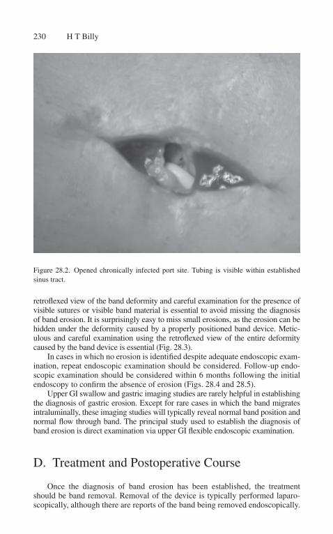
230 H T Billy
retrofl exed view of the band deformity and careful examination for the presence of visible sutures or visible band material is essential to avoid missing the diagnosis of band erosion. It is surprisingly easy to miss small erosions, as the erosion can be hidden under the deformity caused by a properly positioned band device. Metic-ulous and careful examination using the retrofl exed view of the entire deformity caused by the band device is essential (Fig. 28.3).
In cases in which no erosion is identifi ed despite adequate endoscopic exam-ination, repeat endoscopic examination should be considered. Follow-up endo-scopic examination should be considered within 6 months following the initial endoscopy to confi rm the absence of erosion (Figs. 28.4 and 28.5).
Upper GI swallow and gastric imaging studies are rarely helpful in establishing the diagnosis of gastric erosion. Except for rare cases in which the band migrates intraluminally, these imaging studies will typically reveal normal band position and normal fl ow through band. The principal study used to establish the diagnosis of band erosion is direct examination via upper GI fl exible endoscopic examination.
D. Treatment and Postoperative Course
Once the diagnosis of band erosion has been established, the treatment should be band removal. Removal of the device is typically performed laparo-scopically, although there are reports of the band being removed endoscopically.
Figure 28.2. Opened chronically infected port site. Tubing is visible within established sinus tract.

28. Gastric Erosion Following Adjustable Gastric Banding 231
Figure 28.3. Eroded Lap Band, retrofl exed view. Note that the endoscope does not pass within the ring of the device as a result the band is nonfunctional and creates no restrictive mechanism.
Figure 28.4. Subtle erosion barely visible except for hint of suture material.

232 H T Billy
Supporters of endoscopic removal advocate waiting until intragastric migration of the band has occurred in order to remove the device endoscopically. Intra-gastric migration of the band is not a common event and the degree of erosion is not extensive enough to allow successful endoscopic removal of the device in most patients. The overwhelming majority of patients will benefi t from removal of eroded devices laparoscopically without unnecessary delay. Furthermore, the endoscopic removal of an eroded represents an advanced procedure which requires specialized instrumentation and experience. The recommended tech-nique for most erosions encountered will be to utilize a laparoscopic approach to explant the eroded device.
The steps of laparoscopic removal of an eroded adjustable gastric band are as follows:
1. Identifi cation of the lap band tubing 2. Localization of the adjustable gastric band buckle and transaction of
the band 3. Identifi cation and closure of the gastric erosion 4. Drainage of the site with a closed system drain
Laparoscopic removal of an eroded adjustable gastric band is a challenging pro-cedure. It is common to fi nd dense and tenacious adhesions between the omen-tum and abdominal wall, as well as between the left lobe of the liver and the anterior gastric wall. In order to remove the device safely and minimize operative
Figure 28.5. Same erosion as in Fig. 28.4, but with different endoscopic view reveals large visible erosion.

28. Gastric Erosion Following Adjustable Gastric Banding 233
dissection, identifi cation and location of the band tubing are essential. The tubing is typically encased with surrounding omentum and can be diffi cult to local-ize. Once identifi ed, the tubing can be transected and used as a guide to direct the operative dissection directly to the buckle of the adjustable gastric band. It is common to discover a severe infl ammatory reaction along the stomach wall adjoining the left lobe of the liver. These adhesions can be diffi cult to dissect but are progressively released, using the tubing as a guide until the cuff or buckle of the band is exposed and transected which allows easy removal of the device. Limited dissection in and around the buckle in order to free the buckle from sur-rounding tissue is all that is necessary to allow safe removal of the eroded band. There is no need to take down the gastric plication, and this excessive surgical dissection is discouraged. Once transected, the band can be easily removed by slipping it out from its perigastric position and removing it from the abdomen (Figs. 28.6 and 28.7).
The remaining gastric perforation should be located and closed. If possible, the defect is sutured in two layers and then evaluated for leaks. Installation of methylene blue dye mixed into a liter of saline into the gastric lumen by the anesthesiologist is suffi cient to identify any leakage at the site of the erosion. If identifi ed, the site of the leakage should be closed with endoscopic sutures. Addi-tional infl ammatory processes such as subphrenic abscess are relatively uncommon but, if present, should be drained. Drainage of the area with a closed drain should be performed to control any leakage that might develop postoperatively.
The postoperative course following removal of an eroded band should be unremarkable. A postoperative upper GI swallow is performed to rule out persistent
Figure 28.6. Dissection of eroded Lap Band buckle using tubing as a guide.

234 H T Billy
leakage from the erosion site and allow removal of the drain on the fi rst post-operative day. Most patients can be discharged on their fi rst postoperative day and diet can be resumed. We typically keep patients on a liquid diet for the fi rst 5 days following band removal before beginning to advance their diet. Removal of an eroded band is a well-tolerated procedure and rarely develops postopera-tive complications. Infectious complications are rare. Patients are maintained on postoperative antibiotics for several days following band removal.
Figure 28.7. Transection and removal of the eroded band.
Figure 28.8. Oversewing of site of gastric perforation.

28. Gastric Erosion Following Adjustable Gastric Banding 235
Although it is possible to replace an eroded band with a new replacement band simultaneously at the time of band removal, it is not a recommended practice. Most authors support addressing the issue in a delayed approach, postponing any consideration for band replacement or conversion to an alternative bariatric procedure for at least several months. Alternative bariatric procedures or band replacement can be considered at a later date and individualized for the specifi c needs of the individual patient.
E. Summary
Adjustable gastric band erosion is a rare complication, affecting fewer than 1% of patients undergoing this procedure. Erosion should be suspected in patients exhibiting signs of port site infection, unexplained loss of satiety, and nonfunc-tioning bands. The diagnosis is made endoscopically at the time of upper GI endoscopy, and requires careful endoscopic examination of the area.
Erosions can be classifi ed as occurring early and late. The mechanism of early erosion is most likely secondary to surgical trauma resulting in partial thickness injury of the gastric serosa. The presence of the foreign body allows for injury to continue. Band pressure and ischemia secondary to percutaneous adjustment may play a role in promoting development of these partial thickness injuries into full-thickness erosions. Aggressive postoperative adjustments may play a further role in promoting gastric ischemia in the areas around the band, although this has yet to be proved to be a contributing factor in band erosion. Erosions that occur late are thought to develop as a result of multiple factors; smoking, NSAID use, excessive operative dissection, and postoperative ischemia, as well as tight gastric plication, have all been postulated as causes for postoperative erosion. Previous bariatric and gastric procedures such as Nissen fundoplication may contribute to ischemia at the operative site and enhance the development of gastric erosion. The role of pressure necrosis following band adjustment has not been directly associated with the development of late erosions. Judicious use of postoperative adjustments and avoiding the temptation to perform a “large fi ll” may be benefi cial in decreasing the possible association of pressure necrosis and band erosion.
Erosion is rarely a surgical emergency and treatment can be deferred and scheduled electively. Defi nitive treatment of gastric erosion is band removal and can be accomplished laparoscopically in nearly every case, with most patients discharged less than 24 hours after admission.
F. Selected References
Abu-Abeid S, Keidar A, Gavert N, et al. The clinical spectrum of band erosion following laparoscopic adjustable silicone gastric banding for morbid obesity. Surg Endosc 2003;17: 861–863.
Belachew M, Belva PH, Desaive C. Long-term results of laparoscopic adjustable gastric banding for the treatment of morbid obesity. Obes Surg 2002;12:564–568.

236 H T Billy
Belachew M, Legrand MJ, Defechereux TH, et al. Laparoscopic adjustable silicone gastric banding in the treatment of morbid obesity: a preliminary report. Surg Endosc 1994;81354–1365.
Cadiere GB, Himpens J, Vertruyen M, et al. Laparoscopic gastroplasty (adjustable silicone gastric banding). Semin Laparosc Surg 2000;7:55–65.
Dargent J. Laparoscopic adjustable gastric banding: lessons from the first 500 patients in a single institution. Obes Surg 1999;9:446–452.
Favretti F, Cadiere G B, Segato G, et al. Lap-Band for the treatment of morbid obesity: a 6-year experience of 509 patients. Obes Surg 1999;9:327–329.
Favretti F, Cadiere GB, Segato G, et al. Laparoscopic banding: selection and technique in 830 patients. Obes Surg 2002;12:385–390.
Fielding GA, Rhodes M, Nathanson LK. Laparoscopic gastric banding for morbid obesity: surgical outcome in 335 cases. Surg Endosc 1999;13:550–554.
Meir E, Van Baden M. Adjustable silicone gastric banding and band erosion: personal experience and hypotheses. Obes Surg 1999;9:191–193.
Szold A, Abu-Abeid S. Laparoscopic adjustable silicone gastric banding for morbid obes-ity: results and complications in 715 patients. Surg Endosc 2002;16:230–233.
US Food and Drug Administration, Center for Devices and Radiological Health. LAP-BAND Adjustable Gastric Banding (LAGB) System—P000008. Available at http: //www.fda.gov/cdrh/pdf/p000008.htm. Accessed August 23, 2002.
Vertruyen M. Experience with LAP-BAND system up to 7 years. Obes Surg 2002;12:569–572.
Weiner R, Wagner D, Bockhorn H. Laparoscopic gastric banding for morbid obesity. J Laparoendosc Adv Surg Technique 1999;9:23–30.
Weiss H, Nehoda H, Labeck B, et al. Gastroscopic band removal after intragastric migra-tion of adjustable gastric band: a new minimal invasive technique. Obes Surg 2000;10:167–170.

A. Introduction
With the continued growth of bariatric surgery, there has been scrutiny of outcomes. Professional societies, insurance companies, and watch-dog organizations have developed guidelines for the best practices of bariatric surgery and the granting of privileges to qualifi ed surgeons to perform bariatric procedures.
The Society of American Gastrointestinal and Endoscopic Surgeons (SAGES) has a history of authoring privileging and practice guidelines for the promotion of patient safety and improvement of clinical outcomes. Currently, there are 15 such guidelines, ranging from the granting of privileges for gastrointestinal endoscopy to the use of deep venous thrombosis prophylaxis in laparoscopic surgery.
In July 2003, the Board of Governors of SAGES approved the Guidelines for Institutions Granting Bariatric Privileges Utilizing Laparoscopic Techniques. The statement was originally prepared jointly with members of the American Society of Bariatric Surgery (ASBS), now the American Society of Metabolic and Bariatric Surgery (ASMBS). Since then, the ASMBS has published its own guidelines on the granting of privileges for bariatric procedures.
B. The SAGES Guidelines for Institutions Granting Bariatric Privileges Utilizing Laparoscopic Techniques
In the SAGES guideline for the clinical application of laparoscopic bariatric surgery, which was published concurrently with its privileging guidelines, laparoscopic as well as open weight loss procedures were estab-lished as durable treatments for morbid obesity as long as practiced by appropriately trained surgical teams within programs that provide perioperative and long-term management. The indications for weight loss surgery were derived from the 1991 National Institutes of Health Consensus Conference for the surgical management of morbidly obese patients. More recently, the
29. Society of American Gastrointestinal and Endoscopic Surgeons (SAGES) Bariatric Credentialing Guidelines
Shawn Tsuda and Daniel B. Jones

240 S Tsuda and D B Jones
2003 SAGES Appropriateness Conference restated the appropriateness of duodenal switch, banded gastroplasty, laparoscopic adjustable band, and laparoscopic gastric bypass based on current evidence. The SAGES Guidelines for Institutions Granting Bariatric Privileges Utilizing Laparoscopic Techniques addresses who should perform bariatric procedures by stating the purpose, defi nitions, training requirements, institutional support, and maintenance of privileges that would facilitate local hospital staffs to ensure high quality care with a systems-based approach.
1. Purpose
SAGES recommends that their guidelines be used by health care institutions and hospital administrations as an adjunct to the Joint Commission on Accreditation of Healthcare Organizations (JCAHO) to implement privileges for bariatric surgery. In contrast to guidelines developed by the Betsy Lehman Center, ASMBS, and ACS, which establish volume criteria for privileging that range between 25 and 50 weight loss procedures, SAGES states that the decision to grant privileges should not be based solely on the number of procedures per-formed. SAGES places the actual privileging structure on individual institutions, through a process that begins with the chief of surgery and is approved by a com-mittee, board, or other governing body.
2. Defi nitions
The SAGES guidelines for bariatric privileging establishes nomenclature for mandatory recommendation (MUST/SHALL), highly desirable recommendation (SHOULD), optional recommendations (MAY/COULD), documentation of training, privileging, credentialing, competence, complete procedural conduct (ability to perform the operation from patient selection to open conversion if required), laparoscopy, types of bariatric procedures, and what constitutes a formal course of training.
As stated, SAGES places the adequacy (volume number) of the surgeon’s experience on the individual institutions or the chief of surgery. Formal residency and/or fellowship training should be structured, but case numbers are not speci-fi ed. For surgeons without formal training, preceptorship/proctorship and formal courses are offered as alternatives. A preceptor or proctor should be determined by the institution, be familiar with all aspects of the procedure, including patient selection, and be chosen in an unbiased, confi dential, and objective manner. A formal course is not offered as adequate training to perform a procedure independently, but should be done for procedures for which privileges are being sought. Such courses must meet category 1 Continuing Medical Education requirements, include didactics by qualifi ed surgeons, and involve hands-on experience with animate and/or inanimate models. Adjunctive aids include video reviews or computer programs. Examples are the SAGES Grand Rounds DVD for bariatric surgery or the SAGES Pearls series for laparoscopic Roux-en-Y gastric bypass.

29. SAGES Bariatric Credentialing Guidelines 241
C. Minimum Requirements for Privileging of Bariatric Procedures
The recommended mandatory requirements by SAGES consist of: (1) formal residency training in general surgery within an accredited program with subsequent certifi cation by the American Board of Surgery if required by the institution; and (2) documentation that there exists adequate follow-up of patients, including nurs-ing care, dietary care, counseling, support groups, exercise training, psychological care if needed, and a method of identifying and managing complications.
If the surgeon has formal training only in open bariatric surgery, SAGES recommends having a second surgeon who is trained in laparoscopic bariatric surgery, and is therefore complementary to their expertise. Alternatively, the surgeon may participate in a proctored experience deemed adequate by the chief of surgery.
If the surgeon has documented formal training in laparoscopic bariatric surgery, SAGES recommends that the volume of open and laparoscopic cases be demonstrated for the type of procedures to be done, and that a complementary surgeon experienced in open procedures be available if needed. The adequacy of case volume/experience is to be determined by the chief of surgery.
If the surgeon has no documented formal residency training in either laparo-scopic open bariatric surgery, they are expected to take a formal course AND be proctored by a qualifi ed surgeon who is approved by the institution of practice.
D. Institutional Support
Adequate equipment and staff training are expected to be in place prior to starting a bariatric program. Two skilled surgeons are recommended for laparoscopic bariatric procedures, or a surgeon and a skilled fi rst assistant.
E. Maintenance of Privileges
SAGES recommends that the chief of surgery or appropriate institutional body should determine the criteria for provisional privileges, monitoring of performance and outcomes, and continuing education requirements (meetings and courses). The guideline states that outcome data should be reviewed after 6 months of the granting of privileges, and regularly thereafter as compared with published national bench-marks. Any denial of privileges should have an appeal process in place.
F. Selected References
ASBS Bariatric Training Committee. American Society for Bariatric Surgery Guidelines for Granting Privileges in Bariatric Surgery. Surg Obes Rel Dis 2006;2(1):65–67.

242 S Tsuda and D B Jones
Bessler M, Champion K, Cohen R, et al. Laparoscopic Roux-en-Y gastric bypass: SAGES pearls. Cine-Med 2005.
Gagner M, Ren C, Flum D, et al. SAGES grand rounds: bariatric Surgery. CineMed. 2007.Guidelines for the Clinical Application of Laparoscopic Bariatric Surgery. http://www.
sages.org/sagespublicationprint.php?doc=30. Accessed June 25,2006.Jones DB, DeMaria E, Provost DA, et al. Optimal management of the morbidly obese patient:
SAGES appropriateness conference statement. Surg Endosc 2004;18(7):1029–1037.Kelly J, Shikora S, Jones DB, et al. Best practice updates for surgical care in weight loss
surgery. Obes Res 2007; in press.SAGES. Guidelines for institutions granting bariatric privileges utilizing laparoscopic
techniques. Surg Endosc 2003;17:2037–2040.Schirmer B, Jones DB. American College of Surgeons Bariatric Surgery Center Network.
ACS Bull 2007;92(8):21–27.Schirmer BD, Schauer PR, Flum DR, et al. Bariatric surgery training: getting your ticket
punched. J Gastrointe Surg 2007;11:807–812.
A. Introduction
Morbid obesity is rapidly increasing globally and is the leading cause of preventable cause of death in the United States. The only effective and enduring treat-ment for morbid obesity remains bariatric surgery. Both the growth in the preva-lence of obesity and the success of weight loss surgery has led to truly spectacular growth in utilization of bariatric surgery. Outcomes have driven both the rise and fall of bariatric surgery over time. A clear reason for the recent growth of bariatric surgery was the 2004 approval of weight loss surgery by the Centers for Medicare and Medicaid Services, which was driven by outcomes studies. In the 1970s, seri-ous adverse outcomes associated with jejunal-ileal bypass led to its ultimate demise, uniquely becoming the only operation to be banned by the FDA. Currently, both strong consumer and insurer interest in weight loss surgery and increased societal awareness of patient safety have directed intense interest in the outcomes of bariatric surgery. Standardization of outcomes reporting in bariatric surgery has long been an issue of contention; however, recent events have demonstrated that bariatric surgery is entering a watershed in outcomes reporting.
B. Outcomes Defi nitions
Defi ning the problem of obesity and its current solution of surgery is paramount. Obesity is currently defi ned by the National Heart, Lung, and Blood Institute as body mass index (BMI) in four classes: (I) 30–35, (II) 35–40, (III) 40–50, and (IV) >50. BMI has been clearly recognized as the best measure of obesity, surpassing cumbersome measurements such as fat con-tent, waist/hip circumference, or percent of weight over ideal weight. Anthropometric measurements have the disadvantage of recorder variability while ideal weight provides an elusive defi nition. BMI has strong correlation to certain diseases such as type 2 diabetes. In fact, BMI alone may be suffi -cient in documenting the presence of insulin resistance and metabolic syn-drome instead of using the ATP III criteria. By using BMI as a marker for obesity, the Centers for Disease Control have documented a phenomenal growth in obesity in the United States.
30. Reporting of Bariatric Surgery Outcomes
Gavitt A. Woodard and John M. Morton

244 G A Woodard and J M Morton
Correspondingly, weight loss surgery has grown in response to this public health concern. Since 1997, bariatric surgery in the United States has grown sevenfold, with gastric bypass remaining the most common procedure. In the remainder of the world, adjustable gastric banding is the leading weight loss surgery. Defi ning these weight loss procedures appears straightforward, with each falling into the realms of restrictive, malabsorptive, or a combination of the two. These general categories will most likely be amended with the advent of new technology, such as vagal nerve stimulation or NOTES procedures. Prospectively maintained databases for new technologies or procedures such as the NOSCAR database will allow for better recognition of effi cacy and safety for these procedures. Furthermore, even well-established procedures such as gastric bypass have signifi cant variation, which may render comparisons tenuous. For example, a gastric bypass has several points of disparity, such as open vs. laparoscopic, antecolic vs. retrocolic, sparing of vagal branches, limb length, etc. The landmark 1991 National Institutes of Health (NIH) Consensus Conference recognized some of these inherent diffi culties when they wrote, “One of the key problems in the current reports of case series in surgical therapy is the lack of standards for comparison.”
C. Data Sources
Many efforts have been made to collect data for bariatric surgery through individual surgeons, surgical societies, or research initiatives, including the International Bariatric Surgery Registry, Society for Advanced Gastrointestinal Endoscopic Surgery (SAGES) Outcomes Initiative, Swedish Obese Subjects Trial, and the ongoing NIH Longitudinal Assessment of Bariatric Surgery (LABS) trial. Bariatric surgery outcomes have also been reported from existing administrative claims data, such as Medicare, Nationwide In-Patient Sample (NIS), and University Health Consortium (UHC).
A PubMed query including the search terms of surgery, weight loss, and outcomes yielded 465 results. Evaluation of these different data sources should rest on evidence-based criteria. For example, prospective, clinically derived data through standardized defi nitions are clearly ideal. However, prospective data take time to collect and emerging trends of care may not be scrutinized until deleterious effects have already taken place. Administrative claims data, such as exists in Medicare, NIS, or UHC, provide the advantage of easily obtainable data in large numbers that are multi-institutional. In addition, for certain rare proce-dures, such as adolescent bariatric surgery, administrative databases may be the only means of determining procedure trends in sizable numbers. Administrative, claims data certainly supports the proverb, “You search where there is light.” However, these databases can provide confl icting results. These data are not obtained for clinical practice but for billing purposes. The clear shortcomings of administrative claims data are that they are defi cient in clinical data, such as BMI, which are integral for risk adjustment. Administrative claims data do render the essential function of “a canary in a coal mine.” While these claims data do not always render a defi nitive conclusion, they can serve as an early warning system regarding emerging trends in practice patterns and outcomes.

30. Reporting of Bariatric Surgery Outcomes 245
D. Center of Excellence Movement
The infl uence of administrative claims data was clearly seen with the 2005 study which found a 1-year 4.6% all-cause mortality rate for Medicare patients undergoing gastric bypass. The study had the advantages of being multi-institu-tional and having large numbers; however, it lacked robust risk adjustment, applica-bility of results to the general gastric bypass population, and specifi city of cause of death. Despite those limitations, it did demonstrate an important association between surgeon volume and outcomes. These results of higher than expected mortality and positive volume-outcome correlation led to the Centers of Medicare and Medicaid Services (CMMS) mandating two requirements: a prohibition of gastric bypass in Medicare recipients over 65 years of age and an obligation that weight loss surgery be performed at high-volume Centers of Excellence (COE) as defi ned by the two certifying bodies of the American Society of Bariatric and Metabolic Surgery (ASMBS) and American College of Surgeons (ACS). While the prohibition of weight loss surgery in Medicare recip-ients has been overturned, the Center of Excellence initiative has remained in force and gathered strength. Many other payers have followed CMMS’s lead in requiring that weight loss surgery be performed by high-volume surgeons, lead-ing to a preponderance of gastric bypass procedures being performed in high-volume centers. With the advent of the ASMBS and ACS databases, bariatric sur-gery has entered a new era in outcomes reporting. As the offi cial databases for bariatric surgery, there is a heightened interest in the cost and components of the databases as well as a hope that a single standard will be achieved.
One constant in data collection is change. All databases should be organic enough to either grow or diminish over time. There is an essential creative ten-sion in outcomes collection between the desire for covering every aspect of data and the need for brevity to ensure completion of data. A bariatric surgery database should have ideal components and means of collection. Ideally, the data should be collected prospectively and audited by trained clinicians with standardized defi ni-tions entered via the web to allow for ease and speed of entry. Clearly, these means of data collection are costly and cannot be borne by the individual surgeon but should be a hospital-based responsibility to achieve and maintain COE status.
E. Ideal Bariatric Surgery Database
An ideal bariatric surgery database should include the following: patient demographics, hospital characteristics, surgical outcomes, and long-term follow-up. Patient demographics include age, gender, BMI, comorbidities, functional status, quality of life, and socioeconomic status. As mentioned, BMI appears to be a suffi cient measure for weight and avoids the variety of reporting currently seen for ideal body weight. A study by Dixon found a tremendous lack of standardiza-tion for ideal weight. However, BMI may not be appropriate in the adolescent population, in which growth in height is expected and age percentile of weight would be more appropriate. A clear standard for weight loss needs to be set and should not be based on ideal weight given the elusiveness of that ideal.

246 G A Woodard and J M Morton
Comorbidity defi nition and documentation are essential for both risk adjustment of outcomes as well as accurate assessment of resolution. The effects of morbid obesity upon body systems are legion and profound. Ali and colleagues per-formed a detailed taxonomy of the common comorbidities associated with obesity that may serve as an ideal comorbidity scale to embrace. Some comorbidities may defy succinct or concrete defi nition such as musculoskeletal impairment, whereas others such as lipid abnormalities have clear consensus. It has been demonstrated that obesity is an infl ammatory disease and that infl ammation markers such as high-sensitivity C-reactive protein improve dramatically after surgery. As result, clinically defi ned comorbidities may be used in conjunction with biochemical markers to fully defi ne the penumbra of obesity-related diseases.
Furthermore, hospital characteristics should be described in the classic Donabedian model of quality: structure, process, and outcomes. The structural measures of bariatric surgery quality should include the following: Center of Excellence designation, volume of procedures performed, fellowship-trained bari-atric surgeons, and multidisciplinary specialists in anesthesia, gastroenterology, nutrition, and psychology. Process measures of bariatric surgery quality might include adherence to Surgery Care Improvement Project (SCIP) measures, selective use of IVC fi lters, and clinical pathways. Finally, outcomes measures of quality may include weight loss, morbidity including reoperations/readmissions, and mortality. Mortality should be represented as 30 day, 90 day, 1 year, and 5 year to fully comprehend the impact of weight loss surgery upon survival. A telling point in the land mark Adams’ Utah study on gastric bypass and survival demonstrated that the risk of mortality by maintaining a morbidly obese status in the control group was equivalent to the mortality risk of surgery in the fi rst year.
F. Outcomes: Weight Loss
The degree of weight loss associated with bariatric surgery is dependent upon which operation is employed. Recently, several studies have provided considerable information regarding weight loss after surgery. In the Swedish Obese Subjects Study, which compared medically and surgically treated obese patients, there were striking differences between the two groups regarding weight loss. At 10 years the conventionally treated group had a 1.6% increase in total weight, while the surgically treated group saw a 24% decrease in total weight. Among the different surgical procedures performed in the Swedish Obese Subjects (SOS) Study, the highest amount of total weight loss was for gastric bypass (25%), followed by vertical banded gastroplasty (17%), and adjustable gastric banding (13%).
Two important studies have reviewed the entire published medical literature regarding bariatric surgery. These two meta-analyses provide strong validation for the effi cacy of weight loss surgery. The outcomes after surgically induced weight loss published in the last years are impressive. The meta-analysis by Buchwald found that the degree of weight loss is lowest for restrictive proce-dures and highest for malabsorptive procedures. In this meta-analysis of 22,094 patients (mean age: 47 years, mean BMI 46.9, 72.6 % women), the mean per-centage of excess weight loss was 61.2% for all patients. Excessive weight loss was higher for patients who underwent for gastric bypass (61.6%) or gastroplasty

30. Reporting of Bariatric Surgery Outcomes 247
(68.2%) compared with those who received gastric banding (47.5%). Similar results for weight loss were reported by Maggard in the Annals of Internal Medicine. In this study, weight loss in kilograms for at least 3 years postoperatively was as follows: gastric banding (35 kg), gastroplasty (32 kg), biliopancreatic diversion/duodenal switch (53 kg), and gastric bypass (42 kg).
G. Outcomes: Comorbidity Resolution
Weight loss surgery is a singular medical intervention that has the unique ability to reverse or improve the numerous medical conditions associated with obesity. The leading cause of death in the United States remains heart disease and stroke, with 300,000 patients dying annually. The primary medical conditions contributing to this devastating human toll are diabetes, hypertension, and hyper-lipidemia. These three comorbidities, along with visceral obesity, constitute the metabolic syndrome that is a strong risk factor for cardiovascular mortality. All three of these cardiovascular risk factors improved as described in an important Annals of Surgery publication by Schauer et al. regarding laparoscopic gastric bypass with the following resolution rates: diabetes (82%), hypertension (70%), and hyperlipidemia (63%).
The SOS study provided further demonstration of the ameliorative effect of bariatric surgery. At 10 years in the SOS study, surgically treated obese patients had substantial improvements in their cardiac risk factors, in comparison with nonsurgically treated obese patients. Surgically treated obese patients had 25% reduction in hypertension, 43% improvement in HDL, and 75% reduction in diabetes in comparison with the medically treated group. Another study found clear, consistent, and convincing resolution of every conventional risk factor at 1 year after gastric bypass surgery.
Gastric bypass has assembled the most evidence for resolution of comorbidi-ties. However, all weight loss surgeries promote resolution of these important medical problems. Each procedure has a different degree of improvement for different comorbidities as demonstrated by the JAMA article by Buchwald. For diabetes, each procedure has the following resolution rate: banding (48%), gas-troplasty (68%), gastric bypass (84%), and biliopancreatic diversion/duodenal switch (98%). Hyperlipidemia is another cardiac risk factor that is improved by each weight loss surgery by different amounts: banding (71%), gastroplasty (81%), gastric bypass (94%), and biliopancreatic diversion/duodenal switch (99%). Finally, hypertension is also resolved by the following procedures at the accompanying rates: banding (38%), gastroplasty (73%), gastric bypass (75%), and biliopancreatic diversion/duodenal switch (81%).
Beyond the tremendous improvement in cardiac risk factors, weight loss surgery also provides enormous enhancement of the myriad medical problems that obesity engenders. An important health concern associated with obesity is sleep apnea, which carries signifi cant health risks, including premature death. Fortunately, weight loss surgery also improves sleep apnea as demon-strated by Buchwald: banding (95%), gastroplasty (77%), gastric bypass (87%), and biliopancreatic diversion/duodenal switch (95%). Joint disease is a leading health concern that severely decreases both personal satisfaction and

248 G A Woodard and J M Morton
work productivity. Schauer demonstrated 88% resolution or improvement of joint disease for morbidly obese patients undergoing laparoscopic gastric bypass.
In addition, the leading digestive health complaint, gastroesophageal refl ux disease, is cured or improved at a 96% rate. The most prevalent liver disease, nonalcoholic fatty liver disease, is very commonly associated with morbid obesity and is also very often improved by weight loss surgery.
Furthermore, weight loss surgery has been demonstrated to either eliminate or improve the following health conditions: venous stasis disease, gout, asthma, psuedotumor cerebri, urinary incontinence, and infertility.32
H. Outcomes: Quality of Life
Quality of life in the morbidly obese patient is clearly diminished due to self-image, economic discrimination, lack of medical access, and societal lack of accept-ance. Of note, weight loss surgery is also equally powerful in reversing poor quality of life as it is in resolving medical problems. There is ample evidence to demon-strate improvement in quality of life after weight loss surgery. The most accepted survey in medicine for quality of life is the SF36, which has been demonstrated to improve sharply after bariatric surgery. Also, disease-specifi c quality of life instruments have also shown improvement after surgery. Finally, depression is a disease that can severely impair quality of life. A measure of depression, Beck Depression Index, has been demonstrated to decline by half following weight loss surgery.
I. Outcomes: Cost-Effectiveness
Given the dramatic effects upon comorbidities that bariatric surgery renders, it is apparent that bariatric surgery can also provide reduction in health care costs. This reduction in health care costs has been demonstrated in the province of Quebec, where surgically treated patients incurred $CN6,000 less in health care costs versus morbidly obese patients treated nonsurgically. Two cost- effectiveness analyses demonstrated that bariatric surgery is a dominant strategy with a range of $5,000 to $35,600 per Quality Adjusted Life Year (QALY). Both studies show that bariatric surgery provides a QALY at lower rate than $50,000, the common marker of cost-effectiveness.
J. Outcomes: Survival
Weight loss and comorbidity resolution each contribute to increased survival for the surgically treated morbidly obese patient. Several studies have demon-strated improved survival after gastric bypass surgery. Flum et al. noted a 33% reduction in mortality for morbidly obese patients who underwent surgery versus

30. Reporting of Bariatric Surgery Outcomes 249
morbidly obese patients who were treated medically. McDonald et al. also pro-vided clear evidence for a survival benefi t in the surgically treated morbidly obese patient. This retrospective analysis of type 2 diabetic patients with morbid obesity, who underwent either gastric bypass operation or did not undergo surgery, dem-onstrated a mortality rate of only 9% in the surgical group during the 9-year fol-low-up compared with 28% in the nonsurgical control group. A decisive conclu-sion regarding the survival benefi t conferred by gastric bypass surgery is provided by a population-based study from Quebec, Canada. The mortality rate in the bariatric surgery cohort was 0.68% compared with 6.17% in controls, which translated to a tremendous 89% reduction in the relative risk of death.
Finally, two recent articles in the New England Journal of Medicine conclu-sively demonstrated the survival benefi t engendered by bariatric surgery. Sjostrom and colleagues found in the SOS trial, a hazard ratio of 0.71 demonstrating a reduction in mortality for surgically treated morbidly obese patients versus con-trols. The SOS incorporated three different surgical techniques provided 10-year follow-up data. Adams et al. also showed a decided survival benefi t after gastric bypass surgery. From the state of Utah, 7,925 surgical patients and 7,925 severely obese control subjects were matched for age, sex, and body mass index. During a mean follow-up of 7 years, adjusted long-term mortality from any cause in the surgery group decreased by 40%, as compared with that in the control group. In addition, cause-specifi c mortality in the surgery group decreased by 56% for cor-onary artery disease, 92% for diabetes, and 60% for cancer. However, rates of death not caused by disease, such as accidents and suicide, were 58% higher in the surgery group than in the control group. This latter fi nding of increased deaths not caused by disease was not present in the SOS study, in which gastric bypass was not predominant. A common denominator to both accidents and suicides is alcohol abuse: It has been demonstrated that gastric bypass profoundly alters alcohol metab-olism. There are clear and consistent data to support the fact that bariatric surgery renders a survival benefi t to the morbidly obese patient.
K. Conclusions
While more work needs to be done with data collection, defi nitions, and standards, bariatric surgery is a sterling example of outcomes driven care. For the fi rst time in US surgical history, the government mandated a level of performance for a surgical specialty. The bariatric surgery community met that challenge of federal oversight with enthusiasm and results. In a challenging population of patients with multiple medical problems, effective and enduring care is provided. Outcomes have demonstrated that in the right hands and in the right patients, bariatric surgery is a life-saving procedure.
L. Selected References
Adams TD, Gress RE, Smith SC, et al. Long-term mortality after gastric bypass surgery. NEJM 2007;357(8):753–761.

250 G A Woodard and J M Morton
Alhassan S, Kiazand A, Balise RR, et al. Metabolic syndrome: do clinical criteria identify similar individuals among overweight premenopausal women? Metabolism 2008;57(1):49-56.
Ali MR, Maguire MB, Wolfe BM. Assessment of obesity-related comorbidities: a novel scheme for evaluating bariatric surgical patients. J Am Coll Surg 2006;202(1):70–77.
Belle SH, Berk PD, Courcoulas AP, et al. Safety and efficacy of bariatric surgery: longitu-dinal assessment of bariatric surgery. Surg Obes Relat Dis 2007;3(2):116–126.
Best M, Neuhauser D. Avedis Donabedian: father of quality assurance and poet. Qual Saf Health Care 2004;13(6):472–473.
Birkmeyer NJ, Wei Y, Goldfaden A, et al. Characteristics of hospitals performing bariatric surgery. JAMA 2006;295(3):282–284.
Bosy-Westphal A, Geisler C, Onur S, et al. Value of body fat mass vs anthropometric obes-ity indices in the assessment of metabolic risk factors. Int J Obes (Lond) 2006;30(3):475–483.
Buchwald H, Williams SE. Bariatric surgery worldwide 2003. Obes Surg 2004;14(9):1157–1164.
Buchwald H. The future of bariatric surgery. Obes Surg 2005;15(5):598–605.Centers for Medicare and Medicaid Services. Decision memo for bariatric surgery for the
treatment of morbid obesity (CAG-00250R). Available at http://www.cms.hhs.gov. Accessed February 2008.
Christou NV, Sampalis JS, Liberman M, et al. Surgery decreases long-term mortality, morbidity, and health care use in morbidly obese patients. Ann Surg 2004;240(3):416–423; discussion 23–24.
Clegg A, Colquitt J, Sidhu M, et al. Clinical and cost effectiveness of surgery for morbid obesity: a systematic review and economic evaluation. Int J Obes Relat Metab Disord 2003;27(10):1167–1177.
Consensus Development Conference Panel. Gastrointestinal surgery for severe obesity. Ann Int Med 1991;115(12):956–961.
Craig BM, Tseng DS. Cost-effectiveness of gastric bypass for severe obesity. Am J Med 2002;113(6):491–498.
Dixon JB, Dixon ME, O’Brien PE. Depression in association with severe obesity: changes with weight loss. Arch Int Med 2003;163(17):2058–2065.
Dixon JB, McPhail T, O’Brien PE. Minimal reporting requirements for weight loss: cur-rent methods not ideal. Obes Surg 2005;15(7):1034–1039.
Dymek MP, Le Grange D, Neven K, et al. Quality of life after gastric bypass surgery: a cross-sectional study. Obes Res 2002;10(11):1135–1142.
Flum DR, Dellinger EP. Impact of gastric bypass operation on survival: a population-based analysis. J Am Coll Surg 2004;199(4):543–551.
Flum DR, Salem L, Elrod JA, et al. Early mortality among Medicare beneficiaries under-going bariatric surgical procedures. JAMA 2005;294(15):1903–1908.
Griffen WO Jr, Young VL, Stevenson CC. A prospective comparison of gastric and jejunoileal bypass procedures for morbid obesity. Ann Surg 1977;186(4):500–509.
Hagedorn JC, Encarnacion B, Brat GA, et al. Does gastric bypass alter alcohol metabo-lism? Surg Obes Relat Dis 2007;3(5):543–548; discussion 8.
http://www.ncbi.nlm.nih.gov.laneproxy.stanford.edu/sites/entrez. Accessed April 12, 2008.

30. Reporting of Bariatric Surgery Outcomes 251
http://www.nhlbi.nih.gov/guidelines/obesity/ob_home.htm. Accessed April 12, 2008.http://www.sages.org/projects/outcomes/index.php. Accessed April 12, 2008.Klein S, Mittendorfer B, Eagon JC, et al. Gastric bypass surgery improves metabolic and
hepatic abnormalities associated with nonalcoholic fatty liver disease. Gastroenterology 2006;130(6):1564–1572.
Lean ME, Powrie JK, Anderson AS, et al. Obesity, weight loss and prognosis in type 2 diabetes. Diabet Med 1990;7(3):228–233.
MacDonald KG Jr, Long SD, Swanson MS, et al. The gastric bypass operation reduces the progression and mortality of non-insulin-dependent diabetes mellitus. J Gastrointest Surg 1997;1(3):213–220; discussion 20.
Maggard MA, Shugarman LR, Suttorp M, et al. Meta-analysis: surgical treatment of obes-ity. Ann Intern Med 2005;142(7):547–559.
Nguyen NT, Goldman C, Rosenquist CJ, et al. Laparoscopic versus open gastric bypass: a randomized study of outcomes, quality of life, and costs. Ann Surg 2001;234(3):279–289; discussion 89–91.
Nguyen NT, Morton JM, Wolfe BM, et al. The SAGES Bariatric Surgery Outcome Initiative. Surg Endosc 2005;19(11):1429–1438.
Ogden CL, Carroll MD, Curtin LR, et al. Prevalence of overweight and obesity in the United States, 1999-2004. JAMA 2006;295(13):1549–1555.
Pratt GM, McLees B, Pories WJ. The ASBS Bariatric Surgery Centers of Excellence pro-gram: a blueprint for quality improvement. Surg Obes Relat Dis 2006;2(5):497–503.
Russell TR. Yes, we can all get along: American College of Surgeons’ response to edito-rial. Surg Obes Relat Dis 2006;2(5):567–568.
Sampalis JS, Liberman M, Auger S, et al. The impact of weight reduction surgery on health-care costs in morbidly obese patients. Obes Surg 2004;14(7):939–947.
Santry HP, Gillen DL, Lauderdale DS. Trends in bariatric surgical procedures. JAMA 2005;294(15):1909–1917.
Schauer PR, Ikramuddin S, Gourash W, et al. Outcomes after laparoscopic Roux-en-Y gastric bypass for morbid obesity. Ann Surg 2000;232(4):515–529.
Sjostrom L, Lindroos AK, Peltonen M, et al. Lifestyle, diabetes, and cardiovascular risk factors 10 years after bariatric surgery. NEJM 2004;351(26):2683–2693.
Sjostrom L, Narbro K, Sjostrom CD, et al. Effects of bariatric surgery on mortality in Swedish obese subjects. NEJM 2007;357(8):741–752.
Trus TL, Pope GD, Finlayson SR. National trends in utilization and outcomes of bariatric surgery. Surg Endosc 2005;19(5):616–620.
Varela JE, Hinojosa MW, Nguyen NT. Perioperative outcomes of bariatric surgery in adolescents compared with adults at academic medical centers. Surg Obes Relat Dis 2007;3(5):537–540; discussion 41–42.
Williams DB, Hagedorn JC, Lawson EH, et al. Gastric bypass reduces biochemical cardiac risk factors. Surg Obes Relat Dis 2007;3(1):8–13.
Wolfe BM, Morton JM. Weighing in on bariatric surgery: procedure use, readmission rates, and mortality. JAMA 2005;294(15):1960–1963.
Zhang W, Mason EE, Renquist KE, et al. Factors influencing survival following surgical treatment of obesity. Obes Surg 2005;15(1):43–50.
A. Introduction
In 2003, as weight loss surgeries were being performed in increasing numbers, the media and public had signifi cant concerns about the safety of these procedures. At that time, the Massachusetts Department of Public Health charged the Betsy Lehman Center for Patient Safety and Medical Error Reduction with the task of assessing weight loss surgery and developing evidence-based best practice recommendations for the safe and effective performance of these proce-dures. An expert panel on weight loss surgery was convened in 2004, and the resulting recommendations were published in Obesity Research in 2005. Three years later, the Betsy Lehman Center reconvened the Expert Panel on Weight Loss Surgery to update the initial recommendations based on the signifi cant body of literature that had been published in the interim.
The best practice recommendations published by the Betsy Lehman Center have set the standard for weight loss surgery across the state of Massachusetts, and across the nation. The report was abstracted by the Agency for Healthcare Research and Quality (AHRQ) and adopted as the blueprint for the American College of Surgeons Bariatric Surgery Center Network accreditation program, which is recognized by the Centers for Medicare and Medicaid Services.
B. Methodology
The Betsy Lehman Center Expert Panel of Weight Loss Surgery consists of a multidisciplinary expert panel and 11 task groups. The expert panel and task groups were made up of members from all aspects of the care of the obese patient, including patients, surgeons, nurses, anesthetists, psychologists, psychiatrists, ethicists, nutritionists, internal medicine doctors, gastroenterologists, pediatricians, research methodologists, hospital administrators, as well as representatives from the insurers, Medicaid, the state board of registration in medicine, and the Department of Public Health.
A systematic review of the literature was performed, and relevant articles were abstracted using standardized data extraction tables. The available data were ranked according to levels of evidence based on study design (Table 31.1). Prospective, randomized controlled trials, or meta-analyses of such trials are
31. The Betsy Lehman Center Guidelines for Weight Loss Surgery
Matthew M. Hutter

254 M M Hutter
given the highest level of evidence—Category A. Prospective cohort studies or case-control studies are considered Category B. Retrospective cohort studies or case series are considered Category C. For the areas in which there was no credible literature, recommendations were based on expert opinion—Category D.
All recommendations were vetted in an open forum, through a series of meetings, and edits were made until all recommendations were unanimously agreed upon by the expert panel in a formal voting process. Recommendations and supporting references were then published in a peer-reviewed journal.
C. Betsy Lehman I
Convened in 2004, the initial Betsy Lehman Center Expert Panel on Weight Loss Surgery consisted of 24 expert panel members and 9 task groups. Twelve articles were published in a special supplement to Obesity in February 2005, which comprised 102 manuscript pages and a total of 603 literature citations.
Specifi c recommendations are too numerous to list here, but included recom-mendations for the multidisciplinary care of the weight loss surgical patient, including criteria for surgeon and hospital credentialing, as well as preoperative, intraoperative, and postoperative care recommendations for all aspects of the care of the weight loss surgical patient.
D. Impact of Betsy Lehman I
The recommendations were not only published in a peer-review journal, the fi ndings were abstracted by AHRQ and served as the basis for an international symposium on Patient Safety in Obesity Surgery held in Boston in July 2005. This symposium was subsequently developed into a DVD-based education
Table 31.1. Grading system for evidence-based recommendations.
Category A Evidence obtained from at least one well-conducted randomized clinical trial or a systematic review of all relevant RCTs
Category B Evidence from well-conducted prospective cohort studies,registry or meta-analysis of cohort studies, or population-based case-control studies
Category C Evidence obtained from uncontrolled or poorly controlled clinical trials, or retrospective case-control analyses, cross-sectional studies, case series, or case reports
Category D Evidence consisting of opinion from expert panels or the clinical experience of acknowledged authorities
Adapted from the criteria used by the US Preventive Services Task Force (USPSTF) and the American Diabetes Association.

31. The Besty Lehman Center Guidelines for Weight Loss Surgery 255
program and a book. The recommendations have defi ned the standard of care for weight loss surgery in Massachusetts. Certain programs in Massachusetts have ceased to offer weight loss surgery, whereas others have started up in compliance with the recommendations. Since 2004, accreditation programs have been devel-oped by the American Society of Metabolic and Bariatric Surgery/Surgical Review Corporation and the American College of Surgeons. Many of the criteria for certifi cation by these programs are similar, if not the same as the Lehman Center recommendations. The Centers for Medicare and Medicaid Services made a national coverage decision on weight loss surgery in February 2006 in which they will only cover WLS carried out in facilities that meet accredited standards of excellence developed by these organizations.
E. Betsy Lehman II
Due to a large body of new literature, and identifi cation of new issues that needed to be addressed, the Betsy Lehman Center convened the second Expert Panel on Weight Loss Surgery in 2007. The goal was to update the initial recom-mendations based on this new literature. The expert panel was increased to 33 members, and the task groups were increased from 9 to 11 (Table 31.2). The Coding and Reimbursement task group was changed to the Policy and Access group to address concerns with regard to policy implications on access to care. Behavioral and Psychological Care was split out as a separate group from the Multidisciplinary Evaluation task group to refl ect the importance of this area. An Endoscopic Interventions task group was added to refl ect the advent of this new experimental modality. Specifi c considerations were made for the new US litera-ture of the laparoscopic adjustable gastric band, and the advent of the laparo-scopic sleeve gastrectomy as a standalone procedure. A similar methodology to the fi rst panel was followed, and a similar process of expert panel approval was respected. Recommendations will again be published in Obesity.
Table 31.2. Task Groups of Betsy Lehman II.
1) Surgical Care 2) Multidisciplinary Evaluation and Treatment 3) Behavioral and Psychological Care 4) Pediatric/Adolescent Weight loss surgery 5) Anesthetic Perioperative Care and Pain Management 6) Nursing Perioperative Care 7) Informed Consent/Patient Education 8) Policy and Access 9) Specialized Facilities and Resources10) Data Collection11) Endoscopic Interventions

256 M M Hutter
F. Conclusion
Evidence-based recommendations for best practices in weight loss surgery have been developed by the Betsy Lehman Center Expert Panel on Weight Loss Surgery. These recommendations and similar recommendations based on the available literature and expert opinion have been the cornerstone of the accredita-tion programs for weight loss surgery. Recommendations and guidelines will need to be continuously updated to refl ect new information from the literature, the introduction of new modalities, and additional experience with the care of the morbidly obese patient.
G. Selected References
Lehman Center Weight Loss Surgery Expert Panel. Commonwealth of Massachusetts. Betsy Lehman Center for Patient Safety and Medical Error Reduction. Expert Panel on Weight Loss Surgery: executive report. Obes Res 2005;13:206–226.
Lehman Center Weight Loss Surgery Expert Panel. Commonwealth of Massachusetts. Betsy Lehman Center for Patient Safety and Medical Error Reduction. Expert Panel on Weight Loss Surgery: executive report: Update 2007. Obesity, in press.

A. Centers of Excellence Concept
In 2003, in response to the public perception of dramatic variability in outcomes after bariatric surgery, the American Society for Metabolic and Bariatric Surgery (ASMBS) established a quality assurance initiative known as the Bariatric Surgery Center of Excellence (BSCOE). Previously, no minimal standards existed for hospitals or surgeons to perform bariatric surgery. ASMBS estab-lished an independent, nonprofi t organization known as the Surgical Review Corporation (SRC) to establish quality criteria for hospitals and surgeons as well as to evaluate, certify, and monitor individual hospitals and surgeons. The pri-mary goal of the BSCOE is to allow patients, referring physicians, and health insurance carriers to identify hospitals and surgeons that are performing bariatric surgery at a high standard. The secondary goal is to ensure continued quality improvement in all qualifying BSCOE sites. To qualify as a BSCOE, surgeon practices and hospitals are required to submit documentation supporting the fact that they meet facility and practice standards. All sites must pass a rigorous onsite inspection in order to acquire fi nal approval. In addition, all certifi ed BSCOEs must collect and submit selected outcome data periodically for analysis. The following are the specifi c requirements for obtaining full approval as a BSCOE.
The Bariatric COE program is designed to ensure that:
• Surgery is performed by qualifi ed teams in well-equipped hospitals.• Each center selects and standardizes its best care.• Every patient is reported into a uniform database.• Results be compared and applied to create continuous quality improvement.
The specifi c goals of the program are to establish:
• Patient safety and advocacy• A central outcomes database• A consortium for research• Improved health plan coverage, lower medical malpractice premiums,
and better access to quality care• The industry gold standard for bariatric surgery excellence
32. American Society for Metabolic and Bariatric Surgery (ASMBS) Centers of Excellence Program
Stacy A. Brethauer, Bipan Chand, and Philip R. Schauer

258 S A Brethauer et al.
B. Application Process
1. Application is voluntary.2. For each program that applies, separate applications must be submitted
for:
a. Each individual surgeonb. The surgical practice or groupc. The institution
C. Provisional Status
1. Focuses on the resources of the institution and the training and experience of the surgeon and surgical group.
2. Provisional status is designated for 2 years. Prior to the 2-year dead-line, application for full approval is submitted.
3. Requirements for provisional status
a. Institutional commitment (medical staff and administration) to excellence in the care of bariatric surgical patients documented with regular in-service training and credentialing guidelines.
b. Expectation that the institution will perform ≥125 bariatric proce-dures/year.
c. Expectation that each applicant surgeon will have performed at least 125 bariatric procedures (primary or revisions) lifetime and at least 50 in the preceding 12 months. Applicants may include up to 75 operations performed during their fellowship in this to-tal lifetime count.
d. The applicant maintains a designated physician medical director for bariatric surgery
e. The applicant hospital maintains, within 30 minutes of request, a full complement of medical consultants required for the care of the bariatric surgery patient.
f. The applicant maintains a full line of instruments and equipment required for the care of the bariatric surgical patient including furniture, wheelchairs, operating room tables, beds, radiologic capabilities, surgical instruments, and other facilities suitable for morbidly obese patients.
g. The applicant institution has a bariatric surgeon who spends a signifi cant portion of his or her efforts in the fi eld of bariatric surgery and who has qualifi ed coverage and support for patient care.
h. The applicant uses standardized clinical pathways and orders for perioperative care. Each surgeon should have a standard-ized method for performing bariatric procedures.
i. The applicant utilizes designated nurse or physician extenders who are dedicated to serving bariatric surgical patients and are involved in continuing education in the care of bariatric patients.

32. ASMBS Centers of Excellence Program 259
j. The applicant makes available organized and supervised support groups for all patients who have undergone bariatric surgery at the institution.
k. The applicant provides documentation of a program dedicated to a goal of long-term patient follow-up of at least 75% for bariatric procedures at 5 years with a monitoring and tracking system for outcomes, and agreement to provide annual outcome summaries to SRC.
4. Designations for provisional status application
a. Approved. The applicant can apply for full approval within 2 years.b. Denied. The applicant can appeal to the Board of Directors. The
applicant has 6 months to correct defi ciencies.c. Monitoring status. This is assigned when case volume appears to be
insuffi cient to meet the minimum institution and surgeon requirements. Applicants are asked to report case volumes in 6 months.
d. Pending status. Additional information is required to make a decision.
D. Full Approval
1. Focuses on patient population, procedures performed, and outcomes2. Requires a site inspection (tour of facility, interview, chart review,
evaluation of the center and quality of care)3. Approved for 3 years4. Requirements for full approval
a. Must continue to meet 10 requirements for provisional statusb. Defi ciencies noted during provisional status review are correctedc. Changes in the institution or staff since provisional status granted
must be reportedd. Report of academic activities of the surgeonse. Surgeons must be board certifi edf. Outcome data for all bariatric surgery patients must be reported in the
Bariatric Outcomes Longitudinal Database (BOLD™). BOLD is an internet-based patient outcomes tracking database. It can be used to collect basic outcomes to comply with COE requirements (BOLD-Core™ version) or it can be used as a comprehensive data-base system to track patients and provide extended data collection (BOLD- Complete™ version)
5. Designations for full approval application
a. Full approval: Recommendation is forwarded by the Bariatric COE to the ASMBS Executive Council for fi nal consideration and vote
b. Denied: Placed back on provisional status. Can request application be reviewed again, request a second site visit, or appeal to board of directors

260 S A Brethauer et al.
c. Pending: Reviewers request additional information or a second site visit
d. Probationary status: Fail to meet the requirements after previously re-ceiving designation for full approval. Program has 6 months to rectify the defi ciencies to be reinstated
E. Freestanding Outpatient Bariatric Surgery Center of Excellence
1. Surgery center located greater than one mile from tertiary center is considered freestanding.
2. Same provisional status requirements as hospital-based program plus items 3 to 6 below.
3. Low-risk patients only (BMI <55, age <60, weight <425 lbs, no history of deep vein thrombosis or pulmonary embolism, ASA class <4).
4. Outpatient center must perform at least 100 procedures per year.5. No procedures that include stapling or division of gastrointestinal tract.6. Must have a written transfer agreement with a tertiary care facility
capable of managing bariatric surgical patients.
As of this date, over 250 hospitals and nearly 500 surgeons in the United States have met the standards to become an ASMBS Bariatric Surgery Center of Excel-lence. Since most of these sites were required to improve their existing program to meet the high standards of the BSCOE program, the quality of care for the bariatric surgery patient throughout the United States has improved as a direct result of this initiative. Aggregate outcome data of more than 55,000 patients col-lected during qualifying for BSCOE status reveal a hospital mortality of 0.14%, an operative (<30-day) mortality of 0.29%, a readmission rate of 4.75%, and re-operation rate of 2.15%. These perioperative outcomes are quite favorable considering the general high-risk status of many bariatric patients. Continued outcome monitoring will enable further improvement and ultimately result in better care for bariatric surgery patients.
F. Selected References
Champion JK, Pories WJ. Centers of Excellence for Bariatric Surgery. Surg Obes Relat Dis 2005;1(2):148–151.
Pratt GM, McLees B, Pories WJ. The ASBS Bariatric Surgery Centers of Excellence pro-gram: a blueprint for quality improvement. Surg Obes Relat Dis 2006;2(5):497–503; discussion 503.
Surgical Review Corporation website at http://www.surgicalreview.org.

A. Introduction
The American College of Surgeons is the largest organization of surgeons in the United States, and widely held to be the “parent” organization for surgeons in this country. It represents surgeons from all disciplines. The largest component of the membership of the ACS is general surgeons. While general surgeons in the United States have a wide variety of specialty and regional organizations of which they may become members, the ACS remains the single large common organization for the general surgeon in the United States.
Since the ACS holds such a position, its mission includes providing educa-tional and professional services for all its members. The ACS has a Division of Research and Optimal Patient Care. One of the main focal points of that division is programs that help improve the delivery of surgical care in the safest manner possible. In the past, the ACS has, to this end, taken on the role of supervisory organization for quality standards in certain types of facilities that provide sur-gical services. The first of these organizational roles was for the ACS to develop guidelines for treatment of the critically injured patient. Trauma centers in this country are now classified by a system of levels of care, with designations defined by facilities, staff, and capabilities for caring for the injured patient. Subsequently the ACS took on the role as the organization that provided stand-ards for cancer centers in the United States. Criteria for centers included person-nel, facilities, and processes that would improve the delivery of care to the surgical patient with cancer.
The ACS was, through its previous actions in establishing guidelines for and inspections of facilities in trauma and cancer, accepting its role as the single larg-est organization of surgeons with the capacity to supervise such major programs as well as the professional and social responsibility of providing the highest standards of surgical practice for patients. The Board of Regents of the College began discussions in the fall of 2004 regarding the extension of the “centers” concept to other areas in surgery. It was decided that bariatric surgery would be the first such additional area in the realm of general surgery to which such a system would be developed.
In the fall of 2004, a steering committee for the creation of criteria for centers of excellence for the ACS was established. This ACS Bariatric Surgery Network Planning Committee, headed by Dr. R. Scott Jones, Head of the
33. The American College of Surgeons (ACS) Bariatric Surgery Center Network
Bruce Schirmer

262 B Schirmer
Division of Research and Optimal Patient Care of the ACS, began assembling a set of criteria for centers for bariatric surgery. While the committee was in the process of proceeding with determining the final details of the center’s system, the Board of Regents officially voted to endorse and move forward with the initiative in February 2005. Criteria for the ACS Bariatric Surgery Centers Network were finalized shortly thereafter. These are outlined in the following.
It is appropriate to mention that the preceding process of establishing centers for bariatric surgery took place while the Surgical Review Corporation, a corpo-ration separate from but closely affiliated with the then American Society for Bariatric Surgery (ASBS), now known as the American Society for Metabolic and Bariatric Surgery (ASMBS), was engaged in a similar process. Having two competing systems was viewed as undesirable by the majority of bariatric sur-geons in the United States. Sincere and significant efforts to arrive at a common system agreeable to both parties unfortunately did not meet with success. Currently both systems are functioning to serve the purpose of qualifying bariat-ric centers in the United States. The potential for a future merger into one common system is felt by the practicing bariatric surgeon to be best arrangement. The ACS is still amenable to reopening such discussions with the potential to resolving differences and achieving this goal.
A recent report published in the ACS Bulletin also summarizes the criteria for the ACS Bariatric Surgery Center Network centers, and the reader is referred to it for additional information, including a list of ACS-accredited centers as of August 2007 (33 Level 1, one Level 2, and one outpatient center).
B. Guidelines
The ACS Bariatric Surgery Network Planning Committee relied on several documents and published data to arrive at its criteria for bariatric centers. The Betsy Lehman Center for Patient Safety and Medical Error Reduction had already assembled an expert panel of physicians largely from the state of Massachusetts to create a report on best practices in bariatric surgery. The goal was to establish criteria to maximize safe conduct of bariatric surgery. The report was published in Obesity Research. Guidelines for that report were taken from the published literature on bariatric surgery as well as the expertise of the group. In addition to the Betsy Lehman guidelines, a host of other major articles pertaining to patient safety and outcomes regarding bariatric surgery were reviewed by the commit-tee. Available literature that focused on outcomes of bariatric surgery and rela-tionships to factors such as volume, surgeon experience, and others were particularly included for the review by the committee prior to formulating crite-ria for centers. The Planning Committee was composed not only of bariatric surgeons, but also epidemiologists and experts in outcomes research. Senior bariatric surgeons as well as younger bariatric surgeons recognized for expertise in laparoscopic bariatric surgery were included in the committee. The ACS had two dedicated staff assigned to the project as well. Committee composition is listed in Table 33.1.
33. ACS Bariatric Surgery Center Network 263
1. Adopting criteria for centers
The first and major concept that the ACS network embraced was that the system would certify centers, but not individual surgeons. The credentialing and privileging of surgeons to practice in any hospital has traditionally been under the jurisdiction of that hospital, and the ACS felt this practice should continue.
The second and equally major concept adopted by the ACS network was that of “levels” for centers. This levels concept was similar to that used for classifying trauma centers by the ACS. A Level 1 center would be a more comprehensive center, whereas a Level 2 center would be one that performed bariatric surgery in an excellent fashion, but in a smaller or less comprehensive setting. A final cat-egory for outpatient centers was also designated, in keeping with emerging trends of bariatric practice at the time, and anticipation of them in the future.
The ACS Bariatric Surgery Network Planning Committee adopted various criteria for centers based on available best evidence from the bariatric surgical literature. Considerable debate and discussion surrounded several of the points for center qualification. Once center criteria were finally adopted in the spring of 2005, the ACS Bariatric Surgery Network Planning Committee was converted to the ACS Bariatric Surgery Center Network Advisory Board, composed solely of bariatric surgeons and support personnel from the ACS. Applications for center status were accepted, and the process of reviewing applications was begun.
C. Center Levels and Descriptors
Bariatric Centers in the ACS Network are of three types: Level 1, Level 2, and Outpatient. A subsequent modifier or descriptor of each type of center was later adopted, a new center status. In addition, each center was given a descriptor letter to follow the level number, which described whether or not the center used
Table 33.1.
R. Scott Jones, MD, FACS; Head, ACS Division of Research and Optimal Patient CareJohn Alverdy, MD, FACSNancy Birkmeyer, MD, FACSDavid Flum, MD, FACSMatthew Hutter, MD, FACSDaniel Jones, MD, FACSEdward Mason, MD, FACSNinh Nguyen, MD, FACSPatrick O’Leary, MD, FACSHeena Patel, MD, FACSBruce Schirmer, MD, FACSDaniel Smith, MD, FACSKaren Richards, ACS StaffBianca Reyes, ACS Staff
Composition of ACS bariatric surgery network planning committee.

264 B Schirmer
the National Surgery Quality Improvement Program (NSQIP, see the following for further details) or not. Centers that used this program were labeled “A,” whereas those that did not were labeled “B.” While the ACS has strongly pro-moted the use of the NSQIP system for all hospitals, it was felt requiring hospi-tals to adopt this system, which entails considerable expense to the hospital, might prove excessively burdensome to institutions, which could deter them from participating in the Centers Network system.
1. Level 1 center criteria
Level 1 centers are centers located in large, usually tertiary care hospitals, where patient volume is considerable and bariatric surgery is practiced by at least two bariatric surgeons. A Level 1 center needs to have had a bariatric surgery program in existence for at least 1 and preferably 2 years. A total of no fewer than 125 primary bariatric operations must have been done in the previous 12 months. Such operations must be “standard” bariatric operations, or those recognized by the NIH criteria and insurance payers through the CPT code process as being bari-atric surgical procedures. Revisional surgery, involving standard type operations, may also be included. While centers may perform “nonstandard” operations, such operations do not count toward the minimum annual number of operations required, and must not exceed 50% of the volume of bariatric operations at any center.
Level 1 centers must have at least two full-time bariatric surgeons active on the staff of the hospital. The definition of “active” is that the bariatric surgeon performs elective bariatric operations at the hospital and takes call for bariatric surgery cases for the hospital. In order to also qualify as a “bariatric surgeon” in a Level 1 hospital, a surgeon must have performed at least 50 bariatric operations during the previous 12 months. Not all of the procedures need be done at the hos-pital or medical center for which the bariatric surgeon is counted as a qualified surgeon, for it is recognized that some surgeons work at more than one hospital.
Level 1 centers also must have an environment consistent with the highest level of acute patient care for bariatric surgery. Most often such hospitals are tertiary care hospitals, but this need not be the case if the requirements for care of the bariatric surgery patient are provided at the highest acuity level.
Level 1 centers are expected to be able to treat most if not all qualified bariatric patients, and have the capability of treating the high-risk bariatric patient. Personnel who are required for a Level 1 center include not only those comprising the basic bariatric team (Table 33.2), but also medical and surgical subspecialties that may be required to assist in the optimal care of the high-risk
Table 33.2. Composition of the basic bariatric surgery team.
Bariatric surgeonBariatric program coordinator (nurse or physician extender)NutritionistPsychologistAdministrative secretaryData manager (role may be fulfi lled by coordinator)Primary care physician

33. ACS Bariatric Surgery Center Network 265
and challenging bariatric surgery patient. The constant availability of specially trained or dedicated anesthesiologists on call 24 hours a day, 7 days a week (24/7) is one such personnel requirement. The availability of consulting medical subspecialty services in cardiology, pulmonology, gastroenterology/endoscopy, endocrinology, internal medicine, rheumatology, psychiatry, radiology, gynecol-ogy, plastic surgery, thoracic surgery, otorhinolaryngology, orthopedics, and critical care is all expected in a Level 1 center. Level 1 centers are also expected to provide psychologists, bariatric-oriented nurses, nutritionists, and social work-ers as well as make exercise physiologists available to patients. Usually the nutri-tionist and psychologist are represented on the core bariatric team as well.
Facilities at Level 1 centers must be of the highest quality for the care of the bariatric surgical patient. The center must provide and maintain a fully staffed and medically equipped intensive care unit capable of treating bariatric surgery patients. The same is true for the operating room, including minimally invasive procedure capabilities. The recovery room must be fully staffed and always available to treat bariatric surgical patients. The endoscopy unit must be staffed and available for use 24/7. The same is true of the emergency room in terms of being able to accommodate bariatric patients. Dialysis capability is necessary. Appropriate imaging equipment for bariatric patients (oversize CT scanners, high weight capacity tilt tables for fluoroscopy, etc.) is required. There must be adequate numbers of inpatient rooms that are specifically built to accommodate bariatric patients, with features such as wide doors, floor-mounted toilets, bed trapezes, over-size bathrooms with wide showers, and specialty bariatric beds. Equipment in the operating room, recovery room, ICU, inpatient unit, and other areas of the hospital as mentioned must include size-appropriate items for the bariatric patient, including oversize gowns, wide blood pressure cuffs, extra long instruments, extra wide chairs and walkers, special transfer devices, and special beds. Seating capacity in hospital common areas, waiting rooms, and offices must be able to accommodate the bariatric patient, as must examining tables and other equipment in both inpatient and outpatient areas of the patient experience.
In addition to equipment and staffing, the staff that serves the bariatric patient population must have sufficient orientation, special training sessions, and other appropriate preparation for dealing with the psychological and physical issues that these patients face during their hospitalization or visit to the facility as an outpatient.
The individuals that comprise the bariatric team are expected to be even more experienced and appropriately trained in the art and science of caring for the bariatric surgical patient. The required number of bariatric surgeons for a Level 1 center is at least two, as mentioned. Surgeons must of course meet the guide-lines and qualifications of the local hospital privileging and credentialing com-mittee for practice. Board certification or the equivalent or eligibility is necessary. Documentation of ongoing CME in bariatric surgery is needed for recertifica-tion. Case volume must be adequate. Cases done as an assistant may be counted if the assistant plays a role in the perioperative care of the patient (co-manages the patient) as well as participates in the operative procedure. There must be a bariatric program director, who is a bariatric surgeon.
Other key members of the bariatric team include a program coordinator, who is usually a nurse, nurse clinician, or physicians assistant. That person must have adequate medical training and experience in caring for bariatric surgery patients to serve as the major coordinator of care for patients as they go through the process of

266 B Schirmer
preoperative preparation, operative procedure, and postoperative care and follow-up. It is also expected that the Level 1 center provide a full time nutritionist for bariatric patients. A psychologist is also an important member of the team, as is a social worker when needed, and of course the patient’s primary care physician. The office manager and administrative and/or secretarial support for the program are individuals who must be sensitive to the needs and concerns of the bariatric patient while being attentive to and understanding of the high volume of office calls that this patient population usually produces. A designated and trained individual or individuals from either the physician extender group or the office administrative portion of the team must learn and be responsible for recording careful preopera-tive, operative, and postoperative data regarding patient outcomes for the ACS bariatric database. There must be ready availability by referral to exercise thera-pists, plastic surgeons, and medical specialists as listed. Finally, the program must have both the verbal and written pledge of support for its success by the hospital administration, including a documented record of providing appropriate resources when needed.
2. Level 2 center criteria
Level 2 centers differ from Level 1 centers mainly in the size and complexity of the medical facilities and personnel available for the care of the bariatric patient. Level 2 centers must provide appropriate inpatient facilities for all bari-atric surgical patients, including the operating room, recovery room, intensive care unit, and intermediate care unit. Outpatient facilities must also be of the same high standards. However, a Level 2 center is not expected to provide 24/7 availability of the entire array of medical specialists. Certain members of the bariatric team may not be full time, such as the nutritionist, but must be available. Facilities appropriate for the community-sized hospital are acceptable as long as they provide for the safe care of the bariatric patient and emergent situations that may arise for those patients. Capability for transfer if a particularly difficult com-plication arose must be available for patients at a Level 2 center.
3. Patient education and preparation for surgery
While personnel, facilities and equipment are all essential physical and human components of the bariatric program, there must also be a strong proc-ess of patient selection, evaluation, preparation, education, and preparation for surgery. Appropriate written educational material must be given to patients well before surgery, and a process of thorough patient education adopted. While no hard and fast rule exists requiring forms of media for materials, many centers use talks, videos, web sites, written tests, and other means to improve and document patient education and knowledge regarding their upcoming bariatric operation.
The process of informed consent prior to bariatric surgery is particularly emphasized as needing to be well documented, especially in terms of informing the patient of the surgeon’s experience, the details of the proposed procedure, expected outcomes, and potential common complications and risks. This is true for all levels of centers.

33. ACS Bariatric Surgery Center Network 267
4. Patient selection and preparation
Level 1 centers are expected to be able to provide bariatric surgery services for all patients deemed appropriate candidates for surgery. This includes high-risk patients, male patients, patients with previous abdominal operations, and patients with high BMI and older age. Level 2 centers are not expected to provide services for higher-risk patients, and in fact are only approved for providing serv-ices to patients who would not normally be considered high risk. While this latter category has been a challenge to define for the ACS BSCN Committee, current guidelines for appropriate Level 2 center patients are male patients with a BMI less than 50, and female patients with a BMI less than 55. In addition, any patient with a significant history of cardiopulmonary disease, or any major risk factor such as a history of DVT or genetically hypercoagulable state, nonambulatory status, organ failure such as chronic renal failure, or other severe comorbidity that would put the patient in an ASA category 4 status would be grounds for ineligibility at a Level 2 center. Table 33.3 summarizes the major differences between Level 1 and Level 2 centers in terms of facilities, case volume, surgeon number, and patient appropriateness.
The process for selecting patients must be clarified by the center. The surgeon should have the ultimate determination of the decision, but use of a multidisci-pline team for patient selection is encouraged. If such a team is not specifically involved in patient selection, they must be used to evaluate patients as indicated preoperatively to determine their appropriateness for bariatric surgery.
5. Quality assurance
All centers, regardless of level, must have a quality assurance program in place. The process and its application to bariatric surgery outcomes must be
Table 33.3. Major differences between Level 1 and Level 2 centers.
Category Level 1 Level 2
Annual case volume 125 25Number surgeons 2 or more 1Patients treated All eligible Lower risk:
Males BMI <50Females BMI <55ASA 3 or lessNo severe cardiopulmonary
risksNo severe other
comorbiditiesServices 24/7 tertiary care type 24/7 essential serv-
ices, noncritical services available but not 24/7
Facilities All tertiary care type Community hospital type

268 B Schirmer
clarified. Examples of its use are usually asked at the site visit. Protocols for perioperative care of bariatric patients are encouraged, and routine review and modification based on current data and outcomes should be a regular event.
D. Data Collection
Data collection and reporting are requirements for all centers. Whether Level 1 or 2, all bariatric cases must be entered into a database and collected by the ACS. Data belong to the individual center, and comparisons to each center to aggregate data for the entire database are available in order to gauge performance relative to peers. There are currently two systems for collecting bariatric data for either level centers. If a center is already using the National Surgical Quality Improvement Program (NSQIP) program in its hospital, they are a type A center. If the center is not using the NSQIP program, they are a type B center.
The NSQIP involves collection of patient-related data with the ultimate goal of improving patient safety and surgical outcomes through monitoring of results in comparison with others performing similar procedures. The system was initially begun within the Veterans Administration System, and now has been extended to not only tertiary care private hospitals, but hospitals of all varieties and sizes. Patient information includes demographic, preoperative, and postoperative data. Table 33.4 outlines the major components of the data points involved in NSQIP. Trained reviewers record data from an approximately 20% sampling of inpatients. These data are periodically audited for quality assurance. Outcomes for each indi-vidual center are risk adjusted based on the mathematical risk adjustment of preop-erative coexisting medical problems of the patient population. Thus, a NSQIP center receives their own risk-adjusted outcomes for each of the various operative procedures recorded by the database. Currently general surgery and vascular sur-gery are so recorded. Bariatric surgery is included in the general surgery reported outcomes for those medical centers in which NSQIP has already been in place.
For ACS Bariatric Surgery Centers, the major difference between type A and B centers is that for the A centers, data already collected for bariatric patients by
Table 33.4. The ACS NSQIP database.
All patients undergoing major surgery
Preoperative data10 demographic variables40 clinical variables12 laboratory variables
Intraoperative data15 clinical variables
Postoperative data30-day postoperative mortality20 categories of 30 day postoperative morbidityLength of hospital stay

33. ACS Bariatric Surgery Center Network 269
the NSQIP data collectors can be easily transferred into the ACS Bariatric Database. This ACS Bariatric Database is designed to collect patient data, includ-ing demographic information, preoperative medical conditions, operative data, and postoperative follow-up data, including morbidity and mortality, resolution of comorbid problems, and weight loss. Each center must submit data on all bariatric patients. Since the NSQIP data system does not necessarily report data on all patients, the bariatric patients not reported by NSQIP would still need reporting by all type A centers. In addition, the NSQIP program collects only 30-day follow-up, and hence any follow-up beyond that, required for all bariatric surgery patients, would need to be reported in the ACS bariatric database.
Each ACS Bariatric Surgery Center must designate at least one data collector who will input data into the ACS bariatric database. In some centers, more than one data collector may be needed if several bariatric surgical groups practice at the center, and have separate office facilities for follow-up visits for patients. The designated reviewer must undergo a training orientation prior to entering data into the database.
While the database questions for the ACS bariatric database were formulated by the ACS Bariatric Surgery Advisory Board as a whole, Dr. Matthew Hutter was the main figure who brought the database to fruition through his efforts in finalizing the data points and working with the database company contracted by the ACS. The database began enrolling patients in September 2007 after training 10 sites as beta test sites. Full rollout for the remainder of the centers is expected in January 2008.
The data obtained through input to the database by centers will, in the future, be the basis for confirming appropriately good outcomes for centers and deter-mining the ability of centers to retain their status as a center. Until now, arbitrary determination of adequate numbers of cases and surgeons has been used as a sur-rogate for determining center status. Outcomes parameters will also be a more objective means of establishing criteria for becoming a center as well.
1. Patient follow-up
All centers must have a system of patient follow-up that assures adequate safety for the patient and attempts to maximize outcomes of surgical therapy as well. Multiple visits during the first year after surgery are expected, then annual visits at a minimum thereafter. While complete follow-up of all patients is impos-sible in our current environment, a system that will ensure multiple contacts to the patient who has not returned for scheduled appointments is mandatory. Maximizing follow-up in turn maximizes the quantity and accuracy of follow-up data entered into the database, and reflects with the highest degree of accuracy and certainty the outcomes of bariatric surgical therapy at that center.
E. Special Center Designations
1. Outpatient centers
Recognizing that certain bariatric procedures, particularly laparoscopic adjustable gastric banding at the present time, are often done as outpatient

270 B Schirmer
procedures, the ACS designated a special designation for outpatient bariatric sur-gical centers. These centers must be approved by the same hospital review boards as inpatient hospitals (i.e., JCAHO or equivalent), and have similarly appropriate furniture, equipment, and instruments to accommodate bariatric patients. Surgical equipment and operating rooms must be bariatric capable, as well as minimally invasive capable. If the center does outpatient adjustment of laparoscopic adjust-able bands, then the appropriate fluoroscopic radiology equipment to assist with difficult band fills or to determine band tightness must be available, as must per-sonnel to operate them. The required volume of cases for an outpatient center is 100 cases annually. At least one qualified bariatric surgeon must be on the staff. Availability for transfer of patients to an associated inpatient facility should the need arise is necessary. Data reporting and patient follow-up requirements for outpatient centers are similar to those for levels 1 and 2 centers.
2. New centers
Recognizing that there will be growth in the field of bariatric surgery, and that at times an established center could drop from the ranks of the centers if surgeons were to suddenly leave the medical center, the advisory board felt a mechanism needed to be in place for new centers (or old centers that wanted to rejoin the net-work ranks) to begin performing surgery without an excessively long waiting period in order to qualify. Thus, the category of New Center was defined in 2007. A new center must have the appropriate personnel, equipment, facilities, patient selection, patient education, quality assurance program, and follow-up system as established centers, and must have done at least 25 primary bariatric operations before applying to be a new center. Once a center has applied for this designation, then a site visit is immediately scheduled. Data on outcomes during the first year must be updated and reviewed quarterly, and after one year the center then may achieve either Level 1 or Level 2 status depending on volume and patient selectivity.
F. The Application Process
The process of achieving status as a bariatric center in the ACS Bariatric Surgery Center Network involves preparation and fulfilling the criteria for approval of one of the levels of centers. If a medical center feels they have achieved such preparation and have met the criteria for center status, then an application is filed with the ACS office requesting provisional approval as a center. The initial application asks for basic information on the center, whether or not the hospital is an approved (i.e., JCAHO or comparable) hospital, bed capacity, ICU capacity, occupancy rate, and the capacity of those beds for housing bariatric patients. A letter of support for the bariatric surgery program by the administration must accompany the application. The center is asked to list bariatric surgeons and their case volumes for the previous 2 years. Similarly, the volume of bariatric surgical procedures done in each of the 2 previous years at the center is requested. Checklists for the availability of supporting medical services must be completed, and their availability. Specific questions on bariatric patient selection and education, follow-up, and the hospital’s current use or not of the NSQIP system as well as any currently used databases for bariatric

33. ACS Bariatric Surgery Center Network 271
surgery outcomes are asked. Outcomes data in terms of mortality, morbidity, and readmission rates must be supplied in the application as well.
An application fee of $10,000 must accompany the completed application. Once this is done, the application is sent to a subgroup of the ACS consisting of four members. These reviewers vote to approve, approve with conditions or pend-ing correction of issues, or disapprove of the application. Once the application has achieved approval, a site visit is scheduled. With the ACS BSCN system, all site reviews are performed by a trained bariatric surgeon. Site reviewers are bariatric surgeons of known reputation who have volunteered to serve in this role. Prior to performing any reviews, they undergo a full-day training session at ACS head-quarters. Reviewers are selected to minimize likelihood of conflict of interest in terms of geographic proximity to the site, but also the distance from the site visi-tors residence to the site visited is attempted to be kept to a minimum. Any con-cerns regarding objectivity or conflict of interest by the site reviewer that are raised by the site to be visited result in an alternate reviewer being selected. The site and/site visitor arrange a mutually convenient date for the visit.
The actual site visit requires several hours, usually the better part of a work-ing day. The site visitor has an initial interview with the entire group of individu-als providing bariatric surgical services, including surgeons, administrators, program coordinators, nutritionists, and others involved in the program. Overall aspects of the program are discussed, including patient selection process, overall progress of the patient from initial visit to surgery, personnel involved in the bariatric team, operations offered, follow-up plan and data collection, and improvement process. The reviewer often may have specific questions regarding the site based on the initial application that was filed. Once this session is com-pleted, the reviewer then tours and inspects the various areas of the hospital, including the operating rooms, recovery rooms, ICU, endoscopy unit, emergency room, inpatient unit, radiology suite, dialysis unit, and outpatient clinic areas. Interviews with personnel in those areas are conducted to determine the usual approach to handling the bariatric patient. Facilities are inspected with specific interest toward their capability of accommodating the bariatric patient. The site reviewer then reviews a number of medical records of bariatric surgery patients, including any deaths or major complications that have occurred during the past year. A final session with the very key individuals involved in the program usu-ally concludes the visit.
The site reviewer then files a report of the site visit to the ACS office. The site visit report is circulated to a group of advisory board members. These members then review the findings and recommendations and vote to either accredit, accredit with reservations, accredit pending correction of certain issues, or not accredit. The majority vote determines the decision.
G. Center Obligations
Once a center has been accredited as a member of the ACS Bariatric Surgery Center Network, it must fulfill its obligations to adhere to the principles of patient care which it had been doing at the time of the site visit, or improve deficiencies cited. Data on all bariatric surgery patients must be reported to the ACS bariatric

272 B Schirmer
database. A designated data entry person for each approved site must be identi-fied and trained by the ACS before data entry may begin. For most centers, this process is currently just being initiated as the ACS bariatric database has only recently become available for data entry. If the site is an NSQIP center, then the data collected by NSQIP data collectors relative to bariatric patients may be automatically transferred into the ACS bariatric database.
Outcomes data from all centers will be reviewed on a semiannual basis for all centers by the ACS bariatric database oversight group. This group will report significant deviations from expected norms for any center. Centers that are so identified will be notified and given the opportunity to improve their outcomes over the next 6-month period. Failure to do so will result in suspension or poten-tial dismissal of the center from the network, depending on the severity of the deviation. Similarly, at this time, until more outcomes are available, a center whose volume or number of surgeon status falls below the required level for a Level 1 center could be recategorized as a Level 2 center. The ACS BSCN advi-sory committee has set a goal to eventually eliminate volume as a surrogate for quality, but the extent to which quality outcomes must be maintained before volume is dismissed has yet to be determined.
H. Summary
The ACS Bariatric Surgery Center Network has been established. Criteria for center acceptance have been established and the process for admitting and certi-fying centers has proved successful. The function and role of the advisory com-mittee has been determined. These processes have proved to be dynamic ones, with some changes in the initial guidelines being made as the process evolved and role and function of the committee and the system evolving as the system transitions to including data reporting as the main focus of quality. Throughout the process, however, the ACS system has espoused and promoted the goals of the Network as being improvement in the care of the bariatric surgery patient.
I. Selected References
Fink AS, Campbell DA Jr, Mentzer RM Jr, et al. The National Surgical Quality Impro-vement Program in non-veterans administration hospitals: initial demonstration of feasibility. Ann Surg 2002;236:344–353.
Kelly J, Tarnoff M, Shikora S, et al. Best care recommendations for surgical care in weight loss surgery. Obes Res 2005;13:227–233.
Khuri SF, Daley J, Henderson WG. The comparative assessment and improvement of quality of surgical care in the Department of Veterans Affairs. Arch Surg 2002;137:20–27.
Khuri SF, Daley J, Henderson W, et al. The Department of Veterans Affairs’ NSQIP: the first national, validated, outcome-based, risk-adjusted, and peer-controlled program

33. ACS Bariatric Surgery Center Network 273
for the measurement and enhancement of the quality of surgical care. National VA Surgical Quality Improvement Program. Ann Surg 1998;491–507.
Schirmer B, Jones DB. The American College of Surgeons Bariatric Surgery Center Network: establishing standards. Bull Amer Coll Surg 2007;92(8):21–27.

AAdams, T.D., 249Ali, M.R., 246American College of Surgeons, bariatric surgery
networkapplication process, 270–271center designations, 269–270center levels and descriptors, 263–268center obligations, 271–272centers adopting criteria, 263data collection, 268–269guidelines, 262
American Society for Metabolic and Bariatric Surgery (ASMBS)
application process, 258COE program, 257provisional status, 258–259
Anastomotic leaks, laparoscopic gastric bypassdiagnosis, 193–194incidence, 193management, 194–195outcome, 195upper gastrointestinal series (UGI), 194
Anastomotic stricture, 211
BBand erosion
clinical manifestations, 229–230treatment and postoperative course, 230–235
Bariatric surgeryanesthesiologists, 40calcium and vitamin D deficiency, 68Center of Excellence (COE), 245combined restrictive–malabsorptive
procedures, 11–15comorbidity management, 54–55comorbidity resolution, 247–248complications, 5contraindications, 51, 53coordinator and bariatric nurse, 38copper deficiency, 69–70
cost-effectiveness, 248data sources, 244deep vein thrombosis, 60–61dietary guidelines, 61dietician/nutritional consultants, 38–39educational seminars, 40facilities, 37–38global credentialing requirements, 6–7ideal database, 245–246intragastric balloon and gastric
pacemaker placement, 20intraoperative considerations, 55–56iron deficiency, 69life quality outcomes, 248malabsorptive condition, 9–11maxillomandibular fixation (MMF), 20mechanism, 71medication management, 55mineral and vitamin supplementation, 72pain management, 63patients preoperative testing, 51–52postoperative antibiotics, 64postoperative diet progression, 62postoperative tubes and drains, 60preoperative checklist, 53–55psychologists, 39restrictive procedures, 15–19risk factors and assessment, 6, 53SAGES guidelines, 239–241screening tests, 73social programs and electronic resources, 41surgeon’s profile, 39survival outcomes, 248–249thiamine deficiency, 68vitamin A deficiency, 68–69vitamin B
12 and folate deficiency, 67–68
weight loss outcomes, 246–247zinc deficiency, 69
Bariatric surgery guidelinesbusiness outline, 75competitive advantages and priorities, 76–77
Index
276 Index
Bariatric surgery guidelines (cont.)demand and capacity, 79economic benefits, 79environmental scanning, 77–78mission statement and core
competencies, 75–76service location decision, 80service package and process, 78surgeon’s co-payments, 80surgical procedures, 81–82
Bariatric surgical outcomes, adolescentsbiliopancreatic diversion (BPD), 174body mass index (BMI), 167–170comorbidity studies, 170–172perioperative risk and comorbidities, 179–180postoperative adverse events, 172–174
Barnett, S.J., 172Betsy Lehman Center guidelines
Betsy Lehman I impact, 254–255Betsy Lehman II impact, 255methodology, 253–254
Biliopancreatic diversioncharacteristics, 157–158modifications, 158–159
Biliopancreatic diversion with duodenal switch (BPD-DS)
CEEA technique, 121, 122distal ileoileostomy, 123–124duodenal transection and duodenoileostomy,
110, 117–120duodenoileal anastomosis, 121–123hand-sewn and double-stapled
technique, 126, 127indications and contraindications, 109, 111limb closure, 126mesenteric defects closure, 124–126methodology, 131patient position and room setup, 110–112peridoudenal dissection, 118postoperative care, 127–128Roux-en-Y alimentary limb and
common channel, 109, 111sleeve gastrectomy, 110, 113–117small bowel measurement, 120–121trocar placement, 111–113
Biliopancreatic limb obstruction, 163–164Body mass index (BMI)
bariatric surgery, center of excellence, 260changes after LAGB, 167–169changes in RYGB, 169–170definition, 3, 27gastroesophageal reflux disease, 182gastrojejunostomy, 15kinetics, 170, 171mega and super obesity, 3
morbidly obese patients, 67obesity classes, 243obesity hypoventilation syndrome, 254obstructive sleep apnea (OSA), 23overweight, 260SAGES, 29superobese, 180World health organization (WHO), 27–28
Buchwald, H., 160, 246, 247
CCalcium deficiency, 162–163Cardiovascular dysfunction, 23–24Carmody, B., 99Cholelithiasis, 25Cigaina, V., 20Circular stapler technique, 92–96Combined restrictive–malabsorptive
bariatric surgerybiliopancreatic diversion, 11–12duodenal switch, 12–13gastric bypass, 13–15
Comorbidity resolution, 147–148
DDaniels, S., 179Deep vein thrombosis, 24De Maria, E.J., 53, 99De Meester, T.R., 12Duodenal switch (DS) surgery procedures
definition, 158–159nonalcoholic fatty liver disease, 185superobesity, 180type 2 diabetes mellitus, 181–182ventral hernia, 182–183
Duodenal transection and duodenoileostomy, 117–120
EEconomic impact analysis (EIA), obesity,
77–78Ewing, B.T, 79
FFielding, G.A., 169Flum, D.R., 248Frezza, E.E., 79, 81, 82
GGagner, M., 131, 135Garcia, V., 179Gastric erosion
endoscopic band removal, 230–232gastric secretion, 228laparoscopic band removal, 232–233

Index 277
nonsteroidal anti-inflammatory drug (NSAID) usage, 228–229
perigastric technique, 227–228Gastric pouch dilation, 224Gastroesophageal reflux disease (GERD), 182Gastrointestinal bleeding (GIB),
Roux-en-Y gastric bypassdiagnosis, 200–201hemorrhage etiology, 199–200prevention, 202–203treatment, 201–202
Gastrojejunostomyanastomotic leaks, 194circular stapler technique, 92–97hand-sewn technique, 96–97linear stapler technique, 97
Gastroplasty surgerydevelopment and technique, 15outcome and current status, 16vertical banded gastroplasty, 17
HHaparoscopic Roux-en-Y gastric bypass, 205Hess, D.S., 12, 158Hess, D.W., 12, 158Hiatal hernia, 223–224
IIleoileostomy, BPD-DS, 123–124Implanted gastric pacemaker (IGP), 20Inge, T.H., 179Intestinal obstruction, laparoscopic gastric
bypassdiagnosis, 205incidence and etiology, 205–206management and outcome, 207sequential intraoperative prevention, 206–207
Intraoperative endoscopyadvantages, 202–203obesity considerations, 55–56pneumatic testing, 193–194
Ito, C., 87
JJejunoileal bypass (JIB) surgery
complications, 157surgical procedure, 10–11
Jejunojejunostomyanastomotic leaks, 194RYGBP, 97, 98
KKalra, M., 172Kremen, A.J., 10Kuzmak, L.I., 17
LLaparoscopic adjustable banding surgery (LABS)
band infection, 222gastric pouch dilation, 224gastric prolapse, 222–223hiatal hernia, 223–224port infection, 221–222postoperative follow-up guidelines, 219–221postoperative imaging, 224
Laparoscopic adjustable gastric banding (LAGB)
advantages and disadvantages, 104, 153–154, 178
band erosion, 229–235band-related complications, 154BMI changes, 167–169colon/small bowel resection, 181comorbidity resolution, 155complications, 104–105gastric erosion, 227–229Lap-Band® systems, 102, 106nonalcoholic fatty liver disease, 185operative technique, 104, 105pars flaccida technique, 153patient position and room setup, 101postoperative adverse events, 172–173renal transplantation, 183–184superobesity, 180surgical results, 106trocar position for, 103type 2 diabetes mellitus, 181–182weight loss, 154
Laparoscopic duodenal switch. See Biliopancre-atic diversion with duodenal switch
Laparoscopic gastric bypass surgeryanastomotic leaks, 193intestinal obstruction, 205long-term outcomes, 147–149revisional surgery, 150short-term outcomes, 145–147
Laparoscopic sleeve gastrectomy (LSG)laparoscopic biliopancreatic diversion-
duodenal switch, 135patient positioning and room setup, 132port position, 132–133sleeve creation, 134staged procedure, 132
Laparoscopic-staged Roux-en-Ylaparoscope construction and selection,
138, 140patient position and room setup, 137–138sleeve gastrectomy, 140, 141stages, 138–141trocar position, 138, 139
Lap-Band® systems, 101, 102, 106

278 Index
Larrad-Jimenez, A., 158Lawson, M.L., 169, 172Liver transplantation, obesity, 184Long-term outcomes, gastric bypass surgery
comorbidity resolution, 147–148long-term mortality, 149–150metabolic/nutritional complications, 148–149psychological stability, 149surgical complications, 148weight loss and regain, 147
MMalabsorptive bariatric surgery
biliopancreatic diversion (BPD), 157–159duodenal switch (DS), 158–159jejunoileal bypass, 10–11Roux-en-Y gastric bypass outcomes, 160–164
Marceau, P., 131, 158Marcus, C.L., 172Marginal ulceration, Roux-en-Y gastric bypass
clinical presentation and diagnosis, 214etiology, 213medical and surgical treatments, 214–216
Mason, E.E., 12, 87Maxillomandibular fixation (MMF), 20Mesenteric defects closure, BPD-DS, 124–126Metabolic/nutritional complications, 148–149Morbid obesity
in adolescents, 6body mass indices, 9complications, 3–4definition, 3surgical procedures, 177
NNguyen, N.T., 87Nonalcoholic fatty liver disease (NAFLD), 24,
185Nonalcoholic steatohepatitis (NASH), 24Nonsteroidal anti-inflammatory drugs
(NSAIDs), 200, 201
OOakley, R.S., 158Obesity. See also Morbid obesity
bariatric surgery contraindications, 51, 53body mass index (BMI), 3calcium and vitamin D deficiency, 68caloric goals, 32–33in cancer, 4cardiovascular dysfunction, 23–24cholelithiasis, 25comorbidities, 28copper deficiency, 69–70
daily food intake, 46–47deep vein thrombosis, 24definition, 27depression, 48diet progression, 34–35eating behaviors, 35family weight, 46fat supplement, 33–34gastroesophageal reflux disease (GERD), 182intraoperative considerations, 55–56iron deficiency, 69liver disease, 24–25obstructive sleep apnea (OSA), 23patient selection guidelines, 28–29preoperative checklist, 53–55preoperative testing, 51–52protein and carbohydrate supplements, 33SAGES recommendation, 29surgical therapy, 4–7thiamine deficiency, 68transplant procedures, 183–184type 2 diabetes, 24type 2 diabetes mellitus, 181–182ventral hernia, 182–183vitamin A deficiency, 68–69vitamin and mineral supplements, 34vitamin B
12 and folate deficiency, 67–68
weight loss systems, 47zinc deficiency, 69
O’Brien, P.E., 155Obstructive sleep apnea (OSA) syndrome
consequences, 23LAGB studies, 172
Overweight. See also Obesityage factor, 178body mass indices, 9cancerous conditions, 4definition, 27–28quality of life, 172recommendations for adolescents, 179
PPars flaccida technique, 153, 154PAS. See Personality assessment screenPenrose drain, rouxlimb, 97, 98Perioperative morbidity, 146–147Perioperative mortality, 145–146Personality assessment screen, obesity, 44–45Petersen’s defect closure, 125Pneumoperitoneum, 89Port infection, 221–222Postoperative adverse events
on LAGB, 172–173on RYGB, 174

Index 279
Postoperative care pathway, obesityantibiotics role, 64bariatric beds usage, 61deep vein thrombosis prophylaxis, 60–61dietary guidelines, 61diet progression, 62hospitalization and monitoring
guidelines, 59–60pain management, 63postoperative imaging, 62–63postoperative tubes and drains, 60
Postoperative course, band erosionendoscopic band removal, 230–232laparoscopic band removal, 232–233patients role, 234–235
Postoperative follow-up guidelinesdiagnostic radiologic imaging, 221signs and symptoms, 219–220upper endoscopy, 221
Postoperative imaging, 224Postoperative nutritional guidelines, obesity
caloric goals, 32–33diet progression, 34–35eating behaviors, 35fat supplement, 33–34protein and carbohydrate supplements, 33vitamin and mineral supplements, 34
Preoperative nutritional assessment, obesity, 32Preoperative testing, obesity, 51–52Protein-calorie malnutrition, 163Psychological stability, 149
RRegan, J.P., 19Renal transplantation, obesity, 183–184Restrictive bariatric surgery
gastric banding, 17–19gastroplasty, 15–16sleeve gastrectomy (SG), 19
Revisional surgery, 150Roux-en-Y gastric bypass surgery (RYGB)
antecolic vs. retrocolic limb, 97–99biliopancreatic limb obstruction, 163–164BMI change, 169–170calcium deficiency, 162–163clinical vitamin A deficiency, 163diagnosis, 200–201gastric pouch creation, 89–92gastroesophageal reflux disease (GERD), 182gastrojejunostomy, 90, 92–97hemorrhage etiology, 199–200hyperlipidemia and obstructive sleep apnea,
161–162jejunojejunostomy, 97, 98
laparoscopic port placement, 90liver retraction, 89liver transplantation, 184long-term deficiencies, 162marginal ulceration, 213mesenteric defects closure, 99mortality, 172nissen fundoplication, 181nonalcoholic fatty liver disease, 185open vs. laparoscopic approach, 87–88patient position and room setup, 88pneumoperitoneum establishment, 89postoperative adverse events, 174prevention, 202–203renal transplantation, 183–184stomal stenosis, 211superobesity, 180trochars placement, 89type 2 diabetes mellitus, 161, 181–182ventral hernia, 182–183weight loss pathophysiology, 160–161
Roux limb mesentery, 207Rubino, F., 161
SSchauer, P.R., 247, 248Scopinaro, N., 11, 12, 157Shen, R., 154Short-term outcomes
perioperative morbidity, 146–147perioperative mortality, 145–146
Sjostrom, L., 249Sleeve gastrectomy (SG)
gastroepiploic artery and gastric vessels, 115laparoscopic sleeve gastrectomy, 117laparoscopic staged Roux-en-Y, 140, 141mineral and vitamin supplementation, 72nonalcoholic fatty liver disease, 185nutritional and metabolic complications, 71posterior and antero-lateral retraction, 116superobesity, 180surgical procedure, 19
Small bowel measurement, BPD-DS, 120–121Society of American Gastrointestinal
and Endoscopic Surgeons (SAGES)definitions and purpose, 240mandatory requirements, 241privileges maintenance, 241
Stomal stenosis, Roux-en-Y gastric surgery bypass (RYGB)
anastomotic stricture, 211dilatation treatment, 212stricture diagnosis, 211–212
Strauss, R.S., 169

280 Index
Strengths, weaknesses, opportunities, and threats (SWOT) analysis, 77
Sugerman, H.J., 25, 169Superobesity, 180Super-super obese patients
laparoscopic sleeve gastrectomy (LSG), 132–135
laparoscopic staged Roux-en-Y, 137–141technical and functional challenges, 131, 137
Surgical therapy, obesityglobal credentialing requirements, 6–7patient selection guidelines, 28–29risk factors, 6SAGES recommendation, 29selection and indications, 4types and complications, 5
Surgical treatment, marginal ulcerationacute perforation treatment, 215–216gastrogastric fistula closure, 216gastrojejunostomy resection and revision, 215vagotomy, 215
Symptom checklist 90 (SCL-90), obesity, 43–44
TTachycardia, 193Tachypnea, 193Type 2 diabetes mellitus, 161, 172, 181–182
UUpper gastrointestinal series (UGI), 194
VVentral hernia, 182–183Vitamin A deficiency, 163
WWahler Physical Symptoms Inventory (WPSI),
44Weight loss surgery (WLS)
Betsy Lehman Center guidelines, 253–256cardiovascular dysfunction, 23–24Center of Excellence (COE), 245cholelithiasis, 25comorbidity resolution, 247deep vein thrombosis, 24liver disease, 24–25obstructive sleep apnea (OSA) syndrome, 23patient formal interview, 45–46personality assessment screen (PAS),
44–45postoperative nutritional guidelines, 32–35preoperative nutritional assessment, 31–32substance abuse issues, 48symptom checklist 90 (SCL-90), 43–44type 2 diabetes, 24weight loss systems, 47
Wilkinson, L.H., 17Wittgrove, A.C., 145
YYitzhak, A, 169