the second ecvam workshop on phototoxicity testing · merk,13 j. frank nash,14 norbert j....
TRANSCRIPT

Preface
This is the report of the forty-second of aseries of workshops organised by the Euro-pean Centre for the Validation of AlternativeMethods (ECVAM). ECVAM was establishedin 1992 by the Commission of the European
Communities (EC) to carry into effect article23 of Council Directive 86/609/EEC on theprotection of animals used for experimentaland other scientific purposes (1). ECVAM ispart of the Institute for Health & ConsumerProtection (IHCP) of the Joint ResearchCentre of the European Commission, located
ATLA 28, 777–814, 2000 777
The Second ECVAM Workshop onPhototoxicity Testing
The Report and Recommendations of ECVAM Workshop 421,2
Horst Spielmann,3 Lutz Müller, 4 Dietrich Averbeck,5 Michael Balls,6Susanne Brendler-Schwaab,7 José V. Castell,8 Rodger Curren,9 Odile deSilva,10 Neil K. Gibbs,11 Manfred Liebsch,3 Will W. Lovell,12 Hans F.Merk,13 J. Frank Nash,14 Norbert J. Neumann,15 Wolfgang J.W. Pape,16
Peter Ulrich17 and Hans-Werner Vohr7
3ZEBET, BgVV, Diedersdorfer Weg 1, 12277 Berlin, Germany; 4Novartis Pharma AG, WSH2881.228, 4002 Basel, Switzerland; 5Institut Curie — Section de Recherche, UMR218 CNRS,26 Rue d’Ulm, 75248 Paris Cedex 05, France; 6ECVAM, JRC, Institute for Health andConsumer Protection, TP 580, 21020 Ispra (VA), Italy; 7Bayer AG, FB Toxikologie, Abt.Cancerogenität & Genotoxizität, P.O. Box 10 17 18, 42096 Wuppertal, Germany; 8HospitalUniversitario La Fe, Centro de Investigacion, Avda. de Campanar 21, 46009 Valencia,Spain; 9Institute for In Vitro Sciences Inc., 21 Firstfield Road, Suite 220, Gaithersburg, MD20878, USA; 10L’Oréal, Direction des Sciences du Vivant, Relations Extérieures, 1 AvenueEugène Schueller, 93600 Aulnay-sous-Bois, France; 11University of Manchester DermatologyCentre, Photobiology Unit, Hope Hospital, Manchester M6 8HD, UK; 12Unilever Research,SEAC Toxicology, Colworth House, Sharnbrook, Bedford MK44 1LQ, UK;13Universitätsklinikum der Rheinisch-Westfälischen Technischen Hochschule Aachen,Hautklinik, Pauwelsstrasse 30, 52074 Aachen, Germany; 14The Procter & Gamble Company,Human Safety – Skin Care, Sharon Woods Technical Centre, 11511 Reed HartmanHighway, Cincinnati, OH 45241, USA; 15Heinrich-Heine-Universität, Hautklinik,Moorenstrasse 5, 40225 Düsseldorf, Germany;16Beiersdorf AG, Safety Assessment CentreCosmed, K.St. 4284 – Safety Assessment/Scientific Services, Unnastrasse 48, 20245Hamburg, Germany; 17Novartis Pharma AG, PCS-Tox/Path, WSH-2881.3.29, 4002 Basel,Switzerland
Address for correspondence: Professor Horst Spielmann, ZEBET, BgVV, Diedersdorfer Weg 1, 12277 Berlin, Ger-many. e-mail: [email protected]
Address for reprints: ECVAM, JRC Institute for Health & Consumer Protection, TP 580, 21020 Ispra (VA), Italy.1ECVAM — The European Centre for the Validation of Alternative Methods. 2This document represents theagreed report of the participants as individual scientists.

at Ispra (Italy). The main goal of ECVAM isto promote the regulatory acceptance ofalternative methods which are of importanceto the biosciences and which reduce, refineor replace the use of laboratory animals. Theactivities of the centre are focused on facili-tating, coordinating, supporting and/ororganising international studies, preferablyin collaboration with other bodies.
One of the first priorities set by ECVAMwas the implementation of procedures thatwould enable it to become well informedabout the state of the art of non-animal testdevelopment and validation, and the poten-tial for the possible incorporation of alterna-tive tests into regulatory procedures. It wasdecided that this would be best achieved bythe organisation of ECVAM workshops onspecific topics, at which small groups ofinvited experts would review the current sta-tus of in vitro tests and their potential uses,and would make recommendations about thebest ways forward.
The first ECVAM workshop on in vitrophototoxicity testing (2) set the stage for thefirst successful validation study of in vitrophototoxicity tests (3, 4). Meanwhile, the invitro 3T3 neutral red uptake (3T3 NRU)phototoxicity test has been accepted for reg -ulatory purposes within the EuropeanUnion. Since this in vitro photocytotoxicitytest does not cover all areas of phototoxicity,the European Cosmetic, Perfumery and Toi-letry Association (COLIPA) and the ECVAMScientific Advisory Committee (ESAC) sug-gested that additional in vitro tests should bedeveloped and validated to cover the entirefield of phototoxicology.
To set priorities for collaborative studies,the second ECVAM workshop on in vitrophototoxicity testing was held on 22–27 June1999, in Berlin at ZEBET (the National Ger-man Centre for Documentation and Evalua-tion of Alternatives to Testing in Animals),at the BgVV (Federal Institute for HealthProtection of Consumers and VeterinaryMedicine). The workshop was chaired byHorst Spielmann and Lutz Müller (then atBfArM, Berlin, Germany, now at Novartis,Basel, Switzerland), and was attended byphotobiologists, toxicologists and photo-dermatologists from industry, academia andgovernment. The current status of advancedtesting methods was discussed, focusing onboth the safety of new chemicals in humansand the reduction of testing in animals. A
welcome contribution to the workshop wasthe provision by dermatologists from theAustrian, German and Swiss photopatch testgroup of unpublished clinical data collectedover the past 12 years.
Consensus was reached at the workshopthat validation efforts for in vitro photogeno-toxicity tests should be given a high priorityat ECVAM, since international regulatoryagencies — for example, the US Food andDrug Administration (FDA; 5) — have beenfocusing on chemical photocarcinogenicity inrodents. In addition, the development andvalidation of in vitro methods for assessingthe photoallergy potential of chemicals, andthe application of 3-dimensional human skinmodels for the in vitro phototoxicity testingof finished products should be funded. Theworkshop also agreed on a general testingstrategy covering all aspects of in vitrophototoxicity that will ensure the highestlevel of consumer protection and avoid test-ing in animals.
Introduction
In vitro phototoxicity testing was the topic ofthe second ECVAM workshop, in 1993 (2).The task given to participants of the firstphototoxicity workshop was to plan a valida-tion study on the most promising in vitrophototoxicity tests and to identify an opti-mum set of test chemicals, based on high-quality in vivo data in humans. The partici-pants were chosen in collaboration with theCOLIPA task force on phototoxicity testing,and represented an excellent selection ofEuropean experts from industry and acade-mia. For everyone who had the pleasure ofparticipating in this workshop, the stimulat-ing discussions with the late Brian E. John-son from Aberdeen, remain one of the mostmemorable experiences. Whenever our dis-cussion reached an impasse, Brian, who waswheelchair-bound due to multiple sclerosis,would go back to his room and come up withthe publication that we had been missing.Thanks to him, our discussion was thenimmediately back on course, and we wereable to solve almost all of our scientific prob-lems during our time in Angera, near Ispra.
Taking into account the subsequentprogress in photobiology, the participants ofthe second ECVAM workshop on phototox-icity testing were asked to describe, discuss
778 H. Spielmann et al.

and evaluate the current status of phototox-icity testing both in vitro and in vivo. Tocover the most important areas in phototox-icology, firstly, small break-out groups and, ata later stage, all of the participants discussedthe following main topics: terminology,mechanisms of photosensitisation reactions,acute phototoxicity, photopatch testing inhumans, photoallergy and photogenotoxic-ity/photocarcinogenicity. For each of thesetopics, the workshop agreed on recommenda-tions to ECVAM and other funding agencies,as well as on testing strategies for the safetyassessment of chemicals. The workshopreport will cover each of the major aspects ofphototoxicity separately.
Terminology
Chemical photosensitisation as an adversereaction can be induced by a broad spectrumof industrial or therapeutic agents, whichcan enter the body by being swallowed,injected or topically applied. It is essential toclarify the technical terms related to light-induced and, in particular, ultraviolet (UV)radiation-induced toxicology. Although thisdocument will address genotoxic effectsinduced by exposure to light/UV radiationalone, in general, the terminology isrestricted to defining toxicological and tech-nical terms of acute toxicity induced by achemical plus light (Figure 1).
Photosensitisation is defined as a processin which reactions to normally ineffectiveradiation doses are induced in a system bythe introduction of a specific, radiation-absorbing substance (the photosensitiser)that causes another substance (the sub-strate) to be changed by the same dose ofradiation. When used to describe the reac-tion of skin to an exogenous chemical andUV or visible radiation, the term includesboth phototoxic and photoallergic reactions.
Phototoxicity is an acute toxic responseelicited after the first exposure of skin to cer-tain chemicals and subsequent exposure tolight/UV radiation, or that is similarlyinduced by skin irradiation after the sys-temic administration of a chemical (the pho-tosensitiser).
Photoirritation is a particular type ofphototoxicity: the term is used to describeonly those phototoxic skin reactions that are
induced by chemicals 0–72 hours after expo-sure to light/UV radiation (“acute reaction”).
Photoallergy is an acquired immunologicalreactivity, which does not occur on firsttreatment with a photosensitiser and light/UV radiation, and needs an induction periodof one or two weeks before skin reactivitycan be demonstrated by administration ofphotosensitiser and irradiation withlight/UV radiation.
Photogenotoxicity/photomutagenicityis a genotoxic response, which is observedafter exposure to a chemical (a photosensi-tiser) and a (non)-genotoxic dose of light/UVradiation.
Photocarcinogenicity is defined as car-cinogenicity induced by a chemical andrepeated application of light/UV radiation.The term “photo co-carcinogenesis”, is usedif UV-induced tumorigenesis is enhanced bya chemical.
UV and visible light wavebands. Theultraviolet spectral band designations recom-mended by the CIE (Commission Interna-tionale de L’Eclairage) are: UVA =315–400nm; UVB = 280–315nm; and UVC =100–280nm. Other designations are also used;the division between UVB and UVA is oftenplaced at 320nm. Furthermore, UVA may bedivided into UV-A1 and UV-A2, with a divisionmade at about 340nm. The visible-light spec-tral band (400–750nm) covers the rangebetween ultraviolet and infrared radiation.
Dose of radiation is defined as the quan-tity of UV radiation or light incident on asurface, measured in Joules per squaremetre, J/m2 (irradiance: J/s/m2 = W/m2)
Two general types of light sources areused in phototoxicity testing: light/UV radia-tion sources with a limited emission spec-trum, such as fluorescent tubes for UVA orUVB or mercury arc lamps; and sources withan emission spectrum simulating solar light.The approximate solar simulation given byfiltered mercury-metal halide lamps, as usedin the 3T3 NRU phototoxicity test, is usefulin the routine testing laboratory. More-spe-cialist sources can be used in the researchlaboratory, for example, the European Cen-tre for the Ecotoxicology and Toxicology ofAnimals (ECETOC) source for phototrans-formation (photodegradation) studies.
It is important when considering anyphotobiological effect, to take into account
ECVAM Workshop 42: phototoxicity testing 779


the emission spectrum of the lamp and theaction spectrum of the phenomenon in ques-tion. Thus, although accurate radiometryover a broad spectral range is essential forinterlaboratory comparisons, a simple sum-mation of radiation within a waveband isoften misleading. However, information onirradiance over time is essential, for exam-ple, for interlaboratory comparisons.
Mechanisms of Photosensitised Reactions
The chemistry and biology of photosensitisedreactions (Figure 2) have been extensivelyreviewed (2, 6–8). Absorption of light in the300–750nm range indicates the ability of amolecule to be activated to an excited singletstate, and is an essential feature of photo-sensitisers. Following excitation, someabsorbing molecules may dissipate energy inseveral ways, including photolysis/photo-degradation, which result in photoproductsthat may be toxic. The determination of theUV/visible absorption spectrum shouldtherefore precede any testing for photosensi-tising properties in biological systems invitro or in vivo.
After absorption of a photon of UV/visibleradiation, photosensitisers have two systemsof electronically excited states, the singletand triplet states. The singlet state is usuallyshort-lived, but may cross to form a tripletstate with a longer lifetime. With very fewexceptions, photosensitised oxidations pro-ceed by way of the triplet photosensitiser.Therefore, effective photosensitisers are usu-ally those providing a high yield of a long-lived triplet (see energy transfer in Figure 2).The triplet state of the photosensitiser cansubsequently react via two major pathways(6; Figure 2): a) by electron or hydrogentransfer (free radical) processes (Type I reac-tion), which may or may not require oxygen;or b) by energy transfer (typically) to oxygen(Type II reaction), to form excited-state sin-glet oxygen.
The relative contributions of the Type Iand Type II processes depend on the chemi-cal nature of the photosensitiser and the sub-strate, the reaction conditions (solvent, pH,concentrations of photosensitiser, substrateand oxygen) and, in some cases, on whetherthe photosensitiser absorbs light into its firstor second absorption band.
Even though it is longer-lived than thesinglet, the triplet state of a photosensitiserstill typically only has a sub-millisecond life-time. Therefore, for efficient photosensitisa-tion, the photosensitiser needs to be excitednear to its biological target. Cellular targetsfor photosensitised oxidations include theplasma membrane, cytoplasmic organellesand the nucleus, depending on the uptakeand localisation of the photosensitiser.
Stable photodegradation products of somephotosensitisers can act as toxins or as pho-tosensitisers. However, other reaction typesmay proceed efficiently with only short-lived,low-yield excited states, especially when thephotosensitiser is dark-bound to a macro-molecule (for example, 8-MOP to DNA, ortetrachlorosalicylanilide to serum albumin).
Photoallergy is considered to be delayed-type hypersensitivity mediated by the forma-tion of a photosensitiser-protein conjugate(9). Therefore, protein-binding to skin pro-teins is an essential, but not exclusive, fea-ture of photoallergens.
Photogenotoxicity and photocarcino-genicity (Figure 1), as endpoints of chemi-cal UV sensitisation, have recently come tothe attention of regulators and scientists inthe drug and cosmetic industries. Standardmethods for assessing these endpoints haveyet to be established.
Human Photopatch Testing
The photopatch test
The German, Austrian and Swiss photopatchtest group was founded in 1984, to investi-gate photoallergic reactions, and the epi-demiology in central Europe of photoallergy(10). During the first period of the project(1985–1990), 32 substances were tested inpatients suspected to be photosensitive. Thehigh frequency of clinically non-relevantreactions in photopatch testing (mostly pho-totoxic) was identified as a major problem(11). To reduce such clinically non-relevantreactions, in the second period of the multi-centre study (1991–1997), the set of test sub-stances was modified in the following man-ner: a few substances were taken from thetest set, because they did not induce anyresponses when applied topically to the skin,for example, furosemide, or because topicalapplication in the photopatch test was asso-
ECVAM Workshop 42: phototoxicity testing 781


ciated with a high risk of photosensitisation,for example, tiaprofenic acid. In addition, thetest concentration of promethazine wasreduced from 1% to 0.1% to diminish thehigh numbers of phototoxic reactions thatwere observed in the first test period withthis compound. A few new substances wereadded to the test set, most of them sun-screens (12). Thus, a modified set of 26 testsubstances was used in the second testperiod (Table I).
According to the standard procedure ofphotopatch testing used in the first test period
(Table II), it was the aim of this multicentrestudy to apply substances from the modifiedsecond test set to a large group of photosensi-tive patients (10, 11). After evaluation of thedata from the second test period (1991–1997),the results of the first and second periods oftesting were compared (11). In this process,all the data were subjected to computer-assisted analysis, in order to classify all posi-tive test reactions either as contact (non-spe-cific, toxic) or photoinduced reactions. Thephotoinduced reactions were further definedas phototoxic or photoallergic, and a sub-
Table I: Chemicals used in photopatch testing in period II, 1991–1997
Substance Concentration (%)
1. Tetrachlorosalicylanilide 0.12. Bromosalicylchloranilide 1.03. Tribromosalicylanilide 1.04. Buclosamide 5.05. Fenticlor 1.0
6. Hexachlorophene 1.07. Bithionol 1.08. Triclosan 2.09. Sulphanilamide 5.010. Chlorpromazine 0.1
11. Promethazine 0.112. Carprofen 5.013. Quinidine 1.014. Musk ambrette 5.015. Chlorothiazide 1.0
16. Balsam of Peru 25.017. Compositae mix 6.518. p-Aminobenzoic acid 10.019. 2-Hydroxyl-4-methoxybenzophenone 10.020. 3-(4-Methylbenzylidene)camphor 10.0
21. 4-Isopropyldibenzoylmethane 10.022. 4-tert-Butyl-4´-methoxydibenzoylmethanea 10.023. 2-Ethylhexyl p-dimethylaminobenzoatea 10.024. 2-Ethylhexyl p-methoxycinnamatea 10.025. Phenylbenzimidazolesulphonic acida 10.026. Isoamyl p-methoxycinnamatea 10.0
aAdditionally integrated in the photopatch set of test chemicals of study part II.
ECVAM Workshop 42: phototoxicity testing 783

stance-specific reaction pattern was deter -mined for each chemical tested (13).
In the first test period, data from 1129patients were evaluated. Of 2859 positivetest reactions, 28.6% were excluded asplain contact reactions, 71.3% were foundto be photoinduced, 3.8% of which wereclassified as photoallergic. In the secondtest period, data from 1261 patients wereevaluated. 1415 positive test reactions wererecorded, 28.7% of these were excludedfrom further analysis, since they were dueto plain contact reactions. The remaining71.3% of the skin reactions were classifiedas photoinduced, 8.1% of which were pho-toallergic reactions. From the results of thetwo periods of the central European pho-topatch testing project, the major photoal-lergens identified belonged to the followinggroups of chemicals: non-steroidal anti-inflammatory drugs (NSAIDs), disinfec-tants, phenothiazines and sunscreens(Table III).
Photoinduced reactions
All of the substances tested in the two peri-ods of the project induced photoreactions.However, their frequency varied, i.e. someprovoked phototoxic reactions, othersinduced photoallergic reactions, and thereactions of a considerable number could notclearly be assigned to either of these twogroups.
PhotoirritationAll of the test substances induced phototoxicreactions in both periods of the study at dif-ferent levels of intensity. In test period I,tiaprofenic acid induced phototoxic reactionsmost frequently, followed by promethazine,carprofen, chlorpromazine and hexa-chlorophene. Since tiaprofenic acid wasexcluded from the second period of the study,
chlorpromazine induced phototoxic reactionsmost frequently in period II, followed bypromethazine, carprofen, fentichlor andhexachlorophene (Table III).
Photoallergic reactionsPhotoallergic reactions were induced signif-icantly less frequently in comparison tophototoxic reactions, in both periods of thestudy. Photoallergic reactions wereobserved in 3.8% of the patch tests in periodI, and in 8.1% in period II. In period I,tiaprofenic acid headed the list of com-pounds inducing photoallergic reactions,followed by fentichlor, carprofen, 4-iso-propyldibenzoylmethane, and 2-hydroxy-4-methoxybenzophenone. After excludingtiaprofenic acid from period II testing, fen-tichlor, carprofen, chlorpromazine, 2-hydroxy-4-methoxybenzophenone and 4-isopropyldibenzoylmethane were identifiedas the leading photoallergens (Table IV).
Unclassified reactionsIn period I, reactions that could not clearlybe classified as either phototoxic or photo-allergic were induced by tiaprofenic acid,promethazine, fentichlor, carprofen andchlorpromazine. In test period II, the reac-tions were produced by fentichlor, chlorpro-mazine, carprofen, promethazine and 2-hydroxy-4-methoxybenzophenone.
Analysis of photopatch test reactionsaccording to specificity of reaction pattern
The cumulative data of all the photoinducedskin reactions were analysed according tothe changes in reaction pattern over time,from graphs depicting intensity of the skinreaction for all of the test substance of peri-ods I and II. The graphic presentation ofthese data revealed four major patterns ofreaction (13).
Table II: Photopatch testing procedure
Test materials applied via Finn Chambers to the back for 24 hours
Irradiation with 10J/cm2 UVA (320–400nm)
Reading immediately, and 24, 48 and 72 hours later
Controls: non-irradiated patch test, UVA irradiation of normal back skin
784 H. Spielmann et al.

The decrescendo pattern, peaking imme-diately after irradiation and decreasing inintensity thereafter. This pattern is charac-terised by erythema or by a combination oferythema and infiltration, sometimes accom-panied by a few papulovesicles.
A combination of the decrescendoand crescendo patterns. The immediateresponse, which decreased in intensity(decrescendo), is characterised by ery-thema with minor infiltration. In additionto these signs of inflammation, the latereaction is characterised by a fewpapulovesicles.
The plateau reaction pattern is charac-terised by erythema and infiltration, whichdo not change in intensity over time.
The delayed or crescendo pattern peaks48 hours after irradiation, and is charac-terised by papulovesicles in addition to ery-thema and infiltration.
The decrescendo patternIn period I of the study, a classicdecrescendo pattern was elicited by quini-dine. This photoreaction is typically limitedto erythema in a distinctive decrescendoshape. The reaction induced by quinidine is
Table III: Rank order of phototoxic reactions in test period II, 1991–1997
Test chemical Frequency (%)
1. Chlorpromazine 5.632. Promethazine 5.153. Carprofen 5.154. Fenticlor 2.935. Hexachlorophene 2.54
6. Balsam of Peru 2.067. Triclosan 1.748. 2-Hydroxy-4-methoxybenzophenone 1.359. 4-Isopropyldibenzoylmethane 1.3510. Buclosamide 1.11
11. Musk ambrette 1.1112. 2-Ethylhexyl p-methoxycinnamate 1.0313. Bithionol 0.9514. 3-(4-Methylbenzylidene)camphor 0.9515. 2-Ethylhexyl p-dimethylaminobenzoate 0.87
16. Tribromosalicylanilide 0.7917. Sulphanilamide 0.7918. Quinidine 0.7119. p-Aminobenzoic acid 0.7120. Tetrachlorosalicylanilide 0.63
21. Chlorothiazide 0.6322. Compositae mix 0.5623. Isoamyl p-methoxycinnamate 0.5624. 4-tert-Butyl-4´-methoxydibenzoylmethane 0.4025. Phenylbenzimidazolesulphonic acid 0.4026. Monobromosalicylchloranilide 0.24
ECVAM Workshop 42: phototoxicity testing 785

a typical example of a purely phototoxicreaction of immediate onset (Figure 3).Interestingly, most of the compounds thatare phototoxic after systemic administra-tion, and also plant extracts and deriva-tives, induced a photoreaction of thedecrescendo pattern.
The combined reaction patternThe time-course of the photoreaction inducedby tetrachlorosalicylanilide (Figure 4) is a typi-cal example of the reaction pattern of the sec-ond category, i.e. an immediate reaction of thedecrescendo type overtaken by a delayedcrescendo reaction. Most of the halogenated
salicylanilides, disinfectants, p-aminobenzoicacid, musk ambrette, and also fragrance mix-tures, provoked the combined reaction pattern.
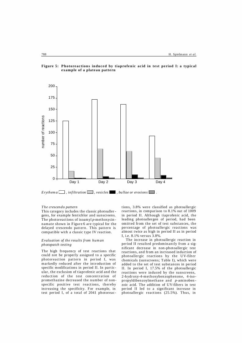
The plateau patternAs reported for period I, tiaprofenic acidinduced photoreactions typical of the plateaupattern, which is characterised by erythemaand infiltration and a few papulovesiclesbetween day 2 and day 4 ( Figure 5). Carpro-fen, promethazine, chlorpromazine and fen-tichlor induce reactions of this type. Themajority of reactions could not be classifiedproperly, although they seem to be inducedby prolonged phototoxic reactions.
Table IV: Rank order of chemicals by photoallergic reaction in test period II,1991–1997
Test chemicals Frequency (%)
1. Fenticlor 1.822. Carprofen 1.193. Chlorpromazine 0.954. 2-Hydroxy-4-methoxybenzophenone 0.635. 4-Isopropyldibenzoylmethane 0.56
6. Tetrachlorosalicylanilide 0.407. Promethazine 0.408. Isoamyl p-methoxycinnamate 0.409. Tribromosalicylanilide 0.3210. Bithionol 0.32
11. Balsam of Peru 0.3212. Monobromosalicylchloranilide 0.2413. Buclosamide 0.2414. Compositae mix 0.2415. p-Aminobenzoic acid 0.24
16. 4-tert-Butyl-4´-methoxydibenzoylmethane 0.1617. 2-Ethylhexyl p-methoxycinnamate 0.1618. Sulphanilamide 0.1619. Quinidine 0.0820. Musk ambrette 0.08
21. 3-(4-Methylbenzylidene)camphor 0.0822. Phenylbenzimidazolesulphonic acid 0.0823. Hexachlorophene 0.0024. Triclosan 0.0025. Chlorothiazide 0.0026. 2-Ethylhexyl p-dimethylaminobenzoate 0.00
786 H. Spielmann et al.

Figure 3: Photoreactions induced by quinidine in test period I: a typical exampleof a decrescendo (phototoxic) pattern
Day 1 Day 2 Day 3 Day 4
7
6
5
4
3
2
1
0
Erythema , infiltration , vesicles , bullae or erosions .
Figure 4: Photoreactions induced by tetrachlorosalicylanilide in test period I: atypical example of a combined pattern
Day 1 Day 2 Day 3 Day 4
12
10
8
6
4
2
0
Erythema , infiltration , vesicles , bullae or erosions .
ECVAM Workshop 42: phototoxicity testing 787
The crescendo patternThis category includes the classic photoaller-gens, for example fentichlor and sunscreens.The photoreactions of isoamyl p-methoxycin-namate shown in Figure 6 are typical for thedelayed crescendo pattern. This pattern iscompatible with a classic type IV reaction.
Evaluation of the results from human photopatch testing
The high frequency of test reactions thatcould not be properly assigned to a specificphotoreaction pattern in period I, wasmarkedly reduced after the introduction ofspecific modifications in period II. In partic-ular, the exclusion of tiaprofenic acid and thereduction of the test concentration ofpromethazine decreased the number of non-specific positive test reactions, therebyincreasing the specificity. For example, intest period I, of a total of 2041 photoreac-
tions, 3.8% were classified as photoallergicreactions, in comparison to 8.1% out of 1009in period II. Although tiaprofenic acid, theleading photoallergen of period, had beenomitted from the set of test substances, thepercentage of photoallergic reactions wasalmost twice as high in period II as in periodI, i.e. 8.1% versus 3.8%.
The increase in photoallergic reaction inperiod II resulted predominantly from a sig-nificant decrease in non-photoallergic testreactions, and from an increased induction ofphotoallergic reactions by the UV-filterchemicals (sunscreens; Table I), which wereadded to the set of test substances in periodII. In period I, 17.5% of the photoallergicreactions were induced by the sunscreens, 2-hydroxy-4-methoxybenzophenone, 4-iso-propyldibenzoylmethane and p-aminoben-zoic acid. The addition of UV-filters in testperiod II led to a significant increase inphotoallergic reactions (25.5%). Thus, in
Figure 5: Photoreactions induced by tiaprofenic acid in test period I: a typicalexample of a plateau pattern
Day 1 Day 2 Day 3 Day 4
200
175
150
125
100
75
50
25
0
Erythema , infiltration , vesicles , bullae or erosions .
788 H. Spielmann et al.

period II, specific modifications in the evalu-ation of the photoreactions resulted in ahigher specificity of the photopatch testingprocedure.
In period I, tiaprofenic acid was the leadingphotoallergen, followed by fentichlor, carpro-fen, 2-hydroxy-4-methoxybenzophenone and4-isopropyldibenzoylmethane. In period II,fentichlor, carprofen, chlorpromazine, and thesunscreens 2-hydroxy-4-methoxybenzophen-one and 4-isopropyldibenzoylmethane, werethe leading photoallergens (Table IV). Insummary, virtually the same chemicalsrevealed a high photoallergic potential in bothperiods of the study. This result confirms thehigh reproducibility of the photopatch testingprocedure.
The reproducibility of the two photoreactionpatterns in the two test periods, and the per-sistence of the plateau pattern of prometh-azine, even after a reduction in the test con-centration, demonstrates the specificity of thereaction pattern for each test chemical (5). Inaddition to the well-known phototoxic (cate-gory 1) and photoallergic (category 4) pattern,computer-assisted analysis of the specificity ofthe reaction pattern over time for each test
chemical revealed a combined pattern (cate-gory 2) and the plateau pattern (category 3).This additional analysis facilitated the classifi-cation of individual photoreactions in one ofthe four photoreaction patterns.
The plateau pattern and the combinedpattern give more insight into clinical photo-reactions, which previously could not be clas-sified. Although the mechanisms underlyingplateau reactions remain to be determined,the analysis of every single individual(plateau) test reaction suggests that theymost probably represent a delayed photo-toxic reaction.
Evaluation of the results from human photopatch testing
It should be noted that photopatch tests aremerely indicative of possible reaction mecha-nisms, i.e. phototoxicity and photoallergy.The underlying mechanism must be con-firmed by experimental laboratory studies.
Since many chemicals are capable of induc-ing both photoallergic and phototoxic effects,a complex photoreaction pattern resultingfrom an immediate (phototoxic) and a delayed
Figure 6: Photoreactions induced by isoamyl p-methoxycinnamate in test period I:a typical example of a crescendo (photoallergic) pattern:
Erythema , infiltration , vesicles , bullae or erosions .
Day 1 Day 2 Day 3 Day 4
6
5
4
3
2
1
0
ECVAM Workshop 42: phototoxicity testing 789

(photoallergic) reaction may be observed.Such a combined pattern was, in fact, elicitedby tetrachlorosalicylanilide. Consequently, afew clinical photoreactions, which hithertocould not be classified, could now be inter -preted as photoallergic reactions superim-posed by immediate phototoxic reactions. Thetwo newly added photoreaction patterns(plateau and combined) seem to hold promisefor improving the specificity of the photopatchtest procedure, and, in particular, for a betterdefinition of photoallergic reactions.
An updated set of test chemicals for thephotopatch test (Table V) should containphotosensitisers that were of significance inthe past and which will remain relevant
today and in the future (14). Thus, a stan-dardised set of test chemicals for photopatchtesting should be continually updated,according to the results of testing and to clin-ical and/or experimental experience.
Acute Phototoxicity Testing
The 3T3 NRU phototoxicity (= photocytotoxicity) test
Background and results of the validationstudyTo validate the 3T3 NRU phototoxicity test,three studies were conducted, all of which
Table V: The photopatch test chemicals: test period III
Test chemical Concentration (%)
Standard set1. Tetrachlorosalicylanilide 0.12. Monobromosalicylchloranilide 1.03. Hexachlorophene 1.04. Bithionol 1.05. Sulphanilamide 5.0
6. Promethazine 0.17. Quinidine 1.08. Musk ambrette 5.09. Perfume mix 8.010. p-Aminobenzoic acid 10.0
11. Ethylhexyl p-dimethylaminobenzoate 10.012. 1-(4-Isopropylphenyl)-3-phenyl-1,3-propanedione 10.013. 4-tert-Butyl-4´-methoxydibenzoylmethane 10.014. Isoamyl p-methoxycinnamate 10.015. 2-Ethylhexyl p-methoxycinnamate 10.0
16. 3-(4-Methylbenzylidene)camphor 10.017. Phenylbenzimidazolesulphonic acid 10.018. Oxybenzone 10.019. Sulisobenzone 10.0
Special set20. Tribosalan 1.021. Chlorpromazine 1.022. Thiourea 0.123. Olaquindox 1.0
790 H. Spielmann et al.

were aimed at different goals and startedwith different levels of information. Thismust be kept in mind, in order to appreciatehow the studies were conducted. A majorproblem in the field of phototoxicity arisesfrom the fact that the number of test chemi-cals backed by high-quality in vivo data ineither animals or humans is very limited.Moreover, when the prevalidation study wasstarted in 1992, the 3T3 NRU phototoxicitytest had just been developed without a pre-diction model, and the concept of a biostatis-tically based prediction model had not yetbeen put forward. It was suggested for thefirst time as an essential element of experi-mental validation at the second Amden(ECVAM/European Research Group forAlternatives in Toxicity Testing [ERGATT])validation workshop in 1994 (15). Later, theInteragency Coordinating Committee on Val-idation of Alternative Methods (ICCVAM)and the Organisation for Economic Coopera-tion and Development (OECD) accepted thata validation study must include an evalua-tion of the reliability and relevance of theprediction model to be used in a new test fora specific purpose (16).
In contrast to all the previous OECD TestGuidelines, the toxicity test described in thisguideline is aimed at correctly predicting thephototoxic potential in relation to clinicalhuman data. Thus, in addition to describinghow to conduct the method appropriately, aprediction model is included as an essentialpart of the method. Taking into account theresults of several experimental validation tri-als, the prediction model allows the photo-toxic potential of new test chemicals inhumans to be predicted with a very highaccuracy. None of the toxicity tests that havebeen accepted into the OECD guidelines sofar have contained a biostatistically basedprediction model that allows toxic potentialin humans to be predicted.
Prevalidation study 1992–1994Before this study was started, a COLIPA taskforce had identified a list of 20 test chemi-cals, 11 of which were phototoxic (+pt) and 9non-phototoxic ( –pt) in animal studies and/orin humans. It was the aim of the study toidentify in vitro phototoxicity tests thatwould correctly predict the phototoxic poten-tial of test chemicals. In order to reduce tech-nical problems at this early stage, the major-ity of the chemicals chosen for testdevelopment and prevalidation were soluble
in water/culture medium. The prevalidationstudy was not conducted as a blind studywith coded chemicals (17).
The 3T3 NRU phototoxicity test study wasconducted in eight laboratories and, from the158 data points obtained, and using discrim-inant analysis, the photoirritation factor(PIF) prediction model was developed to dis-criminate between +pt and –pt chemicals. Acut-off value of PIF 5 resulted from the sta-tistical analysis of the set of test chemicalsused in the prevalidation study. Applyingthis prediction model, all of the test chemi-cals were correctly classified in the 3T3 NRUphototoxicity test.
Identical results were obtained in a studyconducted independently of the EU/COLIPAstudy at the Hatano Research Institute, inJapan, in 1994, with the 3T3 NRU phototox-icity test according to the same test protocol,including the PIF prediction model and withthe same set of chemicals (18).
Formal validation study 1994–1996It became quite obvious during the prevali-dation study that the quality of the availableanimal data was poor. To overcome this prob-lem, in 1993, clinical experts were invited tothe first ECVAM workshop on in vitro photo-toxicity testing, in order to identify a set oftest chemicals backed by high-quality in vivodata. At this ECVAM workshop, a list ofhigh-quality data from standardised humanphotopatch testing was made available, forboth acute phototoxicity and photoallergy.The list of these two groups of chemicals,mostly drugs, including the incidence ofphotochemical side-effects, was published inthe ECVAM workshop report (2). A majorresult of the ECVAM workshop was the com-pilation of a “list of test chemicals with suffi-cient human data”, which covered all classesof currently known phototoxic drugs andchemicals. The list of test chemicals was thesame as that shown in Table VI, whichrelates to the red blood-cell phototoxicity(Photo-RBC) test.
Taking this information into account, aformal validation study on the 3T3 NRUphototoxicity test was conducted under thesponsorship of the European Commissionand COLIPA, as recommended by ECVAM. Itwas the goal of the study to assess whetherthe 3T3 NRU phototoxicity test could cor-rectly predict the phototoxic potentials oftest chemicals in humans. Therefore, anunbalanced set of test chemicals was chosen,
ECVAM Workshop 42: phototoxicity testing 791

with 5 –pt and 25 +pt chemicals. In order toassess problems of bioavailability, threechemicals were used as both water-solublesalts and insoluble free acids or free bases(acridine-hydrochloride and acridine-freebase; nalidixic acid-free acid and nalidixicacid–sodium salt; protoporphyrine IX-freeacid and protoporphyrin IX-disodium salt). Itwas, of course, an essential goal of this partof the study to confirm and/or improve thePIF prediction model with a new set of chem-icals tested under blind conditions.
An interesting result of the study was thatthe 3T3 NRU phototoxicity test was able topredict correctly the phototoxic potential oftest chemicals in humans, irrespective of theirsolubility (3). This proves that solubility isnot, but bioavailability is, a relevant criterionin identifying the phototoxic potential of achemical correctly, and this aspect was care-fully covered by the process laid down in thetest protocol. However, since this study wasconducted under blind conditions and sinceinsufficient guidance was given on appropri-ate solvents and limit concentrations of solu-bility, in many instances the chemicals werenot tested under optimal conditions.
UV-filter chemicals study 1997–1998At the request of the Scientific Committee ofCosmetology and Non-Food Products (SCC-NFP), the expert advisory committee on cos-metics, which reports to the Health and Con-sumer Protection Directorate General of theEuropean Commission, a set of the most com-monly used UV-filter chemicals, which are notphototoxic in vivo and, in most cases, arepoorly soluble in water, was tested in a blindtrial with the 3T3 NRU phototoxicity test. Toobtain information on the optimum test con-centration and on the predictivity of the 3T3NRU phototoxicity test, an equivalent set ofproven phototoxic chemicals was tested. Toavoid the inappropriate use of solvents, infor-mation was provided with each of the codedtest chemicals on both the most appropriatesolvent and the solubility limits in that solvent.
The results of the study confirm that the3T3 NRU phototoxicity test is able to handletest chemicals irrespective of their solubility(4). A thorough biostatistical analysisshowed that no false-positive results wereobtained up to 100µg/ml, whereas somefalse-positive results were obtained at higherconcentrations. The report also shows thatall of the –pt UV-filter chemicals were testedat least up to the highest test concentrations
recommended after the independent assess-ment of solubility.
Taking into account that some test con-centrations reported for the formal valida-tion study were nominal rather than actualconcentrations, they must not be comparedto concentrations at which the same chemi-cal was used in the UV-filter study. Thus, theonly data from which a reliable recommen-dation for maximum test concentrations canbe derived were those produced in the UV-fil-ter study, which shows that false-positiveresults may be obtained at concentrations ofmore than 100µg/ml.
The results from the UV-filter studyclearly demonstrate that the 3T3 NRUphototoxicity test does not provide a yes orno answer, but that the result is clearlydependent on the test concentration applied,with an increasing risk of false-positiveresults at high test concentrations. Thus,expert judgement is required when datafrom the 3T3 NRU phototoxicity test areused for regulatory purposes.
It must be stressed that, among the tox-icity tests currently available, the 3T3 NRUphototoxicity test has a unique position,since it is the only toxicity test that predictsthe situation in humans, due to the avail-ability of high-quality human in vivo photo-patch test data, against which it has beenexperimentally validated. We are not awareof any other toxicity test that has beenaccepted for regulatory purposes, whichmeets this important criterion.
Current use, technical improvements andregulatory acceptanceEarly in the year 2000, the 3T3 NRU photo-toxicity test was officially accepted by theEuropean Commission and the EU MemberStates into Annex V of EU Council Directive67/548/EEC for the classification andlabelling of hazardous chemicals (19). Inaccordance with EU Council Directive86/609/EEC (1), which regulates the use ofexperimental animals, the 3T3 NRU photo-toxicity test must now be used to determinethe phototoxic potential of chemicals, andanimal tests are prohibited for this purposein all EU Member States.
The protocol can be used successfully withhuman keratinocytes, as demonstrated in astudy conducted under blind conditions withthe chemicals of phase II of the EU/COLIPAvalidation study and of the UV-filter study(20).
792 H. Spielmann et al.

ECVAM Workshop 42: phototoxicity testing 793
Other tests
Human 3-D skin models in phototoxicity testingReconstituted human skin models, availablecommercially or in a few experienced labora-tories, are of three different types: dermalmodels (containing skin fibroblasts), epider-mal models (containing skin keratinocytesand a stratum corneum), and full skin mod-els (containing fibroblasts, keratinocytes anda stratum corneum). Since the last two typescontain viable, metabolising primary skincells and a skin barrier, both are frequentlyreferred to as “3-D skin models”. Humanskin models have been used successfully inroutine laboratory investigations, since theyare relevant to the organ of interest. For invitro toxicity testing, standardisation andcontrol of 3-D skin models need to be definedclearly, in order to ensure that reliable andreproducible data are obtained.
In contrast to commonly used cell cul-tures, such as 3T3 mouse fibroblasts, humanskin models permit the topical application ofvarious types of chemicals and preparations,and seem to have fewer limitations relatingto solubility problems. In 3-D skin models,test materials can be applied undiluted, evenat extreme pH values or as “insoluble” sub-stances, as shown, for instance, by the suc-cessful use of such models for testing corro-sives.
The first promising data obtained withskin model phototoxicity tests were reportedin 1994–1995, with a full skin model (21, 22)and an epidermal model (23). The resultsobtained with the full skin model were con-firmed in 1997 (24). Since the commercialproduction of the full skin model, Skin2™,ended in 1996, the test protocol was success-fully adapted to the use of the epidermalmodel, EpiDerm™ (25), and later was evalu-ated in an ECVAM prevalidation study,which revealed promising results from threelaboratories (26). The test is currently estab-lished in several laboratories of the Euro-pean cosmetics industry (27), and has beensuccessfully adapted for the epidermalmodel, SKINETHIC™ (28). Efforts under-taken to optimise the phototoxicity test pro-tocol and the prediction model, when trans-ferring them from the full skin model to theepidermal model (25), revealed that the basictest protocol and prediction model did notneed to be changed. Several studies (22, 24,25) reported that in vivo photoallergens that
are not simultaneously acute photoirritants(for example, coumarin, 6-methylcoumarin,musk ambrette), are classified as negative bythe skin model phototoxicity tests. Finally,dermal models, which do not contain a skinbarrier, show a sensitivity to phototoxicchemicals that is similar to that of photocy-totoxicity tests, such as the 3T3 NRU photo-toxicity test (29). Therefore, they do not pro-vide any advantage in a phototoxicity testingstrategy.
Assuming that the 3-D skin models complywith the best available laboratory and scien-tific standards, they will offer the followingadvantages in comparison to the 3T3 NRUphototoxicity test.
1. Pure chemicals or complex mixtures canbe applied in a way that simulates theapplication of preparations topically tothe skin.
2. The test concentrations applied are closerto real exposure conditions, includingthose used in dermatological patch test-ing.
3. Histology can be performed on exposedand control samples.
4. Exposure to light can be better adaptedto real-life situations; for example, expo-sure time and spectrum of simulated sun-light (a higher dose of short-wave light inthe UVB range).
5. Depending on the barrier function of thestratum corneum, adsorption and pene-tration of the original chemicals or mol-ecules created during the exposure ofskin models will provide more-relevantresults than tests performed on simplermodels (fewer false positives).
The following disadvantages also have to betaken into account.
1. The number of commercial suppliers ofsuch models is limited (for example, Mat-Tek™, SKINETHIC, EPISKIN™, Cell-Systems™).
2. Lack of skin appendices, such as hair fol-licles or sebum and sweat glands, whichmay be sensitive areas in vivo, could be ofrelevance, although there is no experi-mental evidence so far that this is actu-ally a drawback.
3. Until now, there has been no convincingevidence that the photoallergic effects of
chemicals can be predicted in 3-D skinmodels.
4. Since 3-D skin models are still veryexpensive — currently about 25–90 Europer sample — they are not yet suitablefor routine large-scale testing.
In conclusion, during the EU/COLIPA vali-dation project, it was shown that the 3T3NRU phototoxicity test can correctly predictthe phototoxic potentials of chemicals byusing a monolayer cell culture. Since thismay not be relevant when chemicals areapplied topically to the skin at lower concen-trations in finished products, there was aneed for test method development to permitthe application of complex formulationsdirectly to reconstructed skin models.
In the ECVAM prevalidation study on theEpiDerm phototoxicity test, an appropriatetest protocol was developed, successfullyleading to reliable test results in three labo -ratories for ten test chemicals (26). From theresults obtained in this project, there is someevidence that 3-D skin models can be usedfor the assessment of the potency of a photo-toxin applied topically to the skin. Therefore,it can be concluded that 3-D skin models canplay an essential role as adjuncts in a testingstrategy for the sequential evaluation ofphototoxicity.
The combined Photo-RBC testIn the EU/COLIPA validation programme on“photoirritation in vitro”, two core tests anda number of mechanistically based tests wereperformed, to examine their suitability asregulatory tests for phototoxicity testing. Inaddition to the 3T3 NRU phototoxicity test,as second core test, the Photo-RBC test wasevaluated in a prevalidation study within thevalidation programme.
In the protocol for the Photo-RBC test,two endpoints are determined in erythro-cytes, namely, photohaemolysis and met-haemoglobin (met-Hb) formation. The end-points are assessed by measuring changes inoptical density at 525nm for photohaemoly-sis and at 630nm for met-Hb formation. Fur-thermore, a prediction model was used in theStandard Operating Procedure (SOP), tak-ing into account two cut-off values: the pho-tohaemolysis-factor (PHF) ≥ 3.0 for photo-
haemolysis, and optical density (OD) ∆ODmax≥ 0.05 for met-Hb formation. Three laborato-ries agreed on the SOP, and the 30 chemicals(25 phototoxic and five non-phototoxic chem-icals) that had been selected for the valida-tion study (3) were tested under blind condi-tions.
The results of the prevalidation study onthe Photo-RBC suggested that the transferof the SOP between laboratories had notbeen very efficient, and that additional train-ing of the laboratories would be helpful.Most importantly, the results obtained in thelead laboratory (Table VI) clearly indicatethat an experienced laboratory will obtaincorrect classifications for both phototoxicand non-phototoxic test chemicals, when theSOP is applied correctly. The biostatisticalanalysis shows a good overall in vitro/in vivocorrelation of the results, including anacceptable accuracy (83.3%), sensitivity(87.5%), and positive predictivity (91.3%;30). The low specificity (66.7%) and low neg-ative predictivity (57.1%) most probablyresult from the unbalanced number of posi-tive and negative test chemicals, in particu-lar, from the low number of non-phototoxicsubstances in the set of test chemicals.
A test protocol was developed to identifyphotosensitisers and to study phototoxicpotential of light-activated chemicals fromtheir ability to lyse freshly isolated erythro-cytes and/or to oxidise oxyhaemoglobin(oxy-Hb) after exposure to simulated sun-light comprising UVB, UVA and visible light(31, INVITTOX protocol number 811). Incontrast to the established photohaemolysistest (32, 33), the new protocol included pho-tochemically induced met-Hb formation(31, 34, 35) as the second endpoint, sincemet-Hb may be produced during extensiveexposure to sunlight, especially as a resultof type I photodynamic reactions (36). Fur-thermore, in contrast to keratinocytes orfibroblasts, that are used in the 3T3 NRUphototoxicity test protocol, mammalianerythrocytes are readily available. More-over, due to their specific cellular defencemechanisms, erythrocytes can be exposed tomore-intensive UV irradiation, even toUVB, than can cells from other tissue.Finally, since erythrocytes do not contain a
1FRAME,Russell & Burch House, 96–98 North Sherwood Street, Nottingham NG1 4EE, UK.
794 H. Spielmann et al.

ECVAM Workshop 42: phototoxicity testing 795
Table VI: Results of the combined Photo-RBC test in phase II of the EU/COLIPAvalidation project on photoirritation in vitro
Assessment
No. Chemical In vivo PHF ∆ ODmax Final
1. 2-Hydroxy-4-methoxybenzophenone – – – –10. Chlorhexidine dihydrochloride – – – –15. Hexachlorophene – – + +17. Sodium lauryl sulphate – – – –24. p-Aminobenzoic acid (PABA) – – + +
25. Penicillin G – – – –14. Furosemide +? – – –23. Ofloxacin + – – –2. 5-Methoxypsoralene (5-MOP) + – – –3. 6-Methylcoumarin + +/– + +
4. Acridine hydrochloride + +/– + +5. Acridine free base + + + +6. Amiodarone + +/– – +/–7. Anthracene + +/– + +8. Bergamot oil + – + +
9. Bithionol + +/– + +11. Chlorpromazine + +/– + +12. Demeclocycline + – + +13. Fenofibrate + + + +16. Ketoprofen + + + +
18. Musk ambrette + + + +19. Nalidixic acid sodium salt + +/– + +20. Nalidixic acid free acid + – + +21. Neutral red + + + +22. Norfloxacin + – + +
26. Promethazine + – + +27. Protoporphyrin IX free acid + + + +28. Protoporphyrin IX disodium + + – +29. Rose bengal + + + +31. Tiaprofenic acid + + + +
Data from Pape et al. (30).
PHF = photohaemolysis factor, ∆ ODmax = optical density.
+ = positive, – = negative, +/– = equivocal results.

nucleus, they are not susceptible to pho-togenotoxic effects.
Thus, results from the combined Photo-RBC test can be considered as interestingadditional information within an overallphoto-safety testing strategy, when in vitromethods are the initial step of testing. Inaddition, the Photo-RBC test provides mech-anistic information on two different types ofphotodynamic reactions (met-Hb formationfor type I reactions, and photohaemolyticeffects as primary type II reactions). As sum-marised in Table VI, when the resultsobtained with both endpoints in the Photo-RBC test in the final assessment are com-bined, a good overall fit with the in vivo eval-uations is obtained.
The three false-negative results can beexplained in the following manner (31). Forfurosemide, a relevant phototoxic potency invivo is not proven, since the clinical data inhumans are insufficient (3). Ofloxacin wastested as a clinical preparation at a very lowconcentration of the active ingredient, whichmay not reveal its phototoxic potential. 5-MOP may have provided a negative resultdue to the lack of sensitive target moleculesin erythrocytes.
False-positive results were obtained withPABA and hexachlorophene in the Photo-RBC test. PABA is easily oxidised by oxy-Hbin the dark in a pseudo-enzymic reaction,which may be enhanced under light exposure(or increased temperature).
In summary, as a general result of theprevalidation study according to the SOP, thecombined Photo-RBC test can be performedreproducibly, and it provides relevant mech-anistic information on photodynamic reac-tions, which add important information forthe evaluation of the photo-safety of testchemicals, within a wider testing strategystarting with the 3T3 NRU phototoxicitytest, which does not provide information onphotodynamic rections. An additional advan-tage of RBC cells is their resistance to theshort-wave UVB part of sunlight, whichenables RBC cells in the Photo-RBC test tobe exposed to the entire solar spectrum forprolonged periods.
The yeast assayThe use of mammalian cells and bacteria inphototoxicity and photogenotoxicity testingmay be limited by the high sensitivity ofthese organisms to UV light and, in particu-lar, to UVB. The facultative anaerobic yeast
Saccharomyces cerevisiae, a eukaryoticorganism, has the advantage of being rela-tively insensitive to exposure to sunlight orto prolonged exposure to drugs in aqueoussolution, in final galenic forms and oint-ments, even when oxygen tension is low (asin the presence of an oil or cream). Thus,when compared to mammalian cells andother testing organisms, S. cerevisiae offerssome advantages for testing the phototoxicpotentials of chemicals.
In this organism, phototoxicity can easilyand cheaply be tested qualitatively andquantitatively, directly on plates with com-plete growth medium or after plating ofexposed cell suspensions, by measuring thedensity of the outgrowing cells or colonyforming ability (clonogenic survival; 37).
When using yeast, DNA damage can bedemonstrated by comparing the responses ofDNA repair-deficient strains with those ofDNA repair-competent strains (37). In thisway, even the specific mechanism of photoin-duced lethality can be assessed, since mutantstrains are available in all known DNArepair pathways (38). The use of suchmutants can significantly improve the sensi-tivity of the test. Furthermore, photomuta-genicity and mitotic recombination can read-ily be assessed (37, 39).
With regard to testing for the photosensi-tising or photoprotective potentials of med-ical, cosmetic or pharmaceutical compounds,S. cerevisiae offers the advantage that it canwithstand quite long exposures to UVB,UVA, solar simulated or solar radiation(40–42). It is thus useful for testing reciproc-ity between light exposure (fluence) anddrug concentration, and dose-rate effects,and for evaluating the photocytotoxic andphotomutagenic potentials of photosensitis-ing or photoprotecting agents under condi-tions close to clinical use in patients.
In general, the AMES test with Salmo-nella typhimurium is more sensitive than ayeast assay for genotoxicity testing eventhough the latter assay may also provide use-ful information (43–45). Using the yeastassay, Averbeck (46) and Morlière et al. (40)were among the first to clearly demonstratethe specific properties of some bifunctionalpsoralens, for example, 8-methoxypsoralen(8-MOP) or 5-methoxypsoralen (5-MOP), toinduce genetic damage (mutations andmitotic recombination) in the presence ofUVA or simulated solar radiation (40, 41,
796 H. Spielmann et al.

46). These observations were confirmed incultured mammalian and human cells (47).Since the exposure to doses of UV and UVAradiation could be increased in comparisonto those used for bacterial and mammaliancells, it has been possible to test combina-tions of the psoralens with UV-filters byusing conditions which mimic human expo-sure (48). For example, the photoprotectiveeffects of solar filters against the induction ofgenotoxic effects by 5-MOP could be demon-strated (41, 49). Among other assays, yeastwas recently chosen for use within a generalstrategy for photogenotoxicity testing(50–52). Interestingly, the photomutageniceffects of the fluoroquinolones were detectedin this system (51).
In order to examine the effects of metabo-lism of neat compounds, mixed or in a near-final galenic form, the photomutagenic andphotogenotoxic capacities of suction blisterfluids derived from human skin after appli-cation of the test compound to the skin ofvolunteers were determined in yeast. It waspossible to detect the active genotoxic princi-ple bergamot oil in the interstitial liquid ofhuman skin (42). This is of interest, since theimmunosuppressive effects of exposure tosunlight, and photocarcinogenic responses,imply the activation of genes (53, 54).
In summary, in using the yeast assay, ithas to be noted that:
1. in this assay, not only can the neat com-pound be tested, but also its finalgalenic form combined with other com-pounds. Ethanolic or oily solutions oftest compounds can be spread ontoyeast plated on solid growth medium,before exposure to UV radiation, inorder to test for phototoxicity and/orphotogenotoxicity; and
2. the yeast assays can be performed underconditions (for example, prolonged UVexposures) that are relatively close toactual or expected human exposures, butwould be too phototoxic or too pho-togenotoxic for bacteria and human cellsin vitro.
Photoallergy
In photodermatology and immunotoxico-logical risk assessment, the precise predic-tive differentiation of photoallergenic from
acute phototoxic/photoirritation reactionsinduced by low-molecular weight com-pounds, is very important. Although clini-cal criteria in the examination of skinlesions have been, and continue to be, ofproven value, they are not without theirlimitations. The animal models used so farfor the screening of photoallergic proper-ties of compounds are very heterogenousand time-consuming. To achieve properrisk assessment, the animal models cur-rently in use have to be replaced, with theaim of improving hazard identification bypredicting photoreactions in humans moreprecisely.
Human and experimental evidence for photoallergy
The major obstacle in clinical and experi-mental practice is the difficulty of determin-ing the nature of photoreactivity, i.e. ofphotoallergic or phototoxic reactions. Thereare significant human data in the literatureabout photoreactive compounds. However,the classification of the photoallergic andphototoxic properties of chemicals is mostlybased on clinical parameters, which are notalways sufficient. The major differencebetween photoirritancy and photoallergy isthe activation of (photo)antigen-specificimmunocompetent cells. A prerequisite forthe induction of photoallergic reactions isthe presentation of the modified (photo)anti-gen to specific T-cells. Taking this mecha-nism of photoallergy into consideration, theinvolvement of specifically reacting T-lymphocytes or antibodies could be one toolfor use in discriminating between bothphotoreactions. One in vitro assay that couldbe modified for such an approach is the so-called lymphocyte transformation test (LTT;55). However, the LTT is so far a retrospec-tive assay, which needs lymphocytes fromsensitised patients.
For pre-clinical screening purposes, themost promising test is the modified locallymph node assay (UV-LLNA or UV-IMDS[IMDS = integrated model for the differenti-ation of skin reactions]) which takes intoaccount the immunological mechanism ofthe reaction (56–58).
The following list results from studies pro-viding clear evidence for the mechanism ofthe hazardous potential of photoallergy inhumans, guinea-pigs and mice. A summary isgiven in Table VII.
ECVAM Workshop 42: phototoxicity testing 797

Halogenated salicylanilides3,5,3´,4´-Tetrachlorosalicylanilide (TCSA):experimental photoallergy has been reportedin humans (59), guinea-pigs (for example,60–64) and mice (56, 65–68).
3,5,4´-Tribromosalicylanilide (TBS): photo-allergic reactions to TBS were complicatedby the purity of the sample tested. Thereport of Kaidbey & Kligman (59) accordswith marketing experience. They found noreactions to a pure sample of TBS, but read-ily produced reactions to dibromosalicyl-anilides, and concluded that the photosensi-tising potential of TBS was due to thepresence of dibrominated contaminants.
ThiobisphenolsBithionol: in experimental studies, phototoxicreactions were reported in 8 of 18 personsafter using 5% bithionol with 28.5J/cm2 UVA(69). Photoallergy was elicited in 3 of 25 sub-jects after using 1% bithionol with 4J/cm2 UVAin the photomaximisation procedure (59).Photoallergic reactions have been reported inguinea-pigs (61, 63, 70) and mice (66, 67).
Fentichlor: photoallergy in mice wasreported by Gerberick & Ryan (67).
Aromatic nitro-compounds Musk ambrette: in the photomaximisationtest in volunteers, musk ambrette was nega-tive (59), which led to a recommendationthat the guinea-pig could be the animal ofchoice for photoallergy testing (71). Muskambrette has been reported to be photoaller-gic, in the absence of significant phototox-icity, after topical application to guinea-pigs(62, 70, 72–75), and mice (66–68).
Coumarins6-Methylcoumarin (6-MC): in experimentalstudies in volunteers, 6-MC was not photo-toxic, in contrast with 7-MC. However, 6-MCwas photoallergenic in the human photomax-imisation test (76, 77). The US regulatoryposition has been discussed (78). In guinea-pigs, 6-MC has not been reported to be photo-irritant after topical application. In photoal-lergy tests, it has been reported to be negative(63, 70) or weakly active (73, 79, 80). A photo-allergic potential for 6-MC was also obtainedin mice (65, 67, 68), although a further reportin mice was rather equivocal (66).
BenzophenonesKetoprofen: ketoprofen has been reportednot to be photoallergenic in a photomaximi-
sation procedure (81) or in a guinea-pig test(82). However, it showed clear photoaller-genic potential in a sensitive guinea-pig test(T. Maurer, personal communication), as wellas in a mouse model (H-W. Vohr, personalcommunication).
Aromatic aminesSulphanilamide: phototoxic and photoaller-gic reactions were first distinguished afterapplying sulphanilamide.PABA: photoallergy to PABA in guinea-pigswas reported by Gerberick & Ryan (62).
Aromatic nitro-oxidesOmadine: photoallergy to sodium omadinehas been reported after using the photomax-imisation procedure on humans (71) and inmice (65, 67).
Quindoxin: photodermatitis to quindoxinwas reported in the UK in the early 1970s,and to olaquindox in Germany in the 1990s(see 83). Quindoxin was photoallergenic inguinea-pigs (W. Lovell, unpublished observa-tions), and olaquindox has been reported tobe photoallergenic in mice (58, 84).
TetracyclinesTopical tetracycline preparations are rarelycontact sensitisers, but may yellow the skin.In addition, the skin may fluoresce underblack fluorescent light. Together with thephotoreactions found in in vitro investiga-tions with tetracyclines (see Table I), thiseffect led to concern about photoreactions invivo. However, no definite report has beenpublished regarding photoallergic properties.
PhenothiazinesChlorpromazine (CPZ): Phototoxic reactionswere produced by topical application of 0.5%aqueous solutions plus UVA irradiation,whereas photoallergic reactions were elicitedby 0.025% CPZ plus UVA (85). Experimentalphototoxicity was reported after the intra-dermal injection of 0.1mg with about18J/cm2 UVA (76). Photoallergy was elicitedby topical application of 0.2% CPZ with4J/cm2 UVA via the photomaximisation tech-nique (59). In 27 volunteers, phototoxic reac-tions could be induced with a minimal con-centration of 0.5% and 4J/cm2 of UVA,whereas, in patients photoallergic to chlor-promazine, 0.1% CPZ and 2J/cm2 of UVAwere sufficient (86). Photoallergic reactionshave been elicited topically in guinea-pigs(63, 87), and both topically (58, 65–68, 88)and systemically (58, 89) in mice.
798 H. Spielmann et al.

Table VII: Proposed standards for the validation of the integrated model for thedifferentiation of skin reactions test
Animal dataChemical
Class of Patch Guinea- mech-compounds test T-cell pig Mouse PMT anism
Halogenated salicylanilides + + + + + +(for example, TCSA, TBSA) PA
Halogenated thiolic phenols + – + + + ?(for example, bithionol, fentichlor) PT, PA
Aromatic nitro + – + + neg. +(for example, musk ambrette) PA
Coumarins + ? (+) + + –(for example, 6-methylcoumarin) – ? (PT), PA
Benzophenones + – + + – +(for example, ketoprofen, thiaprofen, – PT, PAcarprofen)
Aromatic amines + + + ? + +(for example, sulphonamides, PABA) (PT), PA
Aromatic nitro-oxides + – + + + ?(for example, omadine, quindoxin) PA
Tetracyclines + + ? ? – +(for example, doxycycline, ?oxytetracycline, chlortetracycline, tetracycline)
Phenothiazines + ? + + + +(for example, chlorpromazine, – PT, PApromethazine)
Fluoroquinolones +a – +a +a (+)a –(for example, lomefloxacin, PT, (PA)sparfloxacin)
PT = phototoxic reaction (photoirritation), PA = photoallergic reaction.aOnly after oral/intravenous application; no reaction after topical application.
+ = positive, – = negative, ? = equivocal results.
TCSA = 3,5,3´,4´-tetrachlorosalicylanilide, TBSA = 3,5,4´-tribromosalicylanilide, PMT =photomaximisation test.
ECVAM Workshop 42: phototoxicity testing 799

Promethazine: in a volunteer study,promethazine did not induce phototoxicreactions when one tablet of 25mg wasgiven and light doses of 5–72J/cm2 wereused (90). Promethazine was photoallergic,but was much weaker than chlorpromazinein a guinea-pig test (63). Photoallergicreactions were also reported in guinea-pigsby Guillot et al. (70) and in mice by Vohr etal. (88).
FluoroquinolonesThe fluoroquinolones are a family of broad-spectrum antibiotics, primarily intendedfor systemic use. There is much epidemio-logical evidence from clinical safety trialsthat many of the family of fluoroquinolonesare phototoxic (photoirritant) in humanskin. Carefully conducted trials involvingthe photo-testing of volunteers in definedwavebands of UVR, have supported the epi-demiological evidence and quantified therelative potencies of various fluoro-quinolones (for example, 91–93). The com-pounds primarily absorb in the UVA(315–400nm) range of the solar spectrum,and phototoxic potency varies from theability of compounds such as BAY y3118 tophotosensitise the skin by a factor of over30, to compounds such as moxafloxacin,which have no phototoxic potential (94).Several fluoroquinolones have also beenreported to be phototoxic in guinea-pig skin(95, H-W. Vohr, personal communication)and mice (84, 96). Although most of thequinolones seem to cause phototoxic reac-tions exclusively, there are reports of pho-toallergic properties of some fluoro-quinolones, such as enoxacin (84, 97–99).
Testing strategy
In vivoUntil now, photoallergic hazard identifica-tion has largely been performed in guinea-pig models. Since these animal proceduresare expensive, time-consuming, and harm-ful to the animals, and objective endpointsare still lacking, a new testing strategy isrequired that takes account of objectivity ofhazard identification and animal welfare.Recent reports showed that a combinationof the LLNA and the primary mouse-earswelling test (MEST), the IMDS (58), couldprobably provide a reasonable alternativein terms of animal welfare and objectivity.There has only been intralaboratory pre-
validation of the photo-LLNA until now,which showed that this model also providesthe possibility of discriminating betweenphotoallergic and phototoxic reactions. Toverify the validity and robustness, an inter-laboratory validation of the respective testprotocol, monitored by an internationalpanel of experts, should be conducted. Atesting strategy incorporating the LLNAand the use of structure–activity relation-ships is shown in Figures 7 and 8.
In vitroPhotobinding to protein is a common prop-erty of photoallergens, and a test based onphotobinding of photoallergens to humanserum albumin has been proposed (100).The model has been demonstrated to detectall the photoallergens tested so far. Whilephotobinding to protein is considered a nec-essary condition for photoallergy, it is notsufficient for discrimination betweenphotoallergens and photoirritants. Addi-tional mechanistic tests, including tests forphotooxidation, are required to enable thediscrimination of photoallergens (101). Thephotobinding model, in conjunction with atest of photooxidation of histidine, wasused to test the 30 chemicals selected forthe EU/COLIPA phototoxicity trial. Al-though the test chemicals were heavilybiased toward phototoxicity, the photobind-ing model showed excellent detection ofphotoallergens, and in conjunction with atest for photooxidising potential, differenti-ation between photoallergens and photo-toxins was largely achieved (102).
A hierarchical scheme for the in vitrohazard assessment of substances for pho-toallergic potential should start with areview of the chemistry, structure-activityrelationships and phototoxicology of thetest item (Figure 9). If experimental evi-dence is required, UV/visible absorbancecan be used as a pre-screen; substancesthat absorb significantly have the potentialfor adverse photochemistry and should betested further (101). It has been proposedthat the 3T3 NRU phototoxicity test couldbe used as a general screen for the photo-sensitising potential of absorbing sub-stances (Figure 9). If these screening testsare positive, in vitro screening tests forphotobinding to protein and for photo-oxidation should be performed (100–102).If the substance is an efficient photo-oxi-diser, photoirritancy may be the problem,
800 H. Spielmann et al.

rather than photoallergy. If the photobind-ing test is negative, a potential for photoal-lergy is not expected, and further testingfor photoallergenicity should not berequired. If the test material binds to pro-tein in the absence of significant photo-oxi-dation, a photoallergy potential is pre-dicted. The safety evaluation could bediscontinued, but if there is further inter-est in the test substance, additional studiescan be considered. For example, skin-pene-tration studies can be carried out, as nopenetration may mean there will be noproblem. A further option may be to con-sider the use of radiolabelled test sub-stances, in order to investigate further andto quantify the in vitro photobinding. Invivo testing may be considered.
A biologically based model of photoaller-gic potential could be useful in hazardassessment in practice (Figure 8). Early up-regulation of the inflammatory mediator,IL-1b, by dendritic cells, and their endo-cytic activation by immunogenic haptens,
have been suggested as potential in vitroindicators of immunogenicity (103, 104).Modification of such systems for photoal-lergens could form an in vitro biologicaltest of the present photochemical model.Animal testing, however, would be requiredto address the problem of potency in vivo.
Potency
No experimental approach has been estab-lished to date for assessing potency forphotoallergy. Although one could design atest system for the photoallergy potency oftest chemicals in humans, this is impossi-ble, for ethical reasons. Therefore, riskassessment in humans has to rely on clini-cal case reports and photopatch testing.
A promising preclinical screening test forphotoreactions is the IMDS mentionedabove. At least after oral application, thedetermination of the potency of a test sub-stance to induce photoirritancy or photoal-lergy seems feasible.
Figure 7: Testing for contact (photo) allergenic potential
Testing for contact (photo) allergenic potential
– –
Local lymph node assay
Standardprotocol:
Tier ITesting
Challenge protocol:Tier II Testing
Expert systemanalysis (DEREK)
Comparison withrelated compounds
Partition
Skinmetabolism
Stop
??
??
Challenge
Searching for structural alerts
+
+
–
–
+
+
ECVAM Workshop 42: phototoxicity testing 801

Figure 8: Photoallergy testing scheme
Chemical + UVA
Photoantigen formation
Uptake, processing andassociation with HMLC
IMDS: primaryMEST + LLNA
Pseudo allergicreaction
Irritation
Sensitisation
mouse ear swelling test(MEST)
LN hyperplasia (LLNA)
Protein-binding
albumin-photobinding
Gel electro-phoresis
Protein-modification
Migration to the regional LN
Presentation to and stimulation of LNC
Clonal expansion andimmune response
Clinical manifestation
mastdegranulation
assay
IMDS = integrated model for the differentiation of skin reactions.
LLNA = local lymph node assay.
LNC = lymph node cells
LN = lymph node
MHC = mature Langerhans cells
802 H. Spielmann et al.

ECVAM Workshop 42: phototoxicity testing 803
Metabolism
The generation of photoreactive metabo-lites must not be underestimated andshould, therefore, be considered in the gen-eral toxicological evaluation. A moderntesting strategy for photoallergic potentialshould integrate relevant photosensitisersof the past and of the present, as well asphotosensitisers that may prove to be rele-vant in the future. Thus, the spectrum oftest substances will have to be modifiedover time, according to test results and toclinical or experimental findings. It mustbe taken into account, from a general per-spective, that the metabolic capacity of invitro systems may be limited in comparisonto the situation in vivo, irrespective of theorgan or tissue, and thus may provide aspectrum of metabolites which are differ-ent from the situation in vivo .
Photochemical Genotoxicity/Carcinogenicity (photogenotoxicity/photocarcinogenicity)
Human data
UVB is a known human skin carcinogen, andits direct genotoxic effects are considered to bean important factor in this process (105). UVBis also known to be a skin carcinogen in rodents(106). Conversely, there is a lack of documentedhuman photochemical carcinogens that can beused to evaluate the potential relevance ofphotochemical genotoxicity and rodent co-car-cinogenicity testing. To date, the combinationof 8-methoxypsoralen (8-MOP) and UVA,known as PUVA therapy, as used in the treat-ment of psoriasis, is the only well-documentedhuman photochemical genotoxic carcinogen(107, 108). Although this effect in human skincould be predicted by results in mouse photo-
Figure 9: Phototoxicology: hazard identification
INITIAL EVALUATION
Photochemistry(UV/visible absorption)
QSAR General toxicity(inc. metabolism)
In vitro phototoxicity test(3T3 NRU PT)
In vitrophotobinding to protein?
In vitrophotogenotoxicity testing
depending oncircumstances
Photocarcinogenicitytesting
Photoallergytesting
Classify asnon-phototoxic
+
–
– –+ +

tumorigenesis studies, apart from the pso-ralens and fluoroquinolones (109, 110), noother groups of photo-tumorigens have beenstudied in any detail. Given the scarcity of data,both in animal models and in humans, theexact relevance of this type of modelling is notclear (111). At this time, the limited data sets ofhuman and rodent photochemical carcinogensand non-carcinogens make the evaluation ofthe human relevance of short-term photo-chemical genotoxicity tests problematic.Despite these limitations, genotoxicity can beaddressed by using tests that hold the greatestpromise in assessing potential photochemicalmutagenic/clastogenic effects, akin to standardgenotoxicity/carcinogenicity testing (112).
Current status of photochemical genotoxicity testing
Many different genotoxicity test systems havebeen employed for photochemical genotoxicitytesting. In principle, most standard in vitrogenotoxicity tests can, and have been, adaptedfor testing the consequences of photoactiva-tion of chemicals in terms of damage to thegenetic material. The potential of a fewclasses of chemicals, such as psoralens, phe-nothiazines or compounds used in photody-namic therapy (for example, porphyrin deriv-atives), to induce genotoxic effects afterirradiation, has been known for several years(113–115). More recently, the photochemicalgenotoxicity of fluoroquinolone antibioticshas been reported (116–120). Additionalimportant contributions regarding testmethod development and standardisationhave been made by several groups in recentyears (45, 121–123). Recommendations withregard to the conduct of tests for photochemi-cal genotoxicity have recently been elaboratedby an international expert working group.Their report constitutes a valuable manualfor laboratories involved in the conduct ofthese assays (124).
Regarding product safety testing strategies,the guideline of the SCCNFP stipulates thatboth a bacterial test for gene mutation and anin vitro test for chromosomal aberrations inmammalian cells should be performed in thepresence of UV radiation (125).
Rationale for testing for photochemical genotoxicity
In considering the carcinogenic potentials ofnew chemical entities, an evaluation of geno-
toxic potential has become an integral part ofthe toxicological assessment. A multitude ofin vitro and in vivo mutagenicity test sys-tems have been devised, and regulatoryguidelines for this application have beenissued. From these data and the parallelexperience in standard genotoxicity testing,testing for photochemical genotoxicityshould be considered in the phototoxicologi-cal assessment of chemicals. Importantly, themain purpose of such testing is to make anassessment of the likelihood of a compoundto turn into a photochemical carcinogen afteractivation with UV or visible radiation.
Test systems and strategies to test for photochemical genotoxicity (Figure 10)
Tests for photochemical genotoxicity may beconsidered for compounds that are intendedfor use on the skin or are widely systemicallydistributed with significant skin exposure, ifa phototoxic potential was proven or, for non-phototoxic compounds, if there is evidence ofabsorbance of UV and/or visible light and thecompound belongs to a chemical class con-taining known phototoxic or photogenotoxiccompounds.
An established absence of a phototoxicaction in relevant investigations is consid-ered to be a strong indicator that the respec-tive compound would be unlikely to possessphotogenotoxic properties, although it maynot be a sufficient criterion for forgoing test-ing.
For product safety testing, it seems rea-sonable to focus evaluation and validation onthose in vitro systems that are recommendedas standard genotoxicity systems in regula-tory testing strategies, such as OECD orInternational Conference on Harmonisation(ICH) guidelines. Several arguments can beput forward for a preference for systemsemploying mammalian cells in vitro overthose involving lower eukaryotes or prokary-otes. Such a preference is based, in part, onthe higher complexity of the cellular modeland on the fact that phototoxicity testing invitro is done in mammalian cells. There is nogeneral consensus on which test systemwould constitute a minimal test battery for asufficient assessment of a test compound forphotochemical genotoxicity (124). As nophotochemical genotoxin is known which isexclusively positive for gene mutations, andsince the recognised photochemical mecha-nisms are strongly clastogenic, it may be
804 H. Spielmann et al.

ECVAM Workshop 42: phototoxicity testing 805
Figure 10: A generalised approach to photocarcinogenicity testing
aTests for photomutagenicity are usually not needed if comprehensive testing for phototoxicitydid not indicate a phototoxic potential.
• Dermally applied compounds• Compounds with wide systemic distribution
Photochemistry, photostability data
Photomutagenicity in vivo(transgenic animals, Comet assay)
Photocarcinogenicity testing(hairless mice, XP deficient mice)
Phototoxicityin vitro/in vivo
Photounstable photodynamic effects
negativea
positive positive
positive
not available in the skin
accept photocarcinogenicpotential or testconcerns from
other areassuch asimmuno-
suppression
Problematic group(for example, fluoroquinolone)
Photostable no photocatalytic potential
Photomutagenicityin vitro No further testing
negative

suggested that a test for photochemical clas-togenicity would suffice for screening pur-poses.
Thus, the photochemical genotoxicitytests could follow a general sequence thatincludes:
— a test for induction of chromosome dam-age (for example, aberrations ormicronuclei) in vitro;
— a test for induction of gene mutations inmammalian cells or (less preferable) inbacteria; and
— the Comet assay, assessing photochemi-cally induced DNA damage (116, 117).However, since this test is currently notaccepted in standard batteries for geno-toxicity testing of chemicals, it should beregarded as an ancillary tool.
It can also be noted that the Comet assay isthe only genotoxicity test that has been suc-cessfully applied to testing for photochemi-cal genotoxicity in vivo . Positive resultshave been reported for the fluoroquinoloneclinafloxacin (119).
Metabolic activation
Genotoxicity testing in vitro is usually donewith the inclusion of an external metabolicactivation system such as rat liver S9 frac-tion. There is no evidence that enzymicmetabolic activation plays a crucial role inthe generation of a photochemical geno-toxin. Furthermore, there is evidence thatthe addition of proteinaceous material suchas that present in rat liver S9 can abolishthe photogenotoxic effects of some com-pounds. Thus, the use of an external meta-bolic activation system such as liver S9 isnot advocated for photogenotoxicity testingin vitro. However, there are a limited num-ber of examples where metabolism of theparent compound shifts the absorption spec-trum. Therefore, researchers are encour-aged to look for and to investigate potentialcandidate compounds for which metabolismis suggested to result in increased photo-binding or phototoxicity.
Light source
Light sources, in general, have been dis-cussed in section 3 (Terminology). A solarsimulator has been recommended for use inphotochemical genotoxicity testing. The
characteristics of the irradiation source needto be described in detail. Specifically, it isimportant to give information on the spec-tral output for determining the UVB:UVAratio of the light source. In general, solarirradiation possesses a ratio of around 1:20,although this value depends very much onlocation, time of day and atmospheric condi-tions (the longer the light path through theatmosphere, the lower the ratio; clouds anddust further attenuate the UVB component).It should be noted that the UVB(290–320nm) content of the light source mayneed to be attenuated, in order to increasethe sensitivity of the test method used,notably when monolayer mammalian cellsand, most especially, repair-deficient organ-isms, are used.
In essence, it is important that the charac-teristics of the light source, i.e. the spectraloutput, are known and are provided togetherwith any results from photogenotoxicitystudies.
Test systems for photochemical co-carcinogenesis
A test for photochemical co-carcinogenesis inanimals has been advocated by the FDA, forexample, for drugs that are applied to skin orwhich reach the skin systemically (126). Theonly model that is currently available forassessing such effects in a Good LaboratoryPractice (GLP)-like format is theSKH1(hr/hr) albino hairless mouse model.The endpoints of judgement are the numberand size of papilloma formation on skin andthe latency, after a general duration of treat-ment of 40 weeks. Various UV sources andintensities have been used for the assess-ment of photochemical carcinogenesis. Ascurrently designed, the mechanistic under-standing that is provided by the model isvery limited, since highly tumorigenic dosesof UVB are most often used and histopathol-ogy is not an integrated part of tumourinvestigation (111). In a modified testapproach using a non-tumorigenic protocolof doses from a broad-band fluorescent UVAsource, 8-MOP, as well as some fluoro-quinolones, led to a high incidence of skinpapilloma (110). As stated above, theextremely limited data on photosensitisedhuman skin tumorigenesis, together withonly a marginally larger amount of data frommouse studies, makes the relevance of therodent photocarcinogenesis models difficult
806 H. Spielmann et al.

to assess. Overall, the predictivity of thisrodent photocarcinogenicity model for thehuman situation is not known, and thus therelevance of the rodent studies for humanhealth risk cannot be reliably assessed.
Guidance on testing for photochemical geno-toxicity (Figure 10)
1. The direct genotoxic effects of UVB canlead to high mutation frequencies orchromosomal damage at irradiation dosescomparable to only seconds or minutes ofsolar irradiation at environmental levels.Such a high responsiveness to UVB willlimit the applicable irradiation doses tolevels that might, theoretically, be too lowfor the activation of a comparativelyweak photochemical genotoxin. There-fore, it may be necessary to attenuate theUVB content of the light source used forcompounds that do not exclusively absorbin the UVB region. Irradiation through aplastic lid will generally meet theserequirements, since it predominantlyreduces the UVB part of the spectrum.Note, however, that lids from differentmanufacturers have different UV trans-mission characteristics. It is also possibleto use a quartz glass cover or more sim-ply, food-wrapping plastic film, neither ofwhich affect the spectral composition. Itis recommended that a full dose–responsecurve of the test system to the irradiationsource in the absence of test compound isincluded in the report.
2. A specific, generally applicable irradia-tion dose (for example, equivalent to acertain, environmentally relevant solarirradiation period) cannot be defined forthe various test systems in use, mainlybecause of their different sensitivities tothe UVB effects. The irradiation per seshould yield a small, reproducible geno-toxic or toxic effect. When testing sun-screens, it might be of interest to describethe protective effect of the test com-pound. In such a case, it is advisable touse an irradiation dose which elicits amore sizeable effect, so that attenuationof the UV genotoxicity is measured moreeasily.
3. There is no clear-cut precedent for anexclusively UVB-absorbing compoundwhich is established as a photochemicalgenotoxin. Indeed, for a compound exclu-
sively absorbing in the UVB region, theprotective (shielding) effect is most likelyto predominate over any potential photo-chemical genotoxic action. Similar con-siderations also hold true for phototox-icity and photosensitisation. The EU teston in vitro phototoxicity (19) states that“UVA and visible regions are usuallyassociated with photosensitisation,whereas UVB is of less importance anddirectly highly cytotoxic, increasing itscytotoxicity through 1000 fold from313nm to 280nm.”
4. Doses of UVB and UVA should be deter-mined and reported separately. The sim-plest way that might be done is by usingseparate detectors for UVA and UVB thathave been calibrated for the specific spec-tral outputs of the source employed or bydetermination of the spectral output ofthe irradiation source with the aid of aspectroradiometer and then integratingthe spectral irradiance of the UVB andUVA ranges in parallel with the experi-ment. It has to be noted that, unless themeter is calibrated to the particular spec-tral output of the source (including anychanges that occur due to filtering),measurements with UVA detectors, andeven more so with UVB detectors, arenotoriously unreliable. This is presum-ably due to slight variations in the filtra-tion properties of the filters around315–320nm, as used to discriminate thespectral regions of UVA and UVB, wherethere is a strong increase of irradiationenergy of the solar simulators and slightdifferences could have a large impact.
5. As indicated in Figure 10, photostablechemicals need to be tested, since theymay act as photocatalysts for singlet oxy-gen production, as outlined in the sectionon mechanisms.
Hazard Identification in PhototoxicityTesting
At the first ECVAM workshop on phototox-icity testing (1), a tiered testing strategy wasrecommended for assessing the phototoxicpotential of test chemicals (Figure 9).According to the tiered-testing approach, inthe first step, validated in vitro phototoxicitytests were recommended, although they did
ECVAM Workshop 42: phototoxicity testing 807

not exist when the first workshop was held.It was assumed that, when no phototoxicpotential could be detected in a selected setof validated in vitro phototoxicity tests, onemay proceed to clinical testing in humanswithout any preclinical testing for phototox-icity in animals.
However, the early tiered-testing approachfor phototoxicity did not take into accountadditional endpoints, such as photoallergyand photogenotoxicity/photocarcinogenicity.Due to a lack of experimental and clinicalexperience, the two new endpoints had notbeen considered at the first ECVAM work-shop on phototoxicity. The participants inthe current workshop have thereforeupdated the tiered flow chart for phototox-icity testing, which, today, is more complex.It is derived from the phototoxicity testingstrategy of test guideline B.41 of Annex V ofEU Council Directive 67/548/EEC for theclassification and labelling of hazardouschemicals (19).
Figure 9 indicates that the 3T3 NRUphototoxicity test has the potential todetect most photoallergens and photogeno-toxins, as well as photoirritants. This hasbeen amply shown by the prevalidation andvalidation studies (2, 3). Hence, a negativeresult in the 3T3 tests for a compoundtested at concentrations of up to 100µg/mlis good evidence of the absence of adversephotobiological effects (1, 3, 4). However,additional information on photoallergicand photogenotoxic/photocarcinogenicpotential should be obtained before clinicaltesting of a new chemical in humans is rec-ommended. Unfortunately, there are novalidated in vitro or in vivo tests for theevaluation of photoallergy and photogeno-toxic/photocarcinogenic potential.
Recommendations
Human photopatch testing
1. An updated set of test chemicals for thephotopatch test (Table V) must containphotosensitisers that were of signifi-cance in the past and which will remainrelevant today and into the future (13).Thus, a standardised set of test chemi-cals for photopatch testing should beupdated continually, according to resultsof experimental testing and clinicalexperience.
Acute phototoxicity testing
3T3 NRU phototoxicity test2. The classification results of the valida-
tion of the 3T3 NRU phototoxicity testshould be evaluated against the mostrecent human data set from the German,Austrian and Swiss photopatch testgroup.
3. The need for the inclusion of a metabol-ical activation step, however, could notbe proven with the wide spectrum of testchemicals used during the prevalidationand validation of the 3T3 NRU photo-toxicity test. The use of human ker-atinocytes should be considered as analternative to 3T3 mouse fibroblasts, ifinclusion of a metabolising system seemsappropriate.
Human 3-D skin models4. An exchange of experience and coordina-
tion of the activities within differentevaluation projects using 3-D skin mod-els is recommended. The prevalidationand/or validation of additional assaysbased on 3-D human skin models isimperative.
5. A prevalidation study of the EpiDerm™phototoxicity test model, which has beensuccessfully finished (26), should be pub-lished.
6. The results obtained with the Epi-Derm™ and the SKINETHIK™ modelsin other laboratories (27, 28) should bepublished.
7. Data on the testing of finished productsare urgently needed, to support the needfor including them in a validation study.
8. Comparative data on the metabolisingcapacities of skin models (127) is needed,in order to facilitate their use in routinesafety testing and the regulatory accept-ance of data produced with them.
9. The use of skin models for photopotencytesting should be evaluated.
The need for prevalidation and/or val-idation of additional assays based on 3-Dhuman skin models was stressed by theparticipants of the workshop.
Photoallergy
10. Recent reports showed that a combina-tion of the LLNA and the primary MEST
808 H. Spielmann et al.

(IMDS) might provide a reasonablealternative in terms of animal welfareand objectivity. So far, there has onlybeen an intralaboratory prevalidation ofthis photo-LLNA, which showed that themodel offers the possibility of discrimi-nating between photoallergic and photo-toxic reactions. To verify the validity androbustness of the method, an interlabo-ratory validation of the test protocol,monitored by an international panel ofexperts, should be conducted.
11. Interlaboratory validation of the in vitrophotobinding model, in conjunction witha test for photo-oxidising potential, isrecommended, since this model has beenshown to be able to detect photoaller-genic potential, as distinct from photoir-ritation.
12. Research into the use of dendritic cells,antigen processing and cytokine produc-tion is recommended, to provide addi-tional models for the detection of pho-toallergens as opposed to photoirritants.
Photogenotoxicity studies
13. Thus far, testing for photochemicalgenotoxicity has not been sufficientlystandardised, and only a few laboratoriesare conducting such assays on a regularbasis. Hence, further studies are recom-mended on photogenotoxicity testing invitro, which should mainly address thefollowing topics.
a. Further evaluation of the linksbetween phototoxicity and photogeno-toxicity, for example, through thetesting of compounds that have beenused to validate the 3T3/NRU assay.
b. Evaluation of pairs of phototoxic/non-phototoxic, yet UV light-absorbing,compounds.
c. Clarification of whether metabolismis important for the generation ofphotochemical genotoxins.
d. An exploration of the potential ofphotochemical genotoxins to inducecell transformation in vitro, for exam-ple, in the SHE cell-transformationassays. Potential candidates for suchstudies are established photochemicalgenotoxins that have been shown toinduce skin tumours in animal stud-
ies, such as some psoralens, polycyclichydrocarbons and fluoroquinolones.
e. Evaluation of the photogenotoxicpotential of compounds in short-termin vivo skin assays, i.e. the use of theComet assay with epidermal suspen-sions from phototoxin + UV/visibleradiation-treated mouse or humanskin.
f. Following up patients who are takingknown phototoxins chronically (forexample, fluoroquinolones in cysticfibrosis, or phenothiazines in chronicmental illness) for increased skin can-cer risk.
g. Evaluating whether the use of sources(for example, typical solar simulators)with a high UVB output (known to begenotoxic and carcinogenic in its ownright) limit the ability of current mod-els to detect photogenotoxins andphotocarcinogens.
Acknowledgements
We are indebted to Dieter Traue andSusanne Boy (both at ZEBET) for their skil-ful assistance in the preparation of the man-uscript.
References
1. Anon. (1986). Council Directive 86/609/EEC of24 November 1986 on the approximation of laws,regulations and administrative provisions of theMember States regarding the protection of ani-mals used for experimental and other scientificpurposes. Official Journal of the European Com-munities L358, 1–29.
2. Spielmann, H., Lovell, W.W., Hölzle, E., Johnson,B.E., Maurer, T., Miranda, M.A., Pape, W.J.W.,Sapora, O. & Sladowski, D. (1994). In vitrophoto-toxicity testing. The report and recommenda-tions of ECVAM workshop 2. ATLA 22, 314–348.
3. Spielmann, H., Balls, M., Dupuis, J., Pape, W.J.,Pechovitch, G., de Silva, O., Holzhütter, H-G.,Clothier, R., Desolle, P., Gerberick, F., Liebsch,M., Lovell, W.W., Maurer, T., Pfannenbecker, U.,Potthast, J.M., Csato, M., Sladowski, D., Steiling,W. & Brantom, P. (1998). The internationalEU/COLIPA in vitro phototoxicity validationstudy: results of phase II (blind trial). Part 1: the3T3 NRU phototoxicity test. Toxicology in Vitro12, 305–327.
4. Spielmann, H., Balls, M., Dupuis, J., Pape,W.J.W., de Silva, O., Holzhütter, H-G., Gerberick,F., Liebsch, M., Lovell, W.W. & Pfannenbecker, U.(1998). A study on UV filter chemicals from
ECVAM Workshop 42: phototoxicity testing 809

Annex VII of European Union Directive76/768/EEC, in the in vitro 3T3 NRU phototox-icity test. ATLA 26, 679–708.
5. Anon (2000). Guidance for Industry: PhotosafetyTesting . U.S. Department of Health and HumanServices - Food and Drug Administration, Centerfor Drug Evaluation and Research (CDER), Jan-uary 2000, Pharmacology and Toxicology;Rockville, MD (USA). Web site http://www.fda.gov/cder/guidance/index.htm.
6. Johnson, B.E. (1987). Light sensitivity associ-ated with drugs and chemicals. In The Physiol-ogy and Pathology of the Skin (ed. A. Jarret), pp.2541–2606. San Diego, CA, USA: AcademicPress.
7. Johnson, B.E. (1992). Drug and chemical photo-sensitization. In The Environmental Threat tothe Skin (ed. R. Marks & G. Plewig), pp. 57–66.London, UK: Martin Dunitz.
8. Spikes, J.D. (1989). Photosensitization. In TheScience of Photobiology (ed. K.C. Smith), 2ndedn, pp. 79–110. New York, USA: Plenum Press.
9. Pendlington, R.U. & Barrat, K.R. (1990). Molec-ular basis of photocontact allergy. InternationalJournal of Cosmetic Sciences 12, 91–103.
10. Lehmann, P. (1991). Die deutschsprachigeArbeitsgemeinschaft Photopatch-Test (DAPT).Hautarzt 41, 295–297.
11. Hölzle, E., Neumann, N., Hausen, B., Przybilla,B., Schauder, S. & Honigsmann, H. (1991).Photopatch testing: the 5-year experience of theGerman, Austrian, and Swiss Photopatch TestGroup. Journal of the American Academy of Der-matology 25, 59–68.
12. Neumann, N.J., Hölzle, E., Plewig, G., Schwarz,T., Panizzon, R.G., Breit, R., Ruzicka, T. &Lehmann, P. (2000). Photopatch-testing: the 12-years experience of the German, Austrian, andSwiss photopatch test group. Journal of theAmerican Academy of Dermatology, in press.
13. Neumann, N.J., Hölzle, E., Lehmann, P.,Benedikter, S., Tapernoux, B. & Plewig, G.(1994). Pattern analysis of photopatch test reac-tions. Photodermatology Photoimmunology andPhotomedicine 10, 65–73.
14. Runger, T.M., Lehmann, P., Neumann, N.J.,Matthies, C., Schauder, S. & Ortel, B. (1995).Empfehlungen einer Photopatch-Test Standard-reihe durch die deutschsprachige Arbeitsgruppe“Photopatch-Test”. Hautarzt 46, 240–243.
15. Balls, M., Blaauboer, B.J., Fentem, J., Bruner, L.,Combes, R.D., Ekwall, B., Fielder, R.J., Guil-louzo, A., Lewis, R.W., Lovell, D.P., Reinhardt,C.A., Repetto, G., Sladowski, D., Spielmann, H. &Zucco, F. (1995). Practical aspects of the valida-tion of toxicity test procedures. The report andrecommendations of ECVAM Workshop 5. ATLA23, 129–147.
16. OECD (1996). Final Report of the OECD Work-shop on Harmonization of Validation and Accep-tance Criteria for Alternative Toxicological TestMethods, 60pp. Paris, France: OECD.
17. Spielmann, H., Balls, M., Döring, B., Holzhütter,H.G., Kalweit, S., Klecak, G., L’Eplattenier, H.,Liebsch, M., Lovell, W.W., Maurer, T., Molden-hauer, F., Moore, L., Pape, W., Pfannbecker, U.,Potthast, J., de Silva, O., Steiling, W. & Willshaw,A. (1994). EEC/COLIPA project on in vitrophototoxicity testing: first results obtained witha Balb/c 3T3 cell phototoxicity assay. Toxicology
in Vitro 8, 793–796.18. Wakuri, S., Tanaka, N. & Ono, H. (1995). In vitro
phototoxicity assay using culture cells. Proceed-ings of the 8th Annual Meeting of the JSAAE inTokyo 1994; abstract p.19. AATE (Alternatives toAnimal Testing and Experimentation) 3, 67.
19. Anon (2000). Commission Directive 2000/33/ECof 25 April 2000, adapting to technical progressfor the 27th time Council Directive 67/548/EECon the approximation of laws, regulations andadministrative provisions relating to the classifi-cation, packaging and labelling of dangerous sub-stances. ANNEX II: B.41. In vitro 3T3 NRUPhototoxicity Test. Official Journal of the Euro-pean Communities L136, 90–107.
20. Clothier, R., Willshaw, A., Cox, H., Garle, M.,Bowler, H. & Combes, R. (1999). The use ofhuman keratinocytes in the EU/COLIPA inter-national in vitro phototoxicity test validationstudy and the ECVAM/COLIPA study on UV fil-ter chemicals. ATLA 27, 247–259.
21. Edwards, S.M., Donnelly, T.A., Sayre, R.M.,Rheins, L.A., Spielmann, H. & Liebsch, M.(1994). Quantitative in vitro assessment ofphototoxicity using a human skin model: Skin2.Photodermatology Photoimmunology and Photo-medicine 10, 111–117.
22. Liebsch, M., Döring, B., Donnelly, T.A., Loge-mann, P., Rheins, L.A. & Spielmann, H. (1995).Application of the human dermal model Skin2
ZK 1350 to phototoxicity and skin corrosivitytesting. Toxicology in Vitro 9, 557–562.
23. Roguet, R., Cohen, C. & Rougier, A. (1994). Areconstituted human epidermis to assess cuta-neous irritation, photoirritation and photopro-tection in vitro. In Alternative Methods in Toxi-cology, Vol. 10, In Vitro Skin Toxicology —Irritation, Phototoxicity, Sensitization (ed. A.Rougier, A. Goldberg & H. Maibach), pp.141–149. New York, USA: Mary Ann Liebert.
24. Api, A.M. (1997). In vitro assessment of photo-toxicity. In Vitro Toxicology 10, 339–350.
25. Liebsch, M., Barrabas, C., Traue, D. & Spiel-mann, H. (1997). Entwicklung eines neuen invitro Tests auf dermale Phototoxizität mit einemModell menschlicher Epidermis, EpiDerm™.Alternativen zu Tierexperimenten (ALTEX) 14,165–174.
26. Liebsch, M., Traue, D., Barrabas, C., Spielmann,H., Gerberick, G.F., Cruse, L., Diembeck, W.,Pfannenbecker, U., Spieker, J., Holzhütter, H-G.,Brantom, P., Aspin, P. & Southee, J. (1999).Prevalidation of the EpiDerm Phototoxicity Test.In Alternatives to Animal Testing II: Proceedingsof the Second International Scientific ConferenceOrganised by the European Cosmetic Industry,Brussels, Belgium (ed. D. Clark, S. Lisansky & R.Macmillan), pp. 160–166. Newbury, UK: CPLPress.
27. Jones, P., King, A., Lovell, W. & Earl, L. (1999).Phototoxicity testing using 3-D reconstructedhuman skin models. In Alternatives to AnimalTesting II: Proceedings of the Second Interna-tional Scientific Conference Organised by theEuropean Cosmetic Industry, Brussels, Belgium,(ed. D. Clark, S. Lisansky & R. Macmillan), pp.138–141. Newbury, UK: CPL Press.
28. Bernard, F.X., Barrault, C., Deguery, A., deWever, B. & Rosdy, M. (1999). Development of ahighly sensitive phototoxicity assay using the
810 H. Spielmann et al.

reconstructed human epidermis SkinEthik. InAlternatives to Animal Testing II: Proceedings ofthe Second International Scientific ConferenceOrganised by the European Cosmetic Industry,Brussels, Belgium (ed. D. Clark, S. Lisansky & R.Macmillan), pp. 167–174. Newbury, UK: CPLPress.
29. Augustin, C., Collombel, C. & Damour, O. (1997).Use of dermal equivalent and skin equivalentmodels for identifying phototoxic compounds invitro. Photodermatology Photoimmunology andPhotomedicine 13, 27–36.
30. Pape, W.J.W., Maurer, T., Pfannenbecker, U. &Steiling, W. (2000). The red blood cell phototoxi-citytest (photohaemolysis and haemoglobin oxi-dation): EU/COLIPA validation programme onphototoxicity (Phase II). ATLA, in press.
31. Pape, W.J.W., Brandt, M. & Pfannenbecker, U.(1994). Combined in vitro assay for photo-haemolysis and haemoglobin oxidation as part ofa phototoxicity test system assessed with variousphototoxic substances. Toxicology in Vitro 8,755–757.
32. Kahn, G. & Fleischaker, B.I. (1971). Red bloodcell haemolysis by photosensitizing compounds.Journal of Investigative Dermatology 56, 85–90.
33. Johnson, B.E., Walker, E.M. & Hetherington,A.M. (1986). In vitro models for cutaneous photo -toxicity. In Skin Models: Models to Study Func-tion and Disease of Skin (ed. R. Marks & G.Plewig), pp. 264–281. Berlin, Germany:Springer-Verlag.
34. Traynor, N.J., Johnson, B.E. & Gibbs, N.K.(1996). Photohaemolysis assay for drug photo -toxicity complicated by “bleaching of releasedhaemoglobin”. Toxicology in Vitro 10, 619–624.
35. Okamoto, Y., Ryu, A. & Ohkoshi, K. (1999). Invitro alternatives and phototoxicity testing. I.Evaluation of in vitro phototoxicity assays.ATLA 27, 639–644.
36. Winterbourn, C.C. (1986). Free-radical produc-tion and oxidative reactions of haemoglobin.Environmental Health Perspectives 64, 321–330.
37. Averbeck, D. & Moustacchi, E. (1991). Responseto ultraviolet radiation in a simple eukaryote(yeast): genetic control and biological conse-quences. In Laboratory Manual, NATO ASISeries: Photobiological Techniques (ed. R.H. Pot-tier, R.H. Douglas, P. Mathis & D.P. Valenzeno),pp.165–186. New York, USA: Plenum Press.
38. Friedberg, E.C., Walker, G.C. & Siede, W. (1995).DNA Repair and Mutagenesis, pp. 1–698. Wash-ington, DC, USA: ASM Press.
39. Chételat A., Albertini, S., Dresp, J.H., Strobel, R.& Gocke, E. (1993). Photomutagenesis testdevelopment. I. 8-Methoxypsoralen, chlorpro-mazine and sunscreen compounds in bacterialand yeast assays. Mutation Research 292,241–250.
40. Morlière, P., Hönigsmann, H., Averbeck, D.,Dardalhon, M., Hüppe, G., Ortel, B., Santus, R.& Dubertret, L. (1988). Phototherapeutic, photo -biologic and photosensitizing properties ofkhellin. Journal of Investigative Dermatology 90,720–724.
41. Averbeck, D., Averbeck, S., Dubertret, L., Young,A.R. & Morlière, P. (1990). Genotoxicity ofbergapten and bergamot oil in Saccharomycescerevisiae . Journal of Photochemistry and Photo-biology B: Biology 7, 209–230.
42. Moysan, A., Morlière, P., Averbeck D. &Dubertret, L. (1993). Evaluation of phototoxicand photogenotoxic risk associated with the useof photosensitizers in suntan preparations:application to tanning preparations containingbergamot oil. Skin Pharmacology 6, 282–291.
43. Carls, N. & Schiestl, R.H. (1994). Evaluation ofthe yeast DEL assay with 10 compounds selectedby the International Program on ChemicalSafety for the evaluation of short-term tests forcarcinogens. Mutation Research — DNA Repair320, 293–303.
44. Galli, A. & Schiestl, R.H. (1998). Effect of Sal-monella assay negative and positive carcinogenson intrachromosomal recombination in S-phasearrested yeast cells. Mutation Research 419,53–68.
45. Dean, S.W., Lane, M., Dunmore, R.H., Ruddock,S.P., Martin, C.N., Kirkland, D.J. & Loprieno, N.(1991). Development of assays for the detectionof photomutagenicity of chemicals during expo-sure to UV light. I. Assay development. Mutage-nesis 6, 335–341.
46. Averbeck, D. (1985). Relationship betweenlesions photoinduced by mono- and bifunctionalfurocoumarins in DNA and genotoxic effects indiploid yeast. Mutation Research 148, 217–233.
47. Averbeck, D. (1989). Yearly review: recentadvances in psoralen phototoxicity mechanism.Photochemistry and Photobiology 50, 859–882.
48. Knowland, J., McKenzie, E.A., McHugh, P.J. &Cridland, N.A. (1993). Sunlight-induced muta-genicity of a common sunscreen ingredient.FEBS Letters 324, 309–313.
49. Marrot, L., Belaidi, J.P., Chaubo, C., Meunier,J.R., Perez, P. & Agapakis-Causse, C. (1998). Anin vitro strategy to evaluate the phototoxicity ofsolar UV at the molecular and cellular level:application to photoprotection assessment. Euro-pean Journal of Dermatology 8, 403–412.
50. Marrot, L. & Agapakis-Causse, C. (2000). Differ-ences in the photogenotoxic potential of twofluoroquinolones as shown in diploid yeast strain(Saccharomyces cerevisae) and supercoiled plas-mid DNA. Mutation Research 468, 1–9.
51. Agapakis-Causse, C., Bosca, F., Castell, J.V., Her-nandez, D., Marin, M.L., Marrot, L. & Miranda,M.A. (2000). Tiaprofenic acid-photosensitizeddamage to nucleic acids: a mechanistic studyusing complementary in vitro approaches. Photo-chemistry and Photobiology 71, 499–505.
52. Averbeck, D. & Averbeck, S. (1994). Induction ofthe genes RAD54 and RNR2 by various DNAdamaging agents in Saccharomyces cerevisiae.Mutation Research— DNA Repair 315, 123–138.
53. Dubertret, L., Santus, R. & Morlière, P., eds(1995). Ozone, Sun, Cancer, Molecular and Cel-lular Mechanisms. Prevention, pp. 1–223. Paris,France: INSERM.
54. Krutman, J., Ahrends, C., Roza, L. & Arlett, C.F.(1996). The role of DNA damage and repair inultraviolet B radiation-induced immunomodula-tion: relevance for human photocarcinogenesis.Photochemistry and Photobiology 63, 394–396.
55. Firket, H., Leclercq, J. & Geubelle, F. (1971). Asimplified procedure for the use of the “blast-liketransformation” of lymphocytes in cultures as aroutine clinical test: results in a group of allergicchildren. Acta Allergologica 26, 191–199.
56. Homey, B., von Schilling, C., Bluemel, J.,
ECVAM Workshop 42: phototoxicity testing 811

Schuppe, H.C., Ruzicka, T., Ahr, H.J., Lehmann,P. & Vohr, H.W. (1998). An integrated model forthe differentiation of chemical-induced allergicand irritant skin reactions. Toxicology andApplied Pharmacology 153, 83–94.
57. Ulrich, P., Homey, B. & Vohr, H.W. (1998). A mod-ified local lymph node assay for the differentia-tion of contact photoallergy from phototoxicityby analysis of cytokine expression in skin-drain-ing lymph node cells. Toxicology 125, 149–168.
58. Vohr, H.-W., Blümel, J., Blotz, A., Homey, B. &Ahr, H.J. (2000). An intra-laboratory validationof the integrated model for the differentiation ofskin reactions (IMDS): discrimination between(photo)allergic and (photo)irritant skin reactionsin mice. Archives of Toxicology 73, 501–509.
59. Kaidbey, K.H. & Kilgman, A.M. (1980). Pho-tomaximisation test for identifying photoallergiccontact sensitisers. Contact Dermatitis 6,161–169.
60. Harber, L.C., Targovnik, S.E. & Baer, R.L.(1967). Contact photosensitivity patterns tohalogenated salicylanilides in man and guinea-pigs. Archives of Dermatology 96, 646–656.
61. Morikawa, F., Nakayama, Y., Fukuda, M.,Hamano, M., Yokoyama, Y., Nagura, T., Ishihara,M. & Toda, K. (1974). Techniques for evaluationof phototoxicity and photoallergy in laboratoryanimals and man. In Sunlight and Man (ed. T.B.Fitzpatrick, M.A. Pathak, L.C. Harber, M. Seiji &A. Kukita), pp. 529–557. Tokyo, Japan: Univer-sity of Tokyo Press.
62. Gerberick, G.F. & Ryan, C.A. (1989). Contactphotoallergy testing of sunscreens in guinea-pigs. Contact Dermatitis 20, 251–259.
63. Maurer, T. (1984). Experimental contact photoal-lergenicity: guinea-pig models. Photodermatol-ogy 1, 221–231.
64. Lovell, W.W. & Sanders, D.J. (1990). Dose-response study of ultraviolet-radiation for induc-tion of photoallergy to tetrachlorosalicylanilidein guinea-pigs. Photodermatology 7, 192–197.
65. Maguire, H.C. & Kaidbey, K. (1982). Experimen-tal photoallergic contact dermatitis A mousemodel. Journal of Investigative Dermatology 79,147–152.
66. Miyachi, Y. & Takigawa, M. (1983). Mechanismsof contact photosensitivity in mice III predictivetesting of chemicals with photoallergenic poten-tial in mice. Archives of Dermatology 119,736–739.
67. Gerberick, G.F. & Ryan, C.A. (1990). A predictivemouse ear swelling model for investigating topi-cal photoallergy. Food and Cemical Toxicology28, 361–368.
68. Gerberick, G.F. & Ryan, C.A. (1990). Use of UVBand UVA to induce and elicit contact photoal-lergy in the mouse. Photodermatology 7, 13–19.
69. Kaidbey, K.H. & Kligman, A.M. (1978). Identifi-cation of topical photosensitizing agents inhumans. Journal of Investigative Dermatology70, 149–151.
70. Guillot, J.P. & Martini, M.C. (1985). A newmethod for the assessment of phototoxic andphotoallergic potentials by topical applications inthe albino guinea-pig. Journal of Toxicology –Cutaneous and Ocular Toxicology 4, 117–133.
71. Kligman, A.M. & Kaidbey, K.H. (1982). Humanmodels for identification of photosensitizingchemicals. Journal of the National Cancer Insti-
tute 69, 269–272.72. Kochevar, I.E., Zalar, G.L., Einbinder, J. & Har-
ber, L.C. (1979). Assay of contact photosensitiv-ity to musk ambrette in guinea-pigs. Journal ofInvestigative Dermatology 73, 144–146.
73. Ichikawa, H., Armstrong, R.B. & Harber, L.C.(1981). Photoallergic contact dermatitis inguinea-pigs: improved induction technique usingFreund’s complete adjuvant. Journal of Inves-tigative Dermatology 76, 498–501.
74. Parker, R.D., Buehler, E.V. & Newmann, E.A.(1986). Phototoxicity, photoallergy and contactsensitization of nitro musk perfume raw materi-als. Contact Dermatitis 14, 103–109.
75. Lovell, W.W. & Sanders, D.J. (1988). Photoaller-gic potential in the guinea-pig of the nitromuskperfume ingredients musk ambrette, muskmoskene, musk xylene, musk ketone and musktibetene. International Journal of Cosmetic Sci-ence 10, 271–279.
76. Kaidbey, K.H. & Kligman, A.M. (1978). Photo-contact allergy to 6-methylcoumarin. ContactDermatology 4, 277–282.
77. Kaidbey, K.H. & Kligman, A.M. (1981). Photo-sensitization by coumarin derivatives: structure-activity relationships. Archives of Dermatology117, 259–263.
78. Eiermann, H.J. (1979). Regulatory considera-tions: AETT and 6-methylcoumarin . Drug andCosmetics Industry 124, 37–39.
79. Jordan, W.P. (1982). The guinea-pig as a modelfor predicting photoallergic contact dermatitis.Contact Dermatitis 8, 109–116.
80. Buehler, E.V., Newmann, E.A. & Parker, R.D.(1985). Use of the occlusive patch to evaluate thephotosensitive properties of chemicals in guinea-pigs. Food and Chemical Toxicology 23, 689–694.
81. Mozzanica, N. & Pigatto, P.D. (1990). Contactand photocontact allergy to ketoprofen: clinicaland experimental study. Contact Dermatitis 23,336–340.
82. Endo, T., Katsuta, T. & Inoue, H. (1985). Toxico-logical studies of ketoprofen gel-type ointmentphototoxicity and photocontact sensitivity testsin guinea-pigs. Pharmacometrics 40, 159–164.
83. Schauder, S., Schroder, W. & Geier, J. (1996).Olaquindox-induced airborne photoallergic con-tact dermatitis followed by transient or persist-ent light reactions in 15 pig breeders. ContactDermatitis 35, 344–354.
84. Blotz, A., Blümel, J., Ahr, H.J. & Vohr, H-W.(1998). Analyses of dermal photoreactivity afteroral application of fluoroquinolones with theIMDS. Immunobiology 199, 3–5.
85. Epstein, S. (1968). Chlorpromazine photosensi-tivity: phototoxic and photoallergic reactions.Archives of Dermatology 98, 354–363.
86. Schauder, S. (1985). How to avoid phototoxicreactions in photopatch testing with chlorpro-mazine. Photodermatology 2, 95–100.
87. Hoshino, T., Ishida, K., Irie, T., Uekama, K. &Ono, T. (1989). An attempt to reduce the photo-sensitizing potential of chlorpromazine with thesimultaneous use of beta-cyclodextrins anddimethyl-beta cyclodextrins in guinea-pigs.Archives of Dermatological Research 281, 60–65.
88. Vohr, H-W., Homey, B., Schuppe, H. & Kind, P.(1994). Photoreactions detected in a modifiedlocal lymph node assay in the mouse. Photoder-matology Photoimmunology and Photomedicine
812 H. Spielmann et al.

10, 57–64.89. Giudici, P.A. & Maguire, H.C. (1985). Experimen-
tal photoallergy to systemic drugs. Journal ofInvestigative Dermatology 85, 207–211.
90. Rosen, K. & Swanbeck, G. (1982). Phototoxicreactions from some common drugs provoked bya high-intensity UV-A lamp. Acta Dermato-Venereologica 62, 246–248.
91. Vousden, M., Ferguson, J., Richards, J., Bird, N.& Allen, A. (1999). Evaluation of phototoxicpotential of gemifloxacin in healthy volunteerscompared with ciprofloxacin. Chemotherapy 45,512–20
92. Man, I. , Traynor, N.J. & Ferguson, J. (1999).Recent developments in fluoroquinolone photo -toxicity. Photodermatology Photoimmunologyand Photomedicine 15, 32–33.
93. Man, I., Murphy, J. & Ferguson, J. (1999). Fluo-roquinolone phototoxicity: a comparison of mox-ifloxacin and lomefloxacin in normal volunteers.Journal of Antimicrobial Chemotherapy 43,Suppl. B, 77–82.
94. Traynor, N.J., Barratt, M.D., Lovell, W.W., Fergu-son, J. & Gibbs, N.K. (2000). Comparison of an invitro cellular phototoxicity model against con-trolled clinical trials of fluoroquinolone skinphototoxicity. Toxicology in Vitro 14, 275–283.
95. Akter, U., Niwa, M., Nose, T., Kaida, T., Matsuno,H., Kozawa, O., & Uematsu, T. (1998). Effects ofseveral agents on UVB and UVA plus systemicfluoroquinolone-induced erythema of guinea-pigskin evaluated by reflectance colorimetry. FreeRadical Biology and Medicine 24, 1113–1119.
96. Marutani, K., Matsumoto, M., Otabe, Y., Naga-muta, M., Tanaka, K., Miyoshi, A., Hasegawa, T.,Nagano, H., Matsubara, S. & Kamide, R. (1993).Reduced phototoxicity of a fluoroquinolone anti-bacterial agent with a methoxy group at the 8position in mice irradiated with long-wavelengthUV light. Antimicrobial Agents in Chemotherapy37, 2217–2223.
97. Kawabe, Y., Mizuno, N. & Sakakibara, S. (1989).Photoallergic reaction caused by enoxacin.Photo-Dermatology 6, 57–59.
98. Schauder, S. (1990). Photosensitivität nachEnoxacin und Xipamid: Kombinierte phototoxis-che und photoallergische Reaktion auf Enoxacin,photoallergische Reaktion auf Xipamid mit nach-folgender transienter Lichtreaktion. (Photosen-sitivity following enoxacin, photo-allergic reac-tion to xipamide with subsequent transient lightreaction). Zeitschrift für Hautkrankheiten 65,253–262.
99. Izu, R., Gardeazabal, J., Gonzalez, M., Landa, N.,Raton, J.A. & Diaz-Perez, J.L. (1992). Enoxacin-induced photosensitivity: study of two cases.Photodermatology Photoimmunology and Pho-tomedicine 9, 86–88.
100. Barratt, M.D. & Brown, K.R. (1985). Photochem-ical binding of photoallergens to human serumalbumin: a simple in vitro method for screeningpotential photoallergens. Toxicology Letters 24,1–6.
101. Lovell, W.W. (1993). A scheme for in vitroscreen-ing of substances for photoallergenic potential.Toxicology in Vitro 7, 95–102.
102. Lovell, W.W. & Jones, P.A. (2000). An evaluationof mechanistic in vitro tests for the discrimina-tion of photoallergic and photoirritant potential.ATLA 28, 707–724.
103. De Silva, O., Basketter, D., Barratt, M.D.,Corsini, E., Cronin, M.T.D., Das, P.K., Degwert,J., Enk, A., Garrigue, J.L., Hauser, C., Kimber, I.,Lepoittevin, J-P., Peguet, J. & Ponec, M. (1996).Alternative methods for skin sensitisation test-ing. The report and recommendations of ECVAMworkshop 19. ATLA 24, 683–705.
104. Becker, D., Kuhn, U., Lempertz, U., Enk, A.,Saloga, J. & Knop, J. (1997). Flow-cytometricscreening for the modulation of receptor-medi-ated endocytosis in human dendritic cells: impli-cations for the development of an in vitro tech-nique for predictive testing of contact sensitisers.Journal of Immunological Methods 203,171–180.
105. IARC (1992). Solar and Ultraviolet Radiation:Evaluation of Carcinogenic Risks to Humans.International Agency for Research on Cancer(IARC) Monographs 55, 148 pp. Lyons, France:IARC.
106. Nataraj, A.J., Black, H.S., Kripke, M.L. & Anan-thaswamy, H.N. (1995). Mechanism of inductionof p53 mutation in 8-methoxypsoralen + UV-A(PUVA)-induced mouse skin cancers. Proceed-ings of the American Association for CancerResearch Annual Meeting 36, 192.
107. Stern, R.S. & Laird, N. (1994). The carcinogenicrisk of treatment for severe psoriasis. Cancer 73,2759–2764.
108. Stern, R.S., Nichols, K.T. & Väkevä, L.H. (1997).Malignant melanoma in patients treated for pso-riasis with methoxsalen (psoralen) and ultravio-let A radiation (PUVA). New England Journal ofMedicine 336, 1041–1045.
109. Johnson, B.E., Gibbs, N.K. & Ferguson, J. (1997).Quinolone antibiotic with potential to photosen-sitize skin tumorigenesis. Journal of Photochem-istry and Photobiology B: Biology 37, 171–173.
110. Klecak, G., Urbach, F. & Urwyler, H. (1997).Fluoroquinolone antibacterials enhance UVA-induced skin tumors. Journal of Photochemistryand Photobiology B: Biology 37, 174–181.
111. Forbes, P.D. (1996). Relevance of animal modelsof photocarcinogenesis to humans. Photochem-istry and Photobiology 63, 357–362.
112. Müller, L., Kasper, A., Kersten, B. & Zhang, J.(1998). Photochemical genotoxicity and photo-chemical carcinogenesis: two sides of a coin? Tox-icology Letters 102–103, 383–387.
113. Averbeck D. & Moustacchi E. (1979). Geneticeffects of 3-carboxymethylpsoralen, angelicin,psoralen and 8-methoxypsoralen plus 365 nmirradiation in S. cerevisiae . Mutation Research68, 133–148.
114. Jose, J.G. (1979). Photomutagenesis by chlori-nated phenothiazine tranquilizers . Proceedingsof the National Academy of Sciences USA 76,7445–7449.
115. Gomer, C.J. (1980). DNA damage and repair inCHO cells following hematoporphyrin photoradi-ation. Cancer Letters 11, 161–167.
116. Chételat, A.A., Albertini, S. & Gocke, E. (1996).The photomutagenicity of fluoroquinolones intests for gene mutation, chromosomal aberra-tion, gene conversion and DNA breakage (Cometassay). Mutagenesis 11, 497–504.
117. Reavy, H.J., Traynor, N.J. & Gibbs, N.K. (1997).Photogenotoxicity of skin phototumorigenicfluoroquinolone antibiotics detected using thesingle cell gel electrophoresis (Comet) assay.
ECVAM Workshop 42: phototoxicity testing 813

Photochemistry and Photobiology 66, 368–373.118. Gocke, E., Albertini, S., Chetelat, A.A., Kirchner,
S. & Muster, W. (1998). The photomutagenicityof fluoroquinolones and other drugs. ToxicologyLetters 102–103, 375–381.
119. Bulera, S.J., Theiss, J.C., Festerling, T.A. & de laIglesia, F.A. (1999). in vitro photogenotoxic activ-ity of clinafloxacin: a paradigm predicting photo-carcinogenicity. Toxicology and Applied Pharma-cology 156, 222–230.
120. Snyder, R.D. & Cooper, C.S. (1999). Photogeno-toxicity of fluoroquinolones in Chinese hamsterV79 cells: dependency on active topoisomerase II.Photochemistry and Photobiology 69, 288–293.
121. Chételat, A.A., Albertini, S., Dresp, J.H., Strobel,R. & Gocke, E. (1993). Photomutagenesis testdevelopment. I. 8-Methoxypsoralen, chlorpro-mazine and sunscreen compounds in bacterialand yeast assays. Mutation Research 292,241–250.
122. Chételat, A.A., Dresp, J.H. & Gocke, E. (1993).Photomutagenesis test development. II. 8-Methoxypsoralen, chlorpromazine and sun-screen compounds in chromosomal aberrationassays using CHO cells. Mutation Research 292,251–258.
123. Kersten, B., Zhang, J., Brendler-Schwaab, S.Y.,Kasper, P. & Müller, L. (1999). The application ofthe in vitro micronucleus test in Chinese ham-ster V79 cells to detect drug-induced photogeno-
toxicity. Mutation Research 445, 55–71.124. Gocke, E., Müller, L., Guzzie, P.J., Brendler-
Schwaab, S., Bulera, S., Chignell, C.F., Henderson,L.M., Jacobs, A., Murli, H., Snyder, R.D. &Tanaka, N. (2000). Considerations on photochem-ical genotoxicity: report of the InternationalWorkshop on Genotoxicity Test Procedures Work-ing Group. Environmental and Molecular Muta-genesis 35, 173–184.
125. Anon (1998). European Commission, Directorate-General XXIV. The Scientific Committee On Cos-metic Products And Non-Food Products IntendedFor Consumers — SCCNFP/0069/98 Final . Opin-ion on “in vitromethods to assess phototoxicity inthe safety evaluation of cosmetics ingredients ormixtures of ingredients”; adopted by the plenarysession of the SCCNFP of 25 November 1998. Website: http://europa.eu.int/comm/food/fs/sc/sccp/out49_en.html.
126. Jacobs, A., Avalos, J., Brown, P. & Wilkin, J.(1999). Does photosensitivity predict photocar-cinogenicity? International Journal of Toxicology18, 191–198.
127. Roguet, R., Beck, H., Boelsma, E., Bracher, M.,Faller, C., Harris, I., Lotte, C., Dreher, F. & Ponec,M. (1999). Testing and improvement of recon-structed skin kits in order to elaborate Europeanstandards: first results. ATLA 27, Special Issue,333.
814 H. Spielmann et al.

ECVAM Workshop 42: phototoxicity testing 003