the second receptor binding site of the globular head of the
TRANSCRIPT

The Second Receptor Binding Site of the Globular Head of theNewcastle Disease Virus Hemagglutinin-Neuraminidase Activates theStalk of Multiple Paramyxovirus Receptor Binding Proteins To TriggerFusion
Matteo Porotto,a Zuhair Salah,a,b Ilaria DeVito,a Aparna Talekar,a Samantha G. Palmer,a Rui Xu,c Ian A. Wilson,c,d and Anne Mosconaa
Departments of Pediatrics and of Microbiology and Immunology, Weill Medical College of Cornell University, New York, New York, USAa; Weill Cornell Medical College—Qatar, Cornell University, Qatar Foundation-Education City, Doha, Qatarb; and Department of Molecular Biologyc and Skaggs Institute for Chemical Biology,d The ScrippsResearch Institute, La Jolla, California, USA
The hemagglutinin-neuraminidase (HN) protein of paramyxoviruses carries out three distinct activities contributing to the abil-ity of HN to promote viral fusion and entry: receptor binding, receptor cleavage (neuraminidase), and activation of the fusionprotein. The relationship between receptor binding and fusion triggering functions of HN are not fully understood. For New-castle disease virus (NDV), one bifunctional site (site I) on HN=s globular head can mediate both receptor binding and neur-aminidase activities, and a second site (site II) in the globular head is also capable of mediating receptor binding. The receptoranalog, zanamivir, blocks receptor binding and cleavage activities of NDV HN=s site I while activating receptor binding by site II.Comparison of chimeric proteins in which the globular head of NDV HN is connected to the stalk region of either human para-influenza virus type 3 (HPIV3) or Nipah virus receptor binding proteins indicates that receptor binding to NDV HN site II notonly can activate its own fusion (F) protein but can also activate the heterotypic fusion proteins. We suggest a general model forparamyxovirus fusion activation in which receptor engagement at site II plays an active role in F activation.
Entry of enveloped viruses into host cells requires fusion of theviral and cell membranes. Viral fusion is driven by specialized
fusion proteins that bring the viral and host membranes in closeapposition to form a fusion pore (17, 21, 59, 64, 65). For manyparamyxoviruses, the fusion protein (F) is activated when the re-ceptor binding protein binds to a host receptor (55). Once activa-tion occurs, the F protein undergoes a coordinated series of con-formational changes that bring the two membranes together andpromote membrane fusion (26, 37). The nature of the series ofconformational changes that permit F to mediate membrane fu-sion, as well as the role that the receptor binding protein ofparamyxoviruses plays in this fusion process, has been the subjectof recent studies (16, 25, 31).
Paramyxoviruses possess envelope proteins that provide abinding function and, depending on the specific paramyxovirusfamily member, a receptor-cleaving (neuraminidase) activity. Allof the paramyxovirus receptor binding proteins studied to date,with the possible exception of respiratory syncytial virus (RSV)and human metapneumovirus (hMPV), also possess a third, crit-ical function: they activate the F protein to mediate the merger ofthe viral envelope with the host cell membrane. For measles virusand henipaviruses, the receptor binding proteins (H or G) recog-nize a proteinaceous receptor and do not possess a receptor cleav-age function (6, 22, 42–44, 66). For human parainfluenza viruses,the envelope protein hemagglutinin-neuraminidase (HN) con-tains both sialic acid receptor binding and receptor-cleaving(neuraminidase) activities and, when receptor-bound, activatesthe F protein to initiate the conformational changes leading tofusion (38–40, 52, 53, 55). Crystallographic studies of the HNs ofhuman parainfluenza virus type 3 (HPIV3) (29), Newcastle dis-ease virus (NDV) (14, 69), and parainfluenza virus 5 (PIV5) (68)have shown that a single active/catalytic site on the globular headof the HN molecule, called “site I,” has both receptor binding and
neuraminidase activities. The X-ray crystal structure of the glob-ular head of NDV HN (69) also showed a second binding site(“site II”) in the globular head.
The efficiency of F activation by HN critically influences thedegree of fusion mediated by F as well as the efficiency of viralentry (51, 55). The balance between the three functions of HN—binding, fusion activation, and neuraminidase— ultimately deter-mines the outcome of infection (53). A clear mechanistic compre-hension of how these activities are regulated is key forunderstanding viral entry and for designing strategies to blockinfection (37). We have proposed that a second binding site in theglobular head of HPIV3 and site II observed in the structure ofNDV HN=s globular head plays an important role in F proteintriggering (47, 50, 51, 54).
For NDV HN, mutations in site I can alter sialic acid receptorbinding and neuraminidase activity (11, 32), while mutations atsite II alter receptor binding and fusion promotion without affect-ing neuraminidase activity (9). Small molecule receptor analogsdesigned to inhibit neuraminidase (e.g., zanamivir) inhibit NDVHN=s neuraminidase but not sialic acid receptor binding (54). Wehave previously investigated the functional relationship betweenNDV HN site II and the bifunctional site I and found that theengagement of site I with a receptor analog (zanamivir) leads to
Received 6 December 2011 Accepted 5 March 2012
Published ahead of print 21 March 2012
Address correspondence to Anne Moscona, [email protected], orMatteo Porotto, [email protected].
This is publication 21539 from The Scripps Research Institute.
Copyright © 2012, American Society for Microbiology. All Rights Reserved.
doi:10.1128/JVI.06793-11
5730 jvi.asm.org 0022-538X/12/$12.00 Journal of Virology p. 5730–5741
on April 12, 2018 by guest
http://jvi.asm.org/
Dow
nloaded from

the activation of site II (50). Activated site II has higher receptoravidity than site I (50). It has recently been shown that NDV HNswith mutations in the NDV HN dimer interface, which led todecreased receptor binding, also impaired fusion promotion (13).Adding zanamivir to one of these HN mutants restored binding,suggesting that zanamivir binding to site I induced activation ofsite II in this mutant (33). In the present study, we demonstratethat receptor binding to NDV HN site II efficiently transmits thefusion signal to the stalk regions of not only NDV HN but alsoHPIV3 and Nipah virus (NiV) receptor binding proteins (HN andG). Our data suggest a unified model for paramyxovirus fusionactivation in which receptor engagement at the globular head ofthe receptor binding protein plays an active role in F activation.
MATERIALS AND METHODSChemicals. Zanamivir was prepared from Relenza Rotadisks (5 mg zana-mivir with lactose). A 50 mM stock solution was prepared by dissolvingeach 5-mg blister capsule in 285 �l Optimem medium (Gibco). Stocksolutions were stored at �20°C. Sialyllactose was obtained from Sigma-Aldrich (A0828), and a 10 mM stock solution was prepared by dissolvingin CO2-independent medium (pH 7.3; Gibco).
Plasmids. The genes of NiV wild-type (wt) G and wt F were codonoptimized and synthesized by GeneArt (Germany) and subsequently sub-cloned into the mammalian expression vector pCAGGS using EcoRI orXhoI and BglII restriction enzyme sites. The various chimeric and mu-tated cDNAs were codon optimized and synthesized by Epoch Biolabs andsubcloned into the mammalian expression vector pCAGGS. The NDVHN and F pCAGGS expression vectors were generously provided by Ron-ald Iorio, University of Massachusetts—Worcester.
Transient expression of the HPIV3 and NDV HN/F, NiV G/F, andchimeric cDNA genes. Transfections were performed according to theLipofectamine 2000 manufacturer’s protocols (Invitrogen).
Cell cultures. The human kidney epithelial 293T cell line was grown inDulbecco’s modified Eagle medium (DMEM) supplemented with 10%fetal bovine serum (FBS) and antibiotics at 37°C and 5% CO2.
HAD assays. Hemadsorption (HAD) assays were performed andquantified as previously described (52). Briefly, growth medium from293T monolayers cotransfected with HN/F in 24- or 48-well Biocoatplates (Becton Dickson Labware) was aspirated and replaced with 200 �lof 1% red blood cell (RBC) solution in serum-free, CO2-independentmedium (pH 7.3; Gibco) with or without zanamivir and placed at 4°C for30 min. The wells were then washed three times with 200 �l cold CO2-independent medium. The bound RBCs were lysed with 200 �l RBC lysissolution (0.145 M NH4Cl and 17 mM Tris-HCl), and absorbance was readat 405 nm using a Spectramax M5 (Molecular Devices) microplate reader.
Cell surface expression assay. Monolayers of 293T cells were tran-siently transfected with HN or F constructs. The cells were washedtwice in phosphate-buffered saline (PBS) and then incubated with apool of anti-NDV HN monoclonal antibodies (Santa Cruz Biotechnol-ogy mouse monoclonal IgG1 [IC114], IgG2a [HN14f], and IgG2a
[HN4a]) in PBS containing 3% BSA and 0.1% sodium azide for 1 h.Samples were then washed twice in PBS and incubated with 1:100 ofanti-mouse fluorescein isothiocyanate (FITC) (BD Pharmingen). Toquantify cell surface proteins in each sample, indirect immunofluores-cence was measured by fluorescence-activated cell sorting (FACS)(FACSCalibur; Becton, Dickinson).
Measurements of neuraminidase activity. Assays were performed intransiently transfected 293T cell monolayers as previously described (47,52). Briefly, 293T cells expressing viral glycoproteins were added to 96-well plates in CO2-independent medium at pH 5.0 or 6.5. After addingreaction mixtures containing 20 mM 2=-(4-methylumbelliferyl)-alpha-D-N-acetylneuraminic acid (Toronto Research Chemicals Inc.) substratewith and without 2 mM zanamivir, the plates were incubated at 37°C for1 h. Throughout this period, the fluorescence that occurred as a result of
substrate hydrolysis was read at a 365-nm excitation wavelength and450-nm emission wavelength using a Spectramax M5 microplate reader.
�-Gal complementation-based fusion assay. We previously adapteda fusion assay based on alpha complementation of �-galactosidase (�-Gal) (36, 51). In this assay, receptor-bearing cells expressing the omegapeptide of �-Gal are mixed with cells coexpressing envelope glycoproteinsand the alpha peptide of �-Gal, and cell fusion leads to complementation.Fusion is stopped by lysing the cells, and after addition of the substrate,fusion is quantified on a Spectramax M5 microplate reader.
Measurement of fusion between RBCs and envelope glycoprotein-expressing cells. Monolayers of 293T cells transiently expressing viralglycoproteins were washed and incubated with 1% RBC suspensions (pH7.5) for 30 min at 4°C with or without zanamivir (2 mM). After thesamples were rinsed to remove unbound RBCs, they were placed at 37°Cfor the indicated time with or without 2 mM zanamivir. The plates werethen rocked, and the liquid phase was collected in V-bottom tubes formeasurement of released RBCs. The cells were then incubated at 4°C with200 ml of RBC lysis solution, where the lysis of unfused RBCs with NH4Clremoves RBCs that have not fused with cells coexpressing envelope gly-coproteins. The liquid phase was collected in V-bottom 96-well plates formeasurement of bound RBCs. The cells were then lysed in 200 �l 0.2%Triton X-100-PBS and transferred to flat-bottom 96-well plates for quan-tification of fused RBCs. The amount of RBCs in each of the above threecompartments was determined by measuring the absorption at 405 nm.
Partial removal of sialic acid receptors from RBCs. Partial receptordepletion of RBCs was achieved by treating 2 ml of a 10% RBC solution inserum-free medium for 2 h at 37°C with 0 to 200 mU of Clostridiumperfringens neuraminidase (type V from C. perfringens, catalog no.N-2876; Sigma Scientific, St. Louis, MO) as previously described (41).Neuraminidase was then removed by washing the RBCs 3 times withserum-free medium. Each set of RBCs was then resuspended in serum-free, CO2-independent medium to achieve final 2% RBC stocks.
Assessment of HN receptor binding avidity with receptor-depletedRBCs. RBCs partially depleted of their surface sialic acid receptors (de-scribed above) were used to determine the relative receptor binding avid-ities of variant HN molecules as previously described (41). In each exper-iment, all RBCs were obtained from the same preparation of depletedstocks (as described above). The RBCs were overlaid on 293T cell mono-layers in 48-well plates transiently transfected 24 h prior with wt or variantHN expression vectors as described above. The plates were incubated at4°C for 30 min to allow RBC binding. The cell monolayers were thenwashed at 4°C with cold CO2-independent medium to remove unboundRBCs, bound RBCs were lysed with RBC lysis buffer, and the absorbancewas read at 405 nm on a Spectramax enzyme-linked immunosorbentassay (ELISA) reader. Results were presented as percent retention of RBCsrelative to the control level (undepleted RBCs) versus the degree of deple-tion, expressed as mU of bacterial neuraminidase. For pretreatment ofHN-expressing cells with neuraminidase, the monolayers were treatedwith 25 mU of the enzyme per well in 48-well plates (5 � 105 cells) for 3 hat 37°C, transferred to 4°C until they reach that temperature, and thenwashed.
Homology modeling of HPIV3-NDV chimera. The HPIV HN(1–144)–NDV HN(123–599) chimera model was built from the crystal struc-ture of NDV HN (PDB identifier 3T1E). Part of the HPIV3 HN sequence(residues 97 to 144) was sequence aligned with the stalk region of NDVHN. The HPIV3 HN stalk was then generated from 3T1E.pdb based onsequence alignment. The linker region between the HPIV3 stalk and NDVreceptor binding domain was manually built in the Coot software pro-gram (18), and the complete model was energy minimized in the Phenixprogram (1).
RESULTSRole of NDV HN site II in homotypic fusion activation. Thespecific role of NDV HN site II in driving F-mediated fusion hasnot been explored. To evaluate the roles of site II, we used the
Fusion Activation by Paramyxovirus HN Site II
May 2012 Volume 86 Number 10 jvi.asm.org 5731
on April 12, 2018 by guest
http://jvi.asm.org/
Dow
nloaded from
receptor analog zanamivir to occupy site I and activate site II (50).NDV (Australia-Victoria [AV]) wt HN was coexpressed withNDV F, and we evaluated the impact of the addition of zanamiviron the ability of HN to activate F, using an assay we had previouslydesigned to distinguish between different states of F activation(47, 51, 55). The readout for F activation by HN in each case wasfusion of red blood cells (RBC) with the HN/F-expressing cells.Cells coexpressing NDV F and NDV HN were allowed to bind totheir sialic acid receptors on RBCs at 4°C in the presence or ab-sence of 2 mM zanamivir (Fig. 1). The cells were then washed, andmedium containing 0 or 2 mM zanamivir was added at 4°C. Thecells were then transferred to 37°C to permit F activation. At dif-ferent time points, we determined the amount of target RBCs that(i) were released into the medium, indicating that they had beenattached only through HN and were released by the neuramini-dase, which cleaves the sialic acid receptor (circles), (ii) werebound but had not fused, indicating that they were either boundby HN or by fusion peptide insertion (squares), or (iii) had un-dergone fusion (triangles), indicating that the F activation processproceeded past the transitional intermediate to achieve fusion.The NDV HN-F pair reached a maximum of 50% fusion in theabsence of zanamivir (Fig. 1A). The same pair incubated in thepresence of zanamivir, which blocks site I and activates site II,mediated fusion with 100% of the bound RBCs (Fig. 1B). In theabsence of zanamivir, fusion promotion by NDV HN was mostlikely limited by the neuraminidase activity of site I, which cleavesthe receptor and thereby halts the fusion process (53). In the pres-ence of zanamivir, site I is engaged and cannot mediate receptorbinding or neuraminidase activity, and therefore site II is capableof mediating both binding and fusion activation. In order to ac-complish activation of site II, a functional site I is required; forexample, mutations in site I that abolish neuraminidase activity ofHN (i.e., D198R) also abolish both binding and fusion promotion(11, 32), and zanamivir does not activate site II in this mutant(data not shown). The results in Fig. 1 reveal that fusion occursmore efficiently when NDV HN site II is active and site I is engagedby zanamivir and that NDV HN site II alone is sufficient for bind-ing and fusion activation.
Chimeric receptor binding proteins mediate fusion. We haverecently shown that an NiV G-NDV HN chimeric protein medi-ated fusion in the presence of zanamivir (49). Chimeric HPIV3HN-NDV HN proteins have been described by us and others (19,
63), and we hypothesize that receptor engagement via NDV HN=ssite II renders these chimeric proteins functional. Schematic dia-grams of the two chimeric proteins used in the current studies areshown in Fig. 2. A beta-galactosidase complementation assay wasused to quantify the fusion mediated by the chimeric proteins andtheir respective F proteins (the F protein homotypic to the stalk ofeach chimera). In cells coexpressing HPIV3 F and chimeraHPIV3-NDV or HPIV3 HN (Fig. 3A) or coexpressing NiV F andchimera NiV-NDV or NiV G (Fig. 3B), each receptor bindingprotein activated the fusion protein corresponding to its specificstalk. If Fs were paired with chimeric receptor binding proteinsbearing heterotypic stalks, fusion was not promoted (data notshown). The chimeric proteins promoted less fusion than the na-
FIG 1 Role of HN in initiating the activation of F: requirement for site IIstimulation for F activation. Monolayers of 293T cells coexpressing NDV wtHN and wt F were allowed to bind to receptor-bearing RBCs at 4°C in theabsence (A) or presence (B) of 2 mM zanamivir. Upon transfer to 37°C, me-dium without (A) or with (B) 2 mM zanamivir was added. Values on the y axisreflect quantitation of RBCs that were released, bound, or fused. The values aremeans � standard deviation (SD) of experiments performed in triplicate.
FIG 2 Schematic diagram of chimeric HN proteins. (A) Schematic diagram ofchimera HPIV3-NDV. The stalk region is derived from residues 1 to 166 ofHPIV3 HN, and the globular head is derived from residues 124 to 571 of NDVHN. (B) Schematic diagram of chimera NiV-NDV. The stalk region is derivedfrom residues 1 to 186 of NiV G and the globular head is derived from residues124 to 571 of NDV HN.
FIG 3 Chimeric receptor binding proteins with NDV globular heads mediatefusion promotion. Cell-to-cell fusion mediated by the chimeric HPIV3-NDVprotein with HPIV3 F (A) or chimeric NiV-NDV protein with NiV F (B),compared to the HPIV3 HN/F (A) or NiV G/F (B) proteins. Fusion is mea-sured by a �-Gal complementation assay. The values are means � SD of resultsfrom samples assessed in triplicate and are representative of the experimentrepeated at least 4 times.
Porotto et al.
5732 jvi.asm.org Journal of Virology
on April 12, 2018 by guest
http://jvi.asm.org/
Dow
nloaded from
tive receptor binding proteins, a difference that could result frommultiple changes in the protein’s properties.
Expression, binding, and neuraminidase activity of chimericreceptor binding proteins. To assess the properties of the chime-ric proteins that may affect fusion promotion, we determined theexpression levels of the two chimeric proteins compared to that ofNDV HN. Expression levels were measured using a commercialkit (27, 28, 62) with pooled anti-NDV HN monoclonal antibodies.Expression is presented as the percentage of NDV HN expressionin Fig. 4A. The HPIV3-NDV chimera had a higher expression levelthan NDV HN and the NiV-NDV chimera. To assess binding, cellsexpressing the indicated envelope proteins were allowed to bindRBCs at 4°C in the absence (clear bar) or presence (black bar) ofzanamivir for 30 min, unbound RBCs were washed away, and thebound RBCs were quantified. Binding by the envelope glycopro-teins was somewhat lower in the presence of zanamivir (Fig. 4B).Each protein exhibited neuraminidase activity in the absence(clear bar) or presence (black bar) of zanamivir (Fig. 4C), as cal-culated using our previously published assay (20, 47, 51). Al-
though the cleavage activity was lower for the chimeric proteinsthan for NDV HN, the overall activity was still markedly higherthan that of HPIV3 HN. Note that while the enzymatic site iscontained in the globular head, the neuraminidase activity is in-fluenced by the stalk domain; single-residue stalk alterations canaffect neuraminidase activity (7, 15, 41, 50, 51, 55, 67), and it istherefore not surprising that transposing the NDV head onto adifferent stalk alters neuraminidase activity. The presence of zana-mivir (black bar) decreased the neuraminidase activity of the chi-meric proteins (Fig. 4C) without decreasing binding activity (Fig.4B), suggesting that NDV HN site II likely mediates receptor bind-ing when site I is blocked by the inhibitor.
Zanamivir increases the receptor binding avidity of the chi-meric proteins. We have used a quantitative receptor avidity assay(41, 50, 51, 55) to show that NDV HN has low avidity for receptorson RBCs (either human or avian) compared to HPIV3 HNs (54).We have also compared the avidities of the two NDV HN sites andfound that the receptor binding avidity of site II was higher thanthat of site I (50). RBCs with increasing sialic acid receptor deple-
FIG 4 Chimeric protein expression, binding, and neuraminidase activity. (A) FACS analysis of cell surface expression from cells transfected with the chimericproteins shown in Fig. 2. The results are presented as percentages of NDV HN cell surface expression. (B) Receptor binding in the absence (clear bar) or presence(black bar) of 2 mM zanamivir. The binding results are compared to NDV HN binding under the same conditions. (C) Neuraminidase activity of the receptorbinding proteins, expressed in relative fluorescence intensity units (RFU) in the absence (clear bar) or presence (black bar) of 2 mM zanamivir. The neuramin-idase results are compared to those for NDV HN and HPIV3 HN under the same conditions. The values are means � SD of results from samples assessed intriplicate and are representative of the experiment repeated at least 4 times.
Fusion Activation by Paramyxovirus HN Site II
May 2012 Volume 86 Number 10 jvi.asm.org 5733
on April 12, 2018 by guest
http://jvi.asm.org/
Dow
nloaded from
tion are bound to HN-expressing cells in a quantitative hemad-sorption (HAD) assay, where greater receptor depletion requiredto reduce binding indicates higher avidity. At 4°C, the neuramin-idase activity of NDV HN is not functional and therefore cannotcontribute to RBC release. Thus, in the presence of zanamivir,HAD results from site II binding only, allowing its avidity to beassessed.
The binding avidity of the HPIV3-NDV chimeric protein (Fig.5A) without zanamivir (open squares) declined rapidly, reaching60% binding at a level caused by only 10 mU neuraminidase.However, in the presence of zanamivir, this chimera maintainedapproximately 90% binding, indicating that the receptor avidityof this chimera increased in the presence of zanamivir. Similarly,the binding avidity of the NiV-NDV chimeric protein (Fig. 5B)without zanamivir (open squares) also declined rapidly, reaching60% binding at a level caused by only 10 mU neuraminidase.However, in the presence of zanamivir (filled circles), the declinewas more gradual and remained at 90 to 100% binding at the samedepletion level, indicating that the receptor avidity of NDV HN isincreased in the presence of zanamivir. These results suggest thatNDV HN=s site II is activated by zanamivir in the chimeric recep-tor binding proteins.
NDV HN site II transmits a fusion signal to HPIV3 or NiV Fprotein. In NDV wt HN, the fusion signal is transmitted to thestalk region of the receptor binding protein as a result of activationof site II (47, 51, 55), with or without zanamivir (Fig. 1). Zanami-vir blocks the cleavage activity of site I and establishes constantreceptor engagement through the activation of site II, allowing usto determine whether site II activation is sufficient for the chime-ric proteins to activate F. These conditions remove the variable ofreceptor cleaving activity, which can have variable effects depend-ing on the virus; in the case of NiV, the receptor binding protein Gdoes not have receptor cleaving activity and therefore remainsbound to the receptor during F activation, while for HPIV3, theratio of binding avidity to neuraminidase activity modulates Factivation (53).
Here we used the same fusion assay to study the chimeras. Cellscoexpressing the chimeric receptor binding proteins with the Fproteins homotypic for the specific stalk region were allowed to
bind to RBCs for 30 min at 4°C in the presence or absence ofzanamivir (Fig. 6). The cells were washed, new medium with orwithout zanamivir was added, and the cells were transferred to37°C to determine at different time points the percentages of sialicacid receptor-bearing RBCs that were released into the medium(circles), were bound but had not fused (squares), or had under-gone fusion (triangles).
For the chimeric NiV-NDV and HPIV3-NDV proteins in theabsence of zanamivir (Fig. 6A and C), no fusion was observed.However, in the presence of zanamivir, where site II is activatedand site I is blocked, both chimeric proteins promoted fusion (Fig.6B and D). As we and others have shown (2, 57, 60), the NiVfusion machinery is slower than that of HPIV3. Under the exper-imental conditions used for Fig. 6, the inherent triggering activitythat results from the combination of the F protein and the recep-tor binding protein’s specific stalk domain can be evaluated. Thedata are consistent with a requirement for activation of site II.
Stalk domains of chimeric receptor binding proteins conferspecificity of fusion promotion, irrespective of constant recep-tor engagement or neuraminidase activity. We considered thepossibility that the fusion promoted by the chimeras resulted fromconstant receptor engagement instead of specific activation of Fproteins. In that case, neuraminidase activity of HN could preventthe detection of heterotypic fusion activation, since the nonspe-cific heterotypic activation would be masked by disengagementfrom receptor binding in the absence of zanamivir. To addressthese questions, we performed cross-complementation of eachchimera with each other’s F protein and also included NDV HN,as well as an HPIV3 HN with a mutation that confers constitutivereceptor engagement (D216R HN) (47, 50, 51, 54). Fusion wasassessed in the absence (white bars) or presence (black bars) ofzanamivir as for Fig. 6 at the 1-h time point. For HPIV3 F in thepresence of zanamivir (Fig. 7A, black bars), only the HPIV3-NDVchimeric receptor binding protein promoted fusion, while in theabsence of zanamivir (white bars), only the receptor-engagedHPIV3 HN promoted fusion. Neither NiV-NDV nor NDV HNpromoted HPIV3 F fusion, even though these constructs mediateconstant receptor binding in the presence of zanamivir. For NiV F,only the NiV-NDV chimeric receptor binding protein promotedfusion in the presence of zanamivir (Fig. 7B). No fusion was ob-served in the absence of zanamivir. Receptor binding proteinscontaining the HPIV3 stalk or the NDV stalk did not promote NiVF fusion. For NDV F in the presence of zanamivir (Fig. 7C, blackbars), only the NDV HN promoted fusion, while in the absence ofzanamivir this fusion was markedly reduced (consistent with theexperiment shown in Fig. 1). Neither the chimera containing theHPIV3 stalk, the HPIV3 receptor-engaged HN, nor the chimeracontaining the NiV stalk promoted NDV F fusion. These resultssuggest that the cell-cell fusion observed here is mediated throughthe specific activation of F proteins by the stalk region of receptorbinding proteins.
Mutation at NDV HN site II abolishes the ability of chimericreceptor binding proteins to promote fusion. Mutations thatabolish receptor binding at NDV HN=s site II have been described(9); specifically, a mutation at NDV HN residue R516 ablatedbinding of site II to sialic acid and reduced formation of syncytia.The arginine at position 516 is thus likely an important residue inthis binding pocket. We introduced this mutation, R516A (9),into the globular head of the chimeric receptor binding proteinsdescribed in Fig. 2. Expression levels of the chimeric proteins with
FIG 5 The effects of zanamivir on receptor binding by chimeric proteins. Apanel of RBCs with different degrees of receptor depletion was used to quantifyHAD on cell monolayers expressing HPIV3-NDV (A) or NiV-NDV (B) in theabsence (open squares) or presence (filled circles) of 2 mM zanamivir at 4°C (atemperature at which neuraminidase activity is negligible). The binding ofeach depleted RBC preparation (y axis) is expressed as a percentage of that ofthe control (i.e., of the amount of untreated, nondepleted RBCs bound to cellsexpressing the corresponding receptor binding protein). Data represent themeans of results from triplicate monolayers from 3 representative experi-ments, with bars denoting standard deviations.
Porotto et al.
5734 jvi.asm.org Journal of Virology
on April 12, 2018 by guest
http://jvi.asm.org/
Dow
nloaded from
R516A were measured using a commercial kit (27, 28, 62) withpooled anti-NDV HN monoclonal antibodies. Expression is pre-sented as the percentage of NDV HN expression in Fig. 8A. Eachprotein exhibited low but detectable neuraminidase activity, com-parable to the activities of the chimeric proteins shown in Fig. 4C,in the absence (clear bar) or presence (black bar) of zanamivir(Fig. 8B), as calculated using our previously published assay (20,
47, 51). The binding avidity of the HPIV3-NDV R516A chimericprotein (Fig. 8C) without zanamivir (open squares) declined rap-idly, reaching 50% binding at a receptor depletion level caused byonly 10 mU neuraminidase. In the presence of zanamivir, thischimera’s binding declined similarly, indicating that the recep-tor avidity of this chimera is not increased by zanamivir, asexpected with an ablated site II. Similarly, the binding avidity
FIG 6 NDV HN site II is required for F activation. 293T cells coexpressing NiV F with the NiV-NDV chimeric binding protein (A and B) or expressing HPIV3F with the HPIV3-NDV chimeric binding protein (C and D) were allowed to bind to receptor-bearing RBCs at 4°C in the absence (A and C) or presence (B andD) of zanamivir. Zanamivir was added to activate NDV HN site II. Values on the y axis reflect quantitation of RBCs that were released, bound, or fused at the timepoints of incubation at 37oC indicated on the x axis. The values are means � SD of results from triplicate monolayers from a representative experiment, repeatedat least 3 times.
FIG 7 Stalk domains of chimeric receptor binding proteins confer specificity of fusion promotion. 293T cells coexpressing HPIV3 F (A), NiV F (B), or NDV F(C) with the indicated receptor binding proteins (listed on the x axis) were allowed to bind to receptor-bearing RBCs at 4°C in the absence (white bars) or presence(dark bars) of zanamivir. Zanamivir was added to activate NDV HN site II. Values on the y axis reflect quantitation of RBCs that fused with the glycoprotein-expressing cells. The values are means � SD of results of three experiments performed in triplicate.
Fusion Activation by Paramyxovirus HN Site II
May 2012 Volume 86 Number 10 jvi.asm.org 5735
on April 12, 2018 by guest
http://jvi.asm.org/
Dow
nloaded from
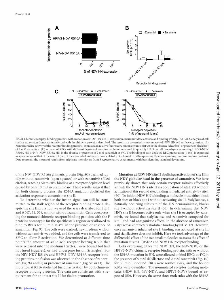
of the NiV-NDV R516A chimeric protein (Fig. 8C) declined rap-idly without zanamivir (open squares) or with zanamivir (filledcircles), reaching 50 to 60% binding at a receptor depletion levelcaused by only 10 mU neuraminidase. These results suggest thatfor both chimeric proteins, the R516A mutation abolished theactivation response to zanamivir at site II.
To determine whether the fusion signal can still be trans-mitted to the stalk region of the receptor binding protein de-spite the site II mutation, we used the assay described for Fig. 1and 6 (47, 51, 55), with or without zanamivir. Cells coexpress-ing the mutated chimeric receptor binding proteins with the Fproteins homotypic for the specific stalk region were allowed tobind to RBCs for 30 min at 4°C in the presence or absence ofzanamivir (Fig. 9). The cells were washed, new medium with orwithout zanamivir was added, and the cells were transferred to37°C to allow F activation. We determined at different timepoints the amount of sialic acid receptor-bearing RBCs thatwere released into the medium (circles), were bound but hadnot fused (squares), or had undergone fusion (triangles). Forthe NiV-NDV R516A and HPIV3-NDV R516A receptor bind-ing proteins, no fusion was observed in the absence of zanami-vir (Fig. 9A and C) or presence of zanamivir (Fig. 9B or D). Themutation at R516 abolished fusion activation by both chimericreceptor binding proteins. The data are consistent with a re-quirement for an intact site II for fusion promotion.
Mutation at NDV HN site II abolishes activation of site II inthe NDV globular head in the presence of zanamivir. We havepreviously shown that only certain receptor mimics effectivelyactivate the NDV HN=s site II via occupation of site I; yet withoutactivation of this second site, binding is mediated entirely by site I(50). To inhibit NDV HN=s binding, a molecule must either blockboth sites or block site I without activating site II. Sialyllactose, anaturally occurring substrate of the HN neuraminidase, blockssite I without activating site II (50). In determining that NDVHN=s site II becomes active only when site I is occupied by zana-mivir, we found that sialyllactose and zanamivir competed forsite I and had antagonistic effects. In the absence of zanamivir,sialyllactose completely abolished binding by NDV HN. However,once zanamivir inhabited site I, binding was activated at site II,and sialyllactose does not inhibit. Here we took advantage of thedifferential effect of the two small molecules to assess the effect ofmutation at site II (R516A) on NDV HN receptor binding.
Cells expressing either the NDV HN, the NiV-NDV, or theHPIV3-NDV chimeric receptor binding protein, with or withoutthe R516A mutation in HN, were allowed to bind RBCs at 4°C inthe presence of 5 mM sialyllactose and 2 mM zanamivir (Fig. 10)for 30 min, unbound RBCs were washed away, and the boundRBCs were quantified. The nonmutated receptor binding mole-cules (NDV HN, NiV-NDV, and HPIV3-NDV) bound as ex-pected (50). However, the same three molecules with the R516A
FIG 8 Chimeric receptor binding proteins with mutation at NDV HN site II: expression, neuraminidase activity, and binding avidity. (A) FACS analysis of cellsurface expression from cells transfected with the chimeric proteins described. The results are presented as percentages of NDV HN cell surface expression. (B)Neuraminidase activity of the receptor binding proteins, expressed in relative fluorescence intensity units (RFU) in the absence (clear bar) or presence (black bar)of 2 mM zanamivir. (C) A panel of RBCs with different degrees of receptor depletion was used to quantify HAD on cell monolayers expressing HPIV3-NDVR516A HN or NiV-NDV R516A HN in the absence or presence of 2 mM zanamivir at 4°C. The binding of each depleted RBC preparation (y axis) is expressedas a percentage of that of the control (i.e., of the amount of untreated, nondepleted RBCs bound to cells expressing the corresponding receptor binding protein).Data represent the means of results from triplicate monolayers from 3 representative experiments, with bars denoting standard deviations.
Porotto et al.
5736 jvi.asm.org Journal of Virology
on April 12, 2018 by guest
http://jvi.asm.org/
Dow
nloaded from
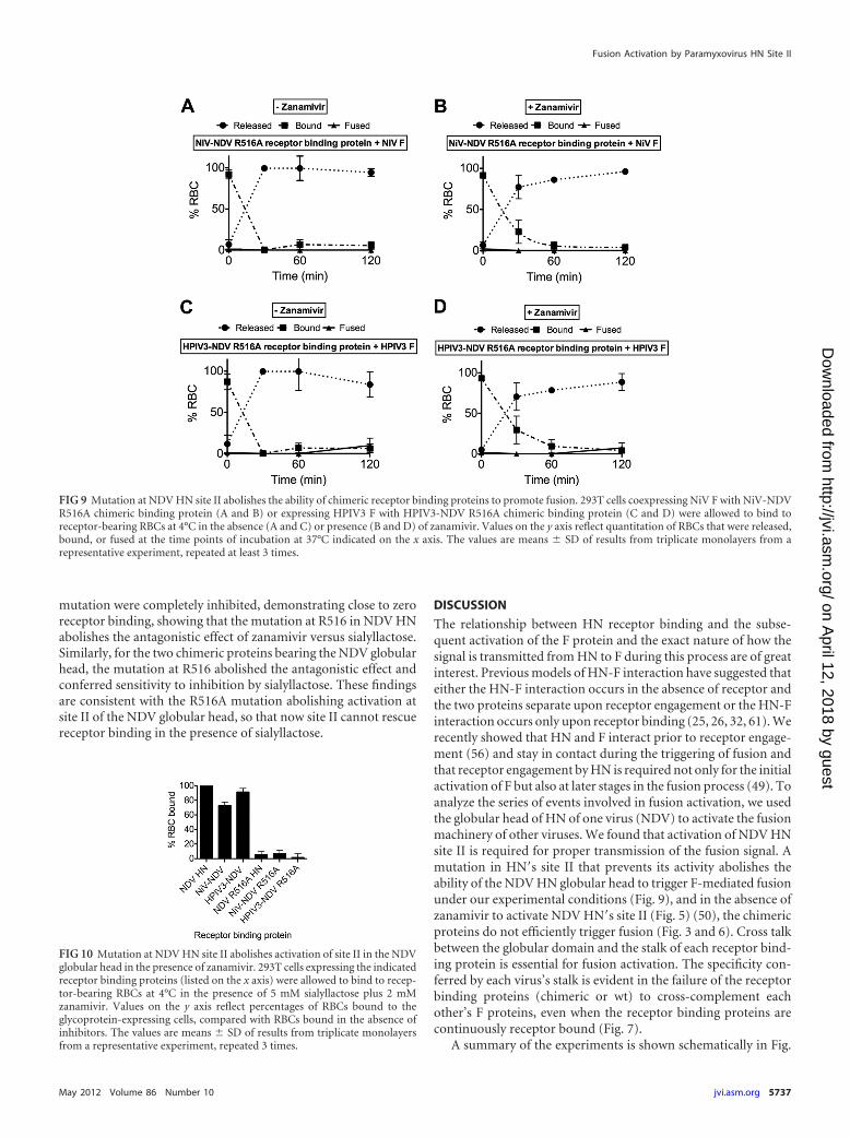
mutation were completely inhibited, demonstrating close to zeroreceptor binding, showing that the mutation at R516 in NDV HNabolishes the antagonistic effect of zanamivir versus sialyllactose.Similarly, for the two chimeric proteins bearing the NDV globularhead, the mutation at R516 abolished the antagonistic effect andconferred sensitivity to inhibition by sialyllactose. These findingsare consistent with the R516A mutation abolishing activation atsite II of the NDV globular head, so that now site II cannot rescuereceptor binding in the presence of sialyllactose.
DISCUSSION
The relationship between HN receptor binding and the subse-quent activation of the F protein and the exact nature of how thesignal is transmitted from HN to F during this process are of greatinterest. Previous models of HN-F interaction have suggested thateither the HN-F interaction occurs in the absence of receptor andthe two proteins separate upon receptor engagement or the HN-Finteraction occurs only upon receptor binding (25, 26, 32, 61). Werecently showed that HN and F interact prior to receptor engage-ment (56) and stay in contact during the triggering of fusion andthat receptor engagement by HN is required not only for the initialactivation of F but also at later stages in the fusion process (49). Toanalyze the series of events involved in fusion activation, we usedthe globular head of HN of one virus (NDV) to activate the fusionmachinery of other viruses. We found that activation of NDV HNsite II is required for proper transmission of the fusion signal. Amutation in HN=s site II that prevents its activity abolishes theability of the NDV HN globular head to trigger F-mediated fusionunder our experimental conditions (Fig. 9), and in the absence ofzanamivir to activate NDV HN=s site II (Fig. 5) (50), the chimericproteins do not efficiently trigger fusion (Fig. 3 and 6). Cross talkbetween the globular domain and the stalk of each receptor bind-ing protein is essential for fusion activation. The specificity con-ferred by each virus’s stalk is evident in the failure of the receptorbinding proteins (chimeric or wt) to cross-complement eachother’s F proteins, even when the receptor binding proteins arecontinuously receptor bound (Fig. 7).
A summary of the experiments is shown schematically in Fig.
FIG 9 Mutation at NDV HN site II abolishes the ability of chimeric receptor binding proteins to promote fusion. 293T cells coexpressing NiV F with NiV-NDVR516A chimeric binding protein (A and B) or expressing HPIV3 F with HPIV3-NDV R516A chimeric binding protein (C and D) were allowed to bind toreceptor-bearing RBCs at 4°C in the absence (A and C) or presence (B and D) of zanamivir. Values on the y axis reflect quantitation of RBCs that were released,bound, or fused at the time points of incubation at 37°C indicated on the x axis. The values are means � SD of results from triplicate monolayers from arepresentative experiment, repeated at least 3 times.
FIG 10 Mutation at NDV HN site II abolishes activation of site II in the NDVglobular head in the presence of zanamivir. 293T cells expressing the indicatedreceptor binding proteins (listed on the x axis) were allowed to bind to recep-tor-bearing RBCs at 4°C in the presence of 5 mM sialyllactose plus 2 mMzanamivir. Values on the y axis reflect percentages of RBCs bound to theglycoprotein-expressing cells, compared with RBCs bound in the absence ofinhibitors. The values are means � SD of results from triplicate monolayersfrom a representative experiment, repeated 3 times.
Fusion Activation by Paramyxovirus HN Site II
May 2012 Volume 86 Number 10 jvi.asm.org 5737
on April 12, 2018 by guest
http://jvi.asm.org/
Dow
nloaded from
11. The receptor binding proteins are comprised of the stalk cor-responding to the F protein in each pair (NDV, HPIV3, or NiV;each has its own color) and the globular head domain of NDV HN.Each row depicts a different receptor binding protein stalk/F pair.In the presence of zanamivir, the neuraminidase activity of site I ofthe NDV HN heads is occupied, and binding site II is activated.Upon engagement of site II with sialic acid receptor, each F istriggered, inserting its fusion peptide into the target membrane,and fusion ensues. The chimeric proteins were found to efficientlyactivate their respective fusion proteins, despite having a headregion belonging to a very different virus, but did not activate theheterologous F protein. We propose that these findings provide astrong indication that the globular head of HN, once receptorbound, transmits the signal to the receptor binding protein’s stalk,permitting fusion to proceed.
Binding site II on NDV HN, revealed by cocrystallization withthiosialoside (69), is at the dimer interface of the molecule andmade up of hydrophobic residues from both monomers. Muta-tions at residue R516, a residue involved in the interaction withsialic acid, resulted in HNs that when coexpressed with F are lessefficient in fusion promotion (9). In a previous report, NDV HNsmutated at site II (R516A or R516S) were found to bind RBCssimilarly to wt HN in hemagglutination (HA) assays, a findinginterpreted to mean that site II did not contribute to receptorbinding activity (9). We have shown that the R516A mutationablates the activation of site II in response to zanamivir andthereby alters binding avidity of site II in the presence of zanami-vir. The finding that zanamivir does not inhibit receptor bindingfor the receptor binding protein bearing the R516A mutation wasunexpected in light of a previous report showing that the small
FIG 11 Schematic representation of fusion activation by NDV site II. See Discussion, describing the use of NDV HN or chimeric HPIV3 HN-NDV HN or NiVG-NDV HN as receptor binding proteins, paired with NDV, HPIV3, or NiV F. Each row depicts a different HN stalk/F pair, as indicated. The NDV HN headsengage sialic acid receptors in the presence of zanamivir (the yellow molecule appearing in the second column). Zanamivir blocks binding by NDV HN=s site Ibut activates binding site II. Upon engagement of site II, each F inserts its fusion peptide into the target membrane, and fusion ensues.
Porotto et al.
5738 jvi.asm.org Journal of Virology
on April 12, 2018 by guest
http://jvi.asm.org/
Dow
nloaded from

transition state analog BCX2798 blocked binding by NDV HNR516A (9). Unavailability of BCX2798 prevents a direct experi-mental comparison; however, we speculate that the Kansas NDVglobular head used in that study differs from the AV NDV globu-lar head used here and/or that it is possible for site I to retainbinding function even when occupied by zanamivir. The experi-ments shown in Fig. 8, 9, and 10 indicate that mutation at site IIabolishes activation of that site and that receptor binding proteinsbearing the mutated site II in the head of NDV fail to efficientlypromote the fusion process for the HPIV3 and NiV paramyxovi-rus F proteins.
Site II of NDV HN is activated in the presence of zanamivir butnot in the presence of the receptor analog sialyllactose (50). Wehypothesize that our results with this artificial addition of zana-mivir are indicative of what may happen during viral infection.Zanamivir was designed based on transition state analogs and issimilar in structure to the unsaturated derivative 2-deoxy-2,3-de-hydro-N-acetylneuraminic acid (DANA), which naturally occursas a by-product of the HN=s neuraminidase activity (58, 69). Wepropose that the generation of a transition state compound inactive site I during receptor interaction leads to activation of siteII. Such a reaction intermediate, which would resemble zanamivirrather than sialyllactose, may activate site II under physiologicalconditions. In this case, after initial binding of NDV HN via itsbifunctional site I to a new cell and initiation of neuraminidasecleavage of receptor moieties, site II, which binds more avidly butlacks neuraminidase, becomes activated (50, 69). Although thishypothesis will need to be confirmed with additional studies in thefuture, this would explain the absolute requirement, at least forNDV, of neuraminidase activity for proper binding of HN andconsequent fusion (23, 32). The interplay between the two NDVsites, which is needed for proper fusion activation, is also a noveltarget for the development of new inhibitors. The mechanism bywhich the binding of zanamivir to site I activates site II is of greatinterest, since it would provide insight into the roles of distinctregions of NDV HN in the multiple functions of this molecule (16,24, 25, 30).
Our laboratory together with others has proposed that a sec-ond receptor binding site for several different HNs exists (8, 35,47, 51), even though crystallographic evidence for viruses otherthan NDV is currently lacking (69). In previous studies, we iden-tified a putative HPIV3 site II that is involved in receptor bindingand fusion activation (47, 51). The HN mutation H552Q con-ferred partial resistance to both zanamivir (54) and another site Ibinding/neuraminidase inhibitor, BCX2855 (35), consistent withthe notion that the mutation enhances binding by site II. Removalof glycans at various positions on HN (4, 35) unmasks receptorbinding at site II, as demonstrated in the presence of small mole-cules that block site I. A conflicting report suggested that forHPIV1, mutation at residue 523 creates a second receptor bindingsite (8, 35, 47, 51). While only structural studies will settle thesematters, there is now clear evidence of the existence of a site II forHPIV3 and HPIV1 HNs. For HPIV3, mutations in site II of HNmodulate viral growth in the natural host (46). Future studies willassess whether the globular head of HPIV3 is able to activate stalkregions of other receptor binding proteins and whether this acti-vation depends on site I or on the putative site II (51).
The recent availability of the crystal structures of the NDV andPIV5 stalk regions confirms that these domains form four-helicalbundles (7, 67). In the NDV crystal, the stalk is connected to the
globular head by an unresolved connecting region, and the glob-ular head is bent to the side (67). The structural data led, in thatreport, to the suggestion that upon receptor engagement, theglobular head is pulled up (67). In Fig. 12, we show a homologymodel of the HPIV3-NDV chimera, based on the crystal structureof NDV HN (67). The stalk region of the chimera was built bysequence alignment of HPIV3 HN (residues 97 to 144) with NDVHN. The conserved Pro111 in the stalk domain of HPIV3 isaligned with Pro93 in NDV HN proteins. The residues in thelinker region (dashed lines) that are disordered in the crystalstructure were manually built to connect the C-terminal end ofthe stalk with the nearest N terminus of the receptor binding do-main. We previously showed that a chimera of HPIV3 HN(1-144)–NDV(124-571) is functional for receptor engagement andfusion promotion (19). The chimera that we present here, with theextended linker region (HPIV3 HN 1 to 166; Fig. 2A), also acti-vates the F protein in the presence of zanamivir. Comparing thesechimeric HPIV3-NDV receptor binding proteins reveals that dif-ferent lengths of loop between the stalk and the globular head aretolerated, consistent with the proposed mechanism that upon re-ceptor engagement, the globular head is pulled up, which thenleads to the transmission of the fusion signal through the receptorbinding protein’s stalk domain (Fig. 12). For measles virus recep-tor binding protein (H) as well, it has been shown that the lengthof the intervening region between the stalk and the globular headcan be altered without complete loss of function (45). A specialcase may occur for respiratory syncytial virus (RSV), where infec-tion does not seem to require any surface protein other than F andthe mechanism of activation of F remains to be determined (10).
It will be important to apply the set of experimental strategiesthat we used here to additional paramyxoviruses that lack receptordestroying (or cleaving) activity and whose receptors are not sialicacid based. For these viruses, including measles virus, it has beensuggested that the receptor binding protein mainly exerts a repres-sive role (3, 5, 12, 25, 30, 34, 42, 48); the receptor binding proteinsstabilize the metastable state of the fusion proteins prior to recep-
FIG 12 Homology model of HPIV3-NDV HN chimera. Fusion-related con-formational changes are relayed from the receptor binding domain to the stalkthrough the domain interface. The chimeras of HPIV3 HN (yellow and or-ange) and NDV HN (green and cyan) likely preserve structural elements at theinterface that are important for fusion activation. The chimera of HPIV3HN(1–166)–NDV HN(124 –571) has a long linker region (indicated by adashed line) compared with the HPIV3 HN(1–144) chimera (in solid line) andwt NDV HN.
Fusion Activation by Paramyxovirus HN Site II
May 2012 Volume 86 Number 10 jvi.asm.org 5739
on April 12, 2018 by guest
http://jvi.asm.org/
Dow
nloaded from
tor engagement, and upon receptor binding, the fusion proteinsare released and proceed independently to fusion (25, 30, 34, 42).Based on the results presented here, taken together with ourprevious data, we favor a model for paramyxovirus fusion ac-tivation in which receptor engagement is required for the acti-vation of F. The finding that one paramyxovirus receptor bind-ing protein’s globular head domain can activate at least twoother paramyxovirus F proteins to proceed through fusion sug-gests that activation proceeds via similar mechanisms for thesedifferent viruses. The sialic acid binding NDV HN head acti-vates the F protein of a virus whose receptor binding protein(G) binds proteinaceous receptors (6, 43), suggesting that de-spite the use of different specific receptor molecules by theseviruses, the mechanism may be similar.
ACKNOWLEDGMENTS
We are grateful to Ashton Kutcher and Jonathan Ledecky for theirsupport, to Dan and Nancy Paduano for support of innovative re-search projects, and to the Friedman Family Foundation for renova-tion of our laboratories at Weill Cornell Medical College. We acknowl-edge flow cytometry support from Sergei Rudchenko in the FlowCytometry Facility of the Hospital for Special Surgery/Weill CornellMedical College.
The work was supported by NIH (NIAID) grants R01 AI31971 to A.M.and R21 EBO11707 to M.P. R.X. and I.A.W. were supported in part byNIAID grant AI058113 (to I.A.W.) and the Skaggs Institute for ChemicalBiology.
REFERENCES1. Afonine PV, Grosse-Kunstleve RW, Adams PD. 2005. A robust bulk-
solvent correction and anisotropic scaling procedure. Acta Crystallogr. DBiol. Crystallogr. 61:850 – 855.
2. Aguilar HC, Aspericueta V, Robinson LR, Aanensen KE, Lee B. 2010. Aquantitative and kinetic fusion protein-triggering assay can discern dis-tinct steps in the Nipah virus membrane fusion cascade. J. Virol. 84:8033–8041.
3. Aguilar HC, et al. 2006. N-glycans on Nipah virus fusion protein protectagainst neutralization but reduce membrane fusion and viral entry. J.Virol. 80:4878 – 4889.
4. Alymova IV, et al. 2008. Loss of the N-linked glycan at residue 173 ofhuman parainfluenza virus type 1 hemagglutinin-neuraminidase exposesa second receptor-binding site. J. Virol. 82:8400 – 8410.
5. Bishop KA, et al. 2008. Residues in the stalk domain of the hendra virusG glycoprotein modulate conformational changes associated with recep-tor binding. J. Virol. 82:11398 –11409.
6. Bonaparte MI, et al. 2005. Ephrin-B2 ligand is a functional receptor forHendra virus and Nipah virus. Proc. Natl. Acad. Sci. U. S. A. 102:10652–10657.
7. Bose S, et al. 2011. Structure and mutagenesis of the parainfluenza virus5 hemagglutinin-neuraminidase stalk domain reveals a four-helix bundleand the role of the stalk in fusion promotion. J. Virol. 85:12855–12866.
8. Bousse T, Takimoto T. 2006. Mutation at residue 523 creates a secondreceptor binding site on human parainfluenza virus type 1 hemagglutinin-neuraminidase protein. J. Virol. 80:9009 –9016.
9. Bousse TL, et al. 2004. Biological significance of the second receptorbinding site of Newcastle disease virus hemagglutinin-neuraminidaseprotein. J. Virol. 78:13351–13355.
10. Chaiwatpongsakorn S, Epand RF, Collins PL, Epand RM, Peeples ME.2011. Soluble respiratory syncytial virus fusion protein in the fully cleaved,pretriggered state is triggered by exposure to low-molarity buffer. J. Virol.85:3968 –3977.
11. Connaris H, et al. 2002. Probing the sialic acid binding site of the hem-agglutinin-neuraminidase of Newcastle disease virus: identification of keyamino acids involved in cell binding, catalysis, and fusion. J. Virol. 76:1816 –1824.
12. Corey EA, Iorio RM. 2007. Mutations in the stalk of the measles virushemagglutinin protein decrease fusion but do not interfere with virus-
specific interaction with the homologous fusion protein. J. Virol. 81:9900 –9910.
13. Corey EA, Mirza AM, Levandowsky E, Iorio RM. 2003. Fusion defi-ciency induced by mutations at the dimer interface in the Newcastle dis-ease virus hemagglutinin-neuraminidase is due to a temperature-dependent defect in receptor binding. J. Virol. 77:6913– 6922.
14. Crennell S, Takimoto T, Portner A, Taylor G. 2000. Crystal structure ofthe multifunctional paramyxovirus hemagglutinin-neuraminidase. Nat.Struct. Biol. 7:1068 –1074.
15. Deng R, Wang Z, Mirza A, Iorio R. 1995. Localization of a domain on theparamyxovirus attachment protein required for the promotion of cellularfusion by its homologous fusion protein spike. Virology 209:457– 469.
16. Dutch RE. 2010. Entry and fusion of emerging paramyxoviruses. PLoSPathog. 6:e1000881.
17. Eckert DM, Kim PS. 2001. Mechanisms of viral membrane fusion and itsinhibition. Annu. Rev. Biochem. 70:777– 810.
18. Emsley P, Lohkamp B, Scott WG, Cowtan K. 2010. Features and devel-opment of Coot. Acta Crystallogr. D Biol. Crystallogr. 66:486 –501.
19. Farzan SF, et al. 2011. Premature activation of the paramyxovirus fusionprotein before target cell attachment with corruption of the viral fusionmachinery. J. Biol. Chem. 286:37945–37954.
20. Greengard O, Poltoratskaia N, Leikina E, Zimmerberg J, Moscona A.2000. The anti-influenza virus agent 4-GU-DANA (Zanamivir) inhibitscell fusion mediated by human parainfluenza virus and influenza virusHA. J. Virol. 74:11108 –11114.
21. Harrison SC. 2008. Viral membrane fusion. Nat. Struct. Mol. Biol. 15:690 – 698.
22. Hashiguchi T, et al. 2011. Structure of the measles virus hemagglutininbound to its cellular receptor SLAM. Nat. Struct. Mol. Biol. 18:135–141.
23. Iorio RM, et al. 2001. Structural and functional relationship between thereceptor recognition and neuraminidase activities of the Newcastle diseasevirus hemagglutinin-neuraminidase protein: receptor recognition is de-pendent on neuraminidase activity. J. Virol. 75:1918 –1927.
24. Iorio RM, Mahon PJ. 2008. Paramyxoviruses: different receptors—different mechanisms of fusion. Trends Microbiol. 16:135–137.
25. Iorio RM, Melanson VR, Mahon PJ. 2009. Glycoprotein interactions inparamyxovirus fusion. Future Virol. 4:335–351.
26. Lamb RA, Paterson RG, Jardetzky TS. 2006. Paramyxovirus membranefusion: lessons from the F and HN atomic structures. Virology 344:30 –37.
27. Latger-Cannard V, et al. 2000. Determination of polymorphonuclearneutrophil adhesion receptors. Effect of pre-analytic factors. J. Mal. Vasc.25:181–186. (In French.)
28. Latger-Cannard V, et al. 2000. Use of standard for quantitation of adhe-sion of polynuclear neutrophils by flow cytometry. Ann. Biol. Clin. (Paris)58:337–343. (In French.)
29. Lawrence MC, et al. 2004. Structure of the haemagglutinin-neuraminidase from human parainfluenza virus type III. J. Mol. Biol.335:1343–1357.
30. Lee B, Ataman ZA. 2011. Modes of paramyxovirus fusion: a Henipavirusperspective. Trends Microbiol. 19:389 –399.
31. Lee KK, et al. 2011. Capturing a fusion intermediate of influenza hem-agglutinin with a cholesterol-conjugated peptide: a new antiviral strategyfor influenza virus. J. Biol. Chem. 286:42141– 42149.
32. Li J, Quinlan E, Mirza A, Iorio RM. 2004. Mutated form of the Newcastledisease virus hemagglutinin-neuraminidase interacts with the homolo-gous fusion protein despite deficiencies in both receptor recognition andfusion promotion. J. Virol. 78:5299 –5310.
33. Mahon PJ, Mirza AM, Iorio RM. 2011. Role of the two sialic acid bindingsites on the Newcastle disease virus HN protein in triggering the interac-tion with the F protein required for the promotion of fusion. J. Virol.85:12079 –12082.
34. Mirza AM, et al. 2011. Triggering of the Newcastle disease virus fusionprotein by a chimeric attachment protein that binds to Nipah virus recep-tors. J. Biol. Chem. 286:17851–17860.
35. Mishin VP, et al. 2010. N-linked glycan at residue 523 of human parain-fluenza virus type 3 hemagglutinin-neuraminidase masks a second recep-tor-binding site. J. Virol. 84:3094 –3100.
36. Moosmann P, Rusconi S. 1996. Alpha complementation of LacZ in mam-malian cells. Nucleic Acids Res. 24:1171–1172.
37. Moscona A. 2005. Entry of parainfluenza virus into cells as a target forinterrupting childhood respiratory disease. J. Clin. Invest. 115:1688 –1698.
38. Moscona A, Peluso R. 1991. Properties of the human parainfluenza virus
Porotto et al.
5740 jvi.asm.org Journal of Virology
on April 12, 2018 by guest
http://jvi.asm.org/
Dow
nloaded from
type 3 RNA polymerase/replicase in vitro: consensus with other negative-stranded RNA viruses. J. Virol. 65:4470 – 4474.
39. Moscona A, Peluso RW. 1992. Fusion properties of cells infected withhuman parainfluenza virus type 3: receptor requirements for viral spreadand virus-mediated membrane fusion. J. Virol. 66:6280 – 6287.
40. Moscona A, Peluso RW. 1993. Relative affinity of the human parainflu-enza virus 3 hemagglutinin-neuraminidase for sialic acid correlates withvirus-induced fusion activity. J. Virol. 67:6463– 6468.
41. Murrell M, Porotto M, Weber T, Greengard O, Moscona A. 2003.Mutations in human parainfluenza virus type 3 HN causing increasedreceptor binding activity and resistance to the transition state sialic acidanalog 4-GU-DANA (zanamivir). J. Virol. 77:309 –317.
42. Navaratnarajah CK, et al. 2011. The heads of the measles virus attach-ment protein move to transmit the fusion-triggering signal. Nat. Struct.Mol. Biol. 18:128 –134.
43. Negrete OA, et al. 2005. EphrinB2 is the entry receptor for Nipah virus, anemergent deadly paramyxovirus. Nature 436:401– 405.
44. Noyce RS, et al. 2011. Tumor cell marker PVRL4 (nectin 4) is an epithelialcell receptor for measles virus. PLoS Pathog. 7:e1002240.
45. Paal T, et al. 2009. Probing the spatial organization of measles virus fusioncomplexes. J. Virol. 83:10480 –10493.
46. Palermo L, et al. 2009. Human parainfluenza virus infection of the airwayepithelium: the viral hemagglutinin-neuraminidase regulates fusion pro-tein activation and modulates infectivity. J. Virol. 83:6900 – 6908.
47. Palermo LM, Porotto M, Greengard O, Moscona A. 2007. Fusionpromotion by a paramyxovirus hemagglutinin-neuraminidase protein:pH modulation of receptor avidity of binding sites I and II. J. Virol. 81:9152–9161.
48. Plemper RK, Hammond AL, Gerlier D, Fielding AK, Cattaneo R. 2002.Strength of envelope protein interaction modulates cytopathicity of mea-sles virus. J. Virol. 76:5051–5061.
49. Porotto M, et al. 2011. Spring-loaded model revisited: Paramyxovirusfusion requires engagement of a receptor binding protein beyond initialtriggering of the fusion protein. J. Virol. 85:12867–12880.
50. Porotto M, et al. 2006. Paramyxovirus receptor-binding molecules: en-gagement of one site on the hemagglutinin-neuraminidase protein mod-ulates activity at the second site. J. Virol. 80:1204 –1213.
51. Porotto M, Fornabaio M, Kellogg G, Moscona A. 2007. A secondreceptor binding site on the human parainfluenza 3 hemagglutinin-neuraminidase contributes to activation of the fusion mechanism. J. Virol.81:3216 –3228.
52. Porotto M, Greengard O, Poltoratskaia N, Horga M-A, Moscona A.2001. Human parainfluenza virus type 3 HN-receptor interaction: theeffect of 4-GU-DANA on a neuraminidase-deficient variant. J. Virol. 75:7481–7488.
53. Porotto M, Murrell M, Greengard O, Doctor L, Moscona A. 2005.Influence of the human parainfluenza virus 3 attachment protein’s neur-aminidase activity on its capacity to activate the fusion protein. J. Virol.79:2383–2392.
54. Porotto M, et al. 2004. Inhibition of parainfluenza type 3 and Newcastledisease virus hemagglutinin-neuraminidase receptor binding: effect of re-ceptor avidity and steric hindrance at the inhibitor binding sites. J. Virol.78:13911–13919.
55. Porotto M, Murrell M, Greengard O, Moscona A. 2003. Triggering ofhuman parainfluenza virus 3 fusion protein(F) by the hemagglutinin-neuraminidase (HN): an HN mutation diminishing the rate of F activa-tion and fusion. J. Virol. 77:3647–3654.
56. Porotto M, Palmer SG, Palermo LM, Moscona A. 2012. Mechanism offusion triggering by human parainfluenza virus type III: communicationbetween the viral glycoproteins during entry. J. Biol. Chem. 287:778 –793.
57. Porotto M, Yokoyama C, Orefice G, Kim H-S, Moscona A. 2009. Kineticdependence of paramyxovirus entry inhibition. J. Virol. 83:6947– 6951.
58. Ryan C, et al. 2006. Structural analysis of a designed inhibitor complexedwith the hemagglutinin-neuraminidase of Newcastle disease virus. Glyco-conj. J. 23:135–141.
59. Sapir A, Avinoam O, Podbilewicz B, Chernomordik LV. 2008. Viral anddevelopmental cell fusion mechanisms: conservation and divergence.Dev. Cell 14:11–21.
60. Smith EC, Dutch RE. 2010. Side chain packing below the fusion peptidestrongly modulates triggering of the Hendra virus F protein. J. Virol. 84:10928 –10932.
61. Smith EC, Popa A, Chang A, Masante C, Dutch RE. 2009. Viral entrymechanisms: the increasing diversity of paramyxovirus entry. FEBS J. 276:7217–7227.
62. Smith KB, Ellis SA. 1999. Standardisation of a procedure for quantifyingsurface antigens by indirect immunofluorescence. J. Immunol. Methods228:29 –36.
63. Wang Z, Mirza AM, Li J, Mahon PJ, Iorio RM. 2004. An oligosaccharideat the C-terminus of the F-specific domain in the stalk of the humanparainfluenza virus 3 hemagglutinin-neuraminidase modulates fusion.Virus Res. 99:177–185.
64. Weissenhorn W, Hinz A, Gaudin Y. 2007. Virus membrane fusion. FEBSLett. 581:2150 –2155.
65. White JM, Delos SE, Brecher M, Schornberg K. 2008. Structures andmechanisms of viral membrane fusion proteins: multiple variations on acommon theme. Crit. Rev. Biochem. Mol. Biol. 43:189 –219.
66. Yanagi Y, Takeda M, Ohno S, Hashiguchi T. 2009. Measles virus recep-tors. Curr. Top. Microbiol. Immunol. 329:13–30.
67. Yuan P, et al. 2011. Structure of the Newcastle disease virus hemaggluti-nin-neuraminidase (HN) ectodomain reveals a four-helix bundle stalk.Proc. Natl. Acad. Sci. U. S. A. 108:14920 –14925.
68. Yuan P, et al. 2005. Structural studies of the parainfluenza virus 5 hem-agglutinin-neuraminidase tetramer in complex with its receptor, sialyllac-tose. Structure 13:803– 815.
69. Zaitsev V, et al. 2004. Second sialic acid binding site in Newcastle diseasevirus hemagglutinin-neuraminidase: implications for fusion. J. Virol. 78:3733–3741.
Fusion Activation by Paramyxovirus HN Site II
May 2012 Volume 86 Number 10 jvi.asm.org 5741
on April 12, 2018 by guest
http://jvi.asm.org/
Dow
nloaded from