the selective arterial calcium injection test is a...
TRANSCRIPT

Hiroshima J. Med. Sci.Vol. 65, No. 1, 13~17, March, 2016HIJM 65–3
13
Gastrinoma is the most common neuroendocrine tumor in patients with multiple endocrine neopla-sia type 1 (MEN 1)6,10,11). Gastrinomas in a patient with MEN 1 tend to be multiple and too small for imaging methods to show them, making the local-ization and diagnosis of these microgastrinomas difficult preoperatively. Endoscopic ultrasonogra-phy (EUS) is a useful method for showing and di-agnosing submucosal microgastrinomas, but in a case of severe stenosis caused by recurrent ulcers, it was impossible for EUS to pass through the ste-nosis and show the microgastrinomas. Some have recommended the use of the selective arterial calci-um injection (SACI) test as a valid diagnostic
method for the localization of these invisible micro-gastrinomas preoperatively4,8,12). We report a rare case of multiple invisible duodenal microgastrino-mas and pancreatic neuroendocrine tumors in a patient with MEN 1 with severe duodenal stenosis diagnosed preoperatively by using the SACI test.
CASE PRESENTATION
A 50-year-old man was admitted to our hospital with recurrent duodenal ulcers causing epigastric pain, nausea and vomiting. He had been treated with proton pump inhibitors for duodenal ulcers for 2 years. Despite continuous proton pump
The Selective Arterial Calcium Injection Test is a Valid Diagnostic Method for Invisible Gastrinoma with Duodenal
Ulcer Stenosis: A Case ReportKenjiro OKADA1), Takeshi SUDO1), Katsunari MIYAMOTO1), Yujiro YOKOYAMA1),
Yoshihiro SAKASHITA1), Yasushi HASHIMOTO1), Hironori KOBAYASHI1), Hiroyuki OTSUKA1), Takuya SAKODA1) and Fumio SHIMAMOTO2)
1) Department of Surgery, Hiroshima Memorial Hospital, Honkawa-cho 1-4-3, Naka-ku, Hiroshima 730-0802, Japan
2) Department of Pathology, Hiroshima Memorial Hospital, Honkawa-cho 1-4-3, Naka-ku, Hiroshima 730-0802, Japan
ABSTRACTThe localization and diagnosis of microgastrinomas in a patient with multiple endocrine neo-
plasia type 1 is difficult preoperatively. The selective arterial calcium injection (SACI) test is a valid diagnostic method for the preoperative diagnosis of these invisible microgastrinomas. We report a rare case of multiple invisible duodenal microgastrinomas with severe duodenal steno-sis diagnosed preoperatively by using the SACI test. A 50-year-old man was admitted to our hospital with recurrent duodenal ulcers. His serum gastrin level was elevated to 730 pg/ml. It was impossible for gastrointestinal endoscopy to pass through to visualize the inferior part of the duodenum, because recurrent duodenal ulcers had resulted in severe duodenal stenosis. The duodenal stenosis also prevented additional endoscopic examinations such as endoscopic ultraso-nography. Computed tomography did not show any tumors in the duodenum and pancreas. The SACI test provided the evidence for a gastrinoma in the vascular territory of the inferior pancre-atic-duodenal artery. We diagnosed a gastrinoma in the peri- ampullary lesion, so we performed Subtotal Stomach-Preserving Pancreatico- duodenectomy with regional lymphadenectomy. His-topathological findings showed multiple duodenal gastrinomas with lymph node metastasis and nonfunctioning pancreatic neuroendocrine tumors. Twenty months after surgery, the patient is alive with no evidence of recurrence and a normal gastrin level. In conclusion, the SACI test can enhance the accuracy of preoperative localization and diagnosis of invisible microgastrino-mas, especially in the setting of severe duodenal stenosis.
Key words: Selective arterial calcium injection test, Gastrinoma, Duodenal stenosis, Multiple endocrine neoplasia type 1
List of abbreviations SACI: Selective arterial calcium injection, MEN 1: Multiple endocrine neoplasm type 1 EUS: Endoscopic ultrasonography, GIS: Gastrointestinal endoscopy, CT: Computed tomography IPDA: Inferior pancreatic-duodenal artery, MRI: Magnetic resonance imagingSSPPD: Subtotal Stomach-Preserving Pancreaticoduodenectomy, DNETs: Duodenal neuroendocrine tumorsPNETs: Pancreatic neuroendocrine tumors, CgA: Chromogranin A

14 K. Okada et al
parathyroid adenoma, but brain magnetic reso-nance imaging (MRI) showed a normal pituitary gland.
We diagnosed a gastrinoma in the peri-ampulla-ry lesion with MEN 1, so we performed Subtotal Stomach-Preserving Pancreaticoduodenectomy (SSPPD) with regional lymphadenectomy (Fig. 3).
Histopathological findings showed 4 small neuroen-docrine tumors sized within 3 mm in the duodenum (DNETs) (Fig. 4a-c), 4 small neuroen docrine tumors sized within 3mm in the head of pancreas (PNETs) (Fig. 5a-c) and 1 lymph node metastasis in the pos-terior part of the head of pancreas (Fig. 6a-b). Im-munohistochemical examination found that the DNETs and the lymph node metastasis were posi-tive for chromogranin A (CgA), synaptophysin and gastrin. On the other hand, the PNETs were posi-tive for CgA and synaptophysin, but negative for gastrin. The Ki-67 labeling index of the DNETs and the metastatic lymph node were about 10%, and that of the PNETs was < 2%. By WHO classi-fication 2010, the DNETs were NET G2, and the PNETs were NET G1. The patient was diagnosed with multiple duodenal gastrinomas with lymph node metastasis and nonfunctioning PNETs with MEN 1.
In the postoperative course, the pancreatic fistula as a complication was treated with conservative measures. He was discharged from the hospital on the 51st postoperative day. Ten months after the surgery, parathyroidectomy was performed for the suspected parathyroid adenoma. Histopathological findings showed the parathyroid adenoma. Now twenty months after the first surgery, SSPPD, the patient is alive with a normal gastrin level and without any evidence of recurrence.
inhibitor treatment, his abdominal symptoms did not improve. There was no other past medical history except for the duodenal ulcers. His father had died of an unspecified pancreatic tumor. Laboratory tests showed elevated levels of serum calcium (12.8 mg/dl), gastrin (730 pg/ml), and parathyroid hormone (2,000 pg/ml), but prolactin was normal (18.3 ng/ml).
Gastrointestinal endoscopy (GIS) showed a giant ulcer filling one third of the lumen of the duodenum. It was impossible to pass the en do scope through this lesion to view the inferior part, because recurrent duodenal ulcers had resulted in severe duodenal ste-nosis. In addition, the duodenal stenosis prevented additional endoscopic examinations such as EUS from showing submucosal tumors such as gastri-nomas and obtaining specimens by fine-needle as-piration of the tumors. The tissue biopsied from these ulcerative lesions was negative for duodenal carcinoma. Contrast-enhanced computed tomog-raphy (CT) showed duodenal wall thickening and edema of the descending part of the duodenum, but did not show tumors in the duodenum and pancreas (Fig. 1a). Only one lymph node sized within 16 mm in diameter in the posterior part of the head of pancreas was enhanced (Fig. 1b). An-giography and the SACI test were performed to identify the arterial supply of the gastrinoma. The gastroduodenal artery, inferior pancreatic- duodenal artery (IPDA), proper hepatic artery and splenic artery were selectively catheterized. This test provided the evidence for a gastrinoma in the vascular territory of the IPDA, the peri-ampullary lesion, and the absence of hepatic metastases (Fig. 2). In addition, parathyroid and pituitary gland evaluation were conducted to diagnose the MEN 1. Thyroid ultrasonography showed a suspected
a bFig. 1. Computed tomography.(a) Arrow indicates duodenal wall thickening and edema.(b) Arrow indicates enhanced lymph node sized within 16 mm in diameter in the posterior part of the head of pancreas.
15SACI Test for Invisible Gastrinoma with DU Stenosis
0 30sec 60sec 90sec
GDA 1300 1700 1600 2300
IPDA 1400 2800 3600 2100
PHA 1500 1600 1800 2000
SPA proximal 2100 1800 2300 2000
SPA distal 1900 1800 2100 2000
0
500
1000
1500
2000
2500
3000
3500
4000
gast
rin
(pg/
ml)
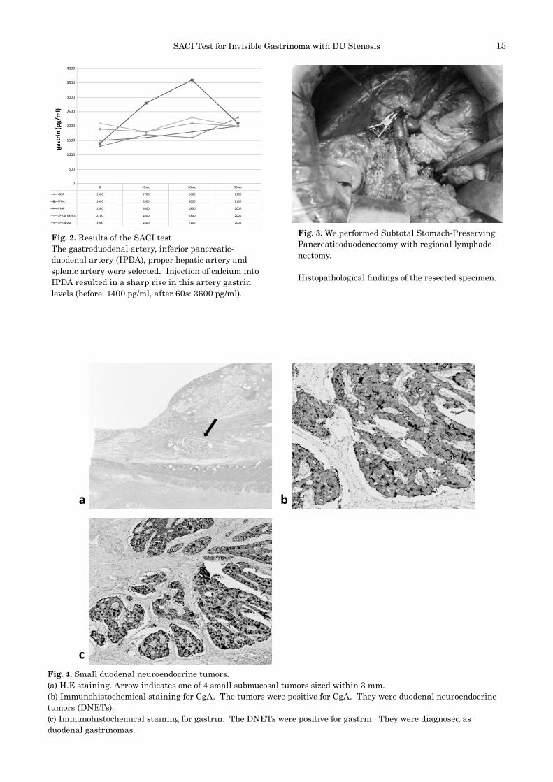
Fig. 2. Results of the SACI test.The gastroduodenal artery, inferior pancreatic-duodenal artery (IPDA), proper hepatic artery and splenic artery were selected. Injection of calcium into IPDA resulted in a sharp rise in this artery gastrin levels (before: 1400 pg/ml, after 60s: 3600 pg/ml).
Fig. 3. We performed Subtotal Stomach-Preserving Pancreaticoduodenectomy with regional lymphade-nectomy.
Histopathological findings of the resected specimen.
a b
cFig. 4. Small duodenal neuroendocrine tumors.(a) H.E staining. Arrow indicates one of 4 small submucosal tumors sized within 3 mm.(b) Immunohistochemical staining for CgA. The tumors were positive for CgA. They were duodenal neuroendocrine tumors (DNETs).(c) Immunohistochemical staining for gastrin. The DNETs were positive for gastrin. They were diagnosed as duodenal gastrinomas.

16 K. Okada et al
contrast-enhanced CT, and MRI are used for preop-erative diagnosis of microgastrinomas, but these imaging studies may fail to show them. EUS is also a useful method for detecting submucosal small tumors as small as 2-3 mm in diameter1) but, in our case, severe duodenal stenosis prevented careful examination with any standard modality. The SACI test was the only valid method for local-ization and diagnosis of the invisible microgastrino-
DISCUSSION
The SACI test is a highly accurate and safe method for the preoperative localization and diag-nosis of gastrinomas with MEN 1 in the duodenum or/and pancreas and is especially helpful in a case of duodenal stenosis. More than half of duodenal gastrinomas with MEN 1 tend to be smaller than 1 cm in diameter3,12). Imaging studies such as GIS,
a b
c
Fig. 5. Small pancreatic neuroendocrine tumors.(a) H.E staining. Arrow indicates one of 4 small pancreatic tumors sized within 3 mm.(b) Immunohistochemical staining for CgA. The tumors were positive for CgA. They were pancreatic neuroendocrine tumors (PNETs).(c) Immunohistochemical staining for gastrin. The PNETs were negative for gastrin. They were diagnosed as nonfunctioning PNETs.
a bFig. 6. The metastatic lymph node.(a) Immunohistochemical staining for CgA. The lymph node was positive for CgA. It was metastatic lymph node of NETs.(b) Immunohistochemical staining for gastrin. The metastatic lymph node was positive for gastrin. It was diagnosed as the metastatic lymph node of duodenal gastrinomas.

17SACI Test for Invisible Gastrinoma with DU Stenosis
studies. All authors read and approved the final manuscript.
(Received January 22, 2016)(Accepted February 23, 2016)
REFERENCES
1. Anderson, M.A., Carpenter, S., Thompson, N.W., Nostrant, T.T., Elta, G.H. and Scheiman, J.M. 2000. Endoscopic ultrasound is highly accurate and directs management in patients with neuroendo-crine tumors of the pancreas. Am. J. Gastroenterol. 95: 2271-2277.
2. Bartsch, D.K., Frendrich, V., Langer, P., Celik, I., Kann, P.H. and Rothmund, M. 2005. Outcome of duodenopancreatic resections in patients with multiple endocrine neoplasia type 1. Ann. Surg. 242: 757-766.
3. Frendrich, V., Langer, P., Waldmann, J., Bartsch, D.K. and Rothmund, M. 2007. Management of spo-radic and multiple endocrine neoplasia type 1 gas-trinomas. Br. J. Surg. 94: 1331-1341.
4. Fujihara, S., Mori, H., Nishiyama, N., Kobayashi, M., Kobara, H. and Masaki, T. 2012. Multiple gi-ant duodenal ulcers associated with duodenal gas-trinoma. Clin. J. Gastroenterol. 5: 64-68.
5. Gibril, F., Venzon, D.J., Ojeaburu, J.V., Bashir, S. and Jensen, R.T. 2011. Prospectivestudy of the natural history of gastrinoma in patients with MEN 1: definition of an aggressive and a nonaggressive form. J. Clin. Endocrinol. Metab. 86: 5282-5293.
6. Goudet, P., Murat, A., Binquet, C., Cardot-Bauters, C., Costa, A., Ruszniewski, P., et al. 2010. Risk factors and causes of death in MEN 1 disease. A GTE (Groupe d’Etude des Tumeurs Endocrines) cohort study among 758 patients. World J. Surg. 34: 249-255.
7. Imamura, M., Komoto, I., Doi, R., Onodera, H., Kobayashi, H. and Kawai, Y. 2005. New pancreas-preserving total duodenectomy technique. World J. Surg. 29: 203-207.
8. Imamura, M., Komoto, I., Ota, S., Hiratsuka, T., Kosugi, S., Doi, R., et al. 2011. Biochemically cura-tive surgery for gastrinoma in multiple endocrine neoplasia type 1 patient. World J. Gastroenterol. 17: 1343-1353.
9. Norton, J.A., Fraker, D.L., Alexander, H.R., Gibril, F., Liewehr, D.J., Venzon, D.J., et al. 2006. Surgery increases survival in patients with gastrinoma. Ann. Surg. 244: 410-419.
10. Sakurai, A., Suzuki, S., Kosugi, S., Okamoto, T., Uchino, S., Miya, A., et al. 2012. Multiple endo-crine neoplasia type 1 in Japan: Establishment and analysis of a multicenter database. Clin. Endoclinol. (Oxf) 76: 533-539.
11. Thakker, R.V., Newey, P.J., Walls, G.V., Bilezikian, J., Dralle, H., Ebeling, H., et al. 2012. Clinical practice guidelines for multiple endocrine neoplasia type 1 (MEN1). J. Clin. Endocrinol. Metab. 97: 2990-3011.
12. Thompson, C. and Courtney, M. 2006. Townsend: endocrine pancreas, p. 625-666. Sabiston textbook of surgery. 18th ed. Saunders Elsevier, Philadelphia.
mas.The treatment strategy for gastrinomas with
MEN 1 has been controversial. We recommend early and aggressive surgical resection of gastri-nomas, because recently published articles suggest that this improves survival rates and the long term biomedical cure of gastrinomas as well as de-creasing the rate of hepatic metastases2,5,7,9). The preoperative diagnosis in our case was invisible gastrinomas in the peri-ampullary lesion, so we selected “total” duodenectomy rather than “par-tial” duodenectomy. In addition, any regional lymph nodes around the head of the pancreas and hepatic artery should be dissected, because duode-nal gastrinomas reportedly metastasize to region-al lymph nodes independent of size7,8). In our case, the preoperative CT showed one highly enhanced lymph node sized within 16 mm in diameter in the posterior part of the head of the pancreas. Pancre-as preserving total duodenectomy to save the head of the pancreas offered a less invasive surgery compared with SSPPD, but this procedure did not allow for regional lymph node dissection and the resection of microgastrinomas in the head of the pancreas. With total duodenectomy, regional lymph node dissection and the possibility of micro-gastrinomas in the head of pancreas, we per-formed SSPPD. As a result, pathological findings showed that one highly enhanced metastatic lymph node was larger than the primary duodenal microgastrinomas, so it was possible for the meta-static lymph node to secrete more gastrin than the duodenal microgastrinomas and to be fed from IPDA, identified by the SACI test as the arterial supply of the gastrinoma. Proper preoperative di-agnosis by the SACI test and early and aggressive surgical resection contributed to curative R0 re-section and recurrence-free survival.
CONCLUSION
The SACI test can enhance the accuracy of preoperative localization and diagnosis of invisible microgastrinomas, especially in the setting of severe duodenal stenosis.
ConsentWritten informed consent was obtained from the
patient for publication of this Case Report and any accompanying images.
Competing interestsThe authors declare that they have no compet-
ing interests.
Authors’ contributionTS conceived of this case presentation and
drafted the presentation. KM, YY, YS, YH, HK, HO and TS participated in the design of this case presentation. FS carried out the pathological