the sequence of the saccharomyces cerevisiae gene ph02 codes
TRANSCRIPT

Volume 15 Number 1 1987 Nucleic Ac ids Research
The sequence of the Saccharomyces cerevisiae gene PH02 codes for a regulatory protein withunusual aminoacid composition
Christian Sengstag and Albert Hinnen
Ciba-Geigy AG, Department of Biotechnology, CH-4002 Basel, Switzerland
Received November 17, 1986; Accepted December 8, 1986
ABSTRACTA new centromere vector for the construction of a Saccharomycescerevisiae gene library, allowing direct selection for DNAinsert, will be described. From that library the gene for theregulatory protein PHO2 involved in PH05 induction has beencloned by complementation of a pho2 mutation. The complementingactivity was shown to be located on a 3.6 kb Hindlll fragment.This fragment was used to evict the genomic copy and withappropriate genetic crosses we proved, that the cloned gene isPHO2. The DNA sequence of PHO2 was determined. Analysis of thesequence data uncovered striking homology regions with PH04.another protein necessary for the induction of PH05. Therelevance of the observed homology will be discussed.
INTRODUCTION
The phosphatase system in yeast Saccharomyces cerevisiae is
a network consisting of structural genes and regulatory genes
(1). The phosphatases can be divided into alkaline (PHO8.) and
acid phosphatases (APase) (PH05. PH03. PH011). The PH03 gene,
known as the constitutive APase, is dependent on the regulatory
genes PH06 and PH07 and seems to be repressed by the PH05 gene
product (2). PH05 and PH011 are in turn regulated APases
- transcriptionally repressed in high Pi medium - that are
induced via the gene products of PH04 and PHO2. The presence of
the PH081 gene is central for the APase induction circuit. In
low Pi medium it binds the negative factors coded by PH080
and/or PH085 dissociating them from the PHO2 and/or PH04 gene
products thus allowing activation of PH05 transcription.
In analogy to GAL4 (3) a model proposes that PH04 binds as a
complex with PHO2 directly to the upstream activator sequence
(UAS) of PH05. Although genetic data put PHO2 on the same
hierarchy level as PH04. it cannot be ruled out that the action
© IRL Press Limited, Oxford, England. 2 3 3
Downloaded from https://academic.oup.com/nar/article-abstract/15/1/233/2382392by gueston 22 March 2018

Nucleic Acids Research
of PH02 is posttranscriptional. With the aim of learning more
about the function of PHO2. we cloned the PHO2 gene from a new
gene library of S. cerevisiae. Here we report the DNA sequence
of PHO2 and show that it exhibits distinct homology regions
with PH04.
MATERIALS AND METHODS
Strains
E. coli HB101 (r. , m, , leu, pro. recA) and JM 109—K ~K
(recA. endAl. qyrA96. thi. hsdR17. supE44. relA.
A(lac-pro) . F1(traD36. proAB. laclq. ZAM15))
were used for standard transformations. B15 (trp.
pyrF. r. , m. ) was used to select for the URA3
gene in E. coli on minimal plates containing
tryptophane.
Yeast S288C is our prototrophic wild-type strain.
YAT104 (a., trpl. pho3. pho2) was a generous gift of
A. Toh-e.
YS104 is a ura3A derivative of YAT104. YS18 is a
ura3A derivative of GRF18 (a, his3-ll. his3-15.
leu2-3. Ieu2-112. canR).
Media
YPD medium contained 2% Bacto Peptone (Difco), 1% Bacto Yeast
Extract (Difco) and 2% glucose. Ura+ selection was done on
synthetic complete medium (4) lacking only uracil. Antibiotics
were used at 50ug/ml for Ampicillin and 4ug/ml for
Tetracyclin. E. coli M9 minimal plates have been described (5).
Yeast transformation
Competent cells were obtained by Li-acetate treatment (5). An
exponentially growing culture in YPD was harvested at
OD,./,=2. washed with TE buffer (10 mM Tris, lmM EDTA. pH 8).600
resuspended in 1/2 vol 1M Li-acetate and snaked at 30° for 1
1/2 h. Then the cells were resuspended in 1/50 vol 1M
Li-acetate and 1-10 u<3 transforming DNA was added to 0.2 ml
competent cells. After 101 incubation at 30°C. PEG 4000 was
added to 40% final volume and incubation continued for lh.
Cells were harvested by centrifugation, washed with 0.8M
sorbitol and spread on appropriate selection plate.
234
Downloaded from https://academic.oup.com/nar/article-abstract/15/1/233/2382392by gueston 22 March 2018

Nucleic Acids Research
Isolation of DNA fragments from gel
DNA bands to recover from agarose gels were electrophoresed
into a suspension of hydroxylappatite. From there the DNA was
eluted with 0.5M phosphate buffer pH 7.5 and purified on a
Sephadex G100 mini column in TE buffer.
Introducing a deletion into the chromosomal URA3 gene of YAT104
Plasmid pUC12::URA3. containing the URA3 gene cloned as 1.1kb
Hindi11 fragment, was digested with Ncol and StuI. the ends
were filled in with Klenow polymerase and the plasmid was
religated. Deletion derivative pUC12::ura3A was digested to
completion with HindiII and used to cotransform YAT104 together
with I11Q YRp7. Trp+ transformants were selected on plate,
washed off and Ura~ mutants were selected in 5-fluoro-orotic
acid as described (7, 8). Correct integration of the ura3A
allele at the URA3 locus has been checked by Southern
hybridization.
In analogy the URA3 gene was mutated in GRF18. except that
Yepl3 was used as cotransforming plasmid.
Construction of PCS19
The l.lkb Hindlll fragment with the URA3 gene was inserted into
the HindiII site of YRP7-PH05 (8). Subsequently the plasmid was
digested with Xbal/BamHI. and the large fragment was ligated
with a lkb Xbal/Bglll fragment carrying CEN15 (9). The Clal
site was converted to a Xhol site by linker insertion giving
pCS14. In parallel the Pstl/BamHI fragment of pUN121 (10)
carrying the cl gene was cloned into Pstl/BamHI digested pUC18
followed by conversion of the PstI site to a Xhol site by
linker insertion. The short Pvul/Xhol fragment was then
replaced by the Pvul/Xhol fragment of pCS14. carrying CEN15.
followed by replacing the short PvuI/BamHI fragment by the ori
containing PvuI/BamHI fragment of pUN121. Finally the short
Pvul/Sall fragment was replaced by the long Pvul/Sall fragment
of pBR322. in which a 158bp HincII/Smal fragment containing the
E. coli trp-terminator had been inserted at the Aval site
behind the Tet gene. This vector (pCS19) has been used for
cloning random yeast DNA.
235
Downloaded from https://academic.oup.com/nar/article-abstract/15/1/233/2382392by gueston 22 March 2018
Nucleic Acids Research
Construction of PCS21
To evict the chromosomal PHO2 gene, the 3.6kb Hindlll fragment
containing PH02 (see Results) was subcloned into the Hindi11
site of pUC19. Subsequently the Hindi site located in the
vector part was eliminated by digesting the plasmid at the
overlapping Sail site and religating the plasmid upon filling
in the Sail ends by Klenow polymerase. The resulting plasmid
was digested with Hindi, religated in the presence of
end-repaired 1.1kb Hindlll fragment carrying URA3 and E. coli
strain B15 (pyrF) was transformed. Clones expressing URA3 were
selected on minimal plates (+ tryptophane) and the correct
construction (plasmid pCS21) was confirmed by restriction
enzyme digests. The yeast DNA part of pCS21 is shown in Fig. 2.
Introducing a frameshift mutation in the N-terminal part of PH02
The 3.6kb Hindlll fragment was cloned into pUC19. This plasmid
was then digested with EcoRI and the protruding 51 ends were
filled in by Klenow polymerase. One half of this end-repaired
DNA was digested with Sail and the 950 bp fragment (Fig. 2) was
isolated from an agarose gel. The other half was digested with
Bqlll and the shortest fragment was isolated. Finally, the
original plasmid was digested with Sall/Bqlll and the large
fragment was ligated with the two DNA fragments isolated
before. Restriction digestion showed, that the EcoRI site at
the N-terminal end of PH02 got lost, and DNA sequencing using a
synthetic oligodeoxynucleotide proved that we had introduced a
+4 frameshift at that particular site. The mutated Hindlll
fragment was then subcloned into pDP39 (see vectors) and
transformed in YS104.
Acid phosphatase assay
The staining assay for APase has been described (11).
Vectors
Plasmid pDP39, a generous gift from D. Pridmore, was used for
subcloning of PHO2 DNA fragments. It was constructed by
inserting URA3. ARS1 and CEN14 into pUC19. All PHO2 fragments
have been cloned into the unique Sail site, after converting it
to a blunt end site by Klenow polymerase.
236
Downloaded from https://academic.oup.com/nar/article-abstract/15/1/233/2382392by gueston 22 March 2018

Nucleic Acids Research
Fig. 1 Restriction map of vector pCS19. The Xcl geneproduct, coded by the vector, represses the XpL promoterwhich controls Tetr expression. Upon cloning into the uniqueBell site. Tetr becomes expressed allowing direct selectionfor recombinant plasmids. The introduced trp terminator isrepresented by the letter t.
RESULTS
Construction of a S. cerevisiae gene library in a centromere
vector
Gene dosage may strongly influence gene expression which can
lead to severe problems in cloning regulatory genes. Therefore
we constructed vector pCS19 for cloning random yeast DNA. Due
to its centromere, this vector (Fig. 1) is present at one to
two copies per yeast cell and is mitotically stable. By cloning
into its unique Bell site, it allows direct selection for
recombinant plasmids by expressing tetracyclin-resistance in E.
coli. For details of construction see Materials and Methods.
DNA of the wild-type yeast strain S288C was partially
digested with Sau3A. fragments in the range of 10kb were
purified from gel and ligated together with Bell cleaved pCS19.
5000 individual tetr E. coli transformants were picked and
stored. Three yeast genome equivalents should be contained in
237
Downloaded from https://academic.oup.com/nar/article-abstract/15/1/233/2382392by gueston 22 March 2018

Nucleic Acids Research
this library, giving about 95% probability for a specific gene
to be represented.
Cloning of yeast DNA sequences that complement the pho2 mutation
Plasmid DNA was purified from pools of each 384 E. coli gene
bank clones and used to transform YS104 (pho2. ura3) to lira .
Transformants selected on synthetic complete medium plates
lacking uracil were replica plated on low Pi and high Pi
medium. Elevated APase activity was monitored by a colony
staining procedure (Materials and Methods). Two transformants
showed correctly expressed APase activity and were chosen for
further analysis. Total DNA was isolated and used to transform
E. coli. Subsequently, plasmid DNA was recovered from
AmprTetr tranformants and used to transform YS104. Correct
expression of APase by the yeast transformants proved that both
plasmids could complement the pho2 mutation.
A 3.6 kb Hindi 11 fragment can complement the pho2 mutation
Restriction enzyme digestion of the two plasmids revealed
that they carried similar but not identical 10 kb long yeast
sequences. In particular a 3.6 kb Hindlll fragment was present
in both plasmids. Cloning of this Hindlll fragment in pDP39 and
subsequent transformation of YS104 demonstrated that this DNA
was sufficient for complementing pho2. A restriction map of the
Hindlll fragment is shown in Fig. 2.
DMA sequence of the 3.6 kb Hindlll fragment
Figure 2 shows the strategy used for sequencing. In brief,
various subfragments of the 3.6 kb Hindlll fragment were
subcloned into M13mpl8 or M13mpl9 and sequenced according to
the dideoxy method (12) using the M13 universal primer.
Subsequently, sequencing was continued by using synthetic
deoxyoligonucleotide-primers.
One long open reading frame was found starting nearby the
leftmost EcoRI site and extending about 200 bp to the right of
a Hindi site. It can code for a 63390 D protein consisting of
559 aminoacids. In the flanking regions open reading frames of
only less than 74 aminoacids are found. The sequence of the
long open reading frame and its 5' and 3' flanking regions is
shown in Fig. 3 together with the deduced protein sequence.
238
Downloaded from https://academic.oup.com/nar/article-abstract/15/1/233/2382392by gueston 22 March 2018

Nucleic Acids Research
pCS21
Fig. 2 A restriction map of the 3.6kb Hindlll fragment isshown in the upper part. The PH02 open reading frame isrepresented by the filled region. Arrows indicate the extent ofDNA sequence determination. Both strands have been sequencedbetween the Hpal and Clal sites. The lower part shows the yeastDNA segment present in pCS21, where the major part of the PH02gene is exchanged against the URA3 gene. The HincII/Hindlllfusion does not restore a Hindlll site. The Sail site of vectorpUC19 is present just left of the Hindlll fragment.
The long open reading frame codes for PH02
To prove that we had cloned the PH02 gene and to exclude that
the observed APase expression was due to a cloned unrelated
factor, different from PHO2. we decided to evict the putative
chromosomal PH02 region. The 3.6 kb Hindlll fragment was
subcloned into pUCl9 and and the internal Hindi fragment was
exchanged against the URA3 gene, giving pCS21 (Fig. 2) (see
Materials and Methods). Yeast strain YS18 (PH02. ura3) was
transformed with Hindlll digested pCS21 plasmid DNA and
selected for Ura+. Total DNA of ten transformants was
isolated, digested with Hindlll and electrophoresed. The gel
was blotted to nitrocellulose and probed with the
nick-translated 3.6 kb Hindlll fragment (putative PHO2.). All
ten transformants showed a unique band at 2.9 kb that was
shifted with respect to the 3.6 kb band of the parental strain
YS18. indicating an efficient gene replacement (data not
shown). Subsequent colony staining clearly showed that APase
expression became eliminated upon transformation. Furthermore
the APase deficient transformants (YS18 pho2::URA3) were
backcrossed with YS104 and the resulting diploids turned out to
lack APase activity.
239
Downloaded from https://academic.oup.com/nar/article-abstract/15/1/233/2382392by gueston 22 March 2018

Nucleic Acids Research
-380 -360 -340TGTTAACGCGTTGCGCGTTTCCTTTATCTCTGGTACTTAACAGTATATGGAGTTAAAAGT
-320 -300 -280GTGGGATTGTTTGTCGTCTAAGTAAAACCATGTGTGAGTAATCTGTGAACGATCAAGGAT
-260 -240 -220AGTGTAAAATAGCTTAGCAACAATAGTCTTTTAACATGCGGACATAAGGTCACTTTTTGG
-200 -180 -160CACAAGATGATAGTAAATGTTGCACATTATGAGCAGTATTTTAATACATCACTTCGGACG
-KD -120 -100ATGCGGCATATCCAGAATTGCAGATTTAGTAAATCACAAGCTCCAGGGAACTGTTGAAAC
-80 -60 -40CTCJTATTAAJJAGACGCAAAGACAACTAAGGAGACATTCAACAGGGCTAGACAAGTCACGG
-20 1 20CTTACTGCTAAATAACGTATACAATACGCTATGATGGAAGAATTCTCGTACGATCACGAT
MetnetGluGluPheSerTyrAspHisAsp
40 60 80TTTAACACACATTTTGCTACAGATTTGGATTATTTGCAACATGACCAACAACAACAACAAPheAsnThcHisPheAlaThrAspLeuAspTyrLeuGlnHisAspGlnGlnGlnGlnGln
100 120 140CAGCAACAACATGATCAACAACATAATCAACAGCAACAACCACAACCACAACCAATTCAAGlnGlnGlnHisAspGlnGlnHisAsnGlnGlnGlnGlnProGlnProGlnProIleGln
160 180 200ACTCAAAACCTGGAGCACGACCACGACCAACATACTAATGATATGAGTGCTTCATCGAATThrGlnAsnLeuGluHisAspHisAspGlnHisThrAsnAspMetSecAlaSerSerAsn
220 240 260GCATCAGATAGTGGACCTCAAAGGCCCAAGAGGACTCGCGCAAAGGGTGAAGCACTAGATAlaSerAspSerGlyProGlnArgProLysArgThrArgAlaLysGlyGluAlaLeuAsp
280 300 320GTGCTAAAGCGTAAATTTGAAATAAATCCAACACCCTCTTTGGTAGAAAGAAAGAAAATAValLeuLysArgLysPheGluIleAsnProThrProSerLeuValGluArgLysLysIle
340 360 3S0TCAGATCTGATAGGAATGCCTGAAAAAAACGTCAGAATTTGGTTTCAGAACAGAAGAGCTSerAspLeuIleGlyMetProGluLysAsnValArglleTrpPheGlnAsnArgArgAla
400 420 440AAATTGAGGAAAAAGCAGCATGGAAGTAATAAGGACACAATCCCCTCGTCACAATCCCGTLysLeuArgLysLysGlnHisGlySerAsnLysAspThrlleProSerSerGlnSerArg
460 480 500GATATTGCCAACGATTACGATCGTGGGAGTACAGACAACAATTTGGTCACTACAACAAGTAspIleAlaAsnAspTyrAspArgGlySerThrAspAsnAsnLeuValThrThrThrSer
520 540 560ACTTCATCCATATTTCACGATGAAGACCTGACTTTTTTCGACCGTATTCCGCTGAACAGCThcSerSerllePheHisAspGluAspLeuThrPhePheAspArglleproLeuAsnSer
5S0 600 620AACAACAACTATTATTTTTTTGACATTTGCTCAATTACTGTGGGAAGTTGGAATAGAATGAsnAsnAsnTytTycPhePheAspIleCysSerlleThrValGlySerTcpAsnArgMet
640 660 680AAAAGCGGCGCACTGCAAAGAAGGAACTTTCAGTCTATAAAGGAGTTGAGAAACCTATCGLysSerGlyAlaLeuGlnArgArgAsnPheGlnSecIleLysGluLeuArgAsnLeuSer
700 720 740CCAATAAAGATTAATAACATAATGTCGAATGCCACAGATTTAATGGTTTTGATATCCAAGPcoIleLysIleAsnAsnl leMetSerAsnAlaThrAspLeuHetvalLeuIleSerLys
760 780 800AAAAACTCAGAAATAAACTATTTTTTTAGTGCCATGGCAAATAATACTAAAATTCTCTTCLysAsnSerGluIleAsnTyrPhePheSerAlanetAlaAsnAsnThrLysIleLeuPhe
620 840 860AGGATCTTTTTCCCATTAAGTTCAGTCACGAATTGCTCTCTAACTTTAGAAACTGACGACArgllePhePheProLeuSerServalThrAsnCysSerLeuThcLeuGluThcAspAsp
GATATAATAAATAGTAACAACACAAGCGATAAAAACAATAGTAATACTAATAATGATGATAspIlelleAsnSerAsnAsnThrSerAspLysAsnAsnSerAsnThtAsnAsnAspAsp
GATAACGACCATAAtSGTAATG^AAGA^AATGATAAfAiSfACTGAtGATAAGAGGAATCcTAspAsnAspAspAsnSerAsnGluAspAsnAspAsnSecSerGluAspLysArgAsnAla
KG^AYAAWffSSXSAXTTGAAGCTAACAGTCACCAGATCACCCACTTTTGCTGTTTACLysAspAsnPheGlyGluLeuLysLcuThrValThrAtgSerProThcPheAlavalTyr
1060 1080 1100TTTTTAAATAATGCTCCTGATGAAGATCCAAATTTGAACAATCAGTGGTCCATATGTGATPheLeuAsnAsnAlaProAspGluAspProAsnLeuAsnAsnGlnTrpSerlleCysAsp
240
Downloaded from https://academic.oup.com/nar/article-abstract/15/1/233/2382392by gueston 22 March 2018

Nucleic Acids Research
1120 1140 1160GATTTCTCAGAAGGTAGACAGGTAAATGACGCATTTGTTGGTGGTTCGAATATTCCTCACAspPheSerGluGlyArgGlnvalAsnAspAlaPhevalGlyGlySerAsnIleProHis
1180 1200 1220ACTTTGAAAGGTTTACAGAAATCATTAAGATTCATGAATTCTCTAATTCTAGACTATAAAThrLeuLysGlyLeuGlnLysSerLeuArgPheMetAsnSecLeuIleLeuAspTyrLys
1240 1260 1280TCATCGAATGAAATATTACCTACGATCAATACAGCGATCCCCACTGCTGCAGTTCCACAASerSerAsnGluIleLeuProThrlleAsnThrAlalleProThrAlaAlaValProGln
1300 1320 1340CAGAATATTGCCCCTCCCTTTCTGAATACAAATTCAAGTGCAACAGACTCAAATCCAAATGlnAsnlleAlaProProPheLeuAsnThcAsnSerSerAlaThrAspSerAsnProAsn
1360 1380 1400ACAAATTTAGAAGATTCTCTCTTCTTCGATCATGATCTGTTATCGAGTTCGATAACCAACThrAsnLeuGluAspSerLeuPhePheAspHisAspLeuLeuSerSerSerlleThrAsn
1420 1440 1460ACCAACAACGGACAAGGCTCTAATAATGGACGTCAAGCTAGCAAGGATGATACGCTCAATThrAsnAsnGlyGlnGlySerAsnAsnGlyArgGlnAlaSerLysAspAspThrLeuAsn
1480 1500 1520TTACTGGATACTACCGTCAACAGCAATAACAATCATAATGCTAATAATGAGGAGAATCAILeuLeuAspThrThrvalAsnSerAsnAsnAsnHisAsnAlaAsnAsnGluGluAsnHis
1540 1560 1580CTAGCGCAAGAACATTTATCCAACGATGCTGATATTGTTGCAAATCCAAATGATCATTTGLeuAlaGlnGluHisLeuSetAsnAspAlaAspIleValAlaAsnProAsnAspHisLeu
1600 1620 1640TTGTCTTTACCGACTGATAGTGAACTCCCAAATACTCCAGATTTTTTGAAGAACACTAACLeuSe rLeuProThrAspSerGluLeuProAsnThrProAspPheLeuLysAsnThrAsn
1660 1680 1700GAACTAACTGACGAGCATAGATGGATATGATCt^AAATAAItrCTGTTTTTTTTTTTTTGCGGluLeuThrAspGluHisArgTrpIle
• • " • • l l i £ • . ^ ...1740 ...1J6JATTGCATTTTCAACTCfrAAATATTOCTCTAACAGTATTTCCTAATTATTTCATAGGTAAC
.. . l Z f ° . . . 1800 182°ATTGTATGACATTTTAATTTTATTGTCCTTATTATTTTACTTACTTAGCTGACTTTTTTG
1840 1860 1880AACATTATCAGATGGGTATATAGATATTTACAAGCTCGCGTTATAGGTGGAGACATGTGT
1900CCTTATATAAATCGCAAAAAGAATCGAT
Fig. 3 DNA sequence of the Hpal/Clal fragment and deducedamino acid sequence of the PH02 gene. The possible TATA elementat -87 and the two motifs at 1683 and 1727 assumed to beinvolved in transcription termination/poly(A) addition areboxed. Dots above the sequence represent the strong homology tothe transcription termination region of the yeast ribosomalprotein L17a and the arrow points to the 3' end of the L17amRNA (19). The region between position 76 and 129 containingalmost exclusively Gin residues is underlined as well as theacidic region between 880 and 987 containing only hydrophilic.mainly Asp and Asn residues. The region showing strong homologyto PHO4 is denoted by a wavy line.
Tetrad analysis of one of these diploids is shown in Table
1. The outcome of a vast majority of Pho~ spores indicates a
tight linkage between the evicted gene and PH02. In addition
YS18 pho2::URA3 was crossed with IH22 (PHO2. ura3). Tetrad
analysis showed that in 14 out of 15 tetrads Pho~ segregated
with Ura . proving that the URA3 gene had inserted into the
PH02 gene. From these results we conclude that the long open
241
Downloaded from https://academic.oup.com/nar/article-abstract/15/1/233/2382392by gueston 22 March 2018

Nucleic Acids Research
Table I Segregation of pho2 in a genetic crossYS18 pho2::URA3 x YS104 (pho2)
Expected if URA3replaced PH02
expected if URA3replaced a DNA segmentunlinked to PH02
observed
Pho-
4:0
100
17
18
:
3:1
0
66
5
Pho +
2:2
0
17
1
Some of the unexpected Pho+ spores showed a constitutiveAPase activity. Those have not been furter analyzed. In alltetrades the Ura+ phenotype segregated 2:2.
reading frame on the 3.6 kb Hindlll fragment codes for the PHO2
protein.
Localization of the PH02 gene within the 3.6kb Hindlll fragment
No sequences further upstream, than the Hpal site, and no
sequences further downstream than the Clal site (Fig. 2) are
essential for PH02 expression. This has been demonstrated by
deleting these segments individually in a pDP39 clone carrying
the 3.6kb Hindlll fragment.
Neither construction gave rise to a reduced APase activity
upon transformation of YS1O4.
Furthermore, a frameshift mutation has been introduced in
vitro at the EcoRl site which is present in the putative
N-terminal end of the protein at position 10 (see Fig. 3). The
fact that this mutation abolishes APase activity proves that
the PHQ2 protein begins with one of the two met residues at
position 1 or 4 (for DNA construction see Materials and
Methods).
Deduced primary structure of the PH02 protein
The protein starts at position + 1 with two met residues, and
two TGA codons in tandem determine the carboxyl end. The
protein is very rich in Asn (78 residues). Asp (54), Gin (34)
and His (17). On the other hand the aminoacids Cys, Gly. Val
and Tyr are underrepresented with regard to an average
aminoacid composition. Two hydrophilic regions of an
extraordinary sequence content emerge. The N-terminal 80
242
Downloaded from https://academic.oup.com/nar/article-abstract/15/1/233/2382392by gueston 22 March 2018
Nucleic Acids Research
PhePhe
LeuLeuLeuLeuLeuLeu
H eHeH e
Met
ValValValVal
TTTTTC*
TTATTG*CTTCTCCTACTG
ATT*ATC*ATA
ATG
GTT*GTC*GTAGTG
188
12130498
12415
9
5522
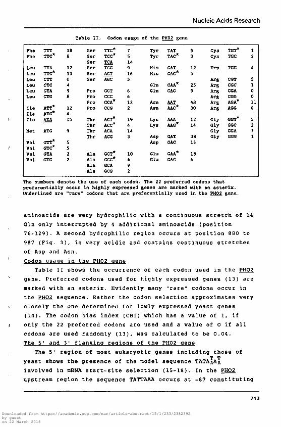
Table II.
SerSerSerSerSerSer
ProProProPro
ThrThrThrThr
AlaAlaAlaAla
Codon
TTC*TCC*TCATCGAGTAGC
CCTCCCCCA*CCG
ACT*ACC*ACAACG
GCT*GCC*GCAGCG
usage
75149165
66122
194143
10492
of the
TyrTyr
HisHis
GinGin
AsnAsn
LysLys
AspAsp
GluGlu
PH02 gene
TATTAC*
CATCAC*
CAA*CAG
AATAAC*
AAAAAG*
GATGAC
GAA*GAG
53
125
259
4830
1214
3816
186
CysCys
Trp
ArgArgArgArgArgArg
GlyGlyGlyGly
TGT*TGC
TGG
CGTCGCCGACGGAGA*AGG
GGT*GGCGGAGGG
12
4
5100116
5271
The numbers denote the use of each codon. The 22 preferred codons thatpreferentially occur in highly expressed genes are marked with an asterix.Underlined are "rare" codons that are preferentially used in the PH02 gene.
aminoacids ate very hydrophilic with a continuous stretch of 14
Gin only interrupted by 4 additional aminoacids (position
76-129). A second hydrophilic region occurs at position 880 to
987 (Fig. 3). is very acidic and contains continuous stretches
of Asp and Asn.
Codon usage in the PH02 gene
Table II shows the occurrence of each codon used in the PHO2
gene. Preferred codons used for highly expressed genes (13) are
marked with an asterix. Evidently many "rare" codons occur in
the PHO2 sequence. Rather the codon selection approximates very
closely the one determined for lowly expressed yeast genes
(14). The codon bias index (CBI) which has a value of 1. if
only the 22 preferred codons are used and a value of 0 if all
codons are used randomly (13), was calculated to be 0.04.
The 5' and 3' flanking regions of the PH02 gene
The 5' region of most eukaryotic genes including those ofT T
yeast shows the presence of the model sequence TATAfiAA
involved in mRNA start-site selection (15-18). In the PHO2
upstream region the sequence TATTAAA occurs at -87 constituting
243
Downloaded from https://academic.oup.com/nar/article-abstract/15/1/233/2382392by gueston 22 March 2018

Nucleic Acids Research
1008
® © © Q © © E <^ © D © © © (E) D K R © A K ^ © F G EPH02 GATGATAACGACGA. . . TAfcACTAATGAAGACAATGATAXTAGTAGTGSCCATAAGAGGAATGCTAAGGATAXCTTTCGAGAA
i i i I I I I I I I I i n n i m i i i i i i i i n u n n H I M I I I I I I H I MG G C C G C G G C G C G TCAAGAGAACCACAACAfiCAGTGAG AATGGGAACGAGAATGAAAATGAACAA
PH04 GAGCAAAACG£CGAGCTGAACAG.. . TCAAGACA^CACMC AfiCAgTC^C . . . AATGGGUCGAGAATGAAAA. . . TCAACAA
204
889© T S
PH02 AACACAAGCGATJM i l l 1 .
PK04 AACAAAGTCACTJAACAAAGTCACTAAAAACAAGA6TAATAGTAGT
© 11 V T © ® K (§)®4? S
Fig. 4 Two homology regions between the PH02 and PH04 genes.Identical bases are denoted by a slash. Identical amino acidsate surrounded by a circle and homologous aminoacids by asquare. In both genes position 1 represents the first base ofthe ATG codon.
a possible TATA box. There is no ATG codon present between this
potential promoter element and the ATG at +1. consistent with
the observation that eukaryots use the 51 proximal ATG codon in
the mRNA as start codon.
The 3'end of the gene contains two derivatives of the
consensus sequence TAAATAAG (Fig. 3). This motif is found in
many yeast genes and is assumed to function as a signal for
transcription termination and/or poly(A)addition (13). Apart
from that, the same region shows striking homology with the
transcription termination region of yeast ribosomal protein
L17a (19) where the 3'end of the mRNA has been mapped (Fig. 3).
In analogy we suggest that the PH02 mRNA ends at position 1713.
DISCUSSION
The 3.6 kb Hindlll fragment with its 559 codon long ORF
carries the PH02 gene, since it is able to complement the pho2
mutation and inactivate the wild-type PH02 gene upon
transplacement of a mutated pho2 sequence into the chromosome.
A frameshift introduced in vitro in the N-terminal part of the
protein abolished PHO2 function. The gene could be narrowd down
to a 2.3kb Hpal/Clal fragment.
The deduced protein sequence shows a very unusual structure
with a segment in the N-terminal part consisting mainly of Gin
residues and another in the middle of PH02 consisting of a
continuous sequence of 36 aminoacids (mainly Asp and Asn) that
are either charged or uncharged, but hydrophilic. Computer
search in a datalibrary revealed that this second charged
244
Downloaded from https://academic.oup.com/nar/article-abstract/15/1/233/2382392by gueston 22 March 2018

Nucleic Acids Research
region is strongly homologous with two regions of the PH04
protein (20) on the aminoacid as well as on the DNA level.
These homology regions are shown in Fig. 4.
Four models can be envisaged: the first states that the
homologous regions are domains within the PH02/PH04 proteins
that are involved in DNA binding at any of the defined (21)
UASp elements of the PH05 promoter. This hypothesis, however,
is very unlikely due to the strong negative charges contained
in these regions. Second, the homology regions could constitute
interaction sites with other still unknown protein(s) (e.g.
initiator proteins), or third, with the RNA polymerase. Fourth
they could be interaction sites with the gene product of the
negative factor PH080. In any case, the observed homology
rather argues for a transcriptional control involvement of PHO2
than for a post transcrlptional action.
Two observations suggest that PHO2 is a lowly expressed
gene. First, the codon bias index (CBI) is near to zero as was
calculated for other regulatory genes like PH04. GAL4 (20, 13)
also expressed at a low level. Second, the 51 flanking region
(100 bp 5' to ATG) does not show a higher A-T content (61%)
than the coding region (63%) as was observed in the structural
genes of the acid phosphatase gene family that are highly
expressed (22).
Our motif for cloning and sequencing PHO2 was the intention
to study the involvement of this regulatory protein in the
phosphatase pathway. With the presented data we are now able to
manipulate the PHO2 gene which should help us to obtain more
insight in the nature of protein/DNA interactions in eukaryotic
gene regulation.
ACKNOWLEDGEMENTSWe like to thank H. Rudolph for fruitful discussions.
D. Primdore for supplying appropriate vector molecules andH. Rink for quick synthesis of oligonucleotides. Furthermore wewish to thank C. Widmer for typing the manuscript.
REFERENCES1. Oshima, Y. (1982) in the Molecular Biology of the Yeast
Saccharomvces. Strathern. J.N., Jones, E.W. and Broach.J.R. Eds., pp 159,-180, Cold Spring Harbor Laboratory Press.Cold Spring Harbor.
245
Downloaded from https://academic.oup.com/nar/article-abstract/15/1/233/2382392by gueston 22 March 2018

Nucleic Acids Research
2.
3 .
4.
5.
6.
7 .
8.
9.
10.
11.
12.
13.
14.
15.
16.
17.
18.
19.
20.
21.22.
Tait-Kamradt, A.G.. Parent. S.A., LeVitre, J., Llfanova. O.and Bostian, K.A. (submitted for publication)Giniger, E., Varnum. S.M. and Ptashne. M. (1985) Cell. 40.767-774.Sherman. F.. Fink. G.R. and Lawrence. C.W. (1983) inMethods in yeast genetics. Cold Spring Harbor Laboratory,Cold Spring Harbor. N.Y.. pp. 61-64.Ito, H., Fukuda. Y., Murata, K. and Kimura. A. (1983) J.Bact. 153. 163-168.Maniatis, T.. Fritsch. E.F. and Sambrook, J. (1982) inMolecular Cloning. Cold Spring Harbor Laboratory. ColdSpring Harbor. N.Y., p. 440.Boeke. J.D.. Lacroute. F. and Fink. G.R. (1984) Mol. Gen.
345-346.Koening-Rauseo. I. and Hinnen. A. (1985) Gene
Genet. 197Rudolph, H36. 87-95.Hieter. P.Davis. R.WNilsson, BPhilipson.Meyhack, BEMBO J. 1,Sanger. F.Natl. AcadBennetzen,
Pridmore. D., Hegemann. J.H., Thomas. M.,and Philippsen. P. (1985) Cell 42. 913-921., Uhlen, M.. Josephson, S., Gatenbeck, S. andL. (1983) Nucl. Acids Res. 11, 8019-8030.. Bajwa. W. , Rudolph, H. and Hinnen, A. (1982)675-680.Nicklen, S. and Coulson, A.R. (1977) Proc.Sci. USA 74. 5463-5467.
J.L. and Hall. D.H. (1982) J. Biol. Chem. 257,
and Mosurski, R. (1986) Nucl.3018-3031.Sharp, P.M., Tuohy, T.M.F.Acids Res. 14, 5125-5143.Dobson, M.J., Tuite, M.F., Roberts, N.A., Kingsman, A.J.and Kingsman. S.M. (1982) Nucl. Acids Res. 10. 2625-2637.Nasmyth. K.A.. Tatchell. K.. Hall. B.D.. Astell. C. andSmith. M. (1981) Nature 289. 244-250.Corden, J., Wasylyk, B., Buckwalder, A., Sassone-Corsi, P.,Kedinger, C. and Chambon P. (1980) Science 209, 1406-1414.Holland, J.P. and Holland, M.J. (1980) J. Biol. Chem. 255,2596-2605.Leer. R.J.Mager. W.H6685-6700.Legrain. M.. De Wilde. M. and Hilger. F. (1986) Nucl. AcidsRes. 14. 3059-3073.Rudolph. H. and Hinnen. A. in press.Bajwa. W., Meyhack. B.. Rudolph. H., Schweingruber. A.M.and Hinnen. A. (1984) Nucl. Acids Res. 12. 7721-7739.
van Raamsdonk-Duin, M.M.C., Hagendoorn, M.J.M..and Planta. R.J. (1984) Nucl. Acids Res. 12,
246
Downloaded from https://academic.oup.com/nar/article-abstract/15/1/233/2382392by gueston 22 March 2018