the single-cell pathology landscape of breast cancer
TRANSCRIPT

1
The Single-Cell Pathology Landscape of Breast Cancer
Hartland W. Jackson1,*, Jana R. Fischer1,2,*, Vito R.T. Zanotelli1,2, H. Raza Ali1,3, Robert
Mechera4, Savas D. Soysal5,6, Holger Moch7, Simone Muenst8, Zsuzsanna Varga7, Walter P.
Weber4, and Bernd Bodenmiller1
1Department of Quantitative Biomedicine, University of Zurich, Switzerland
2Life Science Zurich Graduate School, ETH Zurich and University of Zurich, Switzerland
3CRUK Cambridge Institute, University of Cambridge, Cambridge, UK
4Department of Surgery, University of Basel and University Hospital Basel, Switzerland
5Visceral Surgery Research Laboratory, Clarunis, Department of Biomedicine, University of
Basel, Basel, Switzerland
6Department of Surgery, Clarunis University Center for Gastrointestinal and Liver Diseases
Basel, Basel, Switzerland
7Institute of Pathology and Molecular Pathology, University Hospital Zurich, Zurich, Switzerland.
8Institute of Pathology and Genetics, University Hospital Basel, Switzerland
*Authors contributed equally to this work.
Correspondence should be addressed to
B.B. ([email protected])

2
ABSTRACT
Single-cell analyses have revealed extensive intra- and inter-patient cancer
heterogeneity1, but complex single-cell phenotypes and their spatial context are not yet reflected
in the histologic stratification that is the foundation of many clinical decisions. Here, we used
imaging mass cytometry2 to simultaneously quantify 35 biomarkers resulting in 720 high-
dimension immunohistochemistry pathology images of tumor tissue from 352 breast cancer
patients for whom long-term survival data were available. Spatial, single-cell analysis identified
tumor and stromal single-cell phenotypes, their organization and heterogeneity, and enabled
categorization of breast cancer cellular architecture based on cellular composition and tissue
organization. Our analysis revealed multi-cellular features of the tumor microenvironment and
novel breast cancer subgroups associated with distinct clinical outcomes. Thus, spatially resolved,
single-cell analysis can characterize intra-tumor phenotypic heterogeneity in a disease-relevant
manner with the potential to inform patient-specific diagnosis.
Main
Histologic and phenotypic differences between tumors guide cancer diagnosis, prognosis,
and selection of treatment. Currently, breast cancer patients are graded based on tumor
structure and cellular morphology, and subcategorized when more than 1% of tumor cells
contain hormone receptors or more than 10% express high levels of HER2 or have amplified HER2
3–5. This leaves a large portion of cells uncharacterized even though additional molecular
subclasses and morphologic features have been identified as prognostic6–9. It is clear that clonal
evolution and spatially distinct tumor microenvironments drive inter- and intra-patient cellular

3
heterogeneity and hinder effective treatment10–14. Using highly multiplexed imaging2,15,16,
multiple complex cellular phenotypes have been identified within the context of the tumor
microenvironment, enabling refined histopathology classification of clinical tissue samples17–20.
Here, harnessing topographical single-cell network analysis of high-dimensional mass cytometry
images, we quantified spatial inter- and intra-tumor single-cell heterogeneity and identified
spatially resolved features and novel breast cancer subtypes that are associated with clinical
outcome.
Spatially resolved single-cell phenotypes
To comprehensively quantify the cellular heterogeneity and spatial organization of breast
cancer tissue, we designed a breast-histology-specific imaging mass cytometry (IMC) panel
(Extended Data 1) to image samples from 281 tumors representing all clinical subtypes and
pathology grades (Supplementary Table 1). IMC combines immunohistochemistry staining using
metal isotope-labelled antibodies with laser ablation and mass spectrometry-based detection to
produce high-dimension images (Figure 1a)2. Our 35-antibody panel simultaneously quantified
clinically established breast cancer targets estrogen receptor (ER), progesterone receptor (PR),
and HER2, proliferation marker Ki-67, markers of epithelial, mesenchymal, immune, and
endothelial lineages, and targets that provide insight into signaling pathways, oncogenes, and
epigenetics (Extended Data 1, Supplementary Table 2). IMC produces images comparable to
immunofluorescence or immunohistochemistry but with capacity for highly multiplexed
staining2,21 (Extended Data 2 and 3). Images were segmented into single cells and tumor and
stromal regions using a random forest pixel classifier (Ilastik), and CellProfiler17,20. We identified

4
855,668 cells in 381 images (289 tumor, 87 healthy breast and 5 liver controls), and quantified
marker expression and spatial features of each cell (Figure 1a). Clustering with PhenoGraph22
identified cell phenotype clusters, hereafter referred to as phenotypes, of endothelial, T and B
cell, macrophage, and stromal cell populations as well as 59 diverse tumor cell phenotypes.
Similar to Wagner et al.1, some tumor phenotypes were unique to individual patients (Figure 1b,
c, Extended Data 4a). To identify common cellular subtypes within this diversity, we defined 14
tumor-cell metaclusters by hierarchical clustering of the PhenoGraph-defined tumor single-cell
phenotypes (Figure 1c, Extended Data 5a).
Tumors from every clinical subtype contained fibroblast, endothelial, and immune cell
populations at similar densities but were enriched in tumor cell populations with variable
expression of cytokeratins, hormone receptors, and HER2 reflective of clinical subtype (Figure 1c,
Extended Data 6a). Across all patients, immune cells were excluded from the cohesive tumor
mass, although immune and fibroblast cells did infrequently infiltrate the tumor mass and rare
HRlow/– cells lacking cytokeratins invaded past the tumor-stroma front in some samples (Figure
1c, Extended Data 4). Tumor regions contained various luminal HR+ epithelial cell phenotypes
identified by combinations of ER, PR, GATA3, E-cadherin, and multiple cytokeratins, but hormone
receptors were also expressed without cytokeratins in a few cases (metacluster 26) (Figure 1c,
Extended Data 6b). Of the luminal cytokeratins (CK7, CK8/18, and CK19), only CK7 was associated
with specific luminal tumor cell subsets (metaclusters 19, 20) (Figure 1c). HER2 expression was
not a defining metacluster feature but was observed at different levels in multiple phenotypes.
Phenotypes without hormone and HER2 receptor expression (characteristics of triple-negative
breast cancer (TNBC)) included metaclusters with high levels of Ki-67, p53, EGFR, and hypoxia

5
marker CAIX (metaclusters 15-17), basal cytokeratins (metacluster 18), and even luminal
cytokeratins (PG clusters within metaclusters 19, 22) (Figure 1c).
Multi-cellular breast cancer architecture
Based on these single-cell phenotypes, we sought to define patterns of multi-cellular
architecture in breast tumor tissue (Figure 2a). We evaluated regional correlations between
cellular metaclusters to determine whether cells co-occurred across all images, and used
permutation test-based neighborhood analysis17 to quantify cell co-localization and identify
statistically significant interaction or avoidance between pairs of cell phenotypes (Figure 2b).
Tumor cell phenotypes were rarely correlated: Each individual tumor contained many homotypic
interactions between similar cells and few heterotypic tumor cell interactions (Figure 2b, box #1).
Generally, heterotypic interactions were associated with regionally unique structures such as
blood vessels (Figure 2b, box #2) or with distinct epithelial (box #1) or stromal cell-dense areas
where immune cells interacted (box #3). Uni-directional interactions were also observed where
supporting fibroblasts enclosed endothelial cells in large blood vessels and where stromal cells
surrounded tumor cells at the tumor-stroma interface (Figure 2b, boxes #2 and #4). T cells and
proliferating epithelial cells were observed in the vicinity of endothelial cells, and their
proportions were correlated across images; T cells were surrounded by endothelial cells (e.g.,
inside vessels), not the reverse (Figure 2b, boxes #5 and #6). The neighborhood analysis revealed
interaction signatures that distinguished well-separated or stromal-interactive tumor
architectures that were related to pathologist-scored tumor grade (Extended Data 7)17.

6
Tissue function is implemented by multicellular units, which we call communities, that
consist of higher order, rather than paired, interactions between one or more cell phenotypes.
We identified communities by first constructing a topological neighboring cell interaction
network and then applying a graph-based community detection approach using the Louvain
algorithm (Figure 2a)23. Applied only to tumor cells, community detection identified dense
epithelial patches of different sizes, termed tumor communities (TCs); when applied to all cells
microenvironment communities (MCs) containing tumor and stromal cell components were
identified (Figure 2a). Using PhenoGraph, we grouped multi-cell communities according to
community size and tumor cell phenotypes (TCs) or all cells, but agnostic to tumor cell type (MCs)
. Tumor communities were mostly dominated by a single cellular metacluster and were separated
based on absolute number of cells (Figure 2c, d, TCs 4, 7, 18; Supplementary Images). Some
microenvironment communities consisted of fibroblasts that interacted with a variety of tumor
cells (MCs 2, 5, 8); others showed sparse stroma content (MCs 14, 17, 18, 20, 21, 22) or were
enriched for T cells (MCs 19, 25, 30), macrophages (MC 27), large networks of T and B cells (MC
1), or endothelial cells (MCs 13, 6, 30, 25, 7) (Figure 2e, f). Fibroblast-enriched communities had
few interacting immune cells, aligning with the known roles of fibroblasts as agents of tumor
desmoplasia and immune exclusion24.
Single-cell pathology subgroups are related to clinical outcome
We next investigated how the organization of single cells into communities contributes
to breast cancer tissue architecture and tumor subtypes3. Cells from multiple cellular
metaclusters were found in every clinically defined breast cancer subtype (Extended Data 6a),

7
supporting the conclusion that general pathology classification does not fully elucidate inter- and
intra-patient cellular heterogeneity6,8. We reasoned that the single-cell pathology landscape
would provide a higher resolution patient classification than classic histology-based clinical
subtypes. Patient tumors were grouped based on the tumor cell metacluster composition using
unsupervised clustering, identifying 18 single-cell pathology (SCP) subgroups that split the classic
clinical subtypes (Figure 3a, Extended Data 8a, Supplementary Table 3). SCP subgroups had
various proportions of the epithelial communities (Figure 3b), and individual SCP subgroups had
distinct clinical outcomes when compared to all other patients, to SCP subgroups of the same
clinical classification, and to other SCP subgroups containing similar cellular metaclusters but
different architectures (Figure 4a-h, Supplementary Tables 4 and 5).
HR+ clinically defined tumors were divided into those strongly enriched in cells with high
expression of hormone receptors (SCPs 1-5 and 12) and tumors with few HRhi/+ cells surrounded
by many cells that expressed only low levels or lacked HRs (SCPs 6-10 and 11) that are currently
not clinically classified (Figure 3a, Extended Data 6 and 8a, Supplementary Images). SCP 1, which
contained predominantly metacluster 23 (CK+/HRhi tumor cells), was only associated with
patients who did not succumb to disease. Conversely, SCP 3, which contains the same cellular
metaclusters but differs in structure, with smaller communities and relatively higher proportions
of CKlow/HRlow metacluster 22 and 25 cells, was associated with poor prognosis, as were SCPs 6
and 9, which involve predominantly CKlow/HRlow cells (Figure 3a, Figure 4c, e, Extended Data 9).
SCP 2, containing CK+/HR+ cells, was significantly enriched in the HR+/HER2+ clinical subtype,
which was otherwise dominated by CKlow/HRlow metacluster 22 (Figure 3a, Extended Data 6, and
8a). SCPs 11 and 12 were characterized by CK7+ cells primarily from metaclusters 20 and 19,

8
respectively. SCP 11 overlapped with the clinically assigned HR–/HER2+ tumor type, and, although
this clinical subtype usually has poor outcomes3,5, SCP 11 patients had significantly better
outcomes than other patients in this cohort. In contrast, the small number of CK7+ SCP 12
patients, predominantly clinically assigned as HR+/HER2-, did not survive long term (Figure 3a,
Figure 4a, c, e, Extended Data 6 and 8a). Tumors from patients with high-risk TNBC contained
distinct cell types including cells with cytokeratin expression suggestive of a luminal, not
myoepithelial, cell of origin (Figure 1c, Figure 3a, Extended Data 6 and 8a). TNBC phenotypes
without luminal epithelial markers and with high levels of hypoxic, p53+/EGFR+, basal, or
proliferative markers distinguished SCPs 13, 14, 15, and 17 with poor outcome (Figures 1c, Figure
3a, Extended Data 6 and 8a). SCP 16 tumors were p53+ and expressed apoptotic markers, and
interestingly, patients with tumors of this group did not succumb to disease even though they
were clinically classified as TNBC (Figure 4f, Extended Data 8a).
By mapping the cellular spatial organization of these tumors, we observed variable
structures and cellular densities, and relationships between cellular phenotype and tissue
organization (Figure 3a, b). Heterogeneous tumors consisted of multiple phenotypically pure
communities indicated by many bands on the heatmap, whereas homogeneous tumors
organized in one epithelial sheet or with similar communities of different sizes have only a few
clustered bands (Figure 3b). Most tumors were dominated by a single tumor-cell metacluster and
few community types, but tumors in SCP 8 and some in SCP 10 were unusually heterogeneous,
consisting of multiple epithelial cellular metaclusters at similar proportions localized to spatially
distinct communities (Figure 3b, Extended Data 6). Patients in SCP8 with these heterogeneous
tumors had very poor outcomes (Figure 4d, e). Overall, intra-tumor phenotypic heterogeneity

9
was spatially segregated into separate tumor communities as opposed to heterogeneous tumor
masses, and patients with tumors with greater spatio-phenotypic heterogeneity had poorer
outcomes.
Unlike tumor cell phenotypes, the identified stromal cell phenotypes were present in
every clinical subtype at similar densities (Figure 1c, Extended Data 6). We therefore investigated
whether the tumor-stromal microenvironment communities were more informative than mere
stromal phenotype content. When we hierarchically clustered images by the presence of
microenvironment communities, 11 groups, which we term stromal environments (SEs), were
revealed; some were enriched in one microenvironment community (single column in Extended
Data 7a), whereas others contained mixtures of communities (multiple columns). Neighbor
analysis detected distinct cell-cell interactions within each SE (Extended Data 7b). Some stromal
environments included large epithelial networks with sparse stroma (SEs 7 and 10, made of MCs
17 or 18), others involved vascularized regions (SE9, involving MCs 6 and/or 13), different
fibroblast cell phenotypes such as vimentinhi (SEs 4, 6, and 9, involving metacluster 8 in many
communities) or fibronectinhi (SEs 1 and 2, including metacluster 11), or were poorly cohesive
and made up of many small communities (SEs 2, 5, and 6).
We found that SEs were associated with SCP subgroups and specific tumor cell
phenotypes. For example, hypoxic SCP 17 TNBCs were commonly classified as large, stroma-
deficient epithelial sheets (SEs 7), and TNBCs SCP 13-16 were associated with T cell-enriched (SEs
5 and 8) or macrophage-enriched (SE 3) stromal environments (Extended Data 7 and 8b). HR+
tumors are likely to be immune-cold, but some contained rare and localized immune-enriched
communities (SE 5); the HR+ tumors never contained an exclusively immune-hot stromal

10
environment (SE 8) like those observed in TNBCs and the highly heterogeneous SCP 8 (Extended
Data 7 and 8b). HR+ tumors were associated with a range of fibroblast-enriched stromal
environments with small elongated fibroblasts interacting with CK+/HRlow epithelial cells in SEs 1,
2, and 5 and vimentinhi stroma associated with HRhi tumor cells in SEs 4 and 6 (Figure 2b, c,
Extended Data 8b). Stromal environments were related to tumor phenotype, but only
fragmented SE 6 containing proliferative vimentinhi fibroblasts was independently associated
with poorer disease-free and overall survival (Figure 4h, Extended Data 8b and 9, Supplementary
Tables 6 and 7).
Compared to clinically defined subtypes, SCP grouping improved the ability to predict a
patient’s overall survival using Cox proportional hazards modeling (Supplementary Table 8). In
order to identify features associated with patient risk not captured by clinical grading and
classification, we investigated the epithelial and stromal single-cell and community contributions
to the model. Almost no single-cell phenotypes or cellular metaclusters were independently
associated with outcome (not shown); however, spatially defined cell communities were (Figure
4i). For certain cell types, large tumor cell communities were related to better outcome, whereas
similar networks of small size were related to poor outcome (Figure 4i, TCs 12 vs. 13, 17 vs. 23, 5
vs. 15). In addition, the microenvironment community MC 6, characterized by vascularization
with T cell involvement, was significantly associated with increased risk of death even though it
was more commonly found in the low-risk HR+ clinical subgroups than other subgroups (Figure
4i, Extended Data 7). In contrast, highly T cell infiltrated MC 19 and macrophage-enriched MC 27
were significantly associated with better patient outcomes even though inflammation is more
common in high-risk TNBC tumors than other clinical subgroups (Figure 4i, Extended Data 7).

11
Thus, SCP-defined tumor types and tumor and stromal architectures could inform prognosis
beyond current clinical classifications.
Quantification of intra-tumor heterogeneity
We investigated reproducibility and spatial variability of SCP classifications in two central
and two peripherally located tumor regions from 72 patients in an independent cohort resulting
in 344 additional images containing 411,410 cells (Supplementary Table 9). We used the same
analytical approach described above to independently define single-cell phenotypes, match them
to cellular metaclusters and to classify each imaged region into SCP subgroups and stromal
architectures (Extended Data 5b and 10a). All cellular metaclusters and SCP subgroups identified
in the first cohort were present in the second cohort. Likely due to the patient selection strategy,
which enriched the second cohort in metastatic low-grade tumors, we observed a higher
proportion of subgroups involving CKlow/HRlow single-cell phenotypes such as SCP 3, 6, and 9 in
this cohort (Extended Data 10a). For each region of each tumor, we quantified the spatial
variability of cell phenotypes using Shannon entropy, and the difference in cellular content
relative to the patient’s overall single-cell phenotype distribution (all regions) was determined
using Kullback-Leibler divergence. Most patients had only moderate inter-region heterogeneity
(Extended Data 10b). Distinctly divergent regions were more commonly identified as SCP 2, which
consists of CK+/HR+ cells, or SCP 7, containing Epitheliallow cells (Extended Data 10b, c).
Approximately 40% of tumors had identical classification in all regions, and 60% had one or more
regions that did not agree with the whole tumor classification (Extended Data 10d). However, in

12
the majority of cases SCP classification of individual regions matched the tumor-wide
classification.
SCP subgroups had varying levels of regional heterogeneity. SCP subgroups found in
TNBCs were phenotypically homogenous (the exception being basal CK+ SCP 13). SCP subgroups
associated with HR+ patients with relatively poor prognosis, such as CKlow/HRlow SCPs 6, 7, 9, 10,
had substantial spatial heterogeneity (Extended Data 10e). Different SCP subgroups containing
similar cell types occasionally co-occurred in the same tumor, but SCP 9 was always accompanied
by SCP 6 regions, and the two were associated with similar outcomes (Figure 4e, Extended Data
10e). Spatially heterogeneous HRlow and CKlow SCP subgroups 6-10 have indistinguishable
outcomes from the population average, and based on their regional heterogeneity it is
statistically likely that unsampled phenotypes influenced classification accuracy (Extended Data
10e). We observed that heterogeneous regions classified as SCP 8 or 10 were always
accompanied by multiple additional tumor subtypes (Extended Data 10e). These may be multi-
clonal or highly plastic tumors. Subsampling was sufficient to identify and stratify homogenous
low-risk HRhi and high-risk TNBCs, but increased sampling may be needed to accurately identify
HRlow tumor phenotypes and tumors with considerable intra-tumor heterogeneity.
Discussion
This systematic, multidimensional interrogation of breast cancer histology generated a
detailed spatial map of single-cell phenotypes and cellular communities related to disease. We
demonstrated that single-cell pathology can better segregate patients with distinct clinical
outcomes than the current clinical subtyping strategy. Analysis of multi-cellular structures

13
revealed that phenotypic heterogeneity in tumors was spatially localized to distinct regions or
lesions. Moreover, the multi-cellular structures yielded patient outcome-relevant information
superior to single-cell data alone. We identified co-occurring breast cancer phenotypes and
observed that phenotypic and spatial heterogeneity varied between clinically established
subtypes. This work suggests that multi-cellular spatial information is medically relevant and
provides a basis for future study of how spatial and phenotypic tissue features influence disease
progression.
References
Bibliography
1. Wagner, J. et al. A Single-Cell Atlas of the Tumor and Immune Ecosystem of Human Breast
Cancer. Cell 177, 1330-1345.e18 (2019).
2. Giesen, C. et al. Highly multiplexed imaging of tumor tissues with subcellular resolution by
mass cytometry. Nat. Methods 11, 417–422 (2014).
3. Coates, A. S. et al. Tailoring therapies--improving the management of early breast cancer:
St Gallen International Expert Consensus on the Primary Therapy of Early Breast Cancer
2015. Ann. Oncol. 26, 1533–1546 (2015).
4. Hammond, M. E. H. et al. American Society of Clinical Oncology/College Of American
Pathologists guideline recommendations for immunohistochemical testing of estrogen and
progesterone receptors in breast cancer. J. Clin. Oncol. 28, 2784–2795 (2010).
5. Wolff, A. C. et al. Human epidermal growth factor receptor 2 testing in breast cancer:

14
american society of clinical oncology/college of american pathologists clinical practice
guideline focused update. J. Clin. Oncol. 36, 2105–2122 (2018).
6. Curtis, C. et al. The genomic and transcriptomic architecture of 2,000 breast tumours
reveals novel subgroups. Nature 486, 346–352 (2012).
7. Sørlie, T. et al. Gene expression patterns of breast carcinomas distinguish tumor subclasses
with clinical implications. Proc Natl Acad Sci USA 98, 10869–10874 (2001).
8. Cancer Genome Atlas Network. Comprehensive molecular portraits of human breast
tumours. Nature 490, 61–70 (2012).
9. Beck, A. H. et al. Systematic analysis of breast cancer morphology uncovers stromal
features associated with survival. Sci. Transl. Med. 3, 108ra113 (2011).
10. Bedard, P. L., Hansen, A. R., Ratain, M. J. & Siu, L. L. Tumour heterogeneity in the clinic.
Nature 501, 355–364 (2013).
11. Dagogo-Jack, I. & Shaw, A. T. Tumour heterogeneity and resistance to cancer therapies.
Nat. Rev. Clin. Oncol. 15, 81–94 (2018).
12. McGranahan, N. & Swanton, C. Biological and therapeutic impact of intratumor
heterogeneity in cancer evolution. Cancer Cell 27, 15–26 (2015).
13. Focke, C. M., Decker, T. & van Diest, P. J. Intratumoral heterogeneity of Ki67 expression in
early breast cancers exceeds variability between individual tumours. Histopathology 69,
849–861 (2016).
14. Rye, I. H. et al. Intratumor heterogeneity defines treatment-resistant HER2+ breast tumors.
Mol. Oncol. 12, 1838–1855 (2018).
15. Angelo, M. et al. Multiplexed ion beam imaging of human breast tumors. Nat. Med. 20,

15
436–442 (2014).
16. Gerdes, M. J. et al. Highly multiplexed single-cell analysis of formalin-fixed, paraffin-
embedded cancer tissue. Proc Natl Acad Sci USA 110, 11982–11987 (2013).
17. Schapiro, D. et al. histoCAT: analysis of cell phenotypes and interactions in multiplex image
cytometry data. Nat. Methods 14, 873–876 (2017).
18. Keren, L. et al. A Structured Tumor-Immune Microenvironment in Triple Negative Breast
Cancer Revealed by Multiplexed Ion Beam Imaging. Cell 174, 1373-1387.e19 (2018).
19. Carvajal-Hausdorf, D. E. et al. Multiplexed (18-Plex) Measurement of Signaling Targets and
Cytotoxic T Cells in Trastuzumab-Treated Patients using Imaging Mass Cytometry. Clin.
Cancer Res. 25, 3054–3062 (2019).
20. Damond, N. et al. A map of human type 1 diabetes progression by imaging mass cytometry.
Cell Metab. 29, 755-768.e5 (2019).
21. Bodenmiller, B. Multiplexed Epitope-Based Tissue Imaging for Discovery and Healthcare
Applications. Cell Syst. 2, 225–238 (2016).
22. Levine, J. H. et al. Data-Driven Phenotypic Dissection of AML Reveals Progenitor-like Cells
that Correlate with Prognosis. Cell 162, 184–197 (2015).
23. Blondel, V. D., Guillaume, J.-L., Lambiotte, R. & Lefebvre, E. Fast unfolding of communities
in large networks. J. Stat. Mech. 2008, P10008 (2008).
24. Kalluri, R. The biology and function of fibroblasts in cancer. Nat. Rev. Cancer 16, 582–598
(2016).

16
Figure legends
Figure 1: Single-cell phenotypes in high-dimension histopathology of breast cancer.
(a) Schematic of IMC acquisition of multiplexed images from 281 breast cancer patients and the
analyses of single-cell phenotypes, metaclusters, stromal-cell organization and architecture,
tumor and patient subclassification, and patient overall survival. (b) tSNE map of 171,288
subsampled single cells from high-dimension images of breast tumors colored by cell-type
metacluster identifier. (c) Heatmap showing the z-scored mean marker expression or distance to
tumor-stroma interface for each PhenoGraph cluster, colored according to metacluster identifier.
The absolute cell counts of each PhenoGraph cluster are displayed as a bar plot. In the bubble
plot, circle size shows the relative proportion of all cells in a clinical subtype that come from each
cluster, and circle opacity shows the proportion of each cluster present in the different clinical
subtypes.
Figure 2: A global map of the cellular neighborhoods and cell interaction networks of breast
cancer.
(a) Representative image depicting the different steps in the spatial analysis. Images show (left
to right) pseudo-colored IMC, single-cell mask of the same field of view labeled by cellular
metacluster identifier, the topologic cell interaction network, modular regions of the tumor
network identifying epithelial communities labeled in color, and modular regions in the tumor-
stroma network identified as tumor microenvironment communities. Scale bar = 100 μm. (b)
Heatmap in which squares visualize Pearson correlation of cell phenotype proportions across all
measured tissue regions (n = 367 images) and circles indicate significant pairwise cell type

17
interaction or avoidance summarized across the two-sided permutation tests on the individual
images (n = 367 images, 1000 permutations each). Circle color indicates percentage of images
and size represents number of images with a significant cell-cell interaction or avoidance
(p<0.01). Highlighted interactions include 1) tumor epithelium , 2) endothelium, 3) immune, 4)
surrounding stroma, 5) endothelium and T cells, and 6) proliferating epithelium surrounding
endothelial cells. Non-symmetric highlight colors indicate examples of directional interaction.
Uniquely colored (c-d) epithelial communities (n = 8495) and (e-f) microenvironment
communities (n = 12,854) clustered by PhenoGraph based on min-max normalized absolute
numbers of cells from each cellular metacluster and visualized (c,e) on a tSNE map and (d,f) in
stacked bar plots indicating the average number of cells from each cellular metacluster.
Figure 3: Single-cell pathology identifies subgroups of breast cancer patients.
(a) Hierarchically clustered stacked bar plot of cell-type metacluster densities in each tumor.
Colored columns indicate clinical subtypes, and SCP subgroups. (b) Heatmap indicating
proportions of different epithelial communities present within each image.
Figure 4: Single-cell pathology subgroups have distinct clinical outcomes.
Kaplan-Meier curves of overall survival for each patient group (n = 278 patients total) based on
(a) clinical subtype, (b) clinical grade, (c-f) SCP subgroup, or (g-h) stromal environment. Two-sided
logrank test ✪p<0.05 compared to all other samples, ✭p<0.05 compared to similar subgroups,
✭p<0.05 compared to other HR+/HER2- patients (for exact p-values see SI Tables 4 and 6). (i)
Relative hazard ratios and 95% confidence intervals of disease-specific overall survival for
densities of tumor (T) and microenvironment (ME) cellular communities and clinical categories

18
(molecular subtype and grade) estimated by Cox proportional hazards model (n = 266 patients,
n = 15 patients only containing communities <10 cells were excluded).
Methods
Clinical data
The samples of tumors from the described patient cohorts were obtained from University
Hospital Basel and University Hospital Zurich. The cohort from University Hospital Basel includes
281 patients who were not selected for any clinical or histologic features. Pathologists recorded
the available patient metadata (Supplementary Table 1) and evaluated the suitability of tissue
sections for tissue microarray (TMA) construction25. The TMA contains one 0.8-mm tumor core
per patient, in some cases an additional matched healthy breast tissue sample, and a few control
samples (liver tissue). The cohort from University Hospital Zurich is comprised of 72 patients; the
samples include four 0.6-mm cores from four different regions of each tumor as described in
Kündig et al.26. Tumor cores were punched from two central and two peripheral areas that
averaged 1 cm in distance between regions. Samples were selected to contain equal proportions
of the different tumor grades as well as patients with and without lymph node metastases
(Supplementary Table 9). In total 720 images were acquired that varied in size and localization in
the tumor. This project was approved by the local Commission of Ethics (ref. no. 2014-397 and
2012-0553).
Panel

19
An antibody panel was designed to target epitopes specific for breast cancer as well as
markers for cell cycle and phospho-signaling and to distinguish epithelial, endothelial,
mesenchymal, and immune cell types (Extended Data 1, Supplementary Table 2). Clone
information is available in Supplementary Table 2.
Preparation and staining
Tissue samples were formalin-fixed and paraffin-embedded at the University Hospitals of
Basel and Zurich. The above described antibody panel was used to stain the tissue sections27.
Tissue sections were dewaxed in xylene overnight and rehydrated in a graded series of alcohol
(ethanol:deionized water 100:0, 90:10, 80:20, 70:30, 50:50, 0:100; 5 min each). In a 95 °C water
bath, heat-induced epitope retrieval was conducted in Tris-EDTA buffer at pH 9 for 20 min. The
tissue microarrays were immediately cooled and then blocked with 3% BSA, 5% goat serum in
TBS for 1 h. Samples were incubated overnight at 4 °C in primary antibody at 7.5 g/L diluted in
TBS/0.1% Triton X-100/1% BSA. Tissue samples were washed twice with TBS/0.1% Triton X-100
and twice with TBS and dried before imaging mass cytometry measurements.
For combined immunofluorescence and imaging mass cytometry staining, tissues were
stained overnight at 4 °C with primary metal-conjugated mouse HER2 (151Eu) and rabbit pan-
Cytokeratin (175Lu) antibodies prior to washing and the mixed addition of fluorescent and metal-
conjugated anti-mouse (AF488, 165Ho) and anti-rabbit (AF555, 159Tb) secondary stains for 1 h at
room temperature. A cover slip was added, and tissue was imaged for fluorescence signal.
Subsequently, the cover slip was removed, and samples were washed, dried, and subjected to
mass cytometry laser ablation and acquisition.

20
Imaging mass cytometry
Images were acquired using a Hyperion Imaging System (Fluidigm). The largest square
area from each core of a tissue microarray was laser ablated in a rastered pattern at 200 Hz, and
raw data preprocessing was completed using commercial acquisition software (Fluidigm). IMC
acquisition stability was monitored by interspersed acquisition of isotope-containing polymer
(Fludigm). All successful image acquisitions were processed, and images containing pan-marker
staining variation specific to TMA location were removed. In few cases the acquisition was
interrupted and later continued resulting in 2 tumor images of the same patient. Therefore, the
281-patient cohort resulted in 289 tumor, 87 healthy breast and 5 liver control images. Where
applicable, signal spillover between channels was corrected using functions from the CATALYST
R package (version 1.5.6)28. During analysis of the samples from University Hospital Zurich, cells
from images in one row of the TMA were almost exclusively assigned to the phenotype with the
lowest overall marker expression. These samples were poorly stained and had to be excluded
from further analysis (images Ay.x1). The 72-patient cohort resulted in 263 tumor, 68 healthy
breast and 6 control images used for analysis.
Data processing
Data were converted to .tiff format and segmented into single cells using the flexible
analysis pipeline available at https://github.com/BodenmillerGroup/ImcSegmentationPipeline.
Briefly, individual cells and tumor/stroma regions were segmented using a combination of Ilastik
1.1.929 and CellProfiler 2.1.130. Ilastik was used to generate a probability map by classifying pixels

21
(Single cells - nuclei, membrane and background; Tumor/Stroma - tumor, stroma and
background) based on a combination of membrane and nuclei identifying antibody stains.
Probability maps were then segmented into single cell, or tumor and stroma object masks using
CellProfiler.
Single-cell segmentation masks and tiff images of the 35 channels were overlaid and
single-cell marker expression means and spatial features were extracted using the Matlab
toolbox regionprops, as implemented in histoCAT31. Even with very good quality segmentation,
the imaging of tissue segments results in single-cell data of tissue slices and overlapping cell
fragments that do not always capture the nucleus of a cell, and therefore nuclei-mismatched
signal can be assigned to neighboring cells in densely packed areas. This can lead to rare cases
where data assigned to one cell contains marker expression from the neighborhood.
The single cell IDs of each cell’s direct neighbors within 4 pixels (4 μm) of the cell of
interest was detected and recorded using histoCAT software. The number of pixels expanded to
detect neighbors was chosen such that small gaps in segmentation would be bridged, yet no cells
after the direct neighbor would be recorded (cell minor axis lengths: 5th - 95th percentile 4.84 -
14.59 pixels, average 9.51 pixels).
Individual cell locations inside or outside of a tumor mask were identified and the distance
of each cell to the tumor boundary (from inside and outside of the tumor region) was calculated
using the Matlab toolbox regionprops. Distances were measured between the closest pixels of
the objects in question.
Data transformation and normalization

22
The presented data were not transformed, and all analyses were based on raw IMC
measurements. Single-cell marker expressions are summarized by mean pixel values for each
channel. The single-cell data were censored at the 99th percentile to remove outliers, and Z-
scored cluster means were visualized in heatmaps. For tSNE and PhenoGraph the data were
normalized to the 99th percentile, as suggested by the authors of these algorithms32,33. To
visualize the number of cells per image or patient and for survival modelling, the counts were
normalized by the image area (total number of pixels) and displayed as cell density. For coxph
survival modelling, these densities were multiplied by a factor of 1031 in order to yield values
larger than 1 and then log-transformed.
Analysis workflow
The single-cell analysis pipeline was implemented in R, but image analysis steps were
performed in Matlab. All statistical tests were performed using common R functions.
Clustering and metaclustering
Single cells of the large cohort from University Hospital Basel were clustered into groups
of phenotypically similar cells using a combination of PhenoGraph33 for initial, unsupervised
clustering and an aggregation of these clusters into larger groups based on their mean marker
correlations to identify cellular metaclusters. In a first step, the data were over-clustered to
detect and separate rare cell subpopulations. PhenoGraph (version 2.0) was used with default
parameters (as implemented in histoCAT/Cyt) and 20 nearest neighbors. For high-dimensional
clustering, 29 markers and 4 cell shape features were used: Iridium, Histone, phospho Histone,

23
CK14, CK5, CK8/18, CK19, CK7, panCK, E/P-Cadherin, ER, PR, HER2, GATA3, SMA, Vimentin,
Fibronectin, vWF/CD31, CD44, CD45, CD68, CD3, CD20, cleaved Caspase 3/cleaved PARP,
Carbonic Anhydrase, phospho-S6, Ki67, p53, EGFR, Area, Eccentricity, Extent, and Number
Neighbors. Of the resulting 71 clusters, the 59 epithelial ones were aggregated into larger groups
following the hierarchical clustering (Euclidean distance and Ward’s linkage) of their mean
marker correlations. Multiscale bootstrap resampling was used to assess the uncertainty of each
subtree (R package pvclust, version 2.0), and separation of the hierarchy was assigned so that
significant epithelial subtrees were maintained and known biologic differences were separated.
This resulted in 14 tumor cell metaclusters of varying size and subtree robustness (Extended Data
5a). Clusters showing marker expression typical of stromal and immune cells, which were limited
due to our tumor marker focused panel, were kept as in the original PhenoGraph clustering and
not aggregated into larger groups. This metaclustering yielded 27 cellular subgroups,
representing various immune, stromal, and epithelial cell types. The granularity, the level and
detail at which phenotypes are divided or clustered, of the studied cell types depends on the
selection of both the panel and the choice of parameters. While a more granular distinction of
cell types might elucidate even more subtle difference in the marker expressions of cells, it would
limit comparability between tumors as many tumor cell types would be patient specific.
Cluster matching across cohorts
Single cells from the second cohort from University Hospital Zurich were clustered
unsupervised and independently using PhenoGraph33 with the same settings described above for
the first cohort and a nearest neighbor parameter of 30. The clusters were matched to the most

24
similar metacluster of the previous cohort using Pearson correlation of the z-scored mean marker
expressions. In two special cases (clusters 8 and 15) where the cluster in question was rather
poorly correlated with all metaclusters but most correlated with a stromal cell type, we manually
re-assigned the cluster because upon visual inspection of the images those clusters represented
cells forming clear tumor bulks.
bh-tSNE
For visualization, high-dimension single-cell data were reduced to two dimensions using
the non-linear dimensionality reduction algorithm tSNE32. We applied the Barnes-Hut
implementation of tSNE (bh-tSNE) to 99th-percentile normalized data with default parameters
(initial dimensions, 110; perplexity, 30; theta, 0.5). The algorithm was run on a randomly
subsampled set of cells (20% from each image) in order to not obscure visible patterns in crowded
plots and for better computational performance.
Neighborhood analysis
To identify significantly enriched or depleted pairwise neighbor interactions between cell
types, histoCAT functions were used to perform a permutation test based analysis of spatial
single-cell neighborhoods31. Neighboring cells were defined as those within 4 pixels (4 µm). A p-
value cut-off of <0.01 was used for significance.
Single-cell pathology patient grouping

25
Patients were grouped based on the proportions of tumor cell metaclusters using the
cytofkit R implementation of PhenoGraph33 (version 1.10.0) with 8 nearest neighbors and default
parameters. The parameter number of nearest neighbors was chosen such that small groups of
patients consisting of a distinct predominant cell type could be separated. A choice of a higher
value for this parameter would lead to fewer groups, and hence patients with entirely unrelated
predominant phenotypes grouped together. A lower value of the nearest neighbor parameter
might capture more subtle differences in cellular composition of tumor types but would severely
limit statistical power for group comparison and survival analysis. The composition of each
patient group by their clinically assigned metadata is available in Supplementary Table 3. Patient
group 18 was removed from further downstream analysis, due to lack of statistical power, as it
contains only three patients with distinct tumors strongly dominated by a rare HR+/CK– cell type.
Single-cell pathology group matching
Tumor cores from the second cohort from University Hospital of Zurich were assigned to
the most similar previously defined single-cell pathology group based on their matched tumor
cell type components. The inverse of Pearson correlation was used as distance metric.
Spatial heterogeneity
In the cohort from University Hospital Zurich that contains multiple cores per tumor,
intra-core heterogeneity of tumor and stromal cells were separately quantified using the entropy
based Shannon index on the amounts of the different cell types within each core. Shannon
entropy has been shown to serve as a measure of diversity and homogeneity in various

26
contexts34. It can also be considered a measure for the information content of a string, where in
our case every cell of an image is represented by a letter according to its cell type. The most
compressible string is obtained if every cell is of the same type, and the string with most
information is obtained if every cell is unique (Formula 1, where Pi is the probability of a given
symbol). Inter-core heterogeneity within a tumor was approximated by calculating the Kullback-
Leibler divergence from the cell-type distribution (proportions of each cell type) of an individual
core to the average cell-type distribution across all cores of a patient. Kullback-Leibler divergence
describes the information loss when going from an original distribution to a summary
distribution35. Hence, if all cores of a tumor are composed of identical proportions of the same
cell types, the Kullback-Leibler divergences of every individual core to the patient average will be
minimal. The R package entropy (version 1.2.1) was used for the calculation of both Shannon
entropy and the Kullback-Leibler divergence. Intra-tumor heterogeneity and the consistency of
SCP group assignment of images of the same patient are visualized in Extended Data 10.
H(X) = -∑ pi log2 (pi) (Formula 1)
Spatial communities
The images were converted into topological neighborhood graphs where every cell is
represented by a node (visualized at the centroid), and the nodes are connected by an edge if
the cells directly neighbor each other (Figure 2). Neighboring cells were defined as those within
4 pixels (4 µm) of the outermost pixel assigned to a cell. Subsequently, the Louvain community
detection algorithm36 (C implementation by Lefebvre and Guillaume, version 0.2, wrapped by

27
Matlab as used by the implementation of PhenoGraph 2.0 used by histoCAT/Cyt) was applied to
identify highly interconnected spatial subunits in the tissue graph. While using community
detection algorithms on spatially constrained networks is known to hide underlying non-spatially
driven solutions, the only aim of applying the algorithm here was to extract spatial information
and identify communities based on physical proximity37. This analysis was performed on
epithelial cells only to identify tumor communities (without including stromal or immune cells in
the graph) and again on all cells of a tissue to identify tumor-microenvironment communities. A
tumor-specific cohesiveness score was calculated based on the average sizes of the identified
tumor communities. Communities involving fewer than 10 cells were excluded from further
analysis in order to focus on cohesive cell patches and not individual disconnected cells. 15
patients were excluded from analysis based on tumor communities because the imaged regions
did not contain any tumor communities consisting of at least 10 cells. In order to identify
recurring similar spatial cell type communities, the cytofkit PhenoGraph33 (version 1.10.0) was
run on the min-max normalized, absolute numbers of cells of each cell metacluster in each
community. This analysis was conducted separately for the tumor communities based on only
the epithelial cell types (k=80) and for the microenvironment communities based on all cells but
only taking into account the individual stromal cell types and aggregating all tumor cell types into
one label (cell type group 100: including all tumor cells, k=30). This analysis was conducted
separately for each cohort but based on the matched metacluster cell types.
Stromal environments

28
Based on their microenvironment community compositions, images were grouped into
11 different stromal environments using hierarchical clustering (Euclidean distance and Ward’s
linkage). This analysis was conducted separately for each cohort but based on the matched
metacluster cell types.
Overlapping classifications and enrichments
Fisher’s exact test was used to identify single-cell pathology patient groups enriched for
a specific stromal environment (Extended Data 8). The test was performed using the R function
fisher.test (with parameter enrichment = “greater”) for every potential stromal region of a
patient group. The p-values were corrected for multiple testing using the Bonferroni method.
This enrichment analysis was also conducted with different combinations of single-cell pathology
subgroups, stromal environments, and clinical classifications (Extended Data 8).
Survival curves and coxph regression models
Kaplan-Meier survival curves and coxph survival regression models were generated using
the R package survival (version 2.42-4). The overall survival as well as the disease-free survival of
patients in different clinical or single-cell-defined subgroups was analyzed (Figure 4, Extended
Data 9, Supplementary Table 4-8). Both logrank tests and coxph models were employed to
investigate whether a patient subgroup significantly deviated from the survival of the remaining
patients or from the survival of other patients of similar SCP groups or the same clinical
classification (Figure 4, Supplementary Table 4-7). Log-transformed densities of communities or
single cells, alongside the clinical subgrouping and grading, were provided to a coxph survival

29
model in order to find significant associations of certain community or single-cell types with
patient risk and to investigate the hazard ratios (Figure 4). Nested coxph models were compared
using likelihood ratio tests (anova.coxph) to assess whether additional variables improved the
survival model (Supplementary Table 8).
30
References:
25. Kononen, J. et al. Tissue microarrays for high-throughput molecular profiling of tumor
specimens. Nat. Med. 4, 844–847 (1998).
26. Kündig, P. et al. Limited utility of tissue micro-arrays in detecting intra-tumoral
heterogeneity in stem cell characteristics and tumor progression markers in breast
cancer. Journal of Translational Medicine, 16(1), 118 (2018).
27. Giesen, C. et al. Highly multiplexed imaging of tumor tissues with subcellular resolution
by mass cytometry. Nat. Methods 11, 417–422 (2014).
28. Chevrier, S. et al. Compensation of Signal Spillover in Suspension and Imaging Mass
Cytometry. Cell Syst. 6, 612-620 (2018).
29. Sommer, C., Straehle, C., Köthe, U. & Hamprecht, F.A. in Proc. 2011 8th IEEE
International Symposium on Biomedical Imaging: From Nano to Macro 230–233 (IEEE,
2011).
30. Carpenter A.E., et al. CellProfiler: image analysis software for identifying and quantifying
cell phenotypes. Genome Biology 7:R100. PMID: 17076895 (2006) .
31. Schapiro, D. et al. histoCAT: analysis of cell phenotypes and interactions in multiplex
image cytometry data. Nat. Methods 14, 873–876 (2017).
32. Amir, A.D. et al. viSNE enables visualization of high dimensional single-cell data and
reveals phenotypic heterogeneity of leukemia. Nat. Biotechnol. 31, 545–552 (2013).
33. Levine, J.H. et al. Data-Driven Phenotypic Dissection of AML Reveals Progenitor-like Cells
that Correlate with Prognosis. Cell 162, 184–197 (2015).
34. Angel Martın, M., & Rey, J.-M. (2000). On the role of Shannon’s entropy as a measure of
heterogeneity. Geoderma (Vol. 98).
35. Kullback, S., Leibler, R. A. On Information and Sufficiency. Ann. Math. Statist. 22 (1951).
36. Blondel, V.D. et al. Fast unfolding of communities in large networks. J. Stat. Mech.
(2008) P10008

31
37. Expert, P., Evans, T. S., Blondel, V. D., & Lambiotte, R. Uncovering space-independent
communities in spatial networks. Proceedings of the National Academy of Sciences,
108(19), 7663 LP-7668 (2011).
Acknowledgements
We are grateful for the donation of tumor samples by patients undergoing surgery. We
thank Serenella Eppenberger and Susanne Dettwiler for the coordination of tissue collection and
for construction of tissue microarrays. We thank the Bodenmiller lab, in particular Natalie de
Souza for help with writing the manuscript, and Dr. Daniel Schulz for fruitful discussions. BB’s
research was funded by a SNSF R`Equip grant, a SNSF Assistant Professorship grant, the SystemsX
Transfer Project “Friends and Foes”, the SystemX grants Metastasix and PhosphoNEtX, a NIH
grant (UC4 DK108132), the CRUK IMAXT Grand Challenge, and by the European Research Council
(ERC) under the European Union's Seventh Framework Program (FP/2007-2013)/ERC Grant
Agreement n. 336921. HWJ was funded by SystemsX Transitional Post-Doctoral Fellowship, the
Canadian Institute of Health Research Post-Doctoral Fellowship, and the Cancer Research Society
Scholarship for the Next Generation of Scientists.
Author competing interest
The authors declare that they have no conflicts of interest.

32
Author Information
These authors contributed equally: Hartland W. Jackson and Jana R. Fischer
Affiliations
Department of Quantitative Biomedicine, University of Zurich, Switzerland
Hartland W. Jackson, Jana R. Fischer, Vito R.T. Zanotelli, H. Raza Ali, Bernd Bodenmiller
Life Science Zurich Graduate School, ETH Zurich and University of Zurich, Switzerland
Jana R. Fischer, Vito R.T. Zanotelli
CRUK Cambridge Institute, University of Cambridge, Cambridge, UK
H. Raza Ali
Department of Surgery, University of Basel and University Hospital Basel, Switzerland
Robert Mechera, Walter P. Weber
Visceral Surgery Research Laboratory, Clarunis, Department of Biomedicine, University of Basel,
Basel, Switzerland
Savas D. Soysal
Department of Surgery, Clarunis University Center for Gastrointestinal and Liver Diseases Basel,
Basel, Switzerland
Savas D. Soysal
Institute of Pathology and Molecular Pathology, University Hospital Zurich, Zurich, Switzerland
Holger Moch, Zsuzsanna Varga
Institute of Pathology and Genetics, University Hospital Basel, Switzerland
Simone Muenst

33
Contributions
HWJ and BB conceived the study. HWJ performed all image quantification and IMC
experiments. ZV performed immunohistochemical staining. JRF performed data analysis. VRTZ
constructed image processing and analysis tools. HWJ and JRF performed the biological analysis
and interpretation with input from the co-authors. RA provided input on clinical interpretation
and survival analysis. HM, ZV, RM, SDS, SMS, and WPW provided patient samples and clinical
input throughout the study. HWJ, JRF, and BB wrote the manuscript.
Corresponding Author
Bernd Bodenmiller ([email protected])
Data availability
The data supporting the findings of this study, including high-dimension tiff images,
single-cell and tumor/stroma masks, single-cell and patient data will be available online upon
publication (DOI: 10.5281/zenodo.3518284).
Code availability
All code that produced the results of this study will be available on
https://github.com/BodenmillerGroup/SCPathology_publication upon publication.
Extended data legends
Extended Data 1: Antibody panel and example 6-marker pseudo-colored images.

34
Antigens targeted by the antibodies in the panel of 35 isotope-conjugated antibodies used to
stain the breast cancer tissue. Representative marker images from the analysed cohort generated
by IMC. Every marker is visualized at least once. Each image represents a different tumor of the
analysed cohort. Each marker was individually scaled to enable visualization. Scale bar = 100 μm.
Extended Data 2: IHC and IMC comparison and reproducibility analysis.
(a) Representative IMC and immunohistochemistry (IHC) images of the quantified stains in
sections of the same tumor core. (b) Scatter plot and correlation of total IHC and IMC signal in
sections of the same tumor core (IHC = optical density/μm2, IMC = ion counts/μm2,n = 319 cores).
(c) Scatter plot and correlation of the number of positively stained cells in sections from the same
tumor core (n = 319 cores). (d) Bland-Altman plots for reproducibility of IMC signal in positively
stained cells across images from different regions of the same tumor adapted to visualize the
average across four samples on the x-axis and the difference of every individual sample to the
tumor average on the y-axis. Only images containing positively stained cells and more than 200
cells in total were taken into account for this analysis (ER: n = 280 cores from 72, PR: n = 213
cores from 66 patients, HER2: n = 291 cores from 72 patients, Ki67: n = 281 cores from 72
patients, E/P-Cadherin: n = 200 cores from 65 patients) . The red line represents the overall
average of the differences to the tumor mean, and the blues lines represent the 95% confidence
interval (1.96*standard deviation). The percentage of observations that fall within the confidence
interval is indicated at the top of each plot.
Extended Data 3: Simultaneous IF and IMC imaging.

35
Immunofluorescence (IF) and Mass Cytometry (IMC) imaging of the same tissue sample using
metal-conjugated HER2 and pan-Cytokeratin primary antibodies and both fluorescent and metal-
conjugated secondary stains. Pseudo-color images of (a) individual channels, (b) three marker
images produced from each label type (White: overlap, Red: HER2, Green: pan-Cytokeratin, Blue:
DNA Intercalator), as well as (c) an overlay of the same marker from all three label types (White:
overlap, Red: Secondary IF, Green: Secondary IMC, Blue: Primary IMC). (d) High magnification
image of regions labelled by white squares in panel b comparing immunofluorescence and
imaging mass cytometry resolution.
Extended Data 4: Single-cell localization relative to tumor-stroma interface.
tSNE maps of 171,288 subsampled single cells from high-dimension images of breast tumors
colored by (a) patient, (b) localization relative to the tumor-stroma interface, (c) single-cell
distance to the tumor-stroma interface and (d) number of neighboring cells. (e-g) Representative
image with single-cell mask labeled by (e) metacluster identifier , (f) tumor and stroma masks,
and (g) heatmap representing distances of single cells to the tumor-stroma interface from each
side. Scale bar = 100 μm. (h-j) Log-transformed distances to tumor front of (h) stromal-cell
clusters and (i) tumor-cell metaclusters, and (j) binned distances of all metaclusters to the tumor
front, where bin number 0 contains all cells that are directly touching the interface. Negative
distances represent the distance to the tumor boundary from inside the tumor and positive
values indicate distance outside the tumor.
Extended Data 5: Metaclustering and cluster matching across cohorts.

36
(a) Heatmap displaying z-scored mean marker expressions of single-cell phenotypic clusters
identified by PhenoGraph (Figure 1) with colors on colorbar and hierarchical clustering indicating
the corresponding metacluster. Red stars on the hierarchical clustering tree indicate subgroups
that robustly reappear as separate groups using multiscale bootstrap resampling (R function
pvclust, p < 0.05). (b) Examples of untransformed distributions of cluster marker expressions
differing between metaclusters. (c) Heatmaps showing the z-scored mean marker expression or
distance to tumor-stroma interface for each metacluster defined in the Basel 281-patient cohort
and each matched PhenoGraph cluster from the Zurich multi-core 71-patient cohort.
PhenoGraph clusters of the Zurich cohort were matched to the metaclusters of the Basel cohort
based on Pearson correlation of the mean marker expression (Methods).
Extended Data 6: Densities of single-cell phenotypes in different clinical subtypes and SCP patient
subgroups.
Box plots of cellular metacluster densities in patients of (a) each clinical subtype (HR+HER2-: n =
173, HR+HER2+: n = 29, HR-HER2+: n = 23, TripleNeg: n = 48) and (b) each SCP subgroup (center
line, median; box limits, first and third quartile; whiskers, 1.5x interquartile range; points beyond
whiskers, outliers; SCP1: n = 17, SCP2: n = 21, SCP3: n = 20, SCP4: n = 12, SCP5: n = 32, SCP6: n =
10, SCP7: n = 13, SCP8: n = 11, SCP9: n = 20, SCP10: n = 24, SCP11: n = 31, SCP12: n = 14, SCP13:
n = 15, SCP14: n = 11, SCP15: n = 8, SCP16: n = 10, SCP17: n = 9, SCP18: n = 3).
Extended Data 7: Stromal environments based on microenvironment community composition
and their distinct pairwise cell type interactions.
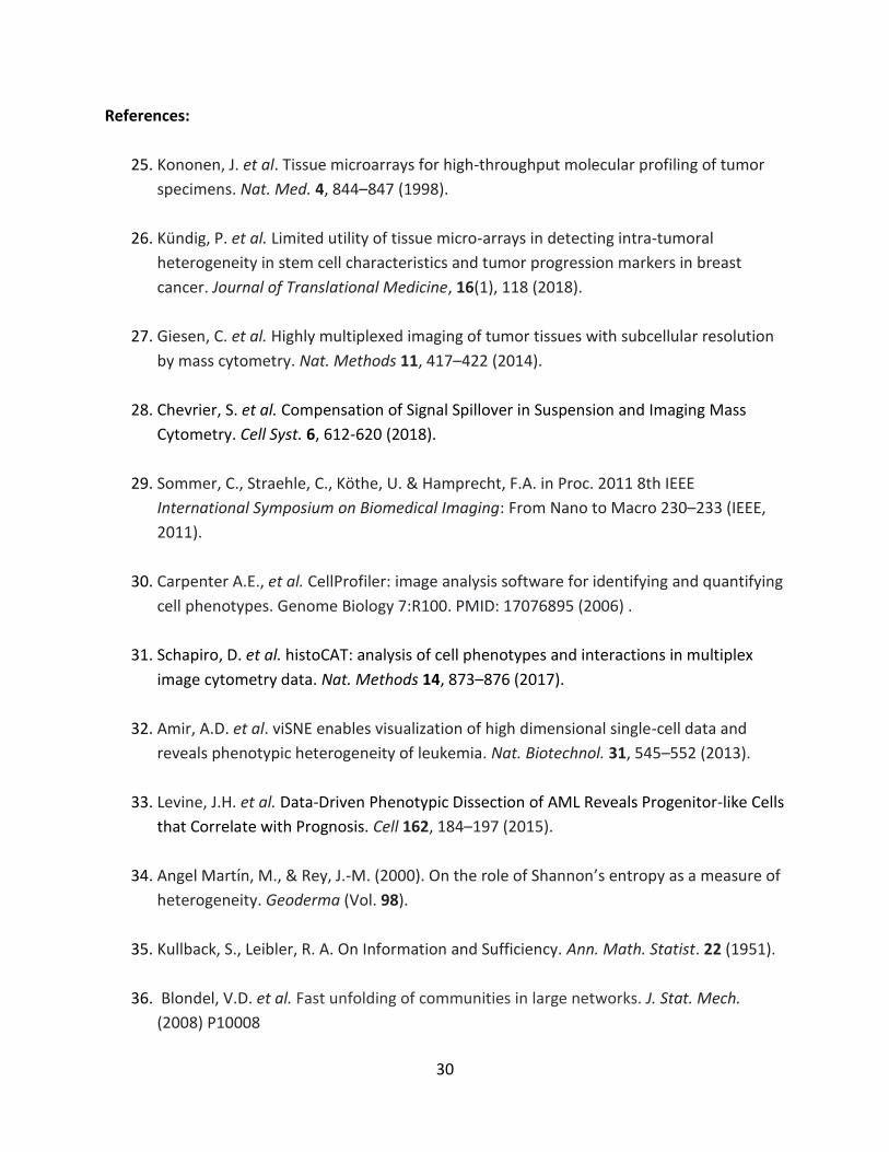
37
(a) Hierarchical clustering of tumor cores (n = 281) according to stromal community content and
splitting into corresponding stromal environments (n = 11). The stacked bar plot at the top
indicates the average number of cells from each cellular metacluster present within each
microenvironment community type. (b) The presence of significant (p<0.01) cell-cell interactions
(red) and cell-cell avoidances (blue) identified per image based on a permutation test (1000
permutations). Black outlined regions indicate significant interactions that are enriched in images
from the respective stromal environments (one-sided Fisher’s exact test for enrichment, p < 0.05
after multiple-testing correction). Color bars on the right indicate the SCP subgroup, grade, and
clinical subtype of the tumor. Cell-type interactions along the top are indicated by the labeled
cell type of interest and neighboring cell.
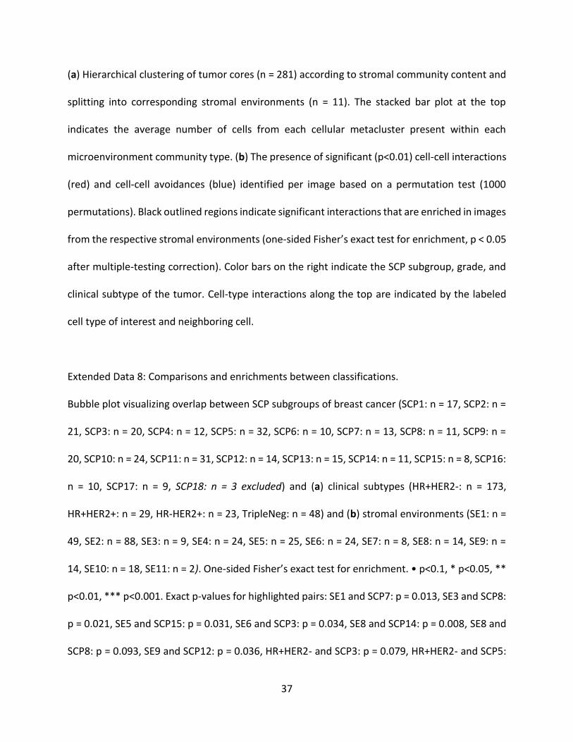
Extended Data 8: Comparisons and enrichments between classifications.
Bubble plot visualizing overlap between SCP subgroups of breast cancer (SCP1: n = 17, SCP2: n =
21, SCP3: n = 20, SCP4: n = 12, SCP5: n = 32, SCP6: n = 10, SCP7: n = 13, SCP8: n = 11, SCP9: n =
20, SCP10: n = 24, SCP11: n = 31, SCP12: n = 14, SCP13: n = 15, SCP14: n = 11, SCP15: n = 8, SCP16:
n = 10, SCP17: n = 9, SCP18: n = 3 excluded) and (a) clinical subtypes (HR+HER2-: n = 173,
HR+HER2+: n = 29, HR-HER2+: n = 23, TripleNeg: n = 48) and (b) stromal environments (SE1: n =
49, SE2: n = 88, SE3: n = 9, SE4: n = 24, SE5: n = 25, SE6: n = 24, SE7: n = 8, SE8: n = 14, SE9: n =
14, SE10: n = 18, SE11: n = 2). One-sided Fisher’s exact test for enrichment. • p<0.1, * p<0.05, **
p<0.01, *** p<0.001. Exact p-values for highlighted pairs: SE1 and SCP7: p = 0.013, SE3 and SCP8:
p = 0.021, SE5 and SCP15: p = 0.031, SE6 and SCP3: p = 0.034, SE8 and SCP14: p = 0.008, SE8 and
SCP8: p = 0.093, SE9 and SCP12: p = 0.036, HR+HER2- and SCP3: p = 0.079, HR+HER2- and SCP5:

38
p = 3.58e-04, HR+HER2+ and SCP2: p = 0.032, HR-HER2+ and SCP11: p = 2.36e-04, HR-HER2- and
SCP8: p = 0.060, HR-HER2- and SCP14: p = 0.008, HR-HER2- and SCP15: p = 6.13e-06, HR-HER2-
and SCP16: p = 0.031.
Extended Data 9: Kaplan-Meier survival curves for overall and disease free survival.
Kaplan-Meier survival curves of (a-b) overall survival for Stromal Environments not shown in
Figure 4 and (c-l) disease-free survival for each patient group based on (c) clinical subtype , (d)
grade , (e-h) SCP subgroup , and (i-l) stromal environment . Two-sided logrank test ✪ p<0.05
compared to all other samples (for exact p-values see SI Tables 5 and 7).
Extended Data 10: Multi-core cohort regional heterogeneity analysis.
Quantification of intra-tumor regional heterogeneity in Zurich multi-core cohort. (a)
Hierarchically clustered stacked bar plot of cell type metacluster densities in each tumor, grouped
by patient. Colored columns indicate patient, clinical subtypes, SCP subgroups, location of core
in the tumor, Shannon entropy (intra-core heterogeneity), and tumor-specific cohesiveness
score. (b) Dot plot of Kullback-Leibler divergence from the cell-type distribution of an individual
tumor region to the patient average distribution, colored according to the SCP subgroup
classification per tumor region (n = 263 tumor cores), grouped by patient (n = 71 patients) and
ordered by the increasing average Kullback-Leibler divergence per patient. (c) Boxplot of the
same Kullback-Leibler divergences of each region to the patient’s average cell-type distribution,
grouped by tumor regions individually identified as the same SCP subgroup, independent of
patient (center line, median; box limits, first and third quartile; whiskers, 1.5x interquartile range;

39
points beyond whiskers, outliers; SCP1: n = 12, SCP2: n = 13, SCP3: n = 11, SCP4: n = 10, SCP6: n
= 76, SCP7: n = 7, SCP8: n = 3, SCP9: n = 5, SCP10: n = 1, SCP11: n = 26, SCP12: n = 51, SCP13: n =
15, SCP14: n = 18, SCP15: n = 4, SCP16: n = 5, SCP17: n = 5, SCP18: n = 1). (d) Bar indicating the
percentage of patients (n = 71) with the indicated fraction of individually classified images
matching the whole tumor classification. (e) Bubble plot visualizing intra-tumor region variation
within patients of each SCP subgroup. Rows represent tumors of each SCP subgroup as identified
by the combined analysis of all imaged regions. Columns represent tumor regions individually
matched to a SCP subgroup. For each whole-tumor classification on the y-axis, circle size indicates
the fraction of corresponding images individually classified as a SCP subgroup. For each image
classification on the x-axis, color indicates the fraction of images within each tumor type.