the small gtpase rab35 is a novel oncogenic …
TRANSCRIPT

THE SMALL GTPASE RAB35 IS A NOVEL ONCOGENIC REGULATOR
OF PI3K/AKT SIGNALING
A Dissertation
Presented to the Faculty of the Weill Cornell Graduate School
of Medical Sciences
in Partial Fulfillment of the Requirements for the Degree of
Doctor of Philosophy
by
Douglas B. Wheeler
May 2015

© 2015 Douglas Berg Wheeler
THE SMALL GTPASE RAB35 IS A NOVEL ONCOGENIC REGULATOR
OF PI3K/AKT SIGNALING
Douglas Berg Wheeler, Ph.D.
Cornell University 2015
The phosphatidylinositol 4,5-bisphosphate 3’-OH kinase (PI3K) is a
lipid kinase that regulates cell survival, proliferation and metabolism in
response to external growth factors. PI3K signaling regulates a diverse set of
effectors via the phosphoinositide dependent kinase 1 (PDK1), AKT/PKB,
and the mechanistic target of rapamycin complexes 1 and 2 (mTORC2). The
components of this pathway are frequently mutated in human cancers, which
give rise to oncogenic signals that drive tumorigenesis. As such, the proteins
involved in PI3K/AKT signaling are attractive targets for targeted therapies.
It is thought that most tumors upregulate PI3K/AKT signaling in some
way. However, a large portion of tumors do not have any identifiable genetic
lesions in the genes that code for proteins that regulate this pathway. Thus,
we reasoned that there are likely to be many proteins that regulate PI3K/AKT
signaling that are currently unappreciated. To identify novel regulators of the
PI3K axis, we undertook an arrayed loss-of-function RNAi screen using a
library of shRNA reagents that targeted all known human kinases and
GTPases to identify proteins whose depletion altered AKT phosphorylation.
Further, we triaged genes from this screen using oncogenomic databases to
identify screen hit genes that were mutated in human tumor samples or cell

lines. We reasoned that this approach could identify novel components of
PI3K/AKT signaling that also play a role in tumorigenesis.
This screen identified the small GTPase RAB35 as a novel positive
regulator of PI3K/AKT signaling. Depletion of RAB35 in a variety of human
and murine cell lines suppressed phosphorylation of PI3K-dependent
proteins like AKT, FOXO1/3A and NDRG1. Moreover, stable expression of a
GTPase-deficient, constitutively active allele of RAB35—but not wildtype
RAB35—potently activated AKT signaling in the absence of growth factors.
Further, we identified two RAB35 mutations in human tumor genome
sequence databases that activated PI3K signaling and transformed NIH-3T3
cells in vitro in a PI3K-dependent manner. Finally, we find that RAB35 is
likely regulating PI3K signaling by trafficking the platelet derived growth
factor receptor (PDGFR) to a RAB7-positive compartment in the cell where
the receptor actively signals PI3K. Thus, RAB35 is a novel oncogenic
regulator of PI3K/AKT signaling.
iii
BIOGRAPHICAL SKETCH
Douglas Berg Wheeler was born in 1981 in Utica, NY to Nancy Berg
and Albert Charles Wheeler. Doug attended Poland Central School in
Poland, NY for elementary, middle and high school. During high school, he
participated annually in the Poland Central School and Utica College
Science Fairs, which gave him an early taste for both biology and failure.
After high school, Doug attended Hamilton College in Clinton, NY from 1999
to 2001, where he worked in the endocrinology lab of Dr. David A. Gapp.
After two years at Hamilton, he transferred to Cornell University in Ithaca, NY
in the fall of 2001. During his time at Cornell, he taught undergraduate
Biochemistry (BIOBM 330) and worked in the lab of yeast geneticist Dr.
Susan A. Henry. He graduated from Cornell in May, 2003 with a Bachelors
of Science in Biology.
After his time at Cornell, Doug moved to Cambridge, MA and began
working as a research technician in the lab of Dr. David M. Sabatini at the
Whitehead Institute for Biomedical Research at the Massachusetts Institute
of Technology. While at the Whitehead, he developed a tool for rapid, high-
throughput loss-of-function RNA interference screens in Drosophila cells [3-
4]. This technology—where RNA reagents are printed onto glass slides and
cells seeded on these spots could be assayed for loss of gene function—
spurred Doug’s interest in high-throughput technology and the examination
of gene function. Further, his time in the Sabatini lab sparked in Doug a
desire to study the genes that regulate survival and growth in cancer cells.

iv
After two years at the Whitehead, Doug moved to New York City in
July of 2005 to begin his time at the Tri-Institutional MD/PhD program.
During the two preclinical years of medical school, Doug decided that he
wanted to start his graduate work with an RNAi screen to discover new
components the PI3K/AKT signaling pathway. In July of 2007 he began his
graduate work in the lab of Dr. Charles L. Sawyers at Memorial Sloan-
Kettering Cancer Center. With the cooperation of Dr. Sawyers and Dr. Olaf
S. Andersen, Doug was fortunate enough to concoct a thesis that allowed
him to be mentored jointly by Dr. Sawyers and his previous mentor, Dr.
Sabatini. Doug spent the first two and a half years of his graduate training
primarily working at the Whitehead and Broad Institutes. While there, he
performed the RNAi screens that nucleate this thesis. After finishing these
screens, Doug returned to New York City in the beginning of 2010 to
continue his thesis work full time in the Sawyers laboratory.
After six years in the lab, Doug will ultimately defend his Ph.D. in
December of 2013, after which he will begrudgingly put down his pipettes
and return to medical school. He is looking forward to spending the rest of
his days studying cancer.

v
This dissertation is dedicated to my parents, Nancy Berg Wheeler and Albert
Charles Wheeler. Without the two of them I would not have had the
inquisitiveness or stubbornness that science demands. They have been very
patient.

vi
ACKNOWLEDGEMENTS
I would first like to thank the director of the Tri-Institutional MD/PhD
program, Dr. Olaf Sparre Andersen. Without Dr. Andersen’s willingness to
allow me to pursue my interests, I would not have been able to design and
carry out my thesis work the way I wanted to. In the middle of my second
year of medical school, I approached Dr. Andersen with the idea for my
thesis project—which involved me moving to Boston for an indeterminate
amount of time. Rather than turn down my request, Dr. Andersen allowed
me to return to the Whitehead Institute without any hesitation. I thank Dr.
Andersen for his flexibility in allowing me to pursue my scientific interests.
I would also like to thank my two thesis advisors: Dr. David M.
Sabatini and Dr. Charles L. Sawyers. I have known David since 2003, when I
joined his lab as a technician. David has always encouraged my pursuit of
science, and his passion and excitement for biology are contagious.
Moreover, his sense of humor taught me that science can be fun, even when
the results are not. I also thank my MSKCC mentor, Dr. Charles Sawyers, for
being willing to take me on as a graduate student when I approached him to
ask if I could be in his lab, but from a distance. In the years that Dr. Sawyers
has been my thesis advisor, I have come to admire his sense of humor, his
patience, and his dedication to helping cancer patients with translational
science and medicine.
Further, I would like to extend my thanks to my thesis committee
members for their patience, scientific insights, and flexible schedules: Dr.

vii
Scott C. Blanchard, Dr. Robert Darnell, Dr. Timothy E. McGraw, Dr. Neal
Rosen and Dr. Agata Smogorzewska. Each member of my committee has
been generous in sharing their time and wisdom with me. They stand as
examples of busy faculty who are nevertheless willing to selflessly extend
themselves to trainees. I am in their debt and will seek to emulate their
generosity throughout my career.
I would also like to acknowledge the many people who have
encouraged my pursuit of science at various stages of my life. During middle
and high school, I was privileged to have teachers like William Newman and
Edward Rosenburgh, my biology and earth science teachers, respectively.
Both Mr. Newman and Mr. Rosenburgh encouraged my science fair projects
all throughout my time at Poland Central School—despite the fact that not
one of my projects ever succeeded. They helped me to learn very early on
that science is not about proving one’s hypothesis right, but rather about
asking interesting questions. Without their involvement in the PCS Science
Fair, it is unlikely that I would have chosen the training path that I have.
I was very fortunate during my undergraduate years to have a number
of teachers who encouraged me to take risks and try new things. In
particular, my advisor from Hamilton College—Dr. David A. Gapp—was
instrumental in my early years at the bench. Another of my mentors at
Hamilton—my organic chemistry professor Dr. Ian Rosenstein—helped to
stoke my passion for biochemistry at an early age. While at Cornell, I was
lucky enough to teach undergraduate biochemistry under the guidance of

viii
James E. Blankenship and Dr. Helen T. Nivison. Their enthusiasm for
teaching left an indelible mark on me. My time at the bench at Cornell was
overseen by Dr. Stephen Jesch in the lab of Dr. Susan A. Henry. I thank
both Steve and Susan for their commitment to teaching and their patience
for my marginal skills at yeast genetics.
I have been lucky to have been a part of two wonderful labs during my
graduate training, and was blessed with many friends from these two groups.
From the Sabatini laboratory I would like to thank Dr. Siraj M. Ali, Dr.
Timothy R. Peterson, Heather Keyes, Dr. Peggy Hsu, Dr. Yoav Shaul, Dr.
Brian Grabiner, Dr. Mathieu LaPlante, Dr. Alejo Efeyan, Dr. Carson Thoreen,
Stephanie Kinkel, Dr. Steve Bailey, and Dr. Caitlin Higgins Bailey. I would
also like to thank the director of the Broad Institute’s RNAi Consortium, Dr.
David E. Root. From the Sawyers laboratory I extend my gratitude to my
bay-mate Dr. Phil Watson, our lab manager John Wongvipat, my friends Dr.
Michael J. Evans, Dr. Minna Balbas, Dr. Rohit Bose, and the technicians that
keep the Sawyers lab running: Taslima Ishmael, Mandy Lee, Emily
Schenkein, and Jacqueline Wanjala. Dr. Watson has been particularly
patient in tolerating my feigned aggression and sarcasm throughout the
years.
I wish to also thank a number of close friends who have encouraged
me, listened to me, supported me and often offered their honest criticisms at
the (many) times when I have been wrong. First and foremost among these
are Matthew A. Domser and his parents Celia and Dr. Mark Domser. The

ix
Domsers have been a part of my life since before I can remember, and I
cannot imagine having pursued science without their encouragement. I must
particularly acknowledge Matt, who has always been there to listen to my
scientific and personal travails and has never hesitated to offer his
friendship, encouragement, and honest criticism.
I have also been privileged to have shared my time with a number of
friends and classmates. I would like to thank my “medical school crew”—Dr.
Dennis Spencer, Dr. Selom Gasinu, Dr. Irina Chaikhoutdinov, Dr. Anand
Nataraj and Dr. Malikah Latmore—for their time, friendship, and especially
their encouragement in pursuing thesis work that would take me to Boston
and unfortunately away from them. Further, my medical school and MD/PhD
program classmates Dr. Natasha D. Novikov, Dr. Sandeep Kishore, Dr. Ankit
B. Patel, Dr. Kirti Magudia, Dr. Nicole Ramsey, Corynn Kasap, and Dr.
Svetlana Pavlovic. Everyone listed here—and many that I am likely
forgetting—have at one point or another offered their advice, friendship and
listened to me talk about cancer and protein phosphorylation ad nauseum. In
particular, Dr. Novikov has been a pillar of support and friendship in the final
year of our training together. Я люблю тебя , мой дорогой Наталья
Дмитриевна Новикова.
Although this paragraph will lack sentimentality, I must certainly
acknowledge and thank the United States taxpayers for funding my work. In
particular, the National Institutes of Health Medical Scientist Training
Program grant GM07739, which helps to fund the Tri-Institutional MD/PhD

x
program. I would also like to thank the Department of Defense Prostate
Cancer Research Program and the Office of Congressionally Directed
Medical Research Programs for awarding me Predoctoral Prostate Cancer
Training Award PC094483. Lastly, the Starr Cancer Consortium generously
awarded us Starr Cancer Consortium Award #I2-A117, which allowed me to
perform the RNAi screens that began my thesis work.
I must extend my deepest and most heartfelt gratitude to my family:
my parents Nancy B. and Albert C. Wheeler, my sister Marie N. Cotier, and
my brother Edward S. Wheeler. My mother and father were my first
teachers, and their influence is evident in my work, if only just to me.
Specifically, I thank my mother for her passion for teaching, learning and
science. As an elementary school science and math teacher, my mother
always led by example with her commitment to teaching. Further, I must
thank my father. My father has been an electrical engineer for most of his
life, and I used to presume that my work as a biologist was as far from
electrical engineering as one could get. However, after years in the lab, I can
see that I primarily inherited my stubbornness and attention to detail from my
Father, and our hours of working together on Gracie often parallel my efforts
that I put forth at the bench.
I must also acknowledge my siblings—Marie Cotier and Edward S.
Wheeler. My sister and brother have always served as examples to the kind
of person that I aspire to be. They are both compassionate and intelligent
human beings that have put up with a lot of annoying behavior from their

xi
younger brother. They have been unwavering supporters of my work, and I
am boundlessly grateful to them.
Finally, I acknowledge a gene who barely even appears in this thesis,
but who was instrumental in the work presented here: RAB25. From
December 2009 to March 2012, most of my work in the lab focused on the
GTPase RAB25. Despite many experiments, I finally came to the conclusion
that RAB25—although a tantalizing hit from my RNAi screens—was not
regulating growth factor signaling the way I initially thought it was. Although
none of my efforts with RAB25 are directly evident in this thesis, the time
spent on this protein taught me how to rigorously design experiments and
execute methods in a way that I could trust my data. Ultimately, this protein
taught me that one must be ready to discard one’s favorite theories in favor
of pursuing biology that is more likely to be real. Thus, the attention to detail
that RAB25 taught me bore fruit immediately when I turned my efforts to
RAB35.
xii
TABLE OF CONTENTS
BIOGRAPHICAL SKETCH ............................................................................. iii
DEDICATION ................................................................................................... v
ACKNOWLEDGEMENTS ............................................................................... vi
TABLE OF CONTENTS ................................................................................. xii
LIST OF FIGURES ......................................................................................... xx
LIST OF TABLES ....................................................................................... xxiv
LIST OF ABBREVIATIONS ......................................................................... xxv
CHAPTER ONE: INTRODUCTION: PI3K/AKT SIGNALING IN HUMAN CANCERS AND RNAi SCREENS TO IDENFITY NOVEL REGULATORS OF THE PI3K/AKT AXIS ............................................ 1
1.1 EXTRACELLULAR SIGNALS REGULATE
INTRACELLULAR SIGNALING ....................................... 1
1.1.1 Growth factor receptor tyrosine kinases initiate
phosphoinositide-dependent signaling .................. 1
1.1.2 Phosphatidylinositol 3’-OH kinases phosphorylate
membrane lipids to transduce growth factor
signals ................................................................... 3
1.1.3 PTEN and lipid phosphatases negatively regulate
PI3K signaling ........................................................ 5
1.1.4 The Ras/Raf/MAPK/ERK signaling axis ................ 7
1.2 SIGNALING DOWNSTREAM OF PIP3 ............................ 9

xiii
1.2.1 PDK1 activates AKT in a phosphoinositide
dependent manner ................................................ 9
1.2.2 AKT/PKB is a major effector of PI3K signaling .... 11
1.2.3 AKT/PKB positively regulates cell metabolism,
growth and survival .............................................. 13
1.2.4 The mechanistic target of rapamycin (mTOR) ..... 15
1.2.5 mTOR Complex 1 (mTORC1) is a nutrient and
growth factor sensitive protein kinase that regulates
cell size and growth through S6K1 and 4EBP1 ... 16
1.2.6 mTOR Complex 2 (mTORC) is a PI3K-dependent
kinase that regulates AKT/PKB and other AGC
kinases ................................................................ 19
1.2.7 Feedback signaling within the PI3K/AKT/mTOR
pathway ............................................................... 21
1.3 THE PI3K/AKT SIGNALING AXIS IS FREQUENTLY
ALTERED IN HUMAN CANCERS.................................. 24
1.4 TARGETING THE PI3K/AKT PATHWAY IN HUMAN
CANCERS .................................................................... 27
1.4.1 From chemotherapy to targeted therapy ............. 27
1.4.2 Strategies for targeting receptor tyrosine
kinases ................................................................ 28
1.4.3 P3K inhibitors ...................................................... 29

xiv
1.4.4. Serine/threonine kinase inhibitors: targeting AKT
and mTOR .......................................................... 30
1.4.6. The promises and risks of targeted therapies ...... 31
1.5 RNAi SCREENS TO IDENTIFY NOVEL COMPONENTS
OF ONCOGENIC SIGNALING ....................................... 32
1.5.1 RNAi interference (RNAi as a loss-of-function tool
to study gene function ......................................... 32
1.5.2 RNAi screens to identify novel oncogenic
regulators of PI3K/AKT signaling ......................... 35
CHAPTER TWO: AN RNAi SCREEN IDENTIFIES THE SMALL GTPASE RAB35 AS A NOVEL ONCOGENIC REGULATOR OF PI3K/AKT SIGNALING ........................................................................................ 37
2.1 ABSTRACT .................................................................... 37
2.2 INTRODUCTION ............................................................ 38
2.3 METHODS ..................................................................... 37
2.3.1 Cell Lines and Tissue Culture .............................. 39
2.3.2 Antibodies ............................................................ 40
2.3.3 Chemicals, Ligands and Small Molecules ........... 40
2.3.4 RNAi Screens ...................................................... 41
2.3.5 Immunofluorescence for RNAi Screens ............... 42
2.3.6 Phospho-AKT and Viability Z-score Calculation .. 43
2.3.7 RNAi Screen Data Analysis and Hit Selection ..... 44

xv
2.3.8 Identification of Hit Gene Mutations and Genomic
Alterations in Human Cancers ............................. 45
2.3.9 Cell Lysis ............................................................. 46
2.3.10 Immunoprecipitations .......................................... 47
2.3.11 SDS-PAGE .......................................................... 47
2.3.12 Immunoblotting .................................................... 48
2.3.13 Plasmids, cDNA Manipulations and
Mutagenesis ........................................................ 48
2.3.14 Lentiviral and Retroviral Production ..................... 49
2.3.15 Lentiviral and Retroviral Concentration ................ 50
2.3.16 Lentiviral shRNA Experiments ............................. 50
2.3.17 Human and Mouse Stable Cell Line Generation.. 51
2.3.18 Lentiviral RNAi Reagents .................................... 51
2.3.19 mTORC2 in vitro Kinase Assays ......................... 54
2.3.20 PI3-Kinase in vitro Kinase Assays ....................... 55
2.3.21 Apoptosis Assays of NIH-3T3 Cell Lines ............. 56
2.3.22 NIH-3T3 Focus Formation Assays ....................... 57
2.4 RESULTS ....................................................................... 58
2.4.1 A quantitative, immunofluorescent assay for AKT
phosphorylation ................................................... 58
2.4.2 An RNAi screen identifies RAB35 as a novel
regulator of PI3K/AKT signaling........................... 60
xvi
2.4.3 RAB35 is necessary for full activation of PI3K/AKT
signaling in response to serum ............................ 67
2.4.4 RAB35 is sufficient to activate PI3K/AKT
signaling .............................................................. 72
2.4.5 RAB35 depletion inhibits mTORC2 kinase activity
towards AKT/PKB in vitro .................................... 73
2.4.6 RAB35 functions above PDK1 and mTORC2 ...... 75
2.4.7 RAB35 depletion upregulates protein levels of the
mTORC1/2 inhibitor DEPTOR ............................. 77
2.4.8 Depletion of RAB35 blunts mTORC1 inhibitor-
induced PI3K/AKT activation ............................... 78
2.4.9 RAB35 is necessary for full activation of PI3K
kinase activity in vitro ........................................... 80
2.4.10 RAB35 is necessary for activation of PI3K/AKT
signaling in response to growth factor receptor
stimulation ........................................................... 81
2.4.11 RAB35 is mutated in human tumors .................... 82
2.4.12 Mutant alleles of RAB35 identified in human tumors
can activate PI3K/AKT signaling.......................... 86
2.4.13 Stable expression of mutant alleles of RAB35
suppresses apoptosis .......................................... 87
2.4.14 Mutant alleles of RAB35 transform NIH-3T3 cells in
vitro in a PI3K-dependent manner ....................... 89

xvii
2.5 DISCUSSION ................................................................. 91
CHAPTER THREE: RAB35 REGULATES THE PI3K/AKT PATHWAY VIA THE PLATELET DERIVED GROWTH FACTER RECEPTOR TYROSINE KINASE ............................................................................ 92
3.1 ABSTRACT .................................................................... 92
3.2 INTRODUCTION ............................................................ 92
3.3 METHODS ..................................................................... 93
3.3.1 In vitro PI3K-RAB35 interaction assays ............... 93
3.3.2 Immunofluorescence for microscopy ................... 95
3.3.3 Immunoprecipitation/mass spectroscopy for the
identification of PI3K interacting proteins ............. 97
3.4 RESULTS ....................................................................... 97
3.4.1 RAB35 interacts with PI3K in a nucleotide-
dependent manner .............................................. 97
3.4.2 GTPase-deficient RAB35 specifically activates the
platelet-derived growth factor receptor .............. 101
3.4.3 Pharmacological inhibition of PDGFRα/β but not
PDGFRβ inhibits PI3K/AKT signaling in cells stably
expressing the GTPase-deficient RAB35Q67L .... 104
3.4.4 RAB35wt and GTPase-deficient RAB35Q67L interact
with PDGFRα and PDGFRβ .............................. 107

xviii
3.4.5 GTPase-deficient RAB35Q67L constitutively
localizes PDGFRα to an internal compartment . 109
3.4.6 RAB35Q67L sequesters PDGFRα to RAB7-positive
endomembranes ................................................ 112
3.5 DISCUSSION ............................................................... 116
CHAPTER FOUR: RAB35, CANCER, AND THE ENDOMEMBRANE TRAFFICKING SYSTEM AS A SOURCE OF ONCOGENIC POTENTIAL ...................................................................................... 118
4.1 THE SMALL GTPASE RAB35 IS A REGULATOR OF
ENDOSOMAL TRAFFICKING AND CYTOSKELETAL
ORGANIZATION .......................................................... 118
4.1.1 RAB35 regulates endomembrane trafficking at the
recycling membrane .......................................... 118
4.1.2 RAB35 controls actin cytoskeletal dynamics and
cell shape .......................................................... 120
4.2 PDGFR TRAFFICKING, RAB35, PI3K/AKT SIGNALING,
AND CANCER ............................................................. 123
4.2.1 Growth factor receptor tyrosine kinases are
trafficked via the endomembrane system .......... 123
4.2.2 RAB35 regulates the PI3K/AKT signaling axis by
controlling the intracellular location of
PDGFRα ............................................................ 125

xix
4.2.3 Is RAB35 a physiological regulator of
PDGFRα/PI3K signaling, and are oncogenic
mutant alleles of RAB35 hypermorphic or
neomorphic? ...................................................... 128
4.3 CONCLUDING REMARKS .......................................... 131
4.3.1 RAB35, cancer, and why “private mutations”
matter ................................................................ 131
4.3.2 Endomembrane trafficking as an oncogenic
force .................................................................. 132
APPENDICES .............................................................................................. 134 APPENDIX A: Human kinases and GTPases targeted by shRNA
screen ............................................................................................... 134 APPENDIX B: Non-lethal shRNAs with phospho-AKT Z-scores greater
than -/+ 1.50 .................................................................................... 142 APPENDIX C: shRNAs from Appendix B with greater than 50% target
mRNA knockdown .......................................................................... 160 APPENDIX D: 48 hit genes from Table S4 that are not known to be
associated with PI3K/AKT signaling whose expression is not tissue-specific ................................................................................ 166
BIBLIOGRAPHY .......................................................................................... 168

xx
LIST OF FIGURES
Figure 1.1: The PI3K/AKT/mTOR Pathway ..................................................... 2
Figure 1.2: Structure of the AKT1, AKT2 and AKT3 isoforms........................ 12
Figure 1.3: mTORC1 or mTORC1/2 inhibition activate PI3K/AKT signaling by
relieving negative feedback circuits to upstream RTK signaling
machinery ........................................................................................... 22
Figure 2.1: A quantitative immunofluorescent assay for PI3K/AKT activity ... 59
Figure 2.2: An arrayed RNAi loss-of-function screen identifies known and
novel regulators of PI3K/AKT signaling ............................................... 59
Figure 2.3: RAB proteins are prevalent in the enriched RNAi screen hitlist,
and RAB35 is somatically mutated in human cancers ........................ 65
Figure 2.4: Validation of RAB35 as a bona fide hit from our RNAi screen .... 67
Figure 2.5: RAB35 is necessary for serum-induced PI3K/AKT signaling ...... 68
Figure 2.6: Depletion of RAB35 in cell lines with various genetic backgrounds
inhibits PI3K/AKT signaling .................................................................. 70
Figure 2.7: Depletion of RAB35 in cell lines with activating PIK3CA or
inactivating PTEN mutations suppresses PI3K/AKT signaling in
response to serum .............................................................................. 71
Figure 2.8: GTPase-deficient RAB35Q67L is sufficient to activate PI3K/AKT
signaling ............................................................................................. 72
Figure 2.9: Stable expression of dominant active, GTPase-deficient
RAB35Q67L does not activate ERK signaling ........................................ 73

xxi
Figure 2.10: RAB35 depletion inhibits the in vitro kinase activity of
immunopurified mTORC2 towards AKT ............................................... 74
Figure 2.11: AKT phosphomimetic mutants to delineate between regulators of
PDK1 and mTORC2 ............................................................................ 76
Figure 2.12: RAB35 acts upstream of both PDK1 and mTORC2 .................. 77
Figure 2.13: Serum deprivation and depletion of either RICTOR, p110α or
RAB35 all lead to accumulation of the mTORC1 and mTORC2 inhibitor
DEPTOR .............................................................................................. 78
Figure 2.14: Depletion of RAB35 prevents PI3K/AKT activation in response to
mTORC1 inhibition .............................................................................. 80
Figure 2.15: RAB35 depletion inhibits the kinase activity of immunopurified
PI3K in vitro ......................................................................................... 81
Figure 2.16: RAB35 depletion inhibits PI3K signaling to AKT downstream of
multiple growth factor receptors ........................................................... 82
Figure 2.17: RAB35 mutants identified in human tumors are similar to known
activating mutations in KRAS .............................................................. 85
Figure 2.18: Mutant RAB35 alleles from human tumors activate PI3K/AKT
signaling .............................................................................................. 86
Figure 2.19: Expression of RAB35 mutants suppresses apoptosis ............... 88
Figure 2.20: RAB35 mutants can transform cells in vitro in a PI3K-dependent
manner ................................................................................................ 90
Figure 2.21: RAB35 regulates PI3K signaling to AKT either through PI3K or
growth factor receptors ........................................................................ 91

xxii
Figure 3.1: Stably expressed RAB35 interacts with endogenous PI3K in a
nucleotide-dependent fashion .............................................................. 98
Figure 3.2: Transiently expressed RAB35 interacts with PI3K in a nucleotide-
dependent fashion ............................................................................... 99
Figure 3.3: Immunopurified PI3K interacts with purified GTPγS-RAB35 but not
GDP-RAB35 ...................................................................................... 100
Figure 3.4: PI3K immunoprecipitated from cells stably expressing RAB35Q67L
is associated with a constitutively elevated phospho-tyrosine signal . 101
Figure 3.5: The platelet derived growth factor receptor—but not other growth
factor receptors—is hyper-phosphorylated at a catalytic domain tyrosine
residue in cells stably expressing GTPase-deficient RAB35Q67L ........ 103
Figure 3.6: Pharmacological blockade of PDGFRα/β inhibits PI3K/AKT
signaling in cells stably expressing GTPase-deficient RAB35Q67L ..... 105
Figure 3.7: Tyrosine kinase inhibitors that do not inhibit PDGFR do not reduce
PI3K/AKT signaling in cells stably expressing GTPase-deficient
RAB35Q67L ......................................................................................... 106
Figure 3.8: RAB35wt and GTPase-deficient RAB35Q67L interact with PDGFRα
and PDGFRβ ..................................................................................... 108
Figure 3.9: Dominant active, GTPase-deficient RAB35Q67L constitutively
internalizes PDGFRα ......................................................................... 110
Figure 3.10: Dominant active RAB35Q67L does not alter the localization of the
epidermal growth factor receptor (EGFR) .......................................... 111

xxiii
Figure 3.11: PDGF-stimulated PDGFRα localizes to a RAB7-positive
compartment in cells stably expressing RAB35wt or in PDGF-deprived
cells stably expressing RAB35Q67L ..................................................... 113
Figure 3.12: PDGFRa does not localize to RAB5 or RAB11-positive
endomembranes in cells expressing RAB35wt or RAB35Q67L............. 115
Figure 4.1: RAB35 regulates the recycling endosome and actin cytoskeletal
dynamics ........................................................................................... 122
Figure 4.2: The fates of internalized growth factor receptor tyrosine
kinases .............................................................................................. 124
Figure 4.3: A model for regulation of PDGFRα-PI3K signaling by the small
GTPase RAB35 ................................................................................. 128

xxiv
LIST OF TABLES
Table 2.1: Criteria for selecting hit shRNAs and genes ................................. 62
Table 2.2: RNAi screen hit genes that are widely expressed and altered in
human tumor samples or cancer cell lines ........................................... 62
Table 2.3: The TCGA, COSMIC, and MSKCC cBIO databases reveal that
RAB35 is mutated in human tumors .................................................... 84
APPENDIX A: Human kinases and GTPases targeted by shRNA screen ... 134
APPENDIX B: Non-lethal shRNAs with phospho-AKT Z-scores greater than
-/+ 1.50 .............................................................................................. 142
APPENDIX C: shRNAs from Appendix B with greater than 50% target mRNA
knockdown ......................................................................................... 160
APPENDIX D: 48 hit genes from Appendix C that are not known to be
associated with PI3K/AKT signaling whose expression is not tissue-
specific ............................................................................................... 165
xxv
LIST OF ABBREVIATIONS
4EBP1 eIF4e-binding protein 1
aa amino acid
ABL Abelson kinase
AGC protein kinase A, G and C
AKT/PKB AK thymoma/Protein Kinase B
ADP adenosine diphosphate
AMP adenosine monophosphate
AMPK AMP-activated kinase
APC adenomatous polyposis coli
ATM ataxia telangiectasia, mutated
ATP adenosine triphosphate
BCS bovine calf serum
BCR breakpoint cluster region homology domain
BCR-ABL breakpoint cluster region-Abelson kinase
BH BCR homology domain
bp base pair
CCLE cancer cell line encyclopedia
CD cluster of differentiation
cDNA complementary DNA
CLIP-170 CAP-GLY domain containing linker protein 170 kDa
CML chronic myelogenous leukemia
COSMIC catalogue of somatic mutations in cancer

xxvi
Da Dalton
DEP dishevelled, egl-10 and pleckstrin domain
DEPTOR DEP domain-containing protein 6
DMEM Dulbecco’s modified eagles media
DMSO dimethylsulfoxide
DNA deoxyribonucleic acid
DNA-PK DNA-dependent protein kinase
EDTA ethylenediaminetetraacetic acid
EE early endosome
EEA1 early endosomal antigen 1
EGF epidermal growth factor
EGFR epidermal growth factor receptor
eIF4e eukaryotic translation initiation factor 4e
EMT epithelial to mesenchymal transition
EPI64A-C EBP50–PDZ interactor of 64 kDa (also known as TBC1D10A-
C)
ERK extracellular signal-regulated kinase(s)
ESC embryonic stem cell
ESCRT endosomal sorting complexes required for transport
FAT FRAP, ATM and TRRAP domain
FBS fetal bovine serum
FGF fetal growth factor
FGFR fetal growth factor receptor
xxvii
FK506 fujimycin (also known as tacrolimus)
FKBP12 FK506 binding protein, 12 kDa
FOXO1/3A forkhead box O 1/3A
FRAP1 FKBP12 and rapamycin associated protein 1 (also known as
mTOR, RAFT1)
FRB FKBP12 and rapamycin binding domain
GAP GTPase activating protein
GβL g beta-like protein, aka “gable” (see mLST8)
gDNA genomic DNA
GDI guanine nucleotide dissociation inhibitor
GDP guanine diphosphate
GEF guanine nucleotide exchange factor
GFP green fluorescent protein
GF growth factor
GPCR G-protein coupled receptor
GRB1 growth factor receptor-bound protein 1 (p85 subunit of PI3K)
GRB2 growth factor receptor-bound protein 2
GSK3 glycogen synthase kinase 3
GST glutathione S-transferase
GTP guanine triphosphate
GTPγS guanosine 5’-3-O-(thio)triphosphate
HEAT Huntingtin, EF3, PP2A, and TOR1
HGF hepatocyte growth factor

xxviii
HGFR hepatocyte growth factor receptor
HSP heat shock protein
ICGC international cancer genome consortium
IF immunofluorescence
IFN interferon
IGF-I insulin-like growth factor I
IGF-IR insulin-like growth factor receptor
IgG immunoglobulin G
IκB nuclear factor of kappa light polypeptide gene enhancer in B-
cells inhibitor
IKK IκB kinase
IL interleukin
ILK integrin-linked kinase
IP immunoprecipitation
IP/MS immunoprecipitation/mass-spectroscopy
IR insulin receptor
IRS-1 insulin receptor substrate 1
IRS-2 insulin receptor substrate 2
IVK assay in vitro kinase assay
kDa kilodalton
KD kinase dead
KDR kinase-insert domain receptor (also known as VEGFR)
LE late endosome

xxix
LKB1 liver kinase B1
M molar
MAPK mitogen-activated protein kinase
MDM2 mouse double minute 2 homolog
MEK MAP kinase/ERK kinase
MERLIN Moesin-Ezrin-Radixin-Like Protein (also known as
neurofibromin 2)
miRNA micro RNA
µg microgram
µm micrometer
µM micromolar
µL microliter
mL milliliter
mLST8 mammalian lethal with SEC13 protein 8 (see GβL)
mM millimolar
mRNA messenger RNA
MTM myotubularin
mTOR mechanistic target of rapamycin (also known as FRAP1 and
RAFT1)
mTORC1 mechanistic target of rapamycin complex 1
mTORC2 mechanistic target of rapamycin complex 2
MS mass spectroscopy
NDRG1 n-myc downstream regulated 1

xxx
NF1 neurofibromin 1
NF2 neurofibromin 2 (also known as MERLIN)
NF-κB nuclear factor kappa-light-chain-enhancer of activated B cells
NIH-3T3 NIH, 3-day transfer, inoculum 3x10⁵ cells
NIK NF-kappa-B-inducing kinase
nM nanomolar
nt nucleotides
OCRL Lowe oculocerebrorenal syndrome protein
OMIM online Mendelian inheritance in man
PA phosphatidic acid
PBS phosphate buffered saline
PCR polymerase chain reaction
PDGF platelet derived growth factor
PDGFR platelet derived growth factor receptor
PDK1 phosphoinositide dependent kinase 1
PDZ PSD-95/DlgA/ZO-1–like
PGC1 peroxisome proliferator-activated receptor gamma coactivator
1-alpha
PH pleckstrin homology
PI phosphatidylinositol
PI3K phosphatidylinositol 4, 5-bisphosphate 3’-OH kinase
PI4K phosphatidylinositol 4’-OH kinase
PI5K phosphatidylinositol 5’-OH kinase
xxxi
PIKK phosphatidylinositol 3’-kinase like kinase family
PIP phosphatidylinositol phosphate
PIP2 phosphatidylinositol 4, 5-bisphosphate
PIP3 phosphatidylinositol 3, 4, 5-trisphosphate
PKA protein kinase A
PKB protein kinase B (also known as AKT)
PKC protein kinase C
PLD1 phospholipase D1
PP2A phosphatase 2A
PPARγ peroxisome proliferator-activated receptor
PRAS40 proline-rich AKT substrate, 40 kDa
Protor-1 protein observed with RICTOR-1 (see PRR5)
Protor-2 protein observed with RICTOR-2 (see PRR5L)
PRR5 proline rich 5 (renal) (see Protor-1)
PRR5L proline rich 5 (renal)-like (see Protor-2)
PTEN phosphatase and tensin homologue
PX P40phox and p47phox domain
RAF rapidly activated fibrosarcoma
RAFT1 rapamycin and FKBP12 target 1 (also known as mTOR and
FRAP1)
RAG Ras-related GTP-binding protein
Raptor regulatory-associated protein of mTOR
RB retinoblastoma

xxxii
RE recycling endosome
REDD1 regulated in development and DNA damage responses 1
RHEB ras homologue expressed in brain
RICTOR rapamycin insensitive companion of TOR
RISC RNA-initiated silencing complex
RHOK 1 RHO kinase 1
RNA ribonucleic acid
RNAi RNA interference
RSK ribosomal S6 kinase
RTK receptor tyrosine kinase
RT-PCR reverse transcription polymerase chain reaction
S6 Ribosomal protein subunit, 6 Svedbergs
S6K1 (ribosomal) S6 kinase 1
S6K2 (ribosomal) S6 kinase 2
SDS-PAGE sodium dodecylsulfate polyacrylamide gel electrophoresis
SGK1 serum and glucocorticoid-induced kinase 1
SH2 src homology domain 2
SH3 src homology domain 3
SHIP1 SH2-domain containing phosphatidylinositol- 3, 4, 5-
trisphosphate 5’-phosphatase 1
SHIP2 SH2-domain containing phosphatidylinositol 3, 4, 5-
trisphosphate 5’-phosphatase 2
shRNA short hairpin RNA

xxxiii
SIN1 SAPK-interacting protein 1
siRNA short interfering RNA
SOS son of sevenless
SREBP1 Sterol regulatory element-binding protein 1
TBC1D10C TBC1 domain family member 10C (also known as EPI64)
TGFβ transforming growth factor beta
TKI tyrosine kinase inhibitor
TNFα tumor necrosis factor, alpha
TOP tract of pyrimidines
TOR target of rapamycin
TRC the RNAi consortium
TRRAP transformation/transcription domain-associated protein
TSC tuberous sclerosis complex
TSC1 tuberous sclerosis complex 1 (hamartin)
TSC2 tuberous sclerosis complex 2 (tuberin)
Ub ubiquitin
VEGF vascular endothelial growth factor
VEGFR vascular endothelial growth factor receptor (see KDR)
VHL Von-Hippel Lindau
WB western blot

1
CHAPTER ONE:
INTRODUCTION: PI3K/AKT SIGNALING IN HUMAN CANCERS AND
RNAi SCREENS TO IDENTIFY NOVEL REGULATORS OF THE PI3K/AKT
AXIS
1.1 EXTRACELLULAR SIGNALS REGULATE INTRACELLULAR
SIGNALING
1.1.1 Growth factor receptor tyrosine kinases initiate phosphoinositide-
dependent signaling
Growth factors and other extracellular factors regulate intracellular
signal transduction networks by transmitting their signals through the cell
membrane via transmembrane receptors. While many mechanisms for
transducing extracellular signals to the inside of cells exist, most growth
factors (e.g. insulin and similar signaling molecules) bind to transmembrane
growth factor receptor tyrosine kinases (RTKs). The family of
transmembrane RTKs is diverse, but most RTKs share a common
mechanism: once bound by their cognate growth factors, RTKs form
homodimers, and their tyrosine kinase activity is upregulated. RTKs then
auto-phosphorylate themselves in trans, which forms phosphotyrosine sites
that can serve as docking sites to recruit a diverse array of proteins that
transduce signals in response to growth factor stimulation.
Activated RTKs can initiate signaling through a number of different
transduction pathways, and one of the most well studied of these is the
phosphatidylinositol 3-OH kinase (PI3K)/AKT pathway, which is responsible

2
for a large portion of the cellular processes that lie downstream of RTK
activation (Figure 1.1). Because signaling through RTKs typically suppresses
apoptosis and activates cell growth, it is not surprising that RTKs and the
signaling machinery they regulate are often altered in human tumors.
Figure 1.1: The PI3K/AKT/mTOR Pathway. Growth factor signaling is initiated at the cell membrane when growth factor ligands bind to their receptor tyrosine kinases (RTKs). RTKs then dimerize, phosphorylate one another in trans, and can recruit effectors to the cell membrane. One effector—phosphatidylinositol 3’-OH kinase (PI3K)—is recruited either directly or by adaptor proteins like the insulin receptor substrate 1 (IRS-1, not shown). PI3K is then proximal to phosphatidylinositol bisphosphate (PIP2), a lipid membrane that PI3K phosphorylates to convert to phosphatidylinositol trisphosphate (PIP3). PIP3 can then recruit effectors such as phosphatidylinositol-dependent kinase 1 (PDK1) or mammalian target of rapamycin complex 2 (mTORC2), which phosphorylate the kinase AKT on distinct sites. Once activated by these phosphorylations, AKT regulates a number of physiological processes such as cell survival, metabolism, and the cell cycle (listed).

3
1.1.2 Phosphatidylinositol 3’-OH kinases phosphorylate membrane lipids to
transduce growth factor signals
The Class I PI3 kinases are medium-sized lipid kinases that were
initially identified to have in vitro lipid kinase activity that was activated in
response to growth factors like insulin and the platelet derived growth factor
(PDGF) [5-11]. Once activated, PI3Ks phosphorylate the 3’ hydroxyl (3’-OH)
group of the inositol ring on phosphatidylinositol 4, 5-bisphosphate (PIP2) to
form phosphatidylinositol 3, 4, 5-trisphosphate (PIP3) [10]. PIP3 is a potent
second messenger that can then activate an array of signaling molecules.
Thus, growth factor stimulation of RTKs activates PI3K signaling to
accumulate PIP3, which in turn activates pro-growth signaling (Figure 1.1).
Several isoforms—α, β, δ and γ—of Class I PI3Ks exist. Further, other
PI3Ks—such as Class II and Class III PI3Ks—are also present in cells, but
their lipid substrates and products do not play as central a role in growth
factor signal transduction [12-14].
PI3Kα is thought to be the PI3K that is primarily responsible for
transducing signals from activated RTKs [15]. PI3Kα exists as an obligate
heterodimer of two proteins: the p110α catalytic subunit, and a regulatory
subunit such as p85α, p55α or p50α (three different products of the PIK3R1
gene) [16]. In resting cells, p110α-p85α heterodimers exist as a tightly
bound complex within the cytosol that are recruited to the cell membrane
upon the activation of growth factor RTKs. PI3Kα then interacts with RTKs
via SRC homology 2 (SH2) domains on p85 [17-18], which bind

4
phosphotyrosine motifs (pYXXM) on either the RTK itself or on adapter
molecules like the insulin receptor substrates IRS1 and IRS2 [19]. Once
recruited to RTKs, PI3Kα is in close proximity to its substrate PIP2 and is
able to generate PIP3. Interestingly, while the p85/p55 adapter subunits are
necessary to link PI3Kα to RTKs, it is clear that p85 actually negatively
regulates the lipid kinase activity of p110α [20-23]. Thus, although the
p85/p55 adapter subunits are necessary to link p110α to RTKs,
The PI3Kβ isoform is distinct in that it can transduce signals
downstream of either RTKs or G-protein coupled receptors (GPCRS) [24-
26]. Interestingly, some data suggest that PI3Kβ is the PI3K isoform that is
primarily responsible for oncogenic signaling in the context of PTEN
inactivation [27-28], which may suggest a complex spatial and temporal
regulation of phosphoinositide pools by the PI3Ks and PTEN. However, this
concept remains controversial, as several groups have published data which
suggests that the loss of PTEN can utilize either PI3Kα or PI3Kβ to drive
transformation [29-30]. The development of isoform specific inhibitors of the
different PI3Ks should help to clarify which genetic contexts (PTEN loss,
etc.) are dependent on which PI3Ks.
While PI3Kα and PI3Kβ are the most well studied class I PI3Ks,
signaling by PI3Kγ and PI3Kδ occurs under various contexts. Dysregulation
of PI3Kγ and PI3Kδ is not a common event in tumors, but overexpression of
these kinases can indeed drive transformation in vitro [31]. PI3Kδ—encoded
by the gene PIK3CD—is primarily expressed in immune cells [32]. Like the

5
other Class IA PI3Ks, PI3Kδ is associated with p85α, p55α, p50α or p85β
regulatory subunits. Although the function of PI3Kδ is not completely
understood, it appears that in leukocytes PI3Kδ is involved in B and T cell
development and antigen receptor signaling [33-36]. The first PI3Kδ isoform-
specific inhibitors were recently approved for chronic lymphocytic leukemia
and may have an additional role in autoimmune and inflammatory disorders
[37-42].
PI3Kγ is the sole member of the Class IB group of PI3Ks [13]. Rather
than being regulated by RTKs, PI3Kγ is activated mostly by G-protein
coupled receptors (GPCRs) [43-44]. Further, PI3Kγ does not interact with the
p85α, p55α, p50α or p85β regulatory subunits, but is instead tightly
associated with either p101 or p87PIKAP [45-46]. Similar to PI3Kβ, PI3Kγ
kinase signaling is activated by the Gβ and Gγ subunits of heterotrimeric
GTPases [47-48]. PI3Kγ is thought to function primarily in cardiac muscle
and immune cells, but these functions are poorly defined.
1.1.3 PTEN and lipid phosphatases negatively regulate PI3K signaling
Signaling events downstream of RTKs and PI3Ks are exquisitely
regulated under normal physiological conditions. Because prolonged growth
factor signaling would result in aberrant growth, there exist a multitude of
negative regulatory mechanisms that are set in motion after signal cascades
are initiated. One mechanism of negative regulation of PI3K/AKT signaling is
the breakdown of PIP3 by lipid phosphatases like PTEN and SHIP2.

6
The phosphatase and tensin homologue (PTEN) protein is coded for
by the gene PTEN, and was originally identified as a tumor suppressor that
is frequently altered in human cancers [49-53]. PTEN was originally thought
to be a protein tyrosine phosphatase [53], but it is now understood that it is a
lipid phosphatase that dephosphorylates PIP3 at the 3’ hydroxyl group of the
inositol ring to form phosphatidylinositol 4,5-bisphosphate (PIP2) [54].
Genetic studies have verified that PTEN is a tumor suppressor, while clinical
studies have implicated PTEN deletion or mutation in tumorigenesis [55-59].
The PTEN protein is localized to the inner leaf of the cell membrane by virtue
of its C2 domain, which allows the phosphatase to reside in proximity to its
substrate PIP3 [60].
Another less commonly mutated lipid phosphatase and tumor
suppressor is the SH2-domain containing phosphatidylinositol-3, 4, 5-
trisphosphate 5’-phosphatase 2 (SHIP2) protein, which is coded for by the
gene INPPL1 [61-62]. Unlike PTEN, SHIP2 dephosphorylates PIP3 at the 5’-
OH group of the inositol ring [61, 63]. Further, rather than being targeted to
membrane lipids, SHIP2 is localized to the cell membrane by recruitment to
growth factor receptors via its phospho-tyrosine binding SH2 domains,
where it is phosphorylated at tyrosine residues by the RTK [64-68]. Its
phosphatase activity is activated by these phosphorylation events, thus
allowing SHIP2 to locally dephosphorylate PIP3 to form phosphatidylinositol
3, 4-bisphosphate. The closely related phosphatase phosphatidylinositol- 3,
7
4, 5-trisphosphate 5’-phosphatase 1 (SHIP1) functions similarly to SHIP2,
but is less well characterized.
In addition to PTEN and the SHIP phosphatases, there exist a
number of other lipid phosphatases: 1.) the Lowe oculocerebrorenal
syndrome protein (OCRL), an inositol polyphosphate 5-phosphatase [69-70],
2.) the phosphatidylinositol 3-phosphate and phosphatidylinositol 3, 5-
bisphosphate 3’-OH phosphatase myotubularin (MTM) [71], and several
others. These proteins are less well characterized within the context of
PI3K/AKT signaling, although it is clear that they may play a role in spatial
and temporally specific regulation of signal transduction. Because their roles
as tumor suppressors are less prominent, they will not be further discussed
here.
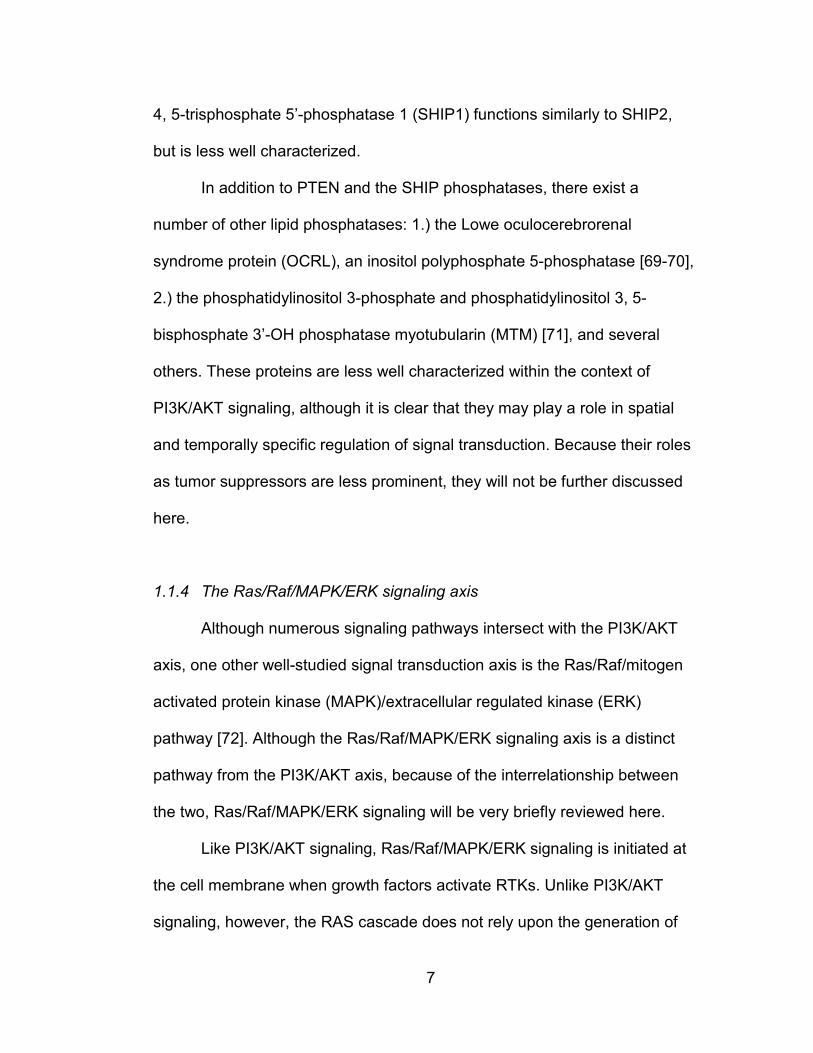
1.1.4 The Ras/Raf/MAPK/ERK signaling axis
Although numerous signaling pathways intersect with the PI3K/AKT
axis, one other well-studied signal transduction axis is the Ras/Raf/mitogen
activated protein kinase (MAPK)/extracellular regulated kinase (ERK)
pathway [72]. Although the Ras/Raf/MAPK/ERK signaling axis is a distinct
pathway from the PI3K/AKT axis, because of the interrelationship between
the two, Ras/Raf/MAPK/ERK signaling will be very briefly reviewed here.
Like PI3K/AKT signaling, Ras/Raf/MAPK/ERK signaling is initiated at
the cell membrane when growth factors activate RTKs. Unlike PI3K/AKT
signaling, however, the RAS cascade does not rely upon the generation of

8
the lipid second messenger PIP3. Instead, tyrosine phosphorylation of
growth factor receptors promotes the recruitment of the adapter protein
growth factor receptor 2 (GRB2) [73], which interacts with the guanine
nucleotide exchange factor (GEF) son of sevenless (SOS) [74]. SOS then
facilitates the exchange of guanine diphosphate (GDP) for guanine
triphosphate (GTP) on the RAS GTPases (HRas, KRAS and NRas). Thus,
growth factor receptor activation leads to activated, GTP-bound RAS
GTPases.
Once loaded with GTP, the RAS proteins regulate a number of
signaling molecules. Key among these is the serine/threonine protein
kinases a-Raf, b-Raf and c-Raf [75-76]. When bound to RAS proteins that
are loaded with GTP, the Raf kinases are activated and phosphorylate the
MAPK or ERK kinases 1 and 2 (MEK1/2) [77]. MEKs are dual-specificity
protein kinases that—when activated by Raf—phosphorylate the
extracellular regulated kinases (ERK1/2) on tyrosine residues [78-80].
ERK—also known as MAPK—then regulates a set of effectors to encourage
cell division and survival. The best characterized of these are ETS family
transcription factors like Elk-1 [81-82], the p90 ribosomal S6 kinase (RSK)
[83-84], and mitogen-and stress-activated protein kinase 1(MSK1) [85].
While they are two distinct signaling cascades, the PI3K and MAPK
pathways are by no means unconnected. Indeed, they intersect at many
points. In some instances, they serve to downregulate the activity of one
another. For example, Raf is phosphorylated at several sites by AKT, which

9
inhibits Raf activity [86], while activated BRAFV600E can inhibit AKT signaling
by inhibiting mTORC2 activity [87]. Moreover, treatment with specific small
molecule inhibitors of MEK1 can induce AKT phosphorylation, while
inhibition of PI3K, mTORC1, mTORC1/2 or AKT can upregulate ERK
phosphorylation in many contexts [88].
However, there exist a number of cooperative circuits between MAPK
and PI3K pathway. Phosphorylation of the TSC1/2 complex by ERK and
RSK at multiple sites can activate mTORC1 [89-93]. A particularly interesting
example of cooperation between PI3K and MAPK lies upstream, where
KRAS can interact with and activate PI3K isoforms [94-100]. Although this
finding was initially controversial, genetic evidence suggests that this
interaction is indeed meaningful in tumors as well as some regular
physiological contexts [101]. Thus, the interaction between the PI3K/AKT
and Ras/MAPK pathways are numerous and complex.
1.2 SIGNALING DOWNSTREAM OF PIP3
1.2.1 PDK1 activates AKT in a phosphoinositide dependent manner
The generation of PIP3 initiates a cascade of signaling events that
positively regulates cell growth, metabolism and survival. One direct effector
of PIP3 is the phosphoinositide-dependent kinase 1 (PDK1) [102-104], which
was originally identified from tissue extracts as a kinase that could
phosphorylate AKT at threonine 308 (T308). This threonine resides in the
catalytic domain of AKT, and its phosphorylation is necessary for AKT kinase

10
activity [105-106]. Interestingly, the inherent kinase activity of PDK1 is very
high regardless of growth factor stimulation or PIP3 levels in the cell [102].
Rather than being catalytically activated by growth factors, the key
determinant in PDK1 signaling to downstream effectors is its localization to
the cell membrane, which is accomplished via PDK1’s C-terminal pleckstrin
homology (PH) domain [107], which binds to PIP3 and the PIP2
phosphatidylinositol 3, 4-bisphosphate with high affinity. At the membrane,
the constitutively active PDK1 can phosphorylate its substrates. The most
well studied PDK1 substrates are the AGC family member kinases AKT/PKB
and PKC.
Unequivocal genetic evidence for PDK1’s role in activating AKT
arrived when murine embryonic stem (ES) cells null for the PDK1 gene were
generated [108]. In PDK1-/- ES cells the phosphorylation of AKT’s kinase
domain residue at T308 was completely absent after growth factor
stimulation. Further, the homologous residue in the catalytic domain of other
AGC kinases (SGK1 and PKC isoforms) was not phosphorylated in these
cells. Lastly, PDK1 is also necessary for activation of a Ras/Raf/MEK/ERK
regulated kinase, the 90 kDa ribosomal S6 kinase (p90 RSK) [109-111].
Unlike S6K1/2, RSK phosphorylates the ribosomal S6 protein in a
rapamycin-independent manner and largely takes its cues through the ERK
signaling cascade. Therefore, although PDK1 is important in PI3K
dependent signaling, it is also able to signal to other effectors outside of the
PI3K/AKT signaling axis.

11
1.2.2 AKT/PKB is a major effector of PI3K signaling
As mentioned above, one of the most well characterized effectors of
PI3K signaling is AKT (also known as protein kinase B (PKB)). AKT was
originally discovered as a viral oncogene that was found to be transforming
in a mouse thymoma model [112-114]. Higher eukaryotes have at least one
conserved homologue of the viral AKT gene. Indeed, mice and humans have
three distinct versions of AKT, coded for by the genes AKT1, AKT2, and
AKT3 [115]. These isoforms are similar in their structure, but the functional
outputs of the signaling by each kinase are unique as well as overlapping.
Nevertheless, the signaling events downstream of AKT—i.e. anti-apoptotic,
pro-metabolic, pro-growth, and pro-cell division—are generally shared by the
AKT isoforms.
The three AKT isoforms are similar in their structure in that they
contain an N-terminal PH domain, a kinase domain similar to other AGC
kinase family members in the middle, and an inhibitory C-terminal domain
that contains a hydrophobic motif (Figure 1.2) [116]. Similar to PDK1, the PH
domain on AKT serves to localize the kinase to the cell membrane when
PIP3 is generated [117]. At the membrane, the kinase activity of AKT is
upregulated by phosphorylation of its catalytic domain at residue T308 by
PDK1, a step which is required for AKT to exhibit enzymatic activity [118].
12
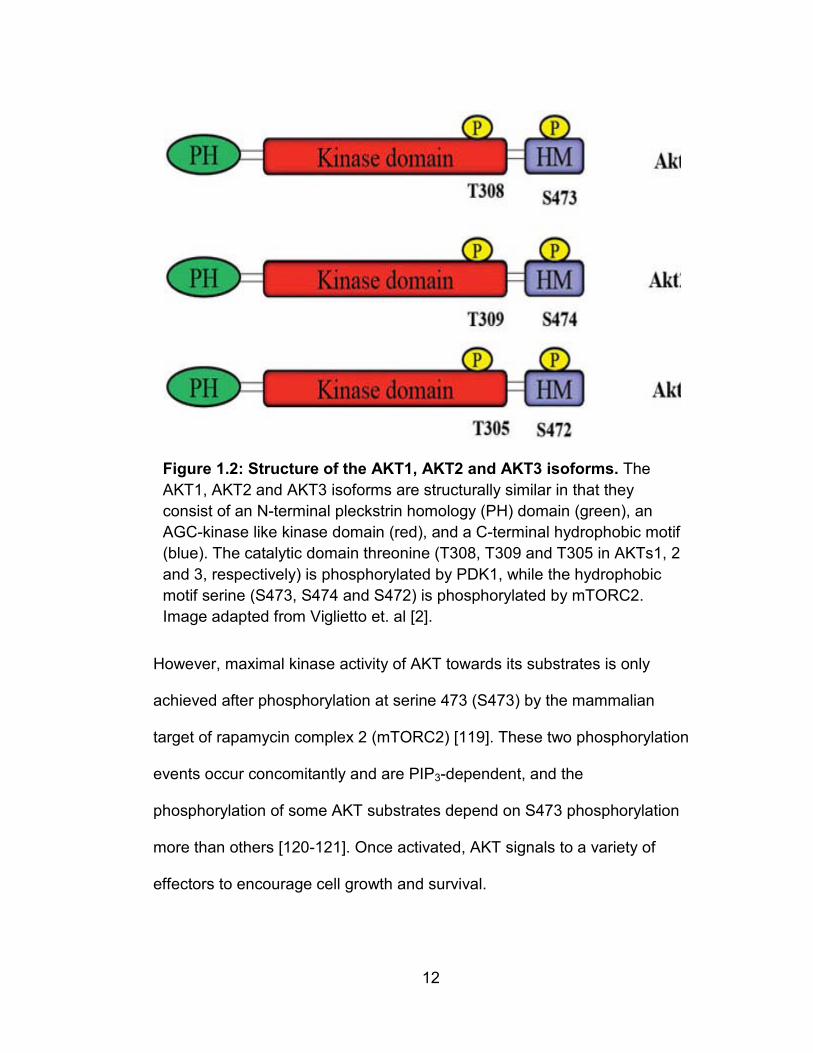
However, maximal kinase activity of AKT towards its substrates is only
achieved after phosphorylation at serine 473 (S473) by the mammalian
target of rapamycin complex 2 (mTORC2) [119]. These two phosphorylation
events occur concomitantly and are PIP3-dependent, and the
phosphorylation of some AKT substrates depend on S473 phosphorylation
more than others [120-121]. Once activated, AKT signals to a variety of
effectors to encourage cell growth and survival.
Figure 1.2: Structure of the AKT1, AKT2 and AKT3 isoforms. The AKT1, AKT2 and AKT3 isoforms are structurally similar in that they consist of an N-terminal pleckstrin homology (PH) domain (green), an AGC-kinase like kinase domain (red), and a C-terminal hydrophobic motif (blue). The catalytic domain threonine (T308, T309 and T305 in AKTs1, 2 and 3, respectively) is phosphorylated by PDK1, while the hydrophobic motif serine (S473, S474 and S472) is phosphorylated by mTORC2. Image adapted from Viglietto et. al [2].

13
1.2.3 AKT/PKB positively regulates cell metabolism, growth and survival
Activated AKT transmits a pro-metabolic signal by regulating several
steps in glucose metabolism. Importantly, AKT plays a critical step in the
insulin-dependent internalization of glucose by the glucose transporter
GLUT4 [122]. Once insulin has activated PI3K, AKT phosphorylates the AKT
Substrate of 160 kDa (AS160), which is a RAB GTPase-activating protein
(RabGAP) [123] that is required for the translocation of GLUT4 to the cell
membrane, where it facilitates glucose uptake into the cell [124]. AKT also
regulates glycogen metabolism by phosphorylating and inhibiting the
glycogen synthase kinase 3 (GSK3) [125-126]. By inhibiting GSK3—which
itself inhibits glycogen synthase—AKT upregulates the incorporation of
glucose into glycogen.
AKT also negatively regulates apoptosis to encourage cell survival.
This is thought to primarily occur through the Forkhead family of transcription
factors, particularly FOXO1 and FOXO3a (referred to here collectively as
FOXO) [127]. FOXO is a transcription factor that activates the expression of
apoptotic proteins like BIM-1, bNIP3, Bcl-6, FasL, and TRAIL [128-129]. AKT
directly phosphorylates FOXO at threonine 24 (T24), serine 256 (S256) and
serine 319 (S319) which prevents FOXO from entering the nucleus and
regulating gene expression. Thus, the phosphorylation of FOXO by AKT
sequesters FOXO in the cytosol, which inhibits the expression of members
of the apoptotic machinery. Further, AKT can regulate the apoptotic
14
machinery more directly by phosphorylating targets like Bad, which AKT
phosphorylates to inhibit the apoptotic functions of Bad [130-131].
The discovery and elucidation of the mechanistic target of rapamycin
(mTOR) signal transduction network has revealed several mechanisms
through which AKT regulates cell growth and protein synthesis. Briefly, AKT
negatively regulates two inhibitors of mTOR signaling, which upregulate
ribosome biogenesis, protein synthesis, and the accumulation of cell mass.
These mechanisms will be reviewed in the section below that discusses
mTORC1 signaling.
In addition to AKT, other members of the AGC kinase family regulate
survival signaling in a PI3K-dependent manner. This is not surprising
considering that AGC kinases share a remarkable amount of overlapping
characteristics in terms of protein structure [132-133]. One prominent
example of a kinase with a similar signaling role to AKT is the serum and
glucocorticoid-induced kinase 1 (SGK1) [134]. Interestingly, SGK1 can
phosphorylate AKT substrates like FOXO1/3A [135]. Further, recent
evidence suggests that SGK1 might take over the PI3K-dependent survival
that is usually regulated by AKT in certain tumors and cell lines [136]. The
reason for this shift in signaling is unclear, but it highlights the fact that
tumors with activated PI3K may not all respond equally to small molecules
that target AKT.

15
1.2.4 The mechanistic target of rapamycin (mTOR) is a master regulator of
cell growth that is downstream of PI3K/AKT
One of the more recently well studied members of PI3K/AKT signaling
is the mechanistic target of rapamycin (mTOR). Rapamycin is a small
molecule (~900 Da) product of Streptomyces hygroscopicus that was
discovered from soil samples collected on Rapa Nui (Easter Island) [137-
140]. The molecular mechanism of rapamycin was initially unclear, but early
studies demonstrated that rapamycin blocks the activation of T and B cells
by interleukin-2 (IL-2) [140-141]. Thus, even before the target of rapamycin
was known, it was appreciated that rapamycin could function as a potent
immunosuppressant.
The precise molecular target was elusive until genetic experiments in
yeast and affinity purification approaches in mammalian cells identified the
kinase mTOR (TOR in yeast) as the binding partner of rapamycin when
rapamycin is bound to the prolyl-isomerase FK506 binding protein, 12 kDa
(FKBP12) [142-146]. mTOR is a large (~289 kDa) protein kinase that is very
similar to the yeast proteins TOR1 (42% homology) and TOR2 (39%
homology), which were identified as the yeast targets of rapamycin prior to
the identification of mammalian mTOR [147]. The mTOR protein shares a
large degree of homology to PI3K family members, and is the founding
member of the phosphatidylinositol kinase-like kinase (PIKK) family [148].
Indeed, it was originally thought to phosphorylate phospholipids at the 4’-OH
group of the inositol ring [149-150]. Despite its amino acid sequence
16
similarity to PI-kinases, mTOR is indeed a serine/threonine kinase [151]. The
most well characterized effectors of mTOR are the protein kinase ribosomal
S6 kinase1/2 (S6K1 and S6K2), and the translation initiation inhibitor 4E-
BP1.
1.2.5 mTOR Complex 1 (mTORC1) is a nutrient and growth factor sensitive
protein kinase that regulates cell size and cell growth through S6K1
and 4EBP1
For some time after the discovery of mTOR, it was apparent that
mTOR phosphorylation of S6K1/2 is regulated by growth factors, amino
acids, and glucose. However, interacting proteins remained elusive until
gentler purification conditions and chemical crosslinking approaches were
used to identify the regulatory associated protein of mTOR (Raptor) [152-
153]. Together with mTOR, Raptor can be considered the founding member
of a protein complex that is now referred to as the mammalian target of
rapamycin complex 1 (mTORC1). mTORC1 is comprised of mTOR, Raptor,
the G-beta like protein GβL, the proline-rich AKT substrate of 40 kDa
(PRAS40), and the mTOR kinase inhibitor protein DEP-domain containing
protein DEPTOR. mTORC1 is a rapamycin-sensitive complex that positively
controls protein translation, cell size, and cell growth. mTORC1 is itself
regulated by several inputs, such as growth factors, redox conditions within
the cell, and glucose and amino acid availability.
17
By far, the most well understood mTORC1 substrates are the AGC
family kinase ribosomal S6 kinase 1/2 (S6K1/2) and eIF4e-binding protein 1
(4EBP1) [151, 154-157]. Both of these proteins are positive regulators of cell
growth, primarily through their regulation of mRNA translation [158]. S6K1
and S6K2 phosphorylate the ribosomal S6 protein to positively regulate
mRNA translation [159-161]. One important consequence of activated
S6K1/2 regulation of S6 is an increase in protein synthesis. Further, S6K-S6
positively regulates both cellular and organismal size [158, 162-164]. In
addition to regulating signaling to the ribosome machinery, S6K1 can
negatively control growth factor RTK signaling in a negative feedback circuit
that will be described below.
mTORC1 is activated by growth factor signaling via AKT through at
least two different mechanisms. The first of these to be described is a circuit
where AKT signals through a GTPase activating protein (GAP) complex
comprised of Tuberous Sclerosis Complex 1 and 2 (TSC1 and TSC2) [165-
166]. Phosphorylation of TSC2 by AKT destabilizes the interaction of the two
subunits and compromises the GAP activity of TSC1/2 towards the small
RAS-like GTPase RHEB (Ras homolog enhanced in brain) [167-172]. When
bound to GTP and in its active state, RHEB catalytically activates the kinase
activity of mTORC1 towards its substrates S6K1 and 4E-BP1 [173-176].
Thus, activated AKT phosphorylates TSC2, which inhibits the GAP activity of
the TSC complex, which allows GTP-bound RHEB to catalytically activate
mTORC1 signaling.

18
A second route through which AKT regulates mTORC1 is the
mTORC1 inhibitor Proline Rich AKT Substrate, 40 kDa (PRAS40). PRAS40
was initially identified as a component of mTORC1 that eluded detection
because the PRAS40-mTORC1 interaction is disrupted by lysis buffers that
contain NaCl [177-178]. Once identified, it was demonstrated that PRAS40
binds mTORC1 via Raptor to suppress mTORC1 kinase activity both in vivo
and in vitro. Phosphorylation of PRAS40 by AKT at threonine 246 (T246)
destabilizes the mTORC1-PRAS40 interaction to allow for mTORC1
activation. PRAS40 has also been described as an mTORC1 substrate [179-
180]. Activated mTORC1 phosphorylates PRAS40 at serine 183 (S183),
which further destabilizes the ability of PRAS40 to interact with Raptor. Thus,
mTORC1 is able to activate itself in response to growth factors via a
mTORC1-PRAS40 feed-forward circuit.
One of the major signaling inputs that regulate mTORC1 signaling is
intracellular amino acids [181]. mTORC1 activity towards S6K1 and 4E-BP1
is tightly regulated by the availability of amino acids. The past several years
have seen a flurry of studies that describe an elegant mechanism for amino
acid sensing by mTORC1 [182]. First, the Rag GTPases were found to be
necessary for amino acid induced activation of mTORC1 [183]. The Rags
exist as heterodimers (RAGA/RAGC and RAGB/RAGD) anchored on the
surface of the lysosome by the Ragulator complex, the whole of which
serves to localize mTORC1 to the lysosome by interacting with Raptor [184].
Once there, mTORC1 is in proximity to the aforementioned GTPase RHEB,

19
which then catalytically activates mTORC1. In a step that is only partially
understood, this sensing of amino acids by the machinery upstream of
mTORC1 relies upon a vacuolar (H+)-ATPase on the lysosome surface
[185]. These data suggest that amino acid sensing by mTORC1 might
actually be initiated within the lumen of the lysosome. Finally, the recently
described GATOR complexes act as Rag GAPs to downregulate mTORC1
signaling at the lysosome [186].
1.2.6 mTOR Complex 2 (mTORC2) is a PI3K-dependent kinase that
regulates AKT/PKB and other AGC kinases
One of the long-missing pieces of phosphoinositide-dependent
signaling was the kinase that phosphorylates AKT at the hydrophobic motif—
PDK2 [132, 187-191]. Whereas PDK1 had been well documented as an
AKT-phosphorylating, phosphoinositide-dependent kinase, PDK2 remained
elusive for several years. In fact, it had been speculated at several points
that PDK2 was one of any number of kinases: PDK1 [187], DNA-dependent
protein kinase (DNA-PK) [192], integrin-linked kinase (ILK) [193-194], or
even AKT itself [118].
The identity of PDK2 was clarified when it was found that mTOR—in a
second, rapamycin-insensitive kinase complex—is the hydrophobic motif
kinase that phosphorylates AKT at S473 [119, 195]. Examination of
immunopurified mTOR complexes revealed the existence of a second
mTORC (mTORC2) that is defined by the presence of the rapamycin-

20
insensitive companion of mTOR, or RICTOR [196]. mTORC2 was initially
described as the hydrophobic-motif kinase for several Protein Kinase C
(PKC) isoforms, but was subsequently found to phosphorylate other AGC
kinases such as AKT and serum and glucocorticoid-induced kinase 1
(SGK1) [197].
Like mTORC1, mTORC2 is a protein complex of several subunits. It
shares with mTORC1 the mTOR kinase itself and GβL (also known as
mLST8) [198], but is distinct from mTORC1 in that it contains RICTOR
instead of Raptor. Further, mTORC2 contains MAP kinase-associated
protein 1 (MAPKAP1)/mammalian stress-activated protein kinase (SAPK)-
interacting protein (mSIN1), a protein that is necessary for mTORC2 stability
and activity towards its substrates [199-201]. It has been recently
appreciated that mSIN1 likely functions to provide substrate specificity for
mTORC2, as it is apparently necessary for mTORC2 to interact with
substrates like AKT and SGK1 [202-204]. mTORC2 also contains one of two
proline-rich proteins—PRR5 and PRR5L—which are also known as Protor-1
and Protor-2 [177, 205-206]. The physiological role of these proteins was
initially unclear, as they are not required for mTORC2 complex stability or
activity towards AKT [206]. Studies of Protor-1 and the similar Protor-2 have
revealed that they can suppress apoptosis through PDGFRβ-dependent
signaling [177, 205], and that Protor-1 is important in regulating SGK1
function and sodium channel activation in the kidney [207].

21
Recently, mTORC2 has been found to share another component with
mTORC1—the previously mentioned DEPTOR, which is a negative regulator
of mTORC1 and mTORC2 kinase activity [208]. Although DEPTOR
intrinsically inhibits mTORC2 activation, increased expression of DEPTOR in
a cell can lead to a somewhat paradoxical increase in mTORC2 signaling, as
the inhibition of mTORC1 triggers activation of PI3K-dependent signaling,
which then activates mTORC2. Thus, DEPTOR could potentially be a
context-dependent tumor suppressor or oncogene. Indeed, elevated
expression of DEPTOR in certain forms of multiple myeloma appears to be
required for elevated PI3K/AKT signaling and survival of these cells [208].
Under normal physiological conditions, DEPTOR appears to positively
regulate adipogenesis [209].
Although many proteins have been described as parts of both the
mTORC1 and mTORC2 complexes, it is likely that even more binding
partners, adapter proteins, and regulators will be described for both of these
large signaling complexes. Interestingly, because of a complex web of
feedback circuits within and around the PI3K/AKT/mTOR axis, the mTOR
kinase can be considered to be both upstream and downstream of itself.
1.2.7 Feedback signaling within the PI3K/AKT/mTOR pathway
As with many biological signaling networks, there exist within the
PI3K/AKT/mTOR axis several mechanisms for downregulation of signaling
once it has been initiated. One of the first circuits to be appreciated was an

22
inhibitory circuit where activation of insulin signaling leads to the degradation
of the IRS1/2 adapter subunits [210-211]. It is now understood that both
S6K1 and mTORC1 phosphorylate IRS1/2 at several residues [212-215],
and that—unlike tyrosine phosphorylation of IRS1/2—these phosphorylation
events serve to destabilize the protein and facilitate its degradation by the
Figure 1.3: mTORC1 or mTORC1/2 inhibition activate PI3K/AKT signaling by relieving negative feedback circuits to upstream RTK signaling machinery. (A) Under normal signaling conditions, RTK activation is inhibited by a negative regulatory circuit where S6K1 inhibits IRS1 stability. (B) mTORC1 inhibition prevents S6K1 phosphorylation of IRS1, which activates mTORC2, PI3K, and AKT. (C) ATP-competitive inhibitors of mTOR potently relieve the aforementioned negative regulatory circuits, which upregulates IRS1/RTK signaling to PI3K, and hyperphosphorylates AKT at T308 despite an ablation in S473 phosphorylation. Adapted from Laplante and Sabatini, 2012 [1].

23
proteasome. Thus, mTORC1 inhibition with rapamycin leads to accumulation
of IRS1/2, activates PI3K, and leads to an increase in membrane PIP3 and
phosphorylation of AKT at T308 and S473 (Figure 1.3). Similarly, the
development of ATP-competitive mTOR kinase inhibitors has demonstrated
that dual mTORC1/2 inhibition also potently de-engages the negative
regulatory circuits between downstream and upstream signaling, despite the
inactivation of mTORC2 by these inhibitors [208]. Thus, simultaneous
inhibition of mTORC1/2 can potently hyperactivate PI3K activity and AKT
T308 phosphorylation, thus upregulating AKT.
Phosphoproteomic approaches have recently uncovered yet another
feedback inhibitory mechanism between mTORC1 and the upstream growth
factor machinery. The recently described mTORC1 substrate GRB10 serves
to inhibit insulin and IGF-I signaling under normal conditions, but this effect is
blunted when mTORC1 is inhibited with rapamycin or ATP-competitive
inhibitors like Torin1 [216-218]. Further, GRB10 is downregulated in some
tumors [218], which suggests that these patients may respond better to
therapies that utilize PI3K inhibitors, rather than ones that rely on mTOR
inhibitors.
These feedback circuits are likely to have consequences for the
efficacy of small molecule inhibitors in the clinic. Indeed,
immunohistochemistry from tumors of rapamycin treated patients has
demonstrated that rapamycin can potently upregulate AKT phosphorylation
in vivo [219-220]. Thus, while certain tumors—for examples, cancers with
24
activating mTOR mutations or inactivating TSC1/2 mutations—may respond
ideally to mTOR inhibitors, great caution should be exercised in determining
which patients would respond best to these therapies [221-222].
1.3 THE PI3K/AKT SIGNALING AXIS IS FREQUENTLY ALTERED IN
HUMAN CANCERS
A number of dysregulations in regular cellular physiology are
necessary in the course of tumorigenesis [223]. For example, cells typically
respond to a number of cues that encourage regular apoptosis, restrict
inappropriate growth, maintain genomic integrity, and keep interactions with
surrounding cells physiologically regular. However, in deregulating one or
several of these characteristics—typically by genetic or epigenetic
mechanisms—cancer cells can escape the normal life and fate of regular
cells and become “masters of their own destinies” [223]. Given the central
role of PI3K/AKT signaling in regular cellular and organismal physiology, it is
unsurprising that a variety of mutations within this pathway are found in
human tumors.
It is now appreciated that direct gain-of-function mutations in
PIK3CA—such as mutations that code for amino acid changes E545K and
H1047R in the p110α protein, among others—are found relatively frequently
in certain tumor types [31, 224]. Indeed, mutations such as these activate
the inherent kinase activity of p110α, which activates downstream kinases
like AKT and mTOR. Further, mutations in the regulatory p85 subunit of PI3K

25
can serve to activate the pathway in tumors [225-227]. The oncogenicitiy of
certain hotspot mutations make possible the specific targeting of mutant
PI3K proteins with small molecule inhibitors [228]. This work is in relatively
early stages, but could be a promising way to target PI3K/AKT signaling in
patients.
The most commonly mutated and well understood tumor suppressor
within the PI3K pathway is the lipid phosphatase PTEN. As described above,
PTEN is a lipid phosphatase that plays a critical role in downregulating
PI3K/AKT signaling by dephosphorylating the 3’-OH of PIP3. Loss of PTEN
function can occur in tumors a number of ways—methylation of the PTEN
promoter can silence PTEN mRNA expression [229], missense or nonsense
mutations can lead to loss-of-function mutant proteins [230-231], and
heterozygous or homozygous deletions of PTEN can give cells a selective
growth advantage [52, 232]. Strikingly, depending on the cancer type, loss of
PTEN function can be found in up to 40-80% of tumors. Further, conditional
knockout studies in the mouse have confirmed that PTEN loss is sufficient to
drive tumorigenesis in the prostate and other tissues [233]. Interestingly,
even haploinsufficiency of PTEN in some systems is sufficient to dysregulate
PI3K/AKT signaling and drive tumor growth [234]. PTEN loss poses a
significant challenge in the clinic because reconstituting the lost function of a
tumor suppressor within diseased cells is far less straightforward than
drugging an oncoprotein that is a kinase or transcription factor. However, the

26
recent description of a soluble PTEN protein that is excreted by cells may
suggest interesting therapeutic approaches available in the future [235].
In addition to mutations in PIK3CA and PTEN, mutations in the
receptor tyrosine kinases upstream of PIP3 generation are able to potently
activate PI3K signaling and drive tumorigenesis. The most commonly
deranged RTK is EGFR, which is found amplified or mutated in a variety of
tumors, most typically in various subtypes of lung and brain tumors [225].
Copy number amplification of the EGFR gene is sufficient to drive oncogenic
signaling [236], and oncogenic mutations in the EGFR gene can arise in the
kinase domain itself, the extracellular domain of the receptor [237], or by
heterozygous deletion of an autoinhibitory domain of the kinase (the so-
called EGFRviii variant) [238-239]. Indeed, these alterations—copy number
gain, activating point mutations, and deletion of inhibitory domains—are
themes that are repeated with other receptor tyrosine kinases in various
tumors (c-Kit, c-Met, PDGFR, etc.).
Finally, several kinases downstream of PIP3 generation can activate
tumorigenic signaling as well. Mutations in AKT1 have recently been
described, where an E17K mutation in the PH domain results in an
increased affinity for the phospholipid membrane, thus localizing AKT to the
membrane even in the absence of accumulated PIP3 [240-243]. Although
once thought rare, the E17K AKT1 mutation is now found relatively regularly
in multiple tumor types. Downstream of AKT, mutations in MTOR have
recently been identified in tumor samples that confer increased kinase

27
activity to mTORC1 and in some cases mTORC2 [244][245]. Interestingly,
many of these MTOR mutants render mTORC1 signaling exquisitely
sensitive to rapamycin, which suggests that tumors harboring these
mutations may be ideal candidates for mTOR inhibitor-based therapies.
1.4 TARGETING THE PI3K/AKT PATHWAY IN HUMAN CANCERS
1.4.1 From chemotherapy to targeted therapy
The elucidation of the various genetic derangements within growth
regulating pathways that can drive tumorigenesis has led to the development
of therapies that can specifically target the causative lesion in a tumor [246].
The prospect of identifying the genetic lesions of an individual’s tumor and
then drugging those lesions has offered a welcome alternative to traditional
radiation- and chemotherapy-based treatments for cancer. Rather than
targeting all growing cells, targeted inhibition of the oncoprotein(s) that drive
the growth of a tumor could ostensibly provide greatly diminished toxicity and
better clinical responses. Indeed, some of the first targeted therapies—
Gleevec® (imatinib) for chronic myelogenous leukemia (CML) and
Herceptin® (trastuzumab) for ERBB2-driven breast cancers—have proved
themselves to be superior to the previous therapies for those tumors.
However, the hope of targeted therapies has been tempered by the
emergence of many mechanisms of resistance. A more complete
understanding of the complex balance of survival signal transduction within
28
the cell (and the mutations that alter these pathways) would greatly improve
the design and deployment of targeted therapies for cancer.
1.4.2 Strategies for targeting tyrosine kinases
As mentioned above, the development of kinase inhibitors like
Imatinib demonstrated the possibility that small molecules targeting specific
signaling proteins could be effectively deployed in the clinic. In the clinic, the
most successful type of small molecule kinase inhibitor for cancer therapy
have been tyrosine kinase inhibitors. Although it was originally developed to
target the BCR-ABL fusion protein, imatinib is also useful for targeting
tumors with other activated tyrosine kinases such as PDGFR. Further,
several imatinib derivatives—like dasatinib, nilotinib and bosutinib—have
been developed to counter the different imatinib-resistant mutations that
arise in BCR-ABL [247-249].
Beyond BCR-ABL, several tyrosine kinase inhibitors have also found
some success in the clinic. The first of these was monoclonal antibody—
trastuzumab—that targets the human epidermal growth factor receptor 2
(HER-2) [250-251]. Trastuzumab was approved by the FDA for use against
breast cancer in 1998, and has also found use as a treatment for some types
of gastric tumors [252-253]. Other antibodies that target HER-2 or EGFR,
like pertuzumab [254], cetuximab [255], and panitumumab [256] are also
FDA approved, mostly for use in different types of HER-2 or EGFR-positive
breast cancers. EGFR and HER-2 are also targeted by a number of small
29
molecule inhibitors such as lapatinib [257], erlotinib [258] and gefitinib [259].
These molecules are typically deployed against various breast tumors, non-
small cell lung cancers, or pancreatic tumors.
1.4.3 PI3K inhibitors
Considering the central role that PI3Ks play in survival signaling, it is
unsurprising that many inhibitors targeting PI3K isoforms are in either
preclinical or clinical development [260]. Many groups and private
companies have developed isoform-specific inhibitors for p110α [261],
p110β [262]. Further, a potentially attractive method for specifically targeting
the aberrant PI3K signaling within tumors that harbor activating PIK3CA
mutations is the use of mutant-specific small molecule inhibitors [228]. These
inhibitors are, however, in their early stages of development and it remains
unclear how clinically useful they will prove.
PI3K inhibitors have great potential as anti-tumor tools in the clinic
[263]. However, that they have yet to gain FDA approval suggests that more
effort must be put into properly identifying which patients are most likely to
respond favorably to treatment with PI3K inhibitors. For example, while
patients with activating EGFR or PIK3CA mutations may both have
hyperactive PI3K/AKT signaling, PI3K blockade would likely be most
advantageous to the patient with the PIK3CA mutant tumor. Since not all
clinical trials evaluating PI3K inhibitors have triaged patients based on the
mutational status of the genes in the PI3K/AKT pathway, it is quite possible

30
that the antitumor benefits of several of these molecules has been masked
by tumors predestined by their genotypes to be refractory to PI3K inhibition.
1.4.4 Serine/threonine kinase inhibitors: targeting AKT and mTOR
Because the genetic lesions that drive tumors can occur at multiple
points within a given pathway, it may not always be advantageous or even
useful to target upstream regulators like RTKs and PI3Ks. For example,
some tumors are likely driven by mutations in AKT or MTOR, and thus
inhibiting PI3K signaling would pose little to no benefit. Indeed, several
groups have described both ATP-competitive and allosteric inhibitors of
AKT1-3 [264-267]. These inhibitors have either shown little advantage over
current therapies in the clinic—like perifosine [268]—or are in very early
stages of clinical evaluation [269]. However, AKT inhibitors could be very
advantageous for patients with tumors driven by oncogenic AKT mutations.
As mentioned above, the mTOR inhibitor rapamycin has been
deployed in the clinic as an immunosuppressant, an anti-restenotic agent,
and most recently as an anti-tumor agent. Although rapamycin and its
“rapalogue” derivatives have been clinically evaluated for activity against a
variety of tumors, mTORC1 inhibitors like CCI-779 (temsirolimus) were first
FDA approved for the treatment of renal cell carcinoma [270-271].
Subsequent studies have led to approval in breast cancer in combination
with hormone therapy [272-275]. As is the case for the other small molecule
inhibitors discussed above, it is likely that clinical trials that specifically target

31
patients based on which genetic lesions are present in their tumors (e.g.
activating MTOR mutations or inactivating TSC1/TSC2 mutations) will be
useful in identifying which patient populations would benefit most from
molecules that inhibit mTOR.
1.4.5 The promises and risks of targeted therapies
The successes of targeted therapies like Gleevec® and Herceptin has
often been heralded as a harbinger of a new era of “personalized medicine”
driven by targeted therapies [276-277]. It should be kept in mind that despite
the early successes of drugs like BCR-ABL inhibitors (Gleevec, etc.) in
treating CML, most cancers are dauntingly complex in the number of genetic
lesions that may drive a tumor. Much of the success of BCR-ABL inhibitors is
likely due to the fact that oncogenesis in CML is driven primarily by the BCR-
ABL fusion protein. In contrast, tumorigenesis in solid tumors and non-CML
liquid tumors is typically driven by a number of genetic lesions [223].
The development of resistance to targeted therapies by tumors is a
complex challenge. For example, CML often develops resistance to BCR-
ABL inhibitors through mutations within or near the ATP-binding pocket of
the kinase domain [276, 278-280]. Similarly, EGFR and ERBB2-driven
tumors can easily develop resistant kinase mutations in response to
treatment with inhibitors like erlotinib or lapatinib [278]. Beyond mutations in
the targeted signaling molecule, resistance to EGFR inhibitors can arise from
compensatory upregulation of ERK/MAPK signaling [281] or downregulation

32
of NF-ΚB signaling [282]. Finally, as mentioned above, mTORC1 inhibition in
the clinic should be pursued with caution, as it is likely that the anti-growth
properties of rapamycin are blunted by the fact that mTORC1 inhibitors
activate PI3K/AKT [219-220].
It is tempting to wish that the success of drugs like Gleevec® could be
generalizable to other tumor types. Unfortunately, the signal transduction
pathways that regulate tumorigenesis are complex, as is the scope of the
genomic alterations that activate these pathways. Thus, the emphasis of the
cancer research community should be focused on the discovery of as many
regulatory steps, circuits and nodes as possible. This would allow for the
design of multitudes of specific inhibitors that could be used to tailor targeted
therapies to each individual tumor with an eye towards circumventing
resistance to targeted therapies.
1.5 RNAi SCREENS TO IDENTIFY NOVEL COMPONENTS OF
ONCOGENIC SIGNALING
1.5.1 RNA interference (RNAi) as a loss-of-function tool to study gene
function
RNA interference (RNAi) is a relatively new technology in the toolkit of
the modern molecular biologist that takes advantage of an evolutionary
conserved mechanism of the regulation of mRNA through the micro RNA
(miRNA) pathway [283-286]. miRNAs are small (22 nt), non-coding RNAs
that are endogenous to the cell that serve to regulate the abundance and

33
translation of mRNA transcripts that code for genes [286-288]. When bound
to their target mRNAs, miRNAs down-regulates translation of the mRNA by
one of two mechanisms: 1.) blocking the ribosome from translating the
mRNA, or 2.) facilitating degradation of the mRNA by the RNA-induced
silencing complex (RISC).
The mechanism for RNAi-mediated gene silencing is similar to the
miRNA pathway with the exception that RNAi is not an endogenous process.
Rather, it simply takes advantage of the miRNA machinery that exists within
the cell. Indeed, RNAi has been tremendously advantageous to investigators
of biology because it provides a relatively easy—albeit imperfect—method of
silencing the mRNA transcript of a gene of interest, thus allowing for loss-of-
function studies of proteins. Further, in contrast to miRNAs, RNAi reagents
are most always perfectly complementary to their target transcripts, which
facilitate destruction of targeted mRNA.
There are many methods of delivering RNAi reagents to mammalian
cells. For example, small interfering RNAs (siRNAs) can be readily
transfected into some cell types. However, this method can induce unwanted
toxicity to the transfected cells [3]. To remedy the issues of transient
transfection and transfection toxicity associated with siRNA delivery, several
groups designed retroviral or lentiviral approaches to facilitate RNAi studies
[289-294]. Although these methods differ in some regards, they are similar in
that 1.) they rely upon viral delivery of DNA that stably integrates into the
host cell’s genome, 2.) stable expression of miRNA-like RNAs or short

34
hairpin RNAs (shRNAs) is achieved from the integrated DNA, and 3.)
suppression of the targeted mRNA transcript can be achieved for longer
periods than with siRNA-based approaches.
Not long after the initial characterization of mammalian and
Drosophila RNAi, several groups undertook efforts to generate large scale
RNAi libraries to facilitate loss-of-function genetic screens [289, 295-308].
The most prevalent deployment of these RNAi libraries has been in pooled
screens, where each shRNA reagent is administered to a population of cells
at a multiplicity of infection (MOI) of <1 [282, 305]. Although well suited for
high-throughput RNAi, pooled screens are limited by the fact that their
readouts rely on the measurement of shRNA or shRNA barcode abundance
within the pool. Hairpin abundance is itself an ersatz readout of the viability
of a cell population that has been transduced with an individual shRNA
reagent. Thus, this approach has been useful in identifying genes that confer
drug resistance [282, 305] or in performing in vivo loss-of-function screens
[306, 309-312].
Although non-pooled, arrayed shRNA screens were described before
pooled approaches were refined, the number of arrayed screens that have
successfully identified novel biology is relatively limited. Arrayed screens are
less technically straightforward than pooled RNAi screens in that they
require resources like plate handling robots and high throughput, often high-
content microscopy. However, arrayed screens do not require the
deconvolution that is inherent to pooled screening. Further, arrayed formats
35
allow for more biologically interesting assays than pooled screens. For
instance, an arrayed RNAi screen can be used to assay the activity of
signaling pathways. Indeed, several groups have used this approach to
successfully identify novel components of Ras/ERK signaling in Drosophila
[298, 313-314] and new regulators of cell division and histone
phosphorylation [302-303]. Thus, arrayed screens are useful tools for
probing signal transduction in ways that pooled approaches are not.
1.5.2 RNAi screens to identify novel oncogenic regulators of PI3K/AKT
signaling
Although discovery of new components of signal transduction
pathways can be achieved a number of ways—through proteomics, small
molecule inhibitor screens, cDNA microarray studies—we reasoned that a
loss-of-function RNAi screen could be a potent tool if combined with a robust
signaling assay. We were fortunate that the Broad Institute RNAi Consortium
(TRC) platform has undertaken a long-term effort to design, synthesize, and
make available lentivirus-delivered shRNAs targeting every known human
and mouse gene [302-303, 315]. This collection provides an impressive
amount of coverage in that a minimum of 5 shRNAs are provided for gene,
and many genes are targeted by 10-20 different, non-overlapping shRNA
reagents. In addition to a library of shRNA reagents, the TRC has performed
extensive validation with quantitative reverse-transcription PCR (qRT-PCR)
to validate which hairpins succeed in depleting their target mRNAs.

36
While the biochemistry and genetics of PI3K/AKT signaling are both
very well studied, we theorized that there are likely a number of
unappreciated proteins that regulate this pathway. Our rationale was that
while most tumors likely upregulate PI3K/AKT in some way, not all tumors in
queried oncogenomic databases display obvious lesions in the genes within
this signaling axis. Although the assumption of PI3K upregulation in all
tumors is not a certain one, we reasoned that tumors with no obvious
RTK/PI3K/PTEN/AKT/MTOR pathway lesions must be activating PI3K/AKT
signaling via genes that regulate—but are currently not understood to be
involved in—this pathway.
This thesis details a set of RNAi screens that we performed at the
Broad Institute to identify novel regulators of PI3K/AKT signaling. Our goal
was to identify novel proteins that regulate the PI3K/AKT pathway, then to
interrogate the mutational status of these genes in oncogenomic databases.
We suggest that this approach—targeted loss-of-function screening whose
results are prioritized by utilizing oncogenomic data—is a useful tactic to
identify proteins that play a role in normal cellular signaling as well as
tumorigenesis.

37
CHAPTER TWO:
AN RNAi SCREEN IDENTIFIES THE SMALL GTPASE RAB35 AS A
NOVEL ONCOGENIC REGULATOR OF PI3K/AKT SIGNALING
2.1 ABSTRACT
In an RNAi screen for genes that affect AKT phosphorylation, we
identified the RAB35 small GTPase—a protein previously implicated in
endomembrane trafficking—as a new regulator of the PI3K pathway. Depletion
of RAB35 in human and mouse cells suppresses AKT phosphorylation in
response to growth factors, whereas expression of a GTPase-deficient mutant
of RAB35 constitutively activates the PI3K/AKT pathway. RAB35 functions
downstream of growth factor receptors and upstream of PDK1 and mTORC2,
two established regulators of AKT, and interacts with PI3K. We identified two
RAB35 mutations in human tumors that activate PI3K/AKT signaling, suppress
apoptosis, and transform cells in a PI3K-dependent manner. Thus, we identify
RAB35 as a proto-oncogene that is both necessary and sufficient for activating
PI3K/AKT signaling. That a RAB GTPase can activate PI3K/AKT signaling and
transform cells suggests that there may be latent oncogenic potential in
dysregulated endomembrane trafficking.

38
2.2 INTRODUCTION
The phosphatidylinositol 3’-OH kinase alpha (PI3Kα) is a lipid kinase
and a key regulator of cell survival, proliferation and growth that is commonly
activated in human cancers [316-317]. PI3Kα is comprised of a 110 kDa
catalytic subunit (p110α) and an 85 kDa adapter subunit (p85) that links
PI3Kα to activated growth factor receptor tyrosine kinases (RTKs) [318]. At
the cell membrane, PI3Kα phosphorylates the membrane lipid
phosphatidylinositol (4, 5)-bisphosphate (PIP2) to form the second
messenger phosphatidylinositol (3, 4, 5)-trisphosphate (PIP3), which then
recruits a number of effectors to the lipid membrane. Key among these is the
serine/threonine kinase AKT, which regulates an array of cellular processes
such as glucose transport, the cell cycle, and anti-apoptotic signaling [133].
Activating mutations in oncogenes (PIK3CA, EGFR, AKT1) or loss-of-
function alterations in tumor suppressors (PTEN) have been well
characterized for their ability to drive tumorigenesis in a PI3K-dependent
fashion [225]. Thus, the components of this pathway are attractive targets for
cancer therapy, and many small molecules that target components of the
PI3K/AKT pathway are either currently deployed in the clinic or in clinical
trials [263, 319].
To identify new regulators of the PI3K/AKT pathway, we performed a
loss-of-function RNA interference (RNAi) screen using lentiviruses that
express short hairpin RNAs (shRNAs) [303]. We reasoned that the proteins
most likely to be regulators of PI3K/AKT signaling would be GTPases and

39
kinases, so we limited the screen to shRNAs targeting genes coding for all
known G-proteins and lipid/protein kinases. Because AKT phosphorylation is
a faithful indicator of PI3K activity, we used an immunofluourescent
approach to quantitatively measure phosphorylation of AKT at serine 473
(S473) in screened HeLa cells. Therefore, we screened a collection of 7,450
shRNAs in an arrayed format to identify kinases or GTPases whose
depletion could alter PI3K-dependent AKT phosphorylation.
2.3 METHODS AND MATERIALS
2.3.1 Cell lines and tissue culture
The following cell lines were obtained from American Type Culture
Collection (ATCC): 22Rv1 (CRL-2505), 786-O kidney adenocarcinoma
(CRL-1932), ACHN kidney adenocarcinoma (CRL-1611), HeLa cervical
adenocarcinoma cells (CCL-2), Caki-2 renal adenocarcinoma (HTB-47), HT-
29 colon adenocarcinoma (HTB-38), NIH-3T3 murine fibroblasts (CRL-
1658), PC3 prostate adenocarcinoma (CRL-1435), SK-CO-1 colorectal
adenocarcinoma (HTB-39), and SW620 colorectal adenocarcinoma (CCL-
227). SN12C kidney carcinoma cells were obtained from the Developmental
Therapeutics Program, NCI/NIH. HEK-293E cells were a generous gift to the
Sabatini lab from Dr. John Blenis (Harvard Medical School). All human cells
were cultured at 37° C (5% CO2) in high-glucose Dulbecco’s Modified Eagle
Medium (DMEM) supplemented with 10% fetal bovine serum (FBS),
penicillin-streptomycin and glutamate. NIH-3T3 murine fibroblasts were

40
cultured in DMEM with 10% bovine calf serum (BCS) from Colorado Serum
Company. All cells were passaged every 3 days as previously described
[208].
2.3.2 Antibodies
The goat anti-RICTOR S-20 antibody was from Santa-Cruz
Biotechnology (sc-50678), the anti-p85 antibody was from Millipore (04-196),
the anti-FLAG M2 antibody was from Sigma-Aldrich (F1804), and the anti-
RAB35 antibody was from Proteintech (11329-2-AP). All other antibodies
were from Cell Signaling Technologies: anti-phospho S473 AKT (4060), anti-
phospho T308 AKT (2965), anti-total pan AKT (2920), anti-phospho T346
NDRG1 (5482), anti-phospho T24/32 FOXO1/3A (9464), anti-RICTOR
(2140), anti-mTOR (2998), anti-p110α (4249), anti-p85 (4257), anti-PARP
(9532), anti-cleaved Caspase 3 (9664), and anti-HA (3724). Secondary
antibodies used for quantitative immunoblotting were obtained from LiCor
Biosciences: IRDye 800CW goat anti-rabbit IgG (926-32211), IRDye 800CW
goat anti-mouse IgG (926-32210), IRDye 680RD goat anti-mouse (926-
68170).
2.3.3 Chemicals, ligands and small molecules
Insulin (I9278) and polybrene (AL-118) were purchased from Sigma-
Aldrich. Dimethylsulfoxide (DMSO, BP231-4) was purchased from Fisher
Scientific. Puromycin (ant-pr-1) and Hygromycin B (ant-hm-1) were from
41
InvivoGen. Epidermal Growth Factor (EGF, AF-100-15), Platelet Derived
Growth Factor AA (PDGF-AA, 100-13A), Insulin-like Growth Factor I (IGF-I,
100-11) and Vascular Endothelial Growth Factor (VEGF, 100-20) were
purchased from Peprotech. Torin1 (S2827), rapamycin (S1039), PIK-90
(S1187) and GDC-0941 (S1065) were all purchased from Sellekchem. Each
drug was resuspended to 10 mM in DMSO, aliquoted and stored at -20° C
until use. 100% Triton TX-100 was purchased from Roche (11 332 481 001).
2.3.4 RNAi screens
High-throughput lentiviral-short hairpin RNA (shRNA) screens were
performed at The RNAi Consortium (TRC) at the Broad Institute as
previously described [303]. The shRNA library screened contained shRNAs
targeting control genes (LUC, GFP, RFP and LACZ), and between 5-15
individual shRNAs targeting each human kinase or GTPase for a total of
7,774 shRNAs targeting 1,012 genes (Table 1). 1 day before infection, 300
HeLa cells were seeded in each well of glass bottomed 384-well plates
(Costar 9120) in 50 µL of regular DMEM. 24 hours later, the medium was
removed and replaced with 50 µL DMEM containing 8 µg/mL polybrene. 1
hour later, 1µL of viral supernatant containing one individual shRNA-
expressing lentivirus was aliquoted into each well, and the plates were
incubated at 37° C. Each individual plate was performed in triplicate, and
each plate contained 15-20 control hairpins (shLUC, shGFP, shRFP,
shLACZ) distributed throughout each plate, 2 shRAPTOR_65 and 2
42
shRICTOR_25 control hairpins, 4 or more empty wells as selection controls
and ~350 shRNA reagents targeting human kinases or GTPases. 24h later,
the media in each well was gently aspirated and replaced with DMEM
supplemented with 1 µg/mL puromycin. Screen plates were incubated at 37°
C for 3 days, treated for 15 minutes with DMEM containing 100 ng/mL
insulin, then immediately processed for immunofluorescence.
2.3.5 Immunofluorescence for RNAi screens
After treatment with insulin, plate media was aspirated, and the cells
were fixed for 15 min. at room temperature with 50 µL 4%
paraformaldehyde. Plates were then washed with PBS 3 times, then
permeabilized with 50 µL 0.1% Triton X-100 in PBS for 15 min, and washed
once again with PBS 3 times. The last rinse was aspirated, and 12.5 µL of
anti-phospho-S473 AKT antibody mix was added to each well (Cell Signaling
Technology #4060, diluted 1:100 in 0.1% TX-100 PBS). Plates were briefly
centrifuged, then incubated overnight at 4° C. The next day, primary
antibody buffer was aspirated, the plates were washed 3 times with PBS,
and the plates were then incubated at room temperature for 2 hours with a
secondary immunofluorescence cocktail of goat anti-rabbit 800 nm antibody
(1:1000) and Sapphire 700 nm whole cell stain (LiCor 928-40022 , 1:1000) in
0.1% TX-100 PBS. Finally, secondary reagents were aspirated, the wells
were washed 3 times with PBS, and each well was filled with 50 µL PBS
after the final wash. Plates were then imaged at 700 nm and 800 nm

43
wavelengths using a LiCor Odyssey near-infrared scanner and LiCor
Odyssey software. For each well, the integrated signal intensity in the 700
nm (nuclear and whole-cell stain) and 800 nm (phospho-S473 AKT)
channels were quantified using the LiCor Odyssey software, and a
normalized phospho-AKT signal of each well was calculated by dividing the
signal value for the 800 nm channel by that of the 700 nm channel. These
values were then used to calculate Z-scores for each well within its plate as
compared to control shRNA reagents.
2.3.6 Phospho-AKT and viability Z-score calculation
In order to compare phospho-AKT levels between different plates, we
converted the normalized phospho-AKT from each well to a Z-score [320]
with the formula z = (x - µ)/σ (x, normalized phospho-AKT signal for a well; µ
and σ, mean and standard deviation (respectively) of the normalized
phospho-AKT values for all shLUC, shRFP, shGFP and shLACZ shRNAs in
an individual plate). Once normalized, we averaged the values of each well
for replicate plates to generate a mean phospho-AKT Z-score for every
screened shRNA reagent. The same calculation was used to score
viability/cell count for each well using the 700 nm whole-cell stain values.
Viability and phospho-AKT Z-scores for each screened shRNA are listed in
Appendix A.
44
2.3.7 RNAi screen data analysis and hit selection
After generating averaged phospho-AKT Z-scores for each hairpin as
described above, we ranked our entire dataset from smallest to largest
average phospho-AKT Z-score. We chose -1.50 and +1.50 as cutoff points
to call shRNAs as hits, thus we discarded from this list any shRNA reagents
with a phospho-AKT Z-score greater than -1.50 and less than +1.50. We
chose to impose a 40% shRNA hit-rate requirement for each gene. Thus, for
a gene represented by 5 hairpins, 2 of those average hairpins must return
phospho-AKT Z-scores below -1.50 or above +1.50 to be considered a hit.
Similarly, genes with 10 screened hairpins must return 4 shRNAs as hits.
shRNAs were only considered for our 40% hit-rate if they scored in the same
direction. Further, to exclude shRNAs that induced undue toxicity, we
excluded shRNAs that caused a decrease of -6.0 or greater in the viability Z-
score. Thus, our primary hit list was comprised of shRNAs that were both
non toxic and scored as hits at a rate of 40% or greater per gene, which are
listed in Appendix B. We next used quantitative RT-PCR data generated by
The RNAi Consortium to further curate our hits. A secondary hit-list was
generated by removing any shRNA that failed to knock down its target
mRNA by more than 50%. Genes that retained a 40% shRNA hit-rate after
this step are listed as secondary hits in supplemental Table 3. We next
excluded from our hits any gene that has been linked to PI3K/AKT signaling
in the literature. This was accomplished by searching NCBI PubMed for PI3K
or AKT and the name of each gene. For example, a PubMed search for

45
“AKT and RAB35” returns no publications and RAB35 would therefore be
retained in our tertiary list of hits. Genes with no prior link to PI3K/AKT
signaling thus comprised our tertiary list of hits and are reported in Table 4.
Finally, tertiary hits were curated for alterations in human tumors and cancer
cell lines as described in the next section.
2.3.8 Identification of hit gene mutations and genomic alterations in human
cancers
To identify genes with mutations or alterations in human cancers we
utilized the MSKCC cBio genomics portal (http://www.cbioportal.org/public-
portal) [321-322], the Sanger Catalog of Somatic Mutations in Cancer
(COSMIC) (http://cancer.sanger.ac.uk/cancergenome/projects/COSMIC/)
[323-326], and the Broad Institute/Novartis Cancer Cell Line Encyclopedia
(CCLE) (http://www.broadinstitute.org/ccle/home) [327-329]. To identify hit
genes mutated in cancers within these databases, we considered
meaningful mutations to be either 1.) recurrent mutations, or 2.) mutations
that resembled known mutations in similar genes that altered protein
function. For example, a Q->L mutation in the catalytic domain of an
uncharacterized hit GTPase that resembles the canonical transforming
KRASQ61L mutation would be considered meaningful even if it was not
recurrent. For copy number alterations, we considered genes to be altered if
they were focally amplified or deleted. For example, a hit gene was not
considered to be “altered” in its copy number if the deletion containing it was

46
comprised of the loss of an entire chromosome arm. We did not inspect
genes for alterations in their mRNA expression levels. Tertiary hits that were
found to be altered in human tumors or cancer cell lines and their respective
alterations comprised our list of prioritized hits and are reported in Table 5.
2.3.9 Cell lysis
Cell lysis and immunoprecipitations were performed as previously
described with some modifications [208]. All cells were rinsed once with ice-
cold PBS before lysis. Unless otherwise noted, cells were lysed with Triton
X-100 lysis buffer (40 mM HEPES [pH 7.4], 2 mM
ethylenediaminetetraacetic acid [EDTA], 10 mM sodium pyrophosphate, 10
mM sodium glycerophosphate, 50 mM NaF, 150 mM NaCl, 1% Triton X-
100). 25 mL of lysis buffer was supplemented with one cOmplete EDTA-free
protease inhibitor chip (Roche, 13971800). The lysates were incubated at 4°
C for 15 minutes, the insoluble fraction was spun in a microcentrifuge at
14,000 rpm for 15 minutes at 4° C, and lysates were collected. The protein
concentrations of cell lysates were then determined using Bio-Rad Bradford
assay reagent (500-0001) and an Eppendorf Biophotometer by reading the
absorption of each sample at 600 nm. Lysates were then mixed with
Laemmli’s buffer (50 mM Tris-HCl [pH 6.8], 2% SDS, 10% glycerol, 100 mM
2-mercaptoethanol, 0.1% bromophenol blue) so that the final protein
concentration in each sample was 1-3 µg/µL, boiled for five minutes, and
47
used for sodium dodecylsulfate polyacrylamide gel electrophoresis (SDS-
PAGE) as described below.
2.3.10 Immunoprecipitations
Cells were lysed in CHAPS-containing lysis buffer (40 mM HEPES
[pH 7.4], 5 mM MgCl2, 10 mM sodium pyrophosphate, 10 mM sodium
glycerophosphate, 50 mM NaF, 0.3% CHAPS). 25 mL of lysis buffers were
supplemented with one EDTA-free protease inhibitor chip. Proteins were
immunoprecipitated from 1000 µg of cell lysates with 1 µg of primary
antibody for 2 hours or overnight at 4° C. 15 µL of a 50% slurry of Protein G-
agarose beads (Pierce) was then added to each tube, and the
immunoprecipitations were incubated for another hour at 4° C with rotation.
Immunoprecipitates were then centrifuged at 5,000 rpm for one minute and
washed 3 times with CHAPS lysis buffer. On the final wash, the lysis buffer
was aspirated completely and 15 µL 1X SDS loading buffer was added to the
beads, which were then boiled for 5 minutes. The immunoprecipitates were
then analyzed by SDS-PAGE as described below.
2.3.11 SDS-PAGE
10-20 µg of protein lysates or immunoprecipitates were loaded onto 4-
12% Bis-Tris gels (Novex) and resolved by sodium dodecylsulfate-
polyacrylamide gel electrophoresis (SDS-PAGE) as previously described
[330]. Proteins were then transferred to Immobilon-FL nitrocellulose
48
membranes (0.45 µm pore size) from Millipore (IPFL00010), and then
processed for immunoblotting.
2.3.12 Immunoblotting
All western blots were processed using the LiCor Odyssey near-
infrared system. After transferring to membranes, blots were rinsed for 1
hour in Odyssey blocking buffer (927-40000) diluted in 50% PBS, then
incubated with primary antibody (1:1000) in antibody incubation buffer
(Odyssey blocking buffer, 50% PBS-T) overnight at 4° C or 2 hours at room
temperature. Blots were then washed 3 times (5 minutes each) in PBS-T
(Tween, 0.1%), and incubated with secondary antibodies (1:10,000, goat
anti-rabbit or mouse conjugated to 700 nm or 800 nm IR-dye) in secondary
antibody incubation buffer (Odyssey blocking buffer, 50% PBS-T, 0.01%
SDS) for 1 hour at room temperature. The blots were rinsed 3 more times in
PBS-T, rinsed with deionized H2O, washed for 5 minutes in PBS, and then
imaged on the LiCor Odyssey near-infrared scanner (resolution 84 µm,
intensity of 7 on both 700 and 800 nm channels). Images were saved,
processed, and quantitated using the LiCor Odyssey software.
2.3.13 Plasmids, cDNA manipulations and mutagenesis
The following plasmids were purchased from Addgene: lentiviral
(psPAX2) and retroviral (pUMVC) packaging plasmids (12260 and 8449,
respectively), pVSV-G (8454), pBabe HA-PIK3CA H1047R (12524), and

49
pBabe KRAS G12V (9052). pMSCV plasmids coding for murine AKT1
(wildtype and S473D) were generous gifts from Dr. Dos Sarbassov (M.D.
Anderson Cancer Center, Houston TX). RAB35 was synthesized by
Genscript (Piscataway, NJ) and subcloned into the SalI-NotI sites in the
pLJM60 vector to generate a plasmid that would express N-terminal FLAG-
tagged RAB35 protein [208], then transformed into TOP10 E. coli cells
(Invitrogen). Mutagenesis of pLJM60 FLAG-RAB35 and pMSCV mAkt1
cDNAs was performed as previously described using the Stratagene
QuickChange II XL site-directed mutagenesis kit (200521) [330].
2.3.14 Lentiviral and retroviral production
Lentiviral particles expressing shRNA hairpins or cDNAs were
generated as previously described using HEK-293CT cells (Clontech).
500,000 HEK-293CT cells were seeded in 5 mL of DMEM containing 10%
IFS and 0.1X penicillin-streptomycin in 6 cm dishes. The next day, each
plate was transfected with 1 µg of pLKO or pLJM60 plasmid expressing the
desired shRNA or gene, 100 ng of pVSV-G plasmid, and 900 ng of psPAX2
packaging plasmid mixed with 6 µL FugeneHD transfection reagent
(Promega, E2311). Retroviral particles expressing cDNAs were generated
similarly, except that 900 ng pUMVC was used for packaging 1 µg pBabe or
pMSCV reagents. 24 hours after transfection, the media on each plate was
aspirated and replaced with regular DMEM supplemented with 10% FBS and
1.1% BSA. Media containing viruses was harvested 24 and 48 hours

50
afterwards. This supernatant was then clarified of cells by filtration through a
0.22 µm Steri-Flip filter (Millipore). Supernatants containing virus were frozen
at -80° C until use or concentrated as described below.
2.3.15 Lentivirus and Retrovirus concentration
Because viral titers from lentiviruses and retroviruses expressing
genes are often low, we concentrated each using Lenti-X and Retro-X
reagents from Clontech (631231 and 631455, respectively). Briefly, viral
supernatants were mixed 4:1 with the appropriate concentration reagent and
incubated at 4° C overnight. The next day, virus was spun at 1,500g for 45
minutes, and the clarified supernatant was gently aspirated. Viral pellets
were resuspended to 1/50th their original volume in fresh DMEM (with 10%
FBS and 1.1% BSA), aliquoted, and stored at -80° C until use.
2.3.16 Lentiviral shRNA experiments
Lentiviral transduction of cells for shRNA experiments was performed
as previously described [119, 291, 303, 331]. Human or mouse cells were
seeded at 50,000 cells per well in 2 mL of media in 6 well plates. The next
day, media was removed and replaced with media containing 8 µg/mL
polybrene. 1 mL of unconcentrated shRNA-expressing lentivirus was then
added to each well. 24 hours later, the media and virus was removed and
replaced with fresh medium containing 1 µg/mL puromycin. Cells were then
cultured for 3 to 5 days, then treated as necessary and lysed.

51
2.3.17 Human and mouse stable cell line generation
HEK-293E, HeLa and NIH-3T3 cell lines stably expressing FLAG-
RHEBwt, FLAG-RAB35wt, FLAG-RAB35Q67L, FLAG-RAB35S22N, FLAG-
RAB35A151T, FLAG-RAB35F161L, HA-p110αH1047R, mAkt1wt, mAkt1S473D,
mAkt1T308D, or FLAG-AKT1myr were generated as previously described [332].
Briefly, 10,000 cells were seeded into one well of a 6-well plate in 2 mL
media. The next day, the media in each well was supplemented with 8
µg/mL polybrene. 1 mL of concentrated lentivirus (FLAG-RHEB or the FLAG-
RAB35 alleles) or retrovirus (HA-p110αH1047R, mAkt1wt, mAkt1S473D,
mAkt1T308D, KRASG12V or FLAG-AKT1myr) was then added to each well. 24
hours later, the media was aspirated and replaced with fresh media
containing the appropriate selection reagent (1 µg/mL puromycin or 100
ng/mL hygromycin B). Cells were grown to 80% confluence, then passaged,
expanded further, and preserved at -80° C as frozen aliquots in media
containing 10% DMSO and 40% serum.
2.3.18 Lentiviral RNAi reagents
We found that the shLUC_221 hairpin served as the best control
hairpin for our experiments because it was not toxic towards cells and did
not alter protein levels or phosphorylation under a number of different
conditions. Thus, we used shLUC_221 as the preferred shRNA control
hairpin for our low-throughput shRNA experiments.
shLUC: shLUC_221: TRCN0000072246; promegaLuc_221s1c1

52
The human shRICTOR_25 and shRAPTOR_65 shRNAs used as
control hairpins in the RNAi screen and throughout this paper can be
obtained from Addgene (Plasmids 1853 and 1857, respectively).
Human shRICTOR_25: Sense:
CCGGTACTTGTGAAGAATCGTATCTTCTCGAGAAGATACGATTCTTCAC
AAGTTTTTTG
Human shRAPTOR_65: Sense:
CCGGAGGGCCCTGCTACTCGCTTTTCTCGAGAAAAGCGAGTAGCAGGG
CCCTTTTTTG
TRC reagents were obtained either directly from the Broad Institute
RNAi Consortium or ordered from Sigma-Aldrich. The TRC identifications for
the shRNAs used here are as follows and can be used to query individual
hairpins in The RNAi Consortium’s public data portal
(http://www.broadinstitute.org/rnai/public/gene/search):
The five shRAB35 reagents screened in our RNAi screen were:
Human shRAB35_207: TRCN0000047796; NM_006861.4-207s1c1
Human shRAB35_451: TRCN0000047793; NM_006861.4-451s1c1
Human shRAB35_486: TRCN0000307737; NM_006861.4-486s1c1
Human shRAB35_649: TRCN0000047794; NM_006861.4-649s1c1
Human shRAB35_709: TRCN0000047797; NM_006861.4-709s1c1
Additionally, we tested the following nine shRAB35 reagents that were
generated by the TRC more recently and were thus not included in our RNAi
screen:

53
Human shRAB35_207b: TRCN0000291620; NM_006861.4-207s21c1
Human shRAB35_260: TRCN0000331177; NM_006861.4-260s21c1
Human shRAB35_359: TRCN0000331153; NM_006861.4-359s21c1
Human shRAB35_453: TRCN0000380080; NM_006861.4-453s21c1
Human shRAB35_475: TRCN000038033; NM_006861.4-475s21c1
Human shRAB35_486: TRCN0000307737; NM_006861.4-486s21c1
Human shRAB35_537: TRCN0000297015; NM_006861.4-537s21c1
Human shRAB35_565: TRCN0000379410; NM_006861.4-565s21c1
Human shRAB35_601: TRCN0000380003; NM_006861.4-601s21c1
We found that three particular shRAB35 reagents were the most
reliable in both depleting RAB35 protein levels and suppressing AKT
phosphorylation. The three hairpins used throughout this paper for all
experiments after validation of RAB35 as our primary hit are represented with
simplified numbers (1, 2, and 3):
shRAB35_1: Human shRAB35_709: TRCN0000047797; NM_006861.4-
709s1c1
shRAB35_2: Human shRAB35_475: TRCN000038033; NM_006861.4-
475s21c1
shRAB35_3: Human shRAB35_601: TRCN0000380003; NM_006861.4-
601s21c1
shRNAs targeting murine RICTOR and human PIK3CA were
purchased from Sigma-Aldrich:

54
shPIK3CA_1: Human shPIK3CA_3234: TRCN0000010407; NM_006218.x-
3234s1c1
shPIK3CA_2: Human shPIK3CA_3254: TRCN0000195304; NM_006218.2-
3254s1c1
shRICTOR_1: Mouse shRICTOR_5030: TRCN0000123398; NM_030168.2-
5030s1c1
shRICTOR_2: Mouse shRICTOR_5031: TRCN0000123395; NM_030168.2-
5031s1c1
2.3.19 mTORC2 in vitro kinase assays
mTORC2 kinase assays were performed in vitro as described
previously [119, 333]. Briefly, cells grown in 15 cm plates were serum
starved overnight, then stimulated, washed once in ice-cold PBS and lysed
in 1 mL 0.3% CHAPS lysis buffer. Lysates were incubated at 4° C for 15
minutes, centrifuged at 14,000 rpm for 15 minutes, the soluble fraction was
collected and the protein concentration was determined. 1000 µg of protein
in a final volume of 750 µL was then used for each immunoprecipitation. 15
µL of goat anti-RICTOR antibody (Santa Cruz) was added to each tube, and
tubes were incubated at 4° C overnight with rotation. The next day, 15 µL of
a 50% slurry of agarose Protein G beads (Pierce) was added to each tube
and incubated for an additional hour with rotation. Immunoprecipitates were
then washed three times with CHAPS lysis buffer, and once with 1X
mTORC2 kinase buffer (25 mM HEPES [pH 7.5], 100 mM potassium

55
acetate, 1 mM MgCl2). The kinase buffer was then completely aspirated and
the following was immediately added: 7.5 µL 2X mTORC2 kinase buffer, 250
ng inactivated AKT1/PKBα (Millipore, #14-279), 0.75 µL 10 mM ATP, and
the reaction volume was then brought up to 15 µL with dH2O. The reactions
were then incubated at 37° C for 20 minutes with gentle agitation (300 rpm).
Every five minutes, the tubes were gently flicked to resuspend the agarose
beads within the reactions. The kinase assay was then stopped with the
addition of 200 µL of 1X Laemmli’s loading buffer, the tubes were boiled for
five minutes, and 20 µL of each reaction was then analyzed by SDS-PAGE.
2.3.20 PI3-kinase in vitro kinase assays
We used Echelon Biosciences PI3K kinase assay ELISA “pico” kits
(K-1000s) for in vitro PI3K reactions using purified p85-p110α. Cells were
lysed in 0.3% CHAPS lysis buffer as described above and PI3K was
immunopurified from 1000 µg of clarified cell lysates using 20 µL agarose-
conjugated anti-p85 antibody (Millipore, 16-107) overnight at 4° C.
Immunoprecipitates were then washed three times in CHAPS lysis buffer
and once in KBZ buffer (Echelon). After the final wash, KBZ buffer was
completely aspirated and 30 µL of KBZ buffer supplemented with 5 mM DTT
and 150 µM ATP was immediately added to the immunoprecipitates. 30 µL
of 100 µM phosphatidylinositol 4, 5-bisphosphate (PIP2) and either DMSO or
PIK-90 were then added to the reaction. Final kinase assay buffer was thus
comprised of KBZ buffer, 50 µM PIP2, 2.5 mM DTT, 75 µM ATP, DMSO or

56
500 µM PIK-90, and 20 µL of agarose anti-p85 beads. Reactions were then
incubated at 37° C and tubes were gently tapped every 15 minutes to
resuspend the agarose beads in the reaction. After 3 hours, reactions were
stopped with the addition of 60 µL KBZ buffer supplemented with 4 mM
EDTA. The reactions were then centrifuged, and the supernatants were
removed and frozen at -20° C. The PIP3 competitive ELISA assay kit was
then used as per the manufacturer’s instructions to determine the
concentration of PIP3 generated in each kinase reaction. The supernatant
from each reaction was split between two individual ELISA wells and each
reaction was run in duplicate, thus each kinase assay condition was assayed
in quadruplicate.
2.3.21 Apoptosis assays of NIH-3T3 cell lines
NIH-3T3 cells were assayed for apoptosis by both western blotting of
whole cell lysates and by cell counting methods. Briefly, cells were split and
each cell line was seeded at 1 million cells per well of a 6-well plate in 2 mL
of DMEM supplemented with 10% BCS. After 6 hours at 37° C, 3 wells of
each plate were aspirated, washed once with serum-free DMEM, and the
media was replaced with serum-free DMEM and the plates were returned to
37° C. 4 hours later, the cells were lysed in TX-100 lysis buffer and cell
lysates were analyzed by immunoblotting. For cell count assays, each cell
line was seeded and treated as just described. Instead of lysis, after 4 hours
of serum withdrawal, cells were rinsed once with PBS, briefly trypsinized with

57
500 µL of trypsin, dissociated into a single cell suspension, resuspended to
1000 µL with 500 µL of DMEM and collected. 500 µL of each cell suspension
was then counted on a Beckman-Dickinson Vi-Cell XR, and the total number
of viable cells per mL was recorded. Each condition (serum complete and
serum withdrawn) was performed in triplicate (3 wells each). Serum
withdrawn cell counts for each cell line were then normalized to their
respective serum complete cell count to arrive at a % of viable cells. Mean
and standard deviation values for each condition were then calculated and
graphed in Microsoft Excel.
2.3.22 NIH-3T3 focus formation assays
Focus formation assays with NIH-3T3 stable cell lines were performed
as previously described [334-335]. For each focus formation assay, freshly
thawed, low-passage NIH-3T3 stable cell lines were used. Briefly, NIH-3T3
cells stably expressing genes indicated throughout this paper were seeded
in 6-well plates in 2 mL of 10% BCS DMEM at 20,000 cells per well. The
next day, the media was aspirated and replaced with 2 mL 2% BCS DMEM
containing either DMSO or 250 nM GDC-0941. Cells were then cultured at
37° C for 4 weeks with media changes every 3 days. After 4 weeks, cells
were fixed and processed for crystal violet staining as follows. Cells were
rinsed once with PBS and fixed for 10 minutes at room temperature in 1 mL
70% EtOH. Next, EtOH was aspirated and cells were stained with crystal
violet reagent (0.5% crystal violet, 25% EtOH) for 10 minutes. Crystal violet

58
was then removed, and each well was rinsed gently with dH2O until
background stain was removed. The plates were then imaged using an
Oxford Optronix plate imager. Because the qualitative aspects of foci formed
by different oncogenes varies widely (i.e. fewer but larger ones for
RAB35Q67L and many small ones for AKTmyr, etc.) and because colony
counting is a subjective process, we chose to quantify the amount of crystal
violet stain in each well as a way of objectively measuring the total amount of
cells present in our focus formation assays. To extract crystal violet from
stained cells, we incubated each well overnight at room temperature in 1 mL
of Sorenson’s buffer (50 mM sodium citrate, 50 mM citric acid, 50% EtOH)
with gentle agitation. The next day, extractions were collected and the
absorbance at 590 nm was recorded using a spectrophotometer. These
values were tabulated in Microsoft Excel, and the values for all samples
were normalized to the average absorbance of DMSO treated FLAG-
RAB35wt NIH-3T3 cells.
2.4 RESULTS
2.4.1 A quantitative, immunofluorescent assay for AKT phosphorylation
To quantitatively assess PI3K/AKT signaling in cultured cells, we designed
an immunofluorescence-based assay that detects levels of AKT
phosphorylation at S473. mTORC2 phosphorylates AKT at this site in a
PI3K-dependent manner, a phosphorylation which both reflects PI3K activity
and is required for full activation of AKT itself [119]. To probe for AKT

59
phosphorylation in HeLa cells grown in 384-well plates, we probed fixed cells
with a monoclonal rabbit antibody directed against AKT phosphorylated at
S473 (Figure 2.1). This antibody was probed with a secondary antibody
tagged with an 800 nm
fluorophore (green), and the cells were counterstained with a 700 nm whole
cell stain (red). We imaged and quantified these two signals with a near-
infrared scanner. These signals could be divided by one another to yield a
normalized value for each well that represents phospho-AKT levels (see
methods for a more complete description of this assay). This assay was
sensitive to inhibition with PI3K and mTOR kinase inhibitors. Thus, we
Figure 2.1: An immunofluorescent assay for phospho-S473 AKT quantitatively reflects PI3K activity. HeLa cells grown in 384-well plates were serum starved overnight, treated with PIK-90 and insulin as indicated, then fixed and processed for immunofluorescence with a whole cell stain (red) and an anti-phospho-S473 AKT monoclonal antibody (green) (top). Signals were imaged and quantified with a LiCor Odyssey near-infrared scanner, and the phospho-AKT signal intensity was divided by the whole-cell stain signal intensity to calculate a normalized phospho-AKT value for each well (bottom).

60
developed a robust assay for PI3K/AKT activity that we next deployed in a
high-throughput screen.
2.4.2 An RNAi screen identifies RAB35 as a novel regulator of PI3K/AKT
signaling
We defined phospho-AKT Z-scores -1.50 and +1.50 as the cutoffs
beyond which individual shRNAs would be considered as low or high hits,
respectively. Fortunately, shRNAs targeting genes whose knockdown should
suppress AKT phosphorylation—RICTOR, PIK3CA, PIK3CB, AKT1, and
Figure 2.2: An arrayed lentiviral loss-of-function RNA interference screen identifies known and novel regulators of the PI3K/AKT axis. Averaged phospho-AKT Z-scores of screened shRNAs. HeLa cells were plated in 384-well plates, transduced with lentiviral reagents expressing control shRNAs or shRNAs targeting human kinases and GTPases, then processed as in Figure 2.1. Phospho-AKT Z-scores for each shRNA were then calculated, averaged (n=3), and arranged in increasing order. Low hits (Z-score < -1.50) are indicated in red, high hits (Z-score > 1.50) are indicated in green. Hit genes that are known to regulate PI3K/AKT signaling are indicated in red or green boxes. For a full list of non-lethal hit genes please see Appendix B.
61
AKT3—all diminished AKT phosphorylation and scored as low hits (Figure
2.2, Appendix B). Conversely, shRNAs targeting genes whose protein
products inhibit AKT signaling—such as RAPTOR and RHEB—elevated
phospho-AKT levels and scored as high hits. Since this assay successfully
identified known regulators of PI3K/AKT signaling, we were confident that
our screening approach was a valid method for discovering new members of
the this pathway.
To reduce the number of shRNAs and genes in our primary hit list to a
manageable number, we discarded shRNAs that were toxic or did not knock
down their target mRNA, and considered as hits only genes for which 40%
or more of the screened hairpins scored in our assay (Table 2.1, Appendix
B). Further, we discarded hit genes that were not ubiquitously expressed or
had been previously studied in the context of PI3K/AKT signaling (Appendix
D). Finally, we used publicly available oncogenomic databases to annotate
the remaining hit genes and discarded any genes that were not altered in
human tumor samples or cell lines. Thus we prioritized our hits for validated,
non-toxic shRNAs that targeted genes with no previous link to PI3K/AKT
signaling that were altered in human cancers. This left us with a manageable
list of 29 genes with which we could perform low-throughput loss-of-function
experiments (Table 2.2).
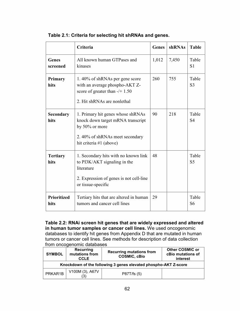
62
Table 2.2: RNAi screen hit genes that are widely expressed and altered in human tumor samples or cancer cell lines. We used oncogenomic databases to identify hit genes from Appendix D that are mutated in human tumors or cancer cell lines. See methods for description of data collection from oncogenomic databases
SYMBOL Recurring
mutations from CCLE
Recurring mutations from COSMIC, cBio
Other COSMIC or cBio mutations of
interest Knockdown of the following 3 genes elevated phospho-AKT Z-score
PRKAR1B V100M (3), A67V (3) P87T/fs (5)
Table 2.1: Criteria for selecting hit shRNAs and genes.
Criteria Genes shRNAs Table
Genes screened
All known human GTPases and kinases
1,012 7,450 Table S1
Primary hits
1. 40% of shRNAs per gene score with an average phospho-AKT Z-score of greater than -/+ 1.50
2. Hit shRNAs are nonlethal
260 755 Table S3
Secondary hits
1. Primary hit genes whose shRNAs knock down target mRNA transcript by 50% or more
2. 40% of shRNAs meet secondary hit criteria #1 (above)
90 218 Table S4
Tertiary hits
1. Secondary hits with no known link to PI3K/AKT signaling in the literature
2. Expression of genes is not cell-line or tissue-specific
48 Table S5
Prioritized hits
Tertiary hits that are altered in human tumors and cancer cell lines
29 Table S6
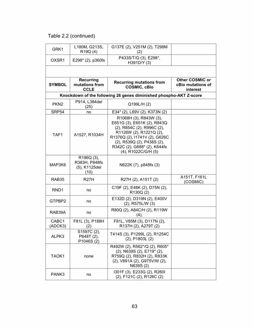
63
Table 2.2 (continued)
GRK1 L180M, G213S, R19Q (4)
G137E (2), V251M (2), T298M (2)
OXSR1 E298* (2), p360fs P433S/T/Q (3), E298*, H391D/Y (3)
SYMBOL Recurring
mutations from CCLE
Recurring mutations from COSMIC, cBio
Other COSMIC or cBio mutations of
interest Knockdown of the following 26 genes diminished phospho-AKT Z-score
PKN2 P914, L384del (25) Q199L/H (2)
SRP54 no E34* (2), L69V (2), K373N (2)
TAF1 A1527, R1034H
R1068H (3), R843W (3), E651G (3), E651K (2), R843Q
(2), R854C (2), R996C (2), R1126W (2), R1221Q (2),
R1376Q (2), I1741V (2), G626C (2), R539Q (2), P438S (2),
R342C (2), G688* (2), K644fs (4), R1022C/G/H (5)
MAP3K6
R196Q (3), R383H, P848fs (5), K1125del
(10)
N622K (7), p848fs (3)
RAB35 R27H R27H (2), A151T (2) A151T, F161L (COSMIC)
RND1 no C19F (2), E48K (2), D75N (2), R130Q (2)
GTPBP2 no E132D (2), D319N (2), E400V (2), R575L/W (3)
RAB39A no R80Q (2), A84C/H (2), R119W (4),
CABC1 (ADCK3)
F81L (3), P188H (2)
F81L, V85M (3), D117N (2), R137H (2), A279T (2)
ALPK3 S1597C (2), P648T (2), P1046S (2)
T414S (3), P1299L (2), R1254C (2), P1803L (2)
TAOK1 none
R492W (2), R562*/Q (2), R605* (2), N639S (2), E719* (2),
R759Q (2), R832H (2), R833K (2), V891A (2), G975V/W (2),
N639S (2)
PANK3 no I301F (3), E233G (2), R260I (2), F121C (2), R126C (2)

64
Table 2.2 (continued)
ALPK1 T1150fs (6), S942del (12)
V62M (3), A86T (2), A144S (2), A152S (2), N175D (3), D253N (2), V296M (2), S832F/N/T (4), I1124M (2), K1125E (2), P293L
(2)
RAB1B no E35K/Q (2), G42W (2), R79fs (2) A152T (COSMIC)
ADPGK no L176I (2), K450fs (3), R475*/Q (3)
GSG2 no R82C (18), K358fs* (2) POMK
(SGK196) R15Q, T146N A13T (5)
PKDCC E408G (9) V131I (2)
RASEF no R262C (2), R332Q (3), H350R (2), R706* (2), G792W (2)
FUK no R235H (2), R767Q (2)
NRK S101F, R1387W, P426A, K1409T,
E898K
S424C (4), A681E (3), S108P (3), S708P (3), R438*, P389H
(2), R1911Q (3)
STK32C S392Y (3) G282W (2), S482L (2)
TSSK4 T338del (12) R281C (2)
NEK8 G602E (2) A206T (2), L621F (2), G605D (2), V690fs (2)
RABL3 no R107C (2)
NME9 no E75K (5), R221W (5), G270D (2), E53K (5), G209D (2)
Next, we were curious as to the types of genes that were present in
this enriched hit list, and whether they shared any functional characteristics.
Because this list was largely comprised of low hits (26 of 29) and because
we were interested in identifying positive (i.e. oncogenic) regulators of
PI3K/AKT signaling, we focused on only the 26 low hit genes from Table 2.2.
We used literature searches to identify the molecular function—or, failing
that, the broader organismal function—of these 26 genes, then asked which
molecular function was the most prominently represented (Figure 2.3A).

65
Interestingly, 5 of these 26 low hit genes were RAB GTPases, which
suggested to us that there may be a previously unappreciated role for RAB
proteins in oncogenic growth factor signaling.
Of the five RAB proteins in this list (RAB1B, RAB35, RAB39, RASEF
and RABL3), we prioritized RAB35 for further study because 1.) it is
somatically mutated in human cancers (Figure 2.3B) and 2.) it is well-
characterized in recycling endosome biology and has widely available
reagents and tools. A member of the RAB family of GTPases, RAB35 has
Figure 2.3: RAB proteins are prevalent in the enriched RNAi screen hitlist, and RAB35 is somatically mutated in human cancers. (A) The 26 hit genes that reduced AKT phosphorylation, are expressed widely, and are mutated in human cancers were grouped by their annotated function from the literature. (B) Mutations found in oncogenomic databases that are predicted to have a medium (black) or high (red) effect on protein function are mapped along the length of the RAB35 protein.

66
been described as a regulator of cytoskeletal organization and trafficking at
the recycling endosome [336-339]. While a number of regulators and
effectors of RAB35 have been identified, we did not find any evidence in the
literature to suggest that RAB35 plays a central role in growth factor
signaling [340]. Further, although several RAS family members have been
implicated in cancer, there is little evidence that suggests that RAB proteins
possess oncogenic potential. Thus, we reasoned that RAB35 was the most
promising candidate for further study.
To confirm that RAB35 depletion with the screened shRNAs could
inhibit AKT phosphorylation, we transduced HeLa cells with lentiviruses
expressing shRNAs targeting luciferase (as a control), the mTORC2
component RICTOR, or RAB35. Indeed, three of the shRAB35 hairpins
simultaneously reduced RAB35 protein levels and diminished the
phosphorylation of AKT and the AKT substrate FOXO1/3A in response to
stimulation with serum (Figure 2.4). Similarly, shRAB35 hairpins that were
not included in our screen that depleted RAB35 protein levels also
suppressed AKT phosphorylation (Data not shown).

67
2.4.3 RAB35 is necessary for full activation of PI3K/AKT signaling in
response to serum
To ensure that the effect of RAB35 knockdown on AKT signaling was
not cell-type specific, we depleted RAB35 protein in two human (HeLa, HEK-
293E) and one mouse (NIH-3T3) cell line(s) (Figure 2.4). Knockdown of
Figure 2.4: Validation of RAB35 as a bona fide hit from our RNAi screen. (A) Phenotype-knockdown correlation of RAB35 mRNA levels and phospho-S473 AKT Z-scores. (B) shRNAs that deplete RAB35 protein levels also suppress phosphorylation of AKT and the AKT substrate FOXO1/3A. HeLa cells were transduced lentiviruses expressing the indicated shRNAs, starved overnight, stimulated as indicated with medium containing serum, lysed, then lysates were analyzed by immunoblotting.

68
RAB35 protein with two different shRNAs diminished AKT phosphorylation
at S473 and T308 more potently than did RICTOR knockdown in both
human and mouse cells. Further, the PI3K-dependent phosphorylation of
two proteins—the AKT substrate FOXO1/3A and the SGK1 substrate
Figure 2.5: RAB35 is necessary for serum-induced PI3K/AKT signaling. (A, B) Depletion of RAB35 inhibits serum-induced activation of AKT. (A) HeLa and HEK-293E cells were transduced with lentiviral reagents expressing the indicated shRNAs. Cells were serum starved overnight, treated as indicated with fetal bovine serum (FBS), lysed, and cell lysates were then analyzed by immunoblotting. (B) Murine NIH-3T3 cells were serum starved then treated as indicated with bovine calf serum (BCS) and analyzed as in (A).

69
NDRG1—was also decreased in cells depleted of RAB35. The same
experiment performed in a variety of cancer cell lines with different
mutational backgrounds (oncogenic PIK3CA or KRAS, deleted PTEN,
mutated NF2 or TP53) yielded similar results (Figures 2.6, 2.7). Thus,
RAB35 is broadly necessary for the efficient activation of PI3K/AKT signaling
in response to serum stimulation.

70
Figure 2.6: Depletion of RAB35 in cell lines with various genetic backgrounds inhibits PI3K/AKT signaling. Cell lines of the indicated mutational backgrounds were transduced with lentiviruses expressing the indicated shRNAs, serum starved overnight, treated as indicated and analyzed as in Figure 2.5. The categories of cell line mutational backgrounds and their relevant cancer gene mutations were obtained from the Sanger COSMIC database and are listed as follows: (A) Cell lines with non-PI3K/PTEN alterations: SN12C (NF2 and TP53 mutations) and Caki-2 (VHL inactivating nonsense mutation). (B) Cell line with a KRAS activating mutation: SW620 (KRAS activating mutation, APC nonsense substitution, TP53 mutations).

71
Figure 2.7: Depletion of RAB35 in cell lines with activating PIK3CA or inactivating PTEN mutations suppresses PI3K/AKT signaling in response to serum. (A) Cell lines with PIK3CA mutations: 22Rv1 (PIK3CA activating mutation, TP53 mutation) and HT-29 (APC inactivating mutations, BRAF activating mutation, PIK3CA mutation, TP53 mutation) (B) cell lines with PTEN inactivation: 786-0 (PTEN nonsense mutation, CDKN2A deletions, VHL deletion, TP53 mutation) and PC-3 (PTEN and TP53 deletions). Cells were treated and analyzed as in Figure 2.5 and 2.6.
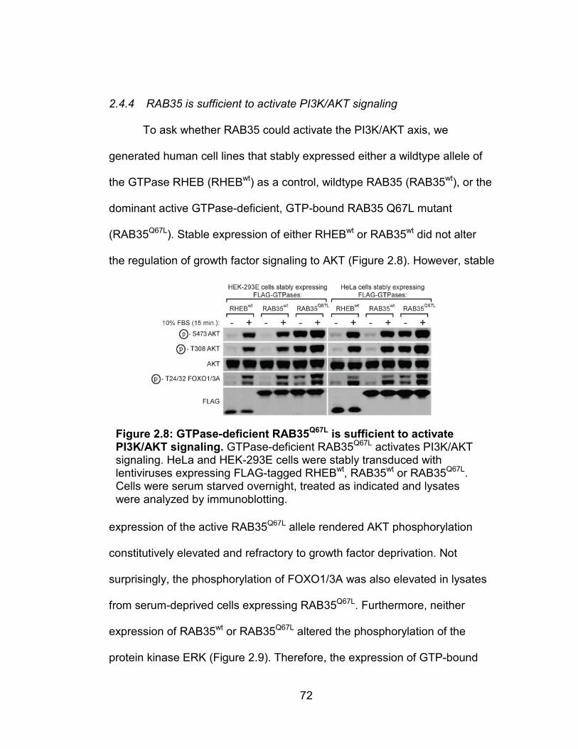
72
2.4.4 RAB35 is sufficient to activate PI3K/AKT signaling
To ask whether RAB35 could activate the PI3K/AKT axis, we
generated human cell lines that stably expressed either a wildtype allele of
the GTPase RHEB (RHEBwt) as a control, wildtype RAB35 (RAB35wt), or the
dominant active GTPase-deficient, GTP-bound RAB35 Q67L mutant
(RAB35Q67L). Stable expression of either RHEBwt or RAB35wt did not alter
the regulation of growth factor signaling to AKT (Figure 2.8). However, stable
expression of the active RAB35Q67L allele rendered AKT phosphorylation
constitutively elevated and refractory to growth factor deprivation. Not
surprisingly, the phosphorylation of FOXO1/3A was also elevated in lysates
from serum-deprived cells expressing RAB35Q67L. Furthermore, neither
expression of RAB35wt or RAB35Q67L altered the phosphorylation of the
protein kinase ERK (Figure 2.9). Therefore, the expression of GTP-bound
Figure 2.8: GTPase-deficient RAB35Q67L is sufficient to activate PI3K/AKT signaling. GTPase-deficient RAB35Q67L activates PI3K/AKT signaling. HeLa and HEK-293E cells were stably transduced with lentiviruses expressing FLAG-tagged RHEBwt, RAB35wt or RAB35Q67L. Cells were serum starved overnight, treated as indicated and lysates were analyzed by immunoblotting.

73
RAB35 is sufficient to activate PI3K/AKT signaling in cells in the absence of
growth factors.
2.4.5 RAB35 depletion inhibits mTORC2 kinase activity towards AKT/PKB
in vitro
Because these data thus far did not indicate where in the pathway
RAB35 might be acting, we asked whether RAB35 regulates AKT activation
via either of the two kinases that phosphorylate AKT—PDK1 and mTORC2
[341-342]. Because one of our motivations for performing our RNAi screen
was to identify regulators of mTORC2, the first question we asked was
whether depletion of RAB35 could inhibit mTORC2 kinase activity in vitro
(Figure 2.10). We generated HeLa cells stably expressing a control shRNA
or shRNAs targeting either RICTOR, PIK3CA, or RAB35, then
immunopurified mTORC2 with an anti-RICTOR antibody and assayed the in
vitro kinase activity of mTORC2 against recombinant, inactivated AKT [333,
341, 343-345]. Not surprisingly, the ability of immunopurified mTORC2 to
Figure 2.9: Stable expression of dominant active, GTPase-deficient RAB35Q67L does not activate ERK signaling. HEK-293E and HeLa cells transduced with FLAG-tagged RHEBwt, RAB35wt, or RAB35Q67L from Fig. 2C were treated as indicated, lysed, and cell lysates were analyzed by immunoblotting for levels of total ERK protein and ERK phosphorylation.
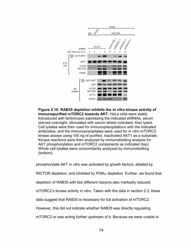
74
phosphorylate AKT in vitro was activated by growth factors, ablated by
RICTOR depletion, and inhibited by PI3Kα depletion. Further, we found that
depletion of RAB35 with two different hairpins also markedly reduced
mTORC2’s kinase activity in vitro. Taken with the data in section 2.3, these
data suggest that RAB35 is necessary for full activation of mTORC2.
However, this did not indicate whether RAB35 was directly regulating
mTORC2 or was acting further upstream of it. Because we were unable to
Figure 2.10: RAB35 depletion inhibits the in vitro kinase activity of immunopurified mTORC2 towards AKT. HeLa cells were stably transduced with lentiviruses expressing the indicated shRNAs, serum starved overnight, stimulated with serum where indicated, then lysed. Cell lysates were then used for immunoprecipitations with the indicated antibodies, and the immunoprecipitates were used for in vitro mTORC2 kinase assays using 100 ng of purified, inactivated AKT1 as a substrate. Kinase reactions were then analyzed by immunoblotting analysis for AKT phosphorylation and mTORC2 components as indicated (top). Whole cell lysates were concomitantly analyzed by immunoblotting (bottom).

75
identify any interaction between endogenous or recombinant RAB35 and
components of mTORC2 (data not shown), we reasoned that RAB35 could
be regulating mTORC2 indirectly. We next sought to delineate whether
RAB35 was regulating both PDK1 and mTORC2 by acting on their common
regulator PI3K.
2.4.6 RAB35 functions above PDK1 and mTORC2
To place RAB35 within the pathway, we asked if RAB35 regulates AKT
activation via either of the two kinases that phosphorylate AKT—PDK1 on
T308 and mTORC2 on S473 [341-342]. Determining whether a particular gene
or small molecule functions through either PDK1 or mTORC2 is challenging
because the phosphorylation states of T308 and S473 in wildtype cells are
influenced by one another [119, 134, 346]. We took advantage of cells that
stably expressed alleles of murine Akt1 (mAkt1) where either T308 or S473
were mutated to phosphomimetic aspartate residues [344] (Figure 2.11) to ask
whether RAB35 was regulating AKT via either PDK1, mTORC2, or both
kinases.

76
As expected, mTOR blockade with the ATP-competitive inhibitor Torin1
or RICTOR depletion suppressed S473 phosphorylation on mAkt1 that had an
intact mTORC2 site (mAkt1wt and mAkt1T308D) (Figure 2.12). Further, in cells
without a regulatable mTORC2 site (mAkt1S473D), RICTOR depletion did not
depress T308 phosphorylation, and treatment with Torin1 elevated
phosphorylation at T308. Interestingly, depletion of RAB35 decreased S473
phosphorylation in mAkt1wt and mAkt1T308D cells as well as T308
phosphorylation in mAkt1wt and mAkt1S473D cells. Together, these data
suggested that RAB35 signals to AKT by acting upstream of both PDK1 and
mTORC2.
Figure 2.11: AKT phosphomimetic mutants to delineate between regulators of PDK1 and mTORC2. Because phosphorylation of the T308 and S473 sites on AKT can affect one another, determining whether an unknown regulator or perturbogen functions by controlling PDK1, mTORC2, or p110α is challenging. By locking one residue into a phosphomimetic state by mutating it to an aspartate, the phosphorylation state of the other can be probed without concern that it is being influenced by the other site. (A) S473D mutation allows measurement of T308 phosphorylation to reflect only PDK1 activity. (B) T308D mutation allows measurement of S473 phosphorylation to reflect only mTORC2 activity.

77
2.4.7 RAB35 depletion upregulates protein levels of the mTORC1/2
inhibitor DEPTOR
In addition to the “biochemical epistasis” experiment described in
Figure 2.12, we performed further experiments to confirm that RAB35 was
acting upstream of mTOR. One indicator of both mTORC1 and mTORC2
activity is the stability of DEPTOR, a negative regulator of both mTOR
complexes [208, 343, 347-349]. In addition to regulating mTOR, DEPTOR is
itself an mTOR substrate, and phosphorylation by mTOR reduces the
stability of DEPTOR protein. Thus, inhibition of PI3K or mTOR—by
pharmacological means or via deprivation of growth factors—leads to
accumulation of DEPTOR levels. Similarly, depletion of PI3K/mTOR pathway
Figure 2.12: RAB35 acts upstream of both PDK1 and mTORC2. HeLa cells stably expressing murine Akt1wt, Akt1S473D or Akt1T308D were treated as indicated with shRNA-expressing lentiviruses and Torin1. Cells were then lysed and cell lysates were analyzed by immunoblotting.

78
components can lead to increased DEPTOR stability. We reasoned that if
RAB35 was regulating PI3K and/or mTOR that RAB35 depletion in the
presence of growth factors would result in increased DEPTOR protein levels.
Indeed, serum deprivation, RICTOR, PI3Kα or RAB35 depletion—in the
presence of complete media—all lead to DEPTOR accumulation (Figure
2.13). This further suggested to us that RAB35 regulates PI3K/mTOR
signaling upstream of mTOR.
2.4.8 Depletion of RAB35 blunts mTORC1 inhibitor-induced PI3K/AKT
activation
Next, we reasoned that if RAB35 was involved in PI3K/AKT/mTOR
signaling, depletion of this protein would blunt the activation of PI3K/AKT
Figure 2.13: Serum deprivation, depletion of RICTOR, p110α, or RAB35 all lead to accumulation of the mTORC1 and mTORC2 inhibitor Deptor. HeLa cells were transduced with lentivirus expressing the indicated shRNAs, then were serum starved for 30 hours and either lysed or re-stimulated with serum for 12 hours, lysed, and lysates were analyzed by immunoblotting.

79
signaling that is induced by rapamycin. As reviewed in chapter 1 of this
thesis, there exists a negative feedback circuit between mTORC1/S6K1 and
upstream components of PI3K signaling—namely through proteins like IRS-1
and GRB10 [218-220, 350-354]. Under regular conditions, mTORC1 and its
substrate S6K1 phosphorylate and inhibit negative regulatory components of
PI3K/AKT signaling like GRB10, or destabilize positive regulators like IRS1.
Thus, mTORC1 inhibition with rapamycin induces an increase in AKT and
AKT substrate phosphorylation. Even mTORC1/2 inhibition with ATP-
competitive mTOR kinase inhibitors potently upregulates PI3K activity and
leads to activation of PI3K, PIP3 accumulation, and hyperphosphorylation of
T308 on AKT even when mTORC2 is disabled [208, 355-356].
We therefore reasoned that if RAB35 was a positive regulator of
signaling from PI3K/AKT to either PDK1 or mTORC2 (or both), then the
increase in AKT phosphorylation induced by mTORC1 inhibition with
rapamycin should be blunted with RAB35 inactivation (Figure 2.14). Not
surprisingly, we found that depletion of the mTORC2 component RICTOR in
HeLa cells prevented any increase of AKT phosphorylation at both T308 and
S473 after 18 hours of mTORC1 inhibition. Consistent with our data that
indicated that RAB35 lay upstream of both mTORC2 and PDK1, depletion of
RAB35 blunted AKT activation by rapamycin to the same extent as did
RICTOR knockdown. Thus, RAB35 likely functions proximally to RTKs and
PI3K rather than at downstream signal nodes like AKT and mTORC2.
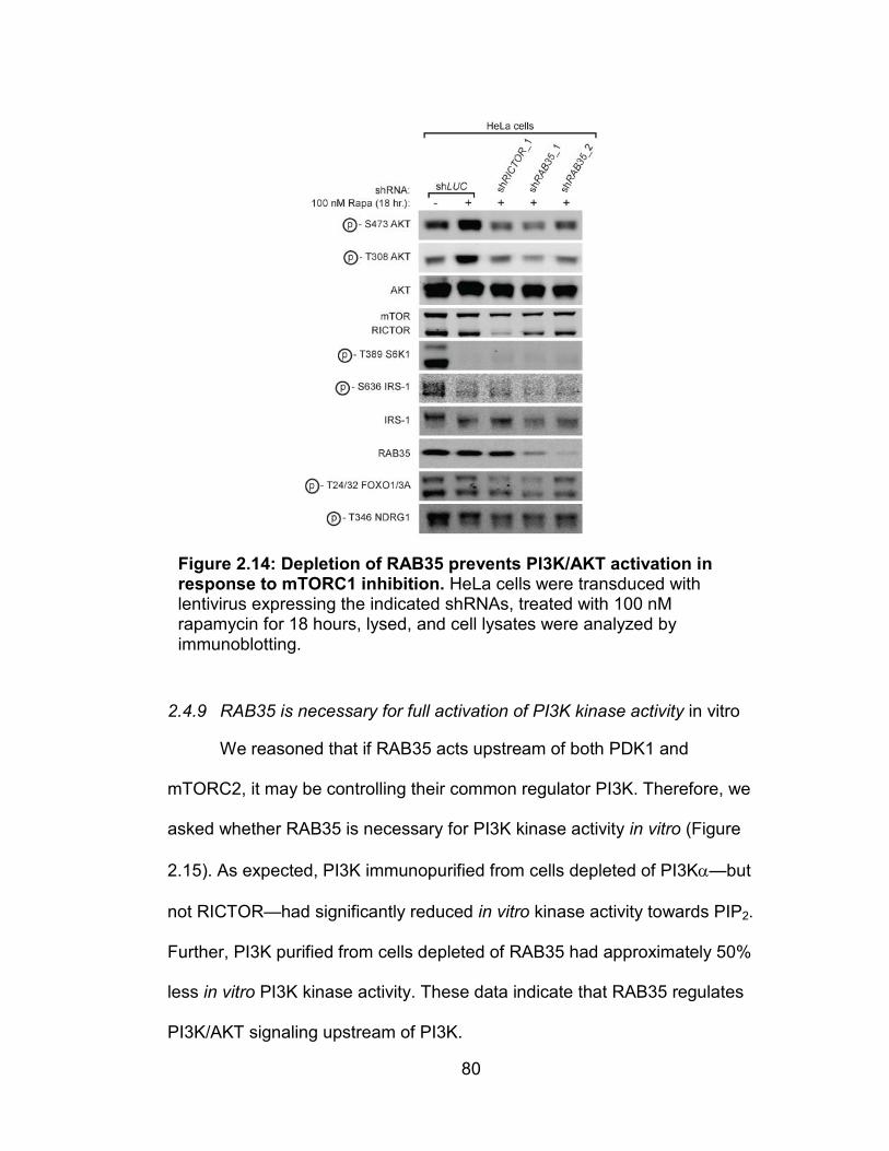
80
2.4.9 RAB35 is necessary for full activation of PI3K kinase activity in vitro
We reasoned that if RAB35 acts upstream of both PDK1 and
mTORC2, it may be controlling their common regulator PI3K. Therefore, we
asked whether RAB35 is necessary for PI3K kinase activity in vitro (Figure
2.15). As expected, PI3K immunopurified from cells depleted of PI3Kα—but
not RICTOR—had significantly reduced in vitro kinase activity towards PIP2.
Further, PI3K purified from cells depleted of RAB35 had approximately 50%
less in vitro PI3K kinase activity. These data indicate that RAB35 regulates
PI3K/AKT signaling upstream of PI3K.
Figure 2.14: Depletion of RAB35 prevents PI3K/AKT activation in response to mTORC1 inhibition. HeLa cells were transduced with lentivirus expressing the indicated shRNAs, treated with 100 nM rapamycin for 18 hours, lysed, and cell lysates were analyzed by immunoblotting.

81
2.4.10 RAB35 is necessary for activation of PI3K/AKT signaling in response
to growth factor receptor stimulation
Next, we investigated whether RAB35 acts downstream of any
particular growth factor receptor. Not surprisingly, depletion of RICTOR or
PI3Kα in cells reduced the phosphorylation of AKT in response to treatment
with insulin-like growth factor (IGF-I), epidermal growth factor (EGF),
platelet-derived growth factor (PDGF-AA) and vascular endothelial growth
factor (VEGF) (Figure 2.16A). Further, depletion of RAB35 also prevented
AKT activation by IGF-I, EGF, PDGF-AA or VEGF. Interestingly, RAB35
depletion did not dramatically alter phosphorylation of ERK or tyrosine
phosphorylation of IGFR, EGFR, PDGFR or VEGFR (Figure 2.16B). Taken
together, the data in Figures 2.10-2.16 suggest that RAB35 functions
Figure 2.15: RAB35 depletion inhibits the kinase activity of immunopurified PI3K in vitro. HEK-293E cells stably expressing shRNAs targeting Luciferase, RICTOR, PI3Kα or RAB35 were treated as indicated. Cells were then lysed, PI3K was immunopurified with an anti-p85 antibody, and purified PI3K was analyzed for in vitro kinase activity using PIP2 as a substrate.

82
upstream of PI3K and downstream of growth factor receptor tyrosine
kinases.
2.4.11 RAB35 is mutated in human tumors
Like the other genes on our prioritized hit list in Table 2.2, we found
that RAB35 was mutated in human tumor samples (Table 2.3). Although
none of the RAB35 mutations that we observed were recurrent, two of the
Figure 2.16: RAB35 depletion inhibits PI3K signaling to AKT downstream of multiple growth factor receptors. (A) HeLa cells were transduced with the indicated shRNA-expressing lentiviruses, serum starved overnight, treated as indicated with either insulin-like growth factor I (IGF-I), epidermal growth factor (EGF), platelet derived growth factor AA (PDGF-AA) or vascular endothelial growth factor (VEGF), then lysed and analyzed by immunoblotting. (B) Lysates from (A) were analyzed by immunoblotting for tyrosine phosphorylation of the indicated growth factor receptors.
83
mutations in RAB35—A151T and F161L—occurred at residues that are
conserved in RAS-like GTPases. We noticed that the A151T and F161L
mutations in RAB35 were strikingly similar to KRAS mutations (A146T and
F156L) that have been previously identified from human tumor samples
(Figure 2.17) [357-358]. While they are not “canonical” KRAS- activating
mutations, stable expression of these two KRAS mutants was sufficient to
activate ERK signaling and transform NIH-3T3 cells in vitro. We therefore
reasoned that the two similar mutations in RAB35 might also be gain-of-
function mutations that could activate RAB35 signaling.

84
Table 2.3: The TCGA, COSMIC and MSKCC cBIO databases reveal somatic RAB35 mutations in human cancers. The Cancer Genome Atlas (TCGA), Catalogue of Somatic Mutations in Cancer (COSMIC), the International Cancer Genomics Consortium (ICGC) and MSKCC computation biology (cBio) data portal web sites were used to search for mutations in RAB35 in human tumor samples. Mutations are listed in order of the amino acid residue that they occur at. Predicted change in protein function was assessed by the cBio mutational assessor with the exception of F161L, whose change in function was predicted manually by literature survey (asterisk).
Tissue Database(s) Amino acid change
Frequency of mutation (%)
Predicted change in function?
Uterus cBio, COSMIC, TCGA
S22N K35N
1 of 248 (0.4%) High NP
Colon (HCT-116 cell line)
cBio, COSMIC R27C 1 of 60 (1.67%) Medium
Stomach COSMIC, TCGA R27H 1 of 386 (0.26%) Medium Lung COSMIC, ICGC A29V 1 of 488 (0.2%) Neutral Breast cBio, COSMIC,
TCGA F45L 1 of 772 (0.1%) Medium
Skin cBio, TCGA S50F 1 of 413 (0.24%) NP Colon (HCT-15 cell line)
cBio, TCGA T76 splice
1 of 60 (1.67%) NP
Colon (HCT-116 cell line)
cBio, COSMIC, TCGA
G80E 1 of 60 (1.67%) Medium
Adrenal cBio, TCGA H82Y 1 of 179 (0.56%) Medium Medulla COSMIC, ICGC V90I Unknown NP Colon COSMIC, TCGA E94K 1 of 600 (0.17%) NP Skin cBio S95F 1 of 228 (0.44%) NP Lung COSMIC, TCGA R101Q 1 of 173 (0.58%) Neutral Breast cBio V129G 1 of 1061 (0.09%) Medium Liver cBio Y136C 1 of 268 (0.37%) Neutral Colon COSMIC A139V 1 of 2132 (0.05%) NP Uterus cBio, COSMIC,
TCGA E150K 1 of 248 (0.4%) NP
Lymphoid (MOLT4 cell line)
cBio, COSMIC A151T 1 of 60 (1.67%) High
Uterus COSMIC, ICGC A151T 1 of 815 (0.12%) High

85
Table 2.3 (continued)
Prostate cBio, COSMIC, TCGA
N156S 1 of 425 (0.24%) Medium
Lung cBio, COSMIC F161L 1 of 183 (0.5%) High* Stomach cBio, TCGA K173fs 1 of 295 (0.34%) NP Stomach cBio, TCGA R196* 1 of 295 (0.34%) NP
Figure 2.17: RAB35 mutants identified in human tumors are similar to known activating mutations in KRAS. Two mutations in RAB35 that code for amino acid changes A151T and F161L were identified in the MSKCC cBio and COSMIC datasets. Alignment of RAB35 with KRAS was performed using ClustalW2.
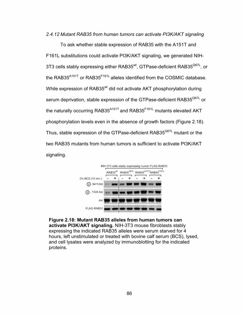
86
2.4.12 Mutant RAB35 from human tumors can activate PI3K/AKT signaling
To ask whether stable expression of RAB35 with the A151T and
F161L substitutions could activate PI3K/AKT signaling, we generated NIH-
3T3 cells stably expressing either RAB35wt, GTPase-deficient RAB35Q67L, or
the RAB35A151T or RAB35F161L alleles identified from the COSMIC database.
While expression of RAB35wt did not activate AKT phosphorylation during
serum deprivation, stable expression of the GTPase-deficient RAB35Q67L or
the naturally occurring RAB35A151T and RAB35F161L mutants elevated AKT
phosphorylation levels even in the absence of growth factors (Figure 2.18).
Thus, stable expression of the GTPase-deficient RAB35Q67L mutant or the
two RAB35 mutants from human tumors is sufficient to activate PI3K/AKT
signaling.
Figure 2.18: Mutant RAB35 alleles from human tumors can activate PI3K/AKT signaling. NIH-3T3 mouse fibroblasts stably expressing the indicated RAB35 alleles were serum starved for 4 hours, left unstimulated or treated with bovine calf serum (BCS), lysed, and cell lysates were analyzed by immunoblotting for the indicated proteins.

87
2.4.13 Stable expression of mutant alleles of RAB35 suppresses apoptosis
Because PI3K/AKT signaling inhibits apoptosis, we next examined
whether cells expressing mutant alleles of RAB35 were resistant to
apoptosis triggered by growth factor withdrawal. 4 hours of serum
deprivation of NIH-3T3 cells stably expressing RAB35wt was sufficient to
elevate cleaved levels of the apoptotic markers PARP and Caspase3 (Figure
2.19A). In comparison, cells stably expressing all three RAB35 mutants or
oncogenic p110αH1047R had decreased levels of cleaved PARP and cleaved
Caspase3 following serum withdrawal. Further, while cells expressing
RAB35wt were sensitive to cell death in response to serum deprivation, cells
expressing mutant alleles of RAB35 or p110αH1047R exhibited significantly
improved cell viability when deprived of growth factors (Figure 2.19B).
Together, these data suggest that mutant RAB35 proteins can suppress cell
death in response to growth factor deprivation.

88
Figure 2.19: Expression of RAB35 mutants suppresses apoptosis. (A) NIH-3T3 cells stably expressing the indicated proteins were plated then treated as indicated for 4 hours with or without serum-containing medium. Cells were then lysed and the cell lysates were immunoblotted for the indicated proteins. (B) NIH-3T3 cells were treated as in (A), trypsinized and the number of viable cells was counted. Cell counts for each cell line were normalized to the number of viable cells for each cell line from non serum starved conditions. Asterisks indicate p-value <0.05.

89
2.4.14 Mutant alleles of RAB35 transform NIH-3T3 cells in vitro in a PI3K-
dependent manner
To determine if the mutant alleles of RAB35 are oncogenic, we asked
whether they could transform non-cancerous NIH-3T3 cells in a focus-
formation assay—a standard in vitro model of density-independent growth
and thus tumorigenicity [334-335]. Indeed, NIH-3T3 cells expressing
oncogenic p110αH1047R, AKT1myr or the three mutant alleles of RAB35—but
not RAB35wt—formed foci (Figure 2.20). Moreover, we found that when
cultured in medium containing 250 nM of the pan-class I PI3K inhibitor GDC-
0941 [359], cells expressing RAB35 mutants and p110αH1047R were unable to
form foci. Not surprisingly, GDC-0941did not inhibit the growth of cells
transduced with AKT1myr. Thus, expression of RAB35 mutants identified from
human cancers can transform NIH-3T3 cells in vitro in a PI3K-dependent
manner.

90
Figure 2.20: RAB35 mutants can transform cells in vitro in a PI3K-dependent manner. (A) NIH-3T3 cells stably expressing the indicated RAB35 alleles, HA-p110αH1047R, or myristoylated FLAG-AKT1myr were cultured for 4 weeks in medium containing 2% BCS with either DMSO or 250 nM GDC-0941, then fixed, stained with crystal violet, and imaged. (B) Crystal violet stain from each well was then solubilized in Sorenson’s Buffer and the absorbance at 590 nm for each was determined.

91
2.5 DISCUSSION
We identify here the small GTPase RAB35 as a positive regulator of
PI3K signaling (Figure 2.21). Although the precise mechanism of how
RAB35 transduces upstream signals to PI3K remains unclear, our data
demonstrate a previously unappreciated role for RAB35 in regulating growth
factor signaling to PI3K/AKT. Further, the ability of mutant alleles of RAB35
found in human cancers to activate PI3K/AKT signaling, protect cells from
apoptosis and transform cells in vitro suggests that RAB35 may be a proto-
oncogene. Finally, that a RAB protein that regulates endomembrane
trafficking possesses oncogenic potential suggests that dysregulation of
membrane trafficking in cells could itself serve as a driver in the
development and survival of tumor cells.
Figure 2.21: RAB35 regulates PI3K signaling to AKT either through PI3K or growth factor receptors. The data presented in this chapter suggest that RAB35 controls PI3K signaling by regulating signaling at the level of growth factor receptor tyrosine kinases (RTKs, left) or at the level of PI3K (right).

92
CHAPTER THREE:
RAB35 REGULATES THE PI3K/AKT PATHWAY VIA THE PLATELET-
DERIVED GROWTH FACTER RECEPTOR TYROSINE KINASE
3.1 ABSTRACT
The data described in Chapter 2 of this thesis suggest that RAB35 is
a bona fide positive regulator of PI3K signaling. Here, we suggest that
RAB35 regulates this pathway specifically via the platelet-derived growth
factor receptor α (PDGFRα). GTPase-deficient RAB35 Q67L potently activates
PI3K/AKT signaling by specifically activating PDGFRα. Further, dominant
active RAB35 constitutively localizes PDGFRα—but not receptor tyrosine
kinases like EGFR—to RAB7-positive late endosomes, which is the same
compartment that active, liganded PDGFRα localizes to after internalization
in cells that express wildtype RAB35. Finally, blockade of PDGFRα kinase
activity with the PDGFRα-specific inhibitor Crenolanib suppresses AKT
activation in cells that express dominant active RAB35Q67L. Thus, RAB35
signals to PI3K/AKT primarily through PDGFRα, likely by serving to traffic
PDGFRα to a RAB7-positive endomembrane where it is active.
3.2 INTRODUCTION
Thus far, our data suggest that RAB35 is a positive regulator of
PI3K/AKT signaling (Chapter 2) [360]. We used loss-of-function studies to
demonstrate that RAB35 is necessary for activation of the PI3K/AKT
pathway. Further, our gain-of-function experiments with a constitutively

93
activated allele of RAB35 provided evidence that RAB35 is a positive
regulator of PI3K/AKT signaling. Finally, biochemical epistasis experiments
strongly suggest that RAB35 functions upstream of PI3K itself.
Because 1.) our data strongly implicate RAB35 as a positive regulator
of PI3K signaling, 2.) RAB35 is well documented to be involved in the
trafficking of several cargoes between endocytic compartments and the cell
membrane, and 3.) growth factor receptors and their associated signaling
machinery are known to actively signal after internalization to
endomembranes, we reasoned that RAB35 might serve to activate or retain
RTK-PI3K complexes in a cellular compartment where they transmit
signaling to PI3K/AKT effectors. Thus, we asked whether RAB35 signaling is
transduced through any particular RTK-PI3K complexes. We investigated
whether these complexes were altered in their trafficking by activated RAB35
proteins. Interestingly, the data we describe in this chapter suggests that
RAB35 activates PI3K through a particular RTK and at a specific
endomembrane within the cell.
3.3 METHODS
3.3.1 In vitro PI3K-RAB35 interaction assays
In vitro interaction assays between immunoprecipitated PI3K and
FLAG-purified RAB35 were adapted from previously described protocols
[183-184, 361-362]. First, we affinity purified FLAG-GTPases as follows.
HEK-293E cell lines stably expressing either FLAG-tagged RHEBwt,
94
RAB35wt, or RAB35Q67L were lysed in 1% TX-100 lysis buffer as described in
Chapter 2.2 of this thesis. The lysates were clarified, and 40 µL of mouse
anti-FLAG M2 antibody conjugated to agarose beads was added to tubes
containing 2 µg of cell lysates. The lysate/antibody mix was incubated over
night at 4° C. The next day, the agarose beads were washed three times
with TX-100 lysis buffer, then twice more with RHEB GTPase buffer (20 mM
HEPES [pH 8.0], 200 mM NaCl, 5 mM MgCl2) [178]. The GTPase buffer was
aspirated completely and 100 µL of 10 mM FLAG peptide in GTPase buffer
was added to the beads. This mixture was then incubated at room
temperature for 1 hour to elute the FLAG-GTPases from the agarose beads.
The eluates were then collected and pooled. To remove the FLAG peptide
and concentrate the GTPases, the pooled eluates were filtered with a 10
kDa Amicon Ultra 0.5 mL centrifuge filter (Millipore, UFC501008).
The same day that FLAG-GTPase expressing cells were lysed,
wildtype HEK-293E cells cultured in complete medium were lysed in 0.3%
CHAPS buffer, and 1 µg of cell lysates were incubated overnight with 20 µL
of anti-p85 antibody conjugated agarose beads. The next day, the complex
of agarose/antibody and immunoprecipitated PI3K was washed three times
with CHAPS lysis buffer.
Prior to combining the immunopurified PI3K and GTPases, FLAG-
RHEB or FLAG-RAB35 were loaded with either GDP or guanosine 5’-3-O-
(thio) triphosphate (GTPγS) as follows [178]. FLAG-RHEB or FLAG-RAB35
GTPases were incubated with 10 mM EDTA and either 1 mM GDP or 0.1

95
mM GTPγS at 30° C for 10 minutes, then 20 mM MgCl2 was added and the
proteins were kept on ice until use.
Once PI3K was immunopurified and washed and FLAG-GTPases
were loaded with either GDP or GTPγS, 40 µL of purified, nucleotide loaded
GTPase was added to each individual PI3K IP. This mixture was incubated
at room temperature for 30 minutes, and then washed three times with
GTPase storage buffer. Finally, the last wash was aspirated, beads were
resuspended in 20 µL Laemmli’s buffer, boiled, resolved by SDS-PAGE and
then analyzed by immunoblotting as described in Chapter 2.
3.3.2 Immunofluorescence for microscopy
Immunofluorescence was performed as described previously [363].
Millicell EZ slide 4-well chamber slides (Millipore, PEZGS0416) were coated
for 30 minutes with 0.01 mg/mL human fibronectin (Calbiochem, 341635) in
PBS, rinsed once with PBS, and seeded with 100,000 HEK-293E cells.
Immediately following the indicated treatments and/or stimulations, slides
were fixed in 4% paraformaldehyde (Electron Microscopy Sciences, 15714-
S) in PBS for 15 minutes at room temperature. The slides were then rinsed 3
times in PBS for 5 minutes each, permeabilized with 0.05% Triton TX-100 in
PBS for 1 minute at room temperature, then rinsed three more times in PBS.
Next, each well was incubated with primary antibody at a dilution of 1:100 in
5% normal goat serum (Vector Laboratories, S-1000) in PBS overnight at 4°
C. When Alexa Fluor 488 conjugated mouse anti-myc antibody (Cell

96
Signaling Technology, 2279) was necessary, it was used at a 1:50 dilution
and incubated as indicated overnight. The next day, the slides were rinsed in
PBS 3 times at room temperature, and incubated for 1 hour at room
temperature with the following mixture: the appropriate fluorophore-
conjugated secondary antibody (either goat anti-rabbit Alexa Fluor 488 nm
(Invitrogen, A11070), goat anti-mouse Alexa Fluor 488 nm (Invitrogen,
A11029), goat anti-mouse Alexa Fluor 555 nm (Invitrogen, A21424), or goat
anti-rabbit Alexa Fluor 555 nm (Invitrogen, A21429)) at a 1:200 dilution, and
Hoechst 33342 (Invitrogen, H3570) at a 1:10,000 dilution. When actin
staining with fluorophore-conjugated phalloidin was performed, Alexa Fluor
594 conjugated phalloidin (Invitrogen, A12381) was included in the
secondary staining mix at a dilution of 1:200. After incubation, the slides
were rinsed 4 times in room temperature PBS, the slide chambers were
removed, slides were mounted with 100 µL ProLong Gold anti-fade reagent
(Invitrogen, P36930) and glass coverslips (Fisher Scientific, 12-545-88).
Slides were then sealed with Sally Hansen® “Super Shine” Shiny Top Coat
(Sally Hansen, 2290), air dried, then stored at 4° C until imaging. Slides were
imaged with an Axio Imager 2 widefield microscope (Zeiss) and AxioVision
4.0 software (Zeiss). Images were exported from AxioVision as TIFF files.

97
3.3.3 Immunoprecipitation/mass spectroscopy for the identification of PI3K
interacting proteins
Immunoprecipitations of PI3K were carried out as described in the
previous chapter. Immunoprecipitates were then resolved by SDS-PAGE
and the gel was stained with Novex Simply Blue stain (Invitrogen, PART #).
The gel was then submitted to the Memorial Sloan-Kettering Cancer Center
Microchemistry and Proteomics Core Facility. The lanes of the gel containing
the immunoprecipitated proteins were then cut into sections, digested with
trypsin, and subjected to mass spectroscopic analysis as previously
described [364-366]. Results of the MS run were analyzed using the
program Scaffold v4.2.1 (Proteome Software). Only peptides of >90%
certainty with 1 or more peptide fragments present were considered in
analysis of MS data.
3.4 RESULTS
3.4.1 RAB35 interacts with PI3K in a nucleotide-dependent manner
The data described in Chapter 2 suggest that RAB35 regulates PI3K
signaling either 1.) at PI3K itself or 2.) at the interface between growth factor
receptor tyrosine kinases and PI3K. However, those data do not delineate
whether RAB35 regulates PI3K directly or indirectly. To first ask whether the
two proteins interact, we used an anti-p85 antibody to immunopurify PI3K
from cells stably expressing FLAG-tagged alleles of RHEB (as a control) or
RAB35 (Figure 3.1). Indeed, we found that FLAG-RAB35wt—but not FLAG-

98
RHEB—co-immunopurified with PI3K. Further, the dominant active GTPase
deficient RAB35Q67L mutant was present in the PI3K immunoprecipitates to a
higher extent than was the wildtype allele. Further, the GDP-bound mutant—
RAB35S22N—was barely detectable in PI3K immunoprecipitates from HEK-
293E cells that stably expressed FLAG-RAB35S22N.
In addition to these data, we also found that transiently expressed
myc-tagged RAB35—but not myc-tagged RHEB—immunoprecipitated with
endogenous PI3Kα (Figure 3.2). Consistent with the interaction data from
the stable cell lines in Figure 3.1, myc-RAB35S22N was nearly undetectable in
PI3K IPs. Taken together, these data suggested to us that RAB35 interacts
with PI3K. However, whether RAB35 interacts with PI3K directly—as has
Figure 3.1: Stably expressed RAB35 interacts with endogenous PI3K in a nucleotide-dependent fashion. HEK-293E cells stably expressing the indicated FLAG-GTPases were serum starved overnight, treated with serum, lysed, and PI3K was immunopurified with an anti-p85 antibody. Lysates and immunoprecipitates were then analyzed by immunoblotting.
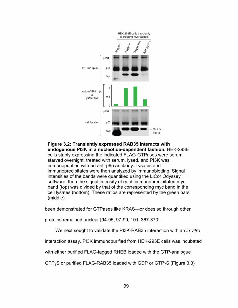
99
been demonstrated for GTPases like KRAS—or does so through other
proteins remained unclear [94-95, 97-99, 101, 367-370].
We next sought to validate the PI3K-RAB35 interaction with an in vitro
interaction assay. PI3K immunopurified from HEK-293E cells was incubated
with either purified FLAG-tagged RHEB loaded with the GTP-analogue
GTPγS or purified FLAG-RAB35 loaded with GDP or GTPγS (Figure 3.3)
Figure 3.2: Transiently expressed RAB35 interacts with endogenous PI3K in a nucleotide-dependent fashion. HEK-293E cells stably expressing the indicated FLAG-GTPases were serum starved overnight, treated with serum, lysed, and PI3K was immunopurified with an anti-p85 antibody. Lysates and immunoprecipitates were then analyzed by immunoblotting. Signal intensities of the bands were quantified using the LiCor Odyssey software, then the signal intensity of each immunoprecipitated myc band (top) was divided by that of the corresponding myc band in the cell lysates (bottom). These ratios are represented by the green bars (middle).

100
[183-184, 361, 371-373]. After incubation together, the agarose beads
bound to PI3K were washed and the remaining proteins were analyzed by
immunoblotting for the presence of FLAG-RAB35wt. Indeed, the only PI3K
immunoprecipitates that retained bound RAB35 were those that had been
incubated with GTP-loaded FLAG-RAB35 wt. These data indicate that PI3K
and RAB35 interact in a GTP-dependent manner. However, because the
PI3K used in this experiment had been immunopurified from cells, it is
possible that adapter proteins or growth factor receptors that associate with
PI3K could serve to interact with RAB35. Thus, whether RAB35 interacts
directly with PI3K remains unclear.
Figure 3.3: Immunopurified PI3K interacts with purified GTPγS-RAB35 but not GDP-RAB35. FLAG-tagged RHEB or RAB35 were affinity purified from lysates of cells stably expressing either GTPase, loaded with either GDP or GTPγS as indicated. Endogenous PI3K was immunopurified from wildtype HEK-293E cells with an agarose-conjugated anti-p85 antibody, washed, and then incubated with the GTPases as indicated above. The GTPase-PI3K complexes were then washed and analyzed by immunoblotting.

101
3.4.2 GTPase-deficient RAB35 specifically activates the platelet-derived
growth factor receptor
In probing the interaction of RAB35 with PI3K, we noticed that a
prominent phospho-tyrosine signal was present in immunoprecipitates of
PI3K from cells stably expressing GTPase-deficient RAB35Q67L (Figure 3.4).
In cells stably expressing RAB35wt, this signal is strongest in PI3K
immunoprecipitates from cells that have been treated with growth factors or
serum. Interestingly, this signal is elevated in PI3K immunoprecipitates from
cells expressing RAB35Q67L regardless of whether cells had been serum
starved or not. Indeed, this signal was apparent even in the lysates of cells
expressing RAB35Q67L but not RHEB or RAB35wt.
The protein responsible for the elevated phospho-tyrosine signal in
PI3K immunoprecipitates and whole cell lysates from cells expressing
Figure 3.4: PI3K immunoprecipitated from cells stably expressing RAB35Q67L is associated with a constitutively elevated phospho-tyrosine signal. HEK-293E cells stably expressing FLAG GTPases were serum starved overnight, treated as indicated with FBS, lysed, and PI3K was immunopurified from the lysates. Immunoprecipitates and whole cell lysates were then analyzed by immunoblotting. White boxes indicate elevated phosphotyrosine signal in RAB35Q67L lysates.

102
dominant active RAB35 was of a relatively high molecular weight (175-200
kDa). Given the prominence of tyrosine phosphorylation in the activation of
receptor tyrosine kinases and their substrates, we thus reasoned that this
signal might be emanating from either a receptor tyrosine kinase (EGFR,
PDGFR, IGF-IR, etc.) or an RTK substrate of a high molecular weight (such
as IRS-1, etc.). Further, we speculated that this was likely one individual
protein, as there were not other phosphotyrosine signals present in lysates
from cells expressing GTPase-deficient RAB35Q67L.
We next asked which growth factor receptor tyrosine kinase might be
the source of the phosphotyrosine signal in cells stably expressing GTP-
bound RAB35Q67L. Once activated, RTKs auto-phosphorylate numerous
tyrosine residues in trans, and tyrosine phosphorylation of the catalytic
domain tyrosines is necessary for activation of most receptor tyrosine
kinases [374-378]. In contrast, the other auto-phosphorylation tyrosine sites
on RTKs serve as sites for adaptor proteins—such as IRS1/2, p85, GRB2,
etc.—to interact with active RTKs. Because each receptor has multiple
binding domains that contain phosphorylatable tyrosine residues, we
reasoned that the most effective way to identify the source of this elevated
phosphotyrosine signal would be to probe lysates for phosphorylation of the
catalytic domain tyrosine residues of different growth factor receptors. Thus,
we probed for multiple RTK catalytic domain phosphorylation levels in
lysates from cells expressing FLAG-tagged RHEBwt, RAB35wt and
RAB35Q67L that had been either serum starved or stimulated with serum

103
(Figure 3.5). Interestingly, we found that only the phosphorylation levels of
Y849/857 on PDGFRα/β were constitutively elevated in cells expressing
RAB35Q67L. The catalytic domain tyrosine phosphorylation sites on other
receptors were either normally activated by serum in all three cell lines (IGF-
IRβ, EGFR, MET) or equally constitutively active in all three cell lines
regardless of serum deprivation/stimulation (VEGFR, FGFR). We also
probed for phosphorylation of other tyrosine kinases (c-Kit, SRC, etc.) and
did not find any difference in phosphotyrosine levels between the three cell
lines examined in Figure 3.5 (data not shown).
Figure 3.5: The platelet-derived growth factor receptor—but not other growth factor receptors—is hyper-phosphorylated at a catalytic domain tyrosine residue in cells stably expressing GTPase-deficient RAB35Q67L. HEK-293E cells expressing the indicated FLAG-GTPases were starved overnight, stimulated with FBS as indicated, lysed, and lysates were analyzed by immunoblotting with the indicated antibodies.

104
3.4.3 Pharmacological inhibition of PDGFRα/β but not PDGFRβ inhibits
PI3K/AKT signaling in cells stably expressing GTPase-deficient
RAB35Q67L
Because the upregulation of growth factor receptors by dominant
active RAB35 seemed so specific to PDGFR, we next asked whether
pharmacological blockade of PDGFR could inhibit PI3K/AKT signaling. Our
first course of action was to attempt PDGFR inhibition with the well
characterized pan-tyrosine kinase inhibitor imatinib/gleevec [278, 379-381],
but this molecule failed to suppress PI3K/AKT signaling in cells that
expressed GTP-bound RAB35Q67L (data not shown). This could be
accounted for by the fact that: 1.) the affinity of imatinib for PDGFR is
relatively low, or 2.) the activated PDGFR is locked in an imatinib-resistant
conformation.
We next asked whether more potent, isoform specific PDGFR
inhibitors could block PDGFR signaling to PI3K/AKT (Figure 3.6A). Indeed,
in cells stably expressing RAB35Q67L the phosphorylation of the PDGF
receptor, AKT, and FOXO1/3A were not suppressed by treatment with the
PDGFRβ-specific small molecule CP-673451[382]. However, an inhibitor
that specifically targets PDGFRα and PDGFRβ with similar potency—CP-
868596 (Crenolanib®) [383-387]—was more effective at suppressing the
phosphorylation of PDGFR, AKT and FOXO1/3A. To ensure that the majority
of the signal through the PI3K/AKT

105
pathway was originating from the mutant allele of RAB35, these experiments
were performed in serum-free conditions. Interestingly, in the same
experiment performed in cells stably expressing RAB35wt in serum replete
conditions, we found that PDGFRα/β inhibition failed to suppress AKT
phosphorylation (Figure 3.6B). This suggests that PDGFR inhibition on its
own in the presence of serum is not sufficient to inhibit PI3K/AKT signaling
(Figure 3.6B).
Finally, to check whether other RTKs might be involved in the
regulation of PI3K/AKT by RAB35, we also tested a number of lipid kinase
and tyrosine kinase inhibitors (TKI’s) on cells expressing GTPase deficient,
Figure 3.6: Pharmacological blockade of PDGFRα/β inhibits PI3K/AKT signaling in cells stably expressing GTPase-deficient RAB35Q67L. (A) Cells stably expressing RAB35Q67L were serum starved and treated overnight with either vehicle (DMSO), the pan-Class I PI3K inhibitor GDC-0941 (0.5 µM), the PI3Kα-specific BYL-719 (1 µM), the PDGFRβ inhibitor CP-673451 (PDGFRβi, 1 µM), or the PDGFRα/β inhibitor CP-868596 (PDGFRα/βi, 1 µM). Cells were then lysed and lysates were analyzed by immunoblotting. (B) Cells stably expressing RAB35wt were treated overnight as indicated in serum replete medium, then lysed and analyzed as in (A).

106
dominant active RAB35Q67L (Figure 3.7 and data not shown). As expected,
the PI3K inhibitors GDC-0941 and BYL-719 suppressed AKT
Figure 3.7: Non-PDGFR targeting kinase inhibitors do not reduce PI3K/AKT signaling in cells expressing GTPase-deficient RAB35Q67L. (A) Cells stably expressing RAB35wt were serum starved and treated with either DMSO or the indicated drugs overnight as noted. The next day, the cells were stimulated with DMEM containing 10% FBS and the indicated inhibitors. (B) Cells stably expressing RAB35Q67L were treated overnight in serum free medium as indicated. Cells were then lysed and lysates were analyzed by immunoblotting.

107
phosphorylation in cells expressing RAB35Qwt or RAB35Q67L. Further, EGFR
inhibitors like Erlotinib and Lapatinib failed to suppress AKT phosphorylation
in cells expressing GTPase-deficient RAB35Q67L. Further, VEGFR-specific
molecules had little to no effect on AKT phosphorylation in cells expressing
RAB35Q67L.
3.4.4 RAB35wt and GTPase-deficient RAB35Q67L interact with PDGFRα and
PDGFRβ
As demonstrated earlier in this chapter, we had found that RAB35
interacts with PI3K in a GTP-dependent manner. This led us to wonder
whether RAB35 was interacting with PI3K in a manner analogous to how the
GTPase KRAS interacts with PI3K—i.e. directly and in a way that activates
the kinase activity of PI3K. However, our findings that 1.) the expression of
dominant active RAB35Q67L specifically upregulates the tyrosine
phosphorylation of PDGFRα/β (Figure 3.5) and 2.) a PDGFRα/β specific
kinase inhibitor suppresses RAB35Q67L-driven PI3K/AKT activity suggested
to us that RAB35 might be acting directly on PDGFR.
To ask whether RAB35 might be interacting with PDGFR, we
transfected cDNAs expressing either myc-tagged PDGFRα or β into cells
stably expressing either RHEBwt, RAB35wt or RAB35Q67L. After lysing these
cells, we then immunopurified either PI3K (using an anti-p85 antibody) or
myc-PDGFR (using an anti-myc antibody) and asked whether FLAG-tagged
RAB35 co-immunoprecipitated with these proteins (Figure 3.8). Not

108
surprisingly, we found that both PDGFRα and β immunoprecipitated with
PI3K, and that FLAG-tagged RAB35wt and RAB35Q67L—but not RHEBwt—
was also present in these purifications (Figure 3.8A). These data were
consistent with 1.) the fact that PDGFRα/β is a well-characterized interaction
partner of PI3K and 2.) our RAB35-PI3K interaction studies in Figures 3.1-
3.3.
Figure 3.8: RAB35wt and GTPase-deficient RAB35Q67L interact with PDGFRα and PDGFRβ. (A) HEK-293E cells stably expressing FLAG-tagged RHEBwt, RAB35wt or RAB35Q67L were transfected with cDNA encoding for either myc-PDGFRα or myc-PDGFRβ. Cells were then lysed and PI3K was immunopurified with an anti-p85 antibody. (B) myc-PDGFR was immunopurified with an anti-myc antibody from the same lysates as in (A).

109
We also immunopurified myc-PDGFRα or β using an anti-myc
antibody. As with our PI3K purifications, we found that both the p85 and
p110α subunits of PI3K were present in both PDGFRα and β
immunoprecipitates (Figure 3.8B). We also found that both alleles of FLAG-
tagged RAB35 were also present in these purifications. Interestingly, the
FLAG-RAB35 signal present in PDGFR purifications was consistently
stronger than the FLAG-RAB35 signal in our PI3K purifications, which
suggested to us that the PDGFR-RAB35 interaction may be more robust
than the PI3K-RAB35 interaction. This is reinforced by the fact that in cells
expressing RAB35Q67L, less p85 and p110α purify with both PDGFRα and β,
but the amount of FLAG-RAB35Q67L is not diminished in these
immunoprecipitations. This suggests to us that the interaction of PI3K and
RAB35 may be indirect and due to the fact that both are interacting with
PDGFR. However, whether RAB35 interacts with PDGFR directly or via
another protein remains unclear.
3.4.5 GTPase-deficient RAB35Q67L constitutively localizes PDGFRα to an
internal compartment
Because RAB35 is a RAB GTPase, we had previously considered
that its role in PI3K/AKT signaling could be due to endomembrane trafficking
of either PDGFR, PI3K or AKT itself. Since we had evidence that dominant
active RAB35Q67L is exerting its effects via PDGFRα—and because
trafficking of RTKs is a well documented phenomenon [388-391]—we next

110
asked whether expression of dominant active, GTPase-deficient RAB35Q67L
was altering the localization of PDGFRα (Figure 3.9). We transiently
transfected cDNAs coding for myc-tagged PDGFRα into cells that
stably expressed either RAB35wt or RAB35Q67L and asked whether the
localization of the receptor was altered by GTPase-deficient RAB35. Not
surprisingly, in cells expressing RAB35wt, PDGFRα was diffusely localized in
the absence of growth factors but reorganized into discreet internalized
punctate structures after 15 minutes of treatment with PDGF-AA. However,
in cells expressing RAB35Q67L, PDGFRα was constitutively internalized
regardless of whether the cells had been stimulated with PDGF or not. Thus,
GTPase-deficient RAB35Q67L dyslocalizes PDGFRα to compartments inside
the cell regardless of growth factor stimulation.
Figure 3.9: Dominant active, GTPase-deficient RAB35Q67L constitutively internalizes PDGFRα. HEK-293E cells stably expressing FLAG-tagged RAB35wt or RAB35Q67L were transiently transfected with cDNAs expressing myc-tagged PDGFRα, then starved, treated with 100 ng/mL PDGF-AA for 15 minutes and processed for immunofluorescence with an anti-myc antibody and stained for actin and DNA/nuclei as indicated. Scale bar represents 10 µm.

111
Although our biochemical evidence and experiments with small
molecules already suggested that RAB35-mediated activation of PI3K/AKT
signaling was explicitly through PDGFRα, we wanted to ask whether
trafficking of RTKs was generally deranged in cells with activated
RAB35Q67L. The epidermal growth factor receptor (EGFR) is well-
characterized in terms of its internalization and recycling after stimulation
with EGF. Thus, we performed a similar experiment to the one in Figure 3.9
using myc-tagged EGFR to visualize EGFR trafficking in cells expressing
either RAB35wt or RAB35Q67L (Figure 3.10). Unlike our observations with
PDGFRα, we did not see any difference in the EGF-stimulated
internalization of EGFR between the two cell lines, which suggested to us
that the trafficking of EGFR is not affected by the GTP-loading state of
RAB35.
Figure 3.10: Dominant active RAB35Q67L does not alter the localization of the epidermal growth factor receptor (EGFR). HEK-293E cells stably expressing the indicated FLAG-RAB35 alleles were transiently transfected with cDNAs expressing the indicated receptor tyrosine kinases, starved overnight, treated with the indicated ligands then processed for immunofluorescence. Scale bar represents 10 µm.

112
Interestingly, the localization of other growth factor receptors also did
not appear to be grossly altered in cells that stably expressed RAB35Q67L
(data not shown). These data are consistent with our biochemical evidence
that PDGFR is the only RTK that is hyper-phosphorylated due to expression
of RAB35Q67L. This further suggested to us that the influence of RAB35 on
PI3K/AKT signaling is transduced by PDGFRα but not other RTKs.
3.4.6 RAB35Q67L sequesters PDGFRα to RAB7-positive endomembranes
We were curious as to whether or not the localization of PDGFRα in
cells that expressed GTP-bound RAB35Q67L was either due to 1.) excessive
accumulation of the receptor in a compartment that PDGFR normally traffics
through, or 2.) aberrant trafficking of the receptor into an abnormal
endomembrane compartment. We thus asked two questions: 1.) which
endosomal compartment is internalized PDGFRα normally associated with
after PDGF-treatment in cells that express RAB35wt? and 2.) which
endosomal compartment PDGFRα is localized to in cells expressing
activated RAB35Q67L? Although endosomes are a diverse population of
endomembrane compartments, they can be roughly functionally categorized
by the populations of proteins that they contain [392-395]. For example, early
endosomes contain early endosomal antigen 1 (EEA1) and the GTPase
RAB5 [396-402], while late endosomes contain RAB7 [403-404], recycling

113
endosomes are marked by RAB11a and RAB11b [405-411], and lysosomes
contain RAB7 and LAMP1 [412-413].
We found that 15 minutes after PDGF-AA treatment in cells expressing
wildtype RAB35, transiently expressed myc-tagged PDGFRα is localized in
part to a RAB7-positive compartment within the cell (Figure 3.11A). This is
not surprising, as RAB proteins (RAB4, in particular) have been implicated in
PDGFR trafficking previously [414]. Interestingly, in cells stably expressing
RAB35Q67L, myc-PDGFRα was also localized to RAB7-positive
compartments even in the absence of stimulation with PDGF-AA (Figure
3.11B). These data suggested to us that RAB35Q67L may be retaining active
PDGFRα in a RAB7-positive compartment.
Figure 3.11: PDGF-stimulated PDGFRα localizes to a RAB7-positive compartment in cells stably expressing RAB35wt or in PDGF-deprived cells stably expressing RAB35Q67L. HEK-293E cells stably expressing FLAG-RAB35wt were transfected with myc-PDGFRα, treated with PDGF-AA for 15 minutes, then processed for immunofluorescence with anti-myc and antibodies against the indicated GTPases.

114
We also asked whether PDGFRα localized to other compartments
that are marked by the RAB proteins RAB5 (present in early endosomes) or
RAB11 (present in recycling endosomes). Interestingly, myc-PDGFRα did
not localize to RAB5 or RAB11-containing compartments in RAB35wt cells
that had been treated with PDGF-AA or in RAB35Q67L expressing cells that
had been deprived of growth factors. This indicated to us that the
RAB35Q67L-driven dyslocalization of PDGFRa is likely specific to RAB7-
positive endomembranes.

115
Figure 3.12: PDGFRα does not localize to RAB5 or RAB11-positive endomembranes in cells expressing RAB35wt or RAB35Q67L. (A) HEK-293E cells stably expressing FLAG-RAB35wt were transfected with myc-PDGFRα, serum starved overnight, then treated with 100 ng/mL PDGF-AA for 15 minutes and processed for immunofluorescence with the indicated antibodies. (B) HEK-293E cells stably expressing FLAG-RAB35Q67L were transfected with cDNA expressing myc-PDGFRα, serum starved overnight, then processed as in (A).
116
3.5 DISCUSSION
Our data suggest that RAB35 interacts with PI3K, and that this
interaction is at least partially GTP-dependent. In searching for mechanisms
by which RAB35 regulates PI3K, we identified PDGFR as aberrantly
activated in cells that stably express a dominant active allele of RAB35.
Further, small molecule inhibitor experiments suggest that RAB35 likely
regulates PI3K/AKT signaling through PDGFRα, although the “division of
labor” between PDGFRα and β when regulated by RAB35 remains unclear.
In addition to the biochemical evidence that RAB35 regulates PI3K via
the PDGFRs, we find that expression of activated RAB35Q67L retains
PDGFRα in a RAB7-positive endomembrane compartment. Interestingly,
this is similar to the location of liganded PDGFRα in cells expressing
wildtype RAB35. Thus, it appears that dominant active RAB35Q67L is not
trafficking PDGFRs to an inappropriate or abnormal endomembrane, rather
retaining these RTKs in a location that they likely inhabit only temporarily
under normal conditions. Whether this RAB7-positive location in the cell is
an unidentified endosome, a late endosome [404], or the lysosome [415] is
at present unclear.
Rab proteins like RAB35 are primarily thought to regulate
endomembrane trafficking, and growth factor receptor trafficking is a
mechanism of regulating RTK signaling. Thus, we hypothesize that GTP-
loaded RAB35 traffics activated PDGFRs to endomembranes where the
PDGFR-PI3K complexes sustain localized PI3K signaling. Although our data

117
do support such a hypothesis, many questions about the nature of this
relationship remain unanswered. For instance, whether—and to what
extent—RAB35 is a normal physiological regulator of PDGFRα trafficking is
unclear. Further, whether PDGFRα is a direct cargo of RAB35 or merely a
“hitchhiker” on RAB35-positive endosomes is also unknown. These
questions can be answered as more is learned about the trafficking life cycle
of PDGFRα itself and as the relationship between RAB35 and PI3K
signaling becomes more clearly defined.

118
CHAPTER FOUR:
RAB35, CANCER, AND THE ONCOGENIC POTENTIAL OF THE
ENDOMEMBRANE TRAFFICKING SYSTEM
4.1 THE SMALL GTPASE RAB35 IS A REGULATOR OF ENDOSOMAL
TRAFFICKING AND CYTOSKELETAL ORGANIZATION
4.1.1 RAB35 regulates endomembrane trafficking at the recycling
endosome
RAB35 was at first thought to be a member of the RAB1 sub-family of
RAB GTPases by virtue of its amino acid sequence, and it was initially called
Ray/RAB1C [416-417]. However, it was soon appreciated that despite
sequence similarity, RAB35 was functionally distinct from RAB1A and
RAB1B, which regulate vesicular trafficking between the endoplasmic
reticulum and the golgi apparatus [417-420]. The first endomembrane
trafficking function ascribed to RAB35 was fast endocytic recycling, which is
necessary for the terminal steps of cytokinesis in dividing cells [421]. Other
early studies of RAB35 localization indicated that although RAB35 is
localized to recycling endosomes, it is located at other endosomes as well as
at the plasma membrane [336, 421-422]. Considering the different effectors
of RAB35 that will be reviewed here, it is likely that there exist within the cell
many distinct cellular pools of RAB35 that allow for fine-tuning of RAB35’s
functions. Thus, the functions of RAB35 are very likely to be quite diverse.
In addition to regulating the recycling endosome, RAB35 has also
been implicated in exosome regulation. One group recently described

119
RAB35 as a positive regulator of exosome secretion, where RAB35 is
necessary for the normal release of exosomes from oligodendrocytes into
the intracellular space [423]. Hsu et. al. further described the proteins
TBC1D10A-C as RAB35 GTPase activating proteins that inhibit RAB35
GTP-loading and exosome release. Further, TBC1D10C has been shown to
negatively regulate the export of transferrin from the recycling endosome
pathway, as well as T-cell receptor (TCR) recycling at the immunological
synapse [373]. Recent data also suggest that GTP-bound RAB35 serves to
retain cadherin molecules at the surface of cells, which promotes cell-to-cell
adhesion [424-426].
RAB35 has also been demonstrated to play a role at the early
endosome. Both RAB35 and its positive regulator DENND1A (a guanine
nucleotide exchange factor (GEF) [427]) have been shown to regulate
trafficking at the early endosome [337]. Specifically, RAB35 and DENND1A
recruit the tubular endosome protein EHD1 to EEA1-positive early
endosomes. Further, the RAB35 GAP Skylwalker—a Drosophila homologue
of the TBC1D10A-C proteins in humans—regulates the pool of synaptic
endosomes in neurons [428]. Thus, in addition to regulating the recycling of
cargoes through the recycling endosome, RAB35 functions at several other
endomembrane sites in a variety of different cellular contexts.
Interestingly, RAB35 can regulate the endosomal localization and
activation of other small GTPases. For example, GTP-bound RAB35 can
activate centaurin-β2 GAPs that negatively regulate the GTPase ARF6 [429],

120
which in turn downregulates ARF6-dependent recycling of β1-integrins and
EGFRs [426]. Conversely, GTP-bound ARF6 can activate the RAB35 GAP
TBC1D10B, which downregulates RAB35-dependent signaling and prevents
loading of RAB35 into the endocytic pathway [430]. More recent evidence
from the Fukuda lab suggests that RAB35 recruits RAB8, RAB13 and
RAB36 to recycling endosomes via the scaffold protein MICAL-L1 [431]
It also appears that the recruitment of the Rho GTPases RAC1 and
CDC42 to phagosomes is dependent on GTP-bound RAB35 [432]. Further,
Shim et. al. suggest that RAB35 is necessary for actin remodeling by RAC1
and CDC42. Thus, the regulation of actin/the cytoskeleton and membrane
trafficking can actually be seen as overlapping functions, as RAB35
regulates the localization of several GTPases that directly govern
cytoskeletal shape.
4.1.2 RAB35 regulates actin cytoskeletal dynamics and cell shape
In addition to its role in trafficking cargoes through the recycling
endosome, RAB35 regulates the shape of the actin cytoskeleton [338]. The
same group that described RAB35 as a regulator of cytokinesis has also
described RAB35 as a regulator of the 5’-OH phosphatase OCRL, which
localizes phosphatidylinositol 4, 5-bisphosphate and F-actin to the cellular
bridge during the abscission steps of cytokinesis [336]. Activated RAB35 has
also been described as a positive regulator of the actin remodeling that
drives neurite outgrowth [433-435]. Indeed, several groups have found that

121
expression of dominant active GTP-bound RAB35 caused remodeling of the
cytoskeleton into spindly, neurite-like portions of cell membrane. Indeed,
stable expression of RAB35Q67L in our hands yielded the same phenotype.
Interestingly, we found that this was PI3K-dependent, as treatment with PI3K
inhibitors reversed this RAB35Q67L-dependent spindly actin phenotype (data
not shown).
It also appears that RAB35 regulates actin via a direct interaction. In
Drosophila and mammalian cells, GTP-bound RAB35 interacts with the
actin-bundling protein fascin to locally recruit actin [339]. These studies are
supported by data from other groups that also identify the DENND1A-C
proteins as guanine nucleotide exchange factors (GEFs) that activate the
actin-related functions of RAB35 [433].
In summary, RAB35 performs a number of functions in a diverse set
of biological pathways. In most cases, these appear to share a similar
mechanism, where GTP-bound RAB35 recruits its effectors to various
cellular locales. The effectors and regulators of RAB35 discussed in this
section are summarized below in Figure 4.1.
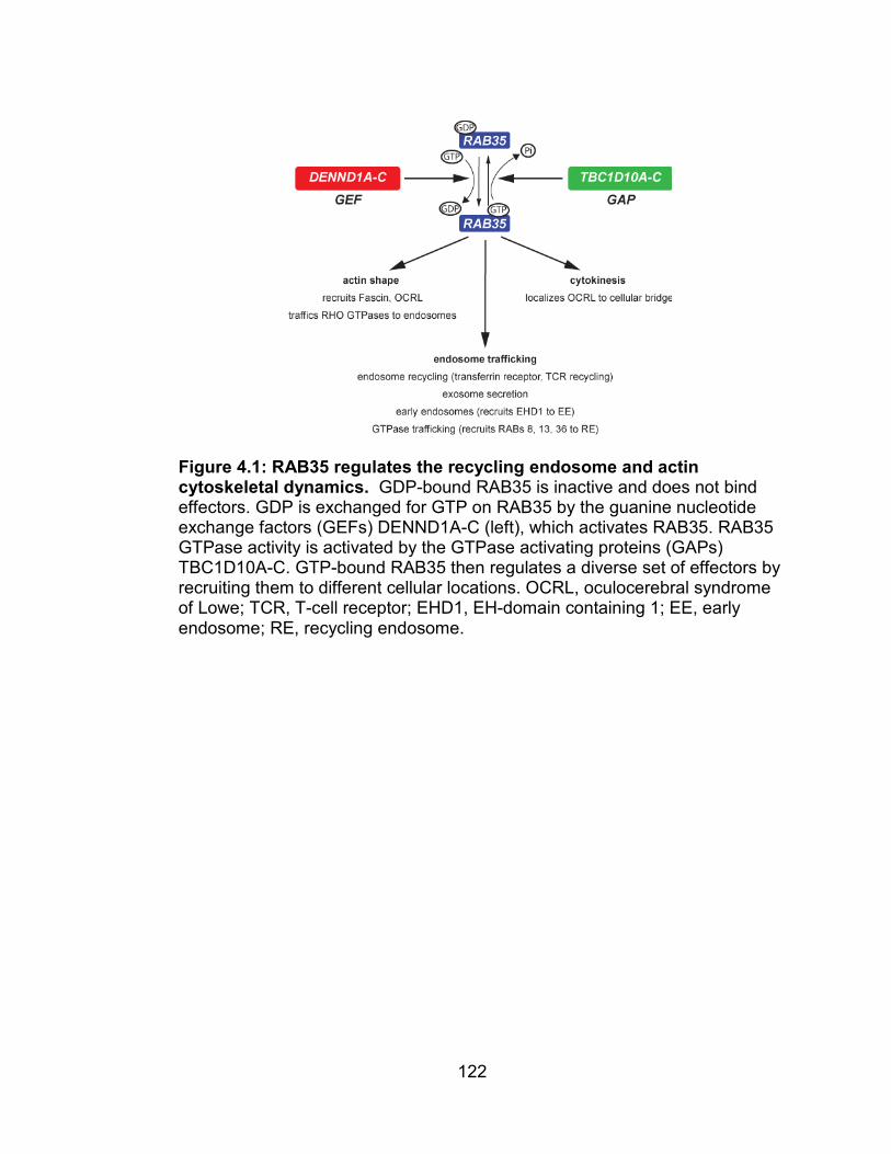
122
Figure 4.1: RAB35 regulates the recycling endosome and actin cytoskeletal dynamics. GDP-bound RAB35 is inactive and does not bind effectors. GDP is exchanged for GTP on RAB35 by the guanine nucleotide exchange factors (GEFs) DENND1A-C (left), which activates RAB35. RAB35 GTPase activity is activated by the GTPase activating proteins (GAPs) TBC1D10A-C. GTP-bound RAB35 then regulates a diverse set of effectors by recruiting them to different cellular locations. OCRL, oculocerebral syndrome of Lowe; TCR, T-cell receptor; EHD1, EH-domain containing 1; EE, early endosome; RE, recycling endosome.

123
4.2 PDGFR TRAFFICKING, RAB35, PI3K/AKT SIGNALING, AND
CANCER
4.2.1 Growth factor receptor tyrosine kinases are trafficked via the
endomembrane system
After growth factor stimulation, RTKs are internalized and either
inactivated and returned to the membrane, allowed to signal in various
subcellular locations, or targeted for destruction by the lysosome and
proteasome system [389]. This mechanism is likely common to
most growth factor receptors, but the epidermal growth factor receptor
(EGFR) is the most thoroughly investigated receptor with regards to its
internalization and trafficking after activation [389]. Because we found that
RAB35 regulates the intracellular location of PDGFRα, it is important to
consider how RTKs are trafficked by the cell after internalization.
One mechanism by which growth factor receptors are downregulated
is through ubiquitinization by the E3 ubiquitin ligase CBL [389, 436-437].
RTKs like EGFR [438], IGFIR [439], PDGFR [440-441], MET [442], KIT
[443], VEGFR [444], and the Ephrin receptors [445-446] are all regulated by
CBL in a similar process. Activated RTKs are internalized by clathrin-coated
pieces of membrane, which often includes the recruitment of CBL to the RTK
or its associated proteins. CBL then mono- and di-ubiquitinylates the target
RTK, which tags the kinase for degradation at the lysosome and/or
proteasome. Alternatively, receptors can be de-liganded, de-ubiquitinylated,

124
and recycled back to the cell membrane where they can be re-activated
later. These scenarios are illustrated in Figure 4.2.
The trafficking and degradation of receptors like EGFR has been
understood for some time, but recent data have demonstrated that instead of
simply being internalized and turned off, EGFR can actively signal while it is
associated with endosomes [389, 391, 447-449] (See example in Figure
4.2). The signaling of internalized EGFR from endomembrane compartments
suggests a mechanism by which the cell can exquisitely govern the spatial
Figure 4.2 The fates of internalized growth factor receptor tyrosine kinases. Once growth factor receptor tyrosine kinases (RTKs) are activated by their growth factor ligands, they initiate signaling at the cell membrane via PI3K and other effectors. They are then trafficked into the cell by endocytosis. Signaling can be attenuated by lysosomal destruction of the RTK, or by de-liganding and recycling the RTK back to the cell membrane. Alternatively, the RTK/PI3K complex can continue to actively signal to effectors like MAPK or AKT while still residing on endomembranes.

125
and temporal activity of signal transduction. Interestingly, similar studies
conducted with respect to PDGFR suggest that much of the signaling
downstream of this receptor occurs after PDGFR is internalized to
endosomes [390].
We speculate that much of the PI3K-dependent signaling by PDGFR
actually happens while receptors are trafficked intracellularly. Interestingly,
we have found that the inhibition of endocytosis with the dynamin GTPase
inhibitor Dynasore [450-453] or N-ethylmaleimide (NEM, which inhibits
vesicular trafficking) [454-456] spares the initial activation of PI3K/AKT by
growth factors (0-5 minutes), but blocks signaling that occurs 15 minutes to
several hours after initial stimulation (data not shown). Given that RTKs are
internalized within minutes after their initial stimulation, this suggests to us
that RTK signaling to oncogenic pathways such as PI3K/AKT is dependent
upon vesicular trafficking.
4.2.2 RAB35 regulates the PI3K/AKT signaling axis by controlling the
intracellular location of PDGFRα
With the data presented in Chapters 2 and 3 of this thesis, we have
provided evidence that GTP-bound RAB35 retains the PDGFRα in a RAB7-
positive endosome where it can drive PI3K/AKT signaling. Because
depletion of RAB35 suppressed AKT and FOXO/NDRG1 phosphorylation in
response to serum and individual growth factors, our data suggested that
RAB35 might function downstream of multiple growth factor receptors. Our

126
first suspicion was that RAB35 either functioned at PI3K or was a general
regulator of RTK activation of PI3K. However, our gain-of-function
experiments indicate that RAB35 might function specifically through
PDGFRα.
Localization studies in cell lines stably expressing wildtype RAB35
suggested to us that PDGFRα is normally trafficked to RAB7-positive
endomembranes after PDGF stimulation. Further, in cells expressing GTPase-
deficient RAB35Q67L, PDGFRα is trafficked to RAB7-positive endomembranes
in the absence of PDGF. Although our data do not indicate whether or not
RAB35 is required for the trafficking of PDGFRα to a RAB7-positive
compartment, it appears that GTP-bound RAB35 somehow retains PDGFRα
at this location, where it actively drives PI3K/AKT signaling in the absence of
ligand. Whether RAB35 is necessary for trafficking PDGFRα to a RAB7-
positive endomembrane will be addressed with RAB35 loss-of-function
studies. Further, it is unclear whether RAB35Q67L is itself driving PDGFRα to
RAB7-positive compartments, or preventing the efficient exit from these
compartments.
It is also possible that GTP-bound RAB35Q67L inhibits a negative
regulator of PDGFR signaling, trafficking, or degradation. For example,
RAB35Q67L could inhibit the ability of the protein tyrosine phosphatase Src
homology 2 domain-containing phosphatase 1 (SHP-1) to dephosphorylate
the phospho-tyrosine residues on PDGFRs [457]. Alternatively—considering
that RAB35 has been demonstrated to negatively regulate GTPases such as

127
ARF6 [426, 430]— RAB35Q67L may inhibit a RAB or ARF GTPase that
determines the trafficking fate(s) of PDGFRs. Finally, RAB35Q67L may
prevent ubiquitin ligases like CBL or CBL2 from ubiquitinylating PDGFRs
[441, 458], which could lead to accumulated, phosphorylated and activated
PDGFRs on endomembranes that can sustain signaling to PI3K/AKT.
Although it is interesting to postulate various scenarios by which
RAB35 may regulate PDGFRα, we suspect that the indirect scenarios
outlined above are less likely, given that we have detected interactions
between wildtype and mutant RAB35 and both PI3Kα and PDGFRα. We
propose that the PDGFRα/PI3Kα complex is bound directly by RAB35,
preferentially when it is loaded with GTP. The data presented in Figure 3.8—
where RAB35 co-purified more robustly with PI3K immunoprecipitates than it
did with PDGFRα immunoprecipitates—suggest to us that RAB35 interacts
with PI3Kα, which interacts with PDGFRα (Figure
4.3). Thus, we suspect that GTP-bound RAB35 directly binds PI3K and
serves to localize PDGFRα/PI3Kα to RAB7-containing endomembranes,
either by “steering” PDGFRα/PI3Kα to this compartment or “trapping” it
there. Whether the exit of PDGFRα from this compartment is dependent on
hydrolysis of the GTP of activated RAB35 is an interesting question that
could be addressed with loss-of-function experiments that ask whether
perturbation of the known RAB35 GAPs (TBC1D10A-C) and GEFs
(DENND1A-C) for RAB35.

128
4.2.3 Is RAB35 a physiological regulator of PDGFRα/PI3K signaling, and
are oncogenic mutant alleles of RAB35 hypermorphic or neomorphic?
At first glance, our gain-of-function data—that RAB35 signal to PI3K
by activating PDGFRα—did not fit with our loss-of-function data, which
suggested that RAB35 was required for PI3K/AKT activation in response to
non-PDGF ligands like VEGF, IGF-I or EGF (Figure 2.16). However, recent
data from the Kwiatkowski lab strongly suggests that these other ligands
actually require PDGFRα/β to efficiently signal to the PI3K/AKT machinery
Figure 4.3: A model for regulation of PDGFRα-PI3K signaling by the small GTPase RAB35. Following stimulation with PDGF-AA, actively signaling PDGFRα/PI3K is internalized to RAB7-positive endomembranes. RAB35 interacts with the PDGFRα/PI3K complex, likely by direct interaction with PI3K (left). In cells that express GTPase-deficient, dominant active RAB35Q67L, PDGFRα/PI3K is retained at RAB7-positive endosomes within the cell even in the absence of ligand, where it constitutively signals to AKT (right).

129
[459]. These findings confirmed earlier ones that indicated that PDGFRs are
activated in a “lateral signaling” fashion: stimulation of IGF-IRs with IGF, for
example, activates a Src-dependent activation of PDGFRs, which then
signal along with IGF-IR to PI3K [460-463]. Further, PDGFR and EGFR can
heterodimerize to activate one another in response to EGF stimulation [464].
Thus, the loss-of-function data that suggests that RAB35 is necessary for
EGF, IGF, PDGF and VEGF-mediated PI3K/AKT activation do not
necessarily conflict with the gain-of-function data that suggest that RAB35
regulation of PI3K/AKT is primarily accomplished via PDGFRα.
It is also interesting to speculate over the nature of the gain-of-
function phenotype we observed in the GTPase-deficient allele of RAB35.
Mutations that alter protein function can be inactivating (hypomorphic),
hyperactivating (hypermorphic), or confer new functions to that protein
altogether (neomorphic) [465]. For example, mutations that result in a
truncated and inactive PTEN protein are hypomorphic, while mutations that
generate constitutively active alleles displaying normal function—like
activating mutations in PI3K—are hypermorphic. Occasionally, mutations
can confer altogether neomorphic functions on proteins. For example, the
PIK3R1/p85 adaptor subunit of PI3K actually serves to inhibit the lipid kinase
activity of PI3K. However, mutations in the PIK3R1 gene have been
identified from human tumors that confer a novel, activating capacity of
PIK3R1 towards PI3K activity [466].

130
The data presented in this thesis suggest that RAB35 is a
physiological regulator of PDGFR-PI3K-AKT signaling and that the
internalization and activation of PDGFRα and PI3K/AKT signaling by
dominant active RAB35Q67L is a hypermorphic—not neomorphic—
phenomenon. We suspect that if the regulation of PDGFRα/PI3K by
RAB35Q67L was non-physiological—i.e. neomorphic—that depletion of
wildtype RAB35 in loss-of-function experiments would not block PI3K/AKT
signaling. Indeed, as demonstrated in Chapter 2, depletion of RAB35 in a
number of contexts serves to blunt the activation of PI3K/AKT as potently as
does the depletion of other core PI3K/AKT signaling components.
Further, our gain-of-function experiments using GTPase-deficient
RAB35Q67L also suggest that the phenotype demonstrated by RAB35Q67L is
an exaggerated and unregulated version of the normal signaling phenotype
of RAB35wt. The internalization of PDGF-stimulated PDGFRα in cells
expressing RAB35wt appears virtually identical to the internalization of
unliganded PDGFRα in cells expressing RAB35Q67L. In both cases, PDGFRα
localizes to RAB7-positive endosomes. If RAB35Q67L were conferring a truly
neomorphic function to PDGFRα signaling in cells, we would expect it to be
re-trafficked into different compartments from those normally occupied by
liganded PDGFRα.
A full assessment of the hypermorphic vs. neomorphic nature of
oncogenic alleles of RAB35 will be further clarified by studying the
relationship between wildtype RAB35 and PDGFRα trafficking. This would
131
likely include loss-of-function experiments using both RNAi and small
molecule inhibitors.
4.3 CONCLUDING REMARKS
4.3.1 RAB35, cancer and why “private mutations” matter
This thesis began with a desire to use RNAi screens to identify novel
regulators of PI3K/AKT signaling. While we used this loss-of-function
screening approach to identify candidate genes that might regulate
PI3K/AKT signaling, we prioritized hit genes that were mutated in a way that
might alter their function (Table 2.2). Although we are interested in
identifying any new members of the PI3K/AKT pathway, we hoped that such
an approach would allow us to identify novel PI3K/AKT components that play
an role in oncogenesis.
As described in Chapter 2 of this thesis, the mutations that we
identified and characterized in RAB35 were not recurrent. Indeed, the
RAB35 mutations that we identified in human tumors (A151T and F161L)
were not canonical mutations that are known to lock RAB35 in a GTP-bound
state (e.g. Q67L). Nevertheless, the fact that the RAB35A151T and
RAB35F161L mutant proteins could activate PI3K/AKT signaling and transform
cells suggests that these mutations are not “passenger” mutations, but that
they instead played a role in the development of the tumors in which they
were found. Recent findings that the same mutations in KRAS at similar
residues are transforming suggest further that these two RAB35 mutations

132
are functionally relevant to tumorigenesis [357-358, 467]. Thus, rare
mutations in proteins like RAB35 that regulate growth factor signaling can
activate oncogenic signaling.
The possibility that RAB35 mutations in patients are activating
PI3K/AKT signaling and aiding in tumor formation or progression could have
actionable clinical implications. For example, patients with activating
mutations in PIK3CA would likely benefit from PI3Kα inhibitors, similar to
BRAFV600E-positive melanoma patients treated with vemurafenib [468-470].
Thus, given that PDGFRα/PI3K/AKT is hyperactivated byRAB35A151T and
RAB35F161L, patients with these mutations could benefit from PI3K-inhibitors.
The scenario outlined above underscores the importance of finding,
identifying, and characterizing rare but biologically relevant mutations in
genes that regulate cell survival pathways. These so-called “private
mutations” [471-473] are the subject of considerable discussion within the
oncology community [474][475] because the number of patients with them is
relatively small. We suggest that the identification of RAB35 as a regulator of
PI3K signaling that is mutated in cancers thus serves as an example for why
rarer “private” mutations should be investigated and understood.
4.3.2 Endomembrane trafficking as an oncogenic force
The concept of dysregulated endomembrane trafficking in cancer is
not a novel one [476]. Indeed, many investigators have documented
dysregulated membrane organization and trafficking downstream of various

133
oncogenic forces. For example, it is well documented that the membrane
polarity of tumor cells is deranged in many contexts [477-479]. In addition, it
has recently been reported that some oncogenic mutations in MET disrupt
the normal trafficking of the receptor [480]. This enhances the oncogenic
potential of MET by preventing its degradation and localizing the protein to
endosomes where it actively signals. Further, as mentioned earlier,
mutations that derange CBL function can also lead to aberrant RTK
localization and signaling.
However, the notion that the direct dysregulation of endomembrane
trafficking may itself lead to transformation is relatively unexamined. We
reason that the oncogenic capability of mutant RAB35 proteins is likely due
to dysregulated membrane trafficking of PDGFRα/PI3K signaling machinery,
which allows sustained oncogenic signaling. The idea that dysregulation of
endomembrane trafficking—in this case by RAB35—could be an oncogenic
force is novel and indeed tantalizing. Further, the fact that the majority (5 of
7) of the GTPases that were tertiary hits in our screen were RAB proteins
(RAB35, RAB1B, RAB39, RASEF, and RABL3) begs further examination of
this idea. If dysregulated endomembrane trafficking possesses tumorigenic
capability, it could reveal an entirely new realm of therapeutic targets for
combating cancer.

134
APPENDIX A: Human kinases and GTPases targeted by shRNA screen.
NCBI ID GENE ID NCBI ID GENE ID NCBI ID GENE ID 25 ABL1 818 CAMK2G 1606 DGKA 27 ABL2 914 CD2 1607 DGKB 90 ACVR1 983 CDC2 1608 DGKG 91 ACVR1B 984 CDC2L1 1609 DGKQ 92 ACVR2A 985 CDC2L2 1613 DAPK3 93 ACVR2B 1017 CDK2 1633 DCK 94 ACVRL1 1018 CDK3 1716 DGUOK 132 ADK 1019 CDK4 1739 DLG1 156 ADRBK1 1020 CDK5 1740 DLG2 157 ADRBK2 1021 CDK6 1741 DLG3 203 AK1 1022 CDK7 1760 DMPK 204 AK2 1024 CDK8 1841 DTYMK 205 AK3L1 1025 CDK9 1859 DYRK1A 207 AKT1 1111 CHEK1 1956 EGFR 208 AKT2 1119 CHKA 1969 EPHA2 238 ALK 1120 CHKB 2011 MARK2 269 AMHR2 1147 CHUK 2041 EPHA1 369 ARAF 1152 CKB 2042 EPHA3 472 ATM 1158 CKM 2043 EPHA4 545 ATR 1159 CKMT1B 2044 EPHA5 558 AXL 1160 CKMT2 2045 EPHA7 613 BCR 1163 CKS1B 2046 EPHA8 640 BLK 1164 CKS2 2047 EPHB1 657 BMPR1A 1195 CLK1 2048 EPHB2 658 BMPR1B 1196 CLK2 2049 EPHB3 659 BMPR2 1198 CLK3 2050 EPHB4 660 BMX 1263 PLK3 2051 EPHB6 673 BRAF 1326 MAP3K8 2064 ERBB2 676 BRDT 1399 CRKL 2065 ERBB3 695 BTK 1432 MAPK14 2066 ERBB4 699 BUB1 1436 CSF1R 2081 ERN1 701 BUB1B 1445 CSK 2185 PTK2B 780 DDR1 1452 CSNK1A1 2241 FER 801 CALM1 1453 CSNK1D 2242 FES 805 CALM2 1454 CSNK1E 2260 FGFR1 808 CALM3 1455 CSNK1G2 2261 FGFR3 814 CAMK4 1456 CSNK1G3 2263 FGFR2 815 CAMK2A 1457 CSNK2A1 2264 FGFR4 816 CAMK2B 1459 CSNK2A2 2268 FGR 817 CAMK2D 1460 CSNK2B 2321 FLT1

135
NCBI ID GENE ID NCBI ID GENE ID NCBI ID GENE ID 2322 FLT3 3815 KIT 5063 PAK3 2324 FLT4 3932 LCK 5105 PCK1 2395 FXN 3984 LIMK1 5106 PCK2 2444 FRK 3985 LIMK2 5127 PCTK1 2475 FRAP1 4058 LTK 5128 PCTK2 2534 FYN 4067 LYN 5129 PCTK3 2580 GAK 4117 MAK 5156 PDGFRA 2584 GALK1 4139 MARK1 5157 PDGFRL 2585 GALK2 4140 MARK3 5159 PDGFRB 2645 GCK 4145 MATK 5163 PDK1 2710 GK 4214 MAP3K1 5164 PDK2 2712 GK2 4215 MAP3K3 5165 PDK3 2868 GRK4 4216 MAP3K4 5166 PDK4 2869 GRK5 4217 MAP3K5 5170 PDPK1 2870 GRK6 4233 MET 5207 PFKFB1 2872 MKNK2 4293 MAP3K9 5208 PFKFB2 2931 GSK3A 4294 MAP3K10 5209 PFKFB3 2932 GSK3B 4296 MAP3K11 5210 PFKFB4 2965 GTF2H1 4342 MOS 5211 PFKL 2984 GUCY2C 4354 MPP1 5213 PFKM 2986 GUCY2F 4355 MPP2 5214 PFKP 2987 GUK1 4356 MPP3 5218 PFTK1 3000 GUCY2D 4486 MST1R 5230 PGK1 3055 HCK 4593 MUSK 5232 PGK2 3098 HK1 4598 MVK 5255 PHKA1 3099 HK2 4638 MYLK 5256 PHKA2 3101 HK3 4750 NEK1 5257 PHKB 3480 IGF1R 4751 NEK2 5260 PHKG1 3551 IKBKB 4752 NEK3 5261 PHKG2 3611 ILK 4830 NME1 5286 PIK3C2A 3643 INSR 4831 NME2 5287 PIK3C2B 3645 INSRR 4832 NME3 5288 PIK3C2G 3654 IRAK1 4833 NME4 5289 PIK3C3 3656 IRAK2 4881 NPR1 5290 PIK3CA 3702 ITK 4882 NPR2 5291 PIK3CB 3705 ITPK1 4914 NTRK1 5292 PIM1 3706 ITPKA 4915 NTRK2 5293 PIK3CD 3707 ITPKB 4916 NTRK3 5294 PIK3CG 3716 JAK1 4919 ROR1 5295 PIK3R1 3717 JAK2 4920 ROR2 5296 PIK3R2 3718 JAK3 4921 DDR2 5297 PI4KA 3791 KDR 5058 PAK1 5298 PI4KB 3795 KHK 5062 PAK2 5305 PIP4K2A

136
NCBI ID GENE ID NCBI ID GENE ID NCBI ID GENE ID 5313 PKLR 5601 MAPK9 6725 SRMS 5315 PKM2 5602 MAPK10 6732 SRPK1 5328 PLAU 5603 MAPK13 6733 SRPK2 5347 PLK1 5604 MAP2K1 6787 NEK4 5361 PLXNA1 5605 MAP2K2 6788 STK3 5362 PLXNA2 5606 MAP2K3 6789 STK4 5364 PLXNB1 5607 MAP2K5 6790 AURKA 5365 PLXNB3 5608 MAP2K6 6792 CDKL5 5394 EXOSC10 5609 MAP2K7 6793 STK10 5562 PRKAA1 5610 EIF2AK2 6794 STK11 5563 PRKAA2 5613 PRKX 6795 AURKC 5564 PRKAB1 5616 PRKY 6850 SYK 5565 PRKAB2 5631 PRPS1 6872 TAF1 5566 PRKACA 5634 PRPS2 6885 MAP3K7 5567 PRKACB 5681 PSKH1 7006 TEC 5568 PRKACG 5747 PTK2 7010 TEK 5571 PRKAG1 5753 PTK6 7016 TESK1 5573 PRKAR1A 5754 PTK7 7046 TGFBR1 5575 PRKAR1B 5756 TWF1 7048 TGFBR2 5576 PRKAR2A 5832 ALDH18A1 7049 TGFBR3 5577 PRKAR2B 5871 MAP4K2 7075 TIE1 5578 PRKCA 5891 RAGE 7083 TK1 5579 PRKCB 5894 RAF1 7084 TK2 5580 PRKCD 5979 RET 7175 TPR 5581 PRKCE 5987 TRIM27 7204 TRIO 5582 PRKCG 6011 GRK1 7272 TTK 5583 PRKCH 6041 RNASEL 7273 TTN 5584 PRKCI 6046 BRD2 7294 TXK 5585 PKN1 6093 ROCK1 7297 TYK2 5586 PKN2 6098 ROS1 7301 TYRO3 5587 PRKD1 6102 RP2 7371 UCK2 5588 PRKCQ 6195 RPS6KA1 7443 VRK1 5590 PRKCZ 6196 RPS6KA2 7444 VRK2 5591 PRKDC 6197 RPS6KA3 7465 WEE1 5592 PRKG1 6198 RPS6KB1 7525 YES1 5593 PRKG2 6199 RPS6KB2 7535 ZAP70 5594 MAPK1 6259 RYK 7786 MAP3K12 5595 MAPK3 6300 MAPK12 7867 MAPKAPK3 5596 MAPK4 6347 CCL2 8019 BRD3 5597 MAPK6 6351 CCL4 8295 TRRAP 5598 MAPK7 6416 MAP2K4 8317 CDC7 5599 MAPK8 6446 SGK1 8382 NME5 5600 MAPK11 6714 SRC 8394 PIP5K1A

137
NCBI ID GENE ID NCBI ID GENE ID NCBI ID GENE ID 8395 PIP5K1B 9113 LATS1 10420 TESK2 8396 PIP4K2B 9149 DYRK1B 10461 MERTK 8408 ULK1 9162 DGKI 10494 STK25 8428 STK24 9175 MAP3K13 10519 CIB1 8444 DYRK3 9201 DCLK1 10595 ERN2 8445 DYRK2 9212 AURKB 10645 CAMKK2 8476 CDC42BPA 9223 MAGI1 10654 PMVK 8491 MAP4K3 9252 RPS6KA5 10733 PLK4 8503 PIK3R3 9261 MAPKAPK2 10746 MAP3K2 8525 DGKZ 9262 STK17B 10769 PLK2 8526 DGKE 9263 STK17A 10783 NEK6 8527 DGKD 9344 TAOK2 10922 FASTK 8536 CAMK1 9414 TJP2 11011 TLK2 8550 MAPKAPK5 9448 MAP4K4 11035 RIPK3 8558 CDK10 9451 EIF2AK3 11040 PIM2 8566 PDXK 9467 SH3BP5 11113 CIT 8569 MKNK1 9475 ROCK2 11139 8573 CASK 9578 CDC42BPB 11183 MAP4K5 8576 STK16 9625 AATK 11184 MAP4K1 8621 CDC2L5 9641 IKBKE 11200 CHEK2 8649 MAPKSP1 9706 ULK2 11213 IRAK3 8711 TNK1 9748 SLK 11284 PNKP 8737 RIPK1 9807 IP6K1 11329 STK38 8767 RIPK2 9829 DNAJC6 11344 TWF2 8780 RIOK3 9833 MELK 22848 AAK1 8798 DYRK4 9874 TLK1 22853 LMTK2 8814 CDKL1 9891 NUAK1 22858 ICK 8844 KSR1 9942 XYLB 22868 FASTKD2 8851 CDK5R1 9943 OXSR1 22901 ARSG 8859 STK19 9950 GOLGA5 22928 SEPHS2 8877 SPHK1 10000 AKT3 22983 MAST1 8895 CPNE3 10020 GNE 23012 STK38L 8899 PRPF4B 10087 COL4A3BP 23031 MAST3 8941 CDK5R2 10110 SGK2 23043 TNIK 8986 RPS6KA4 10114 HIPK3 23049 SMG1 8997 KALRN 10128 LRPPRC 23097 CDC2L6 8999 CDKL2 10154 PLXNC1 23129 PLXND1 9020 MAP3K14 10188 TNK2 23139 MAST2 9024 BRSK2 10201 NME6 23178 PASK 9060 PAPSS2 10221 TRIB1 23227 MAST4 9061 PAPSS1 10290 SPEG 23235 SIK2 9064 MAP3K6 10295 BCKDK 23300 ATMIN 9088 PKMYT1 10298 PAK4 23387

138
NCBI ID GENE ID NCBI ID GENE ID NCBI ID GENE ID 23396 PIP5K1C 51347 TAOK3 56848 SPHK2 23476 BRD4 51422 PRKAG2 56911 C21orf7 23533 PIK3R5 51447 IP6K2 56924 PAK6 23552 CCRK 51678 MPP6 56997 CABC1 23604 DAPK2 51701 NLK 57118 CAMK1D 23617 TSSK2 51727 CMPK1 57143 ADCK1 23636 NUP62 51755 CRKRS 57144 PAK7 23654 PLXNB2 51765 57147 SCYL3 23677 SH3BP4 51776 57172 CAMK1G 23678 SGK3 53354 PANK1 57396 CLK4 23683 PRKD3 53632 PRKAG3 57410 SCYL1 23729 SHPK 53834 FGFRL1 57538 ALPK3 25778 DSTYK 53904 MYO3A 57551 TAOK1 25865 PRKD2 53944 CSNK1G1 57729 25989 ULK3 54101 RIPK4 57761 TRIB3 26007 DAK 54103 PION 57787 MARK4 26289 AK5 54822 TRPM7 58538 MPP4 26353 HSPB8 54861 SNRK 60493 FASTKD5 26524 LATS2 54899 PXK 64080 RBKS 26576 SRPK3 54963 UCKL1 64089 SNX16 26750 RPS6KC1 54981 C9orf95 64122 FN3K 27010 TPK1 54986 ULK4 64149 C17orf75 27102 EIF2AK1 55224 ETNK2 64398 MPP5 27148 STK36 55229 PANK4 64768 IPPK 27231 ITGB1BP3 55277 FGGY 64781 CERK 27330 RPS6KA6 55300 PI4K2B 65018 PINK1 27347 STK39 55312 RFK 65061 PFTK2 28951 TRIB2 55351 STK32B 65125 WNK1 28996 HIPK2 55359 STYK1 65220 NADK 29110 TBK1 55361 PI4K2A 65266 WNK4 29904 EEF2K 55437 STRADB 65267 WNK3 29922 NME7 55500 ETNK1 65268 WNK2 29941 PKN3 55558 PLXNA3 65975 STK33 29959 NRBP1 55561 CDC42BPG 79012 CAMKV 30811 HUNK 55577 NAGK 79072 FASTKD3 30849 PIK3R4 55589 BMP2K 79646 PANK3 50488 MINK1 55681 SCYL2 79672 50808 AK3 55728 N4BP2 79675 FASTKD1 51086 TNNI3K 55750 AGK 79705 LRRK1 51135 IRAK4 55781 RIOK2 79834 51231 VRK3 55872 PBK 79837 PIP4K2C 51265 CDKL3 56155 TEX14 79858 NEK11 51314 TXNDC3 56164 STK31 79877 DCAKD

139
NCBI ID GENE ID NCBI ID GENE ID NCBI ID GENE ID 79906 MORN1 112858 TP53RK 221823 PRPS1L1 79934 ADCK4 114783 LMTK3 225689 MAPK15 80025 PANK2 114836 SLAMF6 253430 IPMK 80122 YSK4 115701 ALPK2 255239 ANKK1 80201 HKDC1 117283 IP6K3 256356 GK5 80216 ALPK1 120892 LRRK2 260425 MAGI3 80271 ITPKC 122011 CSNK1A1L 282974 STK32C 80347 COASY 122481 AK7 283455 KSR2 80851 SH3BP5L 124923 283629 TSSK4 81629 TSSK3 127933 UHMK1 284086 NEK8 81788 NUAK2 130106 CIB4 284656 EPHA10 83440 ADPGK 130399 ACVR1C 285220 EPHA6 83549 UCK1 131890 GRK7 285962 83694 RPS6KL1 132158 GLYCTK 340156 MYLK4 83732 RIOK1 136332 LRGUK 340371 NRBP2 83903 GSG2 138429 PIP5KL1 341676 NEK5 83931 STK40 138474 TAF1L 344387 CDKL4 83942 TSSK1B 139189 DGKK 347359 BMP2KL 83983 TSSK6 139728 PNCK 347736 NME9 84033 OBSCN 140469 MYO3B 374872 C19orf35 84197 POMK 140609 NEK7 375133 PI4KAP2 84206 MEX3B 140803 TRPM6 375298 CERKL 84254 CAMKK1 140901 STK35 375449 MAST4 84284 C1orf57 143098 MPP7 378464 MORN2 84433 CARD11 146057 TTBK2 387851 AK3L2 84446 BRSK1 147746 HIPK4 388228 SBK1 84451 149420 PDIK1L 388259 84630 TTBK1 150094 SIK1 388957 84930 MASTL 152110 NEK10 389840 MAP3K15 85366 MYLK2 157285 389906 85443 DCLK3 158067 C9orf98 390877 85481 PSKH2 160851 DGKH 391295 89882 TPD52L3 166614 DCLK2 391533 90381 C15orf42 167359 392226 90956 ADCK2 169436 C9orf96 392265 91156 IGFN1 197258 FUK 392347 91419 XRCC6BP1 197259 MLKL 400301 91461 200576 PIP5K3 402289 91584 PLXNA4 202374 STK32A 402679 91754 NEK9 203054 ADCK5 415116 PIM3 91807 MYLK3 203447 NRK 440275 EIF2AK4 92335 STRADA 204851 HIPK1 440345 93627 220686 441047

140
NCBI ID GENE ID NCBI ID GENE ID NCBI ID GENE ID 441655 2773 GNAI3 5879 RAC1 441708 2774 GNAL 5880 RAC2 441777 2775 GNAO1 5881 RAC3 441971 2776 GNAQ 5898 RALA 442075 2778 GNAS 5899 RALB 442313 2779 GNAT1 5901 RAN 442558 2780 GNAT2 5906 RAP1A 57521 RAPTOR 2781 GNAZ 5908 RAP1B 253260 RICTOR 2782 GNB1 5911 RAP2A
373 TRIM23 2783 GNB2 5912 RAP2B 375 ARF1 2784 GNB3 6009 RHEB 377 ARF3 2785 GNG3 6014 RIT2 378 ARF4 2786 GNG4 6016 RIT1 379 ARL4D 2787 GNG5 6236 RRAD 381 ARF5 2788 GNG7 6237 RRAS 382 ARF6 2791 GNG11 6729 SRP54 387 RHOA 2792 GNGT1 6734 SRPR 388 RHOB 2793 GNGT2 7284 TUFM 389 RHOC 3265 HRAS 7879 RAB7A 390 RND3 3266 ERAS 8153 RND2 391 RHOG 3845 KRAS 8766 RAB11A 399 RHOH 4218 RAB8A 8934 RAB7L1 400 ARL1 4261 CIITA 9077 DIRAS3 402 ARL2 4599 MX1 9230 RAB11B 403 ARL3 4600 MX2 9363 RAB33A 989 39698 4733 DRG1 9364 RAB28 998 CDC42 4735 39693 9367 RAB9A 1731 39692 4893 NRAS 9545 RAB3D 1759 DNM1 4976 OPA1 9567 GTPBP1 1785 DNM2 5413 39696 9609 RAB36 1819 DRG2 5414 39695 9630 GNA14 1915 EEF1A1 5861 RAB1A 9886 RHOBTB1 1917 EEF1A2 5862 RAB2A 9927 MFN2 1968 EIF2S3 5864 RAB3A 10059 DNM1L 2633 GBP1 5865 RAB3B 10123 ARL4C 2634 GBP2 5867 RAB4A 10124 ARL4A 2635 GBP3 5868 RAB5A 10139 ARFRP1 2669 GEM 5869 RAB5B 10325 RRAGB 2767 GNA11 5870 RAB6A 10633 RASL10A 2768 GNA12 5872 RAB13 10670 RRAGA 2769 GNA15 5873 RAB27A 10672 GNA13 2770 GNAI1 5874 RAB27B 10681 GNB5 2771 GNAI2 5878 RAB5C 10801 39700
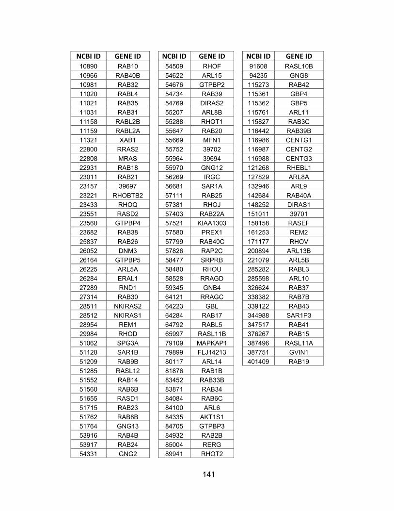
141
NCBI ID GENE ID NCBI ID GENE ID NCBI ID GENE ID 10890 RAB10 54509 RHOF 91608 RASL10B 10966 RAB40B 54622 ARL15 94235 GNG8 10981 RAB32 54676 GTPBP2 115273 RAB42 11020 RABL4 54734 RAB39 115361 GBP4 11021 RAB35 54769 DIRAS2 115362 GBP5 11031 RAB31 55207 ARL8B 115761 ARL11 11158 RABL2B 55288 RHOT1 115827 RAB3C 11159 RABL2A 55647 RAB20 116442 RAB39B 11321 XAB1 55669 MFN1 116986 CENTG1 22800 RRAS2 55752 39702 116987 CENTG2 22808 MRAS 55964 39694 116988 CENTG3 22931 RAB18 55970 GNG12 121268 RHEBL1 23011 RAB21 56269 IRGC 127829 ARL8A 23157 39697 56681 SAR1A 132946 ARL9 23221 RHOBTB2 57111 RAB25 142684 RAB40A 23433 RHOQ 57381 RHOJ 148252 DIRAS1 23551 RASD2 57403 RAB22A 151011 39701 23560 GTPBP4 57521 KIAA1303 158158 RASEF 23682 RAB38 57580 PREX1 161253 REM2 25837 RAB26 57799 RAB40C 171177 RHOV 26052 DNM3 57826 RAP2C 200894 ARL13B 26164 GTPBP5 58477 SRPRB 221079 ARL5B 26225 ARL5A 58480 RHOU 285282 RABL3 26284 ERAL1 58528 RRAGD 285598 ARL10 27289 RND1 59345 GNB4 326624 RAB37 27314 RAB30 64121 RRAGC 338382 RAB7B 28511 NKIRAS2 64223 GBL 339122 RAB43 28512 NKIRAS1 64284 RAB17 344988 SAR1P3 28954 REM1 64792 RABL5 347517 RAB41 29984 RHOD 65997 RASL11B 376267 RAB15 51062 SPG3A 79109 MAPKAP1 387496 RASL11A 51128 SAR1B 79899 FLJ14213 387751 GVIN1 51209 RAB9B 80117 ARL14 401409 RAB19 51285 RASL12 81876 RAB1B 51552 RAB14 83452 RAB33B 51560 RAB6B 83871 RAB34 51655 RASD1 84084 RAB6C 51715 RAB23 84100 ARL6 51762 RAB8B 84335 AKT1S1 51764 GNG13 84705 GTPBP3 53916 RAB4B 84932 RAB2B 53917 RAB24 85004 RERG 54331 GNG2 89941 RHOT2

142
APPENDIX B: Non-lethal shRNAs with phospho-AKT Z-scores greater than -/+ 1.50. NCBI ID Symbol TRC Clone Name mean
%
cell 207 AKT1 NM_005163.x-642s1c1 -2.23 10% A-431
207 AKT1 NM_005163.x-1044s1c1 -1.52 14% A-431 207 AKT1 NM_005163.1-1410s1c1 -1.69 48% A-431 207 AKT1 NM_005163.x-981s1c1 -2.02 17% A-431 207 AKT1 NM_005163.x-642s1c1d2 -1.55 16% A431 207 AKT1 NM_005163.x-1044s1c1d1 -1.70 14% A431 373 TRIM23 NM_001656.3-655s1c1d1 -2.07 37% Jurkat 373 TRIM23 NM_001656.3-301s1c1d1 -1.67 65% Jurkat 378 ARF4 NM_001660.2-614s1c1d2 2.55 1% A549 378 ARF4 NM_001660.2-296s1c1d2 4.14 68% A549 387 RHOA NM_001664.1-382s1c1d2 -2.51 16% A549 387 RHOA NM_001664.1-616s1c1d2 -2.78 3% A549 387 RHOA NM_001664.1-300s1c1d2 -2.67 15% A549 387 RHOA NM_001664.1-199s1c1d2 -2.51 5% A549 388 RHOB NM_004040.2-839s1c1d2 4.69 5% A549 388 RHOB NM_004040.2-452s1c1d2 -1.91 12% A549 388 RHOB NM_004040.2-461s1c1d2 -1.80 22% A549 388 RHOB NM_004040.2-623s1c1d2 -2.74 65% A549 389 RHOC NM_175744.3-539s1c1d3 -1.50 13% A549 389 RHOC NM_175744.3-328s1c1d2 -1.77 12% A549 390 RND3 NM_005168.2-557s1c1d2 -2.61 103% A549 390 RND3 NM_005168.2-338s1c1d2 -2.61 32% A549 391 RHOG NM_001665.2-273s1c1d2 -3.16 #N/A #N/A 391 RHOG NM_001665.2-616s1c1d2 -2.47 #N/A #N/A 391 RHOG NM_001665.2-192s1c1d2 -1.81 #N/A #N/A 399 RHOH NM_004310.2-1177s1c1d2 -2.15 #N/A #N/A 399 RHOH NM_004310.2-828s1c1d2 -2.19 #N/A #N/A 400 ARL1 NM_001177.3-374s1c1d2 -3.57 #N/A #N/A 400 ARL1 NM_001177.3-190s1c1d2 -2.25 #N/A #N/A 402 ARL2 NM_001667.1-531s1c1d3 -1.88 54% A549 402 ARL2 NM_001667.1-182s1c1d3 -2.09 11% A549 403 ARL3 NM_004311.2-480s1c1d3 -2.65 13% A549 403 ARL3 NM_004311.2-290s1c1d3 -1.57 3% A549 472 ATM NM_000051.2-9211s1c1 -1.81 0% MCF7 472 ATM NM_000051.2-9380s1c1 -2.57 0% MCF7 472 ATM NM_000051.2-1717s1c1 -1.76 0% MCF7 472 ATM NM_000051.2-1974s1c1 -2.39 75% MCF7 545 ATR NM_001184.x-3426s1c1 -1.78 33% A549 640 BLK NM_001715.x-1031s1c1 -2.41 16% Jurkat 640 BLK NM_001715.2-746s1c1 -2.12 30% Jurkat 640 BLK NM_001715.2-1626s1c1 -1.66 43% Jurkat

143
640 BLK NM_001715.x-1200s1c1 -2.03 29% Jurkat 676 BRDT NM_001726.1-629s1c1 1.98 36% A549 676 BRDT NM_001726.2-2905s1c1 2.15 80% A549 676 BRDT NM_001726.1-2954s1c1 2.95 36% A549 780 DDR1 NM_001954.x-961s1c1 2.00 20% MCF7 780 DDR1 NM_001954.x-3795s1c1 1.64 100% A549 983 CDC2 NM_001786.x-820s1c1 -1.76 10% A549 983 CDC2 NM_001786.x-906s1c1 -2.31 52% A549 983 CDC2 NM_001786.2-748s1c1 -1.92 51% A549 998 CDC42 NM_001791.2-493s1c1d2 -1.73 73% A549 998 CDC42 NM_001791.2-328s1c1d2 -2.18 24% A549 1119 CHKA NM_001277.1-1557s1c1 1.98 71% A549 1119 CHKA NM_001277.1-1136s1c1 2.11 92% A549 1152 CKB NM_001823.3-1250s1c1 -2.50 8% MCF7 1152 CKB NM_001823.3-877s1c1 -1.70 35% MCF7 1152 CKB NM_001823.3-722s1c1 -2.70 34% MCF7 1152 CKB NM_001823.3-326s1c1 -1.68 60% MCF7 1152 CKB NM_001823.3-1251s1c1 -1.80 20% MCF7 1453 CSNK1D NM_139062.x-991s1c1 2.52 74% A549 1453 CSNK1D NM_139062.x-1027s1c1 2.11 85% A549 1453 CSNK1D NM_001893.x-991s1c1 2.57 58% A549 1453 CSNK1D NM_001893.x-1027s1c1 3.61 69% A549 1606 DGKA NM_001345.4-592s1c1 2.41 22% Jurkat 1606 DGKA NM_001345.4-382s1c1 2.52 42% Jurkat 1633 DCK NM_000788.x-252s1c1 -2.09 113% MCF7 1633 DCK NM_000788.x-440s1c1 -1.60 0% MCF7 1915 EEF1A1 NM_001402.4-807s1c1d1 -1.76 2% A549 1915 EEF1A1 NM_001402.4-338s1c1d1 -2.53 4% A549 1968 EIF2S3 NM_001415.2-1225s1c1d1 -4.87 136% A549 1968 EIF2S3 NM_001415.2-646s1c1d1 -1.85 11% A549 1968 EIF2S3 NM_001415.2-782s1c1d1 -2.28 45% A549 2041 EPHA1 NM_005232.2-3118s1c1 -1.94 8% MCF7 2041 EPHA1 NM_005232.2-2176s1c1 -2.01 61% MCF7 2049 EPHB3 NM_004443.3-3611s1c1 2.14 43% MCF7 2049 EPHB3 NM_004443.3-1895s1c1 1.58 46% MCF7 2050 EPHB4 NM_004444.x-459s1c1 -1.76 27% A549 2050 EPHB4 NM_004444.x-2076s1c1 -1.55 8% A549 2064 ERBB2 NM_004448.x-1214s1c1 -2.37 0% A-431 2064 ERBB2 NM_004448.x-1365s1c1 -2.23 37% A-431 2064 ERBB2 NM_004448.1-466s1c1 -1.78 135% A549 2260 FGFR1 NM_000604.2-2340s1c1 1.99 56% MCF7 2260 FGFR1 NM_000604.2-1013s1c1 1.67 121% MCF7 2260 FGFR1 NM_000604.2-1237s1c1 1.77 38% MCF7 2321 FLT1 NM_002019.2-5483s1c1 -2.57 #N/A #N/A
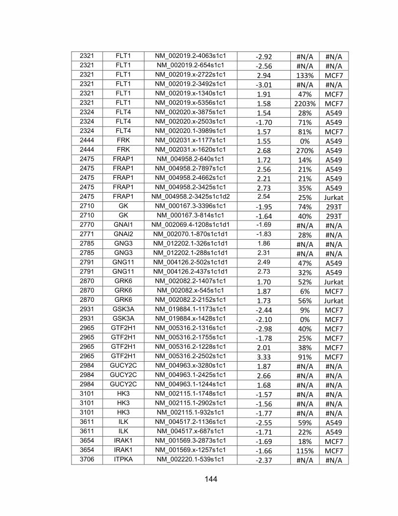
144
2321 FLT1 NM_002019.2-4063s1c1 -2.92 #N/A #N/A 2321 FLT1 NM_002019.2-654s1c1 -2.56 #N/A #N/A 2321 FLT1 NM_002019.x-2722s1c1 2.94 133% MCF7 2321 FLT1 NM_002019.2-3492s1c1 -3.01 #N/A #N/A 2321 FLT1 NM_002019.x-1340s1c1 1.91 47% MCF7 2321 FLT1 NM_002019.x-5356s1c1 1.58 2203% MCF7 2324 FLT4 NM_002020.x-3875s1c1 1.54 28% A549 2324 FLT4 NM_002020.x-2503s1c1 -1.70 71% A549 2324 FLT4 NM_002020.1-3989s1c1 1.57 81% MCF7 2444 FRK NM_002031.x-1177s1c1 1.55 0% A549 2444 FRK NM_002031.x-1620s1c1 2.68 270% A549 2475 FRAP1 NM_004958.2-640s1c1 1.72 14% A549 2475 FRAP1 NM_004958.2-7897s1c1 2.56 21% A549 2475 FRAP1 NM_004958.2-4662s1c1 2.21 21% A549 2475 FRAP1 NM_004958.2-3425s1c1 2.73 35% A549 2475 FRAP1 NM_004958.2-3425s1c1d2 2.54 25% Jurkat 2710 GK NM_000167.3-3396s1c1 -1.95 74% 293T 2710 GK NM_000167.3-814s1c1 -1.64 40% 293T 2770 GNAI1 NM_002069.4-1208s1c1d1 -1.69 #N/A #N/A 2771 GNAI2 NM_002070.1-870s1c1d1 -1.83 28% #N/A 2785 GNG3 NM_012202.1-326s1c1d1 1.86 #N/A #N/A 2785 GNG3 NM_012202.1-288s1c1d1 2.31 #N/A #N/A 2791 GNG11 NM_004126.2-502s1c1d1 2.49 47% A549 2791 GNG11 NM_004126.2-437s1c1d1 2.73 32% A549 2870 GRK6 NM_002082.2-1407s1c1 1.70 52% Jurkat 2870 GRK6 NM_002082.x-545s1c1 1.87 6% MCF7 2870 GRK6 NM_002082.2-2152s1c1 1.73 56% Jurkat 2931 GSK3A NM_019884.1-1173s1c1 -2.44 9% MCF7 2931 GSK3A NM_019884.x-1428s1c1 -2.10 0% MCF7 2965 GTF2H1 NM_005316.2-1316s1c1 -2.98 40% MCF7 2965 GTF2H1 NM_005316.2-1755s1c1 -1.78 25% MCF7 2965 GTF2H1 NM_005316.2-1228s1c1 2.01 38% MCF7 2965 GTF2H1 NM_005316.2-2502s1c1 3.33 91% MCF7 2984 GUCY2C NM_004963.x-3280s1c1 1.87 #N/A #N/A 2984 GUCY2C NM_004963.1-2425s1c1 2.66 #N/A #N/A 2984 GUCY2C NM_004963.1-1244s1c1 1.68 #N/A #N/A 3101 HK3 NM_002115.1-1748s1c1 -1.57 #N/A #N/A 3101 HK3 NM_002115.1-2902s1c1 -1.56 #N/A #N/A 3101 HK3 NM_002115.1-932s1c1 -1.77 #N/A #N/A 3611 ILK NM_004517.2-1136s1c1 -2.55 59% A549 3611 ILK NM_004517.x-687s1c1 -1.71 22% A549 3654 IRAK1 NM_001569.3-2873s1c1 -1.69 18% MCF7 3654 IRAK1 NM_001569.x-1257s1c1 -1.66 115% MCF7 3706 ITPKA NM_002220.1-539s1c1 -2.37 #N/A #N/A

145
3706 ITPKA NM_002220.1-1420s1c1 -1.94 #N/A #N/A 3706 ITPKA NM_002220.1-783s1c1 -2.10 #N/A #N/A 3706 ITPKA NM_002220.1-808s1c1 1.97 #N/A #N/A 3706 ITPKA NM_002220.1-1076s1c1 3.36 #N/A #N/A 3706 ITPKA NM_002220.1-368s1c1 3.37 #N/A #N/A 3717 JAK2 NM_004972.2-3669s1c1 1.66 #N/A #N/A 3717 JAK2 NM_004972.x-4564s1c1 3.73 23% A549 3717 JAK2 NM_004972.2-4773s1c1 1.98 #N/A #N/A 3795 KHK NM_000221.1-359s1c1 -2.19 #N/A #N/A 3795 KHK NM_000221.1-1328s1c1 -2.83 #N/A #N/A 3795 KHK NM_000221.1-458s1c1 -3.11 #N/A #N/A 3984 LIMK1 NM_002314.x-1512s1c1 -2.04 5% MCF7 3984 LIMK1 NM_002314.x-1374s1c1 -1.78 3% MCF7 4058 LTK NM_002344.x-2132s1c1 -2.46 0% A549 4058 LTK NM_002344.x-1509s1c1 -2.00 69% A549 4058 LTK NM_002344.x-593s1c1 -1.88 99% A549 4117 MAK NM_005906.x-982s1c1 2.78 #N/A #N/A 4117 MAK NM_005906.x-2562s1c1 4.33 #N/A #N/A 4139 MARK1 NM_018650.2-1916s1c1 -1.83 86% A549 4139 MARK1 NM_018650.2-2586s1c1 -1.58 86% A549 4139 MARK1 NM_018650.2-2974s1c1 4.07 117% A549 4139 MARK1 NM_018650.2-1564s1c1 1.59 99% A549 4140 MARK3 NM_002376.x-518s1c1 -2.69 36% MCF7 4140 MARK3 NM_002376.x-1363s1c1 -2.47 59% MCF7 4214 MAP3K1 XM_042066.8-5517s1c1 1.73 173% A549 4214 MAP3K1 XM_042066.11-7305s1c1 1.53 49% A549 4294 MAP3K10 NM_002446.x-3247s1c1 -3.27 84% MCF7 4294 MAP3K10 NM_002446.x-1420s1c1 -2.37 83% MCF7 4294 MAP3K10 NM_002446.2-1329s1c1 -2.30 #N/A #N/A 4294 MAP3K10 NM_002446.x-2033s1c1 -1.54 94% MCF7 4354 MPP1 NM_002436.2-1286s1c1 -3.06 26% A549 4354 MPP1 NM_002436.2-1216s1c1 -1.73 24% A549 4355 MPP2 NM_005374.2-178s1c1 1.95 46% Jurkat 4355 MPP2 NM_005374.2-755s1c1 2.84 57% Jurkat 4486 MST1R NM_002447.x-4136s1c1 -3.05 58% A549 4486 MST1R NM_002447.1-2829s1c1 -2.00 57% A549 4486 MST1R NM_002447.1-2397s1c1 -2.14 20% A549 4733 DRG1 NM_004147.2-665s1c1d1 1.51 18% A549 4733 DRG1 NM_004147.2-514s1c1d1 1.89 10% A549 4750 NEK1 NM_012224.1-1427s1c1 1.52 71% Jurkat 4750 NEK1 NM_012224.1-2206s1c1 2.59 92% Jurkat 4750 NEK1 NM_012224.1-2620s1c1 1.55 89% Jurkat 4832 NME3 NM_002513.2-310s1c1 -1.88 11% 293T 4832 NME3 NM_002513.2-414s1c1 -2.96 15% 293T

146
4976 OPA1 NM_130832.1-435s1c1d1 -4.29 55% A549 4976 OPA1 NM_130832.1-434s1c1d1 -2.45 12% A549 5062 PAK2 NM_002577.x-2531s1c1 2.20 9% MCF7 5062 PAK2 NM_002577.3-1555s1c1 2.63 7% MCF7 5062 PAK2 NM_002577.x-454s1c1 2.70 15% MCF7 5062 PAK2 NM_002577.3-4034s1c1 2.77 96% MCF7 5165 PDK3 NM_005391.1-1322s1c1 -2.02 83% A549 5165 PDK3 NM_005391.1-440s1c1 -1.96 16% A549 5165 PDK3 NM_005391.1-1354s1c1 -1.60 106% A549 5170 PDPK1 NM_002613.1-1373s1c1 -1.88 86% MCF7 5170 PDPK1 NM_002613.1-542s1c1 -2.46 118% MCF7 5170 PDPK1 NM_002613.x-1306s1c1 -1.57 110% MCF7 5211 PFKL NM_002626.x-1630s1c1 -2.08 10% A549 5211 PFKL NM_002626.4-2301s1c1 -1.69 11% A549 5211 PFKL NM_002626.4-2309s1c1 -1.69 0% A549 5213 PFKM NM_000289.3-1883s1c1 -1.52 35% MCF7 5213 PFKM NM_000289.3-429s1c1 -2.57 16% MCF7 5213 PFKM NM_000289.3-1708s1c1 -1.71 #N/A #N/A 5213 PFKM NM_000289.3-1571s1c1 -3.01 #N/A #N/A 5291 PIK3CB NM_006219.x-61s1c1 -1.63 40% MCF7 5291 PIK3CB NM_006219.x-2289s1c1 -1.60 25% MCF7 5291 PIK3CB NM_006219.x-748s1c1 -1.56 55% MCF7 5291 PIK3CB NM_006219.x-1306s1c1 -1.57 29% MCF7 5291 PIK3CB NM_006219.x-61s1c1d2 -1.74 40% MCF7 5291 PIK3CB NM_006219.x-748s1c1d1 -1.89 56% MCF7 5347 PLK1 NM_005030.3-1986s1c1 -1.50 48% MCF7 5347 PLK1 NM_005030.3-1374s1c1 -1.93 35% MCF7 5347 PLK1 NM_005030.3-1893s1c1 -2.01 25% MCF7 5347 PLK1 NM_005030.3-1361s1c1 -1.89 0% MCF7 5361 PLXNA1 NM_032242.2-4255s1c1 2.36 31% A549 5361 PLXNA1 NM_032242.2-4656s1c1 2.82 38% A549 5364 PLXNB1 NM_002673.3-6053s1c1 -2.85 43% Jurkat 5364 PLXNB1 NM_002673.3-1116s1c1 -1.91 66% Jurkat 5394 EXOSC10 NM_002685.1-1833s1c1 -1.87 24% A549 5394 EXOSC10 NM_002685.1-1274s1c1 -3.51 7% A549 5394 EXOSC10 NM_002685.1-2560s1c1 -1.64 25% A549 5563 PRKAA2 NM_006252.x-2127s1c1 -2.05 #N/A #N/A 5563 PRKAA2 NM_006252.x-1028s1c1 -2.37 #N/A #N/A 5575 PRKAR1B NM_002735.1-157s1c1 3.79 #N/A #N/A 5575 PRKAR1B NM_002735.1-593s1c1 2.77 8% MCF7 5575 PRKAR1B NM_002735.1-544s1c1 4.01 61% MCF7 5575 PRKAR1B NM_002735.1-620s1c1 3.24 7% MCF7 5580 PRKCD NM_006254.3-2389s1c1 -2.77 102% MCF7 5580 PRKCD NM_006254.x-826s1c1 -2.55 56% A549

147
5580 PRKCD NM_006254.3-1145s1c1 -1.74 57% MCF7 5586 PKN2 NM_006256.1-2250s1c1 -2.06 39% A549 5586 PKN2 NM_006256.1-309s1c1 -2.26 28% A549 5608 MAP2K6 NM_002758.x-592s1c1 1.89 77% MCF7 5608 MAP2K6 NM_002758.x-503s1c1 1.61 191% MCF7 5631 PRPS1 NM_002764.x-592s1c1 1.70 42% MCF7 5631 PRPS1 NM_002764.x-819s1c1 2.05 0% MCF7 5634 PRPS2 NM_002765.x-871s1c1 1.81 16% MCF7 5634 PRPS2 NM_002765.x-320s1c1 1.96 13% MCF7 5832 ALDH18A1 NM_002860.2-568s1c1 -2.13 44% A549 5832 ALDH18A1 NM_002860.2-658s1c1 -2.59 8% A549 5861 RAB1A NM_004161.3-698s1c1d1 -3.70 9% A549 5861 RAB1A NM_004161.3-1607s1c1d1 -2.02 57% A549 5862 RAB2A NM_002865.1-523s1c1d1 -2.40 143% A549 5864 RAB3A NM_002866.3-266s1c1d4 -2.11 161% Jurkat 5864 RAB3A NM_002866.3-447s1c1d2 -1.67 132% Jurkat 5865 RAB3B NM_002867.x-907s1c1d1 -1.54 6% A549 5865 RAB3B NM_002867.x-447s1c1d1 -2.71 8% A549 5865 RAB3B NM_002867.x-446s1c1d1 -2.98 5% A549 5867 RAB4A NM_004578.2-500s1c1d1 -3.32 71% Jurkat 5867 RAB4A NM_004578.2-608s1c1d1 -2.33 69% Jurkat 5867 RAB4A NM_004578.2-346s1c1d3 -2.10 56% Jurkat 5868 RAB5A NM_004162.3-659s1c1d1 -3.94 46% #N/A 5868 RAB5A NM_004162.3-2139s1c1d3 -1.86 16% #N/A 5868 RAB5A NM_004162.3-406s1c1d1 -1.53 69% #N/A 5870 RAB6A NM_002869.4-824s1c1d3 1.97 103% Jurkat 5870 RAB6A NM_002869.4-1073s1c1d3 2.07 42% Jurkat 5872 RAB13 NM_002870.2-336s1c1d2 -3.60 #N/A #N/A 5872 RAB13 NM_002870.2-196s1c1d2 -2.18 #N/A #N/A 5873 RAB27A NM_004580.3-735s1c1d2 -1.72 5% A549 5873 RAB27A NM_004580.3-477s1c1d2 -2.09 25% A549 5874 RAB27B NM_004163.3-697s1c1d2 -2.83 24% A549 5874 RAB27B NM_004163.3-217s1c1d2 -1.61 72% A549 5874 RAB27B NM_004163.3-566s1c1d2 -2.18 133% A549 5881 RAC3 NM_005052.2-544s1c1d2 -2.77 17% 293T 5881 RAC3 NM_005052.2-664s1c1d2 -3.31 20% 293T 5881 RAC3 NM_005052.2-331s1c1d2 -2.78 14% 293T 6009 RHEB NM_005614.x-624s1c1d2 2.48 #N/A #N/A 6009 RHEB NM_005614.x-754s1c1d2 2.57 #N/A #N/A 6009 RHEB NM_005614.2-1100s1c1d2 2.28 #N/A #N/A 6011 GRK1 NM_002929.x-388s1c1 2.79 #N/A #N/A 6011 GRK1 NM_002929.x-945s1c1 1.73 #N/A #N/A 6011 GRK1 NM_002929.2-1682s1c1 2.82 #N/A #N/A 6011 GRK1 NM_002929.2-463s1c1 1.61 #N/A #N/A

148
6046 BRD2 NM_005104.2-2401s1c1 -2.12 40% 293T 6046 BRD2 NM_005104.2-2059s1c1 -2.57 21% 293T 6729 SRP54 NM_003136.2-1839s1c1d1 -2.80 25% A549 6729 SRP54 NM_003136.2-1903s1c1d1 -2.61 22% A549 6729 SRP54 NM_003136.2-849s1c1d1 -2.71 15% A549 6733 SRPK2 NM_003138.1-749s1c1 -1.79 9% MCF7 6733 SRPK2 NM_003138.1-2730s1c1 -1.61 3% MCF7 6734 SRPR NM_003139.2-202s1c1d1 -4.08 4% A549 6734 SRPR NM_003139.2-1086s1c1d1 -1.86 5% A549 7083 TK1 NM_003258.x-530s1c1 -1.74 14% A549 7083 TK1 NM_003258.x-610s1c1 -1.94 11% A549 7083 TK1 NM_003258.x-327s1c1 -1.51 3% A549 7284 TUFM NM_003321.3-1266s1c1d1 -1.71 2% A549 7284 TUFM NM_003321.3-668s1c1d1 -2.68 5% A549 7297 TYK2 NM_003331.x-3209s1c1 -2.87 13% A549 7297 TYK2 NM_003331.x-4078s1c1 -2.54 15% A549 7879 RAB7A NM_004637.5-347s1c1d1 -1.96 151% Jurkat 7879 RAB7A NM_004637.5-781s1c1d1 -2.40 41% Jurkat 8153 RND2 NM_005440.2-244s1c1d3 -2.11 #N/A #N/A 8153 RND2 NM_005440.2-189s1c1d2 -2.77 #N/A #N/A 8153 RND2 NM_005440.2-286s1c1d3 -2.16 #N/A #N/A 8476 CDC42BPA NM_014826.x-2749s1c1 2.49 3% A549 8476 CDC42BPA NM_014826.x-4292s1c1 1.55 44% A549 8476 CDC42BPA NM_003607.x-4535s1c1 2.58 6% A549 8476 CDC42BPA NM_003607.x-2992s1c1 2.33 5% A549 8503 PIK3R3 NM_003629.2-752s1c1d1 3.29 #N/A #N/A 8503 PIK3R3 NM_003629.2-1826s1c1d1 2.82 #N/A #N/A 8503 PIK3R3 NM_003629.2-1543s1c1d1 5.15 #N/A #N/A 8503 PIK3R3 NM_003629.2-3516s1c1 1.56 #N/A #N/A 8503 PIK3R3 NM_003629.2-1826s1c1 2.56 #N/A #N/A 8503 PIK3R3 NM_003629.2-1126s1c1 1.95 #N/A #N/A 8526 DGKE NM_003647.1-853s1c1 -1.70 20% MCF7 8526 DGKE NM_003647.1-952s1c1 -1.54 5% MCF7 8527 DGKD NM_003648.x-1118s1c1 -1.82 42% Jurkat 8527 DGKD NM_003648.2-2201s1c1 -1.96 81% Jurkat 8527 DGKD NM_003648.x-1812s1c1 -1.95 40% Jurkat 8527 DGKD NM_003648.x-350s1c1 -1.56 56% Jurkat 8566 PDXK NM_003681.x-889s1c1 -1.81 112% A549 8566 PDXK NM_003681.x-684s1c1 -1.73 9% A549 8649 MAPKSP1 NM_021970.2-415s1c1 2.44 40% A-431 8649 MAPKSP1 NM_021970.2-577s1c1 2.44 69% A-431 8766 RAB11A NM_004663.3-209s1c1d1 -2.46 #N/A #N/A 8766 RAB11A NM_004663.3-387s1c1d1 -1.52 #N/A #N/A 8798 DYRK4 NM_003845.1-1163s1c1 -1.99 21% Jurkat

149
8798 DYRK4 NM_003845.1-1342s1c1 -2.22 37% Jurkat 8798 DYRK4 NM_003845.1-814s1c1 -1.84 76% Jurkat 8798 DYRK4 NM_003845.x-1755s1c1 -1.81 94% Jurkat 8877 SPHK1 NM_182965.1-1640s1c1 -2.43 36% 293T 8877 SPHK1 NM_182965.1-1116s1c1 -1.68 21% 293T 8877 SPHK1 NM_182965.1-1063s1c1 -3.09 13% 293T 8934 RAB7L1 NM_003929.1-533s1c1d2 -2.70 157% A549 8934 RAB7L1 NM_003929.1-307s1c1d2 -1.66 33% A549 9060 PAPSS2 NM_004670.2-177s1c1 -1.83 11% MCF7 9060 PAPSS2 NM_004670.2-1768s1c1 -1.58 7% MCF7 9064 MAP3K6 NM_004672.3-2887s1c1 -1.61 #N/A #N/A 9064 MAP3K6 NM_004672.x-3795s1c1 -2.40 24% A549 9064 MAP3K6 NM_004672.3-4159s1c1 -1.84 #N/A #N/A 9064 MAP3K6 NM_004672.3-3541s1c1 -1.64 #N/A #N/A 9223 MAGI1 NM_004742.2-5513s1c1 1.99 91% A549 9223 MAGI1 NM_004742.2-1636s1c1 2.34 76% A549 9230 RAB11B NM_004218.1-288s1c1d1 -2.40 #N/A #N/A 9230 RAB11B NM_004218.1-469s1c1d1 -2.75 #N/A #N/A 9263 STK17A NM_004760.x-1998s1c1 -1.55 65% A549 9263 STK17A NM_004760.x-854s1c1 -2.64 150% A549 9363 RAB33A NM_004794.2-630s1c1d2 -3.23 20% Jurkat 9363 RAB33A NM_004794.2-778s1c1d2 -2.14 89% Jurkat 9363 RAB33A NM_004794.2-749s1c1d2 -2.79 55% Jurkat 9364 RAB28 NM_004249.1-265s1c1d2 -2.74 #N/A #N/A 9364 RAB28 NM_004249.1-75s1c1d2 -2.85 #N/A #N/A 9545 RAB3D NM_004283.2-296s1c1d2 -2.10 #N/A #N/A 9545 RAB3D NM_004283.2-745s1c1d2 -1.67 #N/A #N/A 9567 GTPBP1 NM_004286.3-884s1c1d2 -4.45 23% 293T 9567 GTPBP1 NM_004286.3-1015s1c1d2 -1.90 31% 293T 9567 GTPBP1 NM_004286.3-362s1c1d2 -1.92 61% 293T 9641 IKBKE NM_014002.x-941s1c1 -2.06 40% MCF7 9641 IKBKE NM_014002.x-1628s1c1 -2.32 62% MCF7 9829 DNAJC6 NM_014787.x-355s1c1 -3.04 56% Jurkat 9829 DNAJC6 NM_014787.x-2453s1c1 -2.01 26% Jurkat 9891 NUAK1 NM_014840.x-2796s1c1 -2.20 0% A549 9891 NUAK1 NM_014840.x-1712s1c1 -1.67 56% A549 9943 OXSR1 NM_005109.x-551s1c1 2.16 #N/A #N/A 9943 OXSR1 NM_005109.x-1189s1c1 1.52 #N/A #N/A 9943 OXSR1 NM_005109.x-1969s1c1 2.54 #N/A #N/A 10000 AKT3 NM_005465.x-569s1c1 -1.82 21% A549 10000 AKT3 NM_005465.x-770s1c1 -3.04 33% A549 10000 AKT3 NM_005465.x-1757s1c1 -1.83 47% A549 10000 AKT3 NM_005465.x-1307s1c1 -1.57 58% A549 10139 ARFRP1 NM_003224.2-265s1c1d2 4.15 66% A549

150
10139 ARFRP1 NM_003224.2-571s1c1d2 3.86 27% A549 10154 PLXNC1 NM_005761.1-3289s1c1 -2.25 31% Jurkat 10154 PLXNC1 NM_005761.1-1236s1c1 -2.26 32% Jurkat 10221 TRIB1 NM_025195.x-1691s1c1 2.66 95% A549 10221 TRIB1 NM_025195.x-641s1c1 1.67 60% A549 10519 CIB1 NM_006384.2-504s1c1 -2.80 7% A549 10519 CIB1 NM_006384.2-376s1c1 -2.04 31% A549 10783 NEK6 NM_014397.x-445s1c1 -2.40 3% MCF7 10783 NEK6 NM_014397.x-484s1c1 -2.11 22% MCF7 10890 RAB10 NM_016131.2-997s1c1d1 -3.78 #N/A #N/A 10890 RAB10 NM_016131.2-874s1c1d1 -2.49 #N/A #N/A 10966 RAB40B NM_006822.1-513s1c1d2 -3.48 40% A549 10966 RAB40B NM_006822.1-354s1c1d1 -2.30 7% A549 10966 RAB40B NM_006822.1-243s1c1d1 -2.99 53% A549 11020 RABL4 NM_006860.2-605s1c1d2 -2.14 #N/A #N/A 11020 RABL4 NM_006860.2-527s1c1d2 -2.69 #N/A #N/A 11021 RAB35 NM_006861.4-486s1c1d2 -3.27 27% A549 11021 RAB35 NM_006861.4-451s1c1d2 -3.67 64% A549 11021 RAB35 NM_006861.4-207s1c1d2 -2.58 19% A549 11021 RAB35 NM_006861.4-709s1c1d2 -3.93 37% A549 11035 RIPK3 NM_006871.x-1280s1c1 -3.02 98% Jurkat 11035 RIPK3 NM_006871.x-612s1c1 -1.59 104% Jurkat 11158 RABL2B NM_001003789.1-319s1c1d2 -2.18 #N/A #N/A 11158 RABL2B NM_001003789.1-270s1c1d2 -2.83 #N/A #N/A 11158 RABL2B NM_001003789.1-418s1c1d2 -3.69 #N/A #N/A 11158 RABL2B NM_001003789.1-625s1c1d2 -2.37 #N/A #N/A 11159 RABL2A NM_007082.2-312s1c1d2 -1.55 #N/A #N/A 11159 RABL2A NM_007082.2-190s1c1d2 -2.63 #N/A #N/A 11159 RABL2A NM_007082.2-705s1c1d1 -2.76 #N/A #N/A 11200 CHEK2 NM_007194.x-2219s1c1 -1.67 110% A549 11200 CHEK2 NM_007194.x-2394s1c1 -1.70 49% A549 11321 XAB1 NM_007266.1-835s1c1d2 2.33 4% A549 11321 XAB1 NM_007266.1-220s1c1d2 2.73 18% A549 11344 TWF2 NM_007284.3-522s1c1 -2.09 22% A549 11344 TWF2 NM_007284.3-610s1c1 -1.50 9% A549 23227 MAST4 XM_291141.3-2703s1c1 -1.95 #N/A #N/A 23227 MAST4 XM_291141.3-4700s1c1 -2.33 #N/A #N/A 23433 RHOQ NM_012249.1-96s1c1d1 -3.06 #N/A #N/A 23433 RHOQ NM_012249.1-385s1c1d1 -2.69 #N/A #N/A 23560 GTPBP4 NM_012341.1-114s1c1d2 -3.01 #N/A #N/A 23560 GTPBP4 NM_012341.1-485s1c1d2 -1.51 #N/A #N/A 23636 NUP62 NM_012346.3-302s1c1 -3.18 50% Jurkat 23636 NUP62 NM_012346.3-925s1c1 -2.62 110% Jurkat 23636 NUP62 NM_012346.3-533s1c1 -1.97 87% Jurkat

151
23729 SHPK NM_013276.2-3662s1c1 -2.00 85% Jurkat 23729 SHPK NM_013276.2-1334s1c1 -1.92 50% Jurkat 23729 SHPK NM_013276.2-558s1c1 -1.85 18% Jurkat 25837 RAB26 NM_014353.4-721s1c1d2 -4.06 40% A549 25837 RAB26 NM_014353.4-512s1c1d2 -2.21 48% A549 26007 DAK NM_015533.2-1015s1c1 -1.70 10% A549 26007 DAK NM_015533.2-1597s1c1 -1.51 0% A549 26164 GTPBP5 NM_015666.2-372s1c1d2 -3.99 #N/A #N/A 26164 GTPBP5 NM_015666.2-747s1c1d2 -1.77 #N/A #N/A 26164 GTPBP5 NM_015666.2-1174s1c1d2 -2.10 #N/A #N/A 26164 GTPBP5 NM_015666.2-1250s1c1d2 -2.00 #N/A #N/A 26225 ARL5A NM_177985.1-335s1c1d1 1.95 #N/A #N/A 26225 ARL5A NM_177985.1-556s1c1d1 3.44 #N/A #N/A 26284 ERAL1 NM_005702.1-539s1c1d1 -2.37 #N/A #N/A 26284 ERAL1 NM_005702.1-1693s1c1d1 -1.59 #N/A #N/A 27289 RND1 NM_014470.2-741s1c1d2 -2.73 #N/A #N/A 27289 RND1 NM_014470.2-285s1c1d2 -1.86 #N/A #N/A 27289 RND1 NM_014470.2-424s1c1d2 -2.72 #N/A #N/A 27314 RAB30 NM_014488.3-341s1c1d2 -2.92 26% Jurkat 27314 RAB30 NM_014488.3-571s1c1d2 -3.07 240% Jurkat 28996 HIPK2 NM_022740.x-1389s1c1 -1.86 104% A549 28996 HIPK2 NM_022740.x-1940s1c1 -1.63 227% A549 28996 HIPK2 NM_022740.x-3587s1c1 -1.53 51% A549 29110 TBK1 NM_013254.x-1212s1c1 -2.46 35% A549 29110 TBK1 NM_013254.x-1536s1c1 -1.75 33% A549 29110 TBK1 NM_013254.x-1773s1c1 -1.98 74% A549 29904 EEF2K NM_013302.2-281s1c1 -3.44 74% Jurkat 29904 EEF2K NM_013302.2-1449s1c1 -3.78 136% Jurkat 51128 SAR1B NM_016103.2-267s1c1d2 -3.50 9% A549 51128 SAR1B NM_016103.2-569s1c1d2 -1.95 8% A549 51135 IRAK4 NM_016123.x-2142s1c1 1.64 71% A549 51135 IRAK4 NM_016123.x-374s1c1 1.88 29% A549 51135 IRAK4 NM_016123.x-197s1c1 2.81 53% A549 51135 IRAK4 NM_016123.x-820s1c1 1.53 10% A549 51209 RAB9B NM_016370.1-444s1c1d2 -3.12 79% Jurkat 51209 RAB9B NM_016370.1-361s1c1d2 -1.83 63% Jurkat 51231 VRK3 NM_016440.x-354s1c1 1.74 149% MCF7 51231 VRK3 NM_016440.x-209s1c1 1.70 11% MCF7 51285 RASL12 NM_016563.2-1552s1c1d1 2.76 #N/A #N/A 51285 RASL12 NM_016563.2-341s1c1d1 1.58 #N/A #N/A 51552 RAB14 NM_016322.2-304s1c1d1 1.50 21% Jurkat 51552 RAB14 NM_016322.2-505s1c1d1 2.84 55% Jurkat 51678 MPP6 NM_016447.x-528s1c1 -1.89 14% A549 51678 MPP6 NM_016447.x-527s1c1 -1.57 27% A549

152
51701 NLK NM_016231.2-2245s1c1 -2.56 #N/A #N/A 51701 NLK NM_016231.2-3634s1c1 -3.95 #N/A #N/A 51701 NLK NM_016231.x-1422s1c1 -3.18 60% A549 51701 NLK NM_016231.x-2413s1c1 -2.86 38% A549 51715 RAB23 NM_016277.3-1262s1c1d2 -1.96 #N/A #N/A 51715 RAB23 NM_016277.3-762s1c1d2 -1.69 #N/A #N/A 53632 PRKAG3 NM_017431.2-1075s1c1 1.56 #N/A #N/A 53632 PRKAG3 NM_017431.1-564s1c1 2.05 #N/A #N/A 53632 PRKAG3 NM_017431.2-183s1c1 2.52 #N/A #N/A 53632 PRKAG3 NM_017431.1-156s1c1 1.92 #N/A #N/A 53834 FGFRL1 NM_001004356.1-2412s1c1 -2.87 136% Jurkat 53834 FGFRL1 NM_001004356.1-1103s1c1 -2.25 29% Jurkat 54509 RHOF NM_019034.2-565s1c1d2 -3.14 #N/A #N/A 54509 RHOF NM_019034.2-376s1c1d2 -2.90 #N/A #N/A 54676 GTPBP2 NM_019096.3-1522s1c1d2 -2.27 #N/A #N/A 54676 GTPBP2 NM_019096.3-464s1c1d2 -1.74 #N/A #N/A 54676 GTPBP2 NM_019096.3-1308s1c1d2 -4.43 #N/A #N/A 54734 RAB39 NM_017516.1-1624s1c1d2 -2.80 #N/A #N/A 54734 RAB39 NM_017516.1-533s1c1d2 -1.81 #N/A #N/A 54734 RAB39 NM_017516.1-485s1c1d2 -1.71 #N/A #N/A 55277 FGGY NM_018291.2-1232s1c1 1.73 58% Jurkat 55277 FGGY NM_018291.2-863s1c1 2.03 45% Jurkat 55288 RHOT1 NM_018307.2-900s1c1d2 -2.05 17% Jurkat 55288 RHOT1 NM_018307.2-457s1c1d2 -2.15 71% Jurkat 55288 RHOT1 NM_018307.2-169s1c1d2 -3.02 23% Jurkat 55312 RFK NM_018339.3-731s1c1 1.71 10% A549 55312 RFK NM_018339.3-595s1c1 3.05 3% A549 55312 RFK NM_018339.3-550s1c1 1.87 5% A549 55351 STK32B NM_018401.x-185s1c1 -1.81 12% A549 55351 STK32B NM_018401.x-570s1c1 -2.36 34% A549 55500 ETNK1 NM_018638.x-714s1c1 -1.75 11% A549 55500 ETNK1 NM_018638.x-581s1c1 2.65 15% A549 55577 NAGK NM_017567.1-369s1c1 2.62 65% A549 55577 NAGK NM_017567.1-621s1c1 3.09 10% A549 55577 NAGK NM_017567.1-999s1c1 2.05 34% A549 55750 AGK NM_018238.2-457s1c1 2.72 47% Jurkat 55750 AGK NM_018238.2-1634s1c1 2.07 35% Jurkat 55970 GNG12 NM_018841.2-238s1c1d1 2.21 4% A549 55970 GNG12 NM_018841.2-272s1c1d1 1.79 32% A549 56681 SAR1A NM_020150.3-398s1c1d3 -2.29 34% A549 56681 SAR1A NM_020150.3-566s1c1d3 -2.07 9% A549 56997 CABC1 NM_020247.3-1678s1c1 -1.56 47% MCF7 56997 CABC1 NM_020247.3-1093s1c1 -1.93 0% MCF7 56997 CABC1 NM_020247.3-683s1c1 -2.17 35% MCF7

153
57111 RAB25 NM_020387.1-571s1c1d1 -2.66 2% MCF7 57111 RAB25 NM_020387.1-706s1c1d1 -3.05 2% MCF7 57111 RAB25 NM_020387.1-387s1c1d1 -3.65 14% MCF7 57381 RHOJ NM_020663.2-878s1c1d2 -4.05 #N/A #N/A 57381 RHOJ NM_020663.2-1007s1c1d2 -2.22 #N/A #N/A 57403 RAB22A NM_020673.2-500s1c1d2 5.07 127% Jurkat 57403 RAB22A NM_020673.2-356s1c1d2 2.92 84% Jurkat 57521 Raptor NM_020761.1-65s1c1 4.06 #N/A #N/A 57521 Raptor NM_020761.1-65s1c1 2.38 #N/A #N/A 57521 Raptor NM_020761.1-65s1c1 1.76 #N/A #N/A 57521 Raptor NM_020761.1-65s1c1 4.59 #N/A #N/A 57521 Raptor NM_020761.1-65s1c1 2.14 #N/A #N/A 57521 Raptor NM_020761.1-65s1c1 4.78 #N/A #N/A 57521 Raptor NM_020761.1-65s1c1 2.05 #N/A #N/A 57521 Raptor NM_020761.1-65s1c1 2.72 #N/A #N/A 57521 Raptor NM_020761.1-65s1c1 1.55 #N/A #N/A 57521 Raptor NM_020761.1-65s1c1 1.53 #N/A #N/A 57521 Raptor NM_020761.1-65s1c1 1.65 #N/A #N/A 57521 Raptor NM_020761.1-65s1c1 2.01 #N/A #N/A 57521 Raptor NM_020761.1-65s1c1 2.05 #N/A #N/A 57521 Raptor NM_020761.1-65s1c1 1.60 #N/A #N/A 57521 Raptor NM_020761.1-65s1c1 1.75 #N/A #N/A 57521 Raptor NM_020761.1-65s1c1 1.71 #N/A #N/A 57521 Raptor NM_020761.1-65s1c1 2.42 #N/A #N/A 57521 Raptor NM_020761.1-65s1c1 2.03 #N/A #N/A 57521 Raptor NM_020761.1-65s1c1 1.66 #N/A #N/A 57521 Raptor NM_020761.1-65s1c1 1.67 #N/A #N/A 57521 KIAA1303 NM_020761.1-4689s1c1d1 2.53 28% A549 57521 KIAA1303 NM_020761.1-2564s1c1d1 2.52 35% A549 57521 Raptor NM_020761.1-65s1c1 2.49 #N/A #N/A 57521 Raptor NM_020761.1-65s1c1 1.79 #N/A #N/A 57521 Raptor NM_020761.1-65s1c1 2.49 #N/A #N/A 57521 Raptor NM_020761.1-65s1c1 2.73 #N/A #N/A 57521 Raptor NM_020761.1-65s1c1 2.17 #N/A #N/A 57521 Raptor NM_020761.1-65s1c1 2.63 #N/A #N/A 57521 Raptor NM_020761.1-65s1c1 1.60 #N/A #N/A 57521 Raptor NM_020761.1-65s1c1 1.85 #N/A #N/A 57538 ALPK3 NM_020778.1-1004s1c1 -2.51 #N/A #N/A 57538 ALPK3 NM_020778.1-2799s1c1 -1.82 #N/A #N/A 57538 ALPK3 NM_020778.1-3638s1c1 -1.83 #N/A #N/A 57799 RAB40C NM_021168.2-388s1c1d2 -2.25 50% Jurkat 57799 RAB40C NM_021168.2-253s1c1d2 -1.66 91% Jurkat 58480 RHOU NM_021205.4-972s1c1d2 -3.88 #N/A #N/A 58480 RHOU NM_021205.4-1218s1c1d2 -3.20 #N/A #N/A

154
60493 FASTKD5 NM_021826.4-1341s1c1 -2.47 7% A549 60493 FASTKD5 NM_021826.4-1602s1c1 -2.00 5% A549 64080 RBKS NM_022128.1-837s1c1 2.12 10% A549 64080 RBKS NM_022128.x-101s1c1 2.29 5% A549 64122 FN3K NM_022158.2-860s1c1 -1.93 #N/A #N/A 64122 FN3K NM_022158.2-413s1c1 -3.33 #N/A #N/A 64122 FN3K NM_022158.2-848s1c1 -1.60 #N/A #N/A 64122 FN3K NM_022158.2-257s1c1 -2.32 #N/A #N/A 64792 RABL5 NM_022777.1-178s1c1d1 -3.10 #N/A #N/A 64792 RABL5 NM_022777.1-626s1c1d1 -2.38 #N/A #N/A 64792 RABL5 NM_022777.1-451s1c1d1 -1.52 #N/A #N/A 64792 RABL5 NM_022777.1-439s1c1d1 -2.66 #N/A #N/A 65018 PINK1 NM_032409.1-785s1c1 -2.37 0% MCF7 65018 PINK1 NM_032409.1-1601s1c1 -1.84 0% MCF7 65018 PINK1 NM_032409.1-2473s1c1 -1.98 99% MCF7 65018 PINK1 NM_032409.1-784s1c1 -2.10 10% MCF7 65266 WNK4 NM_032387.2-723s1c1 3.60 #N/A #N/A 65266 WNK4 NM_032387.2-534s1c1 4.00 #N/A #N/A 65267 WNK3 NM_020922.x-5208s1c1 -2.15 122% MCF7 65267 WNK3 NM_020922.x-2746s1c1 -1.98 74% MCF7 65267 WNK3 NM_020922.x-4699s1c1 -1.70 60% MCF7 65267 WNK3 NM_020922.x-2356s1c1 -2.35 95% MCF7 65975 STK33 NM_030906.x-2319s1c1 -2.89 37% A549 65975 STK33 NM_030906.x-3035s1c1 -2.19 95% A549 65975 STK33 NM_030906.x-1436s1c1 -1.61 60% A549 65975 STK33 NM_030906.x-2398s1c1 -1.89 49% A549 79109 MAPKAP1 NM_024117.x-1308s1c1d1 -1.50 19% MCF7 79109 MAPKAP1 NM_024117.x-526s1c1d1 -1.94 23% MCF7 79675 FASTKD1 NM_024622.2-770s1c1 -1.65 28% Jurkat 79675 FASTKD1 NM_024622.2-862s1c1 -1.61 42% Jurkat 79877 DCAKD NM_024819.3-590s1c1 3.06 21% Jurkat 79877 DCAKD NM_024819.3-419s1c1 3.28 35% Jurkat 79877 DCAKD NM_024819.3-420s1c1 3.44 22% Jurkat 79877 DCAKD NM_024819.3-997s1c1 1.55 10% Jurkat 79899 FLJ14213 NM_024841.2-1143s1c1d1 1.96 #N/A #N/A 79899 FLJ14213 NM_024841.2-963s1c1d1 2.87 #N/A #N/A 79906 MORN1 NM_024848.1-117s1c1 1.69 #N/A #N/A 79906 MORN1 NM_024848.1-118s1c1 2.73 #N/A #N/A 80216 ALPK1 NM_025144.2-3416s1c1 -1.96 #N/A #N/A 80216 ALPK1 NM_025144.2-1319s1c1 -2.36 #N/A #N/A 80216 ALPK1 NM_025144.2-4083s1c1 -1.99 #N/A #N/A 80347 COASY NM_025233.4-547s1c1 -2.01 37% A549 80347 COASY NM_025233.4-2299s1c1 -2.74 7% A549 81876 RAB1B NM_030981.1-260s1c1d2 -2.11 2% A549

155
81876 RAB1B NM_030981.1-374s1c1d1 -2.51 3% A549 81876 RAB1B NM_030981.1-290s1c1d1 -1.93 47% A549 83440 ADPGK NM_031284.3-507s1c1 -3.50 8% A549 83440 ADPGK NM_031284.3-995s1c1 -3.55 3% A549 83440 ADPGK NM_031284.3-1498s1c1 -2.18 6% A549 83452 RAB33B NM_031296.1-801s1c1d1 -1.92 70% Jurkat 83452 RAB33B NM_031296.1-2153s1c1d1 -2.66 42% Jurkat 84100 ARL6 NM_032146.3-445s1c1d3 2.10 #N/A #N/A 84100 ARL6 NM_032146.3-702s1c1d3 2.88 #N/A #N/A 84100 ARL6 NM_032146.3-713s1c1d3 1.83 #N/A #N/A 84206 MEX3B NM_032246.3-597s1c1 -1.66 165% Jurkat 84206 MEX3B NM_032246.3-1299s1c1 -1.91 212% Jurkat 84284 C1orf57 NM_032324.1-569s1c1 2.68 20% Jurkat 84284 C1orf57 NM_032324.1-561s1c1 2.66 26% Jurkat 84451 NM_032435.x-1423s1c1 -3.68 0% MCF7 84451 NM_032435.x-4199s1c1 -2.02 14% MCF7 84451 NM_032435.x-2263s1c1 -1.58 26% MCF7 84705 GTPBP3 NM_032620.1-1158s1c1d2 -1.82 #N/A #N/A 84705 GTPBP3 NM_032620.1-645s1c1d2 -2.74 #N/A #N/A 84930 MASTL NM_032844.x-2725s1c1 -1.93 16% A549 84930 MASTL NM_032844.x-1818s1c1 -1.95 17% A549 84932 RAB2B NM_032846.2-502s1c1d2 -2.37 92% Jurkat 84932 RAB2B NM_032846.2-405s1c1d2 -2.04 88% Jurkat 89882 TPD52L3 NM_033516.4-396s1c1 -2.17 22% MCF7 89882 TPD52L3 NM_033516.4-266s1c1 -1.74 40% MCF7 89882 TPD52L3 NM_033516.4-563s1c1 -1.94 36% MCF7 89941 RHOT2 NM_138769.1-227s1c1d1 -1.84 32% A549 89941 RHOT2 NM_138769.1-1585s1c1d1 -2.50 13% A549 89941 RHOT2 NM_138769.1-499s1c1d1 -3.56 ND A549 94235 GNG8 NM_033258.1-51s1c1d1 2.13 #N/A #N/A 94235 GNG8 NM_033258.1-48s1c1d1 1.74 #N/A #N/A 115273 RAB42 NM_152304.1-586s1c1d2 -2.48 #N/A #N/A 115273 RAB42 NM_152304.1-472s1c1d2 -1.95 #N/A #N/A 115273 RAB42 NM_152304.1-336s1c1d2 -2.70 #N/A #N/A 115827 RAB3C NM_138453.2-361s1c1d2 -1.83 #N/A #N/A 115827 RAB3C NM_138453.2-683s1c1d1 -2.89 #N/A #N/A 115827 RAB3C NM_138453.2-566s1c1d1 -1.72 #N/A #N/A 116442 RAB39B NM_171998.1-594s1c1d2 -1.60 47% Jurkat 116442 RAB39B NM_171998.1-393s1c1d2 -2.02 61% Jurkat 116442 RAB39B NM_171998.1-468s1c1d2 -2.27 53% Jurkat 127829 ARL8A NM_138795.2-535s1c1d1 3.04 93% Jurkat 127829 ARL8A NM_138795.2-208s1c1d1 1.78 67% Jurkat 130106 CIB4 XM_059399.4-218s1c1 2.51 #N/A #N/A 130106 CIB4 XM_059399.4-491s1c1 1.58 #N/A #N/A

156
130106 CIB4 XM_059399.4-101s1c2 1.51 #N/A #N/A 132158 GLYCTK NM_145262.2-178s1c1 -1.91 55% A549 132158 GLYCTK NM_145262.2-1501s1c1 -2.15 36% A549 132158 GLYCTK NM_145262.2-1510s1c1 -1.85 59% A549 139728 PNCK NM_198452.1-229s1c1 2.22 21% MCF7 139728 PNCK NM_198452.1-1391s1c1 1.64 27% MCF7 143098 MPP7 NM_173496.2-493s1c1 -1.87 37% MCF7 143098 MPP7 NM_173496.2-610s1c1 -2.44 44% MCF7 158067 C9orf98 NM_152572.2-1877s1c1 -1.56 73% Jurkat 158067 C9orf98 NM_152572.2-700s1c1 -2.17 14% Jurkat 158067 C9orf98 NM_152572.2-1099s1c1 -2.30 10% Jurkat 158158 RASEF NM_152573.2-907s1c1d2 -1.79 #N/A #N/A 158158 RASEF NM_152573.2-1153s1c1d2 -2.06 #N/A #N/A 158158 RASEF NM_152573.2-2189s1c1d2 -2.12 #N/A #N/A 197258 FUK NM_145059.1-1966s1c1 -2.54 23% Jurkat 197258 FUK NM_145059.1-3135s1c1 -2.34 9% Jurkat 197258 FUK NM_145059.1-525s1c1 -3.21 16% Jurkat 200894 ARL13B NM_182896.1-656s1c1d1 -2.40 37% Jurkat 200894 ARL13B NM_182896.1-1057s1c1d1 -1.80 102% Jurkat 203447 NRK NM_198465.1-2037s1c1 -1.51 #N/A #N/A 203447 NRK NM_198465.1-6369s1c1 -1.81 #N/A #N/A 203447 NRK NM_198465.1-3444s1c1 -1.79 #N/A #N/A 203447 NRK NM_198465.1-1263s1c1 -1.94 #N/A #N/A 204851 HIPK1 NM_152696.3-3218s1c1 -1.62 #N/A #N/A 204851 HIPK1 NM_152696.3-2649s1c1 -1.78 29% MCF7 204851 HIPK1 NM_152696.3-3011s1c1 -2.07 #N/A #N/A 204851 HIPK1 NM_152696.3-2105s1c1 -2.60 #N/A #N/A 220686 NM_199283.4-2143s1c1 -1.58 #N/A #N/A 220686 NM_199283.4-1290s1c1 -1.82 #N/A #N/A 220686 NM_199283.4-699s1c1 1.99 #N/A #N/A 221079 ARL5B NM_178815.2-272s1c1d2 5.15 76% Jurkat 221079 ARL5B NM_178815.2-589s1c1d2 3.83 249% Jurkat 253260 RICTOR NM_152756.3-25s1c1 -1.60 #N/A #N/A 253260 RICTOR NM_152756.3-25s1c1 -2.01 #N/A #N/A 253260 RICTOR NM_152756.3-25s1c1 -2.20 #N/A #N/A 253260 RICTOR NM_152756.3-25s1c1 -1.50 #N/A #N/A 253260 RICTOR NM_152756.3-25s1c1 -2.30 #N/A #N/A 253260 RICTOR NM_152756.3-25s1c1 -1.51 #N/A #N/A 253260 RICTOR NM_152756.3-25s1c1 -1.73 #N/A #N/A 253260 RICTOR NM_152756.3-25s1c1 -2.19 #N/A #N/A 253260 RICTOR NM_152756.3-25s1c1 -2.09 #N/A #N/A 253260 RICTOR NM_152756.3-25s1c1 -2.06 #N/A #N/A 253260 RICTOR NM_152756.3-25s1c1 -1.67 #N/A #N/A 253260 RICTOR NM_152756.3-25s1c1 -2.24 #N/A #N/A

157
253260 RICTOR NM_152756.3-25s1c1 -1.75 #N/A #N/A 253260 RICTOR NM_152756.3-25s1c1 -2.59 #N/A #N/A 253260 RICTOR NM_152756.3-25s1c1 -2.19 #N/A #N/A 253260 RICTOR NM_152756.3-25s1c1 -1.90 #N/A #N/A 253260 RICTOR NM_152756.3-25s1c1 -1.64 #N/A #N/A 253260 RICTOR NM_152756.2-5035s1c1d1 -1.59 26% A549 253260 RICTOR NM_152756.3-25s1c1 -1.52 #N/A #N/A 253260 RICTOR NM_152756.3-25s1c1 -1.83 #N/A #N/A 253260 RICTOR NM_152756.3-25s1c1 -1.60 #N/A #N/A 253260 RICTOR NM_152756.3-25s1c1 -2.34 #N/A #N/A 253260 RICTOR NM_152756.3-25s1c1 -1.67 #N/A #N/A 253260 RICTOR NM_152756.3-25s1c1 -1.69 #N/A #N/A 253260 RICTOR NM_152756.3-25s1c1 -1.86 #N/A #N/A 253260 RICTOR NM_152756.3-25s1c1 -1.54 #N/A #N/A 253260 RICTOR NM_152756.3-25s1c1 -2.02 #N/A #N/A 253260 RICTOR NM_152756.3-25s1c1 -1.71 #N/A #N/A 253260 RICTOR NM_152756.3-25s1c1 -1.52 #N/A #N/A 253260 RICTOR NM_152756.3-25s1c1 -2.05 #N/A #N/A 253260 RICTOR NM_152756.3-25s1c1 -2.64 #N/A #N/A 253260 RICTOR NM_152756.3-25s1c1 -2.05 #N/A #N/A 253430 IPMK NM_152230.2-691s1c1 2.46 #N/A #N/A 253430 IPMK NM_152230.2-1075s1c1 2.44 #N/A #N/A 253430 IPMK NM_152230.2-1502s1c1 2.43 #N/A #N/A 256356 GK5 NM_152776.1-1237s1c1 -4.51 69% Jurkat 256356 GK5 NM_152776.1-356s1c1 -2.65 60% Jurkat 260425 MAGI3 NM_020965.2-2984s1c1 -3.05 15% A549 260425 MAGI3 NM_020965.2-1964s1c1 -2.08 8% A549 282974 STK32C NM_173575.2-311s1c1 -2.72 16% A549 282974 STK32C NM_173575.2-1898s1c1 -1.84 23% A549 282974 STK32C NM_173575.2-839s1c1 -2.04 29% A549 282974 STK32C NM_173575.2-1386s1c1 -1.93 21% A549 283629 TSSK4 NM_174944.x-1134s1c1 -1.79 #N/A #N/A 283629 TSSK4 NM_174944.2-217s1c1 -1.56 #N/A #N/A 283629 TSSK4 NM_174944.2-408s1c1 -1.95 #N/A #N/A 283629 TSSK4 NM_174944.x-495s1c1 -1.80 #N/A #N/A 284086 NEK8 NM_178170.2-2231s1c1 -3.55 #N/A #N/A 284086 NEK8 NM_178170.2-84s1c1 -3.78 #N/A #N/A 284086 NEK8 NM_178170.2-2321s1c1 -2.08 #N/A #N/A 284086 NEK8 NM_178170.2-1734s1c1 -1.77 #N/A #N/A 284086 NEK8 NM_178170.2-210s1c1 -1.89 #N/A #N/A 284086 NEK8 NM_178170.2-1494s1c1 -2.72 #N/A #N/A 284656 EPHA10 NM_173641.1-546s1c1 -2.39 64% Jurkat 284656 EPHA10 NM_173641.1-199s1c1 -2.30 107% Jurkat 285282 RABL3 NM_173825.1-253s1c1d1 -1.66 #N/A #N/A

158
285282 RABL3 NM_173825.1-288s1c1d1 -2.69 #N/A #N/A 285282 RABL3 NM_173825.1-511s1c1d1 -1.57 #N/A #N/A 326624 RAB37 NM_175738.2-1059s1c1d2 -2.67 36% Jurkat 326624 RAB37 NM_175738.2-711s1c1d2 -3.07 36% Jurkat 339122 RAB43 NM_198490.1-326s1c1d1 3.32 106% Jurkat 339122 RAB43 NM_198490.1-323s1c1d1 1.72 72% Jurkat 344988 SAR1P3 XM_293671.3-48s1c1d2 1.73 #N/A #N/A 344988 SAR1P3 XM_293671.3-90s1c1d2 4.57 #N/A #N/A 347359 BMP2KL XM_293293.1-316s1c1 -1.56 #N/A #N/A 347359 BMP2KL XM_293293.1-829s1c1 -2.68 #N/A #N/A 347736 TXNDC6 NM_178130.2-862s1c1 -1.81 #N/A #N/A 347736 TXNDC6 NM_178130.2-695s1c1 -2.86 #N/A #N/A 347736 TXNDC6 NM_178130.2-1004s1c1 -2.14 #N/A #N/A 374872 C19orf35 NM_198532.1-1942s1c1 -2.13 #N/A #N/A 374872 C19orf35 NM_198532.1-163s1c2 -2.77 #N/A #N/A 374872 C19orf35 NM_198532.1-2156s1c1 -1.74 #N/A #N/A 375133 PI4KAP2 NM_199345.1-570s1c2 -1.67 #N/A #N/A 375133 PI4KAP2 NM_199345.1-721s1c1 -2.02 #N/A #N/A 375133 PI4KAP2 NM_199345.2-868s1c1 -1.52 #N/A #N/A 387751 GVIN1 XM_495863.1-787s1c1d2 -2.09 #N/A #N/A 387751 GVIN1 XM_495863.1-3014s1c1d2 -2.37 #N/A #N/A 390877 XM_372705.1-33s1c1 1.80 #N/A #N/A 390877 XM_372705.1-306s1c1 1.91 #N/A #N/A 392226 XM_498286.1-954s1c1 -2.73 #N/A #N/A 392226 XM_498286.1-166s1c1 -2.51 #N/A #N/A 392226 XM_498286.1-790s1c1 1.69 #N/A #N/A 392265 XM_498294.1-79s1c1 -2.42 #N/A #N/A 392265 XM_498294.1-502s1c1 -2.77 #N/A #N/A 392347 XM_373298.2-623s1c1 2.46 #N/A #N/A 392347 XM_373298.2-598s1c1 -3.01 #N/A #N/A 392347 XM_373298.2-411s1c1 -1.88 #N/A #N/A 392347 XM_373298.2-94s1c1 3.29 #N/A #N/A 402679 XM_380022.4-429s1c1 -2.18 #N/A #N/A 402679 XM_380022.2-441s1c1 -3.15 #N/A #N/A 402679 XM_380022.4-298s1c1 -3.98 #N/A #N/A 402679 XM_380022.4-27s1c1 -2.35 #N/A #N/A 440275 EIF2AK4 XM_496066.1-495s1c1 2.55 73% A549 440275 EIF2AK4 XM_496066.1-405s1c1 2.05 56% A549 440275 EIF2AK4 XM_496066.1-304s1c1 4.62 21% A549 440345 XM_496125.1-7371s1c1 2.08 #N/A #N/A 440345 XM_496125.1-7211s1c1 2.60 #N/A #N/A 440345 XM_496125.1-352s1c1 1.96 #N/A #N/A 441655 XM_497366.1-217s1c1 -2.37 #N/A #N/A 441655 XM_497366.1-331s1c1 -1.55 #N/A #N/A

159
441971 XM_497790.1-98s1c1 -1.63 #N/A #N/A 441971 XM_497790.1-373s1c1 -1.56 #N/A #N/A 441971 XM_497790.1-446s1c1 -3.89 #N/A #N/A 441971 XM_497790.1-119s1c1 -3.04 #N/A #N/A 442313 XM_498204.1-610s1c1 -1.73 #N/A #N/A 442313 XM_498204.1-650s1c1 -2.39 #N/A #N/A 442558 XM_499301.1-934s1c1 -1.58 #N/A #N/A 442558 XM_499301.1-514s1c1 -1.84 #N/A #N/A 442558 XM_499301.1-568s1c1 2.11 #N/A #N/A 442558 XM_499301.1-496s1c1 2.09 #N/A #N/A

160
APPENDIX C: shRNAs from Appendix B with greater than 50% target mRNA knockdown.
NCBI ID Symbol TRC Clone Name mean pAKT Z-
score % mRNA cell line
207 AKT1 NM_005163.x-642s1c1 -2.23 10% A-431 207 AKT1 NM_005163.x-981s1c1 -2.02 17% A-431 207 AKT1 NM_005163.x-1044s1c1d1 -1.70 14% A431 207 AKT1 NM_005163.1-1410s1c1 -1.69 48% A-431 207 AKT1 NM_005163.x-642s1c1d2 -1.55 16% A431 207 AKT1 NM_005163.x-1044s1c1 -1.52 14% A-431 387 RHOA NM_001664.1-616s1c1d2 -2.78 3% A549 387 RHOA NM_001664.1-300s1c1d2 -2.67 15% A549 387 RHOA NM_001664.1-382s1c1d2 -2.51 16% A549 387 RHOA NM_001664.1-199s1c1d2 -2.51 5% A549 388 RHOB NM_004040.2-452s1c1d2 -1.91 12% A549 388 RHOB NM_004040.2-461s1c1d2 -1.80 22% A549 389 RHOC NM_175744.3-328s1c1d2 -1.77 12% A549 389 RHOC NM_175744.3-539s1c1d3 -1.50 13% A549 403 ARL3 NM_004311.2-480s1c1d3 -2.65 13% A549 403 ARL3 NM_004311.2-290s1c1d3 -1.57 3% A549 640 BLK NM_001715.x-1031s1c1 -2.41 16% Jurkat 640 BLK NM_001715.2-746s1c1 -2.12 30% Jurkat 640 BLK NM_001715.x-1200s1c1 -2.03 29% Jurkat 640 BLK NM_001715.2-1626s1c1 -1.66 43% Jurkat 1152 CKB NM_001823.3-722s1c1 -2.70 34% MCF7 1152 CKB NM_001823.3-1250s1c1 -2.50 8% MCF7 1152 CKB NM_001823.3-1251s1c1 -1.80 20% MCF7 1152 CKB NM_001823.3-877s1c1 -1.70 35% MCF7 1606 DGKA NM_001345.4-592s1c1 2.41 22% Jurkat 1606 DGKA NM_001345.4-382s1c1 2.52 42% Jurkat 1915 EEF1A1 NM_001402.4-338s1c1d1 -2.53 4% A549 1915 EEF1A1 NM_001402.4-807s1c1d1 -1.76 2% A549 1968 EIF2S3 NM_001415.2-782s1c1d1 -2.28 45% A549 1968 EIF2S3 NM_001415.2-646s1c1d1 -1.85 11% A549 2049 EPHB3 NM_004443.3-1895s1c1 1.58 46% MCF7 2049 EPHB3 NM_004443.3-3611s1c1 2.14 43% MCF7 2050 EPHB4 NM_004444.x-459s1c1 -1.76 27% A549 2050 EPHB4 NM_004444.x-2076s1c1 -1.55 8% A549 2475 FRAP1 NM_004958.2-640s1c1 1.72 14% A549 2475 FRAP1 NM_004958.2-4662s1c1 2.21 21% A549 2475 FRAP1 NM_004958.2-3425s1c1d2 2.54 25% Jurkat 2475 FRAP1 NM_004958.2-7897s1c1 2.56 21% A549
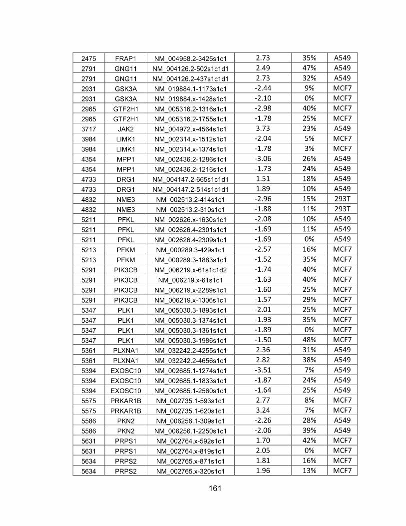
161
2475 FRAP1 NM_004958.2-3425s1c1 2.73 35% A549 2791 GNG11 NM_004126.2-502s1c1d1 2.49 47% A549 2791 GNG11 NM_004126.2-437s1c1d1 2.73 32% A549 2931 GSK3A NM_019884.1-1173s1c1 -2.44 9% MCF7 2931 GSK3A NM_019884.x-1428s1c1 -2.10 0% MCF7 2965 GTF2H1 NM_005316.2-1316s1c1 -2.98 40% MCF7 2965 GTF2H1 NM_005316.2-1755s1c1 -1.78 25% MCF7 3717 JAK2 NM_004972.x-4564s1c1 3.73 23% A549 3984 LIMK1 NM_002314.x-1512s1c1 -2.04 5% MCF7 3984 LIMK1 NM_002314.x-1374s1c1 -1.78 3% MCF7 4354 MPP1 NM_002436.2-1286s1c1 -3.06 26% A549 4354 MPP1 NM_002436.2-1216s1c1 -1.73 24% A549 4733 DRG1 NM_004147.2-665s1c1d1 1.51 18% A549 4733 DRG1 NM_004147.2-514s1c1d1 1.89 10% A549 4832 NME3 NM_002513.2-414s1c1 -2.96 15% 293T 4832 NME3 NM_002513.2-310s1c1 -1.88 11% 293T 5211 PFKL NM_002626.x-1630s1c1 -2.08 10% A549 5211 PFKL NM_002626.4-2301s1c1 -1.69 11% A549 5211 PFKL NM_002626.4-2309s1c1 -1.69 0% A549 5213 PFKM NM_000289.3-429s1c1 -2.57 16% MCF7 5213 PFKM NM_000289.3-1883s1c1 -1.52 35% MCF7 5291 PIK3CB NM_006219.x-61s1c1d2 -1.74 40% MCF7 5291 PIK3CB NM_006219.x-61s1c1 -1.63 40% MCF7 5291 PIK3CB NM_006219.x-2289s1c1 -1.60 25% MCF7 5291 PIK3CB NM_006219.x-1306s1c1 -1.57 29% MCF7 5347 PLK1 NM_005030.3-1893s1c1 -2.01 25% MCF7 5347 PLK1 NM_005030.3-1374s1c1 -1.93 35% MCF7 5347 PLK1 NM_005030.3-1361s1c1 -1.89 0% MCF7 5347 PLK1 NM_005030.3-1986s1c1 -1.50 48% MCF7 5361 PLXNA1 NM_032242.2-4255s1c1 2.36 31% A549 5361 PLXNA1 NM_032242.2-4656s1c1 2.82 38% A549 5394 EXOSC10 NM_002685.1-1274s1c1 -3.51 7% A549 5394 EXOSC10 NM_002685.1-1833s1c1 -1.87 24% A549 5394 EXOSC10 NM_002685.1-2560s1c1 -1.64 25% A549 5575 PRKAR1B NM_002735.1-593s1c1 2.77 8% MCF7 5575 PRKAR1B NM_002735.1-620s1c1 3.24 7% MCF7 5586 PKN2 NM_006256.1-309s1c1 -2.26 28% A549 5586 PKN2 NM_006256.1-2250s1c1 -2.06 39% A549 5631 PRPS1 NM_002764.x-592s1c1 1.70 42% MCF7 5631 PRPS1 NM_002764.x-819s1c1 2.05 0% MCF7 5634 PRPS2 NM_002765.x-871s1c1 1.81 16% MCF7 5634 PRPS2 NM_002765.x-320s1c1 1.96 13% MCF7

162
5832 ALDH18A1 NM_002860.2-658s1c1 -2.59 8% A549 5832 ALDH18A1 NM_002860.2-568s1c1 -2.13 44% A549 5865 RAB3B NM_002867.x-446s1c1d1 -2.98 5% A549 5865 RAB3B NM_002867.x-447s1c1d1 -2.71 8% A549 5865 RAB3B NM_002867.x-907s1c1d1 -1.54 6% A549 5868 RAB5A NM_004162.3-659s1c1d1 -3.94 46% #N/A 5868 RAB5A NM_004162.3-2139s1c1d3 -1.86 16% #N/A 5873 RAB27A NM_004580.3-477s1c1d2 -2.09 25% A549 5873 RAB27A NM_004580.3-735s1c1d2 -1.72 5% A549 5881 RAC3 NM_005052.2-664s1c1d2 -3.31 20% 293T 5881 RAC3 NM_005052.2-331s1c1d2 -2.78 14% 293T 5881 RAC3 NM_005052.2-544s1c1d2 -2.77 17% 293T 6046 BRD2 NM_005104.2-2059s1c1 -2.57 21% 293T 6046 BRD2 NM_005104.2-2401s1c1 -2.12 40% 293T 6729 SRP54 NM_003136.2-1839s1c1d1 -2.80 25% A549 6729 SRP54 NM_003136.2-849s1c1d1 -2.71 15% A549 6729 SRP54 NM_003136.2-1903s1c1d1 -2.61 22% A549 6733 SRPK2 NM_003138.1-749s1c1 -1.79 9% MCF7 6733 SRPK2 NM_003138.1-2730s1c1 -1.61 3% MCF7 6734 SRPR NM_003139.2-202s1c1d1 -4.08 4% A549 6734 SRPR NM_003139.2-1086s1c1d1 -1.86 5% A549 7083 TK1 NM_003258.x-610s1c1 -1.94 11% A549 7083 TK1 NM_003258.x-530s1c1 -1.74 14% A549 7083 TK1 NM_003258.x-327s1c1 -1.51 3% A549 7284 TUFM NM_003321.3-668s1c1d1 -2.68 5% A549 7284 TUFM NM_003321.3-1266s1c1d1 -1.71 2% A549 7297 TYK2 NM_003331.x-3209s1c1 -2.87 13% A549 7297 TYK2 NM_003331.x-4078s1c1 -2.54 15% A549 8476 CDC42BPA NM_014826.x-4292s1c1 1.55 44% A549 8476 CDC42BPA NM_003607.x-2992s1c1 2.33 5% A549 8476 CDC42BPA NM_014826.x-2749s1c1 2.49 3% A549 8476 CDC42BPA NM_003607.x-4535s1c1 2.58 6% A549 8526 DGKE NM_003647.1-853s1c1 -1.70 20% MCF7 8526 DGKE NM_003647.1-952s1c1 -1.54 5% MCF7 8877 SPHK1 NM_182965.1-1063s1c1 -3.09 13% 293T 8877 SPHK1 NM_182965.1-1640s1c1 -2.43 36% 293T 8877 SPHK1 NM_182965.1-1116s1c1 -1.68 21% 293T 9060 PAPSS2 NM_004670.2-177s1c1 -1.83 11% MCF7 9060 PAPSS2 NM_004670.2-1768s1c1 -1.58 7% MCF7 9064 MAP3K6 NM_004672.x-3795s1c1 -2.40 24% A549 9567 GTPBP1 NM_004286.3-884s1c1d2 -4.45 23% 293T 9567 GTPBP1 NM_004286.3-1015s1c1d2 -1.90 31% 293T

163
10000 AKT3 NM_005465.x-770s1c1 -3.04 33% A549 10000 AKT3 NM_005465.x-1757s1c1 -1.83 47% A549 10000 AKT3 NM_005465.x-569s1c1 -1.82 21% A549 10154 PLXNC1 NM_005761.1-1236s1c1 -2.26 32% Jurkat 10154 PLXNC1 NM_005761.1-3289s1c1 -2.25 31% Jurkat 10519 CIB1 NM_006384.2-504s1c1 -2.80 7% A549 10519 CIB1 NM_006384.2-376s1c1 -2.04 31% A549 10783 NEK6 NM_014397.x-445s1c1 -2.40 3% MCF7 10783 NEK6 NM_014397.x-484s1c1 -2.11 22% MCF7 10966 RAB40B NM_006822.1-513s1c1d2 -3.48 40% A549 10966 RAB40B NM_006822.1-354s1c1d1 -2.30 7% A549 11021 RAB35 NM_006861.4-709s1c1d2 -3.93 37% A549 11021 RAB35 NM_006861.4-486s1c1d2 -3.27 27% A549 11021 RAB35 NM_006861.4-207s1c1d2 -2.58 19% A549 11321 XAB1 NM_007266.1-835s1c1d2 2.33 4% A549 11321 XAB1 NM_007266.1-220s1c1d2 2.73 18% A549 11344 TWF2 NM_007284.3-522s1c1 -2.09 22% A549 11344 TWF2 NM_007284.3-610s1c1 -1.50 9% A549 25837 RAB26 NM_014353.4-721s1c1d2 -4.06 40% A549 25837 RAB26 NM_014353.4-512s1c1d2 -2.21 48% A549 26007 DAK NM_015533.2-1015s1c1 -1.70 10% A549 26007 DAK NM_015533.2-1597s1c1 -1.51 0% A549 29110 TBK1 NM_013254.x-1212s1c1 -2.46 35% A549 29110 TBK1 NM_013254.x-1536s1c1 -1.75 33% A549 51128 SAR1B NM_016103.2-267s1c1d2 -3.50 9% A549 51128 SAR1B NM_016103.2-569s1c1d2 -1.95 8% A549 51135 IRAK4 NM_016123.x-820s1c1 1.53 10% A549 51135 IRAK4 NM_016123.x-374s1c1 1.88 29% A549 51678 MPP6 NM_016447.x-528s1c1 -1.89 14% A549 51678 MPP6 NM_016447.x-527s1c1 -1.57 27% A549 55288 RHOT1 NM_018307.2-169s1c1d2 -3.02 23% Jurkat 55288 RHOT1 NM_018307.2-900s1c1d2 -2.05 17% Jurkat 55312 RFK NM_018339.3-731s1c1 1.71 10% A549 55312 RFK NM_018339.3-550s1c1 1.87 5% A549 55312 RFK NM_018339.3-595s1c1 3.05 3% A549 55577 NAGK NM_017567.1-999s1c1 2.05 34% A549 55577 NAGK NM_017567.1-621s1c1 3.09 10% A549 55750 AGK NM_018238.2-1634s1c1 2.07 35% Jurkat 55750 AGK NM_018238.2-457s1c1 2.72 47% Jurkat 55970 GNG12 NM_018841.2-272s1c1d1 1.79 32% A549 55970 GNG12 NM_018841.2-238s1c1d1 2.21 4% A549 56681 SAR1A NM_020150.3-398s1c1d3 -2.29 34% A549
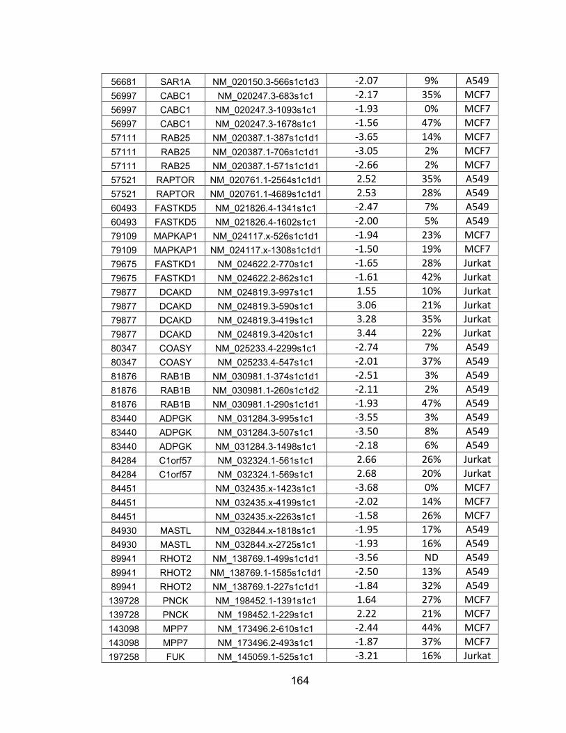
164
56681 SAR1A NM_020150.3-566s1c1d3 -2.07 9% A549 56997 CABC1 NM_020247.3-683s1c1 -2.17 35% MCF7 56997 CABC1 NM_020247.3-1093s1c1 -1.93 0% MCF7 56997 CABC1 NM_020247.3-1678s1c1 -1.56 47% MCF7 57111 RAB25 NM_020387.1-387s1c1d1 -3.65 14% MCF7 57111 RAB25 NM_020387.1-706s1c1d1 -3.05 2% MCF7 57111 RAB25 NM_020387.1-571s1c1d1 -2.66 2% MCF7 57521 RAPTOR NM_020761.1-2564s1c1d1 2.52 35% A549 57521 RAPTOR NM_020761.1-4689s1c1d1 2.53 28% A549 60493 FASTKD5 NM_021826.4-1341s1c1 -2.47 7% A549 60493 FASTKD5 NM_021826.4-1602s1c1 -2.00 5% A549 79109 MAPKAP1 NM_024117.x-526s1c1d1 -1.94 23% MCF7 79109 MAPKAP1 NM_024117.x-1308s1c1d1 -1.50 19% MCF7 79675 FASTKD1 NM_024622.2-770s1c1 -1.65 28% Jurkat 79675 FASTKD1 NM_024622.2-862s1c1 -1.61 42% Jurkat 79877 DCAKD NM_024819.3-997s1c1 1.55 10% Jurkat 79877 DCAKD NM_024819.3-590s1c1 3.06 21% Jurkat 79877 DCAKD NM_024819.3-419s1c1 3.28 35% Jurkat 79877 DCAKD NM_024819.3-420s1c1 3.44 22% Jurkat 80347 COASY NM_025233.4-2299s1c1 -2.74 7% A549 80347 COASY NM_025233.4-547s1c1 -2.01 37% A549 81876 RAB1B NM_030981.1-374s1c1d1 -2.51 3% A549 81876 RAB1B NM_030981.1-260s1c1d2 -2.11 2% A549 81876 RAB1B NM_030981.1-290s1c1d1 -1.93 47% A549 83440 ADPGK NM_031284.3-995s1c1 -3.55 3% A549 83440 ADPGK NM_031284.3-507s1c1 -3.50 8% A549 83440 ADPGK NM_031284.3-1498s1c1 -2.18 6% A549 84284 C1orf57 NM_032324.1-561s1c1 2.66 26% Jurkat 84284 C1orf57 NM_032324.1-569s1c1 2.68 20% Jurkat 84451 NM_032435.x-1423s1c1 -3.68 0% MCF7 84451 NM_032435.x-4199s1c1 -2.02 14% MCF7 84451 NM_032435.x-2263s1c1 -1.58 26% MCF7 84930 MASTL NM_032844.x-1818s1c1 -1.95 17% A549 84930 MASTL NM_032844.x-2725s1c1 -1.93 16% A549 89941 RHOT2 NM_138769.1-499s1c1d1 -3.56 ND A549 89941 RHOT2 NM_138769.1-1585s1c1d1 -2.50 13% A549 89941 RHOT2 NM_138769.1-227s1c1d1 -1.84 32% A549 139728 PNCK NM_198452.1-1391s1c1 1.64 27% MCF7 139728 PNCK NM_198452.1-229s1c1 2.22 21% MCF7 143098 MPP7 NM_173496.2-610s1c1 -2.44 44% MCF7 143098 MPP7 NM_173496.2-493s1c1 -1.87 37% MCF7 197258 FUK NM_145059.1-525s1c1 -3.21 16% Jurkat

165
197258 FUK NM_145059.1-1966s1c1 -2.54 23% Jurkat 197258 FUK NM_145059.1-3135s1c1 -2.34 9% Jurkat 204851 HIPK1 NM_152696.3-2649s1c1 -1.78 29% MCF7 260425 MAGI3 NM_020965.2-2984s1c1 -3.05 15% A549 260425 MAGI3 NM_020965.2-1964s1c1 -2.08 8% A549 282974 STK32C NM_173575.2-311s1c1 -2.72 16% A549 282974 STK32C NM_173575.2-839s1c1 -2.04 29% A549 282974 STK32C NM_173575.2-1386s1c1 -1.93 21% A549 282974 STK32C NM_173575.2-1898s1c1 -1.84 23% A549 326624 RAB37 NM_175738.2-711s1c1d2 -3.07 36% Jurkat 326624 RAB37 NM_175738.2-1059s1c1d2 -2.67 36% Jurkat

166
APPENDIX D: 48 hit genes from Table S4 that are not known to be associated with PI3K/AKT signaling whose expression is not tissue-specific. nd, no data.
NCBI ID SYMBOL Expressed widely? Result of KD? 5575 PRKAR1B Yes ↑ phospho-AKT 6011 GRK1 Yes ↑ phospho-AKT 9943 OXSR1 Yes ↑ phospho-AKT 55312 RFK Yes ↑ phospho-AKT 79877 DCAKD Yes ↑ phospho-AKT 1163 CKS1B nd ↓ phospho-AKT 3706 ITPKA Yes ↓ phospho-AKT 5394 EXOSC10 Yes ↓ phospho-AKT 5586 PKN2 Yes ↓ phospho-AKT 5881 RAC3 Yes ↓ phospho-AKT 6729 SRP54 Yes ↓ phospho-AKT 6872 TAF1 Yes ↓ phospho-AKT 7083 TK1 Yes ↓ phospho-AKT 9064 MAP3K6 nd ↓ phospho-AKT 11021 RAB35 Yes ↓ phospho-AKT 11158 RABL2B nd ↓ phospho-AKT 11159 RABL2A nd ↓ phospho-AKT 26164 GTPBP5 Yes ↓ phospho-AKT 27289 RND1 nd ↓ phospho-AKT 54676 GTPBP2 nd ↓ phospho-AKT 54734 RAB39 nd ↓ phospho-AKT 56997 CABC1 nd ↓ phospho-AKT 57538 ALPK3 Yes ↓ phospho-AKT 57551 TAOK1 Yes ↓ phospho-AKT 64122 FN3K Yes ↓ phospho-AKT 64792 RABL5 Yes ↓ phospho-AKT 79646 PANK3 Yes ↓ phospho-AKT 80216 ALPK1 Yes ↓ phospho-AKT 81876 RAB1B Yes ↓ phospho-AKT 83440 ADPGK Yes ↓ phospho-AKT 83903 GSG2 Yes ↓ phospho-AKT 84100 ARL6 Yes ↓ phospho-AKT 84197 POMK Yes ↓ phospho-AKT 89941 RHOT2 Yes ↓ phospho-AKT 91461 PKDCC Yes ↓ phospho-AKT 127933 UHMK1 Yes ↓ phospho-AKT 130106 CIB4 Yes ↓ phospho-AKT 158158 RASEF Yes ↓ phospho-AKT 197258 FUK Yes ↓ phospho-AKT 203447 NRK low ↓ phospho-AKT 282974 STK32C Yes ↓ phospho-AKT

167
283629 TSSK4 Yes ↓ phospho-AKT 284086 NEK8 Yes ↓ phospho-AKT 285282 RABL3 Yes ↓ phospho-AKT 347736 TXNDC6 Yes ↓ phospho-AKT 374872 C19orf35 nd ↓ phospho-AKT 402679 MARK2P10 nd ↓ phospho-AKT 440345 NPIPB4 nd ↓ phospho-AKT

168
BIBLIOGRAPHY
1. Laplante, M. and D.M. Sabatini, mTOR signaling in growth control and disease. Cell, 2012. 149: p. 274-293.
2. Viglietto, G., et al., Contribution of PKB/AKT signaling to thyroid cancer. Front Biosci (Landmark Ed), 2011. 16: p. 1461-87.
3. Wheeler, D.B., A.E. Carpenter, and D.M. Sabatini, Cell microarrays and RNA interference chip away at gene function. Nat Genet, 2005. 37 Suppl: p. S25-30.
4. Wheeler, D.B., et al., RNAi living-cell microarrays for loss-of-function screens in Drosophila melanogaster cells. Nat Methods, 2004. 1(2): p. 127-32.
5. Ruderman, N.B., et al., Activation of phosphatidylinositol 3-kinase by insulin. Proc Natl Acad Sci U S A, 1990. 87: p. 1411-1415.
6. Cantley, L.C., et al., Oncogenes and signal transduction. Cell, 1991: p. 281-302.
7. Soltoff, S.P., et al., Phosphatidylinositol-3 kinase and growth regulation. Cold Spring Harb Symp Quant Biol, 1992. 57: p. 75-80.
8. Auger, K.R., et al., Polyoma virus middle T antigen-pp60c-src complex associates with purified phosphatidylinositol 3-kinase in vitro. J Biol Chem, 1992. 267: p. 5408-5415.
9. Auger, K.R., et al., Phosphatidylinositol 3-kinase and its novel product, phosphatidylinositol 3-phosphate, are present in Saccharomyces cerevisiae. J Biol Chem, 1989. 264: p. 20181-20184.
10. Whitman, M., et al., Type I phosphotidylinositol kinase makes a novel inositol phospholipid. Nature, 1988. 332: p. 644-646.
11. Auger, K.R., et al., PDGF-dependent tyrosine phosphorylation stimulates production of novel polyphosphoinositides in intact cells. Cell, 1989. 57: p. 167-175.
12. Vanhaesebroeck, B., et al., Phosphoinositide 3-kinases: a conserved family of signal transducers. Trends Biochem Sci, 1997. 22(7): p. 267-72.
13. Wymann, M.P. and L. Pirola, Structure and function of phosphoinositide 3-kinases. Biochimica et biophysica acta, 1998. 1436: p. 127-50.
14. Leevers, S.J., B. Vanhaesebroeck, and M.D. Waterfield, Signalling through phosphoinositide 3-kinases: the lipids take centre stage. Curr Opin Cell Biol, 1999. 11(2): p. 219-25.
15. Knight, Z.A., et al., A pharmacological map of the PI3-K family defines a role for p110alpha in insulin signaling. Cell, 2006. 125: p. 733-47.
16. Okkenhaug, K. and B. Vanhaesebroeck, New responsibilities for the PI3K regulatory subunit p85 alpha. Sci STKE, 2001. 2001(65): p. pe1.
17. Klippel, A., et al., The interaction of small domains between the subunits of phosphatidylinositol 3-kinase determines enzyme activity. Mol Cell Biol, 1994. 14: p. 2675-2685.
18. Karlsson, T., et al., Molecular interactions of the Src homology 2 domain protein Shb with phosphotyrosine residues, tyrosine kinase

169
receptors and Src homology 3 domain proteins. Oncogene, 1995. 10: p. 1475-1483.
19. White, M.F., The IRS-signaling system: a network of docking proteins that mediate insulin and cytokine action. Recent Prog Horm Res, 1998. 53: p. 119-38.
20. Ueki, K., et al., Molecular balance between the regulatory and catalytic subunits of phosphoinositide 3-kinase regulates cell signaling and survival. Mol Cell Biol, 2002. 22: p. 965-977.
21. Mauvais-Jarvis, F., et al., Reduced expression of the murine p85alpha subunit of phosphoinositide 3-kinase improves insulin signaling and ameliorates diabetes. J Clin Invest, 2002. 109: p. 141-149.
22. Ueki, K., et al., Positive and negative roles of p85 alpha and p85 beta regulatory subunits of phosphoinositide 3-kinase in insulin signaling. J Biol Chem, 2003. 278: p. 48453-48466.
23. Taniguchi, C.M., et al., Phosphoinositide 3-kinase regulatory subunit p85alpha suppresses insulin action via positive regulation of PTEN. Proc Natl Acad Sci U S A, 2006. 103: p. 12093-12097.
24. Stoyanov, B., et al., Cloning and characterization of a G protein-activated human phosphoinositide-3 kinase. Science, 1995. 269: p. 690-693.
25. Hooshmand-Rad, R., et al., The PI 3-kinase isoforms p110(alpha) and p110(beta) have differential roles in PDGF- and insulin-mediated signaling. J Cell Sci, 2000. 113 Pt 2: p. 207-14.
26. Arcaro, A., et al., Two distinct phosphoinositide 3-kinases mediate polypeptide growth factor-stimulated PKB activation. EMBO J, 2002. 21(19): p. 5097-108.
27. Jia, S., et al., Essential roles of PI(3)K-p110beta in cell growth, metabolism and tumorigenesis. Nature, 2008. 454: p. 776-9.
28. Jiang, X., et al., Phosphoinositide 3-kinase pathway activation in phosphate and tensin homolog (PTEN)-deficient prostate cancer cells is independent of receptor tyrosine kinases and mediated by the p110beta and p110delta catalytic subunits. J Biol Chem, 2010. 285(20): p. 14980-9.
29. Foukas, L.C., et al., Activity of any class IA PI3K isoform can sustain cell proliferation and survival. Proc Natl Acad Sci U S A, 2010. 107(25): p. 11381-6.
30. Berenjeno, I.M., et al., Both p110alpha and p110beta isoforms of PI3K can modulate the impact of loss-of-function of the PTEN tumour suppressor. Biochem J, 2012. 442(1): p. 151-9.
31. Kang, S., et al., Oncogenic transformation induced by the p110beta, -gamma, and -delta isoforms of class I phosphoinositide 3-kinase. Proc Natl Acad Sci U S A, 2006. 103(5): p. 1289-94.
32. Vanhaesebroeck, B., et al., P110delta, a novel phosphoinositide 3-kinase in leukocytes. Proc Natl Acad Sci U S A, 1997. 94: p. 4330-4335.

170
33. Aksoy, E., et al., The p110delta isoform of the kinase PI(3)K controls the subcellular compartmentalization of TLR4 signaling and protects from endotoxic shock. Nat Immunol, 2012. 13(11): p. 1045-54.
34. Okkenhaug, K. and B. Vanhaesebroeck, PI3K in lymphocyte development, differentiation and activation. Nat Rev Immunol, 2003. 3(4): p. 317-30.
35. Okkenhaug, K. and B. Vanhaesebroeck, PI3K-signalling in B- and T-cells: insights from gene-targeted mice. Biochem Soc Trans, 2003. 31(Pt 1): p. 270-4.
36. Okkenhaug, K., et al., Impaired B and T cell antigen receptor signaling in p110delta PI 3-kinase mutant mice. Science, 2002. 297(5583): p. 1031-4.
37. Maxwell, M.J., et al., Attenuation of phosphoinositide 3-kinase delta signaling restrains autoimmune disease. J Autoimmun, 2012. 38(4): p. 381-91.
38. Yamany, T., et al., Erythema multiforme-like reaction with mucosal involvement following administration of idelalisib for relapse of chronic lymphocytic leukemia. Leuk Lymphoma, 2014: p. 1-2.
39. Chung, C. and R. Lee, Ibrutinib, obinutuzumab, idelalisib, and beyond: review of novel and evolving therapies for chronic lymphocytic leukemia. Pharmacotherapy, 2014. 34(12): p. 1298-316.
40. Brown, J.R., Idelalisib for chronic lymphocytic leukemia. Clin Adv Hematol Oncol, 2014. 12(12): p. 846-8.
41. Khan, M., et al., Idelalisib for the treatment of chronic lymphocytic leukemia. ISRN Oncol, 2014. 2014: p. 931858.
42. Brown, J.R., et al., Idelalisib, an inhibitor of phosphatidylinositol 3-kinase p110delta, for relapsed/refractory chronic lymphocytic leukemia. Blood, 2014. 123(22): p. 3390-7.
43. Stephens, L., et al., A heterotrimeric GTPase-regulated isoform of PI3K and the regulation of its potential effectors. Philosophical transactions of the Royal Society of London. Series B, Biological sciences, 1996. 351: p. 211-5.
44. Lopez-Ilasaca, M., et al., Linkage of G protein-coupled receptors to the MAPK signaling pathway through PI 3-kinase gamma. Science, 1997. 275: p. 394-397.
45. Stephens, L.R., et al., The G beta gamma sensitivity of a PI3K is dependent upon a tightly associated adaptor, p101. Cell, 1997. 89(1): p. 105-14.
46. Voigt, P., M.B. Dorner, and M. Schaefer, Characterization of p87PIKAP, a novel regulatory subunit of phosphoinositide 3-kinase gamma that is highly expressed in heart and interacts with PDE3B. J Biol Chem, 2006. 281(15): p. 9977-86.
47. Brock, C., et al., Roles of G beta gamma in membrane recruitment and activation of p110 gamma/p101 phosphoinositide 3-kinase gamma. J Cell Biol, 2003. 160(1): p. 89-99.

171
48. Czupalla, C., et al., Identification and characterization of the autophosphorylation sites of phosphoinositide 3-kinase isoforms beta and gamma. J Biol Chem, 2003. 278(13): p. 11536-45.
49. Steck, P.A., et al., Identification of a candidate tumour suppressor gene, MMAC1, at chromosome 10q23.3 that is mutated in multiple advanced cancers. Nat Genet, 1997. 15(4): p. 356-62.
50. Tashiro, H., et al., Mutations in PTEN are frequent in endometrial carcinoma but rare in other common gynecological malignancies. Cancer Res, 1997. 57(18): p. 3935-40.
51. Rasheed, B.K., et al., PTEN gene mutations are seen in high-grade but not in low-grade gliomas. Cancer Res, 1997. 57(19): p. 4187-90.
52. Wang, S.I., et al., Somatic mutations of PTEN in glioblastoma multiforme. Cancer Res, 1997. 57(19): p. 4183-6.
53. Li, J., et al., PTEN, a putative protein tyrosine phosphatase gene mutated in human brain, breast, and prostate cancer. Science, 1997. 275(5308): p. 1943-7.
54. Li, J., et al., The PTEN/MMAC1 tumor suppressor induces cell death that is rescued by the AKT/protein kinase B oncogene. Cancer Res, 1998. 58(24): p. 5667-72.
55. Simpson, L. and R. Parsons, PTEN: life as a tumor suppressor. Exp Cell Res, 2001. 264(1): p. 29-41.
56. Sulis, M.L. and R. Parsons, PTEN: from pathology to biology. Trends Cell Biol, 2003. 13(9): p. 478-83.
57. Parsons, R. and L. Simpson, PTEN and cancer. Methods Mol Biol, 2003. 222: p. 147-66.
58. Parsons, R., Human cancer, PTEN and the PI-3 kinase pathway. Semin Cell Dev Biol, 2004. 15(2): p. 171-6.
59. Gericke, A., M. Munson, and A.H. Ross, Regulation of the PTEN phosphatase. Gene, 2006. 374: p. 1-9.
60. Georgescu, M.M., et al., Stabilization and productive positioning roles of the C2 domain of PTEN tumor suppressor. Cancer Res, 2000. 60(24): p. 7033-8.
61. Wisniewski, D., et al., A novel SH2-containing phosphatidylinositol 3,4,5-trisphosphate 5-phosphatase (SHIP2) is constitutively tyrosine phosphorylated and associated with src homologous and collagen gene (SHC) in chronic myelogenous leukemia progenitor cells. Blood, 1999. 93(8): p. 2707-20.
62. Hejna, J.A., et al., Cloning and characterization of a human cDNA (INPPL1) sharing homology with inositol polyphosphate phosphatases. Genomics, 1995. 29(1): p. 285-7.
63. Pesesse, X., et al., The SH2 domain containing inositol 5-phosphatase SHIP2 displays phosphatidylinositol 3,4,5-trisphosphate and inositol 1,3,4,5-tetrakisphosphate 5-phosphatase activity. FEBS Lett, 1998. 437(3): p. 301-3.
172
64. Habib, T., et al., Growth factors and insulin stimulate tyrosine phosphorylation of the 51C/SHIP2 protein. J Biol Chem, 1998. 273(29): p. 18605-9.
65. Bruyns, C., et al., The two SH2-domain-containing inositol 5-phosphatases SHIP1 and SHIP2 are coexpressed in human T lymphocytes. Biol Chem, 1999. 380(7-8): p. 969-74.
66. Blero, D., et al., The SH2 domain containing inositol 5-phosphatase SHIP2 controls phosphatidylinositol 3,4,5-trisphosphate levels in CHO-IR cells stimulated by insulin. Biochem Biophys Res Commun, 2001. 282(3): p. 839-43.
67. Pesesse, X., et al., The Src homology 2 domain containing inositol 5-phosphatase SHIP2 is recruited to the epidermal growth factor (EGF) receptor and dephosphorylates phosphatidylinositol 3,4,5-trisphosphate in EGF-stimulated COS-7 cells. J Biol Chem, 2001. 276(30): p. 28348-55.
68. Clement, S., et al., The lipid phosphatase SHIP2 controls insulin sensitivity. Nature, 2001. 409(6816): p. 92-7.
69. Leahey, A.M., L.R. Charnas, and R.L. Nussbaum, Nonsense mutations in the OCRL-1 gene in patients with the oculocerebrorenal syndrome of Lowe. Hum Mol Genet, 1993. 2(4): p. 461-3.
70. Ungewickell, A., et al., The inositol polyphosphate 5-phosphatase Ocrl associates with endosomes that are partially coated with clathrin. Proc Natl Acad Sci U S A, 2004. 101(37): p. 13501-6.
71. Razidlo, G.L., D. Katafiasz, and G.S. Taylor, Myotubularin regulates Akt-dependent survival signaling via phosphatidylinositol 3-phosphate. The Journal of biological chemistry, 2011. 286: p. 20005-19.
72. Karnoub, A.E. and R.A. Weinberg, Ras oncogenes: split personalities. Nat Rev Mol Cell Biol, 2008. 9(7): p. 517-31.
73. Marengere, L.E., et al., SH2 domain specificity and activity modified by a single residue. Nature, 1994. 369: p. 502-505.
74. Sakaue, M., D. Bowtell, and M. Kasuga, A dominant-negative mutant of mSOS1 inhibits insulin-induced Ras activation and reveals Ras-dependent and -independent insulin signaling pathways. Mol Cell Biol, 1995. 15: p. 379-388.
75. Wood, K.W., et al., ras mediates nerve growth factor receptor modulation of three signal-transducing protein kinases: MAP kinase, Raf-1, and RSK. Cell, 1992. 68(6): p. 1041-50.
76. Avruch, J., X.F. Zhang, and J.M. Kyriakis, Raf meets Ras: completing the framework of a signal transduction pathway. Trends Biochem Sci, 1994. 19(7): p. 279-83.
77. Macdonald, S.G., et al., Reconstitution of the Raf-1-MEK-ERK signal transduction pathway in vitro. Mol Cell Biol, 1993. 13(11): p. 6615-20.
78. Crews, C.M., A.A. Alessandrini, and R.L. Erikson, Mouse Erk-1 gene product is a serine/threonine protein kinase that has the potential to phosphorylate tyrosine. Proc Natl Acad Sci U S A, 1991. 88: p. 8845-8849.

173
79. Crews, C.M., A. Alessandrini, and R.L. Erikson, Erks: their fifteen minutes has arrived. Cell Growth Differ, 1992. 3: p. 135-142.
80. Crews, C.M., A. Alessandrini, and R.L. Erikson, The primary structure of MEK, a protein kinase that phosphorylates the ERK gene product. Science, 1992. 258(5081): p. 478-80.
81. Marais, R., J. Wynne, and R. Treisman, The SRF accessory protein Elk-1 contains a growth factor-regulated transcriptional activation domain. Cell, 1993. 73(2): p. 381-93.
82. Cruzalegui, F.H., E. Cano, and R. Treisman, ERK activation induces phosphorylation of Elk-1 at multiple S/T-P motifs to high stoichiometry. Oncogene, 1999. 18(56): p. 7948-57.
83. Grove, J.R., et al., Regulation of an epitope-tagged recombinant Rsk-1 S6 kinase by phorbol ester and erk/MAP kinase. Biochemistry, 1993. 32: p. 7727-7738.
84. Dalby, K.N., et al., Identification of regulatory phosphorylation sites in mitogen-activated protein kinase (MAPK)-activated protein kinase-1a/p90rsk that are inducible by MAPK. J Biol Chem, 1998. 273(3): p. 1496-505.
85. Deak, M., et al., Mitogen- and stress-activated protein kinase-1 (MSK1) is directly activated by MAPK and SAPK2/p38, and may mediate activation of CREB. EMBO J, 1998. 17(15): p. 4426-41.
86. Guan, K.L., et al., Negative regulation of the serine/threonine kinase B-Raf by Akt. J Biol Chem, 2000. 275(35): p. 27354-9.
87. Chen, B., et al., BRAFV600E negatively regulates the AKT pathway in melanoma cell lines. PLoS One, 2012. 7(8): p. e42598.
88. Hayashi, H., et al., Down-regulation of the PI3-kinase/Akt pathway by ERK MAP kinase in growth factor signaling. Genes Cells, 2008. 13(9): p. 941-7.
89. Naegele, S. and S.J. Morley, Molecular cross-talk between MEK1/2 and mTOR signaling during recovery of 293 cells from hypertonic stress. J Biol Chem, 2004. 279: p. 46023-46034.
90. Ma, L., et al., Phosphorylation and functional inactivation of TSC2 by Erk implications for tuberous sclerosis and cancer pathogenesis. Cell, 2005. 121: p. 179-193.
91. Arvisais, E.W., et al., AKT-independent phosphorylation of TSC2 and activation of mTOR and ribosomal protein S6 kinase signaling by prostaglandin F2alpha. J Biol Chem, 2006. 281: p. 26904-26913.
92. Ma, L., et al., Identification of S664 TSC2 phosphorylation as a marker for extracellular signal-regulated kinase mediated mTOR activation in tuberous sclerosis and human cancer. Cancer Res, 2007. 67: p. 7106-7112.
93. Roux, P.P., et al., Tumor-promoting phorbol esters and activated Ras inactivate the tuberous sclerosis tumor suppressor complex via p90 ribosomal S6 kinase. Proc Natl Acad Sci U S A, 2004. 101: p. 13489-13494.

174
94. Rodriguez-Viciana, P., et al., Phosphatidylinositol-3-OH kinase as a direct target of Ras. Nature, 1994. 370: p. 527-532.
95. Kodaki, T., et al., The activation of phosphatidylinositol 3-kinase by Ras. Curr Biol, 1994. 4: p. 798-806.
96. Downward, J., KSR: a novel player in the RAS pathway [comment]. Cell, 1995. 83: p. 831-834.
97. Rodriguez-Viciana, P., et al., Activation of phosphoinositide 3-kinase by interaction with Ras and by point mutation. EMBO J, 1996. 15: p. 2442-2451.
98. Rodriguez-Viciana, P., et al., Phosphatidylinositol 3' kinase: one of the effectors of Ras. Philos Trans R Soc Lond B Biol Sci, 1996. 351: p. 222-225.
99. Rubio, I., et al., Interaction of Ras with phosphoinositide 3-kinase gamma. Biochem J, 1997. 326: p. 891-895.
100. Rodriguez-Viciana, P., et al., Role of phosphoinositide 3-OH kinase in cell transformation and control of the actin cytoskeleton by Ras. Cell, 1997. 89: p. 457-467.
101. Gupta, S., et al., Binding of ras to phosphoinositide 3-kinase p110alpha is required for ras-driven tumorigenesis in mice. Cell, 2007. 129: p. 957-68.
102. Alessi, D.R., et al., 3-Phosphoinositide-dependent protein kinase-1 (PDK1): structural and functional homology with the Drosophila DSTPK61 kinase. Curr Biol, 1997. 7: p. 776-789.
103. Alessi, D.R., et al., Characterization of a 3-phosphoinositide-dependent protein kinase which phosphorylates and activates protein kinase Balpha. Curr Biol, 1997. 7: p. 261-269.
104. Cohen, P., D.R. Alessi, and D.A. Cross, PDK1, one of the missing links in insulin signal transduction? FEBS Lett, 1997. 410: p. 3-10.
105. Alessi, D.R., et al., Mechanism of activation of protein kinase B by insulin and IGF-1. EMBO J, 1996. 15: p. 6541-6551.
106. Datta, K., et al., Akt is a direct target of the phosphatidylinositol 3-kinase. Activation by growth factors, v-src and v-Ha-ras, in Sf9 and mammalian cells. J Biol Chem, 1996. 271: p. 30835-30839.
107. Currie, R.A., et al., Role of phosphatidylinositol 3,4,5-trisphosphate in regulating the activity and localization of 3-phosphoinositide-dependent protein kinase-1. Biochem J, 1999. 337 ( Pt 3): p. 575-83.
108. Williams, M.R., et al., The role of 3-phosphoinositide-dependent protein kinase 1 in activating AGC kinases defined in embryonic stem cells. Curr Biol, 2000. 10: p. 439-48.
109. Frodin, M., et al., A phosphoserine-regulated docking site in the protein kinase RSK2 that recruits and activates PDK1. EMBO J, 2000. 19(12): p. 2924-34.
110. Jensen, C.J., et al., 90-kDa ribosomal S6 kinase is phosphorylated and activated by 3-phosphoinositide-dependent protein kinase-1. J Biol Chem, 1999. 274: p. 27168-76.
175
111. Richards, S.A., et al., Ribosomal S6 kinase 1 (RSK1) activation requires signals dependent on and independent of the MAP kinase ERK. Curr Biol, 1999. 9: p. 810-20.
112. Staal, S.P., Molecular cloning of the akt oncogene and its human homologues AKT1 and AKT2: amplification of AKT1 in a primary human gastric adenocarcinoma. Proc Natl Acad Sci U S A, 1987. 84: p. 5034-5037.
113. Staal, S.P., et al., The AKT1 proto-oncogene maps to human chromosome 14, band q32. Genomics, 1988. 2: p. 96-98.
114. Staal, S.P. and J.W. Hartley, Thymic lymphoma induction by the AKT8 murine retrovirus. J Exp Med, 1988. 167: p. 1259-1264.
115. Dummler, B. and B.a. Hemmings, Physiological roles of PKB/Akt isoforms in development and disease. Biochemical Society transactions, 2007. 35: p. 231-5.
116. Bellacosa, A., et al., Structure, expression and chromosomal mapping of c-akt: relationship to v-akt and its implications. Oncogene, 1993. 8: p. 745-754.
117. Bellacosa, A., et al., Akt activation by growth factors is a multiple-step process: the role of the PH domain. Oncogene, 1998. 17: p. 313-325.
118. Toker, A. and A.C. Newton, Akt/protein kinase B is regulated by autophosphorylation at the hypothetical PDK-2 site. J Biol Chem, 2000. 275: p. 8271-8274.
119. Sarbassov, D.D., et al., Phosphorylation and regulation of Akt/PKB by the rictor-mTOR complex. Science, 2005. 307: p. 1098-1101.
120. Guertin, D.A. and D.M. Sabatini, Defining the role of mTOR in cancer. Cancer Cell, 2007. 12: p. 9-22.
121. Guertin, D.A., et al., Ablation in mice of the mTORC components raptor, rictor, or mLST8 reveals that mTORC2 is required for signaling to Akt-FOXO and PKCalpha, but not S6K1. Dev Cell, 2006. 11: p. 859-871.
122. Zeigerer, A. and M. Lampson, GLUT4 retention in adipocytes requires two intracellular insulin-regulated transport steps. Molecular biology of …, 2002. 13: p. 2421-2435.
123. Zhou, Q.L., et al., Akt substrate TBC1D1 regulates GLUT1 expression through the mTOR pathway in 3T3-L1 adipocytes. Biochem J, 2008. 411: p. 647-655.
124. Ramm, G., et al., A role for 14-3-3 in insulin-stimulated GLUT4 translocation through its interaction with the RabGAP AS160. J Biol Chem, 2006. 281(39): p. 29174-80.
125. Srivastava, A.K. and S.K. Pandey, Potential mechanism(s) involved in the regulation of glycogen synthesis by insulin. Mol Cell Biochem, 1998. 182(1-2): p. 135-41.
126. Cross, D.A., et al., Inhibition of glycogen synthase kinase-3 by insulin mediated by protein kinase B. Nature, 1995. 378: p. 785-789.
127. Brunet, A., et al., Akt promotes cell survival by phosphorylating and inhibiting a Forkhead transcription factor. Cell, 1999. 96: p. 857-868.

176
128. Greer, E.L. and A. Brunet, FOXO transcription factors in ageing and cancer. Acta Physiol (Oxf), 2008. 192(1): p. 19-28.
129. Greer, E.L. and A. Brunet, FOXO transcription factors at the interface between longevity and tumor suppression. Oncogene, 2005. 24(50): p. 7410-25.
130. Franke, T.F. and L.C. Cantley, Apoptosis. A Bad kinase makes good. Nature, 1997. 390: p. 116-117.
131. Datta, S.R., et al., Akt phosphorylation of BAD couples survival signals to the cell-intrinsic death machinery. Cell, 1997. 91: p. 231-241.
132. Peterson, R.T. and S.L. Schreiber, Kinase phosphorylation: Keeping it all in the family. Curr Biol, 1999. 9: p. R521-4.
133. Pearce, L.R., D. Komander, and D.R. Alessi, The nuts and bolts of AGC protein kinases. Nature reviews. Molecular cell biology, 2010. 11: p. 9-22.
134. Biondi, R.M., et al., The PIF-binding pocket in PDK1 is essential for activation of S6K and SGK, but not PKB. EMBO J, 2001. 20: p. 4380-90.
135. Brunet, A., et al., Protein kinase SGK mediates survival signals by phosphorylating the forkhead transcription factor FKHRL1 (FOXO3a). Mol Cell Biol, 2001. 21: p. 952-965.
136. Vasudevan, K.M., et al., AKT-independent signaling downstream of oncogenic PIK3CA mutations in human cancer. Cancer cell, 2009. 16: p. 21-32.
137. Vezina, C., A. Kudelski, and S.N. Sehgal, Rapamycin (AY-22,989), a new antifungal antibiotic. I. Taxonomy of the producing streptomycete and isolation of the active principle. J Antibiot (Tokyo), 1975. 28: p. 721-726.
138. Sehgal, S.N., H. Baker, and C. Vezina, Rapamycin (AY-22,989), a new antifungal antibiotic. II. Fermentation, isolation and characterization. J Antibiot (Tokyo), 1975. 28: p. 727-732.
139. Baker, H., et al., Rapamycin (AY-22,989), a new antifungal antibiotic. III. In vitro and in vivo evaluation. J Antibiot (Tokyo), 1978. 31: p. 539-545.
140. Singh, K., S. Sun, and C. Vezina, Rapamycin (AY-22,989), a new antifungal antibiotic. IV. Mechanism of action. J Antibiot (Tokyo), 1979. 32: p. 630-645.
141. Martel, R.R., J. Klicius, and S. Galet, Inhibition of the immune response by rapamycin, a new antifungal antibiotic. Can J Physiol Pharmacol, 1977. 55: p. 48-51.
142. Brown, E.J., et al., A mammalian protein targeted by G1-arresting rapamycin-receptor complex. Nature, 1994. 369: p. 756-758.
143. Sabatini, D.M., et al., RAFT1: a mammalian protein that binds to FKBP12 in a rapamycin-dependent fashion and is homologous to yeast TORs. Cell, 1994. 78: p. 35-43.
144. Koser, P.L., et al., The tyrosine89 residue of yeast FKBP12 is required for rapamycin binding. Gene, 1993. 129: p. 159-165.
177
145. Chiu, M.I., H. Katz, and V. Berlin, RAPT1, a mammalian homolog of yeast Tor, interacts with the FKBP12/rapamycin complex. Proc Natl Acad Sci U S A, 1994. 91: p. 12574-12578.
146. Stan, R., et al., Interaction between FKBP12-rapamycin and TOR involves a conserved serine residue. J Biol Chem, 1994. 269: p. 32027-32030.
147. Heitman, J., et al., FK506-binding protein proline isomerase is a target for the immunosuppressant rapamycin. Proc. Natl. Acad. Sci., 1991. 88: p. 1948-1952.
148. Lempiainen, H. and T.D. Halazonetis, Emerging common themes in regulation of PIKKs and PI3Ks. EMBO J, 2009. 28(20): p. 3067-73.
149. Sabatini, D.M., et al., The rapamycin and FKBP12 target (RAFT) displays phosphatidylinositol 4-kinase activity. J Biol Chem, 1995. 270: p. 20875-20878.
150. Kunz, J., et al., Target of rapamycin in yeast, TOR2, is an essential phosphatidylinositol kinase homolog required for G1 progression. Cell, 1993. 73: p. 585-596.
151. Burnett, P.E., et al., RAFT1 phosphorylation of the translational regulators p70 S6 kinase and 4E-BP1. PNAS, 1998. 95: p. 1432-1437.
152. Hara, K., et al., Raptor, a Binding Partner of Target of Rapamycin (TOR), Mediates TOR Action. Cell, 2002. 110: p. 177-89.
153. Kim, D.-H.H., et al., mTOR Interacts with Raptor to Form a Nutrient-Sensitive Complex that Signals to the Cell Growth Machinery. Cell, 2002. 110: p. 163-175.
154. Lee-Fruman, K.K., et al., Characterization of S6K2, a novel kinase homologous to S6K1. Oncogene, 1999. 18: p. 5108-14.
155. Dufner, a. and G. Thomas, Ribosomal S6 kinase signaling and the control of translation. Experimental cell research, 1999. 253: p. 100-9.
156. Khaleghpour, K., et al., Translational homeostasis: eukaryotic translation initiation factor 4E control of 4E-binding protein 1 and p70 S6 kinase activities. Molecular and cellular biology, 1999. 19: p. 4302-10.
157. Iiboshi, Y., et al., Amino acid-dependent control of p70(s6k). Involvement of tRNA aminoacylation in the regulation. J Biol Chem, 1999. 274: p. 1092-9.
158. Fingar, D.C., et al., Mammalian cell size is controlled by mTOR and its downstream targets S6K1 and 4EBP1/eIF4E. Genes Dev, 2002. 16: p. 1472-87.
159. Clohessy, J.G., M. Reschke, and P.P. Pandolfi, Found in translation of mTOR signaling. Cell Res, 2012. 22: p. 1315-1318.
160. Thoreen, C.C., et al., A unifying model for mTORC1-mediated regulation of mRNA translation. Nature, 2012. 485: p. 109-113.
161. Thomas, G. and M.N. Hall, TOR signalling and control of cell growth. Curr Opin Cell Biol, 1997. 9: p. 782-7.
178
162. Ohanna, M., et al., Atrophy of S6K1(-/-) skeletal muscle cells reveals distinct mTOR effectors for cell cycle and size control. Nat Cell Biol, 2005. 7: p. 286-294.
163. Stocker, H., et al., Rheb is an essential regulator of S6K in controlling cell growth in Drosophila. Nat Cell Biol, 2003. 5: p. 559-565.
164. Kozma, S.C. and G. Thomas, Regulation of cell size in growth, development and human disease: PI3K, PKB and S6K. Bioessays, 2002. 24: p. 65-71.
165. Potter, C.J., L.G. Pedraza, and T. Xu, Akt regulates growth by directly phosphorylating Tsc2. Nat Cell Biol, 2002. 4: p. 658-65.
166. Inoki, K., et al., TSC2 is phosphorylated and inhibited by Akt and suppresses mTOR signalling. Nat Cell Biol, 2002. 12: p. 12.
167. Castro, A.F., et al., Rheb binds tuberous sclerosis complex 2 (TSC2) and promotes S6 kinase activation in a rapamycin- and farnesylation-dependent manner. J Biol Chem, 2003. 278: p. 32493-32496.
168. Manning, B.D. and L.C. Cantley, Rheb fills a GAP between TSC and TOR. Trends Biochem Sci, 2003. 28: p. 573-576.
169. Kwiatkowski, D.J., Rhebbing up mTOR: new insights on TSC1 and TSC2, and the pathogenesis of tuberous sclerosis. Cancer Biol Ther, 2003. 2: p. 471-476.
170. Inoki, K., et al., Rheb GTPase is a direct target of TSC2 GAP activity and regulates mTOR signaling. Genes Dev, 2003. 17: p. 1829-1834.
171. Zhang, Y., et al., Rheb is a direct target of the tuberous sclerosis tumour suppressor proteins. Nat Cell Biol, 2003. 5: p. 578-581.
172. Tee, A.R., et al., Tuberous sclerosis complex gene products, Tuberin and Hamartin, control mTOR signaling by acting as a GTPase-activating protein complex toward Rheb. Curr Biol, 2003. 13: p. 1259-1268.
173. Saucedo, L.J., et al., Rheb promotes cell growth as a component of the insulin/TOR signalling network. Nat Cell Biol, 2003. 5: p. 566-571.
174. Garami, A., et al., Insulin activation of Rheb, a mediator of mTOR/S6K/4E-BP signaling, is inhibited by TSC1 and 2. Mol Cell, 2003. 11: p. 1457-1466.
175. Long, X., et al., Rheb binds and regulates the mTOR kinase. Curr Biol, 2005. 15: p. 702-713.
176. Tee, A.R., J. Blenis, and C.G. Proud, Analysis of mTOR signaling by the small G-proteins, Rheb and RhebL1. FEBS Lett, 2005. 579: p. 4763-4768.
177. Thedieck, K., et al., PRAS40 and PRR5-like protein are new mTOR interactors that regulate apoptosis. PloS one, 2007. 2: p. e1217.
178. Sancak, Y., et al., PRAS40 is an insulin-regulated inhibitor of the mTORC1 protein kinase. Mol Cell, 2007. 25: p. 903-915.
179. Oshiro, N., et al., The proline-Rich Akt substrate of 40 kDa (PRAS40) is a physiological substrate of mTOR complex 1. J Biol Chem, 2007.

179
180. Fonseca, B.D., et al., PRAS40 is a target for mammalian target of rapamycin complex 1 and is required for signaling downstream of this complex. The Journal of biological chemistry, 2007. 282: p. 24514-24.
181. Laplante, M. and D.M. Sabatini, mTOR Signaling. Cold Spring Harb Perspect Biol, 2012. 4.
182. Efeyan, A., R. Zoncu, and D.M. Sabatini, Amino acids and mTORC1: from lysosomes to disease. Trends Mol Med, 2012. 18: p. 524-533.
183. Sancak, Y., et al., The Rag GTPases bind raptor and mediate amino acid signaling to mTORC1. Science (New York, N.Y.), 2008. 320: p. 1496-501.
184. Sancak, Y., et al., Ragulator-Rag complex targets mTORC1 to the lysosomal surface and is necessary for its activation by amino acids. Cell, 2010. 141: p. 290-303.
185. Zoncu, R., et al., mTORC1 senses lysosomal amino acids through an inside-out mechanism that requires the vacuolar H(+)-ATPase. Science, 2011. 334: p. 678-683.
186. Bar-Peled, L., et al., A Tumor suppressor complex with GAP activity for the Rag GTPases that signal amino acid sufficiency to mTORC1. Science, 2013. 340(6136): p. 1100-6.
187. Balendran, A., et al., PDK1 acquires PDK2 activity in the presence of a synthetic peptide derived from the carboxyl terminus of PRK2. Curr Biol, 1999. 9: p. 393-404.
188. Chan, T.O. and P.N. Tsichlis, PDK2: a complex tail in one Akt. Sci STKE, 2001. 2001: p. PE1.
189. Hresko, R.C., H. Murata, and M. Mueckler, Phosphoinositide-dependent kinase-2 is a distinct protein kinase enriched in a novel cytoskeletal fraction associated with adipocyte plasma membranes. J Biol Chem, 2003. 278: p. 21615-21622.
190. Partovian, C. and M. Simons, Regulation of protein kinase B/Akt activity and Ser473 phosphorylation by protein kinase Calpha in endothelial cells. Cellular signalling, 2004. 16: p. 951-7.
191. Viniegra, J.G., et al., Full activation of PKB/Akt in response to insulin or ionizing radiation is mediated through ATM. The Journal of biological chemistry, 2005. 280: p. 4029-36.
192. Feng, J., et al., Identification of a PKB/Akt Hydrophobic Motif Ser-473 Kinase as DNA-dependent Protein Kinase. J Biol Chem, 2004. 279: p. 41189-41196.
193. Persad, S., et al., Regulation of protein kinase B/Akt-serine 473 phosphorylation by integrin-linked kinase: critical roles for kinase activity and amino acids arginine 211 and serine 343. J Biol Chem, 2001. 276: p. 27462-27469.
194. Persad, S., et al., Inhibition of integrin-linked kinase (ILK) suppresses activation of protein kinase B/Akt and induces cell cycle arrest and apoptosis of PTEN-mutant prostate cancer cells. Proceedings of the National Academy of Sciences of the United States of America, 2000. 97: p. 3207-12.

180
195. Hresko, R.C. and M. Mueckler, mTOR/RICTOR is the Ser473 kinase for Akt/PKB in 3T3-L1 adipocytes. J Biol Chem, 2005. 280: p. 40406-40416.
196. Sarbassov, D.D., et al., Rictor, a novel binding partner of mTOR, defines a rapamycin-insensitive and raptor-independent pathway that regulates the cytoskeleton. Curr Biol, 2004. 14(14): p. 1296-302.
197. Yan, L., V. Mieulet, and R.F. Lamb, mTORC2 is the hydrophobic motif kinase for SGK1. The Biochemical journal, 2008. 416: p. e19-21.
198. Kim, D.-H.H., et al., GbetaL, a positive regulator of the rapamycin-sensitive pathway required for the nutrient-sensitive interaction between raptor and mTOR. Molecular cell, 2003. 11: p. 895-904.
199. Yang, Q., et al., Identification of Sin1 as an essential TORC2 component required for complex formation and kinase activity. Genes Dev, 2006. 20: p. 2820-2832.
200. Jacinto, E., et al., SIN1/MIP1 maintains rictor-mTOR complex integrity and regulates Akt phosphorylation and substrate specificity. Cell, 2006. 127: p. 125-37.
201. Frias, M.a., et al., mSin1 is necessary for Akt/PKB phosphorylation, and its isoforms define three distinct mTORC2s. Current biology : CB, 2006. 16: p. 1865-70.
202. Lu, M., et al., mSIN1 protein mediates SGK1 protein interaction with mTORC2 protein complex and is required for selective activation of the epithelial sodium channel. J Biol Chem, 2011. 286(35): p. 30647-54.
203. Facchinetti, V., et al., The mammalian target of rapamycin complex 2 controls folding and stability of Akt and protein kinase C. The EMBO journal, 2008. 27: p. 1932-43.
204. Hall, M.N., mTOR-what does it do? Transplant Proc, 2008. 40: p. S5-8. 205. Woo, S.-Y.Y., et al., PRR5, a novel component of mTOR complex 2,
regulates platelet-derived growth factor receptor beta expression and signaling. J Biol Chem, 2007. 282: p. 25604-25612.
206. Pearce, L.R., et al., Identification of Protor as a novel Rictor-binding component of mTOR complex-2. Biochem J, 2007. 405: p. 513-522.
207. Pearce, L.R., et al., Protor-1 is required for efficient mTORC2-mediated activation of SGK1 in the kidney. Biochem J, 2011. 436(1): p. 169-79.
208. Peterson, T.R., et al., DEPTOR is an mTOR inhibitor frequently overexpressed in multiple myeloma cells and required for their survival. Cell, 2009. 137: p. 873-886.
209. Laplante, M., et al., DEPTOR cell-autonomously promotes adipogenesis, and its expression is associated with obesity. Cell Metab, 2012. 16: p. 202-212.
210. Pederson, T.M., D.L. Kramer, and C.M. Rondinone, Serine/threonine phosphorylation of IRS-1 triggers its degradation: possible regulation by tyrosine phosphorylation. Diabetes, 2001. 50(1): p. 24-31.
211. Sun, X.J., et al., Insulin-induced insulin receptor substrate-1 degradation is mediated by the proteasome degradation pathway. Diabetes, 1999. 48: p. 1359-1364.

181
212. Tzatsos, A. and K.V. Kandror, Nutrients suppress phosphatidylinositol 3-kinase/Akt signaling via raptor-dependent mTOR-mediated insulin receptor substrate 1 phosphorylation. Mol Cell Biol, 2006. 26: p. 63-76.
213. Gual, P., et al., MAP kinases and mTOR mediate insulin-induced phosphorylation of insulin receptor substrate-1 on serine residues 307, 612 and 632. Diabetologia, 2003. 46: p. 1532-1542.
214. Carlson, C.J., M.F. White, and C.M. Rondinone, Mammalian target of rapamycin regulates IRS-1 serine 307 phosphorylation. Biochem Biophys Res Commun, 2004. 316: p. 533-539.
215. Hiratani, K., et al., Roles of mTOR and JNK in serine phosphorylation, translocation, and degradation of IRS-1. Biochem Biophys Res Commun, 2005. 335: p. 836-842.
216. Yu, Y., et al., Phosphoproteomic analysis identifies Grb10 as an mTORC1 substrate that negatively regulates insulin signaling. Science, 2011. 332: p. 1322-1326.
217. Yea, S.S. and D.A. Fruman, Cell signaling. New mTOR targets Grb attention. Science, 2011. 332: p. 1270-1271.
218. Yu, Y., S. Yoon, and G. Poulogiannis, Quantitative Phosphoproteomic Analysis Identifies the Adaptor Protein Grb10 as an mTORC1 Substrate that Negatively Regulates Insulin Signaling. Science (New York, …, 2011. 332: p. 1322-1326.
219. O'Reilly, K.E., et al., mTOR inhibition induces upstream receptor tyrosine kinase signaling and activates Akt. Cancer Res, 2006. 66: p. 1500-1508.
220. Easton, J.B., R.T. Kurmasheva, and P.J. Houghton, IRS-1: auditing the effectiveness of mTOR inhibitors. Cancer Cell, 2006. 9: p. 153-155.
221. Sawyers, C.L., Will mTOR inhibitors make it as cancer drugs? Cancer Cell, 2003. 4: p. 343-348.
222. Gera, J.F., et al., AKT activity determines sensitivity to mammalian target of rapamycin (mTOR) inhibitors by regulating cyclin D1 and c-myc expression. J Biol Chem, 2004. 279: p. 2737-2746.
223. Hanahan, D. and R.a. Weinberg, Hallmarks of cancer: the next generation. Cell, 2011. 144: p. 646-74.
224. Bader, A.G., S. Kang, and P.K. Vogt, Cancer-specific mutations in PIK3CA are oncogenic in vivo. Proc Natl Acad Sci U S A, 2006. 103(5): p. 1475-9.
225. Vivanco, I. and C.L. Sawyers, The phosphatidylinositol 3-Kinase AKT pathway in human cancer. Nat Rev Cancer, 2002. 2: p. 489-501.
226. Philp, A.J., et al., The phosphatidylinositol 3'-kinase p85alpha gene is an oncogene in human ovarian and colon tumors. Cancer Res, 2001. 61(20): p. 7426-9.
227. Jimenez, C., et al., Identification and characterization of a new oncogene derived from the regulatory subunit of phosphoinositide 3-kinase. EMBO J, 1998. 17: p. 743-753.
228. Ndubaku, C.O., et al., Discovery of 2-{3-[2-(1-isopropyl-3-methyl-1H-1,2-4-triazol-5-yl)-5,6-dihydrobenzo[f]imidazo[1 ,2-d][1,4]oxazepin-9-yl]-
182
1H-pyrazol-1-yl}-2-methylpropanamide (GDC-0032): a beta-sparing phosphoinositide 3-kinase inhibitor with high unbound exposure and robust in vivo antitumor activity. J Med Chem, 2013. 56(11): p. 4597-610.
229. Mueller, S., et al., PTEN promoter methylation and activation of the PI3K/Akt/mTOR pathway in pediatric gliomas and influence on clinical outcome. Neuro Oncol, 2012. 14: p. 1146-1152.
230. Vlietstra, R.J., et al., Frequent inactivation of PTEN in prostate cancer cell lines and xenografts. Cancer Res, 1998. 58: p. 2720-2723.
231. Liaw, D., et al., Germline mutations of the PTEN gene in Cowden disease, an inherited breast and thyroid cancer syndrome. Nat Genet, 1997. 16(1): p. 64-7.
232. Dahia, P.L., et al., Somatic deletions and mutations in the Cowden disease gene, PTEN, in sporadic thyroid tumors. Cancer Res, 1997. 57(21): p. 4710-3.
233. Trotman, L.C., et al., Pten dose dictates cancer progression in the prostate. PLoS biology, 2003. 1: p. E59.
234. Ma, L., et al., Genetic analysis of Pten and Tsc2 functional interactions in the mouse reveals asymmetrical haploinsufficiency in tumor suppression. Genes Dev, 2005. 19: p. 1779-1786.
235. Hopkins, B.D., et al., A secreted PTEN phosphatase that enters cells to alter signaling and survival. Science, 2013. 341(6144): p. 399-402.
236. Kadota, M., et al., Identification of novel gene amplifications in breast cancer and coexistence of gene amplification with an activating mutation of PIK3CA. Cancer Res, 2009. 69(18): p. 7357-65.
237. Lee, J.C., et al., Epidermal growth factor receptor activation in glioblastoma through novel missense mutations in the extracellular domain. PLoS medicine, 2006. 3: p. e485.
238. Gong, H., et al., A comparative study of affibody, panitumumab, and EGF for near-infrared fluorescence imaging of EGFR- and EGFRvIII-expressing tumors. Cancer Biol Ther, 2013. 15(2).
239. Tanaka, K., et al., Oncogenic EGFR signaling activates an mTORC2-NF-κB pathway that promotes chemotherapy resistance. Cancer discovery, 2011. 1: p. 524-38.
240. Carpten, J.D., et al., A transforming mutation in the pleckstrin homology domain of AKT1 in cancer. Nature, 2007. 448: p. 439-44.
241. Bleeker, F.E., et al., AKT1(E17K) in human solid tumours. Oncogene, 2008. 27: p. 5648-50.
242. Malanga, D., et al., Activating E17K mutation in the gene encoding the protein kinase AKT1 in a subset of squamous cell carcinoma of the lung. Cell Cycle, 2008: p. 665-669.
243. Lauring, J., et al., Knock in of the AKT1 E17K mutation in human breast epithelial cells does not recapitulate oncogenic PIK3CA mutations. Oncogene, 2010. 29(16): p. 2337-45.

183
244. Murugan, A.K., A. Alzahrani, and M. Xing, Mutations in critical domains confer the human mTOR gene strong tumorigenicity. J Biol Chem, 2013. 288(9): p. 6511-21.
245. Brian Grabiner and David M. Sabatini`, personal communication. 246. Leyland-Jones, B., Trastuzumab: hopes and realities. Lancet Oncol,
2002. 3(3): p. 137-44. 247. Hegedus, C., et al., Interaction of nilotinib, dasatinib and bosutinib with
ABCB1 and ABCG2: implications for altered anti-cancer effects and pharmacological properties. Br J Pharmacol, 2009. 158(4): p. 1153-64.
248. Redaelli, S., et al., Activity of bosutinib, dasatinib, and nilotinib against 18 imatinib-resistant BCR/ABL mutants. J Clin Oncol, 2009. 27(3): p. 469-71.
249. Remsing Rix, L.L., et al., Global target profile of the kinase inhibitor bosutinib in primary chronic myeloid leukemia cells. Leukemia, 2009. 23(3): p. 477-85.
250. Perry, C.M. and L.R. Wiseman, Trastuzumab. BioDrugs, 1999. 12(2): p. 129-35.
251. Albanell, J. and J. Baselga, Trastuzumab, a humanized anti-HER2 monoclonal antibody, for the treatment of breast cancer. Drugs Today (Barc), 1999. 35(12): p. 931-46.
252. Gong, S.J., et al., Growth inhibitory effects of trastuzumab and chemotherapeutic drugs in gastric cancer cell lines. Cancer Lett, 2004. 214(2): p. 215-24.
253. Rebischung, C., et al., The effectiveness of trastuzumab (Herceptin) combined with chemotherapy for gastric carcinoma with overexpression of the c-erbB-2 protein. Gastric Cancer, 2005. 8(4): p. 249-52.
254. Nahta, R., M.C. Hung, and F.J. Esteva, The HER-2-targeting antibodies trastuzumab and pertuzumab synergistically inhibit the survival of breast cancer cells. Cancer Res, 2004. 64(7): p. 2343-6.
255. Kawaguchi, Y., et al., Targeting EGFR and HER-2 with cetuximab- and trastuzumab-mediated immunotherapy in oesophageal squamous cell carcinoma. Br J Cancer, 2007. 97(4): p. 494-501.
256. Tyagi, P., Recent results and ongoing trials with panitumumab (ABX-EGF), a fully human anti-epidermal growth factor receptor antibody, in metastatic colorectal cancer. Clin Colorectal Cancer, 2005. 5(1): p. 21-3.
257. Konecny, G.E., et al., Activity of the dual kinase inhibitor lapatinib (GW572016) against HER-2-overexpressing and trastuzumab-treated breast cancer cells. Cancer Res, 2006. 66(3): p. 1630-9.
258. Herbst, R.S., et al., TRIBUTE: a phase III trial of erlotinib hydrochloride (OSI-774) combined with carboplatin and paclitaxel chemotherapy in advanced non-small-cell lung cancer. Journal of clinical oncology : official journal of the American Society of Clinical Oncology, 2005. 23: p. 5892-9.

184
259. Paez, J.G., et al., EGFR mutations in lung cancer: correlation with clinical response to gefitinib therapy. Science (New York, N.Y.), 2004. 304: p. 1497-500.
260. Owonikoko, T.K. and F.R. Khuri, Targeting the PI3K/AKT/mTOR Pathway. Am Soc Clin Oncol Educ Book, 2013: p. 395-401.
261. Furet, P., et al., Discovery of NVP-BYL719 a potent and selective phosphatidylinositol-3 kinase alpha inhibitor selected for clinical evaluation. Bioorg Med Chem Lett, 2013. 23(13): p. 3741-8.
262. Sturgeon, S.A., et al., Advantages of a selective beta-isoform phosphoinositide 3-kinase antagonist, an anti-thrombotic agent devoid of other cardiovascular actions in the rat. Eur J Pharmacol, 2008. 587(1-3): p. 209-15.
263. Martini, M., et al., Targeting PI3K in Cancer: Any Good News? Front Oncol, 2013. 3: p. 108.
264. Hudis, C., et al., A phase 1 study evaluating the combination of an allosteric AKT inhibitor (MK-2206) and trastuzumab in patients with HER2-positive solid tumors. Breast Cancer Res, 2013. 15(6): p. R110.
265. LoPiccolo, J., et al., Targeting the PI3K/Akt/mTOR pathway: effective combinations and clinical considerations. Drug Resist Updat, 2008. 11: p. 32-50.
266. Lin, J., et al., Targeting activated Akt with GDC-0068, a novel selective Akt inhibitor that is efficacious in multiple tumor models. Clin Cancer Res, 2013. 19(7): p. 1760-72.
267. Blake, J.F., et al., Discovery and preclinical pharmacology of a selective ATP-competitive Akt inhibitor (GDC-0068) for the treatment of human tumors. J Med Chem, 2012. 55(18): p. 8110-27.
268. Cho, D.C., et al., Two phase 2 trials of the novel Akt inhibitor perifosine in patients with advanced renal cell carcinoma after progression on vascular endothelial growth factor-targeted therapy. Cancer, 2012. 118(24): p. 6055-62.
269. Yan, Y., et al., Evaluation and Clinical Analyses of Downstream Targets of the Akt Inhibitor GDC-0068. Clin Cancer Res, 2013.
270. Hudes, G., et al., Temsirolimus, interferon alfa, or both for advanced renal-cell carcinoma. The New England journal of medicine, 2007. 356: p. 2271-81.
271. Voss, M.H., A.M. Molina, and R.J. Motzer, mTOR inhibitors in advanced renal cell carcinoma. Hematol Oncol Clin North Am, 2011. 25: p. 835-852.
272. Wheler, J.J., et al., Anastrozole and everolimus in advanced gynecologic and breast malignancies: activity and molecular alterations in the PI3K/AKT/mTOR pathway. Oncotarget, 2014. 5(10): p. 3029-38.
273. Piccart, M., et al., Everolimus plus exemestane for hormone-receptor-positive, human epidermal growth factor receptor-2-negative advanced breast cancer: overall survival results from BOLERO-2dagger. Ann Oncol, 2014. 25(12): p. 2357-62.

185
274. Rugo, H.S., et al., Incidence and time course of everolimus-related adverse events in postmenopausal women with hormone receptor-positive advanced breast cancer: insights from BOLERO-2. Ann Oncol, 2014. 25(4): p. 808-15.
275. Bachelot, T., et al., Comparative efficacy of everolimus plus exemestane versus fulvestrant for hormone-receptor-positive advanced breast cancer following progression/recurrence after endocrine therapy: a network meta-analysis. Breast Cancer Res Treat, 2014. 143(1): p. 125-33.
276. Burgess, M.R. and C.L. Sawyers, Treating imatinib-resistant leukemia: the next generation targeted therapies. TheScientificWorldJournal, 2006. 6: p. 918-30.
277. Furic, L., et al., Targeting mTOR-dependent tumours with specific inhibitors: a model for personalized medicine based on molecular diagnoses. Curr Oncol, 2009. 16: p. 59-61.
278. Carter, T.a., et al., Inhibition of drug-resistant mutants of ABL, KIT, and EGF receptor kinases. Proceedings of the National Academy of Sciences of the United States of America, 2005. 102: p. 11011-6.
279. Young, M.a., et al., Structure of the kinase domain of an imatinib-resistant Abl mutant in complex with the Aurora kinase inhibitor VX-680. Cancer research, 2006. 66: p. 1007-14.
280. Skaggs, B.J., et al., Phosphorylation of the ATP-binding loop directs oncogenicity of drug-resistant BCR-ABL mutants. Proceedings of the National Academy of Sciences of the United States of America, 2006. 103: p. 19466-71.
281. Ercan, D., et al., Reactivation of ERK signaling causes resistance to EGFR kinase inhibitors. Cancer Discov, 2012. 2(10): p. 934-47.
282. Bivona, T.G., et al., FAS and NF-κB signalling modulate dependence of lung cancers on mutant EGFR. Nature, 2011. 471: p. 523-6.
283. Hunter, C.P., Genetics: a touch of elegance with RNAi. Curr Biol, 1999. 9(12): p. R440-2.
284. Sharp, P.A., RNAi and double-strand RNA. Genes Dev, 1999. 13(2): p. 139-41.
285. Murchison, E.P. and G.J. Hannon, miRNAs on the move: miRNA biogenesis and the RNAi machinery. Curr Opin Cell Biol, 2004. 16(3): p. 223-9.
286. Tijsterman, M. and R.H. Plasterk, Dicers at RISC; the mechanism of RNAi. Cell, 2004. 117(1): p. 1-3.
287. Hutvagner, G., Small RNA asymmetry in RNAi: function in RISC assembly and gene regulation. FEBS Lett, 2005. 579(26): p. 5850-7.
288. Filipowicz, W., RNAi: the nuts and bolts of the RISC machine. Cell, 2005. 122(1): p. 17-20.
289. Brummelkamp, T.R., R. Bernards, and R. Agami, A system for stable expression of short interfering RNAs in mammalian cells. Science (New York, N.Y.), 2002. 296: p. 550-3.

186
290. Paddison, P.J., A.A. Caudy, and G.J. Hannon, Stable suppression of gene expression by RNAi in mammalian cells. Proc Natl Acad Sci U S A, 2002. 99(3): p. 1443-8.
291. Stewart, S.A., et al., Lentivirus-delivered stable gene silencing by RNAi in primary cells. RNA, 2003. 9: p. 493-501.
292. Rubinson, D.A., et al., A lentivirus-based system to functionally silence genes in primary mammalian cells, stem cells and transgenic mice by RNA interference. Nat Genet, 2003. 33: p. 401-406.
293. Shi, Y., Mammalian RNAi for the masses. Trends Genet, 2003. 19(1): p. 9-12.
294. Sumimoto, H. and Y. Kawakami, Lentiviral vector-mediated RNAi and its use for cancer research. Future Oncol, 2007. 3(6): p. 655-64.
295. Berns, K., et al., A large-scale RNAi screen in human cells identifies new components of the p53 pathway. Nature, 2004. 428: p. 431-7.
296. Brummelkamp, T.R., et al., Loss of the cylindromatosis tumour suppressor inhibits apoptosis by activating NF-kappaB. Nature, 2003. 424: p. 797-801.
297. Brummelkamp, T.R. and R. Bernards, New tools for functional mammalian cancer genetics. Nat Rev Cancer, 2003. 3: p. 781-789.
298. Friedman, A. and N. Perrimon, A functional RNAi screen for regulators of receptor tyrosine kinase and ERK signalling. Nature, 2006. 444: p. 230-4.
299. Philips, J.a., E.J. Rubin, and N. Perrimon, Drosophila RNAi screen reveals CD36 family member required for mycobacterial infection. Science (New York, N.Y.), 2005. 309: p. 1251-3.
300. Boutros, M., et al., Genome-wide RNAi analysis of growth and viability in Drosophila cells. Science, 2004. 303: p. 832-835.
301. Kiger, A., et al., A functional genomic analysis of cell morphology using RNA interference. J Biol, 2003. 2: p. 27.
302. Moffat, J. and D.M. Sabatini, Building mammalian signalling pathways with RNAi screens. Nat Rev Mol Cell Biol, 2006. 7: p. 177-187.
303. Moffat, J., et al., A lentiviral RNAi library for human and mouse genes applied to an arrayed viral high-content screen. Cell, 2006. 124: p. 1283-1298.
304. Brummelkamp, T.R., et al., An shRNA barcode screen provides insight into cancer cell vulnerability to MDM2 inhibitors. Nature chemical biology, 2006. 2: p. 202-6.
305. Luo, B., et al., Highly parallel identification of essential genes in cancer cells. Proc Natl Acad Sci U S A, 2008. 105: p. 20380-20385.
306. Zender, L., et al., An oncogenomics-based in vivo RNAi screen identifies tumor suppressors in liver cancer. Cell, 2008. 135: p. 852-64.
307. Hannon, G.J. and J.J. Rossi, Unlocking the potential of the human genome with RNA interference. Nature, 2004. 431: p. 371-378.
308. Paddison, P.J., et al., A resource for large-scale RNA-interference-based screens in mammals. Nature, 2004. 428: p. 427-431.

187
309. Mohr, S.E. and N. Perrimon, RNAi screening: new approaches, understandings, and organisms. Wiley Interdiscip Rev RNA, 2012. 3(2): p. 145-58.
310. Possemato, R., et al., Functional genomics reveal that the serine synthesis pathway is essential in breast cancer. Nature, 2011. 476: p. 346-350.
311. Perrimon, N., J.Q. Ni, and L. Perkins, In vivo RNAi: today and tomorrow. Cold Spring Harb Perspect Biol, 2010. 2(8): p. a003640.
312. Lee, A.J., R. Kolesnick, and C. Swanton, RNAi-mediated functional analysis of pathways influencing cancer cell drug resistance. Expert Rev Mol Med, 2009. 11: p. e15.
313. Friedman, A. and N. Perrimon, Genetic screening for signal transduction in the era of network biology. Cell, 2007. 128: p. 225-31.
314. Perrimon, N., et al., Drug-target identification in Drosophila cells: combining high-throughout RNAi and small-molecule screens. Drug discovery today, 2007. 12: p. 28-33.
315. Root, D.E., et al., Genome-scale loss-of-function screening with a lentiviral RNAi library. Nat Methods, 2006. 3: p. 715-719.
316. Shaw, R.J. and L.C. Cantley, Ras, PI(3)K and mTOR signalling controls tumour cell growth. Nature, 2006. 441: p. 424-430.
317. Cantley, L.C., The phosphoinositide 3-kinase pathway. Science, 2002. 296: p. 1655-1657.
318. Katso, R., et al., Cellular function of phosphoinositide 3-kinases: implications for development, homeostasis, and cancer. Annu Rev Cell Dev Biol, 2001. 17: p. 615-75.
319. Hennessy, B.T., et al., Exploiting the PI3K/AKT pathway for cancer drug discovery. Nature reviews. Drug discovery, 2005. 4: p. 988-1004.
320. Zhang, J.-h.H., T.D.Y. Chung, and K.R. Oldenburg, A Simple Statistical Parameter for Use in Evaluation and Validation of High Throughput Screening Assays. J Biomol Screen, 1999. 4: p. 67-73.
321. Gao, J., et al., Integrative Analysis of Complex Cancer Genomics and Clinical Profiles Using the cBioPortal. Science Signaling, 2013. 6: p. pl1-pl1.
322. Cerami, E., et al., The cBio cancer genomics portal: an open platform for exploring multidimensional cancer genomics data. Cancer discovery, 2012. 2: p. 401-4.
323. Forbes, S.A., et al., COSMIC: mining complete cancer genomes in the Catalogue of Somatic Mutations in Cancer. Nucleic Acids Res, 2011. 39(Database issue): p. D945-50.
324. Forbes, S.A., et al., COSMIC (the Catalogue of Somatic Mutations in Cancer): a resource to investigate acquired mutations in human cancer. Nucleic Acids Res, 2010. 38(Database issue): p. D652-7.
325. Forbes, S.A., et al., The Catalogue of Somatic Mutations in Cancer (COSMIC). Curr Protoc Hum Genet, 2008. Chapter 10: p. Unit 10 11.
326. Bamford, S., et al., The COSMIC (Catalogue of Somatic Mutations in Cancer) database and website. Br J Cancer, 2004. 91(2): p. 355-8.

188
327. Sonkin, D., et al., Tumor suppressors status in cancer cell line encyclopedia. Mol Oncol, 2013. 7(4): p. 791-8.
328. Marum, L., Cancer Cell Line Encyclopedia launched by Novartis and Broad Institute. Future Med Chem, 2012. 4(8): p. 947.
329. Barretina, J., et al., The Cancer Cell Line Encyclopedia enables predictive modelling of anticancer drug sensitivity. Nature, 2012. 483(7391): p. 603-7.
330. Sancak, Y., et al., The Rag GTPases Bind Raptor and Mediate Amino Acid Signaling to mTORC1. Science, 2008. 320: p. 1496-1501.
331. Ali, S.M. and D.M. Sabatini, Structure of S6 kinase 1 determines whether raptor-mTOR or rictor-mTOR phosphorylates its hydrophobic motif site. J Biol Chem, 2005. 280: p. 19445-19448.
332. Zhao, J.J., et al., Human mammary epithelial cell transformation through the activation of phosphatidylinositol 3-kinase. Cancer cell, 2003. 3: p. 483-95.
333. Sarbassov, D.D., et al., Isolation of the mTOR complexes by affinity purification. Methods Mol Biol, 2012. 821: p. 59-74.
334. Clark, G.J., et al., Biological assays for Ras transformation. Methods Enzymol, 1995. 255: p. 395-412.
335. Cox, A.D. and C.J. Der, Biological assays for cellular transformation. Methods Enzymol, 1994. 238: p. 277-94.
336. Dambournet, D., et al., Rab35 GTPase and OCRL phosphatase remodel lipids and F-actin for successful cytokinesis. Nat Cell Biol, 2011. 13: p. 981-988.
337. Allaire, P.D., et al., The Connecdenn DENN domain: a GEF for Rab35 mediating cargo-specific exit from early endosomes. Mol Cell, 2010. 37: p. 370-382.
338. Chua, C.E., Y.S. Lim, and B.L. Tang, Rab35--a vesicular traffic-regulating small GTPase with actin modulating roles. FEBS Lett, 2010. 584: p. 1-6.
339. Zhang, J., et al., Rab35 controls actin bundling by recruiting fascin as an effector protein. Science, 2009. 325: p. 1250-1254.
340. Chaineau, M., M.S. Ioannou, and P.S. McPherson, Rab35: GEFs, GAPs and Effectors. Traffic, 2013.
341. Bayascas, J.R. and D.R. Alessi, Regulation of Akt/PKB Ser473 phosphorylation. Mol Cell, 2005. 18(2): p. 143-5.
342. Mora, A., et al., PDK1, the master regulator of AGC kinase signal transduction. Semin Cell Dev Biol, 2004. 15: p. 161-170.
343. Huang, J., An in vitro assay for the kinase activity of mTOR complex 2. Methods Mol Biol, 2012. 821: p. 75-86.
344. Sarbassov, D.D., et al., Prolonged rapamycin treatment inhibits mTORC2 assembly and Akt/PKB. Mol Cell, 2006. 22: p. 159-168.
345. Sarbassov, D.D., et al., Phosphorylation and regulation of Akt/PKB by the rictor-mTOR complex. Science, 2005. 307(5712): p. 1098-101.

189
346. Scheid, M.P., P.A. Marignani, and J.R. Woodgett, Multiple phosphoinositide 3-kinase-dependent steps in activation of protein kinase B. Mol Cell Biol, 2002. 22: p. 6247-6260.
347. Zhao, Y., X. Xiong, and Y. Sun, DEPTOR, an mTOR inhibitor, is a physiological substrate of SCF(betaTrCP) E3 ubiquitin ligase and regulates survival and autophagy. Mol Cell, 2011. 44: p. 304-316.
348. Kazi, A.A., et al., Deptor knockdown enhances mTOR Activity and protein synthesis in myocytes and ameliorates disuse muscle atrophy. Mol Med, 2011. 17: p. 925-936.
349. Gao, D., et al., mTOR drives its own activation via SCF(betaTrCP)-dependent degradation of the mTOR inhibitor DEPTOR. Mol Cell, 2011. 44: p. 290-303.
350. Sun, S.-Y.Y., et al., Activation of Akt and eIF4E survival pathways by rapamycin-mediated mammalian target of rapamycin inhibition. Cancer Res, 2005. 65: p. 7052-7058.
351. Harrington, L.S., G.M. Findlay, and R.F. Lamb, Restraining PI3K: mTOR signalling goes back to the membrane. Trends Biochem Sci, 2005. 30: p. 35-42.
352. Manning, B.D., et al., Feedback inhibition of Akt signaling limits the growth of tumors lacking Tsc2. Genes Dev, 2005. 19: p. 1773-1778.
353. Wan, X., et al., Rapamycin induces feedback activation of Akt signaling through an IGF-1R-dependent mechanism. Oncogene, 2007. 26: p. 1932-40.
354. Hsu, P.P., et al., The mTOR-regulated phosphoproteome reveals a mechanism of mTORC1-mediated inhibition of growth factor signaling. Science, 2011. 332: p. 1317-1322.
355. Liu, Q., et al., Development of ATP-competitive mTOR inhibitors. Methods Mol Biol, 2012. 821: p. 447-460.
356. Schenone, S., et al., ATP-competitive inhibitors of mTOR: an update. Curr Med Chem, 2011. 18: p. 2995-3014.
357. Tyner, J.W., et al., High-throughput sequencing screen reveals novel, transforming RAS mutations in myeloid leukemia patients. Blood, 2009. 113(8): p. 1749-55.
358. Janakiraman, M., et al., Genomic and biological characterization of exon 4 KRAS mutations in human cancer. Cancer Res, 2010. 70(14): p. 5901-11.
359. Folkes, A.J., et al., The identification of 2-(1H-indazol-4-yl)-6-(4-methanesulfonyl-piperazin-1-ylmethyl)-4-morpholin-4-yl-t hieno[3,2-d]pyrimidine (GDC-0941) as a potent, selective, orally bioavailable inhibitor of class I PI3 kinase for the treatment of cancer. J Med Chem, 2008. 51(18): p. 5522-32.
360. Wheeler et. al`, IDENTIFICATION OF AN ONCOGENIC RAB PROTEIN`, in submission
361. Davey, J.R., et al., TBC1D13 is a RAB35 specific GAP that plays an important role in GLUT4 trafficking in adipocytes. Traffic (Copenhagen, Denmark), 2012. 13: p. 1429-41.

190
362. Davey, J.R., et al., TBC1D13 is a RAB35 specific GAP that plays an important role in GLUT4 trafficking in adipocytes. Traffic, 2012. 13(10): p. 1429-41.
363. Zoncu, R., et al., mTORC1 senses lysosomal amino acids through an inside-out mechanism that requires the vacuolar H(+)-ATPase. Science, 2011. 334(6056): p. 678-83.
364. Sarbassov, D.D., et al., Rictor, a novel binding partner of mTOR, defines a rapamycin-insensitive and raptor-independent pathway that regulates the cytoskeleton. Curr Biol, 2004. 14: p. 1296-1302.
365. Lockwood, W.W., et al., The novel ubiquitin ligase complex, SCF(Fbxw4), interacts with the COP9 signalosome in an F-box dependent manner, is mutated, lost and under-expressed in human cancers. PLoS One, 2013. 8(5): p. e63610.
366. Moulick, K., et al., Affinity-based proteomics reveal cancer-specific networks coordinated by Hsp90. Nat Chem Biol, 2011. 7(11): p. 818-26.
367. Downward, J., Ras signalling and apoptosis. Current Opinion in Genetics and Development, 1998. 8: p. 49-54.
368. Downward, J., Role of phosphoinositide-3-OH kinase in Ras signaling. Adv Second Messenger Phosphoprotein Res, 1997. 31: p. 1-10.
369. Khwaja, A., et al., Matrix adhesion and Ras transformation both activate a phosphoinositide 3-OH kinase and protein kinase B/Akt cellular survival pathway. EMBO J, 1997. 16: p. 2783-2793.
370. Marte, B.M., et al., R-Ras can activate the phosphoinositide 3-kinase but not the MAP kinase arm of the Ras effector pathways [published erratum appears in Curr Biol 1997 Mar 1; 7(3):197]. Curr Biol, 1997. 7: p. 63-70.
371. Bar-Peled, L., et al., Ragulator is a GEF for the rag GTPases that signal amino acid levels to mTORC1. Cell, 2012. 150: p. 1196-1208.
372. Chamberlain, M.D., et al., The p85alpha subunit of phosphatidylinositol 3'-kinase binds to and stimulates the GTPase activity of Rab proteins. The Journal of biological chemistry, 2004. 279: p. 48607-14.
373. Patino-Lopez, G., et al., Rab35 and its GAP EPI64C in T cells regulate receptor recycling and immunological synapse formation. J Biol Chem, 2008. 283: p. 18323-18330.
374. Kazlauskas, A. and J.A. Cooper, Autophosphorylation of the PDGF receptor in the kinase insert region regulates interactions with cell proteins. Cell, 1989. 58(6): p. 1121-33.
375. Cama, A., et al., Substitution of glutamic acid for alanine 1135 in the putative "catalytic loop" of the tyrosine kinase domain of the human insulin receptor. A mutation that impairs proteolytic processing into subunits and inhibits receptor tyrosine kinase activity. J Biol Chem, 1993. 268(11): p. 8060-9.
376. Ward, W.H., et al., Epidermal growth factor receptor tyrosine kinase. Investigation of catalytic mechanism, structure-based searching and discovery of a potent inhibitor. Biochem Pharmacol, 1994. 48(4): p. 659-66.

191
377. Xu, B., V.G. Bird, and W.T. Miller, Substrate specificities of the insulin and insulin-like growth factor 1 receptor tyrosine kinase catalytic domains. J Biol Chem, 1995. 270(50): p. 29825-30.
378. Songyang, Z., et al., Catalytic specificity of protein-tyrosine kinases is critical for selective signalling. Nature, 1995. 373(6514): p. 536-9.
379. Sawyers, C.L., et al., Imatinib induces hematologic and cytogenetic responses in patients with chronic myelogenous leukemia in myeloid blast crisis: results of a phase II study. Blood, 2002. 99: p. 3530-9.
380. Holtz, M.S., et al., Imatinib mesylate (STI571) inhibits growth of primitive malignant progenitors in chronic myelogenous leukemia through reversal of abnormally increased proliferation. Blood, 2002. 99: p. 3792-800.
381. Fabian, M.A., et al., A small molecule-kinase interaction map for clinical kinase inhibitors. Nat Biotechnol, 2005. 23: p. 329-336.
382. Chintalgattu, V., et al., Coronary microvascular pericytes are the cellular target of sunitinib malate-induced cardiotoxicity. Sci Transl Med, 2013. 5(187): p. 187ra69.
383. Heinrich, M.C., et al., Crenolanib inhibits the drug-resistant PDGFRA D842V mutation associated with imatinib-resistant gastrointestinal stromal tumors. Clin Cancer Res, 2012. 18(16): p. 4375-84.
384. Fathi, A.T., Emergence of crenolanib for FLT3-mutant AML. Blood, 2013. 122(22): p. 3547-8.
385. Galanis, A., et al., Crenolanib is a potent inhibitor of FLT3 with activity against resistance-conferring point mutants. Blood, 2013.
386. Crenolanib Is Effective Against Secondary FLT3 Mutations in AML. Cancer Discov, 2013. 3(11): p. 1215.
387. Zimmerman, E.I., et al., Crenolanib is active against models of drug-resistant FLT3-ITD-positive acute myeloid leukemia. Blood, 2013. 122(22): p. 3607-15.
388. Haugh, J.M., et al., Effect of epidermal growth factor receptor internalization on regulation of the phospholipase C-gamma1 signaling pathway. The Journal of biological chemistry, 1999. 274: p. 8958-65.
389. Burke, P., K. Schooler, and H.S. Wiley, Regulation of epidermal growth factor receptor signaling by endocytosis and intracellular trafficking. Molecular biology of the cell, 2001. 12: p. 1897-910.
390. Wang, Y., et al., Platelet-derived growth factor receptor-mediated signal transduction from endosomes. The Journal of biological chemistry, 2004. 279: p. 8038-46.
391. McCaffrey, G., et al., High-resolution fractionation of signaling endosomes containing different receptors. Traffic (Copenhagen, Denmark), 2009. 10: p. 938-50.
392. Stenmark, H. and V. Olkkonen, The Rab GTPase family. Genome Biol, 2001: p. 1-7.
393. Jean, S. and A.A. Kiger, Coordination between RAB GTPase and phosphoinositide regulation and functions. Nat Rev Mol Cell Biol, 2012. 13(7): p. 463-70.

192
394. Zerial, M. and H. Stenmark, Rab GTPases in vesicular transport. Curr Opin Cell Biol, 1993. 5(4): p. 613-20.
395. Stenmark, H., Rab GTPases as coordinators of vesicle traffic. Nat Rev Mol Cell Biol, 2009. 10(8): p. 513-25.
396. Gorvel, J.P., et al., rab5 controls early endosome fusion in vitro. Cell, 1991. 64(5): p. 915-25.
397. Li, G. and P.D. Stahl, Post-translational processing and membrane association of the two early endosome-associated rab GTP-binding proteins (rab4 and rab5). Arch Biochem Biophys, 1993. 304(2): p. 471-8.
398. Barbieri, M.A., et al., Rab5, an early acting endosomal GTPase, supports in vitro endosome fusion without GTP hydrolysis. J Biol Chem, 1994. 269(29): p. 18720-2.
399. Li, G., et al., Evidence for phosphatidylinositol 3-kinase as a regulator of endocytosis via activation of Rab5. Proc Natl Acad Sci U S A, 1995. 92: p. 10207-10211.
400. Barbieri, M.A., et al., Rab5 regulates the dynamics of early endosome fusion. Biocell, 1996. 20(3): p. 331-8.
401. Gaullier, J.-m., et al., EEA1 links PI(3)K function to Rab5 regulation of endosome fusion. Nature, 1998. 394: p. 2-6.
402. Christoforidis, S., et al., Phosphatidylinositol-3-OH kinases are Rab5 effectors. Nature cell biology, 1999. 1: p. 249-52.
403. Lebrand, C., et al., Late endosome motility depends on lipids via the small GTPase Rab7. EMBO J, 2002. 21(6): p. 1289-300.
404. Ceresa, B.P. and S.J. Bahr, rab7 activity affects epidermal growth factor:epidermal growth factor receptor degradation by regulating endocytic trafficking from the late endosome. J Biol Chem, 2006. 281(2): p. 1099-106.
405. Urbe, S., et al., Rab11, a small GTPase associated with both constitutive and regulated secretory pathways in PC12 cells. FEBS letters, 1993. 334: p. 175-182.
406. Ren, M., et al., Hydrolysis of GTP on rab11 is required for the direct delivery of transferrin from the pericentriolar recycling compartment to the cell surface but not from sorting endosomes. Proceedings of the National Academy of Sciences of the United States of America, 1998. 95: p. 6187-92.
407. Chen, W., et al., Rab11 is required for trans-golgi network-to-plasma membrane transport and a preferential target for GDP dissociation inhibitor. Molecular biology of the cell, 1998. 9: p. 3241-57.
408. Casanova, J.E., et al., Association of Rab25 and Rab11a with the apical recycling system of polarized Madin-Darby canine kidney cells. Molecular biology of the cell, 1999. 10: p. 47-61.
409. Wang, X., et al., Regulation of vesicle trafficking in madin-darby canine kidney cells by Rab11a and Rab25. The Journal of biological chemistry, 2000. 275: p. 29138-46.
193
410. Hales, C.M., et al., Identification and characterization of a family of Rab11-interacting proteins. The Journal of biological chemistry, 2001. 276: p. 39067-75.
411. Lapierre, L.a., et al., Rab11b resides in a vesicular compartment distinct from Rab11a in parietal cells and other epithelial cells. Experimental Cell Research, 2003. 290: p. 322-331.
412. Kannan, K., et al., Lysosome-associated membrane proteins h-LAMP1 (CD107a) and h-LAMP2 (CD107b) are activation-dependent cell surface glycoproteins in human peripheral blood mononuclear cells which mediate cell adhesion to vascular endothelium. Cell Immunol, 1996. 171(1): p. 10-9.
413. Cook, N.R., P.E. Row, and H.W. Davidson, Lysosome associated membrane protein 1 (Lamp1) traffics directly from the TGN to early endosomes. Traffic, 2004. 5(9): p. 685-99.
414. Chamberlain, M.D., et al., Deregulation of Rab5 and Rab4 proteins in p85R274A-expressing cells alters PDGFR trafficking. Cell Signal, 2010. 22(10): p. 1562-75.
415. Bucci, C., et al., Rab7: a key to lysosome biogenesis. Mol Biol Cell, 2000. 11(2): p. 467-80.
416. Abe, Y., et al., A Small Ras-like protein Ray/Rab1c modulates the p53-regulating activity of PRPK. Biochemical and biophysical research communications, 2006. 344: p. 377-85.
417. Zhao, H., O. Ettala, and H.K. Vaananen, Intracellular membrane trafficking pathways in bone-resorbing osteoclasts revealed by cloning and subcellular localization studies of small GTP-binding rab proteins. Biochem Biophys Res Commun, 2002. 293(3): p. 1060-5.
418. Mihai Gazdag, E., et al., Mechanism of Rab1b deactivation by the Legionella pneumophila GAP LepB. EMBO Rep, 2013. 14(2): p. 199-205.
419. Nuoffer, C., et al., A GDP-bound of rab1 inhibits protein export from the endoplasmic reticulum and transport between Golgi compartments. J Cell Biol, 1994. 125(2): p. 225-37.
420. Pind, S.N., et al., Rab1 and Ca2+ are required for the fusion of carrier vesicles mediating endoplasmic reticulum to Golgi transport. J Cell Biol, 1994. 125(2): p. 239-52.
421. Kouranti, I., et al., Rab35 regulates an endocytic recycling pathway essential for the terminal steps of cytokinesis. Curr Biol, 2006. 16: p. 1719-1725.
422. Echard, A., Membrane traffic and polarization of lipid domains during cytokinesis. Biochem Soc Trans, 2008. 36(Pt 3): p. 395-9.
423. Hsu, C., et al., Regulation of exosome secretion by Rab35 and its GTPase-activating proteins TBC1D10A-C. J Cell Biol, 2010. 189: p. 223-232.
424. Charrasse, S., et al., Rab35 regulates cadherin-mediated adherens junction formation and myoblast fusion. Molecular biology of the cell, 2013. 24: p. 234-45.

194
425. Allaire, P.D., et al., Interplay between Rab35 and Arf6 controls cargo recycling to coordinate cell adhesion and migration. J Cell Sci, 2013. 126(Pt 3): p. 722-31.
426. Allaire, P.D., et al., Interplay between Rab35 and Arf6 controls cargo recycling to coordinate cell adhesion and recycling. Journal of cell science, 2012.
427. Yoshimura, S.-i., et al., Family-wide characterization of the DENN domain Rab GDP-GTP exchange factors. The Journal of cell biology, 2010. 191: p. 367-81.
428. Uytterhoeven, V., et al., Loss of skywalker reveals synaptic endosomes as sorting stations for synaptic vesicle proteins. Cell, 2011. 145(1): p. 117-32.
429. Kobayashi, H. and M. Fukuda, Rab35 regulates Arf6 activity through centaurin beta2/ACAP2 during neurite outgrowth. J Cell Sci, 2012.
430. Chesneau, L., et al., An ARF6/Rab35 GTPase cascade for endocytic recycling and successful cytokinesis. Curr Biol, 2012. 22: p. 147-153.
431. Kobayashi, H., et al., Rab35 promotes the recruitment of Rab8, Rab13 and Rab36 to recycling endosomes through MICAL-L1 during neurite outgrowth. Biol Open, 2014. 3(9): p. 803-14.
432. Shim, J., et al., Rab35 mediates transport of Cdc42 and Rac1 to the plasma membrane during phagocytosis. Molecular and cellular biology, 2010. 30: p. 1421-33.
433. Marat, A.L., M.S. Ioannou, and P.S. McPherson, Connecdenn 3/DENND1C binds actin linking Rab35 activation to the actin cytoskeleton. Mol Biol Cell, 2012. 23: p. 163-175.
434. Prekeris, R., Actin regulation during abscission: unexpected roles of Rab35 and endocytic transport. Cell Res, 2011. 21: p. 1283-1285.
435. Chevallier, J., et al., Rab35 regulates neurite outgrowth and cell shape. FEBS Lett, 2009. 583: p. 1096-1101.
436. Vandenbroere, I., et al., The c-Cbl-associated protein and c-Cbl are two new partners of the SH2-containing inositol polyphosphate 5-phosphatase SHIP2. Biochem Biophys Res Commun, 2003. 300(2): p. 494-500.
437. Grossmann, A.H., et al., Catalytic domains of tyrosine kinases determine the phosphorylation sites within c-Cbl. FEBS Lett, 2004. 577(3): p. 555-62.
438. Muthuswamy, S.K., M. Gilman, and J.S. Brugge, Controlled dimerization of ErbB receptors provides evidence for differential signaling by homo- and heterodimers. Mol Cell Biol, 1999. 19(10): p. 6845-57.
439. Sehat, B., et al., Identification of c-Cbl as a new ligase for insulin-like growth factor-I receptor with distinct roles from Mdm2 in receptor ubiquitination and endocytosis. Cancer Res, 2008. 68(14): p. 5669-77.
440. Miyake, S., et al., The tyrosine kinase regulator Cbl enhances the ubiquitination and degradation of the platelet-derived growth factor receptor alpha. Proc Natl Acad Sci U S A, 1998. 95(14): p. 7927-32.

195
441. Miyake, S., et al., Cbl-mediated negative regulation of platelet-derived growth factor receptor-dependent cell proliferation. A critical role for Cbl tyrosine kinase-binding domain. J Biol Chem, 1999. 274(23): p. 16619-28.
442. Peschard, P., et al., A conserved DpYR motif in the juxtamembrane domain of the Met receptor family forms an atypical c-Cbl/Cbl-b tyrosine kinase binding domain binding site required for suppression of oncogenic activation. J Biol Chem, 2004. 279(28): p. 29565-71.
443. Zeng, S., et al., Regulation of stem cell factor receptor signaling by Cbl family proteins (Cbl-b/c-Cbl). Blood, 2005. 105(1): p. 226-32.
444. Duval, M., et al., Vascular endothelial growth factor-dependent down-regulation of Flk-1/KDR involves Cbl-mediated ubiquitination. Consequences on nitric oxide production from endothelial cells. J Biol Chem, 2003. 278(22): p. 20091-7.
445. Truitt, L., et al., The EphB6 receptor cooperates with c-Cbl to regulate the behavior of breast cancer cells. Cancer Res, 2010. 70(3): p. 1141-53.
446. Sharfe, N., et al., Ephrin-A1 induces c-Cbl phosphorylation and EphA receptor down-regulation in T cells. J Immunol, 2003. 170(12): p. 6024-32.
447. Stasyk, T., et al., Identification of endosomal epidermal growth factor receptor signaling targets by functional organelle proteomics. Molecular & cellular proteomics : MCP, 2007. 6: p. 908-22.
448. Murphy, J.E., et al., Endosomes: a legitimate platform for the signaling train. Proceedings of the National Academy of Sciences of the United States of America, 2009. 106: p. 17615-22.
449. Wang, Y., et al., Endosomal Signaling of Epidermal Growth Factor Receptor Stimulates Signal Transduction Pathways Leading to Cell Survival. Molecular & Cellular Biology, 2002. 22: p. 7279-7290.
450. Girard, E., et al., The dynamin chemical inhibitor dynasore impairs cholesterol trafficking and sterol-sensitive genes transcription in human HeLa cells and macrophages. PLoS One, 2011. 6(12): p. e29042.
451. Douthitt, H.L., et al., Dynasore, an inhibitor of dynamin, increases the probability of transmitter release. Neuroscience, 2011. 172: p. 187-95.
452. Young, A., et al., Dynasore inhibits removal of wild-type and DeltaF508 cystic fibrosis transmembrane conductance regulator (CFTR) from the plasma membrane. Biochem J, 2009. 421(3): p. 377-85.
453. Chung, C.L., et al., Dynasore, a dynamin inhibitor, induces PAI-1 expression in MeT-5A human pleural mesothelial cells. Am J Respir Cell Mol Biol, 2009. 40(6): p. 692-700.
454. Hay, J.C. and R.H. Scheller, SNAREs and NSF in targeted membrane fusion. Current opinion in cell biology, 1997. 9: p. 505-12.
455. Subramaniam, V.N., E. Loh, and W. Hong, N-Ethylmaleimide-sensitive factor (NSF) and alpha-soluble NSF attachment proteins (SNAP) mediate dissociation of GS28-syntaxin 5 Golgi SNAP receptors (SNARE) complex. J Biol Chem, 1997. 272(41): p. 25441-4.

196
456. Sollner, T., et al., SNAP receptors implicated in vesicle targeting and fusion. Nature, 1993. 362(6418): p. 318-24.
457. Tibaldi, E., et al., The tyrosine phosphatase SHP-1 inhibits proliferation of activated hepatic stellate cells by impairing PDGF receptor signaling. Biochim Biophys Acta, 2013.
458. Bonita, D.P., et al., Phosphotyrosine binding domain-dependent upregulation of the platelet-derived growth factor receptor alpha signaling cascade by transforming mutants of Cbl: implications for Cbl's function and oncogenicity. Mol Cell Biol, 1997. 17(8): p. 4597-610.
459. Zhang, H., et al., PDGFRs are critical for PI3K/Akt activation and negatively regulated by mTOR. J Clin Invest, 2007. 117: p. 730-738.
460. Rosenkranz, S., et al., Src family kinases negatively regulate platelet-derived growth factor alpha receptor-dependent signaling and disease progression. The Journal of biological chemistry, 2000. 275: p. 9620-7.
461. Bae, Y.S., et al., Platelet-derived growth factor-induced H(2)O(2) production requires the activation of phosphatidylinositol 3-kinase. The Journal of biological chemistry, 2000. 275: p. 10527-31.
462. Baxter, R.M., et al., Full activation of the platelet-derived growth factor beta-receptor kinase involves multiple events. The Journal of biological chemistry, 1998. 273: p. 17050-5.
463. Lei, H. and A. Kazlauskas, Growth factors outside of the platelet-derived growth factor (PDGF) family employ reactive oxygen species/Src family kinases to activate PDGF receptor alpha and thereby promote proliferation and survival of cells., in The Journal of biological chemistry. 2009. p. 6329-36.
464. Saito, Y., et al., Receptor heterodimerization: essential mechanism for platelet-derived growth factor-induced epidermal growth factor receptor transactivation. Mol Cell Biol, 2001. 21(19): p. 6387-94.
465. Meneely, P.M. and M.R. Willmann, Advanced genetic analysis : genes, genomes, and networks in eukaryotes. 2009, Oxford: Oxford University Press. xxvii, 542 p.
466. Cheung, L.W., et al., Naturally occurring neomorphic PIK3R1 mutations activate the MAPK pathway, dictating therapeutic response to MAPK pathway inhibitors. Cancer Cell, 2014. 26(4): p. 479-94.
467. Quilliam, L.A., et al., Biological and structural characterization of a Ras transforming mutation at the phenylalanine-156 residue, which is conserved in all members of the Ras superfamily. Proc Natl Acad Sci U S A, 1995. 92(5): p. 1272-6.
468. Poulikakos, P.I., et al., RAF inhibitor resistance is mediated by dimerization of aberrantly spliced BRAF(V600E). Nature, 2011. 480: p. 387-90.
469. Atefi, M., et al., Reversing melanoma cross-resistance to BRAF and MEK inhibitors by co-targeting the AKT/mTOR pathway. PLoS One, 2011. 6: p. e28973.

197
470. Poulikakos, P.I., et al., RAF inhibitor resistance is mediated by dimerization of aberrantly spliced BRAF(V600E). Nature, 2011. 480(7377): p. 387-90.
471. Tuchman, M., et al., The ornithine transcarbamylase gene: new "private" mutations in four patients and study of a polymorphism. Hum Mutat, 1994. 3(3): p. 318-20.
472. Tuchman, M., et al., Identification of 'private' mutations in patients with ornithine transcarbamylase deficiency. J Inherit Metab Dis, 1997. 20(4): p. 525-7.
473. Hoglund, P., et al., Clustering of private mutations in the congenital chloride diarrhea/down-regulated in adenoma gene. Hum Mutat, 1998. 11(4): p. 321-7.
474. Sawyers`, C.L.`; personal communication 475. Rahner, N., et al., Nine novel pathogenic germline mutations in MLH1,
MSH2, MSH6 and PMS2 in families with Lynch syndrome. Acta Oncol, 2007. 46(6): p. 763-9.
476. Mosesson, Y., G.B. Mills, and Y. Yarden, Derailed endocytosis: an emerging feature of cancer. Nat Rev Cancer, 2008. 8(11): p. 835-50.
477. Yan, Z.F., et al., Oncogenic c-Ki-ras but not oncogenic c-Ha-ras up-regulates CEA expression and disrupts basolateral polarity in colon epithelial cells. Journal of Biological Chemistry, 1997. 272(44): p. 27902-27907.
478. Rossi, F. and C. Gonzalez, Synergism between altered cortical polarity and the PI3K/TOR pathway in the suppression of tumour growth. EMBO Rep, 2012. 13(2): p. 157-62.
479. Larue, L. and A. Bellacosa, Epithelial-mesenchymal transition in development and cancer: role of phosphatidylinositol 3' kinase/AKT pathways. Oncogene, 2005. 24(50): p. 7443-54.
480. Joffre, C., et al., A direct role for Met endocytosis in tumorigenesis. Nature cell biology, 2011. 13: p. 827-37.