the spleen may be an important target of stem cell therapy
TRANSCRIPT

REVIEW Open Access
The spleen may be an important target ofstem cell therapy for strokeZhe Wang1,2, Da He1, Ya-Yue Zeng1, Li Zhu1, Chao Yang1, Yong-Juan Lu1, Jie-Qiong Huang1, Xiao-Yan Cheng1,Xiang-Hong Huang1 and Xiao-Jun Tan1*
Abstract
Stroke is the most common cerebrovascular disease, the second leading cause of death behind heart disease and isa major cause of long-term disability worldwide. Currently, systemic immunomodulatory therapy based on intravenous cellsis attracting attention. The immune response to acute stroke is a major factor in cerebral ischaemia (CI) pathobiology andoutcomes. Over the past decade, the significant contribution of the spleen to ischaemic stroke has gained considerableattention in stroke research. The changes in the spleen after stroke are mainly reflected in morphology, immune cells andcytokines, and these changes are closely related to the stroke outcomes. Autonomic nervous system (ANS) activation, releaseof central nervous system (CNS) antigens and chemokine/chemokine receptor interactions have been documented to beessential for efficient brain-spleen cross-talk after stroke. In various experimental models, human umbilical cord blood cells(hUCBs), haematopoietic stem cells (HSCs), bone marrow stem cells (BMSCs), human amnion epithelial cells (hAECs), neuralstem cells (NSCs) and multipotent adult progenitor cells (MAPCs) have been shown to reduce the neurological damagecaused by stroke. The different effects of these cell types on the interleukin (IL)-10, interferon (IFN), and cholinergic anti-inflammatory pathways in the spleen after stroke may promote the development of new cell therapy targets and strategies.The spleen will become a potential target of various stem cell therapies for stroke represented by MAPC treatment.
Keywords: Stroke, Spleen, Stem cells, IL-10, Multipotent adult progenitor cells
IntroductionStroke is the most common cerebrovascular disease andthe second leading cause of death behind heart diseaseand is a major cause of long-term disability worldwide[1]. Our understanding of the pathophysiological cascadefollowing ischaemic injury to the brain has greatly im-proved over the past few decades. Cell therapy, as a newstrategy addition to traditional surgery and thrombolytictherapy, has attracted increasing attention [2]. Thetherapeutic options for stroke are limited, especially afterthe acute phase. Cell therapies offer a wider therapeutictime window, may be available for a larger number ofpatients and allow combinations with other rehabilitativestrategies.The immune response to acute stroke is a major factor
in cerebral ischaemia (CI) pathobiology and outcomes [3].In addition to the significant increase in inflammatory
levels in the brain lesion area, the immune status of otherperipheral immune organs (PIOs, such as the bone mar-row, thymus, cervical lymph nodes, intestine and spleen)also change to varying degrees following CI, especially inthe spleen [4]. Over the past decade, the significant contri-bution of the spleen to ischaemic stroke has gained con-siderable attention in stroke research. At present, thespleen is becoming a potential target in the field of stroketherapy for various stem cell treatments represented bymultipotent adult progenitor cells (MAPCs).
Two cell therapy strategiesTwo distinct cell therapy strategies have emerged fromclinical data and animal experiments (Fig. 1). The first isthe nerve repair strategy, which uses different types ofstem cells with the ability to differentiate into cells thatmake up nerve tissue and thus can replace damagednerves to promote recovery during the later stages afterstroke [5–11]. This strategy usually involves cell deliveryto the injury site by intraparenchymal brain implantationand stereotaxic injection into unaffected deep brain
* Correspondence: [email protected] Central Hospital, Clinical Practice Base of Central South University,Xiangtan 411100, ChinaFull list of author information is available at the end of the article
© The Author(s). 2019 Open Access This article is distributed under the terms of the Creative Commons Attribution 4.0International License (http://creativecommons.org/licenses/by/4.0/), which permits unrestricted use, distribution, andreproduction in any medium, provided you give appropriate credit to the original author(s) and the source, provide a link tothe Creative Commons license, and indicate if changes were made. The Creative Commons Public Domain Dedication waiver(http://creativecommons.org/publicdomain/zero/1.0/) applies to the data made available in this article, unless otherwise stated.
Wang et al. Journal of Neuroinflammation (2019) 16:20 https://doi.org/10.1186/s12974-019-1400-0

structures adjacent to the injury site. The main problemwith this strategy is that we should not only ensure theefficient delivery of cells to the injury site but also try toreduce the invasive damage caused by the mode of deliv-ery. Moreover, evaluation of the extent to which cellssurvive over the long term, the differentiation fates of
the surviving cells and whether survival results in func-tional engraftment is difficult. This strategy mainly in-cludes intracerebral [12–15], intrathecal [16] andintranasal administration [17] (Fig. 2).The second strategy is an immunoregulatory strategy
(typically therapeutic cells are injected intravenously),
Fig. 1 Two cell therapeutic strategies for stroke. Replacement of necrotic cells and immunomodulation. Therapeutic stem cells have traditionally beenknown to differentiate into cells that make up nerve tissue to replace necrotic cells, thereby promoting nerve regeneration and angiogenesis. Recentstudies have shown that the immune regulatory capacity of stem cells provides a favourable environment for nerve and vascular regeneration
Fig. 2 The main routes of administration of stem cell therapy for stroke. Although many preclinical studies and clinical applications have beencarried out, the most adequate administration route for stroke is unclear. Each administration route has advantages and disadvantages for clinicaltranslation to stroke patients. a Intranasal, b intracerebral, c intrathecal, d intra-arterial, e intraperitoneal and f intravenous
Wang et al. Journal of Neuroinflammation (2019) 16:20 Page 2 of 24

which takes advantage of the release of trophic factors topromote endogenous stem cell (NSC/neural progenitorcell (NPC)) mobilisation and anti-apoptotic effects inaddition to the anti-inflammatory and immunomodula-tory effects encountered after systemic cell delivery. Themechanism of action appears to be reliant on “by-stander” effects; these effects are likely to include immu-nomodulatory and anti-inflammatory effects mediatedby the systemic release of trophic factors [18, 19], sinceneither animal nor human data have found any signs ofactual engraftment of intravenously delivered cells in thebrain [20–22]. In addition, many therapeutic stem cellshave been found to migrate to PIOs, such as the spleen,following brain injury to play an immunoregulatory role,thus providing a good environment for nerve and vascu-lar regeneration in vivo. This strategy mainly includesintra-arterial [23–26], intraperitoneal [27] and intraven-ous administration [28–31] (Fig. 2). Currently, systemicimmunomodulatory therapy based on intravenous cellsis attracting increasing attention [29].
Immunoregulation may be a better strategyFurther insight into the role of the two strategies hasbeen provided by studies using cellular therapies in ex-perimental models of brain ischaemia. All cells are moreefficacious when administered systemically than whendelivered via intracerebral administration [32–37], prob-ably because intracerebral administration does not guar-antee the extent to which cells can migrate from theirimplantation site in human subjects. Placing cells withinthe cystic space left as a long-term consequence of is-chaemic damage in the absence of some type ofbio-scaffold will be unlikely to promote cell adherenceor persistence. Moreover, gliosis on the margins of thedamaged region may impede cell migration or axonaloutgrowth in the same manner as encountered afterspinal cord injury.The pathological progression of stroke is a complex
systemic process, and changes in state occur in tissuesbesides intracranial tissues. Studies have shown that theimmune response/regulation after stroke plays an im-portant role in the pathological progression of stroke.The immune response is an important endogenousmechanism of post-stroke activation. Although the im-mune response following stroke, including cytokine pro-duction and inflammatory cell infiltration into damagedbrain tissues, has been known for many years [38–40],the complexity of the mechanisms involved inpost-stroke immune activation, inflammatory damageand tissue repair are unknown. In the future, immuno-modulation will be an important potential therapeuticstrategy for stroke. Moreover, finding the most appropri-ate therapeutic target for therapeutic cells may further
improve the effectiveness of immunomodulatorytreatment.
Stem cells and immunoregulation after strokeAt present, most cells used for immunoregulation ther-apy after stroke are various types of stem cells. However,animal experiments have shown that anti-inflammatoryimmune cells (such as regulatory T cells (Tregs), helperT (Th)-2 cells and regulatory B cells (Bregs)) can also al-leviate brain damage [41–44]. In addition, some immunecells are activated after stroke, such as monocytes insome PIOs or astrocytes and have been shown to haveprotective effects in experimental animals [45–47].Stem cell therapy has received considerable attention
and application because of the easy access, strong prolif-eration and low immunogenicity of the cells. Treatmentsbased on different types of stem cells have been studiedfor years and even decades in animal models of stroke.Included in the following subsections are specific exam-ples of cell therapies that have been extensively studiedin animal models and taken forward to clinical trials. Forinstance, human umbilical cord blood cells (hUCBs)[32–34], haematopoietic stem cells (HSCs) [35], bonemarrow stem cells (BMSCs) [36], human amnion epithe-lial cells (hAECs) [48] and neural stem cells (NSCs) [37]have all been shown to reduce neural injury in experi-mental models of stroke.Interestingly, almost all studies have found that when
administered systemically, stem cells migrate to the in-jured brain and PIOs and in some cases have beenshown to modulate the immune response to stroke [32,35–37, 48], which may be one reason that this injectionroute is more efficacious. Studies have also shown thatonly a small number of stem cells injected intravenouslyafter a stroke can be transported through theblood-brain barrier to damaged brain tissue [31]. Thisfinding suggests that regulation of the peripheral im-mune status after stroke may be a potentially importanttherapeutic strategy, especially for improvement of thelong-term prognosis in stroke patients.In addition to stem cells themselves, exosomes derived
from some stem cells have been found to have thera-peutic effects on haemorrhagic stroke [49]. For instance,transplantation of pluripotent mesenchymal stem cell(MSC)-derived exosomes promoted functional recoveryin an experimental intracerebral haemorrhage (ICH) ratmodel [50]. MSC-derived exosomes can amplify en-dogenous brain repair mechanisms and induce neurores-torative effects after CI [51]. Exosomes carry aconcentrated group of functional molecules (DNA, ribo-somal RNA, circular RNA, long noncoding RNA, micro-RNA, proteins and lipids) that serve as intercellularcommunicators not only locally but also systemically.These molecules may be part of the long-distance
Wang et al. Journal of Neuroinflammation (2019) 16:20 Page 3 of 24

cell-to-cell communication that operates by paracrinefunction through secretory factors in the extracellularenvironment and is responsible for the long-distance ef-fects during cell therapy.
Stroke and inflammationThe pathophysiological process of stroke is very complexand involves energy metabolism disorders, acidosis, lossof cellular homeostasis, excitotoxicity, activation of neu-rons and glial cells, blood-brain barrier (BBB) destruc-tion and accompanying leukocyte infiltration [52].Evidence suggests that the immune system is involved inthe various pathological stages of stroke [53]. CI initiatesan inhibitory effect on lymphatic organs through theautonomic nervous system (ANS), which increases therisk of infection after stroke. Infection after stroke is amajor cause of disability and death after stroke [54]. Onthe other hand, the innate immune system also contrib-utes to repair of brain tissue [55] (Fig. 3).
Inflammatory cell infiltration and tissue damageThe inflammatory response to stroke starts immediatelyin the lacuna after arterial occlusion, and production ofreactive oxygen species (ROS) increases rapidly in thecoagulation-promoting state, accompanied by activationof complement, platelets and endothelial cells [56, 57].
Increased cyclooxygenase-2 (COX-2) activity in inflam-matory cells and neurons may lead to increased ROSproduction in the injured tissues and severe prostaglan-din toxicity [58, 59]. ROS also help reduce nitric oxide(NO) activity, leading to platelet aggregation andleukocyte adhesion and thus aggravation of ischaemic in-jury [60]. After a few minutes of arterial occlusion, therelevant intracellular and extracellular regulation beginsimmediately. Acute local injury is sensed by pattern rec-ognition receptors (PRRs) by interaction withpathogen-associated molecular patterns (PAMPs) anddamage-associated molecular patterns (DAMPs) [61–63]. These factors are released by stressed cells in theblood cascade, and PRRs in neurons and glial cells canactivate intracellular signal transduction pathways to in-crease the expression of different pro-inflammatorygenes [64, 65]. This mechanism activates immune sys-tem factors that cause mast cells to release vasoactivemediators, such as histamine, protease and tumour ne-crosis factor (TNF), whereas macrophages releasepro-inflammatory factors [66]. After the rapid produc-tion of inflammatory signals, the interaction between ad-hesion molecules and integrins is mediated by adhesionreceptors to facilitate leukocyte infiltration into the brainparenchyma [67, 68]. After ischaemia, these cells rapidlyrelease pro-inflammatory mediators into the area, and
Fig. 3 Inflammation after stroke. DAMPs released from necrotic neurons activate macrophages through PRRs and the inflammasome. Activatedmacrophages enhance inflammation by releasing pro-inflammatory cytokines and recruiting T cells, which contribute to maintenance of inflammationthrough IL-17. DCs also activate and enhance antigen presentation to T cells. Gelatinase released by activated mast cells and MPP-9 produced byinfiltrating neutrophils destroy the function of the BBB, resulting in brain oedema. Then, under the action of chemokines, leukocytes infiltrate into thedamaged brain tissue, thereby expanding inflammation and injury. Several days after acute stroke, the cytokines produced by the innate immunesystem change to an anti-inflammatory phenotype, which contributes to inhibition of inflammation. The ratio and biodistribution of M1 and M2microglia also changes, with anti-inflammatory M2 microglia becoming dominant again. Debris is cleaned up by microglia and macrophages. NSCs/NPCs are mobilised and migrate to the lesion. The environment becomes conducive to nerve regeneration, angiogenesis and BBB restructuring
Wang et al. Journal of Neuroinflammation (2019) 16:20 Page 4 of 24

these cytokines contribute to leukocyte infiltration intodamaged tissues and activate antigen presentation be-tween dendritic cells (DCs) and T cells [69, 70]. T cellscause tissue damage through IFN-γ and ROS. IL-23 re-leased by microglia and macrophages activates T cells toproduce IL-17, which aggravates the acute ischaemicbrain injury [71]. Ultimately, this neuroimmune imbal-ance leads to an early downregulation of systemic cellu-lar immune responses, resulting in functionaldeactivation of monocytes, Helper T (Th) cells and in-variable natural killer T cells (iNKTs) [72]. This stage isoften accompanied by increased lymphocyte apoptosis,inhibition of peripheral cytokine release and helper Th1cells and changes in the Th1/Th2 ratio. Stroke-inducedimmunosuppression helps to increase the risk of infec-tion, leading to adverse functional outcomes [73].
Phenotypic and spatial distributions of microgliaMicroglia can be regarded as resident immune cells inthe central nervous system (CNS) that are activated bylocal and systemic infections, neurodegenerative diseasesand tissue damage. Microglia respond quickly to strokeinjury. Microglia enter the ischaemic centre within60 min after focal ischaemia, and the number of acti-vated microglia increases significantly for up to 24 h.Pro-inflammatory M1 microglia (which release TNF-α,IL-1β, IL-6 and IL-18) [74] can be observed in the is-chaemic core within 24 h after CI, and the number ofM1 microglia increases gradually within 2 weeks of CI[75]. Inhibitory M2 microglia (which participate in neu-roprotection and promote repair of damaged cellsthrough production of transforming growth factor(TGF)-β, nerve growth factor (NGF) and IL-4) [74]begin to appear 24 h after injury, and their number grad-ually increases over time for up to 1 week after ischae-mia [38]. The phenotypes and spatial distributions ofmicroglia change with the expansion of damaged braintissue [76, 77].
Astrocytic proliferation and activationAstrocytes are the most abundant cell type in the CNSand perform multiple functions that are both detrimen-tal and beneficial for neuronal survival from the acutephase to the recovery phase after ischaemic stroke [78].IL-15 expression is increased in astrocytes in mouse andhuman brains after CI, which elevates the level and acti-vation of CD8+ T cells and natural killer cells (NKs),resulting in aggravation of brain tissue damage [79, 80].IL-15 blockade reduces the effects of NKs, CD8+ T cellsand CD4+ T cells in the brains of mice after ischaemia/reperfusion (I/R), resulting in a reduction of the infarctsize and improvement in motor and locomotor activity[80]. During the recovery phase, IFN-α is mainly in-volved in regulation of astrocytic proliferation through
blocking and activation [29]. Astrocytes regulate the for-mation and maintenance of synapses, cerebral bloodflow and BBB integrity [81]. Astrocytes also indirectlyregulate inflammation by affecting neuronal survivalduring acute injury and axonal regrowth [81]. Activatedastrocytes are beneficial for the recovery of neurologicalfunction after stroke [82]. Recent studies have suggestedthat this endogenous protective mechanism may involvemitochondrial transport from astrocytes to neurons afterbrain injury, which is mediated by a calcium-dependentmechanism involving CD38 and cyclic ADP ribose sig-nalling [83].
Mast cells and BBB breakdownMast cells, which are located in the perivascular spacesurrounding the brain parenchymal vessels and in thedura mater of the meninges, are activated during theearly stage after stroke and contribute to BBB break-down and brain oedema by releasing gelatinase [84, 85].Pharmacological mast cell stabilisation with cromogly-cate reduces haemorrhage formation and mortality afteradministration of thrombolytics in experimental ischae-mic stroke [86], which may involve promotion of BBBbreakdown and neutrophil infiltration by mast cells [87].
Inflammasome activationInflammatory reactions lead to the production of inflam-matory cytokines and the death of neurons and glialcells, which are regulated by a multiprotein complexcalled the inflammasome [67]. Nod-like receptors (NLR)in neurons and glial cells may mediate production of theinflammasome, which participates in the inflammatoryresponse to aseptic tissue damage during CI [64]. Theinflammasome in damaged brain tissue produces IL-1βand IL-18 after activation, which can cause specific celldeath called inflammatory necrosis [88]. In this way, theinflammasome not only helps activate and support in-nate immunity but also aggravates tissue damage.
Inflammation relief and tissue repairInflammation after stroke is also inhibited byauto-suppression, and its remission is regulated by manyimmunosuppressive factors. The termination of inflam-mation also triggers structural and functional remodel-ling of damaged brain tissue. The first mechanisminvolved in this stage is the clearance of dead cells and isaccomplished by microglia and infiltrating macrophages,which are mainly composed of phagocytes [76, 89]. Im-munoglobulins targeting CNS antigens may promote therelease of IL-10 and TGF-β, thereby inhibiting the im-mune response and the production of adhesion mole-cules and inflammatory cytokines [90]. Thesemultipotent immunoregulatory factors can inhibit in-flammation and contribute to tissue repair, and their
Wang et al. Journal of Neuroinflammation (2019) 16:20 Page 5 of 24

protective effects are conducive to cell survival in ischae-mic areas [60]. These growth factors are released by in-flammatory cells, neurons and astrocytes and supportcell budding, neuronal growth, angiogenesis and eventissue remodelling after ischaemic injury [91].Insulin-like growth factor (IGF)-1 plays a key role in theneurogenesis process after ischaemic injury, and theastrocyte response is also necessary for the functional re-covery of damaged tissues [92]. The roles of vascularendothelial growth factor (VEGF) and neutrophil metal-loproteinase are also required for angiogenesis; together,they support the joint activity of inflammatory cells andastrocytes [93].
Changes in peripheral immune organs after strokeThe pathological process after stroke is a complex sys-temic immune state change. In addition to severe in-flammation in brain tissues (including inflammatorychemokine production, inflammatory cell infiltration,microglial activation and inflammasome production)[94], the state of PIOs also changes significantly afterstroke [95] (Fig. 4).
Bone marrowCD34+ HSCs/ haematopoietic progenitor cells (HPCs) inbone marrow are mobilised rapidly into the peripheralblood circulation under post-stroke pathological stressand play an important protective role in the pathologicalprocess of CI [96]. The prognosis can be effectively im-proved by accelerating the mobilisation of protective
cells in bone marrow or increasing their levels in periph-eral circulation after stroke [97, 98]. Clinical trials havealso shown that intra-arterial injection of bonemarrow-derived CD34+ haematopoietic stem cells/pro-genitor cells can significantly improve the prognosis ofacute ischaemic stroke patients and greatly reduce theirmortality and disability rates [26]. In addition, CI regu-lates the elevation of CD4+CD25+FoxP3+ Tregs frombone marrow via the sympathetic nervous system (SNS)[95]. Stroke reduces C-X-C chemokine ligand (CXCL)12 expression in bone marrow but increases C-X-C che-mokine receptor (CXCR) 4 expression in Tregs andother bone marrow cells. Destruction of theCXCR4-CXCL12 axis in bone marrow promotes mobil-isation of Tregs and other CXCR4+ cells into peripheralcirculation and eventually migration to damaged braintissues to facilitate tissue repair [95].
ThymusAnimal data have shown that the thymus exhibits loss ofa large number of lymphocytes within 12 h after ischae-mia/reperfusion (I/R). Cytokine production also changesfrom the Th1 to the Th2 phenotype [99, 100]. Lympho-cytes, such as B cells, T cells and natural killer cells(NKs), were found to be highly apoptotic [101], and thethymic morphology of the tested mice exhibited signifi-cant atrophy after I/R [102]. The non-toxic apoptosis in-hibitor Q-VD-OPH significantly reduced programmeddeath of thymocytes after I/R, effectively reducing the
Fig. 4 Changes in PIOs after stroke. Morphological and biochemical changes occur in the bone marrow, thymus, cervical lymph nodes andintestine after stroke and play respective roles in the stroke outcome
Wang et al. Journal of Neuroinflammation (2019) 16:20 Page 6 of 24

incidence of bacteraemia after CI injury and improvingthe survival rate of the mice [103].
Cervical lymph nodesTreg levels in brain tissue and cervical lymph nodes in-crease significantly after CI [104]. This increase may bedue to changes in BBB permeability after stroke as wellas other pathological causes, resulting in a large numberof efflux cells and soluble proteins migrating from thebrain tissue to the cervical lymph nodes. These cells andproteins migrate to the cervical lymph nodes and playan important role in regulating the pathological immuneresponse after stroke [105, 106]. Many brain-derived an-tigens that migrate to the cervical lymph nodes afterstroke may promote autoimmunity and Treg-basedimmunomodulation [107]. In addition, antigen-specificT lymphocytes may circulate from other parts of thebody to the cervical lymph nodes, where they enter tar-geted cells via integrin expression on their surfaces andare transported to the damaged hemisphere [108].
IntestineExperimental and clinical evidence has shown that tem-porary impairment of the immune response is an im-portant factor in the high post-stroke infection rate [53,109]. The intestine is often exposed to a large number ofmicroorganisms and thus provides potential access topathogens. Therefore, intestinal barrier dysfunction maybe an important risk factor for bacterial translocationand endogenous infection. The numbers of T and B cellsin aggregated lymph nodes have been shown to decreasesignificantly after CI, whereas the numbers of NKs andmacrophages do not differ significantly. Compared withthat of the control group, no significant change occurredin the lymphocyte subsets of the intestinal epitheliumand lamina propria in rats with CI [110]. Stroke mayhave different effects on the immune cell composition inthe intestinal lymphoid tissue, and this change may in-crease the susceptibility to infection after stroke [110].In addition to the immune cell structure, the intestinal
flora also plays an important role in the stroke progno-sis. The interaction between the immune system and in-testinal epithelial surface symbiotic microorganisms isessential for the development, maintenance and functio-nalization of immune cells [111, 112]. Intestinal symbi-otic microorganisms are the most abundant symbioticchambers in the human body and have potential as amethod to regulate the levels of lymphocytes, includingTregs and γδT cells, which play key roles in the patho-logical process of stroke [67]. Altering the intestinalsymbiotic microbial structure of mice usingamoxicillin-clavulanic acid compound antibiotics in-duces tolerance and protection of the mice against I/Rinjury [112]. This protective effect can be transferred
directly between mice through faecal feeding behaviour.Other antibiotics, such as vancomycin, can play a similarrole in altering the structure of the intestinal flora andinducing tolerance to I/R in mice [112]. This protectivemechanism may be due to alteration of the intestinalsymbiotic microflora structure, resulting in the produc-tion of Tregs in intestinal lymph nodes derived from thesmall intestine. Treg homing in the intestine inhibits thedifferentiation of IL-17+ γδT cells via IL-10 secretion.After stroke, effector T cells migrate from the intestineto the meninges, because the decrease in IL-17+ γδTcells reduces CXCL1 and CXCL2 expression in ischae-mic brain tissue, thereby reducing the migration and in-filtration of leukocytes into the ischaemic brain tissueand the resulting brain tissue damage [112].
The role of the spleen in strokeSplenectomy has been shown to play a protective role invarious brain injury models, including permanent/tem-porary middle cerebral artery occlusion (p/t MCAO),ICH and traumatic brain injury (TBI) [113–118]. Splen-ectomy before pMCAO significantly reduces the infarctsize, numbers of neutrophils and activated microglia inthe damaged brain tissue [113], IFN-γ level and numberof infiltrating immune cells [119]. Splenectomy beforetMCAO results in a significantly lower cerebral infarc-tion volume and IFN-γ level after ischaemia and doesnot increase the risk of post-stroke infection [114].Splenectomy immediately after different TBI injurymodels can also reduce nerve injury. For instance, vascu-lar injury and brain oedema in the cerebral ischaemic re-gion were significantly reduced in the splenectomygroup [116–118]. Similar protective effects were ob-served in aged rats either before tMCAO or immediatelyafter reperfusion with splenectomy [120]. However,splenectomy fails to provide long-term protectionagainst I/R. In one study, splenectomy was performed3 days after reperfusion, and the infarct volume, nervefunction and peripheral blood immune cell count wereassessed 28 days after stroke. The results showed thatdelayed splenectomy neither reduced brain tissue lossnor alleviated sensorimotor and cognitive impairment[121]. Although splenectomy immediately upon reperfu-sion significantly reduced the infarct size and immunecell infiltration 3 days after MCAO, the procedure failedto promote long-term recovery [121]. This finding indi-cates that the acute neuroprotective effect achieved byimmediate splenectomy after stroke does not providelong-term protection and that immune regulation by thespleen may play different roles in different pathologicalstages of stroke. As an alternative to splenectomy, ex-posure of the spleen to radiation 4 h after tMCAO hassimilar protective effects. Exposure of the spleen to
Wang et al. Journal of Neuroinflammation (2019) 16:20 Page 7 of 24

radiation causes a temporary decrease in splenic cellsand does not cause extensive immunosuppression [122].The changes in the spleen after stroke are mainly
reflected in three aspects: first, the splenic morphology;second, the numbers of immune cells derived from thespleen; and third, inflammatory cytokine production bythe spleen.
Splenic morphologyIn different stroke animal models, splenic atrophy simi-lar to that of the thymus appears after brain injury [102,114]. The splenic morphology decreases gradually 24 to72 h after pMCAO. The increase in catecholamines inthe insular cortex after I/R may be an important causeof splenic atrophy after injury. Activation of alpha 1 ad-renergic receptor (α1-AR) in the splenic smooth musclesac causes contraction of the splenic envelope, whichleads to a reduction of the splenic volume. Prazosin,which is an α1-AR antagonist, can effectively alleviatesplenic atrophy after CI [123]. Clinical studies have alsoassessed changes in the shape of the spleen in stroke pa-tients. One study showed that loss of splenic volume inischaemic stroke patients began less than 6 h after strokeand that the process of splenic atrophy continued untilapproximately the third day after stroke, gradually in-creased from the fourth day to the eighth day and thenbasically returned to the pre-stroke state. At the sametime, the size of the spleen after tMCAO is negativelycorrelated with the infarct volume, and more severe at-rophy of the spleen is associated with a larger infarctvolume [32]. The spleen of patients with ischaemicstroke may initially contract after onset and thenre-expand, which contributes to ischaemic brain damagevia splenic cell components [124]. Atrophy of the spleenis also accompanied by apoptosis of splenic cells and lossof B cells in the germinal centre. Moreover, this studyalso showed that the only subset of immune cells thatdecreased after tMCAO was B cells [102].
Immune cellsThe spleen is the largest natural reservoir of immune cells,many of which also change in the spleen after stroke, in-cluding lymphocytes, monocytes, neutrophils and NKs.These cells are mobilised from the spleen to the brain afterCI and play an important role in the pathological processafter stroke [125]. Removal of blood neutrophils with ananti-neutrophil antibody has been shown to alleviate nervedamage and splenic atrophy in hypoxic ischaemic neonatalrats [126]. Neutrophils are the first immune cells to re-spond to ischaemic injury and infiltrate into the damagedbrain within hours of stroke. Animal models show thatneutrophil infiltration reaches a peak during days 1–3 afterCI. In acute ischaemic stroke patients, neutrophils are re-cruited within 24 h after symptom onset [127]. Studies have
shown that neutrophil infiltration plays an important rolein the pathological process of stroke [113, 127]. Clinicaldata show that the splenic volume is negatively correlatedwith the percentage of blood lymphocytes and positivelycorrelated with the percentage of neutrophils after acutestroke [128]. In addition, neutrophils may increase BBBpermeability by releasing matrix metalloproteinase(MMP)-9, resulting in more leukocyte infiltration and in-creased neuroinflammation [129, 130]. Animal data alsoshow that arginase I released by activated neutrophils afterCI can induce peripheral immunosuppression [131]. As ex-pected, the protective effect of splenectomy 2 weeks beforepMCAO is also reflected in a reduction of neutrophils atthe injury site [113].Peripheral blood monocytes/macrophages have also been
shown to migrate to and infiltrate into ischaemic brain tissueunder the action of C-C chemokine ligand 2 (CCL-2) and topromote inflammation and tissue damage in the brain afterstroke [132]. Researchers have promoted tissue repair and re-modelling after brain ischaemia by injecting clodronate lipo-somes into mice to deplete macrophages and especially byreducing the numbers of macrophages in the spleen [133].Interestingly, both pro-inflammatory Ly-6Chi andanti-inflammatory Ly-6Clow were mobilised to migrate fromthe spleen to ischaemic brain tissue. However, another studyreported that complete removal of splenic-derived mono-cytes/macrophages by splenectomy did not provide any pro-tection against ischaemic brain tissue [45]. Therefore, onlyselective clearance of pro-inflammatory Ly-6Chi andanti-inflammatory Ly-6Clow monocytes/macrophages can de-termine the different effects of different groups of cells onthe stroke prognosis. However, systemic injection oflow-dose lipopolysaccharide (LPS) induces a Ly6Chi mono-cyte response that protects the brain after tMCAO in mice[134]. Remarkably, adoptive transfer of monocytes isolatedfrom LPS-preconditioned mice into naïve mice 7 h aftertMCAO reduces brain injury, although the protective effectstill depends on an intact spleen [134].Many studies have confirmed that T lymphocytes play
a harmful role in I/R damage [135–137]. T cells contrib-ute to the lymphopaenia induced by CI and are the mostcrucial lymphocytes for immunodepression after stroke[135]. In young mice, RTL1000 (a type of recombinant Tcell receptor ligand) therapy inhibited the splenocyte ef-flux while reducing the frequency of T cells and macro-phages in the spleen. Older mice treated with RTL1000exhibited a significant reduction in inflammatory cells inthe brain and inhibition of splenic atrophy [138]. Theprotective effect of splenectomy on the brain is also ac-companied by a decrease in the number of T cells at thebrain injury site [139]. However, some studies have alsoshown that certain T cell subsets, such as Tregs, mayplay a protective role in the pathological process ofstroke [140, 141]. Animal data showed that the
Wang et al. Journal of Neuroinflammation (2019) 16:20 Page 8 of 24

therapeutic effect of adoptive injection of Tregs could bemaintained for at least 12 days [41]. The increase in thenumber of Tregs in the spleen after stroke is known andmay reflect an endogenous protective mechanism [102].Intraperitoneal injection of CD28SA (a CD28 superago-nistic monoclonal antibody) after MCAO in mice in-creased the Treg levels in the brain and spleen, therebyattenuating the inflammatory response and improvingthe outcome after experimental stroke [142]. Gu et al.studied the effects of the absence of T cell subsets onbrain infarction after in vivo stroke and then used an invitro coculture system of splenocytes and neurons tofurther identify the roles of T cell subsets in neuronaldeath. The data displayed the detrimental versus benefi-cial effects of Th1 and Th2 cells both in vivo and in vitro[143].B cells are the main type of splenic lymphocyte, but
few studies have focused on the role of B cells in strokeinjury. The lack of B cells does not improve brain injuryin mice after I/R, suggesting that endogenous B cells donot have harmful effects after acute CI [145]. However,the animal data showed a genetic deficiency, andpharmacologic ablation of B lymphocytes using ananti-CD20 antibody prevented the appearance of delayedcognitive deficits [144]. Similar to Tregs, Bregs that se-crete IL-10 also protect against I/R injury [145], but fur-ther studies are needed to confirm whether these Bregsare released by the spleen after stroke. However, exogen-ous transplantation of spleen-derived Bregs via intraven-ous injection has been shown to protect against CI [42,146]. In addition, adoptive injection of Bregs intotMCAO rats can increase the level of CD8+CD122+
Tregs in the spleen and CNS after CI [147].NKs, which also travel from the spleen into the ischae-
mic brain, are a type of cytotoxic cell that forms part ofthe innate immune system. Studies have shown that che-mokines produced by ischaemic neurons cause NKs tomigrate and infiltrate into the brain, where they promotefurther brain damage [148, 149]. In addition, splenic Tlymphocytes are known to be activated byantigen-presenting cells (APCs), especially DCs. An in-crease in the number of DCs has been observed in bothpMCAO and tMCAO [150]. Immature DCs patrol theblood and invade injured tissues, where they pick up an-tigens and acquire the ability to stimulate T cells inlymphoid tissues, such as the lymph nodes and spleen[151, 152]. Therefore, DCs can present antigens to Tlymphocytes in the spleen and activate adaptive immun-ity after stroke. However, the exact role of splenic DCsin the prognosis of stroke is unknown.From these studies, we can deduce that brain-spleen cell
cycling after stroke can affect systemic inflammation and thebrain inflammatory milieu, which may be a target for a noveltherapeutic strategy.
CytokinesImmune cells in the spleen contribute to the rise of cy-tokines in the blood and in turn in the brain after stroke.For example, spleen cells collected from stroke modelmice show a stronger ability to secrete inflammatory cy-tokines, including TNF-α, IL-6, monocyte chemoattract-ant protein-1 (MCP-1) and IFN-γ [153], than thosecollected from normal mice. Many of these inflamma-tory cytokines and chemokines, such as IFN-γ andIFN-induced protein 10 (IP-10), have been shown to bekey factors in stroke-induced neurodegenerative diseases[119, 153, 154]. Offner et al. also confirmed that the IL-2and IL-10 levels in the spleen of experimental animalsincreased to varying degrees after CI [155]. In a clinicaltrial involving 158 healthy volunteers and 158 stroke pa-tients, the levels of various inflammatory factors were el-evated in patients with splenic contraction, withsignificant differences in IFN-γ, IL-6, IL-10, IL-12 andIL-13 [156].
Effects of splenic cells on strokeThese responses in the spleen after CI may provide newopportunities for the development of novel stroke ther-apies. What effect does adoptive reinfusion of spleniccells have on stroke? Syngeneic transplantation of new-born splenocytes in a murine model of neonatal I/Rachieved long-term survival of the grafts, exerted an in-fluence on the microenvironment in the injured brainand showed improvement in behavioural tests 2 weeksafter onset, but these parameters were not significantlydifferent from those of the control groups after fourweeks of brain injury [157]. The effect of adoptive trans-fer of splenic immune subsets on CI outcomes still de-pends on the splenic integrity of the mice [158]. Fromthe above research, we can deduce that different subsetsof splenic cells may play distinct roles in different patho-logical stages of stroke through the release of variouscytokines.
Brain-spleen cross-talk after strokeThe mechanism underlying the splenic responses afterstroke and methods to improve the prognosis of strokeby intervening with the splenic reactions have becomeurgent issues in need of clarification. Although the exactmechanisms underlying the initiation of splenocyte re-sponses to stroke have not been identified, severalevents, including ANS activation, release of CNS anti-gens and chemokine/chemokine receptor interactions,have been documented to be essential for efficientbrain-spleen cross-talk after stroke.
ANSClinical and experimental studies have shown that theANS is also involved in the regulation of immune-related
Wang et al. Journal of Neuroinflammation (2019) 16:20 Page 9 of 24

pathological processes and neuroprotective effects afterstroke [159, 160]. Symptoms of ANS dysfunction oftenoccur after stroke, and in many cases, they show a specialassociation with the location and exacerbation of the braindamage. Because many pathological risk factors of strokeare closely related to changes in ANS function, we canspeculate that an interdependence exists between theANS and stroke. The ANS may affect the immune systemthrough many pathways. Many PIOs, including the spleen,are controlled by the ANS (mainly the SNS). At the sametime, immune cells and organs also express various recep-tors for sympathetic neurotransmitters [159, 160]. SNSmarkers are increased and parasympathetic nervous sys-tem (PNS) markers are decreased after stroke [161]. In-creasing SNS activity or decreasing PNS activity isassociated with a worsening prognosis in patients withacute stroke [162–165]. Blocking SNS by chemicalmethods can effectively relieve the immunosuppressionand the reduction in the splenic volume, which improvethe outcomes of experimental animals after CI [166–168].The CNS regulates immune system activity mainly
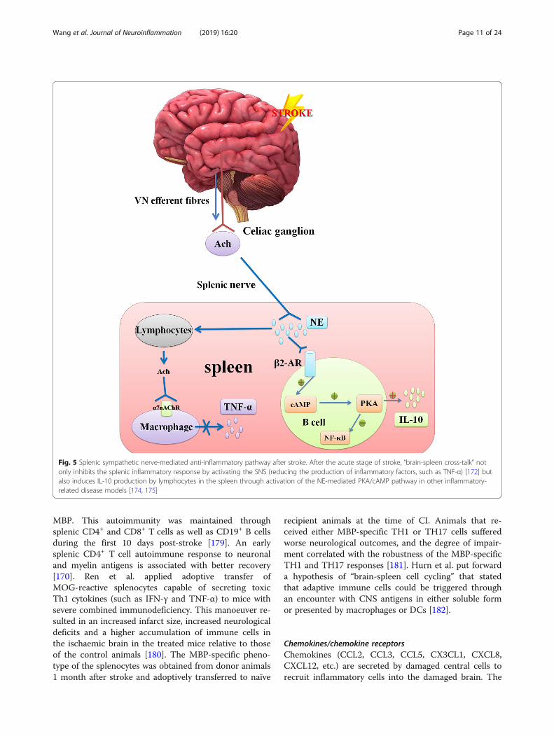
through complex humoural and neural pathways, in-cluding the hypothalamic-pituitary-adrenal (HPA) axis,vagus nerve (VN) and SNS [169]. Elevated cortisone,corticosterone and metanephrine levels and associatedlymphocytopaenia are often observed after extendedbrain infarction. The differential effects and complexinterplay between the SNS and the HPA axis on sys-temic immune cells have to be considered when target-ing the neurohormonal systems in the acute phase ofsevere stroke [170]. The hypothalamus is associated withthe central function of the ANS by synchronising theneuroendocrine (glucocorticoid) response and choliner-gic pathways, which together inhibit the release of in-flammatory cytokines from peripheral T cells, monocytesand macrophages and promote the release ofanti-inflammatory cytokines, such as IL-10. Similarly,norepinephrine (NE) released from dense neural net-works throughout the brain and from peripheral organs,including the spleen, induces significant anti-inflamma-tory phenotypes in lymphocytes, monocytes and macro-phages. In addition, the release of catecholamine fromnerve endings can induce the release of acetylcholine(ACh) from splenic T memory cells, which can inhibitinflammation and increase the risk of infection afterstroke [169]. A recent work showed that transcutaneousauricular VN stimulation (ta-VNS) reduced the infarctvolume and induced angiogenesis in focal cerebral I/Rrats. Ma suggested that the neurobehavioural recoveryinduced by ta-VNS might involve spleen-brain commu-nication triggered by redistribution of growth differenti-ation factor (GDF)-11 [171].The brain and viscera interact through the ANS, and
the VN, which contains 80% afferent fibres and 20%
efferent fibres, plays multiple key roles in regulation ofvisceral homeostasis and anti-inflammatory processes[172]. These VN functions are mediated by many path-ways, some of which are controversial. In the splenicsympathetic nerve-mediated anti-inflammatory pathway,VN fibres stimulate the splenic sympathetic nerve, caus-ing NE release from the distal splenic nerve to act dir-ectly on the β2-AR of splenic lymphocytes, therebyinducing the release of ACh. Finally, ACh inhibits the re-lease of TNF-α from spleen macrophages through theα-7-nicotinic ACh receptor (α7nAChR) [172]. At thesame time, activation of the β2-AR receptor on somesplenic lymphocytes may trigger activation of thecAMP-PKA pathway [173], which is related to inhibitionof the NF-κB pathway and IL-10 production in thespleen [174, 175] (Fig. 5).Generally, the immune response to stroke can be di-
vided into two stages. The immune response at the earlystage of acute stroke is pro-inflammatory and is drivenby an increase in SNS activity. Later, immunosuppres-sion starts when the spleen has depleted its immune cellreserve, and the risk of infection increases after strokeduring this window [117]. Rasouli et al. also proposed asimilar viewpoint of “brain-spleen inflammatory coup-ling” by which autonomic control of splenic macro-phages could modulate systemic inflammation afterinjury. Stimulation of α/β-AR located on splenic macro-phages leads to the release of TNF-α and IL-1β, whichenhance and exacerbate inflammation. Conversely, para-sympathetic stimulation of α7nAChR inhibits the releaseof these cytokines, thereby attenuating the inflammatoryresponse to injury [176].
CNS antigensIn response to stroke, the ischaemic brain may secrete avariety of antigens that activate adaptive immune re-sponses and recruit immune cells from the spleen. Dur-ing acute stroke, neo-antigens, such as microtubule-associated protein 2 (MAP2), N-methyl-D-aspartic acidreceptor subunit 2 (NR-2A), myelin basic protein (MBP)and myelin oligodendrocyte glycoprotein (MOG), can allbe released into the periphery and captured by APCs, es-pecially DCs and macrophages. This response is thoughtto eventually trigger the activation of T cell-dependentadaptive immune responses in the T cell zone [177, 178].Although the immune system does not directly initiate
the pathological process of stroke, stroke-induced im-mune activation has been increasingly recognised to en-hance the neuropathological outcomes or nerve repair.Elucidating novel neo-antigens that are targets for im-mune cells offers unique insights into potential cellularand systemic consequences of autoimmunity duringpost-stroke neuronal plasticity. Mice with small infarctvolumes exhibited high autoreactivity to MAP2 and
Wang et al. Journal of Neuroinflammation (2019) 16:20 Page 10 of 24
MBP. This autoimmunity was maintained throughsplenic CD4+ and CD8+ T cells as well as CD19+ B cellsduring the first 10 days post-stroke [179]. An earlysplenic CD4+ T cell autoimmune response to neuronaland myelin antigens is associated with better recovery[170]. Ren et al. applied adoptive transfer ofMOG-reactive splenocytes capable of secreting toxicTh1 cytokines (such as IFN-γ and TNF-α) to mice withsevere combined immunodeficiency. This manoeuver re-sulted in an increased infarct size, increased neurologicaldeficits and a higher accumulation of immune cells inthe ischaemic brain in the treated mice relative to thoseof the control animals [180]. The MBP-specific pheno-type of the splenocytes was obtained from donor animals1 month after stroke and adoptively transferred to naïve
recipient animals at the time of CI. Animals that re-ceived either MBP-specific TH1 or TH17 cells sufferedworse neurological outcomes, and the degree of impair-ment correlated with the robustness of the MBP-specificTH1 and TH17 responses [181]. Hurn et al. put forwarda hypothesis of “brain-spleen cell cycling” that statedthat adaptive immune cells could be triggered throughan encounter with CNS antigens in either soluble formor presented by macrophages or DCs [182].
Chemokines/chemokine receptorsChemokines (CCL2, CCL3, CCL5, CX3CL1, CXCL8,CXCL12, etc.) are secreted by damaged central cells torecruit inflammatory cells into the damaged brain. The
Fig. 5 Splenic sympathetic nerve-mediated anti-inflammatory pathway after stroke. After the acute stage of stroke, “brain-spleen cross-talk” notonly inhibits the splenic inflammatory response by activating the SNS (reducing the production of inflammatory factors, such as TNF-α) [172] butalso induces IL-10 production by lymphocytes in the spleen through activation of the NE-mediated PKA/cAMP pathway in other inflammatory-related disease models [174, 175]
Wang et al. Journal of Neuroinflammation (2019) 16:20 Page 11 of 24
corresponding chemokine receptors are also increased insplenocytes after I/R [102].CCL2 can effectively mediate monocyte/macrophage
and neutrophil infiltration during CI [183]. InadequateCCR2 expression results in decreased monocyte andneutrophil infiltration into the ischaemic brain, followedby decreased inflammation and cerebral infarction. Baoalso showed that the CCL2-CCR2 interaction might playan important role in the distribution and migration ofmonocytes from the spleen to the injured brain [132].Moxifloxacin treatment can effectively inhibit CCR2 ex-pression in monocytes, thereby significantly reducing theinfarct size after CI [183].The CXCR4-CXCL12 axis is closely related to the path-
ology of ischaemic stroke. CI leads to a rapid andlong-lasting increase in CXCL12 in the ischaemic penum-bra. Transplanted GFP-labelled bone marrow cells are re-cruited in proximity to these CXCL12+ vessels and displaycharacteristics of activated microglial cells. Therefore, wecan speculate that CXCL12 plays an important role inhoming of bone marrow-derived monocytes, which trans-form into microglia at the site of ischaemic injury [184].The CXCR4 antagonist AMD3100 blocks the interactionbetween CXCR4 and CXCL12, which not only alleviatescerebral inflammation and cerebral infarction but alsoprevents splenic atrophy after tMCAO. Therefore,CXCR4-CXCL12 may play a regulatory role in the splenicresponse after stroke [185].Many other cytokines have also been shown to play
important roles in the recruitment of immune cells intothe ischaemic brain. For example, CCL3 is closely re-lated to the accumulation of monocytes and neutrophilsin damaged brain tissue [186, 187]. CCL5 is involved inleukocyte infiltration after I/R [188]. CX3CR1-knockoutmice show reduced neuroinflammation after focal CI,suggesting that CX3CL1 promotes post-stroke inflam-mation, which may be related to chemotaxis of mono-cytes, T cells and NKs [148, 189, 190]. CXCL8 has beenconsidered as an important chemotactic factor for neu-trophil recruitment after ischaemic stroke [191]. IP-10expands the NK-induced damage of the BBB throughCXCR3 [134]. The increased CXCL-1 and CXCL-2 levelsin the brain tissue after stroke lead to accelerated leuko-cytes and particularly granulocyte accumulation and ag-gravate ischaemic tissue damage [192]. CCL20 isupregulated in the thymus and spleen 24 h after TBIand in the cortex and hippocampus 48 h after TBI [116].The roles of these cytokines in splenic cell mobilisationwarrant further study.
Stem cell therapy targeting the spleenIn various experimental models, hUCBs [32–36], HSCs[35], BMSCs [36, 97, 193], hAECs [48] and NSCs [37]have been shown to reduce the neurological damage
caused by stroke. Compared with that of intracerebraladministration, all stem cells show better therapeutic ef-fects when administered systemically. Stem cells migrateto the injured brain and spleen and in some cases havebeen shown to modulate the immune response to stroke[32, 35–37, 48], which may be one reason that this injec-tion route is more efficacious. Ninety-five percent ofBMSCs are found in the spleen following systemic ad-ministration after MCAO [36] (Fig. 6).Intravenous hBMSCs preferentially migrate to the
spleen and alleviate chronic inflammation in rats withCI. hBMSC treatment reduces the TNF-α level in thespleen after CI by 40%. Correlation analysis revealednegative correlations between hBMSC migration in thespleen and the infarct areas, peri-infarct areas, volumeof MHCII− activated cells in the striatum and TNF-αlevel in the spleen [31]. Even NSCs migrate to the spleenfollowing ICH, but the therapeutic effect disappears aftersplenectomy. NSCs have been found to be in direct con-tact with CD11b+ cells in the spleen [37], which partlydemonstrates that the neuroprotective effect of NSCsduring stroke involves their interaction with the spleen.hUCBs are another cell type that has been shown to
affect the spleen during the pathological process ofstroke. hUCBs alter cell populations in the peripheralcirculation and spleen after pMCAO due to the interac-tions of all subpopulations together [196] (Fig. 7). Sys-temic administration of hUCBs 24 h after MCAOsignificantly alters splenic T cell responses to concanava-lin A, decreases the proliferation activity of splenic Tcells, decreases production of the inflammatory factorsTNF-a and IFN-γ and increases production of theanti-inflammatory cytokine IL-10 [32, 196]. hUCBs alsoinhibit splenic atrophy in rats 48 h after MCAO. This ef-fect is thought to be achieved by regulation of immunecells in the spleen by hUCBs after MCAO and inhibitionof their release into the systemic circulation [24]. Kadamet al. used intravenous injection of CD34+-enrichedhUCBs to treat CI mice; the experimental results showedthat neurogenic niche proliferation and glial brain re-sponses to CD34+-enriched hUCBs after neonatal strokemight involve interactions with the spleen and weresex-dependent [197].hAECs are derived from the epithelial layer of the am-
nion, which is the sac that encloses the developing foetusand is attached to the placenta. Evans recently tested theefficacy of systemically delivered hAECs to improve anumber of outcome measures in four animal models of is-chaemic stroke [205]. Based on their experimental results,they put forward the hypothesis that administration dur-ing the acute phase (within 1.5 h) after ischaemic attackallowed the hAECs to migrate preferentially to the spleenand damaged brain; subsequently, cell apoptosis and in-flammation were inhibited. Early brain infiltration of
Wang et al. Journal of Neuroinflammation (2019) 16:20 Page 12 of 24

immune cells, aggravation of infarction and systemic im-munosuppression were also alleviated [48].HSCs injected intravenously 24 h after reperfusion
were first detected at 24 h after injection in the spleenand later in the ischaemic brain parenchyma [35]. Inaddition, compared with that of the sham-operated con-trol group, the immune environment after CI increasedHSC migration to the spleen 72 h after reperfusion. Inthe absence of induction of an injury, the cells did notpreferentially accumulate in the spleen [35]. HSC treat-ment reduced the infarct volumes, apoptotic neuronalcell death in the peri-infarct areas and immune cell (Tcells and macrophages) infiltration into the ischaemichemispheres. Moreover, HSC therapy decreased theTNF-α, IL-1β, CCR2 and CX3CR1 levels in the spleenafter CI [35].These experiments suggest that stem cell therapy
works to some extent by regulating the immune re-sponse after stroke, especially at the spleen level, whichmay be crucial and is an important potential therapeutictarget.
Important therapeutic targetsMany factors and regulatory pathways change signifi-cantly during the entire pathophysiological process ofstroke. They may play different roles in different patho-logical stages after CI. Fully understanding their effects
on stroke may identify new targets for development ofnovel therapeutic strategies.
IL-10IL-10 is a multicellular, multifunctional cytokine that reg-ulates cell growth and differentiation and participates ininflammatory and immune responses. It is mainly pro-duced by cells such as Tregs, Th2 cells and Bregs and cur-rently is recognised as an anti-inflammatory andimmunosuppressive factor. IL-10 is an important compo-nent of the endogenous repair mechanism after stroke.The IL-10 levels in the brain and spleen increase afterstroke [90, 155, 156]. The use of exogenous IL-10 foranti-inflammatory therapeutic approaches has been shownto provide neuroprotection during ischaemic stroke [206].However, an excessive IL-10 response can contribute toimmunosuppression after CI, which worsens outcomes.Additionally, sex differences may exist in the role of IL-10in stroke recovery [207].Adoptive transfer of IL-10-secreting cells is widely used
for the treatment of experimental stroke. However, afterthe earlier use of Tregs [41], Bregs are attracting increas-ing attention. Adoptive reinfusion of spleen-derived Bregscan improve neurological injury and motor dysfunctionafter CI in experimental rats [34, 140]. Elevated Breg andTreg levels in the spleen at different time points after CIhave been found in some studies investigating the precon-ditioning protection of I/R [208, 209]. In addition to their
Fig. 6 Therapeutic stem cells after stroke migrate to peripheral organs, such as the spleen. a After stroke, the patient was treated by intravenousinjection of stem cells. b, c Stem cells in circulation are stimulated by IL-1β, IL-6 and TNF-α after stroke, and their chemokine ligand levels areelevated, thus enhancing the capability of stem cells to recruit inflammatory cells [194, 195]. d Through migration and adhesion, stem cellsmigrate rapidly to PIOs, such as the spleen, to play a regulatory role
Wang et al. Journal of Neuroinflammation (2019) 16:20 Page 13 of 24
anti-inflammatory effects, Bregs can also promote the ac-tivation and proliferation of other anti-inflammatory im-mune cells and have a cascade amplification effects on therepair mechanism after CI [147, 210] (Fig. 8). Therefore,we have proposed that Bregs may represent an importantpotential strategy to rapidly start the endogenous protect-ive mechanism during the early stage of stroke and in-crease the Breg levels in the body, especially in the spleen.
InterferonIFN-γ is the only member of the type II IFNs and is pro-duced only by activated T cells, NKs and natural killer Tcells (NKTs). IFN-γ is a marker cytokine of Th1 cellsthat can activate APCs and promote the differentiationof Th1 cells by upregulating related transcription factors.
Deletion of the IFN-γ gene has been shown to reducebrain damage after CI [137]. In the first 3 days after I/R,intraventricular administration of an IFN-γ neutralisingantibody can protect the brain from CI injury [140].IFN-γ participates in the Th1 inflammatory response byactivating monocytes, microglia and macrophages. Sinceactivation of microglia/macrophages is part of the causeof delayed cell injury following ischaemic injury, IFN-γmay play a role in the splenic response by aggravatingthe inflammation associated with ischaemic injury. Sei-fert et al. previously proposed that the spleen contrib-uted to stroke-induced neurodegeneration throughIFN-γ signalling [119]. IFN-γ is increased early in thespleen but later in the brain following I/R. Splenectomyreduces the IFN-γ level in the infarct after MCAO. The
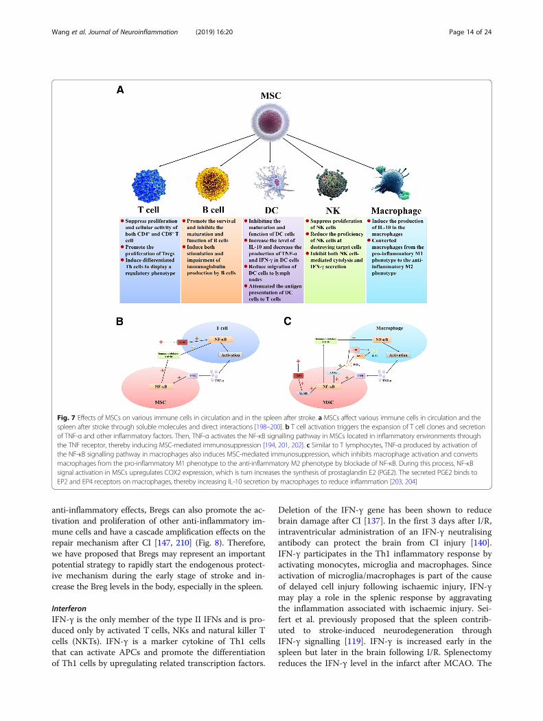
Fig. 7 Effects of MSCs on various immune cells in circulation and in the spleen after stroke. a MSCs affect various immune cells in circulation and thespleen after stroke through soluble molecules and direct interactions [198–200]. b T cell activation triggers the expansion of T cell clones and secretionof TNF-α and other inflammatory factors. Then, TNF-α activates the NF-κB signalling pathway in MSCs located in inflammatory environments throughthe TNF receptor, thereby inducing MSC-mediated immunosuppression [194, 201, 202]. c Similar to T lymphocytes, TNF-α produced by activation ofthe NF-κB signalling pathway in macrophages also induces MSC-mediated immunosuppression, which inhibits macrophage activation and convertsmacrophages from the pro-inflammatory M1 phenotype to the anti-inflammatory M2 phenotype by blockade of NF-κB. During this process, NF-κBsignal activation in MSCs upregulates COX2 expression, which is turn increases the synthesis of prostaglandin E2 (PGE2). The secreted PGE2 binds toEP2 and EP4 receptors on macrophages, thereby increasing IL-10 secretion by macrophages to reduce inflammation [203, 204]
Wang et al. Journal of Neuroinflammation (2019) 16:20 Page 14 of 24

protective effect of splenectomy was eliminated by sys-temic recombinant IFN-γ accompanied by an increase inIFN-γ expression in the brain post-pMCAO [119].Moreover, IP-10 has been shown to be a key factor instroke-induced neurodegenerative diseases [154]. NKsparticipate in CI and promote neuronal necrosis throughIFN-γ. IP-10 expands the damage of the BBB induced byNKs through CXCR3 [149]. Furthermore, hUCB therapyinhibits the proliferation of splenic T cells after MCAOby increasing IL-10 and IFN-γ production [32].IFN-β is a type I IFN that binds to the IFN-α/β recep-
tor. IFN-β exerts anti-inflammatory effects, and systemicadministration of recombinant IFN-β has been used forthe treatment of multiple sclerosis, which is a neuroin-flammatory condition of the CNS [211]. Therefore,IFN-β has been considered to be able to decrease neur-onal death and promote functional recovery after CI bylimiting inflammation. In agreement with this view,IFN-β has been put forward as a candidate drug for thetreatment of stroke. Several animal experiments haveshown that systemic administration of recombinantIFN-β at different time points before and after MCAO
results in neuroprotection [212–214]. This outcome maybe related to endogenous IFN-β signalling not only toreduce CNS inflammation but also to reduce autoreac-tive T cell proliferation via inhibiting the antigen pre-senting capacity of astrocytes and microglia. Inácio’sresearch also showed that endogenous IFN-β signallingcould alleviate local inflammation and regulate periph-eral immune cells, thereby contributing positively tostroke outcomes [215].
Cholinergic anti-inflammatory pathwayAs mentioned above, VNS causes prominent attenuationof the systemic inflammatory response evoked by CI inexperimental animals. This effect is mediated by AChstimulation of acetylcholine receptors on splenic macro-phages. Therefore, the circuit is known as the “choliner-gic anti-inflammatory pathway”, which casts the spleenas the major effector [216]. α7nAChR is considered tobe an important target for alleviating the release ofpro-inflammatory cytokines from macrophages and DCs.Noradrenergic neurons provide innervation to all primary
and secondary lymphoid tissues, including the bone marrow,
Fig. 8 Effects of Breg cells on other immune cells after stroke. Through IL-10, IL-35 and TGF-β production, Bregs can inhibit the differentiation of pro-inflammatory lymphocytes, such as TNF-α-secreting monocytes, IL-12-secreting dendritic cells, Th17 cells, Th1 cells, IL-17+ γδT cells and cytotoxic CD8+
T cells. Bregs can also induce the differentiation of immunosuppressive T cells, such as Foxp3+ Tregs and T regulatory 1 (Tr1) cells and contribute tothe maintenance of iNKTs. Therefore, Bregs result in immune regulation at sites of inflammation, such as the CNS
Wang et al. Journal of Neuroinflammation (2019) 16:20 Page 15 of 24

spleen and lymph nodes [217, 218]. Most immune cells ex-press one or more ARs, but β2-AR is the most widely dis-tributed and mediates most of the effects of sympatheticnerves on immune function [219–222]. Sympathetic nervesaffect the innate and adaptive immune responses throughstimulating β2-AR. Most evidence shows that β2-AR activa-tion has immunosuppressive effects on monocytes and mac-rophages [221, 222]. Stimulation of β2-AR-naïve CD4+ Tcells (Th0) results in their differentiation into Th1 cells,which enhance cellular immunity, or Th2 cells, which de-crease cellular immunity [221, 222]. Norepinephrine (NE)can also produce β2-mediated anti-inflammatory effects byreleasing ACh from cholinergic spleen cells [221, 222].Blocking adrenocortical receptors has been shown to inhibitthe splenic response after pMCAO and reduce injury [123].The spleen receives noradrenergic innervation from thepostganglionic sympathetic neurons [223]. Splenic T cells arethe source of ACh and may be a link between NE andsplenic macrophage suppression [224]. β2-AR on T cells isessential for the anti-inflammatory effect of VNS [225, 226].Splenectomy eliminates the role of VNS in increasing plasmaNE, which supports the conclusion that plasma NE is re-leased from the spleen into circulation during VNS [227].The spleen plays a central role in the pathophysiology of thehyperinflammatory state triggered by threatening conditions,such as stroke, and splenic macrophages are the dominantsource of pro-inflammatory cytokines [226].Targeted therapy for α7nAChR on microglia and mac-
rophages after stroke has long been considered an im-portant potential strategy. α7nAChR expression onactivated microglia and infiltrated macrophages after CIplays an important role in the pathological process ofstroke. Nicotine therapy has been shown to significantlyattenuate the increase in microglia and inflammatory cy-tokines (i.e., TNF-α and IL-1β) induced by CI throughmediation of α7nAChR [228]. In addition, α7 agonistscan reduce the infarct volume and functional deficits indifferent animal models of stroke [229, 230]. In contrast,blockade of α7nAChRs with a selective antagonist in-creases the infarct volume, which suggests some degreeof α7nAChR stimulation by the endogenous agonist[230]. The endogenous choline released from damagedbrain tissue may fulfil this role. The use of an allostericmodulator of α7nAChRs 6 h after tMCAO can reducethe infarct volume and improve neurological perform-ance [231, 232]. In addition, regulation of α7nAChRshas also been shown to be associated with changes inM1 and M2 microglia/macrophages after CI [229].
Multipotent adult progenitor cellsMAPCs are a unique type of adult adhesion cells(MSC-like) that extend understanding of how intravenouscell therapy participates in and regulates the peripheralimmune system after stroke. MAPCs can be isolated from
bone marrow and other tissues [233], are well charac-terised and can be easily distinguished from bone marrowmononuclear cells (BMMNCs) and MSCs based on theirsize [234], transcriptome [235], microRNA profile [236],differentiation capability [237] and secretome [238].Intravenous injection of MAPCs has been shown to
have beneficial effects on other CNS injuries, such asTBI [239]. MAPC therapy attenuates activated micro-glial/macrophage responses, preserves the BBB, re-duces cerebral oedema and improves spatial learningafter TBI [118, 239, 240], and MSPC treatment within24 h after injury can achieve better outcomes [241].After TBI, intravenous MAPCs also tend to migrateto the spleen [234].MAPCs have become a new hotspot of cell therapy for
stroke after BMSCs, hUCBs and HSCs. MAPCs can sig-nificantly inhibit the inflammatory reaction of injuredbrain tissue [242, 243] and improve the motor functionand neurological outcomes of experimental stroke ani-mals [242, 243]. Moreover, MAPCs exhibit more robusttissue sparing and mitigation of glial activation thanMSCs regardless of whether intravenous or intrapar-enchymal administration is used [244, 245]. Similarly, re-cent animal data support a role for intravenous MAPCsin regulation of the peripheral immune system throughspecific interactions between MAPCs and splenocytes,thereby promoting stroke recovery and inhibiting splenicatrophy in the 24 h after CI. Similar to results obtainedfrom testing hUCBs, stroke can also accelerate the mi-gration of splenocyte MAPCs to the spleen and inhibitsplenocyte apoptosis. In addition, MAPCs decreased thelevels of CD3+, CD4+ and CD8+ cells in the spleen andincrease Tregs after tMCAO compared with the levels inthe vehicle-treated group [246]. In terms of inflamma-tory factors, IL-1β and TNF-α were significantly lowerin the splenic cells of rats with I/R after MAPC treat-ment than those of the saline-treated animals, whereasthe IL-10 level was higher [246].MAPCs not only have a good protective effect on
acute CI but also maintain a stable and sustained thera-peutic effect on chronic stroke. Compared with those ofthe saline-treated group, the MAPC treatment group ex-hibited advantages in locomotor and neurological out-comes that persisted for more than 28 days. In addition,the results of a comparative trial of MAPCs for stroke insplenectomized and sham splenectomized mice alsodemonstrated that MAPCs could inhibit expansion ofthe infarct volume through a non-spleen-mediatedmechanism, but the MAPC-induced IL-10 productionafter tMCAO was still dependent on an intact spleen.Moreover, no significant difference was found in the im-provement of neurological outcomes between the splen-ectomy group treated with MAPCs 24 h after tMCAOand the splenectomy group treated with saline [246].
Wang et al. Journal of Neuroinflammation (2019) 16:20 Page 16 of 24

Preclinical animal data support the benefits of intra-venous MAPC therapy for stroke. A phase I/II clinicaltrial is under way to test the safety and efficacy of Multi-Stem (the MAPC clinical-grade product) for treatmentof patients with acute ischaemic stroke. Previous in vitrostudies have shown that MACPs inhibit CD8+ T cells,which are harmful to stroke [247]. The MASTERS trial(MultiStem in Acute Stroke Treatment to Enhance Re-covery Study) was conducted in 33 clinical centres in theUSA and UK from October 2011 through December2015. First, MultiStem treatment was proven to be safe.No infusion-related allergic reaction was observed in theMultiStem and placebo-treated groups, and no casesshowed neurological worsening. MultiStem treatmentcan reduce the risk of life-threatening adverse events ordeath and secondary infections in stroke patients. Fur-thermore, MultiStem treatment also greatly shortenedthe time in the intensive care unit and the overall hospi-talisation time compared with those of theplacebo-treated patients. Compared with those of theplacebo, MultiStem treatment significantly reduced thebiomarkers of post-stroke inflammation (circulatingCD3+ T cells and inflammatory factors). Importantly,MultiStem treatment significantly improved the chancefor an excellent outcome at 1 year of onset [248, 250].Currently, the phase III MASTERS-2 trial aims to ex-
pand our knowledge and understanding of treatment ofischaemic stroke patients with MultiStem and plans tostart recruiting patients in 2018. Another MultiStemclinical study named TREASURE (Treatment evaluationof acute stroke for using in regenerative cell elements)was officially launched in 31 medical centres in Japan in2017. The research project recruited 220 patients withacute ischaemic stroke, including speech or motor defi-cits, as defined by a National Institution of HealthStroke Scale (NIHSS) score of 8–20 at baseline. TREAS-URE is a randomised, double-blind, placebo-controlled,multicentre phase 2/3 trial to evaluate the efficacy andsafety of intravenous administration of MultiStem® com-pared with those of a placebo in patients with ischaemicstroke [249].
DiscussionIntravenous cell therapy can modulate the acute and ad-verse contributions of the peripheral immune systemafter ischaemic stroke [250]. A single intravenous ad-ministration of cells 24 to 36 h after stroke onset thatcan mitigate and rebalance the immune response to theinitial focal ischaemic injury is sufficient for nerve repairand improvement of long-term outcomes [29].As mentioned earlier, the activation, migration and
participation of peripheral immune cells, and perhapsmost importantly immune cells from the spleen, are crit-ical steps in the pathophysiological progression after
stroke. Therefore, we believe that targeted inhibition ofperipheral blood immune cell (especially splenic cell) ac-tivation, reduction of inflammatory cytokine productionand inhibition of their entry into the brain parenchymathrough the BBB are key steps in attenuating the expan-sion of pro-inflammatory microglial activation, neuronaldie-back and tissue loss.Simply inhibiting the participation of splenic compo-
nents of the peripheral immune system in post-strokepathophysiological processes allows tissue sparing but isnot sufficient to enable neurological and locomotor ben-efits [246]. As mentioned above, splenectomy fails toprovide long-term protection against ischaemic stroke,and delayed splenectomy neither reduces brain tissueloss nor alleviates sensorimotor and cognitive impair-ment [121]. These results suggest that the spleen may bea double-edged sword that plays completely oppositeroles during different pathological stages of stroke. Inthe early stage of stroke, the spleen is mainly charac-terised by inflammation and harmfulness but then grad-ually transforms into an anti-inflammatory andprotective phenotype. Therefore, inhibiting the inflam-matory immune response of the spleen in the early stageof stroke and accelerating the initiation of itsanti-inflammatory mechanism have become the keys forthe use of stem cells in the treatment of acute stroke.We also summarise that many stem cells, includingMAPCs, can simultaneously inhibit potentially harmfulaspects of the innate immune system response to strokewhile speeding up beneficial aspects or reparative re-sponses, which confirms our viewpoint (Fig. 9).Through a number of preclinical and clinical studies,
we have obtained a certain understanding of the bio-logical distribution of stem cells after injection for thetreatment of stroke and their effects on many immunecell subsets and cytokines in the CNS and PIOs. How-ever, little is known about the effects of stem cell therapyon the immune regulation mediated by the ANS andSNS. Many trials will provide an opportunity to betterevaluate and examine hypothetical mechanisms, and webelieve that stem cell therapy provides benefits forstroke; however, further preclinical and clinical studiesare needed to advance a more comprehensive under-standing. Evaluating the safety and efficacy of variousstem cell therapies at multiple doses and at differenttime points may also yield new information.
ConclusionIn summary, we can draw the following conclusions.First, the splenic response after stroke is critical forpathological damage and tissue repair. The key of immu-nomodulatory therapy for stroke may be to inhibitsplenic inflammation at the early stage of pathology andaccelerate the initiation of its anti-inflammatory/repair
Wang et al. Journal of Neuroinflammation (2019) 16:20 Page 17 of 24

mechanism. Second, during the course of stem cell ther-apy for stroke, allowing more stem cells to migrate tothe spleen may inhibit the splenic inflammatory re-sponse and accelerate the initiation of its anti-inflamma-tory/repair mechanism, which will have a bettertherapeutic effect than increasing the number of stemcells delivered to the injured brain tissue. Finally, inflam-matory factors, such as TNF-α and IL-1β, produced afterstroke can activate inflammatory pathways, such asNF-κB, in stem cells, thereby enabling stem cells to ac-quire stronger immune regulation potential. Therefore,stroke-like stimuli can be applied to stem cells for treat-ment before injection, which may lead to a better thera-peutic effect. In the future, we believe that the spleenwill become a potential target of various stem cell ther-apies for stroke represented by MAPC treatment. Theresearch results will move closer to clinical applicationand ultimately benefit the majority of stroke patients.
AbbreviationsAch: Acetylcholine; ANS: Autonomic nervous system; APCs: Antigen-presenting cells; AR: Adrenergic receptor; BBB: Blood-brain barrier;BMMNCs: Bone marrow mononuclear cells; BMSCs: Bone marrow stem cells;Bregs: Regulatory B cells; CCL: C-C chemokine ligand; CI: Cerebral ischaemia;
CNS: Central nervous system; COX: Cyclooxygenase; CXCL: C-X-C chemokineligand; CXCR: C-X-C chemokine receptor; DAMPs: Damage-associatedmolecular patterns; DCs: Dendritic cells; GDF: Growth differentiation factor;hAECs: Human amnion epithelial cells; HPA: Hypothalamic-pituitary-adrenal;HPCs: Haematopoietic progenitor cells; HSCs: Haematopoietic stem cells;hUCBs: Human umbilical cord blood cells; I/R: Ischaemia/reperfusion;ICH: Intracerebral haemorrhage; IFN: Interferon; IGF: Insulin-like growth factor;IL: Interleukin; iNKTs: Invariable natural killer T cells; IP: IFN-induced protein;LPS: Lipopolysaccharide; MAP: Microtubule-associated protein;MAPCs: Multipotent adult progenitor cells; MBP: Myelin basic protein;MCP: Monocyte chemoattractant protein; MMP: Matrix metalloproteinase;MOG: Myelin oligodendrocyte glycoprotein; MSCs: Mesenchymal stem cells;MultiStem: The MAPC clinical product; NE: Norepinephrine; NGF: Nervegrowth factor; NIHSS: National Institution of Health Stroke Scale; NKs: Naturalkiller cells; NKTs: Natural killer T cells; NLR: Nod-like receptor; NO: Nitric oxide;NPCs: Neural progenitor cells; NR-2A: N-methyl-D-aspartic acid receptorsubunit 2; NSCs: Neural stem cells; p/t MCAO: Permanent/temporary middlecerebral artery occlusion; PAMPs: Pathogen-associated molecular patterns;PGE: Prostaglandin E; PIOs: Peripheral immune organs; PNS: Parasympatheticnervous system; PRR: Pattern-recognition receptors; ROS: Reactive oxygenspecies; SNS: Sympathetic nervous system; ta-VNS: Transcutaneous auricularVN stimulation; TBI: Traumatic brain injury; TGF: Transforming growth factor;Th: Helper T cell; TNF: Tumour necrosis factor; Tr1: T regulatory 1 cells;Tregs: Regulatory T cells; VEGF: Vascular endothelial growth factor; VN: Vagusnerve; α7nAChR: α-7-nicotinic ACh receptors
AcknowledgementsSincere thanks to La-Mei Cheng, Professor of Reproductive and Stem Cell Re-search Institute of Central South University, for guidance on this subject.
Fig. 9 Stem cell therapy enhances recovery after stroke. In the untreated scenario, ischaemic stroke leads to activation of the peripheral immune system.During this process, the spleen atrophies, lymphocytes undergo apoptosis in the spleen, the inflammatory factor levels in the spleen increase, andinflammatory cells are released from the spleen into circulation. The antigen presentation of DCs is enhanced, and the levels of various chemokines areelevated. These pro-inflammatory mediators contribute to M1 microglia-mediated destruction of the BBB and CNS inflammation. Infiltration of leukocytesfurther aggravates inflammatory necrosis of neurons. Intravenous administration of stem cells reverses splenic atrophy and converts macrophages from thepro-inflammatory M1 phenotype to the anti-inflammatory M2 phenotype and Th cells from the pro-inflammatory Th1 phenotype to the anti-inflammatoryTh2 phenotype. The inflammatory cytokine and cell levels in the spleen decrease, and anti-inflammatory cytokines and cells begin to be produced andreleased into circulation, which ultimately lead to less BBB restructuring and CNS inflammation and provide a favourable environment for nerveregeneration and angiogenesis
Wang et al. Journal of Neuroinflammation (2019) 16:20 Page 18 of 24

FundingFunding was obtained through the National High Technology Research andDevelopment Program of China (863 project) (No. 2011AA020113), HunanProvince Key Scientific and Technological Project (No. 2013SK5070), theNational Natural Science Fund of China (No. 81460244) and ScientificResearch Plan of Hunan Provincial Health Department (No. B2010-107).
Availability of data and materialsData sharing is not applicable for this article, because no datasets weregenerated or analysed during the current study.
Authors’ contributionsZW and XJT designed the study, carried out the literature review and draftedthe manuscript. DH and YYZ carried out the literature review andparticipated in drafting the manuscript. LZ and CY critically edited themanuscript. YJL and JQH conceived of the study and participated in itsdesign and coordination. XYC and XHH are responsible for correctinggrammatical errors in the revised manuscript. All authors read and approvedthe final manuscript.
Ethics approval and consent to participateNot applicable.
Consent for publicationNot applicable.
Competing interestsThe authors declare that they have no competing interests.
Publisher’s NoteSpringer Nature remains neutral with regard to jurisdictional claims inpublished maps and institutional affiliations.
Author details1Xiangtan Central Hospital, Clinical Practice Base of Central South University,Xiangtan 411100, China. 2Institute of Reproductive and Stem Cell Research,School of Basic Medical Science, Central South University, Changsha 410000,China.
Received: 16 November 2018 Accepted: 7 January 2019
References1. Benjamin EJ, Blaha MJ, Chiuve SE, Cushman M, Das SR, Deo R, de Ferranti
SD, Floyd J, Fornage M, Gillespie C, Isasi CR, Jiménez MC, Jordan LC, JuddSE, Lackland D, Lichtman JH, Lisabeth L, Liu S, Longenecker CT, Mackey RH,Matsushita K, Mozaffarian D, Mussolino ME, Nasir K, Neumar RW,Palaniappan L, Pandey DK, Thiagarajan RR, Reeves MJ, Ritchey M, RodriguezCJ, Roth GA, Rosamond WD, Sasson C, Towfighi A, Tsao CW, Turner MB,Virani SS, Voeks JH, Willey JZ, Wilkins JT, Wu JH, Alger HM, Wong SS,Muntner P, American Heart Association Statistics Committee and StrokeStatistics Subcommittee. Heart Disease and Stroke Statistics-2017 Update: AReport From the American Heart Association. Circulation. 2017;135(10):e146–603.
2. Wei L, Wei ZZ, Jiang MQ, Mohamad O, Yu SP. Stem cell transplantationtherapy for multifaceted therapeutic benefits after stroke. Prog Neurobiol.2017;157:49–78.
3. Anrather J, Iadecola C. Inflammation and stroke: an overview.Neurotherapeutics. 2016;13(4):661–70.
4. Liu ZJ, Chen C, Li FW, Shen JM, Yang YY, Leak RK, et al. Splenic responses inischemic stroke: new insights into stroke pathology. CNS Neurosci Ther.2015;21(4):320–6.
5. Maya-Espinosa G, Collazo-Navarrete O, Millán-Aldaco D, Palomero-Rivero M,Guerrero-Flores G, Drucker-Colín R, et al. Mouse embryonic stem cell-derived cells reveal niches that support neuronal differentiation in the adultrat brain. Stem Cells. 2015;33(2):491–502.
6. Rosenblum S, Smith TN, Wang N, Chua JY, Westbroek E, Wang K, et al.BDNF pretreatment of human embryonic-derived neural stem cellsimproves cell survival and functional recovery after transplantation inhypoxic-ischemic stroke. Cell Transplant. 2015;24(12):2449–61.
7. Huang L, Wong S, Snyder EY, Hamblin MH, Lee JP. Human neural stem cellsrapidly ameliorate symptomatic inflammation in early-stage ischemic-reperfusion cerebral injury. Stem Cell Res Ther. 2014;5(6):129.
8. Chen L, Qiu R, Li L, He D, Lv H, Wu X, et al. The role of exogenous neuralstem cells transplantation in cerebral ischemic stroke. J BiomedNanotechnol. 2014;10(11):3219–30.
9. Cheng Y, Zhang J, Deng L, Johnson NR, Yu X, Zhang N, et al. Intravenouslydelivered neural stem cells migrate into ischemic brain, differentiate andimprove functional recovery after transient ischemic stroke in adult rats. IntJ Clin Exp Pathol. 2015;8(3):2928–36.
10. Wakao S, Kuroda Y, Ogura F, Shigemoto T, Dezawa M. Regenerative effects ofmesenchymal stem cells: contribution of muse cells, a novel pluripotent stemcell type that resides in mesenchymal cells. Cell. 2012 Nov 8;1(4):1045–60.
11. Chen J, Li Y, Katakowski M, Chen X, Wang L, Lu D, et al. Intravenous bonemarrow stromal cell therapy reduces apoptosis and promotes endogenouscell proliferation after stroke in female rat. J Neurosci Res. 2003;73(6):778–86.
12. Kondziolka D, Steinberg GK, Wechsler L, Meltzer CC, Elder E, Gebel J, et al.Neurotransplantation for patients with subcortical motor stroke: a phase 2randomized trial. J Neurosurg. 2005;103(1):38–45.
13. Li Y, Chopp M, Chen J, Wang L, Gautam SC, Xu YX, et al. Intrastriataltransplantation of bone marrow nonhematopoietic cells improvesfunctional recovery after stroke in adult mice. J Cereb Blood Flow Metab.2000;20(9):1311–9.
14. Steinberg GK, Kondziolka D, Wechsler LR, Lunsford LD, Coburn ML,Billigen JB, et al. Clinical outcomes of transplanted modified bonemarrow-derived mesenchymal stem cells in stroke: a phase 1/2a study.Stroke. 2016;47(7):1817–24.
15. Otero L, Zurita M, Bonilla C, Aguayo C, Vela A, Rico MA, et al. Latetransplantation of allogeneic bone marrow stromal cells improvesneurologic deficits subsequent to intracerebral hemorrhage. Cytotherapy.2011;13(5):562–71.
16. Wang L, Ji H, Li M, Zhou J, Bai W, Zhong Z, et al. Intrathecal administrationof autologous CD34 positive cells in patients with past cerebral infarction: asafety study. ISRN Neurol. 2013;2013:128591.
17. Danielyan L, Schäfer R, von Ameln-Mayerhofer A, Buadze M, Geisler J,Klopfer T, et al. Intranasal delivery of cells to the brain. Eur J Cell Biol. 2009;88(6):315–24.
18. Janowski M, Wagner D-C, Boltze J. Stem cell-based tissue replacement afterstroke: factual necessity or notorious fiction? Stroke. 2015;46(8):2354–63.
19. Eckert A, Huang L, Gonzalez R, Kim HS, Hamblin MH, Lee JP. Bystandereffect fuels human induced pluripotent stem cell-derived neural stem cellsto quickly attenuate early stage neurological deficits after stroke. Stem CellsTransl Med. 2015;4(7):841–51.
20. Pendharkar AV, Chua JY, Andres RH, Wang N, Gaeta X, Wang H, et al.Biodistribution of neural stem cells after intravascular therapy for hypoxic-ischemia. Stroke. 2010;41(9):2064–70.
21. Detante O, Valable S, de Fraipont F, Grillon E, Barbier EL, Moisan A, et al.Magnetic resonance imaging and fluorescence labeling of clinical-grademesenchymal stem cells without impacting their phenotype: study in a ratmodel of stroke. Stem Cells Transl Med. 2012;1(4):333–41.
22. Rosado-de-Castro PH, Schmidt Fda R, Battistella V, Lopes de Souza SA,Gutfilen B, Goldenberg RC, Kasai-Brunswick TH, Vairo L, Silva RM, WajnbergE, Alvarenga Americano do Brasil PE, Gasparetto EL, Maiolino A, Alves-LeonSV, Andre C, Mendez-Otero R, Rodriguez de Freitas G, Barbosa da FonsecaLM. Biodistribution of bone marrow mononuclear cells after intra-arterial orintravenous transplantation in subacute stroke patients. RegenMed. 2013;8(2):145–55.
23. Watanabe M, Yavagal D. Intra-arterial delivery of mesenchymal stem cells.Brain Circ. 2016;2:114–7.
24. Gutiérrez-Fernández M, Rodríguez-Frutos B, Alvarez-Grech J, Vallejo-Cremades MT, Expósito-Alcaide M, Merino J, et al. Functional recovery afterhematic administration of allogenic mesenchymal stem cells in acuteischemic stroke in rats. Neuroscience. 2011;175:394–405.
25. Li Y, Chen J, Wang L, Lu M, Chopp M. Treatment of stroke in rat withintracarotid administration of marrow stromal cells. Neurology. 2001;56(12):1666–72.
26. Banerjee S, Bentley P, Hamady M, Marley S, Davis J, Shlebak A, et al. Intra-arterial Immunoselected CD34+ stem cells for acute ischemic stroke. StemCells Transl Med. 2014;3(11):1322–30.
27. Ohshima M, Taguchi A, Tsuda H, Sato Y, Yamahara K, Harada-Shiba M, et al.Intraperitoneal and intravenous deliveries are not comparable in terms of
Wang et al. Journal of Neuroinflammation (2019) 16:20 Page 19 of 24
drug efficacy and cell distribution in neonatal mice with hypoxia-ischemia.Brain Dev. 2015;37(4):376–86.
28. Vahidy FS, Rahbar MH, Zhu H, Rowan PJ, Bambhroliya AB, Savitz SI.Systematic review and meta-analysis of bone marrow-derived mononuclearcells in animal models of ischemic stroke. Stroke. 2016;47:1632–9.
29. Hess DC, Wechsler LR, Clark WM, Savitz SI, Ford GA, Chiu D, et al. Safety andefficacy of multipotent adult progenitor cells in acute ischaemic stroke(MASTERS): a randomised, double-blind, placebo-controlled, phase 2 trial.Lancet Neurol. 2017;16:360–8.
30. Bacigaluppi M, Pluchino S, Peruzzotti-Jametti L, Kilic E, Kilic U, Salani G, et al.Delayed post-ischaemic neuroprotection following systemic neural stem celltransplantation involves multiple mechanisms. Brain. 2009;132:2239–51.
31. Acosta SA, Tajiri N, Hoover J, Kaneko Y, Borlongan CV. Intravenous bonemarrow stem cell grafts preferentially migrate to spleen and abrogatechronic inflammation in stroke. Stroke. 2015;46(9):2616–27.
32. Vendrame M, Gemma C, Pennypacker KR, Bickford PC, Davis Sanberg C,Sanberg PR, et al. Cord blood rescues stroke-induced changes in splenocytephenotype and function. Exp Neurol. 2006;199(1):191–200.
33. Vendrame M, Cassady J, Newcomb J, Butler T, Pennypacker KR, Zigova T, etal. Infusion of human umbilical cord blood cells in a rat model of strokedosedependently rescues behavioral deficits and reduces infarct volume.Stroke. 2004;35(10):2390–5.
34. Yan T, Venkat P, Ye X, Chopp M, Zacharek A, Ning R, et al. HUCBCs increaseangiopoietin 1 and induce neurorestorative effects after stroke in T1DM rats.CNS Neurosci Ther. 2014;20(10):935–44.
35. Schwarting S, Litwak S, Hao W, Bahr M, Weise J, Neumann H. Hematopoieticstem cells reduce postischemic inflammation and ameliorate ischemic braininjury. Stroke. 2008;39(10):2867–75.
36. Keimpema E, Fokkens MR, Nagy Z, Agoston V, Luiten PG, Nyakas C, et al.Early transient presence of implanted bone marrow stem cells reduceslesion size after cerebral ischaemia in adult rats. Neuropathol ApplNeurobiol. 2009;35(1):89–102.
37. Lee ST, Chu K, Jung KH, Kim SJ, Kim DH, Kang KM, et al. Anti-inflammatorymechanism of intravascular neural stem cell transplantation inhaemorrhagic stroke. Brain. 2008;131(Pt 3):616–29.
38. Barone FC, Feuerstein GZ. Inflammatory mediators and stroke: new opportunitiesfor novel therapeutics. J Cereb Blood Flow Metab. 1999;19(8):819–34.
39. del Zoppo G, Ginis I, Hallenbeck JM, Iadecola C, Wang X, Feuerstein GZ.Inflammation and stroke: putative role for cytokines, adhesion moleculesand iNOS in brain response to ischemia. Brain Pathol. 2000;10(1):95–112.
40. Iadecola C, Alexander M. Cerebral ischemia and inflammation. Curr OpinNeurol. 2001;14(1):89–94.
41. Li P, Mao L, Zhou G, Leak RK, Sun BL, Chen J, et al. Adoptive regulatory Tcell therapy preserves systemic immune homeostasis following cerebralischemia. Stroke. 2013;44(12):3509–15.
42. Ren X, Akiyoshi K, Dziennis S, Vandenbark AA, Herson PS, Hurn PD, et al.Regulatory B-cells limit CNS inflammation and neurologic deficits in murineexperimental stroke. J Neurosci. 2011;31(23):8556–63.
43. Bodhankar S, Chen Y, Vandenbark AA, Murphy SJ, Offner H. Treatment ofexperimental stroke with IL-10-producing B-cells reduces infarct size andperipheral and CNS inflammation in wild-type B-cell-sufficient mice. MetabBrain Dis. 2014;29(1):59–73.
44. Luo Y, Zhou Y, Xiao W, Liang Z, Dai J, Weng X, et al. Interleukin-33ameliorates ischemic brain injury in experimental stroke through promotingTh2 response and suppressing Th17 response. Brain Res. 2015;1597:86–94.
45. Kim E, Yang J, Beltran CD, Cho S. Role of spleen-derived monocytes/macrophages in acute ischemic brain injury. J Cereb Blood Flow Metab. 2014;34(8):1411–9.
46. Yang B, Strong R, Sharma S, Brenneman M, Mallikarjunarao K, Xi X, et al.Therapeutic time window and dose response of autologous bone marrowmononuclear cells for ischemic stroke. J Neurosci Res. 2011;89(6):833–9.
47. Becerra-Calixto A, Cardona-Gómez GP. The role of astrocytes in neuroprotectionafter brain Stroke_ potential in cell therapy. Front Mol Neurosci. 2017;10:88.
48. Evans MA, Broughton BRS, Drummond GR, Ma H, Phan TG, Wallace EM, etal. Amnion epithelial cells - a novel therapy for ischemic stroke? NeuralRegen Res. 2018;13(8):1346–9.
49. Otero-Ortega L, Gómez de Frutos MC, Laso-García F, Rodríguez-Frutos B,Medina-Gutiérrez E, López JA, Vázquez J, Díez-Tejedor E, Gutiérrez-Fernández M. Exosomes promote restoration after an experimentalanimal model of intracerebral hemorrhage. J Cereb Blood Flow Metab.2018;38(5):767–79.
50. Han Y, Seyfried D, Meng Y, Yang D, Schultz L, Chopp M, et al. Multipotentmesenchymal stromal cell-derived exosomes improve functional recovery afterexperimental intracerebral hemorrhage in the rat. J Neurosurg. 2018 Jul;20:1–11.
51. Venkat P, Chen J, Chopp M. Exosome-mediated amplification ofendogenous brain repair mechanisms and brain and systemic organinteraction in modulating neurological outcome after stroke. J Cereb BloodFlow Metab. 2018;11. https://doi.org/10.1177/0271678X18782789.
52. Woodruff TM, Thundyil J, Tang SC, Sobey CG, Taylor SM, Arumugam TV.Pathophysiology, treatment, and animal and cellular models of humanischemic stroke. Mol Neurodegener. 2011;6:11.
53. Meisel C, Schwab JM, Prass K, Meisel A, Dirnagl U. Central nervous systeminjury-induced immune deficiency syndrome. Nat Rev Neurosci. 2005;6:775–86.
54. Famakin BM. The immune response to acute focal cerebral ischemia andassociated post-stroke immunodepression: a focused review. Aging Dis.2014;5(5):307–26.
55. Wattananit S, Tornero D, Graubardt N, Memanishvili T, Monni E, Tatarishvili J,et al. Monocyte-derived macrophages contribute to spontaneous long-termfunctional recovery after stroke in mice. J Neurosci. 2016;36:4182–95.
56. Carden DL, Granger DN. Pathophysiology of ischaemia-reperfusion injury. JPathol. 2000;190:255–66.
57. Peerschke EI, Yin W, Ghebrehiwet B. Complement activation on platelets:implications for vascular inflammation and thrombosis. Mol Immunol. 2010;47:2170–5.
58. Hurley SD, Olschowka JA, O’Banion MK. Cyclooxygenase inhibition as astrategy to ameliorate brain injury. J Neurotrauma. 2002;19:1–15.
59. Minghetti L. Role of COX-2 in inflammatory and degenerative braindiseases. Subcell Biochem. 2007;42:127–41.
60. Iadecola C, Abe T, Kunz A, Hallembeck J. Cerebral ischemia andinflammation. In: Mohr JP, Wolf PA, Grotta JC, Moskowitz MA, Mayberg M,von Kummer R, editors. Stroke: pathophysiology, diagnosis andmanagement. 5th ed. Philadelphia: Elsevier Health Sciences; 2011. p. 138–53.
61. Zhang Q, Raoof M, Chen Y, Sumi Y, Sursal T, Junger W, et al. Circulatingmitochondrial DAMPs cause inflammatory responses to injury. Nature. 2010;464:104–7.
62. Matzinger P. The evolution of the danger theory. Interview by Laurenconstable, commissioning editor. Expert Rev Clin Immunol. 2012;8:311–7.
63. Caso JR, Pradillo JM, Hurtado O, Lorenzo P, Moro MA, Lizasoain I. Toll-likereceptor 4 is involved in brain damage and inflammation after experimentalstroke. Circulation. 2007;115:1599–608.
64. Savage CD, Lopez-Castejon G, Denes A, Brough D. NLRP3-Inflammasomeactivating DAMPs stimulate an inflammatory response in glia in the absenceof priming which contributes to brain inflammation after injury. FrontImmunol. 2012;3:288.
65. Fann DY, Lee SY, Manzanero S, Chunduri P, Sobey CG, Arumugam TV.Pathogenesis of acute stroke and the role of inflammasomes. Ageing ResRev. 2013;12:941–66.
66. Strbian D, Karjalainen-Lindsberg ML, Tatlisumak T, Lindsberg PJ. Cerebralmast cells regulate early ischemic brain swelling and neutrophilaccumulation. J Cereb Blood Flow Metab. 2006;26:605–12.
67. Iadecola C, Anrather J. The immunology of stroke: from mechanisms totranslation. Nat Med. 2011;17:796–808.
68. Huang J, Choudhri TF, Winfree CJ, McTaggart RA, Kiss S, Mocco J, et al.Postischemic cerebrovascular E-selectin expression mediates tissue injury inmurine stroke. Stroke. 2000;31(12):3047–53.
69. Allan SM, Rothwell NJ. Cytokines and acute neurodegeneration. Nat RevNeurosci. 2001;2(10):734–44 Review.
70. Burnstock G. Purinergic signalling and disorders of the central nervoussystem. Nat Rev Drug Discov. 2008;7:575–90.
71. Shichita T, Sugiyama Y, Ooboshi H, Sugimori H, Nakagawa R, Takada I, et al.Pivotal role of cerebral interleukin-17-producing gammadeltaT cells in thedelayed phase of ischemic brain injury. Nat Med. 2009;15:946–50.
72. Meisel A, Meisel C, Harms H, Hartmann O, Ulm L. Predicting poststrokeinfections and outcome with blood-based immune and stress markers.Cerebrovasc Dis. 2012;33(6):580–8.
73. Hannawi Y, Hannawi B, Rao CP, Suarez JI, Bershad EM. Stroke-associatedpneumonia: major advances and obstacles. Cerebrovasc Dis. 2013;35(5):430–43.
74. Kawanokuchi J, Shimizu K, Nitta A, Yamada K, Mizuno T, Takeuchi H, et al.Production and functions of IL-17 in microglia. J Neuroimmunol. 2008;194:54–61.
75. Morrison HW, Filosa JA. A quantitative spatiotemporal analysis of microgliamorphology during ischemic stroke and reperfusion. J Neuroinflammation.2013;10:4.
Wang et al. Journal of Neuroinflammation (2019) 16:20 Page 20 of 24
76. Denes A, Vidyasagar R, Feng J, Narvainen J, McColl BW, Kauppinen RA, et al.Proliferating resident microglia after focal cerebral ischaemia in mice. JCereb Blood Flow Metab. 2007;27:1941–53.
77. Villarreal A, Rosciszewski G, Murta V, Cadena V, Usach V, Dodes-Traian MM, etal. Isolation and characterization of ischemia-derived astrocytes (IDAs) withability to transactivate quiescent astrocytes. Front Cell Neurosci. 2016;10:139.
78. Liu Z, Chopp M. Astrocytes, therapeutic targets for neuroprotection andneurorestoration in ischemic stroke. Prog Neurobiol. 2016;144:103–20.
79. Fathali N, Ostrowski RP, Hasegawa Y, Lekic T, Tang J, Zhang JH. Splenicimmune cells in experimental neonatal hypoxia-ischemia. Transl Stroke Res.2013;4(2):208–19.
80. Li M, Li Z, Yao Y, Jin WN, Wood K, Liu Q, et al. Astrocyte-derived interleukin-15 exacerbates ischemic brain injury via propagation of cellular immunity.Proc Natl Acad Sci U S A. 2017;114(3):E396–405.
81. Cuadrado E, Jansen MH, Anink J, De Filippis L, Vescovi AL, Watts C, et al.Chronic exposure of astrocytes to interferon-α reveals molecular changesrelated to Aicardi-Goutieres syndrome. Brain. 2013;136(Pt 1):245–58.
82. Sims NR, Yew WP. Reactive astrogliosis in stroke: contributions of astrocytesto recovery of neurological function. Neurochem Int. 2017;107:88–103.
83. Hayakawa K, Esposito E, Wang X, Terasaki Y, Liu Y, Xing C, et al. Transfer ofmitochondria from astrocytes to neurons after stroke. Nature. 2016;535(7613):551–5.
84. Mattila OS, Strbian D, Saksi J, Pikkarainen TO, Rantanen V, Tatlisumak T, et al.Cerebral mast cells mediate blood-brain barrier disruption in acuteexperimental ischemic stroke through perivascular gelatinase activation.Stroke. 2011;42:3600–5.
85. Strbian D, Kovanen PT, Karjalainen-Lindsberg M-L, Tatlisumak T, LindsbergPJ. An emerging role of mast cells in cerebral ischemia and hemorrhage.Ann Med. 2009;41:438–50.
86. Strbian D, Karjalainen-Lindsberg ML, Kovanen PT, Tatlisumak T, Lindsberg PJ.Mast cell stabilization reduces hemorrhage formation and mortality afteradministration of thrombolytics in experimental ischemic stroke. Circulation.2007;116(4):411–8.
87. McKittrick CM, Lawrence CE, Carswell HV. Mast cells promote blood brainbarrier breakdown and neutrophil infiltration in a mouse model of focalcerebral ischemia. J Cereb Blood Flow Metab. 2015;35(4):638–47.
88. Fink SL, Cookson BT. Caspase-1-dependent pore formation duringpyroptosis leads to osmotic lysis of infected host macrophages. CellMicrobiol. 2006;8:1812–25.
89. Schilling M, Besselmann M, Müller M, Strecker JK, Ringelstein EB, Kiefer R.Predominant phagocytic activity of resident microglia over hematogenousmacrophages following transient focal cerebral ischemia: an investigationusing green fluorescent protein transgenic bone marrow chimeric mice.Exp Neurol. 2005;196:290–7.
90. Nathan C, Ding A. Nonresolving inflammation. Cell. 2010;140:871882.91. Li S, Overman JJ, Katsman D, Kozlov SV, Donnelly CJ, Twiss JL, et al. An age-
related sprouting transcriptome provides molecular control of axonalsprouting after stroke. Nat Neurosci. 2010;13:1496–504.
92. Hayakawa K, Nakano T, Irie K, Higuchi S, Fujioka M, Orito K, et al. Inhibitionof reactive astrocytes with fluorocitrate retards neurovascular remodelingand recovery after focal cerebral ischemia in mice. J Cereb Blood FlowMetab. 2010;30:871–82.
93. Hao Q, Chen Y, Zhu Y, Fan Y, Palmer D, Su H, et al. Neutrophil depletiondecreases VEGF-induced focal angiogenesis in the mature mouse brain. JCereb Blood Flow Metab. 2007;27:1853–60.
94. Vidale S, Consoli A, Arnaboldi M, Consoli D. Postischemic inflammation inacute stroke. J Clin Neurol. 2017 Jan;13(1):1–9.
95. Wang J, Yu L, Jiang C, Fu X, Liu X, Wang M, et al. Cerebral ischemia increasesbone marrow CD4+CD25+FoxP3+ regulatory T cells in mice via signals fromsympathetic nervous system. Brain Behav Immun. 2015;43:172–83.
96. Hori E, Hayakawa Y, Hayashi T, Hori S, Okamoto S, Shibata T, et al.Mobilization of pluripotent multilineage-differentiating stress-enduring cellsin ischemic stroke. J Stroke Cerebrovasc Dis. 2016;25(6):1473–81.
97. Wang LL, Chen D, Lee J, Gu X, Alaaeddine G, Li J, et al. Mobilization ofendogenous bone marrow derived endothelial progenitor cells andtherapeutic potential of parathyroid hormone after ischemic stroke in mice.PLoS One. 2014;9(2):e87284.
98. Shyu WC, Lin SZ, Yen PS, Su CY, Chen DC, Wang HJ, et al. Stromal cell-derived factor-1α promotes neuroprotection, angiogenesis, andmobilization/homing of bone marrow derived cells in stroke rats. JPharmacol Exp Ther. 2008;324(2):834–49.
99. Gendron A, Teitelbaum J, Cossette C, Nuara S, Dumont M, Geadah D, et al.Temporal effects of left versus right middle cerebral artery occlusion onspleen lymphocyte subsets and mitogenic response in Wistar rats. Brain Res.2002;955(1–2):85–97.
100. Prass K, Meisel C, Höflich C, Braun J, Halle E, Wolf T, et al. Stroke-induced immunodeficiency promotes spontaneous bacterial infectionsand is mediated by sympathetic activation reversal by poststroke Thelper cell type 1-like immunostimulation. J Exp Med. 2003;198(5):725–36.
101. Prass K, Ruscher K, Karsch M, Isaev N, Megow D, Priller J, et al.Desferrioxamine induces delayed tolerance against cerebral ischemia in vivoand in vitro. J Cereb Blood Flow Metab. 2002;22(5):520–5.
102. Offner H, Subramanian S, Parker SM, Wang C, Afentoulis ME, Lewis A, etal. Splenic atrophy in experimental stroke is accompanied by increasedregulatory T cells and circlating macrophages. J Immunol. 2006;176(11):6523–31.
103. Braun JS, Prass K, Dirnagl U, Meisel A, Meisel C. Protection from braindamage and bacterial infection in murine stroke by the novel caspase-inhibitor Q-VD-OPH. Exp Neurol. 2007;206(2):183–91.
104. Ishibashi S, Maric D, Mou Y, Ohtani R, Ruetzler C, Hallenbeck JM. Mucosaltolerance to E-selectin promotes the survival of newly generatedneuroblasts via regulatory T-cell induction after stroke in spontaneouslyhypertensive rats. J Cereb Blood Flow Metab. 2009;29(3):606–20.
105. Laman JD, Weller RO. Drainage of cells and soluble antigen from the CNSto regional lymph nodes. J NeuroImmune Pharmacol. 2013;8(4):840–56.
106. Urra X, Miró F, Chamorro A, Planas AM. Antigen-specific immune reactionsto ischemic stroke. Front Cell Neurosci. 2014;8:278.
107. van Zwam M, Huizinga R, Melief MJ, Wierenga-Wolf AF, van Meurs M,Voerman JS, Biber KP, Boddeke HW, Höpken UE, Meisel C, Meisel A,Bechmann I, Hintzen RQ, ‘t Hart BA, Amor S, Laman JD, Boven LA. Brainantigens in functionally distinct antigen-presenting cell populations incervical lymph nodes in MS and EAE. J Mol Med (Berl). 2009;87(3):273–86.
108. Weller RO, Massey A, Kuo YM, Roher AE. Lymphatic drainage of the brain andthe pathophysiology of neurological. Ann N Y Acad Sci. 2000;903:110–7.
109. Dirnagl U, Klehmet J, Braun JS, Harms H, Meisel C, Ziemssen T, et al. Stroke-induced immunodepression: experimental evidence and clinical relevance.Stroke. 2007;38(2 Suppl):770–3.
110. Schulte-Herbrüggen O, Quarcoo D, Meisel A, Meisel C. Differential affectionof intestinal immune cell populations after cerebral Ischemiia in mice.Neuroimmunomodulation. 2009;16(3):213–8.
111. Mazmanian SK, Liu CH, Tzianabos AO, Kasper DL. An immunomodulatorymolecule of symbiotic bacteria directs maturation of the host immunesystem. Cell. 2005;122:107–18.
112. Benakis C, Brea D, Caballero S, Faraco G, Moore J, Murphy M, et al.Commensal microbiota affects ischemic stroke outcome by regulatingintestinal γδT cells. Nat Med. 2016;22:516–23.
113. Ajmo CT Jr, Vernon DO, Collier L, Hall AA, Garbuzova-Davis S, Willing A, etal. The spleen contributes to stroke-induced neurodegeneration. J NeurosciRes. 2008;86(10):2227–34.
114. Jin R, Zhu X, Liu L, Nanda A, Granger DN, Li G. Simvastatin attenuatesstroke-induced splenic atrophy and lung susceptibility to spontaneousbacterial infection in mice. Stroke. 2013;44(4):1135–43.
115. Lin JN, Lin CL, Lin MC, Lai CH, Lin HH, Yang CH, et al. Increased risk ofhemorrhagic and ischemic strokes in patients with splenic injury andsplenectomy: a Nationwide cohort study. Medicine (Baltimore). 2015;94(35):e1458.
116. Das M, Leonardo CC, Rangooni S, Mohapatra SS, Mohapatra S, PennypackerKR. Lateral fluid percussion injury of the brain induces CCL20 inflammatorychemokine expression in rats. J Neuroinflammation. 2011;8:148.
117. Li M, Li F, Luo C, Shan Y, Zhang L, Qian Z, et al. Immediate splenectomydecreases mortality and improves cognitive function of rats after severetraumatic brain injury. J Trauma. 2011;71(1):141–7.
118. Walker PA, Shah SK, Jimenez F, Gerber MH, Xue H, Cutrone R, et al.Intravenous multipotent adult progenitor cell therapy for traumatic braininjury: preserving the blood brain barrier via an interaction withsplenocytes. Exp Neurol. 2010;225(2):341–52.
119. Seifert HA, Leonardo CC, Hall AA, Rowe DD, Collier LA, Benkovic SA, et al.The spleen contributes to stroke induced neurodegeneration throughinterferon gamma signaling. Metab Brain Dis. 2012;27(2):131–41.
120. Chauhan A, Al Mamun A, Spiegel G, Harris N, Zhu L, McCullough LD.Splenectomy protects aged mice from injury after experimental stroke.Neurobiol Aging. 2018;61:102–11.
Wang et al. Journal of Neuroinflammation (2019) 16:20 Page 21 of 24
121. Ran Y, Liu Z, Huang S, Shen J, Li F, Zhang W, et al. Splenectomy fails to providelong-term protection against ischemic stroke. Aging Dis. 2018;9(3):467–79.
122. Ostrowski RP, Schulte RW, Nie Y, Ling T, Lee T, Manaenko A, et al. Acutesplenic irradiation reduces brain injury in the rat focal ischemic strokemodel. Transl Stroke Res. 2012;3:473–81.
123. Ajmo CT Jr, Collier LA, Leonardo CC, Hall AA, Green SM, Womble TA, et al.Blockade of Adrenoreceptors inhibits the splenic response to stroke. ExpNeurol. 2009;218(1):47–55.
124. Sahota P, Vahidy F, Nguyen C, Bui TT, Yang B, Parsha K, et al. Changes inspleen size in patients with acute ischemic stroke: a pilot observationalstudy. Int J Stroke. 2013;8(2):60–7.
125. Jin R, Yang G, Li G. Inflammatory mechanisms in ischemic stroke: role ofinflammatory cells. J Leukoc Biol. 2010;87:779–89.
126. Doycheva DM, Hadley T, Li L, Applegate RL, Zhang JH, Tang J. Anti-neutrophil antibody enhances the neuroprotective effects of G-CSF bydecreasing number of neutrophils in hypoxic ischemic neonatal rat model.Neurobiol Dis. 2014;69:192–9.
127. Price CJ, Menon DK, Peters AM. Cerebral neutrophil recruitment, histology,and outcome in acute ischemic stroke: an imaging-based study. Stroke.2004;35:1659–64.
128. Chiu NL, Kaiser B, Nguyen YV, Welbourne S, Lall C, Cramer SC. The volumeof the spleen and its correlates after acute stroke. J Stroke Cerebrovasc Dis.2016;25(12):2958–61.
129. Amantea D, Nappi G, Bernardi G, Bagetta G, Corasaniti MT. Post-ischemicbrain damage: pathophysiology and role of inflammatory mediators. FEBS J.2009;276:13–26.
130. Justicia C, Panés J, Solé S, Cervera A, Deulofeu R, Chamorro A, et al.Neutrophil infiltration increases matrix metalloproteinase-9 in the ischemicbrain after occlusion/reperfusion of the middle cerebral artery in rats. JCereb Blood Flow Metab. 2003;23:1430–40.
131. Sippel TR, Shimizu T, Strnad F, Traystman RJ, Herson PS, Waziri A. Arginase Irelease from activated neutrophils induces peripheral immunosuppression in amurine model of stroke. J Cereb Blood Flow Metab. 2015;35(10):1657–63.
132. Bao Y, Kim E, Bhosle S, Mehta H, Cho S. A role for spleen monocytes in post-ischemic brain inflammation and injury. J Neuroinflammation. 2010;7:92.
133. Ma Y, Li Y, Jiang L, Wang L, Jiang Z, Wang Y, et al. Macrophage depletionreduced brain injury following middle cerebral artery occlusion in mice. JNeuroinflammation. 2016;13:38.
134. Garcia-Bonilla L, Brea D, Benakis C, Lane DA, Murphy M, Moore J, et al.Endogenous protection from ischemic brain injury by preconditionedmonocytes. J Neurosci. 2018;38(30):6722–36.
135. Gu L, Xiong X, Wei D, Gao X, Krams S, Zhao H. T cells contribute to stroke-induced lymphopenia in rats. PLoS One. 2013;8(3):e59602.
136. Hurn PD, Subramanian S, Parker SM, Afentoulis ME, Kaler LJ, Vandenbark AA,et al. T-and B-cell-deficient mice with experimental stroke have reducedlesion size and inflammation. J Cereb Blood Flow Metab. 2007;27:1798–805.
137. Yilmaz G, Arumugam TV, Stokes KY, Granger DN. Role of T lymphocytes andinterferon-gamma in ischemic stroke. Circulation. 2006;113:2105–12.
138. Dotson AL, Zhu W, Libal N, Alkayed NJ, Offner H. Different immunologicalmechanisms govern protection from experimental stroke in young and oldermice with recombinant TCR ligand therapy. Front Cell Neurosci. 2014;8:284.
139. Zhang BJ, Men XJ, Lu ZQ, Li HY, Qiu W, Hu XQ. Splenectomy protectsexperimental rats from cerebral damage after stroke due to anti-inflammatory effects. Chin Med J. 2013;126(12):2354–60.
140. Liesz A, Suri-Payer E, Veltkamp C, Doerr H, Sommer C, Rivest S, et al.Regulatory T cells are key cerebroprotective immunomodulators in acuteexperimental stroke. Nat Med. 2009;15:192–9.
141. Li P, Gan Y, Sun BL, Zhang F, Lu B, Gao Y, et al. Adoptive regulatory T-celltherapy protects against cerebral ischemia. Ann Neurol. 2013;74:458–71.
142. Na SY, Mracsko E, Liesz A, Hünig T, Veltkamp R. Amplification of regulatory Tcells using a CD28 superagonist reduces brain damage after ischemic strokein mice. Stroke. 2015;46(1):212–20.
143. Gu L, Xiong X, Zhang H, Xu B, Steinberg GK, Zhao H. Distinctive effects of Tcell subsets in neuronal injury induced by cocultured splenocytes in vitroand by in vivo stroke in mice. Stroke. 2012;43(7):1941–6.
144. Doyle KP, Quach LN, Solé M, Axtell RC, Nguyen TV, Soler-Llavina GJ, et al. B-lymphocyte-mediated delayed cognitive impairment following stroke. JNeurosci. 2015;35(5):2133–45.
145. Bodhankar S, Chen Y, Vandenbark AA, Murphy SJ, Offner H. IL-10-producingB-cells limit CNS inflammation and infarct volume in experimental stroke.Metab Brain Dis. 2013;28:375–86.
146. Chen Y, Bodhankar S, Murphy SJ, Vandenbark AA, Alkayed NJ, Offner H.Intrastriatal B-cell administration limits infarct size after stroke in B-celldeficient mice. Metab Brain Dis. 2012;27(4):487–93.
147. Bodhankar S, Chen Y, Lapato A, Vandenbark AA, Murphy SJ, Saugstad JA, etal. Regulatory CD8(+) CD122 (+) T-cells predominate in CNS after treatmentof experimental stroke in male mice with IL-10-secreting B-cells. MetabBrain Dis. 2015;30(4):911–24.
148. Gan Y, Liu Q, Wu W, Yin JX, Bai XF, Shen R, et al. Ischemic neurons recruitnatural killer cells that accelerate brain infarction. Proc Natl Acad Sci U S A.2014;111:2704–9.
149. Zhang Y, Gao Z, Wang D, Zhang T, Sun B, Mu L, Li H, Wang G, et al.Accumulation of natural killer cells in ischemic brain tissues and thechemotactic effect of IP-10. J Neuroinflammation. 2014;11:79.
150. Gelderblom M, Leypoldt F, Steinbach K, Behrens D, Choe CU, Siler DA, et al.Temporal and spatial dynamics of cerebral immune cell accumulation instroke. Stroke. 2009;40:1849–57.
151. Alvarez D, Vollmann EH, von Andrian UH. Mechanisms and consequences ofdendritic cell migration. Immunity. 2008;29:325–42.
152. Banchereau J, Briere F, Caux C, Davoust J, Lebecque S, Liu YJ, et al.Immunobiology of dendritic cells. Annu Rev Immunol. 2000;18:767–811.
153. Offner H, Subramanian S, Parker SM. Experimental stroke induces massive,rapid activation of the peripheral immune system. J Cereb Blood FlowMetab. 2006;26:654–65.
154. Seifert HA, Collier LA, Chapman CB, Benkovic SA, Willing AE, Pennypacker KR.Pro-inflammatory interferon gamma signaling is directly associated with strokeinduced neurodegeneration. J NeuroImmune Pharmacol. 2014;9:679–89.
155. Offner H, Vandenbark AA, Hurn PD. Effect of experimental stroke onperipheral immunity: CNS ischemia induces profound immunosuppression.Neuroscience. 2009;158(3):1098–111.
156. Vahidy FS, Parha KN, Rahbar MH, Lee M, Bui TT, Nguyen C, Barreto AD,Bambhroliya AB, Sahota P, Yang B, Aronowski J, Savitz SI. Acute splenicresponses in patients with ischemic stroke and intracerebral hemorrhage. JCereb Blood Flow Metab. 2016;36(6):1012–21.
157. Wang F, Shen Y, Tsuru E, Yamashita T, Baba N, Tsuda M, et al. Authorinformation syngeneic transplantation of newborn splenocytes in a murinemodel of neonatal ischemia-reperfusion brain injury. J Matern FetalNeonatal Med. 2015;28(7):842–7.
158. Wang J, Dotson AL, Murphy SJ, Offner H, Saugstad JA. Adoptive transfer ofimmune subsets prior to MCAO does not exacerbate stroke outcome insplenectomized mice. J Syst Integr Neurosci. 2015;1(1):20–8.
159. Mignini F, Streccioni V, Amenta F. Autonomic innervation of immuneorgans and neuroimmune modulation. Auton Autacoid Pharmacol. 2003;23(1):1–25.
160. Kenney MJ, Ganta CK. Autonomic nervous system and immune systeminteractions. Compr Physiol. 2014;4(3):1177–200.
161. Sörös P, Hachinski V. Cardiovascular and neurological causes of suddendeath after ischaemic stroke. Lancet Neurol. 2012;11(2):179–88.
162. Bassi A, Colivicchi F, Santini M, Caltagirone C. Cardiac autonomicdysfunction and functional outcome after ischaemic stroke. Eur J Neurol.2007;14(8):917–22.
163. Colivicchi F, Bassi A, Santini M, Caltagirone C. Prognostic implications ofright-sided insular damage, cardiac autonomic derangement, andarrhythmias after acute ischemic stroke. Stroke. 2005;36(8):1710–5.
164. Korpelainen JT, Sotaniemi KA, Huikuri HV, Myllyä VV. Abnormal heart ratevariability as a manifestation of autonomic dysfunction in hemispheric braininfarction. Stroke. 1996;27(11):2059–63.
165. Sander D, Winbeck K, Klingelhöfer J, Etgen T, Conrad B. Prognosticrelevance of pathological sympathetic activation after acutethromboembolic stroke. Neurology. 2001;57(5):833–8.
166. Deng QW, Yang H, Yan FL, Wang H, Xing FL, Zuo L, et al. Blockingsympathetic nervous system reverses partially stroke-inducedimmunosuppression but does not aggravate functional outcome afterexperimental stroke in rats. Neurochem Res. 2016;41(8):1877–86.
167. Zierath D, Olmstead T, Stults A, Shen A, Kunze A, Becker KJ. ChemicalSympathectomy, but not adrenergic blockade, improves stroke outcome. JStroke Cerebrovasc Dis. 2018;S1052-3057(18):30384–7.
168. Yan FL, Zhang JH. Role of the sympathetic nervous system and spleen inexperimental stroke-induced Immunodepression. Med Sci Monit. 2014;20:2489–96.
169. Chamorro Á, Meisel A, Planas AM, Urra X, van de Beek D, Veltkamp R. Theimmunology of acute stroke. Nat Rev Neurol. 2012;8(7):401–10.
Wang et al. Journal of Neuroinflammation (2019) 16:20 Page 22 of 24
170. Mracsko E, Liesz A, Karcher S, Zorn M, Bari F, Veltkamp R. Differential effectsof sympathetic nervous system and hypothalamic-pituitary-adrenal axis onsystemic immune cells after severe experimental stroke. Brain Behav Immun.2014;41:200–9.
171. Ma J, Zhang L, He G, Tan X, Jin X, Li C. Transcutaneous auricular vagusnerve stimulation regulates expression of growth differentiation factor 11and activin-like kinase 5 in cerebral ischemia/reperfusion rats. J Neurol Sci.2016 Oct;369:27–35.
172. Bonaz B, Sinniger V, Pellissier S. Anti-inflammatory properties of the vagusnerve: potential therapeutic implications of vagus nerve stimulation. JPhysiol. 2016;594(20):5781–90.
173. Zuo L, Shi L, Yan F. The reciprocal interaction of sympathetic nervoussystem and cAMP-PKA-NF-kB pathway in immune suppression afterexperimental stroke. Neurosci Lett. 2016;627:205–10.
174. Pongratz G, Melzer M, Straub RH. The sympathetic nervous systemstimulates anti-inflammatory B cells in collagen-type II-induced arthritis. AnnRheum Dis. 2012;71(3):432–9.
175. Pongratz G, Straub RH. The B cell, arthritis, and the sympathetic nervoussystem. Brain Behav Immun. 2010;24(2):186–92.
176. Rasouli J, Lekhraj R, Ozbalik M, Lalezari P, Casper D. Brain-spleen inflammatorycoupling: a literature review. Einstein J Biol Med. 2011;27(2):74–7.
177. Planas AM, Gómez-Choco M, Urra X, Gorina R, Caballero M, Chamorro Á.Brain-derived antigens in lymphoid tissue of patients with acute stroke. JImmunol. 2012;188:2156–63.
178. Tsuchida T, Parker KC, Turner RV, McFarland HF, Coligan JE, Biddison WE.Autoreactive CD8+ T-cell responses to human myelin protein-derivedpeptides. Proc Natl Acad Sci U S A. 1994;91:10859–63.
179. Ortega SB, Noorbhai I, Poinsatte K, Kong X, Anderson A, Monson NL, et al.Stroke induces a rapid adaptive autoimmune response to novel neuronalantigens. Discov Med. 2015;19(106):381–92.
180. Ren X, Akiyoshi K, Grafe MR, Vandenbark AA, Hurn PD, Herson PS, et al.Myelin specific cells infiltrate MCAO lesions and exacerbate stroke severity.Metab Brain Dis. 2012;27(1):7–15.
181. Zierath D, Schulze J, Kunze A, Drogomiretskiy O, Nhan D, Jaspers B, et al.The immunologic profile of adoptively transferred lymphocytes influencesstroke outcome of recipients. J Neuroimmunol. 2013;263(1–2):28–34.
182. Hurn PD. 2014 Thomas Willis award lecture: sex, stroke, and innovation.Stroke. 2014;45(12):3725–9.
183. Dimitrijevic OB, Stamatovic SM, Keep RF, Andjelkovic AV. Absence of thechemokine receptor CCR2 protects against cerebral ischemia/reperfusioninjury in mice. Stroke. 2007;38:1345–53.
184. Hill WD, Hess DC, Martin-Studdard A, Carothers JJ, Zheng J, Hale D, et al.SDF-1 (CXCL12) is upregulated in the ischemic penumbra following stroke:association with bone marrow cell homing to injury. J Neuropathol ExpNeurol. 2004;63(1):84–96.
185. Ruscher K, Kuric E, Liu Y, Walter HL, Issazadeh-Navikas S, Englund E, et al.Inhibition of CXCL12 signaling attenuates the postischemic immuneresponse and improves functional recovery after stroke. J Cereb Blood FlowMetab. 2013;33(8):1225–34.
186. Kim JS, Gautam SC, Chopp M, Zaloga C, Jones ML, Ward PA, et al.Expression of monocyte chemoattractant protein-1 and macrophageinflammatory protein-1 after focal cerebral ischemia in the rat. JNeuroimmunol. 1995;56(2):127–34.
187. Reichel CA, Rehberg M, Lerchenberger M, Berberich N, Bihari P, KhandogaAG, et al. Ccl2 and Ccl3 mediate neutrophil recruitment via induction ofprotein synthesis and generation of lipid mediators. Arterioscler ThrombVasc Biol. 2009;29(11):1787–93.
188. Terao S, Yilmaz G, Stokes KY, Russell J, Ishikawa M, Kawase T, et al. Blood cell-derived RANTES mediates cerebral microvascular dysfunction, inflammation,and tissue injury after focal ischemia-reperfusion. Stroke. 2008;39(9):2560–70.
189. Bazan JF, Bacon KB, Hardiman G, Wang W, Soo K, Rossi D, et al. A new class ofmembrane-bound chemokine with a CX3C motif. Nature. 1997;385(6617):640–4.
190. Dénes A, Ferenczi S, Halász J, Környei Z, Kovács KJ. Role of CX3CR1 (fractalkinereceptor) in brain damage and inflammation induced by focal cerebralischemia in mouse. J Cereb Blood Flow Metab. 2008;28(10):1707–21.
191. Domac FM, Misirli H. The role of neutrophils and interleukin-8 in acuteischemic stroke. Neurosciences (Riyadh). 2008;13(2):136–41.
192. Herz J, Hagen SI, Bergmüller E, Sabellek P, Göthert JR, Buer J, et al.Exacerbation of ischemic brain injury in hypercholesterolemic mice isassociated with pronounced changes in peripheral and cerebral immuneresponses. Neurobiol Dis. 2014;62:456–68.
193. Goldmacher GV, Nasser R, Lee DY, Yigit S, Rosenwasser R, Iacovitti L.Tracking transplanted bone marrow stem cells and their effects in the ratMCAO stroke model. PLoS One. 2013;8(3):e60049.
194. Carrero R, Cerrada I, Lledó E, Dopazo J, García-García F, Rubio MP, et al. IL1βinduces mesenchymal stem cells migration and leucocyte chemotaxisthrough NF-κB. Stem Cell Rev. 2012;8(3):905–16.
195. Bai X, Xi J, Bi Y, Zhao X, Bing W, Meng X, et al. TNF-α promotes survival andmigration of MSCs under oxidative stress via NF-κB pathway to attenuateintimal hyperplasia in vein grafts. J Cell Mol Med. 2017;21(9):2077–91.
196. Golden JE, Shahaduzzaman M, Wabnitz A, Green S, Womble TA, Sanberg PR,et al. Human umbilical cord blood cells alter blood and spleen cellpopulations after stroke. Transl Stroke Res. 2012;3(4):491–9.
197. Kadam SD, Chen H, Markowitz GJ, Raja S, George S, Verina T, et al. Systemicinjection of CD34(+)-enriched human cord blood cells modulatespoststroke neural and glial response in a sex-dependent manner in CD1mice. Stem Cells Dev. 2015;24(1):51–66.
198. Peroni JF, Borjesson DL. Anti-inflammatory and immunomodulatoryactivities of stem cells. Vet Clin North Am Equine Pract. 2011;27(2):351–62.
199. Ulrich H, do Nascimento IC, Bocsi J, Tárnok A. Immunomodulation in stem celldifferentiation into neurons and brain repair. Stem Cell Rev. 2015;11(3):474–86.
200. Racz GZ, Kadar K, Foldes A, Kallo K, Perczel-Kovach K, Keremi B, et al.Immunomodulatory and potential therapeutic role of mesenchymal stemcells in periodontitis. J Physiol Pharmacol. 2014;65(3):327–39.
201. Zhang R, Liu Y, Yan K, Chen L, Chen XR, Li P, et al. Anti-inflammatory andimmunomodulatory mechanisms of mesenchymal stem cell transplantationin experimental traumatic brain injury. J Neuroinflammation. 2013;10:106.
202. Dorronsoro A, Ferrin I, Salcedo JM, Jakobsson E, Fernández-Rueda J, Lang V,et al. Human mesenchymal stromal cells modulate T-cell responses throughTNF-α-mediated activation of NF-κB. Eur J Immunol. 2014;44(2):480–8.
203. Prockop DJ, Oh JY. Mesenchymal stem/stromal cells (MSCs): role asguardians of inflammation. Mol Ther. 2012;20(1):14–20.
204. Gao S, Mao F, Zhang B, Zhang L, Zhang X, Wang M, et al. Mouse bonemarrow-derived mesenchymal stem cells induce macrophage M2 polarizationthrough the nuclear factor-κB and signal transducer and activator oftranscription 3 pathways. Exp Biol Med (Maywood). 2014;239(3):366–75.
205. Evans MA, Lim R, Kim HA, Chu HX, Gardiner-Mann CV, Taylor KWE, Kwan W,Teo L, Bourne JA, Neumann S, Young S, Gowing EK, Drummond GR,Clarkson AN, Wallace EM, Sobey CG, Broughton BRS, et al. Acute or delayedsystemic administration of human amnion epithelial cells improvesoutcomes in experimental stroke. Stroke. 2018;49(3):700–9.
206. Spera PA, Ellison JA, Feuerstein GZ, Barone FC. IL-10 reduces rat brain injuryfollowing focal stroke. Neurosci Lett. 1998;251(3):189–92.
207. Conway SE, Roy-O'Reilly M, Friedler B, Staff I, Fortunato G, McCullough LD.Sex differences and the role of IL-10 in ischemic stroke recovery. Biol SexDiffer. 2015;6:17.
208. Wang Z, Zhou Y, Yu Y, He K, Cheng LM. Lipopolysaccharide PreconditioningIncreased the Level of Regulatory B cells in the Spleen after AcuteIschaemia/Reperfusion in Mice. Brain Res. 2018;(18):30293–2.
209. Yang H, Zhang A, Zhang Y, Ma S, Wang C. Resveratrol pretreatmentprotected against cerebral ischemia/reperfusion injury in rats via expansionof T regulatory cells. J Stroke Cerebrovasc Dis. 2016;25(8):1914–21.
210. Mauri C, Bosma A. Immune regulatory function of B cells. Annu RevImmunol. 2012;30:221–41.
211. Dhib-Jalbut S, Marks S. Interferon-beta mechanisms of action in multiplesclerosis. Neurology. 2010;74(Suppl 1):S17–24.
212. Liu H, Xin L, Chan BP, Teoh R, Tang BL, Tan YH. Interferon-betaadministration confers a beneficial outcome in a rabbit model ofthromboembolic cerebral ischemia. Neurosci Lett. 2002;327:146–8.
213. Veldhuis WB, Derksen JW, Floris S, Van Der Meide PH, De Vries HE, SchepersJ, et al. Interferon-beta blocks infiltration of inflammatory cells and reducesinfarct volume after ischemic stroke in the rat. J Cereb Blood Flow Metab.2003;23:1029–39.
214. Marsh B, Stevens SL, Packard AE, Gopalan B, Hunter B, Leung PY, et al.Systemic lipopolysaccharide protects the brain from ischemic injury byreprogramming the response of the brain to stroke: a critical role for IRF3. JNeurosci. 2009;29:9839–49.
215. Inácio AR, Liu Y, Clausen BH, Svensson M, Kucharz K, Yang Y, et al. EndogenousIFN-β signaling exerts anti-inflammatory actions in experimentally inducedfocal cerebral ischemia. J Neuroinflammation. 2015;12:211.
216. Hoover DB. Cholinergic modulation of the immune system presents newapproaches for treating inflammation. Pharmacol Ther. 2017;179:1–16.
Wang et al. Journal of Neuroinflammation (2019) 16:20 Page 23 of 24
217. Jung WC, Levesque JP, Ruitenberg MJ. It takes nerve to fight back: thesignificance of neural innervation of the bone marrow and spleen forimmune function. Semin Cell Dev Biol. 2017;61:60–70.
218. Nance DM, Sanders VM. Autonomic innervation and regulation of theimmune system (1987-2007). Brain Behav Immun. 2007;21(6):736–45.
219. Bellinger DL, Lorton D. Autonomic regulation of cellular immune function.Auton Neurosci. 2014;182:15–41.
220. Elenkov IJ, Wilder RL, Chrousos GP, Vizi ES. The sympathetic nerve--anintegrative interface between two supersystems: the brain and the immunesystem. Pharmacol Rev. 2000;52(4):595–638.
221. Lorton D, Bellinger DL. Molecular mechanisms underlying β-adrenergicreceptor-mediated cross-talk between sympathetic neurons and immunecells. Int J Mol Sci. 2015;16(3):5635–65.
222. Padro CJ, Sanders VM. Neuroendocrine regulation of inflammation. SeminImmunol. 2014;26(5):357–68.
223. Bellinger DL, Felten SY, Lorton D, Felten DL. Origin of noradrenergicinnervation of the spleen in rats. Brain Behav Immun. 1989;3(4):291–311.
224. Rosas-Ballina M, Olofsson PS, Ochani M, Valdés-Ferrer SI, Levine YA, ReardonC, et al. Acetylcholine-synthesizing T cells relay neural signals in a vagusnerve circuit. Science. 2011;334(6052):98–101.
225. Peña G, Cai B, Ramos L, Vida G, Deitch EA, Ulloa L. Cholinergic regulatorylymphocytes re-establish neuromodulation of innate immune responses insepsis. J Immunol. 2011;187(2):718–25.
226. Vida G, Peña G, Kanashiro A, Thompson-Bonilla Mdel R, Palange D, DeitchEA, et al. β2-Adrenoreceptors of regulatory lymphocytes are essential forvagal neuromodulation of the innate immune system. FASEB J. 2011;25(12):4476–85.
227. Vida G, Peña G, Deitch EA, Ulloa L. α7-cholinergic receptor mediates vagalinduction of splenic norepinephrine. J Immunol. 2011;186(7):4340–6.
228. Guan YZ, Jin XD, Guan LX, Yan HC, Wang P, Gong Z, et al. Ncotine inhibitsmicroglial proliferation and is neuroprotective in global ischemia rats. MolNeurobiol. 2015;51(3):1480–8.
229. Han Z, Shen F, He Y, Degos V, Camus M, Maze M, et al. Activation of α-7nicotinic acetylcholine receptor reduces ischemic stroke injury throughreduction of pro-inflammatory macrophages and oxidative stress. PLoS One.2014;9(8):e105711.
230. Parada E, Egea J, Buendia I, Negredo P, Cunha AC, Cardoso S, et al. Themicroglial α7-acetylcholine nicotinic receptor is a key element in promotingneuroprotection by inducing heme oxygenase-1 via nuclear factorerythroid-2-related factor 2. Antioxid Redox Signal. 2013;19(11):1135–48.
231. Sun F, Jin K, Uteshev VV. A type-II positive allosteric modulator of α7nAChRs reduces brain injury and improves neurological function after focalcerebral ischemia in rats. PLoS One. 2013;8(8):e73581.
232. Sun F, Johnson SR, Jin K, Uteshev VV. Boosting endogenous resistance ofbrain to ischemia. Mol Neurobiol. 2017;54(3):2045–59.
233. Mays RW. Clinical development of MultiStem® for treatment of injuries anddiseases of the central nervous system. In: Hess DC, editor. Cell therapy forbrain injury. Switzerland: Springer International; 2015. p. 47–64.
234. Fischer UM, Harting MT, Jimenez F, Monzon-Posadas WO, Xue H, Savitz SI,et al. Pulmonary passage is a major obstacle for intravenous stem celldelivery: the pulmonary first-pass effect. Stem Cells Dev. 2009;18(5):683–92.
235. Roobrouck VD, Clavel C, Jacobs SA, Ulloa-Montoya F, Crippa S, Sohni A, etal. Differentiation potential of human postnatal mesenchymal stem cells,mesoangioblasts, and multipotent adult progenitor cells reflected in theirtranscriptome and partially influenced by the culture conditions. Stem Cells.2011;29(5):871–82.
236. Boozer S, Lehman N, Lakshmipathy U, Love B, Raber A, Maitra A, et al.Global characterization and genomic stability of human MultiStem, amultipotent adult progenitor cell. J Stem Cells. 2009;4(1):17–28.
237. Aranda P, Agirre X, Ballestar E, Andreu EJ, Román-Gómez J, Prieto I, et al.Epigenetic signatures associated with different levels of differentiationpotential in human stem cells. PLoS One. 2009;4(11):e7809.
238. Burrows GG, Van’t Hof W, Newell LF, Reddy A, Wilmarth PA, David LL, et al.Dissection of the human multipotent adult progenitor cell secretome byproteomic analysis. Stem Cells Transl Med. 2013;2(10):745–57.
239. Bedi SS, Hetz R, Thomas C, Smith P, Olsen AB, Williams S, et al. Intravenousmultipotent adult progenitor cell therapy attenuates activated microglial/macrophage response and improves spatial learning after traumatic braininjury. Stem Cells Transl Med. 2013;2(12):953–60.
240. Walker PA, Bedi SS, Shah SK, Jimenez F, Xue H, Hamilton JA, et al.Intravenous multipotent adult progenitor cell therapy after traumatic brain
injury: modulation of the resident microglia population. JNeuroinflammation. 2012;9:228.
241. Rahimzadeh A, Mirakabad FS, Movassaghpour A, Shamsasenjan K,Kariminekoo S, Talebi M, et al. Biotechnological and biomedical applicationsof mesenchymal stem cells as a therapeutic system. Artif Cells NanomedBiotechnol. 2016;44(2):559–70.
242. Brenneman M, Sharma S, Harting M, Strong R, Cox CS Jr, Aronowski J, et al.Autologous bone marrow mononuclear cells enhance recovery after acuteischemic stroke in young and middle-aged rats. J Cereb Blood Flow Metab.2010;30(1):140–9.
243. Liu N, Chen R, Du H, Wang J, Zhang Y, Wen J. Expression of IL-10 and TNF-alpha in rats with cerebral infarction after transplantation withmesenchymal stem cells. Cell Mol Immunol. 2009;6(3):207–13.
244. Mora-Lee S, Sirerol-Piquer MS, Gutiérrez-Pérez M, Gomez-Pinedo U,Roobrouck VD, López T, et al. Therapeutic effects of hMAPC and hMSCtransplantation after stroke in mice. PLoS One. 2012;7(8):e43683.
245. Mays R, Borlongan C, Yasuhara T, Hara K, Maki M, Carroll J, et al.Development of an allogeneic adherent stem cell therapy for treatment ofischemic stroke. J Exp Stroke Transl Med. 2010;3:34–46.
246. Yang B, Hamilton JA, Valenzuela KS, Bogaerts A, Xi X, Aronowski J, et al.Multipotent adult progenitor cells enhance recovery after stroke bymodulating the immune response from the spleen. Stem Cells. 2017;35(5):1290–302.
247. Plessers J, Dekimpe E, Van Woensel M, Roobrouck VD, Bullens DM, PinxterenJ, et al. Clinical-grade human multipotent adult progenitor cells block CD8+cytotoxic T lymphocytes. Stem Cells Transl Med. 2016;5(12):1607–19.
248. Mays R, Deans R. Adult adherent cell therapy for ischemic stroke: clinicalresults and development experience using MultiStem. Transfusion. 2016;56(4):6S–8S.
249. Osanai T, Houkin K, Uchiyama S, Minematsu K, Taguchi A, Terasaka S.Treatment evaluation of acute stroke for using in regenerative cell elements(TREASURE) trial: rationale and design. Int J Stroke. 2018;13(4):444–8.
250. Mays RW, Savitz SI. Intravenous cellular therapies for acute ischemic stroke.Stroke. 2018;49(5):1058–65.
Wang et al. Journal of Neuroinflammation (2019) 16:20 Page 24 of 24