the structure and function of large biological...
TRANSCRIPT

LECTURE PRESENTATIONS
For CAMPBELL BIOLOGY, NINTH EDITION Jane B. Reece, Lisa A. Urry, Michael L. Cain, Steven A. Wasserman, Peter V. Minorsky, Robert B. Jackson
© 2011 Pearson Education, Inc.
Lectures by
Erin Barley
Kathleen Fitzpatrick
The Structure and Function of
Large Biological Molecules
Chapter 5

Overview: The Molecules of Life
• All living things are made up of four classes of large biological molecules: carbohydrates, lipids, proteins, and nucleic acids
• Macromolecules are large molecules composed of thousands of covalently connected atoms
• Molecular structure and function are inseparable
© 2011 Pearson Education, Inc.
Figure 5.1
Concept 5.1: Macromolecules are polymers,
built from monomers
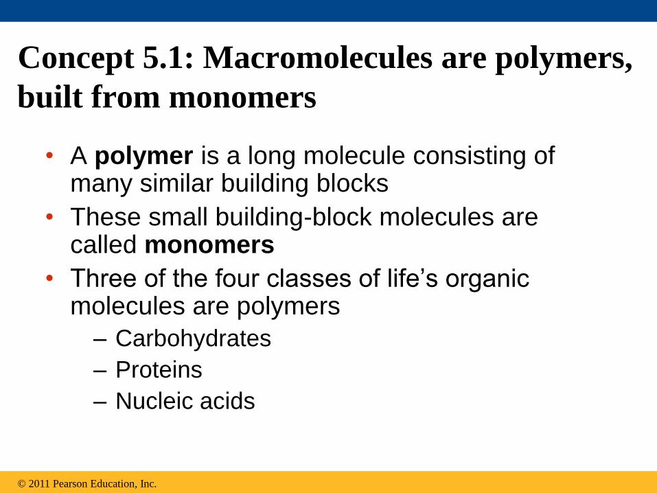
• A polymer is a long molecule consisting of many similar building blocks
• These small building-block molecules are called monomers
• Three of the four classes of life’s organic molecules are polymers
– Carbohydrates
– Proteins
– Nucleic acids
© 2011 Pearson Education, Inc.

• A dehydration reaction occurs when two
monomers bond together through the loss of a
water molecule
• Polymers are disassembled to monomers by
hydrolysis, a reaction that is essentially the
reverse of the dehydration reaction
The Synthesis and Breakdown of Polymers
© 2011 Pearson Education, Inc.
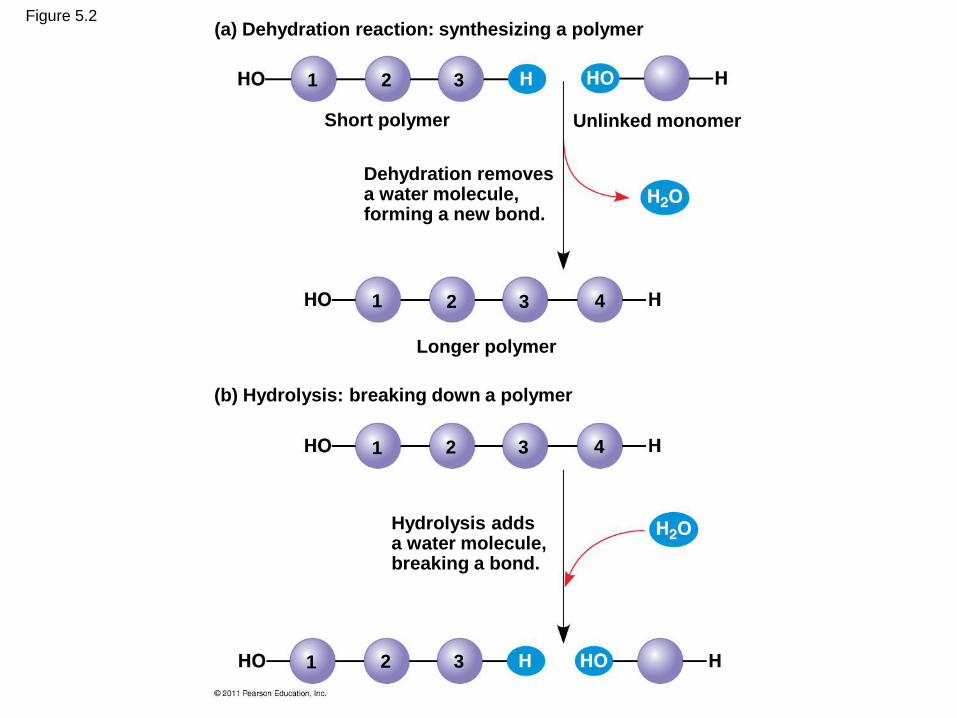
Figure 5.2 (a) Dehydration reaction: synthesizing a polymer
Short polymer Unlinked monomer
Dehydration removes a water molecule, forming a new bond.
Longer polymer
(b) Hydrolysis: breaking down a polymer
Hydrolysis adds a water molecule, breaking a bond.
1
1
1
2 3
2 3 4
2 3 4
1 2 3

The Diversity of Polymers
• Each cell has thousands of different
macromolecules
• Macromolecules vary among cells of an
organism, vary more within a species, and
vary even more between species
• An immense variety of polymers can be built
from a small set of monomers
HO
© 2011 Pearson Education, Inc.

Concept 5.2: Carbohydrates serve as fuel
and building material
• Carbohydrates include sugars and the
polymers of sugars
• The simplest carbohydrates are
monosaccharides, or single sugars
• Carbohydrate macromolecules are
polysaccharides, polymers composed of
many sugar building blocks
© 2011 Pearson Education, Inc.

Sugars
• Monosaccharides have molecular formulas
that are usually multiples of CH2O
• Glucose (C6H12O6) is the most common
monosaccharide
• Monosaccharides are classified by
– The location of the carbonyl group (as aldose
or ketose)
– The number of carbons in the carbon skeleton
© 2011 Pearson Education, Inc.
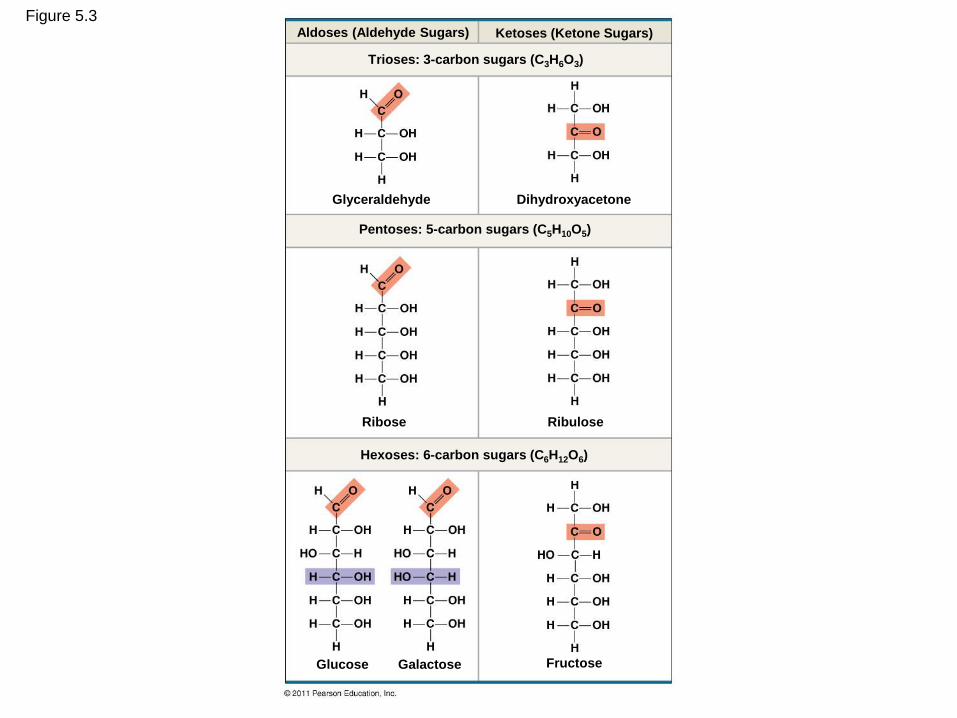
Figure 5.3 Aldoses (Aldehyde Sugars) Ketoses (Ketone Sugars)
Glyceraldehyde
Trioses: 3-carbon sugars (C3H6O3)
Dihydroxyacetone
Pentoses: 5-carbon sugars (C5H10O5)
Hexoses: 6-carbon sugars (C6H12O6)
Ribose Ribulose
Glucose Galactose Fructose
• Though often drawn as linear skeletons, in
aqueous solutions many sugars form rings
• Monosaccharides serve as a major fuel for
cells and as raw material for building
molecules
© 2011 Pearson Education, Inc.
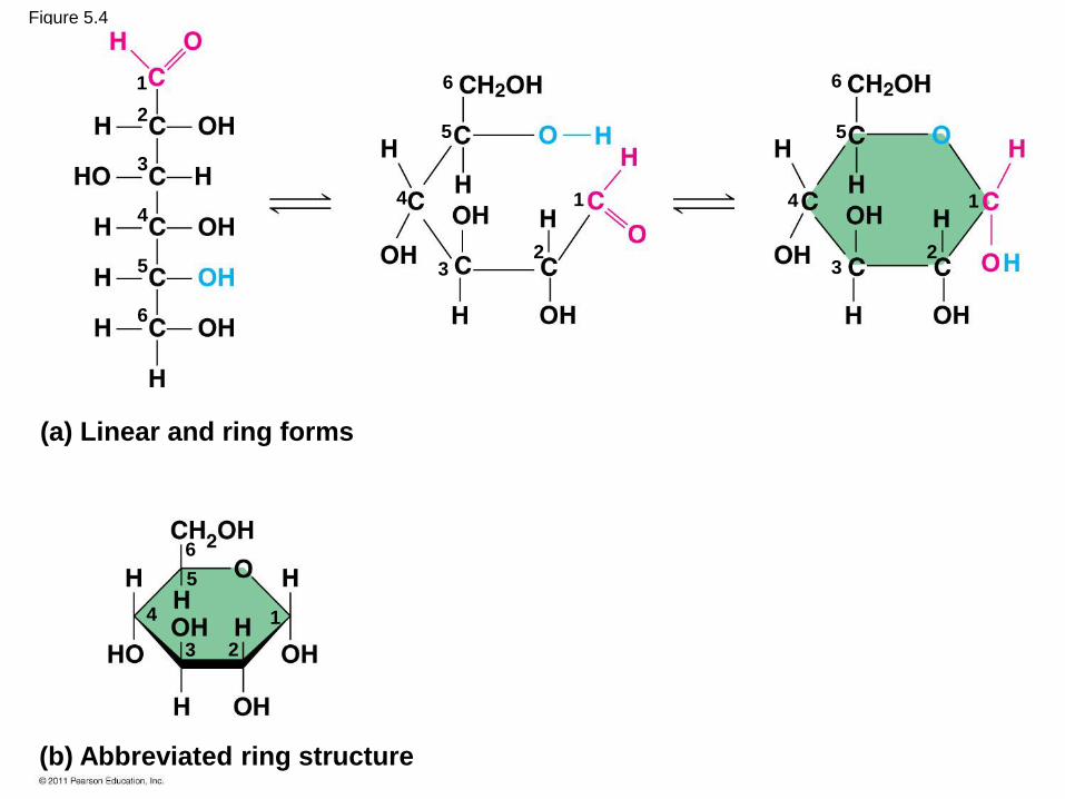
Figure 5.4
(a) Linear and ring forms
(b) Abbreviated ring structure
1
2
3
4
5
6
6
5
4
3 2
1 1
2 3
4
5
6
1
2 3
4
5
6
• A disaccharide is formed when a dehydration
reaction joins two monosaccharides
• This covalent bond is called a glycosidic
linkage
© 2011 Pearson Education, Inc.
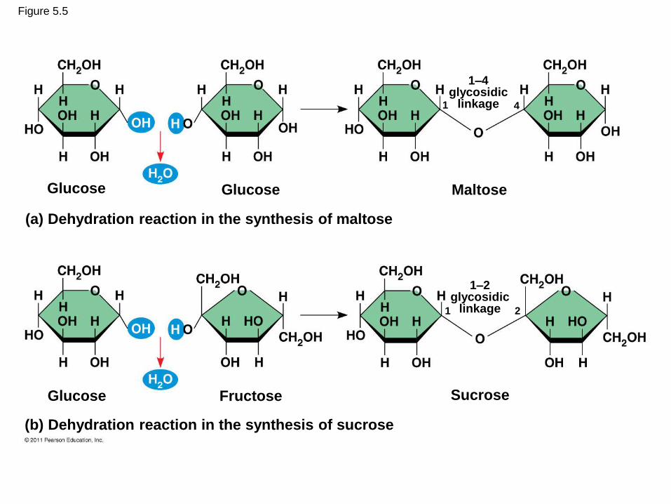
Figure 5.5
(a) Dehydration reaction in the synthesis of maltose
(b) Dehydration reaction in the synthesis of sucrose
Glucose Glucose
Glucose
Maltose
Fructose Sucrose
1–4 glycosidic
linkage
1–2 glycosidic
linkage
1 4
1 2
Polysaccharides
• Polysaccharides, the polymers of sugars,
have storage and structural roles
• The structure and function of a polysaccharide
are determined by its sugar monomers and the
positions of glycosidic linkages
© 2011 Pearson Education, Inc.
Storage Polysaccharides
• Starch, a storage polysaccharide of plants, consists entirely of glucose monomers
• Plants store surplus starch as granules within chloroplasts and other plastids
• The simplest form of starch is amylose
© 2011 Pearson Education, Inc.

Figure 5.6
(a) Starch: a plant polysaccharide
(b) Glycogen: an animal polysaccharide
Chloroplast Starch granules
Mitochondria Glycogen granules
Amylopectin
Amylose
Glycogen
1 m
0.5 m

• Glycogen is a storage polysaccharide in
animals
• Humans and other vertebrates store
glycogen mainly in liver and muscle cells
© 2011 Pearson Education, Inc.
Structural Polysaccharides
• The polysaccharide cellulose is a major
component of the tough wall of plant cells
• Like starch, cellulose is a polymer of glucose,
but the glycosidic linkages differ
• The difference is based on two ring forms for
glucose: alpha () and beta ()
© 2011 Pearson Education, Inc.

Figure 5.7
(a) and glucose ring structures
(b) Starch: 1–4 linkage of glucose monomers (c) Cellulose: 1–4 linkage of glucose monomers
Glucose Glucose
4 1 4 1
4 1 4 1

© 2011 Pearson Education, Inc.
• Polymers with glucose are helical
• Polymers with glucose are straight
• In straight structures, H atoms on one
strand can bond with OH groups on other
strands
• Parallel cellulose molecules held together
this way are grouped into microfibrils,
which form strong building materials for
plants
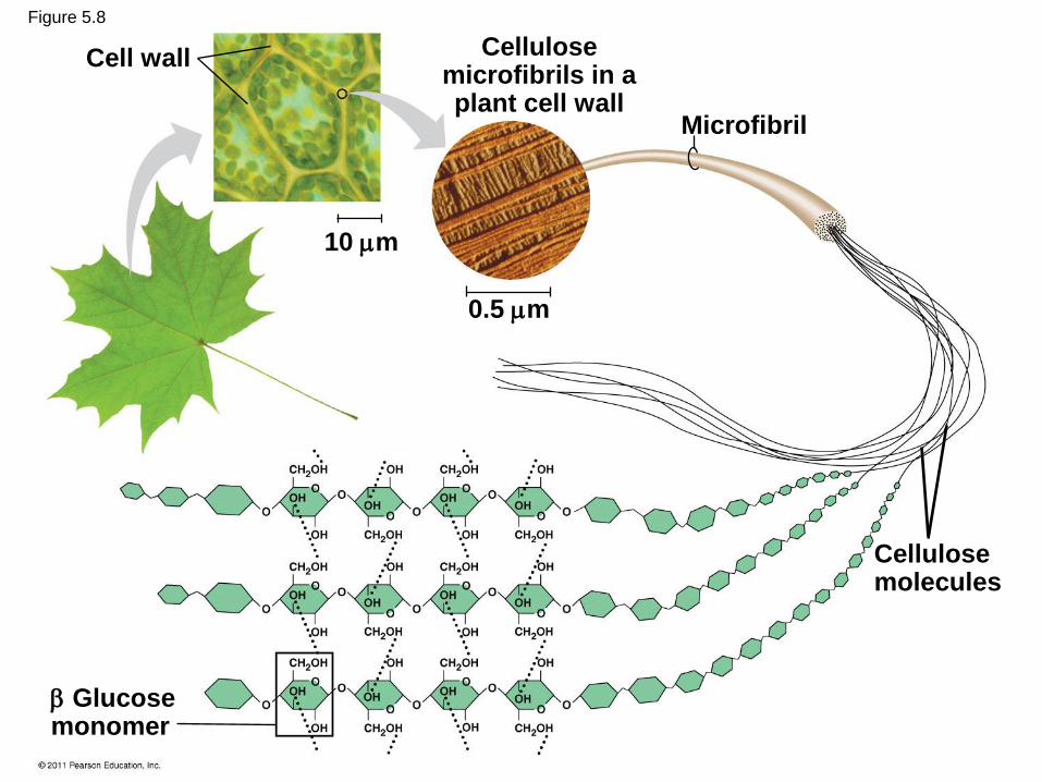
Cell wall
Microfibril
Cellulose microfibrils in a plant cell wall
Cellulose molecules
Glucose monomer
10 m
0.5 m
Figure 5.8

• Enzymes that digest starch by hydrolyzing
linkages can’t hydrolyze linkages in cellulose
• Cellulose in human food passes through the
digestive tract as insoluble fiber
• Some microbes use enzymes to digest
cellulose
• Many herbivores, from cows to termites, have
symbiotic relationships with these microbes
© 2011 Pearson Education, Inc.
• Chitin, another structural polysaccharide, is
found in the exoskeleton of arthropods
• Chitin also provides structural support for the
cell walls of many fungi
© 2011 Pearson Education, Inc.

Figure 5.9
Chitin forms the exoskeleton of arthropods.
The structure of the chitin monomer
Chitin is used to make a strong and flexible surgical thread that decomposes after the wound or incision heals.
Concept 5.3: Lipids are a diverse group of
hydrophobic molecules
• Lipids are the one class of large biological
molecules that do not form polymers
• The unifying feature of lipids is having little or
no affinity for water
• Lipids are hydrophobic becausethey consist
mostly of hydrocarbons, which form nonpolar
covalent bonds
• The most biologically important lipids are fats,
phospholipids, and steroids
© 2011 Pearson Education, Inc.
Fats
• Fats are constructed from two types of smaller
molecules: glycerol and fatty acids
• Glycerol is a three-carbon alcohol with a
hydroxyl group attached to each carbon
• A fatty acid consists of a carboxyl group
attached to a long carbon skeleton
© 2011 Pearson Education, Inc.

Figure 5.10
(a) One of three dehydration reactions in the synthesis of a fat
(b) Fat molecule (triacylglycerol)
Fatty acid (in this case, palmitic acid)
Glycerol
Ester linkage
© 2011 Pearson Education, Inc.
• Fats separate from water because water
molecules form hydrogen bonds with each
other and exclude the fats
• In a fat, three fatty acids are joined to
glycerol by an ester linkage, creating a
triacylglycerol, or triglyceride

• Fatty acids vary in length (number of carbons)
and in the number and locations of double
bonds
• Saturated fatty acids have the maximum
number of hydrogen atoms possible and no
double bonds
• Unsaturated fatty acids have one or more
double bonds
© 2011 Pearson Education, Inc.
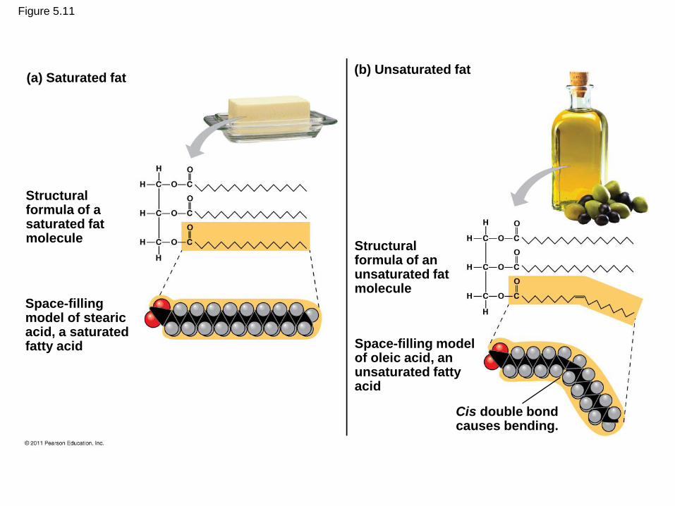
Figure 5.11
(a) Saturated fat (b) Unsaturated fat
Structural formula of a saturated fat molecule
Space-filling model of stearic acid, a saturated fatty acid
Structural formula of an unsaturated fat molecule
Space-filling model of oleic acid, an unsaturated fatty acid
Cis double bond causes bending.
• Fats made from saturated fatty acids are
called saturated fats, and are solid at room
temperature
• Most animal fats are saturated
• Fats made from unsaturated fatty acids are
called unsaturated fats or oils, and are liquid
at room temperature
• Plant fats and fish fats are usually unsaturated
© 2011 Pearson Education, Inc.
• A diet rich in saturated fats may contribute to
cardiovascular disease through plaque deposits
• Hydrogenation is the process of converting
unsaturated fats to saturated fats by adding
hydrogen
• Hydrogenating vegetable oils also creates
unsaturated fats with trans double bonds
• These trans fats may contribute more than
saturated fats to cardiovascular disease
© 2011 Pearson Education, Inc.

• Certain unsaturated fatty acids are not
synthesized in the human body
• These must be supplied in the diet
• These essential fatty acids include the omega-3
fatty acids, required for normal growth, and
thought to provide protection against
cardiovascular disease
© 2011 Pearson Education, Inc.
• The major function of fats is energy storage
• Humans and other mammals store their fat in
adipose cells
• Adipose tissue also cushions vital organs and
insulates the body
© 2011 Pearson Education, Inc.
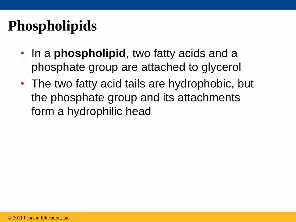
Phospholipids
• In a phospholipid, two fatty acids and a
phosphate group are attached to glycerol
• The two fatty acid tails are hydrophobic, but
the phosphate group and its attachments
form a hydrophilic head
© 2011 Pearson Education, Inc.

Figure 5.12
Choline
Phosphate
Glycerol
Fatty acids
Hydrophilic head
Hydrophobic tails
(c) Phospholipid symbol (b) Space-filling model (a) Structural formula
Hyd
rop
hil
ic h
ea
d
Hyd
rop
ho
bic
ta
ils
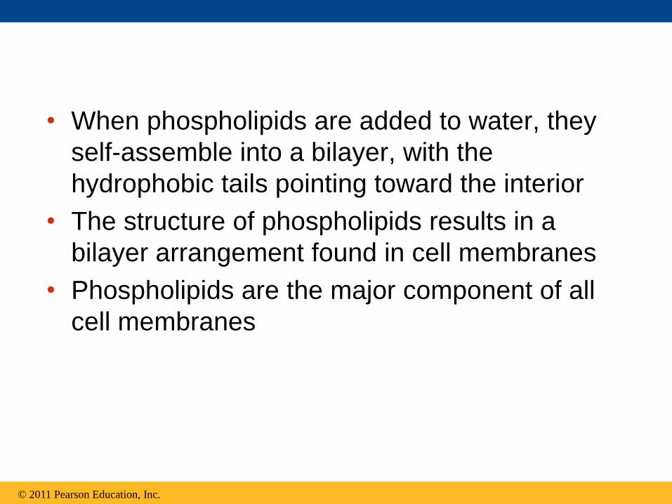
• When phospholipids are added to water, they
self-assemble into a bilayer, with the
hydrophobic tails pointing toward the interior
• The structure of phospholipids results in a
bilayer arrangement found in cell membranes
• Phospholipids are the major component of all
cell membranes
© 2011 Pearson Education, Inc.

Figure 5.13
Hydrophilic head
Hydrophobic tail
WATER
WATER

Steroids
• Steroids are lipids characterized by a carbon
skeleton consisting of four fused rings
• Cholesterol, an important steroid, is a
component in animal cell membranes
• Although cholesterol is essential in animals,
high levels in the blood may contribute to
cardiovascular disease
© 2011 Pearson Education, Inc.

Figure 5.14

Concept 5.4: Proteins include a diversity of
structures, resulting in a wide range of
functions
• Proteins account for more than 50% of the dry
mass of most cells
• Protein functions include structural support,
storage, transport, cellular communications,
movement, and defense against foreign
substances
© 2011 Pearson Education, Inc.

Figure 5.15-a
Enzymatic proteins Defensive proteins
Storage proteins Transport proteins
Enzyme Virus
Antibodies
Bacterium
Ovalbumin Amino acids for embryo
Transport protein
Cell membrane
Function: Selective acceleration of chemical reactions Example: Digestive enzymes catalyze the hydrolysis
of bonds in food molecules.
Function: Protection against disease
Example: Antibodies inactivate and help destroy
viruses and bacteria.
Function: Storage of amino acids Function: Transport of substances
Examples: Casein, the protein of milk, is the major
source of amino acids for baby mammals. Plants have
storage proteins in their seeds. Ovalbumin is the
protein of egg white, used as an amino acid source
for the developing embryo.
Examples: Hemoglobin, the iron-containing protein of
vertebrate blood, transports oxygen from the lungs to
other parts of the body. Other proteins transport
molecules across cell membranes.

Figure 5.15-b
Hormonal proteins
Function: Coordination of an organism’s activities
Example: Insulin, a hormone secreted by the
pancreas, causes other tissues to take up glucose,
thus regulating blood sugar concentration
High blood sugar
Normal blood sugar
Insulin secreted
Signaling molecules
Receptor protein
Muscle tissue
Actin Myosin
100 m 60 m
Collagen
Connective tissue
Receptor proteins
Function: Response of cell to chemical stimuli
Example: Receptors built into the membrane of a
nerve cell detect signaling molecules released by
other nerve cells.
Contractile and motor proteins
Function: Movement
Examples: Motor proteins are responsible for the
undulations of cilia and flagella. Actin and myosin
proteins are responsible for the contraction of
muscles.
Structural proteins
Function: Support
Examples: Keratin is the protein of hair, horns,
feathers, and other skin appendages. Insects and
spiders use silk fibers to make their cocoons and webs,
respectively. Collagen and elastin proteins provide a
fibrous framework in animal connective tissues.

• Enzymes are a type of protein that acts as a
catalyst to speed up chemical reactions
• Enzymes can perform their functions
repeatedly, functioning as workhorses that
carry out the processes of life
© 2011 Pearson Education, Inc.
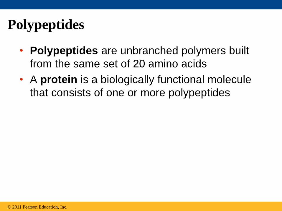
Polypeptides
• Polypeptides are unbranched polymers built
from the same set of 20 amino acids
• A protein is a biologically functional molecule
that consists of one or more polypeptides
© 2011 Pearson Education, Inc.

Amino Acid Monomers
• Amino acids are organic molecules with
carboxyl and amino groups
• Amino acids differ in their properties due to
differing side chains, called R groups
© 2011 Pearson Education, Inc.

Figure 5.UN01
Side chain (R group)
Amino group
Carboxyl group
carbon
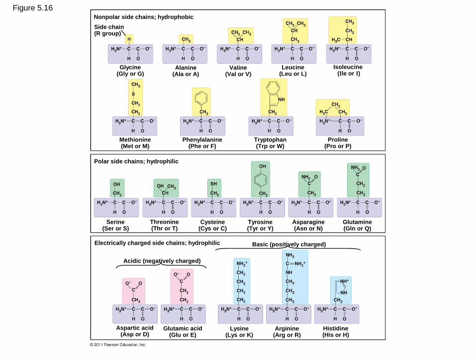
Figure 5.16 Nonpolar side chains; hydrophobic
Side chain (R group)
Glycine (Gly or G)
Alanine (Ala or A)
Valine (Val or V)
Leucine (Leu or L)
Isoleucine (Ile or I)
Methionine (Met or M)
Phenylalanine (Phe or F)
Tryptophan (Trp or W)
Proline (Pro or P)
Polar side chains; hydrophilic
Serine (Ser or S)
Threonine (Thr or T)
Cysteine (Cys or C)
Tyrosine (Tyr or Y)
Asparagine (Asn or N)
Glutamine (Gln or Q)
Electrically charged side chains; hydrophilic
Acidic (negatively charged)
Basic (positively charged)
Aspartic acid (Asp or D)
Glutamic acid (Glu or E)
Lysine (Lys or K)
Arginine (Arg or R)
Histidine (His or H)

Amino Acid Polymers
• Amino acids are linked by peptide bonds
• A polypeptide is a polymer of amino acids
• Polypeptides range in length from a few to
more than a thousand monomers
• Each polypeptide has a unique linear
sequence of amino acids, with a carboxyl end
(C-terminus) and an amino end (N-terminus)
© 2011 Pearson Education, Inc.

Figure 5.17
Peptide bond
New peptide bond forming
Side chains
Back- bone
Amino end (N-terminus)
Peptide bond
Carboxyl end (C-terminus)
Protein Structure and Function
• A functional protein consists of one or more
polypeptides precisely twisted, folded, and
coiled into a unique shape
© 2011 Pearson Education, Inc.
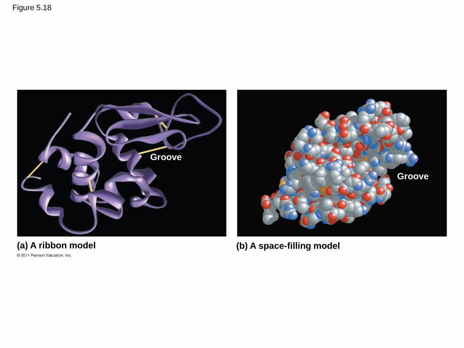
Figure 5.18
(a) A ribbon model (b) A space-filling model
Groove
Groove

• The sequence of amino acids determines a
protein’s three-dimensional structure
• A protein’s structure determines its function
© 2011 Pearson Education, Inc.

Figure 5.19
Antibody protein Protein from flu virus

Four Levels of Protein Structure
• The primary structure of a protein is its unique
sequence of amino acids
• Secondary structure, found in most proteins,
consists of coils and folds in the polypeptide
chain
• Tertiary structure is determined by interactions
among various side chains (R groups)
• Quaternary structure results when a protein
consists of multiple polypeptide chains
© 2011 Pearson Education, Inc.

Figure 5.20a Primary structure
Amino acids
Amino end
Carboxyl end
Primary structure of transthyretin

• Primary structure, the sequence of amino
acids in a protein, is like the order of letters
in a long word
• Primary structure is determined by inherited
genetic information
© 2011 Pearson Education, Inc.
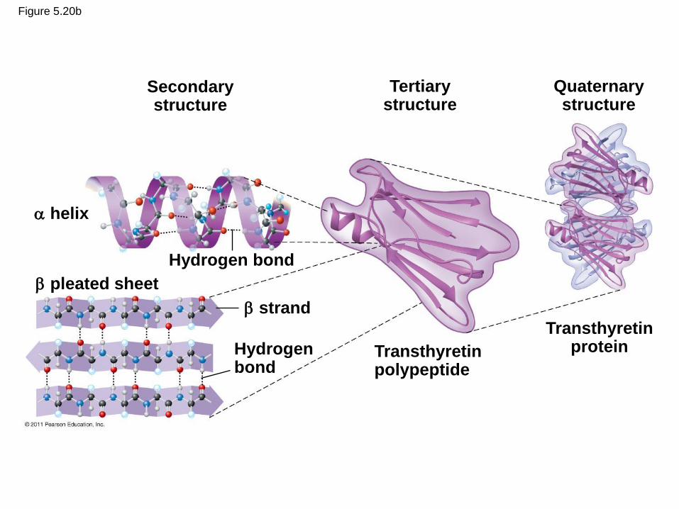
Figure 5.20b
Secondary structure
Tertiary structure
Quaternary structure
Hydrogen bond
helix
pleated sheet
strand
Hydrogen bond
Transthyretin polypeptide
Transthyretin protein

• The coils and folds of secondary structure
result from hydrogen bonds between repeating
constituents of the polypeptide backbone
• Typical secondary structures are a coil called an helix and a folded structure called a pleated sheet
© 2011 Pearson Education, Inc.
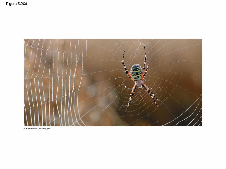
Figure 5.20d
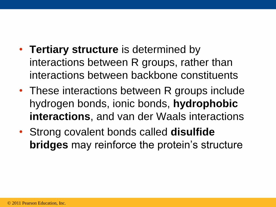
• Tertiary structure is determined by
interactions between R groups, rather than
interactions between backbone constituents
• These interactions between R groups include
hydrogen bonds, ionic bonds, hydrophobic
interactions, and van der Waals interactions
• Strong covalent bonds called disulfide
bridges may reinforce the protein’s structure
© 2011 Pearson Education, Inc.

Figure 5.20f
Hydrogen bond
Disulfide bridge
Polypeptide backbone
Ionic bond
Hydrophobic
interactions and
van der Waals
interactions
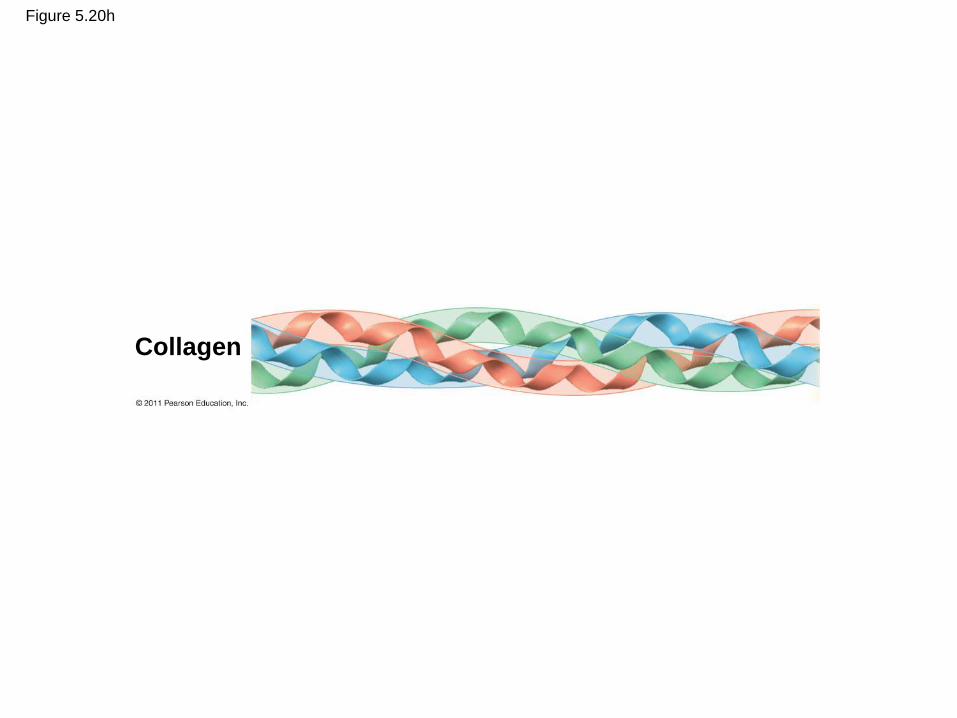
Figure 5.20h
Collagen

Hemoglobin
Heme
Iron
subunit
subunit
subunit
subunit
Figure 5.20i

• Quaternary structure results when two or
more polypeptide chains form one
macromolecule
• Collagen is a fibrous protein consisting of three
polypeptides coiled like a rope
• Hemoglobin is a globular protein consisting of
four polypeptides: two alpha and two beta
chains
© 2011 Pearson Education, Inc.

Sickle-Cell Disease: A Change in Primary
Structure
• A slight change in primary structure can affect
a protein’s structure and ability to function
• Sickle-cell disease, an inherited blood
disorder, results from a single amino acid
substitution in the protein hemoglobin
© 2011 Pearson Education, Inc.
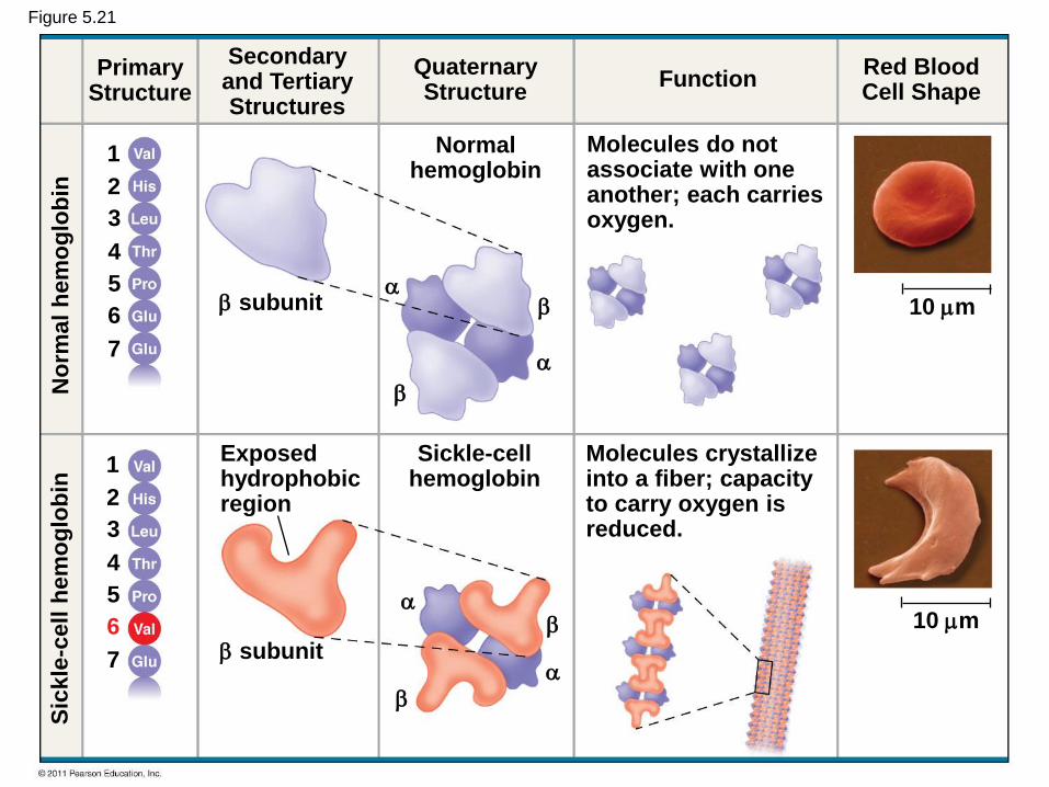
Figure 5.21
Primary Structure
Secondary and Tertiary Structures
Quaternary Structure
Function Red Blood Cell Shape
subunit
subunit
Exposed hydrophobic region
Molecules do not associate with one another; each carries oxygen.
Molecules crystallize into a fiber; capacity to carry oxygen is reduced.
Sickle-cell hemoglobin
Normal hemoglobin
10 m
10 m
Sic
kle
-cell h
em
og
lob
in
No
rma
l h
em
og
lob
in
1
2
3
4
5
6
7
1
2
3
4
5
6
7
What Determines Protein Structure?
• In addition to primary structure, physical and
chemical conditions can affect structure
• Alterations in pH, salt concentration,
temperature, or other environmental factors
can cause a protein to unravel
• This loss of a protein’s native structure is
called denaturation
• A denatured protein is biologically inactive
© 2011 Pearson Education, Inc.

Figure 5.22
Normal protein Denatured protein
tu
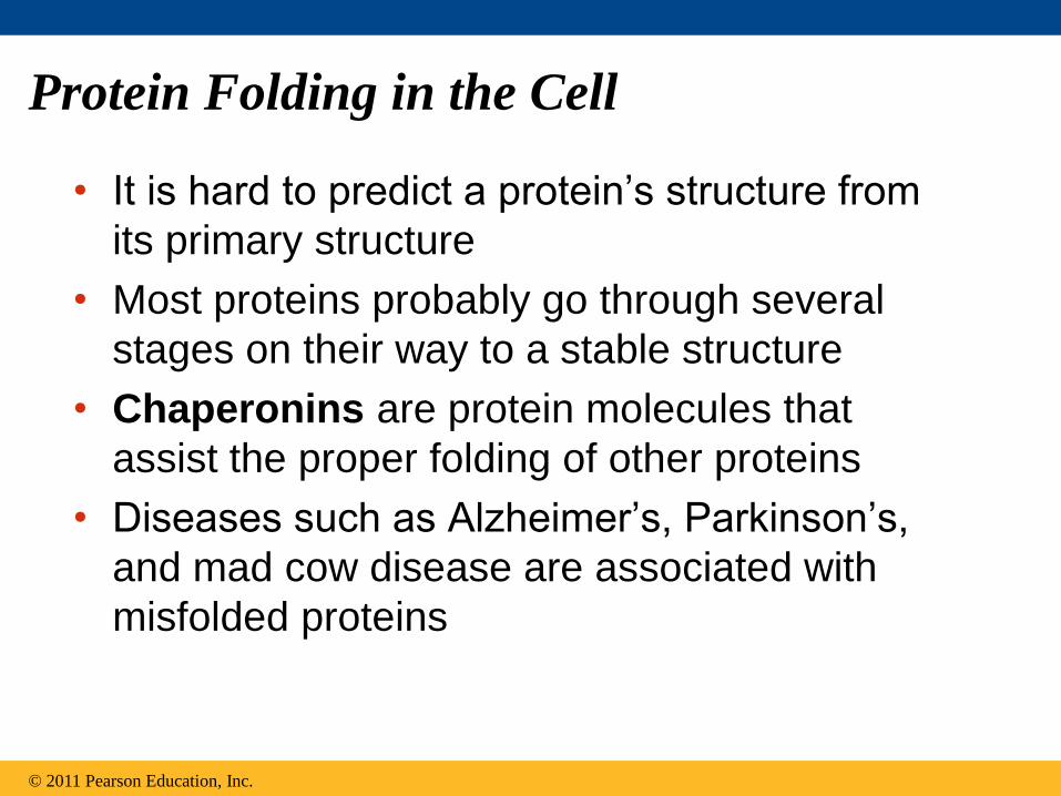
Protein Folding in the Cell
• It is hard to predict a protein’s structure from
its primary structure
• Most proteins probably go through several
stages on their way to a stable structure
• Chaperonins are protein molecules that
assist the proper folding of other proteins
• Diseases such as Alzheimer’s, Parkinson’s,
and mad cow disease are associated with
misfolded proteins
© 2011 Pearson Education, Inc.

Figure 5.23
The cap attaches, causing the cylinder to change shape in such a way that it creates a hydrophilic environment for the folding of the polypeptide.
Cap
Polypeptide
Correctly folded protein
Chaperonin (fully assembled)
Steps of Chaperonin Action:
An unfolded poly- peptide enters the cylinder from one end.
Hollow cylinder
The cap comes off, and the properly folded protein is released.
1
2 3
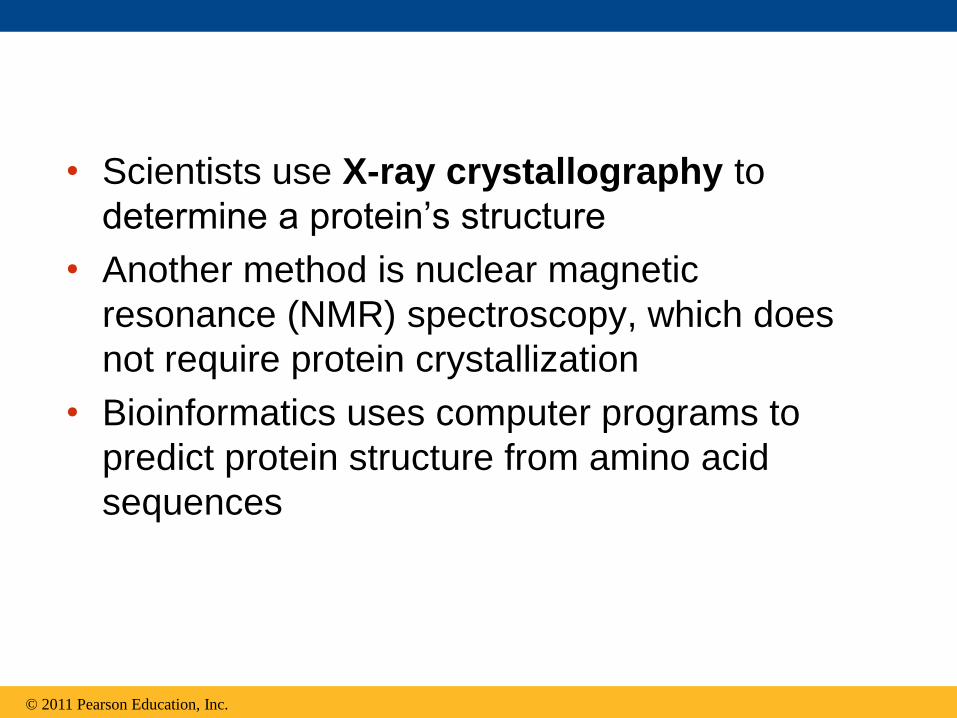
• Scientists use X-ray crystallography to
determine a protein’s structure
• Another method is nuclear magnetic
resonance (NMR) spectroscopy, which does
not require protein crystallization
• Bioinformatics uses computer programs to
predict protein structure from amino acid
sequences
© 2011 Pearson Education, Inc.

Figure 5.24
Diffracted X-rays
X-ray source X-ray
beam
Crystal Digital detector X-ray diffraction pattern
RNA DNA
RNA polymerase II
EXPERIMENT
RESULTS

Concept 5.5: Nucleic acids store, transmit,
and help express hereditary information
• The amino acid sequence of a polypeptide is
programmed by a unit of inheritance called a
gene
• Genes are made of DNA, a nucleic acid
made of monomers called nucleotides
© 2011 Pearson Education, Inc.
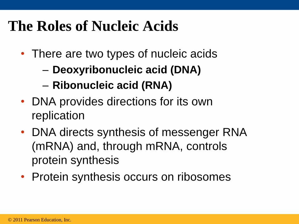
The Roles of Nucleic Acids
• There are two types of nucleic acids
– Deoxyribonucleic acid (DNA)
– Ribonucleic acid (RNA)
• DNA provides directions for its own
replication
• DNA directs synthesis of messenger RNA
(mRNA) and, through mRNA, controls
protein synthesis
• Protein synthesis occurs on ribosomes
© 2011 Pearson Education, Inc.
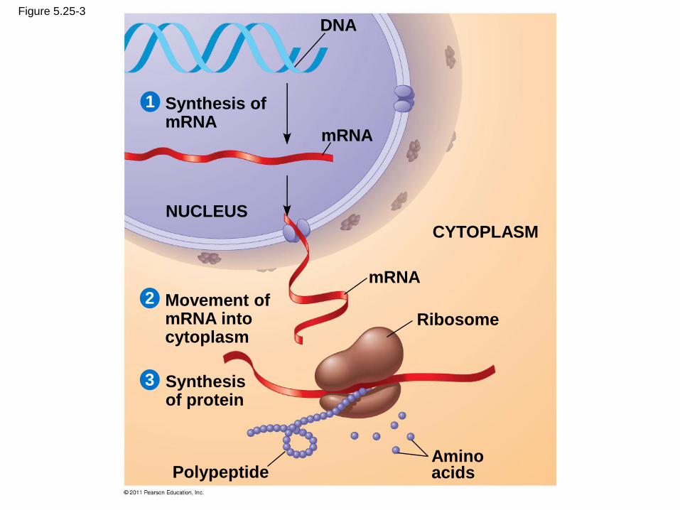
Figure 5.25-3
Synthesis of mRNA
mRNA
DNA
NUCLEUS
CYTOPLASM
mRNA
Ribosome
Amino acids Polypeptide
Movement of mRNA into cytoplasm
Synthesis of protein
1
2
3
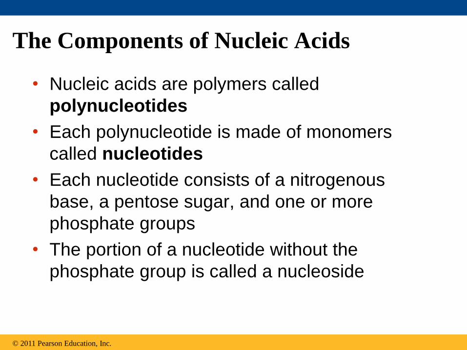
The Components of Nucleic Acids
• Nucleic acids are polymers called
polynucleotides
• Each polynucleotide is made of monomers
called nucleotides
• Each nucleotide consists of a nitrogenous
base, a pentose sugar, and one or more
phosphate groups
• The portion of a nucleotide without the
phosphate group is called a nucleoside
© 2011 Pearson Education, Inc.

Figure 5.26
Sugar-phosphate backbone 5 end
5C
3C
5C
3C
3 end
(a) Polynucleotide, or nucleic acid
(b) Nucleotide
Phosphate group Sugar
(pentose)
Nucleoside
Nitrogenous base
5C
3C
1C
Nitrogenous bases
Cytosine (C) Thymine (T, in DNA) Uracil (U, in RNA)
Adenine (A) Guanine (G)
Sugars
Deoxyribose (in DNA) Ribose (in RNA)
(c) Nucleoside components
Pyrimidines
Purines

• Nucleoside = nitrogenous base + sugar
• There are two families of nitrogenous bases
– Pyrimidines (cytosine, thymine, and uracil)
have a single six-membered ring
– Purines (adenine and guanine) have a six-
membered ring fused to a five-membered ring
• In DNA, the sugar is deoxyribose; in RNA, the
sugar is ribose
• Nucleotide = nucleoside + phosphate group
© 2011 Pearson Education, Inc.

Nucleotide Polymers
• Nucleotide polymers are linked together to build
a polynucleotide
• Adjacent nucleotides are joined by covalent
bonds that form between the —OH group on the
3 carbon of one nucleotide and the phosphate
on the 5 carbon on the next
• These links create a backbone of sugar-
phosphate units with nitrogenous bases as
appendages
• The sequence of bases along a DNA or mRNA
polymer is unique for each gene
© 2011 Pearson Education, Inc.

The Structures of DNA and RNA Molecules
• RNA molecules usually exist as single
polypeptide chains
• DNA molecules have two polynucleotides
spiraling around an imaginary axis, forming a
double helix
• In the DNA double helix, the two backbones
run in opposite 5→ 3 directions from each
other, an arrangement referred to as
antiparallel
• One DNA molecule includes many genes
© 2011 Pearson Education, Inc.

• The nitrogenous bases in DNA pair up and form
hydrogen bonds: adenine (A) always with
thymine (T), and guanine (G) always with
cytosine (C)
• Called complementary base pairing
• Complementary pairing can also occur between
two RNA molecules or between parts of the same
molecule
• In RNA, thymine is replaced by uracil (U) so A
and U pair
© 2011 Pearson Education, Inc.
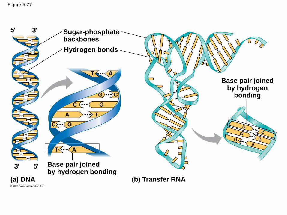
Figure 5.27
Sugar-phosphate backbones
Hydrogen bonds
Base pair joined by hydrogen bonding
Base pair joined by hydrogen
bonding
(b) Transfer RNA (a) DNA
5 3
5 3

DNA and Proteins as Tape Measures of
Evolution
• The linear sequences of nucleotides in DNA
molecules are passed from parents to offspring
• Two closely related species are more similar in
DNA than are more distantly related species
• Molecular biology can be used to assess
evolutionary kinship
© 2011 Pearson Education, Inc.
The Theme of Emergent Properties in the
Chemistry of Life: A Review
• Higher levels of organization result in the
emergence of new properties
• Organization is the key to the chemistry of life
© 2011 Pearson Education, Inc.
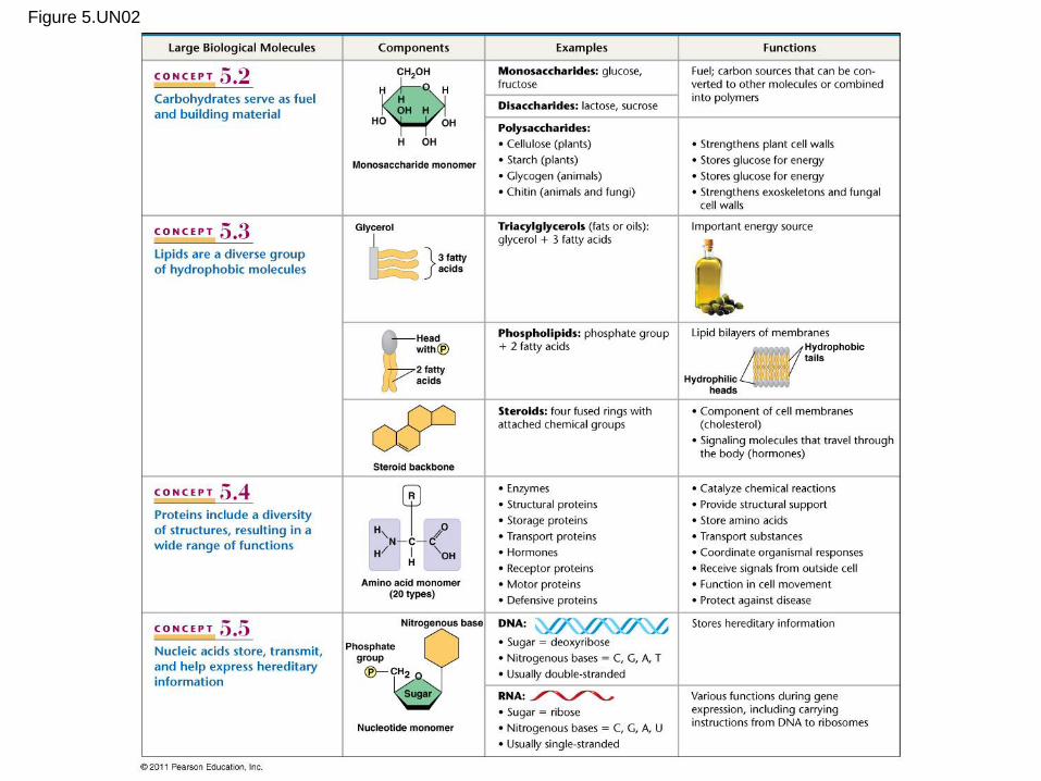
Figure 5.UN02

Figure 5.UN02a

Figure 5.UN02b