the structure of rauvolfia serpentina strictosidine ...the structure of rauvolfia serpentina...
TRANSCRIPT
The Structure of Rauvolfia serpentina Strictosidine Synthase Isa Novel Six-Bladed b-Propeller Fold in Plant Proteins W
Xueyan Ma,a,1 Santosh Panjikar,b,1 Juergen Koepke,c Elke Loris,a and Joachim Stockigta,d,2
a Department of Pharmaceutical Biology, Institute of Pharmacy, Johannes Gutenberg-University, D-55099 Mainz, Germanyb European Molecular Biology Laboratory Hamburg Outstation DESY, D-22603 Hamburg, Germanyc Department of Molecular Membrane Biology, Max-Planck-Institute of Biophysics, 60438 Frankfurt, Germanyd College of Pharmaceutical Sciences, Zhejiang University, Hangzhou 310031, China
The enzyme strictosidine synthase (STR1) from the Indian medicinal plant Rauvolfia serpentina is of primary importance for
the biosynthetic pathway of the indole alkaloid ajmaline. Moreover, STR1 initiates all biosynthetic pathways leading to the
entire monoterpenoid indole alkaloid family representing an enormous structural variety of ;2000 compounds in higher
plants. The crystal structures of STR1 in complex with its natural substrates tryptamine and secologanin provide structural
understanding of the observed substrate preference and identify residues lining the active site surface that contact the
substrates. STR1 catalyzes a Pictet-Spengler–type reaction and represents a novel six-bladed b-propeller fold in plant
proteins. Structure-based sequence alignment revealed a common repetitive sequence motif (three hydrophobic residues
are followed by a small residue and a hydrophilic residue), indicating a possible evolutionary relationship between STR1 and
several sequence-unrelated six-bladed b-propeller structures. Structural analysis and site-directed mutagenesis experi-
ments demonstrate the essential role of Glu-309 in catalysis. The data will aid in deciphering the details of the reaction
mechanism of STR1 as well as other members of this enzyme family.
INTRODUCTION
Themonoterpenoid-derived indole alkaloids compose one of the
structurally largest and pharmacologically most diverse alkaloid
families in higher plants.Of the;2000members,whichare divided
into various structural classes, several have long-standing med-
ical applications. Prominent examples of developed therapeutics
include the treatment of cancer (vinblastine or the camptothecin
derivative topotecan), malaria (quinine), hypertension (raubasine
and reserpine), schizophrenia (reserpine in high dosage), dis-
turbed cerebral blood flow (vincamine), and antiarrhythmic heart
disorders (ajmaline, from the Indian medicinal plant Rauvolfia
serpentina). Strictosidine synthase (STR1; EC 4.3.3.2) is involved
in the biosynthesis of all these alkaloids by catalyzing the con-
densation of the two initial building blocks, tryptamine and the
monoterpenoid secologanin, leading to the glucoalkaloid stric-
tosidine. As the first committed step, STR1 seems to play the
principal role in nature’s strategy to generate the entire mono-
terpenoid indole alkaloid family (Figure 1; see reviews in Kutchan,
1993; Stockigt and Ruppert, 1999).
The reaction type catalyzed by STR1 is so far an exceptional
example in the biosynthesis of natural products. It was hitherto
known only from synthetic chemistry (Pictet-Spengler–type re-
action), where it is applied in alkaloid synthesis, especially of
tetrahydroisoquinolinesbycondensationofanamineandanalde-
hyde under acidic conditions. The only enzymes known to date
that are functionally related to STR1 are deacetylisoipecoside
synthase, deacetylipecoside synthase, and norcoclaurine syn-
thase. The first two enzymes catalyze the condensation of dopa-
mine instead of tryptamine with secologanin leading to the family
of monoterpenoid tetrahydroisoquinoline alkaloids (De-Eknamkul
et al., 2000). Norcoclaurine synthase catalyzes the condensation
of dopamine and 4-hydroxyphenylacetaldehyde, resulting in the
biosynthesis of ;6000 benzylisoquinoline alkaloids (such as
morphine, sanguinarine, or berberine; Samanani et al., 2004).
Although STR1 and its functional related enzymes are key en-
zymes in the biosynthesis of ;50% of all alkaloids, very little is
known either about their reaction mechanisms or about the
amino acids essential for enzymatic activity.
STR1 was first isolated from plant cell suspensions of Cathar-
anthus roseus and R. serpentina and was later detected and
purified from many other species of the plant families Apocyna-
ceae and Rubiaceae, such as Vinca, Cinchona, and Ophiorrhiza
(Treimer and Zenk, 1979; Hampp and Zenk, 1988; Bracher and
Kutchan, 1992; Stevens et al., 1993; DeWaal et al., 1995;
Yamazaki et al., 2003). STR1 from R. serpentina is a monomeric
precursor protein with 344 amino acids (Kutchan, 1993) that
exhibits 100, 79, and 58% identity to STR1 fromRauvolfiamannii,
C. roseus, andOphiorrhiza pumila, respectively. STR1 from these
medicinal plants together with >100 STR1-related genes derived
from UniProt protein database constitute a sequence-related
strictosidine synthase family, which is namedafter the first enzyme
found for this family. Except for STR1, the biological functions of
1 These authors contributed equally to this work.2 To whom correspondence should be addressed. E-mail [email protected]; fax 49-6131-39-23752.The author responsible for distribution of materials integral to thefindings presented in this article in accordance with the policy describedin the Instructions for Authors (www.plantcell.org) is: Joachim Stockigt([email protected]).WOnline version contains Web-only data.Article, publication date, and citation information can be found atwww.plantcell.org/cgi/doi/10.1105/tpc.105.038018.
The Plant Cell, Vol. 18, 907–920, April 2006, www.plantcell.orgª 2006 American Society of Plant Biologists

most of the other family members from both plants and animals
have not yet been identified.Since no significant sequencehomol-
ogy could be detected between STR1 and known protein struc-
tures, the three-dimensional structure of STR1 might help to
understand the structural relationship between STR1 and the
other family members and would provide a solid base for
modeling of other family members.
We recently described an expression system for the produc-
tion of STR1 from R. serpentina in the milligram range and the
protocol for crystallization of this enzyme (Ma et al., 2004; Koepke
et al., 2005). Here, we report the crystal structure ofR. serpentina
STR1 and also the structure of STR1 in complex with each of the
natural substrates tryptamine and secologanin. The structure
represents a novel six-bladed b-propeller fold in plant proteins.
The common features of b-propeller fold proteins are their se-
quence and functional diversity, which led people to believe that
sequence-unrelated propeller structures have evolved from dif-
ferent precursors. However, based on the structure of STR1 and
structure-based sequence alignments, we find a possible evo-
lutionary relationship of several sequence-unrelated six-bladed
b-propeller structures. The complex structures also allow us to
examine issues relating to catalysis, including the architecture of
the active site and the nature of the substrate binding pocket,
which provide significant hints and the structural basis to unravel
the unexplored reactionof this uniqueplant enzyme. These struc-
turesmight also serve as a starting point for the rational construc-
tion of new STR1 variants with various substrate acceptances,
which could lead to novel biologically valuable indole alkaloids.
RESULTS AND DISCUSSION
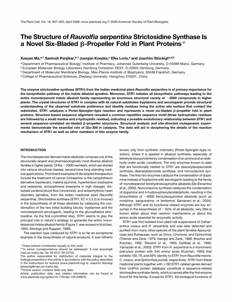
Overall Architecture of STR1
The overall structure of STR1 fromR. serpentina resembles a six-
bladed four-stranded b-propeller fold (the six blades are indi-
cated from 1 to 6 in Figures 2A and 2B). All six blades are radially
arranged around a pseudo six-fold symmetry axis. Each blade
contains a twisted four-stranded antiparallel b-sheet. The
b-strands in each blade are labeled A to D from the inside to
Figure 1. STR1 Catalyzes the Stereo-Specific Condensation of Tryptamine and Secologanin Leading to 3a(S)-Strictosidine: A Central Reaction in the
Biosynthesis of the Entire Family of Monoterpenoid Indole Alkaloids in Plants.
908 The Plant Cell
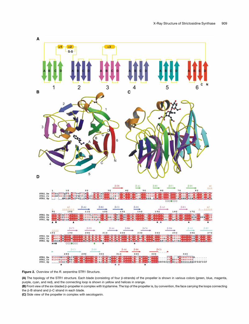
Figure 2. Overview of the R. serpentina STR1 Structure.
(A) The topology of the STR1 structure. Each blade (consisting of four b-strands) of the propeller is shown in various colors (green, blue, magenta,
purple, cyan, and red), and the connecting loop is shown in yellow and helices in orange.
(B) Front view of the six-bladed b-propeller in complex with tryptamine. The top of the propeller is, by convention, the face carrying the loops connecting
the b-B strand and b-C strand in each blade.
(C) Side view of the propeller in complex with secologanin.
X-Ray Structure of Strictosidine Synthase 909

outside of the molecule. The innermost b-strand A (b-A) is
closest to the pseudo six-fold axis. The last blade is formed by
three strands b-A, b-B, and b-C from the C terminus and one (the
outermost strand, b-D) from the N terminus. This type of Velcro
closure has already been observed for several other six-bladed
b-propeller structures exhibiting different catalytic functions
(Scharff et al., 2001; Harel et al., 2004). There are two STR1 mol-
ecules in an asymmetric unit. The root mean square deviation
(RMSD) of all protein atoms, after superposition of both mole-
cules in the native and ligand complexes, is between 0.19 and
0.24 A. The contacts between the two molecules are dominated
by main chain hydrogen bonds between the b-D strands of the
last blade from both monomers. Interfacial area between the two
molecules is 1937 A2, indicating that STR1 might be a dimer.
However, STR1 is active as amonomer in solution as determined
by size exclusion chromatography (Hampp and Zenk, 1988).
There are three helices in the STR1 b-propeller structure. The
first two helices are located between the loops connecting the
outermost strand b-D of blade 1 and the innermost strand b-A of
blade 2 (Figures 2A to 2C). The two small helices are pulled closer
by a disulfide bridge betweenCys-89 andCys-101. This disulfide
bridge is conserved throughout the STR1 family, which seems to
be a distinct feature of the family (Figure 2D; see Supplemental
Figure1online). ThecovalentlyboundCys residuesplayan impor-
tant role in the integrity of the substrate binding pocket and
ultimately to the overall structure. This is confirmed by mutation
of Cys-89 to Ser, which resulted in poor expression in Esche-
richia coli and complete loss of enzyme activity. A striking feature
of the STR1 structure is addition of a helix (a3) between strands
b-B and b-C in blade 3. The helix together with a loop connecting
strands b-B and b-C in blade 5 forms a cap over the active site,
thus shaping the substrate binding pocket (Figures 2A to 2C).
STR1 is expressed in the plant as a precursor protein and is
glycosylated (Hampp and Zenk, 1988). Signal peptide and gly-
cosylation is not essential for the activity of STR1 but may be
important in subcellular compartmentalization of the enzyme to
the vacuole (DeWaal et al., 1995;Ma et al., 2004). There is only one
potential N-glycosylation site Asn-91 on STR1 from R. serpentina
[Nx(S/T) sites]. Interestingly,Asn-91 is locatedon the surfaceof the
first specific insertion of the STR1propeller structure between two
helices (a1 and a2). This N-glycosylation site is conserved in all
three STR1 from different plant species (Figure 2D). In contrast
with STR1 in R. serpentina, different isoforms of STR1 have been
purified and characterized inC. roseus; these isoforms have been
suggested to result fromdifferent glycosylation stages since STR1
frombothC. roseusandR. serpentina is encodedbya single-copy
gene (DeWaal et al., 1995). Analysis of the sequence of STR1 from
C. roseus results in an extra potential N-glycosylation site Asn-
187, the corresponding position inR. serpentina is Asp-181, that is
located on helix a3 (Figure 2D). Therefore, different glycosylation
stages at the N-glycosylation site Asn-187 might contribute the
different STR1 isoforms from C. roseus.
STR1 Active Site and the Binding of Tryptamine
and Secologanin
The substrate binding pocket of STR1 is located near the pseudo
six-fold symmetry axis (Figures 2B and 2C). The binding pocket
links the active center to the surface of themolecule along a helix
(a3) and a connecting loop between strands b-B and b-C of the
fifth blade (Figure 2C). The main residues involved in forming
the pocket are Tyr-105, Trp-149, Tyr-151, Val-176, Met-180,
Val-208, Phe-226, Ser-269, Met-276, Phe-308, His-307, Glu-309,
Leu-323, and Phe-324 (Figures 3A to 3C and 4A to 4C). The
overall nature of the pocket is primarily hydrophobic (Figures 4A
to 4C). Positively charged residues (His-307 and His-277) and
hydrophobic residues (Met-276 and Phe-324) are located at the
entrance of the pocket.
In the tryptamine complex (STR1-TAM), substrate tryptamine
is situated at the bottom of the substrate binding pocket, and its
location is fully occupied in the A-molecule (Figure 3A) but par-
tially in the B-molecule, which is observed to have a higher
B-factor. The amine group of the tryptamine is clearly coordinated
with the side chain of the Glu-309 residue, and water (w41) is
bound to the nitrogen atom of the tryptamine indole ring. Res-
idues Phe-226, Val-208, His-307, Glu-309, and Tyr-151 are lo-
cated in close proximity (4.0 A) of the tryptaminemolecule (Figure
3A). The aromatic ring of the tryptamine ring is stacked between
Phe-226 and Tyr-151.
In the secologanin complex (STR1-SEL), the second substrate
secologanin is also bound in the same pocket (Figures 2C, 3B,
and 4B) but does not occupy the tryptamine position. The loca-
tion of secologanin is well defined in both molecules. This is
perhaps due to soaking of the secologanin complex crystals in
6 mM secologanin solution (see Methods). The secologanin mol-
ecule is bound in the substrate binding pocket in extended form.
Its ester group is facing toward the bottom of the pocket, with the
terpenoid ring in the middle and the aldehyde group pointing
toward Glu-309 (Figures 3B, 4B, and 4C). The hydrophilic glu-
cose ring of secologanin is pointing away from the pocket (Figure
4C). It is accessible to the solvent, and two of the oxygen atoms
are coordinated with the nitrogen atoms of His-307 (Figure 3D).
The aldehyde group of the secologanin is in close proximity to the
amine group of tryptamine (Figure 4C).
In the native structure, the substrate binding pocket is occu-
pied by a tartrate molecule that is present in the crystallization
Figure 2. (continued).
(D) Sequence alignment of STR1 from different plant species. STR1_Rs, STR1 from R. serpentina and from R. mannii; STR1_Cr, STR1 from C. roseus
(sequence identity 79%); STR1_Op, STR1 from O. pumila (sequence identity 58%). Four residues that were mutated to Met are marked with green
circles; internal repetitive sequences that were used to design the fourth Met mutation I65M are indicted with open circles below the sequence.
Residues that form the hydrophobic active site are highlighted with black asterisks; two polar residues within the active site are marked with blue
asterisks; the catalytic residue Glu-309 is marked with a red asterisk. The conserved disulfide bridge is indicated by black arrowheads below the Cys
residues. Putative N-glycosylation residues are indicated by black circles.
910 The Plant Cell

conditions. Ineachmolecule, tartrate iscoordinatedwith thehydroxyl
group of Tyr-151 and the carbonyl oxygen atom of Phe-308. The
tartratemolecule occupies an identical position to the tryptamine
ring. Similar to the tryptamine complex, the tartrate molecule is
fully occupied in the A-molecule and partially in the B-molecule.
The tryptamine or the tartrate for the B-molecule is partially
occupied as suggested by the high B-factor, and this is either
due to inadequate ligand concentration during cocrystallization
or to the crystal packing environment of the second molecule.
The helix (a3) and the loop between strandsb-B andb-C of blade
5 contribute to form an entrance for the substrates or the ligand.
The loop of A-molecule is involved in crystal contacts and there-
fore gives rise to amore ordered environment for the pocket. The
same loop in the B-molecule is not involved in the crystal con-
tacts, and, consequently, its binding pocked is more flexible.
STR1-SEL complex crystalswere soaked in 6mMsecologanin
solution together with precipitant and glycerol solution before
flash freezing. In the resulting structure, secologanin was found
to be well ordered in both molecules, indicating adequate sub-
strate concentration needed for full occupation.
Analysis of Active-Site Amino Acid Residues and
Implications for Catalysis
The reaction of STR1 resembles a Pictet-Spengler condensation
involving the intramolecular addition of carbon 2 of tryptamine,
representing the CH-acidic center, to the Schiff base formed
between the aldehyde group of secologanin and the primary
amine group of tryptamine. Since the primary alkylamines, such
as serotonin and tryptamine, have pKa values close to 10, it is
clear that amine deprotonation must occur before Schiff base
formation. The substrate binding pocket of STR1 is rather hy-
drophobic, and there are only three polar residues, Glu-309, Tyr-
151, and His-307, around the active site. In the STR1-TAM
complex structure (Figure 3C), the amine group of tryptamine is
hydrogen bonded with Glu-309. Mutation of Glu-309 to Ala
significantly reduces the turnover rate (kcat) (879-fold) but sur-
prisingly has almost no influence on the Km for tryptamine (Table
1), indicating a decisive role for Glu in the amine deprotonation
(Figure 4D). After proton abstraction, the free amine function
could easily react with the aldehyde group of secologanin to form
the enzymatic product, strictosidine, through the step of a Schiff
base by loss of a water molecule. The Schiff base and the ring
formation steps occur very quickly, since stable intermediates
cannot be isolated and are not observed in HPLC analyses. The
superposition of both complexes shows that both substrates are
clearly arranged in close vicinity but do not overlap. In the
superposition, the amine group of tryptamine is only 1.23 A away
from the aldehyde group of secologanin. Moreover, in the su-
perposition, the distance between carbon 2 of tryptamine and
the carbon of the aldehyde function of secologanin, which would
form the C2-C3 bond in strictosidine, is only 1.03 A (Figures 4C
Figure 3. Overview of Active Site.
(A) Stereoview of tryptamine binding site region. The (Fo-Fc) SIGMAA-weighted electron density of tryptamine contoured at 4 s is shown in gray.
Residues within 4 A distance from tryptamine are shown in purple and tryptamine in magenta. The water molecule is drawn as a red sphere.
(B) Stereoview of secologanin binding site region. The (Fo-Fc) SIGMAA-weighted electron density of secologanin contoured at 4 s is shown in gray.
Residues within 4 A distance from secologanin are shown in orange and secologanin in yellow.
(C) Active site interactions of the tryptamine complex.
(D) Active site interactions of the secologanin complex.
X-Ray Structure of Strictosidine Synthase 911
and 4D). However, the structures of tryptamine and secologanin
complex are of rather low resolution (2.8 and 3.00 A, respec-
tively), and the mean positional error of the atoms as estimated
from the Luzatti plot (Luzatti, 1952) is 0.37 and 0.46 A, respec-
tively. Additionally, the RMSD of all protein atoms, after super-
position of STR1-TAMcomplex andSTR1-SEL complex, is 0.4 A.
Therefore, the close contacts between tryptamine and secolo-
ganin do not necessarily imply substantial structural rearrange-
ment in the transition state. Relative to the tryptamine complex,
there are small side chain movements of the surrounding resi-
dues upon secologanin binding in the secologanin complex. It is
expected that the binding pocket of strictosidine should be
similar as shown in Figure 4C, but there could be a minor posi-
tional shift of the tryptamine and/or secologanin during the
catalysis to form strictosidine.
The other polar residue close to tryptamine is Tyr-151. How-
ever, a minor role for this residue in the catalysis is suggested
because a mutation of Tyr-151 to Phe (Table 1) causes an
Figure 4. Surface Representation of STR1-Ligand Complexes.
Hydrophobic residues (Tyr, Trp, Phe, Leu, Met/Mse, Cys, Ile, and Val), positively charged residues (Arg, Lys, and His), negatively charged residues (Asp
and Glu), and hydrophilic residues (Ala, Gly, Ser, Thr, Pro, Gln, and Asn) are shown in green, blue, red, and gray, respectively. The surrounding residues
are labeled with single-letter codes.
(A) Close-up view of the substrate binding pocket with tryptamine in stick representation.
(B) Close-up view of the substrate binding pocket with secologanin in stick representation.
(C) Stereoview of tryptamine and secologanin together and superposition of the surrounding residues. The color code is as described in Figure 3.
(D) Schematic presentation of the reaction pathway for the Pictet-Spengler–type reaction with Glu-309 involved in the amine deprotonation.
912 The Plant Cell

increase in the Km for tryptamine (2.8-fold) without significantly
altering the kcat. The 25% decrease in kcat might reflect a subtle
influence on the orientation of the catalytic residue Glu-309 since
the structure the OH group of Tyr-151 is hydrogen bonded with
the carboxyl group of Glu-309. The closest imidazole nitrogen of
His-307 is still 4.5 A away from the amine group of bound trypt-
amine in the complex structures, too far for His-307 to be directly
involved in the catalysis. Site-directed mutagenesis studies also
support the conclusion that His-307 is not involved in the binding
of tryptamine since mutation of this residue to Ala has no sub-
stantial effect on theKm value for tryptamine. Instead, themutant
H307A significantly increases the Km for secologanin (130-fold),
suggesting an important role of His-307 in the binding of this
substrate or inmaintaining the geometry of the binding pocket for
secologanin. In the secologanin complex structure, His-307 is
hydrogen bonded with two glucose oxygens of secologanin
(Figure 3D). The significant decrease in kcat caused by the H307A
mutationmight reflect a substantial conformational change in the
active site that could influence the orientation of the proposed
catalytic residue Glu-309.
Therefore, Glu-309 is the only essential catalytic residue of STR1
we have identified to date, although apart from amine depro-
tonation, other specific functions of Glu-309 remain unclear. This
result is also in agreement with our preliminary biochemical
results from selective modification of reactive amino acid resi-
duesofSTR1byaddingvariousgroup-selective labeling reagents.
Among the reagents used to chemically modify STR1, only a few
reduced the enzyme activity (Table 2). STR1 was not inactivated
by Tyr, Lys/Arg, and Ser/Cys-selective reagents and was only
partially inactivated by Cys- and His-selective reagents. By con-
trast, STR1 was totally inactivated by the Asp/Glu-selective rea-
gent N,N9-dicyclohexylcarbodiimide (DCC). Interestingly, STR1
activity was affected differently by the carboxylate-selective
reagents DCC and N-ethyl-5-phenylisoxazolium-39-sulfonate
(Woodward’s reagent K) and was more effectively inactivated
by the hydrophobic reagent DCC than by the hydrophilic Wood-
ward’s reagent K. Inactivation by DCC could be avoided by addi-
tion of the substrate tryptamine or secologanin (Table 3). These
results suggest that at least one essential Asp or Glu is located in
the active site of STR1, and it is likely that the active site is hydro-
phobic, which is consistent with our crystal structure analysis.
It is also noteworthy that STR1 exhibits high substrate spec-
ificity, accepting only a few hydroxylated, fluorinated, or meth-
ylated tryptamine derivatives (Table 4), but it does not accept
dopamine. Dopamine is, however, the substrate for similar
reactions catalyzed by the STR1-related enzymes deacetylisoi-
pecoside synthase, deacetylipecoside synthase, and norcoclaur-
ine synthase. Deacetylipecoside synthase, which catalyzes the
condensation of dopamine and secologanin to form deacetyli-
pecoside, has recently been purified from Alangium lamarckii
(Alangiaceae) (De-Eknamkul et al., 2000). Both STR1 and deace-
tylipecoside synthase are similar with respect to molecular size,
temperature, and pH optimum, exhibiting high substrate spec-
ificity and sharing one common substrate secologanin, but they
exist in different plant species and have different substrate
specificity. Whether both enzymes, leading in each species to
different alkaloid types, are evolved from the same ancestor is
not known. This can only be elucidated when the amino acid
sequence of deacetylipecoside synthase and the structures
of both enzymes are available. Norcoclaurine synthase cata-
lyzes the Pictet-Spengler–type condensation of dopamine and
4-hydroxyphenylacetaldehyde to form (S)-norcoclaurine. Molecu-
lar cloning and characterization of norcoclaurine synthase from
Thalictrum flavum cell culture have been reported recently
(Samanani et al., 2004). The open reading frame encoded a pro-
tein of 210 amino acids. However, the amino acid sequence of
norcoclaurine synthase showed no sequence identity with STR1;
instead, it belongs to thepathogenesis-related 10 andBet v1 pro-
tein family. These results strongly suggest that the same reaction
type catalyzed by STR1 and norcoclaurine synthase could also
be convergently evolved from different ancestors. But the ques-
tion whether norcoclaurine synthase also uses the same reaction
Table 1. Kinetic Parameters for Wild-Type STR1 and Its Mutants
Km (mM)
STR1 kcat (min�1) Tryptamine Secologanin
Wild type 78.200 6.2 39
Y151F 57.700 17.2 44
H307A 1.800 7.9 5070
E309A 0.089 5.4 95
Table 2. Effects of Group-Selective Reagents on STR1 Activity
Reagent
[Reagent]
(mM)
[Reagent]/
[STR1]
(mol/mol)
Group
Selectivitya
Inactivation (%)
30 min 2 h Buffer
DCC 0.05 50 Asp/Glu 100 A
WK 1.00 1,000 Asp/Glu 48 53 A
5.00 5,000 80 100
DEPC 1.00 1,000 His 49 63 B
PCMB 0.20 200 Cys 34 35 B
PMSF 1.00 1,000 Ser/Cys 0 0 C
TPCK 0.12 120 Ser/Cys 0 0 C
TLCK 0.25 250 Ser/Cys 0 0 C
AEBSF 4.00 4,000 Ser/Cys 4 12 C
NAI 10.00 10,000 Tyr 5 12 D
DNFB 5.00 5,000 Lys/Arg 0 15 D
Selective modification of reactive residues of STR1 was performed by add-
ing various amounts (0.05 to 10 mM) of reagents to 1 mM enzyme so-
lution in appropriate buffers. Aliquots of incubation mixture were taken
at time intervals, and STR1 activity was measured. The pH of the re-
action as well as the molar excess of reagent over the enzyme was
designed to optimize the group selectivity of each reagent. DCC, N,N9-
dicyclohexylcarbodiimide; WK, N-ethyl-5-phenylisoxazolium-39-sulfo-
nate (Woodward’s reagent K); DEPC, diethylpyrocarbonate; PCMB,
p-chloromercuribenzoate; PMSF, phenylmethanesulfonyl fluoride; TPCK,
L-chloro-3-(4-tosyl-amido)-4-phenyl-2-butanone; TLCK, L-chloro-3-(4-tosyl-
amido)-7-amino-2-heptanone; AEBSF, 4-(2-aminoethyl)-benzenesulfonyl-
fluoride; NAI, N-acetylimidazole; DNFB, dinitrofluorobenzene. Buffer A,
50 mM MES, pH 6.0; buffer B, 100 mM potassium phosphate buffer, pH
6.0; buffer C, 100 mM potassium phosphate buffer, pH 7.0; buffer D, 10 mM
Tris-HCl, pH 8.0.a The most likely side chains modified under the conditions of the
experiment.
X-Ray Structure of Strictosidine Synthase 913
mechanismas that ofSTR1canonlybeansweredwhen thestruc-
ture of norcoclaurine synthase becomes available.
Recently, it has been proven by isotope-labeled precursors
that not only plants but also human cells are capable of synthe-
sizingmorphine alkaloids, and the biosynthetic pathway involves
a Pictet-Spengler–type reaction, similar to the catalytic mecha-
nism of STR1 (Boettcher et al., 2005). It seems that nature is
utilizing this reaction type more universally in both the plant and
animal kingdoms.
Gene-Related STR1 Family Members
Enzymes with STR1 activity have been found in several species
of plant families Apocynaceae and Rubiaceae (Kutchan, 1993).
To date, only the cDNA sequences of STR1 from C. roseus
(McKnight et al., 1990), R. serpentina (Kutchan et al., 1988), R.
mannii (Bracher and Kutchan, 1992), and O. pumila (Yamazaki
et al., 2003) have been reported. The residues maintaining the
hydrophobic core of the b-propeller, the hydrophobic active site,
and two prominent helix insertions are highly conserved through-
out these family members. The characteristic disulfide bond and
the catalytic residue Glu-309 are completely conserved (Figure
2D). Therefore, it is likely that STR1s from different plant species
diverged while maintaining their overall active site architecture
and using a similar catalytic mechanism.
It seems that nature has used the STR1 scaffold for a great
variety of functions since many new members of STR1-related
genes have been found both in plants and animals that are known
to be unrelated to alkaloid biosynthesis (appropriate sequence
alignments with STR1 are available in Supplemental Figure 1
online). For example, in Arabidopsis thaliana, STR1-like genes
(26 to 39% identity to STR1) form a multigene family, which may
be divided into different groups, and may perform different func-
tions and are involved in more than one biochemical pathway
(Fabbri et al., 2000). In Drosophila melanogaster, a novel cell sur-
facemolecule hemomucinwas isolated fromahemocyte-like cell
line (Theopold et al., 1996). Hemomucin, which may be involved
in the induction of immune responses mediated by lectin in
insects, is composed of two domains, one with a mucin-like
sequence and the other domain similar to STR1 (31% identity to
STR1). An adipocyte plasmamembrane–associatedproteinBSCv
(see Supplemental Figure 1 online), which may play a role in adi-
pocytedifferentiation, hasalsobeen found in human (Morita et al.,
2000). These STR1-related proteins maintain the key structural
elements of STR1,mostly, the C89-C101 disulfide bridge and the
hydrophobic core residues. However, the catalytic residue Glu-
309 of STR1 is no longer present in the family members of STR1-
related genes (see Supplemental Figure 1 online).
Evolutionary Relevance to Other Six-Bladed Four-Stranded
b-Propeller Structures
STR1 does not show clear sequence similarities nor does it
share functional homologies with other six-bladed b-propeller
structures. Among all the six-bladed b-propeller structures, the
closest structures that could be aligned to STR1 include diiso-
propylfluorophosphatase (DFPase) from Loligo vulgaris (Protein
Data Bank [PDB] code 1e1a; Scharff et al., 2001), brain tumor
NHL domain (PDB code 1q7f; Edwards et al., 2003), serum
paroxonase (PON1; PDB code 1v04; Harel et al., 2004), and low-
density lipoprotein receptorYWTDdomain (LDLR;PDBcode 1ijq;
Jeon et al., 2001) (Figure 5A). Structures of these proteins could
be aligned to STR1 with an RMSD between 1.89 and ;2.41 A
over 207 to ;241 amino acids, but structure-based sequence
alignments show only 11 to ;16% sequence identities (Figure
5B). Both DFPase and PON1 are calcium-dependent phospho-
triesterases. The residues used to bind catalytic calcium Ca1 are
structurally conserved in both enzymes (Figure 5B), although
sequence similarity between them is only 25%. Another signif-
icant difference between the two enzymes is that the helices H2
and H3 in PON1 are not present in DFPase, leaving DFPase with
an open active site andPON1with a closed one (Figure 5A). In the
PON1 structure, the closure of the active site is achieved by the
two helices H2 and H3. H2 is located between strands b-B and
b-C in blade 3, which corresponds to the helix a3 of the STR1
structure. H3 of PON1 is situated between the strands b-B and
b-C in blade 5. Although the corresponding helix is not present in
STR1, the loop at the same position contributes to the cap
formation by extending to the top of the active site. The compa-
rable closed active site observed in STR1, especially in respect
to the unique helix addition in STR1 used to cover the active site,
Table 3. Protection of STR1 against Chemical Modification of DCC
Residual Activity (%) after
15 and 30 min Reaction
Experiment 15 min 30 min
No protection 0 0
þTryptamine (5 mM) 81 52
þSecologanin (5 mM) 91 81
The chemical inactivation of STR1 was started after 20-min preincuba-
tion of the enzyme with either 5 mM tryptamine or 5 mM secologanin.
Molar excess of DCC (100 mM) over enzyme was 100-fold in 50 mM
MES, pH 6.0.
Table 4. Substrate Specificity of STR1
Substrate Relative Activity (%)
Tryptamine 100.0
5-Hydroxytryptamine (serotonin) 9.9
5-Fluorotryptamine 9.5
6-Fluorotryptamine 6.6
7-Methyltryptamine 8.9
5,6-Dihydroxytryptamine 7.0
6-Methoxytryptamine 2.1
5-Methyltryptamine 0.0
5,7-Dihydroxytryptamine 0.0
N-Acetyl-5-hydroxytryptamine 0.0
Phenylalanine 0.0
Histamine 0.0
Dopamine 0.0
5-Methoxytryptamine 0.0
N-Methyltryptamine 0.0
N-v-Methyltryptamine 0.0
2-Methyl-5-hydroxytryptamine 0.0
914 The Plant Cell
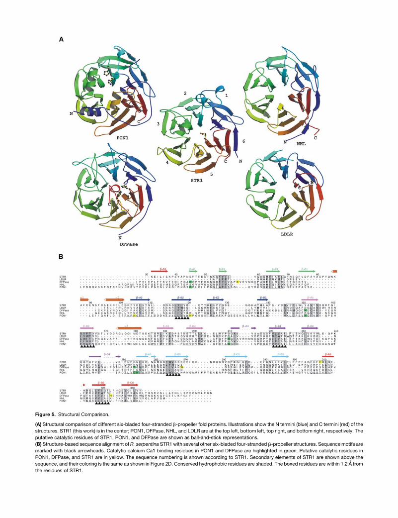
Figure 5. Structural Comparison.
(A) Structural comparison of different six-bladed four-stranded b-propeller fold proteins. Illustrations show the N termini (blue) and C termini (red) of the
structures. STR1 (this work) is in the center; PON1, DFPase, NHL, and LDLR are at the top left, bottom left, top right, and bottom right, respectively. The
putative catalytic residues of STR1, PON1, and DFPase are shown as ball-and-stick representations.
(B) Structure-based sequence alignment ofR. serpentina STR1 with several other six-bladed four-stranded b-propeller structures. Sequence motifs are
marked with black arrowheads. Catalytic calcium Ca1 binding residues in PON1 and DFPase are highlighted in green. Putative catalytic residues in
PON1, DFPase, and STR1 are in yellow. The sequence numbering is shown according to STR1. Secondary elements of STR1 are shown above the
sequence, and their coloring is the same as shown in Figure 2D. Conserved hydrophobic residues are shaded. The boxed residues are within 1.2 A from
the residues of STR1.
also exists at the same position in the PON1 structure, suggest-
ing that both enzymes might be evolutionarily related. Both en-
zymes have a similar molecular architecture; however, they differ
greatly in their primary sequences and catalytic function. STR1
catalyzes the Pictet-Spengler–type reaction, while PON1 is an
esterase. Incubation of STR1 with 10 mM EDTA for even 1 week
has no influence on the activity, indicating that metal ions are not
required for the catalytic activity or for the structural stability of
STR1. This result is also supported by the crystal structure of
STR1, where there is no trace ofmetal ions in the electron density
near the binding pocket, while PON1contains two calciumatoms
in its central tunnel. One of them is catalytic calciumand the other
is structurally important.HelixH2 inPON1 is thought tobe involved
in high-density lipoprotein anchoring by pointing its hydrophobic
face toward the solvent, while in STR1, the a3 helix is important
to create a highly hydrophobic environment around the active site,
with its hydrophobic face pointing toward the substrate binding
pocket. Interestingly, the ligand binding residues in these struc-
tures are located either between the strands b-D and b-A or b-B
and b-C (Figure 5A).
Among all the structures, only LDLR contains an easily iden-
tifiable internal repetitive sequence XYWTD (X is a hydrophobic
residue). STR1 also exhibits a similar XYFTD repeat on b-B3
(Figure 5B).Structure-based sequencealignment showed that this
XYFTD repeat is located at the equivalent position of the XYWTD
repeat in LDLR, which led us to consider whether there is any
evolutionary relationship between the sequences of STR1 and
LDLR. Further analysis of sequences in STR1 was performed at
those sectionswhere five XYWTD repeats are located in LDLR. In
fact, two other similar sequence repeats XYIVD (b-B2) andXYIGT
(b-B6) were detected in STR1, which correspond to XYWSD
(b-B2) andXFWTD (b-B6) in LDLR (Figure 5B). In addition, the rest
of the equivalent positions in STR1 contain the following se-
quences: XLVAE (b-B4) and XWVSS (b-B5). At first glance, it
seems that the last two sequences are unrelated to the XYWTD
repeat. However, further inspection shows that there is a
common motif for these sequences, XXX#§, in which three
hydrophobic residues (XXX) are followed by a small residue (#;
Ser/Thr/Ala/Val/Gly) and a hydrophilic residue (§; in most cases
Asp or Glu). Surprisingly, this common motif also fits all the
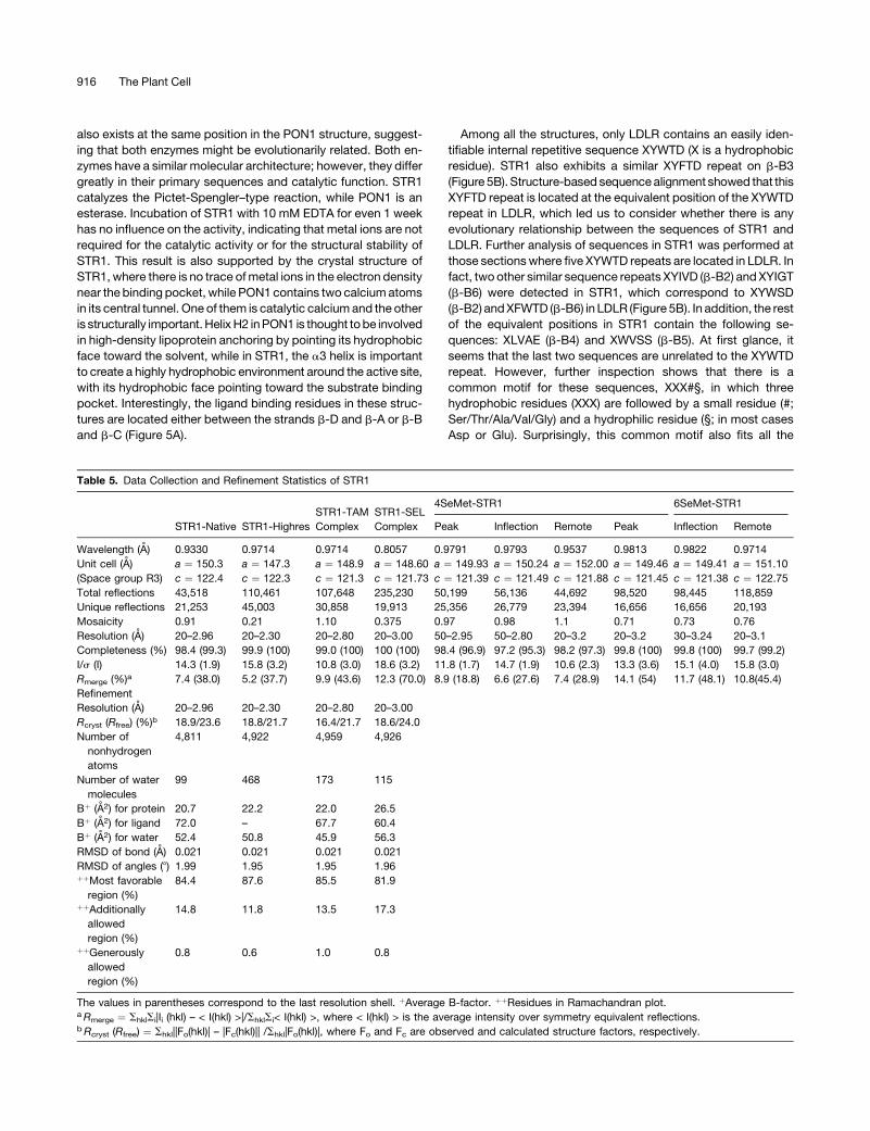
Table 5. Data Collection and Refinement Statistics of STR1
STR1-Native STR1-Highres
STR1-TAM
Complex
STR1-SEL
Complex
4SeMet-STR1 6SeMet-STR1
Peak Inflection Remote Peak Inflection Remote
Wavelength (A) 0.9330 0.9714 0.9714 0.8057 0.9791 0.9793 0.9537 0.9813 0.9822 0.9714
Unit cell (A) a ¼ 150.3 a ¼ 147.3 a ¼ 148.9 a ¼ 148.60 a ¼ 149.93 a ¼ 150.24 a ¼ 152.00 a ¼ 149.46 a ¼ 149.41 a ¼ 151.10
(Space group R3) c ¼ 122.4 c ¼ 122.3 c ¼ 121.3 c ¼ 121.73 c ¼ 121.39 c ¼ 121.49 c ¼ 121.88 c ¼ 121.45 c ¼ 121.38 c ¼ 122.75
Total reflections 43,518 110,461 107,648 235,230 50,199 56,136 44,692 98,520 98,445 118,859
Unique reflections 21,253 45,003 30,858 19,913 25,356 26,779 23,394 16,656 16,656 20,193
Mosaicity 0.91 0.21 1.10 0.375 0.97 0.98 1.1 0.71 0.73 0.76
Resolution (A) 20–2.96 20–2.30 20–2.80 20–3.00 50–2.95 50–2.80 20–3.2 20–3.2 30–3.24 20–3.1
Completeness (%) 98.4 (99.3) 99.9 (100) 99.0 (100) 100 (100) 98.4 (96.9) 97.2 (95.3) 98.2 (97.3) 99.8 (100) 99.8 (100) 99.7 (99.2)
I/s (I) 14.3 (1.9) 15.8 (3.2) 10.8 (3.0) 18.6 (3.2) 11.8 (1.7) 14.7 (1.9) 10.6 (2.3) 13.3 (3.6) 15.1 (4.0) 15.8 (3.0)
Rmerge (%)a 7.4 (38.0) 5.2 (37.7) 9.9 (43.6) 12.3 (70.0) 8.9 (18.8) 6.6 (27.6) 7.4 (28.9) 14.1 (54) 11.7 (48.1) 10.8(45.4)
Refinement
Resolution (A) 20–2.96 20–2.30 20–2.80 20–3.00
Rcryst (Rfree) (%)b 18.9/23.6 18.8/21.7 16.4/21.7 18.6/24.0
Number of
nonhydrogen
atoms
4,811 4,922 4,959 4,926
Number of water
molecules
99 468 173 115
Bþ (A2) for protein 20.7 22.2 22.0 26.5
Bþ (A2) for ligand 72.0 – 67.7 60.4
Bþ (A2) for water 52.4 50.8 45.9 56.3
RMSD of bond (A) 0.021 0.021 0.021 0.021
RMSD of angles (8) 1.99 1.95 1.95 1.96þþMost favorable
region (%)
84.4 87.6 85.5 81.9
þþAdditionally
allowed
region (%)
14.8 11.8 13.5 17.3
þþGenerously
allowed
region (%)
0.8 0.6 1.0 0.8
The values in parentheses correspond to the last resolution shell. þAverage B-factor. þþResidues in Ramachandran plot.aRmerge ¼ ShklSi|Ii (hkl) – < I(hkl) >|/ShklSi< I(hkl) >, where < I(hkl) > is the average intensity over symmetry equivalent reflections.bRcryst (Rfree) ¼ Shkl||Fo(hkl)| – |Fc(hkl)|| /Shkl|Fo(hkl)|, where Fo and Fc are observed and calculated structure factors, respectively.
916 The Plant Cell
relevant sequences in PON1, DFPase, and NHL (Figure 5B).
These observations indicate that all these structurally related
proteins are in fact evolutionary related; it is likely that they have
divergently evolved from a common ancestral b-sheet gene. The
internal repeats may suggest that a six-bladed b-propeller might
be first formed by duplication of an ancestral b-sheet gene that
contains the YWTD repeat. During evolution, the ancestral struc-
ture undergoes insertions and deletions to shape the central
tunnel and to adopt different functions, for example, the cap
region in PON1 and STR1. As suggested before, propeller topol-
ogy does not impose specific constraints on sequence (Murzin,
1992), so these sequence repeats gradually lose their sequence
identity, but they still keep some common sequence homology
features that are important to maintain the overall structural
stability of the fold, as can be seen by the XXX#§ consensus at
the second strand of each blade (Figure 5B).
Extreme sequence diversity accompanied by greatly different
functions and phylogenetic origins usually makes people believe
that these sequence-unrelated propeller structures are unlikely
to have a common precursor and that the evolution may have
taken place from distinct ancestral b-sheet genes (Jawad and
Paoli, 2002). Now, based on the structure of STR1 and structure-
basedsequencealignmentbetweenSTR1and fourother six-bladed
b-propeller structures, a possible evolutionary relationship of
several sequence-unrelated six-bladed b-propeller structures is
proposed.
METHODS
Expression, Purification, Mutagenesis, and Enzyme Activity Assay
Rauvolfia serpentina STR1 excluding the signal peptide (first 28 residues)
was subcloned into pQE-2 vector. Only two additional residues (Gly and
Ala) are left on the N terminus of STR1 when the N-terminal His tag is
cleaved with dipeptidyl aminopeptidase (Qiagen). Expression in Esche-
richia coli strain M15 and purification of native STR1 have been described
previously (Ma et al., 2004). Site-directedmutantswere constructed using
the Quickchange method (Stratagene), verified by sequencing the com-
plete gene and purified as for the wild-type enzyme.
Selenomethionyl-STR1 (SeMet-STR1) was obtained by the Met path-
way inhibition technique (VanDuyne et al., 1993) with the same vector and
E. coli strain used for native STR1. The purification procedure for SeMet-
STR1 was the same as that used for the native enzyme, except that 1 mM
DTT was added to all of the buffers.
Native STR1 contains only two Met residues. Crystals of this SeMet-
STR1 were proven to be insufficient to result in the useful anomalous sig-
nal in solving the structure. Therefore, twomutants with fourMet residues
(STR1-L116MI190M) andsixMet residues (STR1-L116M I190ML203MI65M)
were prepared; these two mutants are designated here as 4Met-STR1
and 6Met-STR1, respectively. Three positions of Leu or Ile for mutations
(Leu-116, Ile-190, and Leu-203) were chosen based on the sequence
alignment with STR1 from species Catharanthus roseus and Ophiorrhiza
pumila (see the sequence alignment in Figure 2D). The fourth one (Ile-65)
was chosen based on the high sequence similarity between 62GRVI65-
KYE68 and 187GRLI190KYD193 inside theR. serpentinaSTR1sequencealone,
since mutation of Ile-190 to Met in the mutant 4Met-STR1 does not influ-
ence the activity or change the crystallization behavior of thismutant. Both
4Met-STR1 and 6Met-STR1mutants are activewith only a slight decrease
on the activity (;10%) compared with native STR1. Selenomethionyl-
4Met-STR1 (4SeMet-STR1) and selenomethionyl-6Met-STR1 (6SeMet-
STR1) were prepared as mentioned above for the SeMet-STR1.
The activity of STR1 was determined by incubating 1 mM tryptamine,
2 mM secologanin, and 50 mM phosphate buffer, pH 7.0 (total volume
50 mL), for 15 min at 358C while shaking (400 rpm). The reaction was
terminated by addition of 100 mLmethanol. After centrifugation (11,000g;
5 min), the supernatant was analyzed by HPLC. For HPLC, a Merck
Hitachi instrument and a Lichrospher 60RP select B column (1253 4mm;
5 mm) were used: injection volume 60 mL; flow rate 1 mL min�1;
absorption measured at 250 nm; acetonitrile/H2O (pH 2.3 adjusted by
H3PO4) as solvent system, gradient 10:90/ 50:50 within 8 min/ 80:20
within 3 min/ 10:90 within 0.5 min/ 10:90 for 3.5 min. Retension time
values are 5.47 min (tryptamine), 6.57 min (secologanin), and 8.65 min
(strictosidine). To determine Km and kcat values, the enzyme assay was
performed using different concentrations of tryptamine and secologanin.
Crystallization
Crystals of STR1 and its complexes were grown by the hanging drop
vapor diffusion method (Ma et al., 2004; Koepke et al., 2005). The res-
ervoir solution contained 0.8 M potassium sodium tartrate tetrahydrate
and 0.1 M HEPES, pH 7.5. The enzyme concentration was 4 to 5 mg/mL.
Crystals of STR1 in complex with substrates tryptamine (STR1-TAM
complex) and secologanin (STR1-SEL complex) were obtained by cocrys-
tallization in the presence of 1.0 mM tryptamine and secologanin, re-
spectively, in the enzyme solution.
Data Collection and Processing
Native, SeMet, and tryptamine complex crystals were cryo-protected by
addition of 25% glycerol to the precipitant buffer before being flash-
cooled in a stream of cold nitrogen at 100K. The secologanin complex
crystals were flash-cooled after 8 min of soaking in the solution of 25%
glycerol, 6.0 mM secologanin, and the precipitant buffer. Three multi-
wavelengthanomalous diffraction (MAD) experimentswereperformednear
the selenium edge. The MAD data sets from 4SeMet-STR1 were col-
lected at the PX beamline at the Swiss Light Source in Villigen, Switzer-
land, and another three wavelength MAD data sets from 6SeMet-STR1
were collected at the BW7A beamline of EMBL Hamburg at the DORIS
storage ring of the Deutsches Elektronen Synchrotron DESY (Hamburg,
Germany). All the x-ray data from cocrystals were collected either on the
BW7A, BW7B, X11, or X13 beamline of EMBL Hamburg. The diffraction
data were processed and scaled using HKL (Otwinowski and Minor,
1997) in the R3 space group. The data collection statistics are illustrated
in Table 2. The preliminary data analysis showed that the crystals contain
;66% solvent content with a Matthews coefficient of 3.6 and two mol-
ecules of STR1 in an asymmetric unit.
Structure Determination, Refinement, and Quality of the Models
The structure was solved by the combination of MAD and molecular re-
placement methods using SeMet-substituted STR1. Initially, the interpret-
able electrondensitywas obtained at 3.2-A resolution for the6SeMet-STR1
data set using the three wavelengths MAD protocol of AUTO-RICKSHAW:
an automated crystal structure determination platform (Panjikar et al.,
2005) in the R3 space group. Within this procedure, the structure factors
were calculated from the measured intensity by employing the CCP4
programs (Collaborative Computation Project Number 4, 1994), and 8 out
of 12 selenium atoms were located using SHELXD (Schneider and
Sheldrick, 2002). The positions of these sites were refined using the
programMLPHARE (CollaborativeComputationProject Number 4, 1994).
Density modification and twofold noncrystallographic symmetry (NCS)
averaging were performed using DM (Cowtan, 1994) in the correct
enantiomorph. Subsequently, it was possible to trace most of the protein
backboneusing themolecular graphicsprogramXTALVIEW/XFIT (McRee,
1999). However, side chains and some loops could not be placed
X-Ray Structure of Strictosidine Synthase 917
unambiguously in the electron density. Therefore, MAD phases were
calculated with the 4SeMet-STR1 data set in the similar manner as
described above and were significantly improved when it was combined
with the phase generated from the 6SeMet-STR1 data set using the
program SIGMAA (Collaborative Computation Project Number 4, 1994)
and continued with the phase extension to 2.95 A by NCS averaging and
densitymodification. Themodel was further improved by buildingmissing
loops. Some of the side chains could be recognized and were placed in
the electron density. At this stage of themodel building, it became evident
that the structure looked like a six-bladed b-propeller fold. A quick search
with Structural Classification of Proteins database (http://scop.berkeley.
edu/) showedmany structures (i.e., PDBcodes 1v04, 1ijq, 1e1a, and 1q7f)
containing such a fold. Althoughmolecular replacement against the STR1
data was unsuccessful using any of the structures as a search model, the
1v04 structure could be placed in the experimental electron density suc-
cessfully using the phased molecular replacement protocol of the pro-
gram MOLREP (Vagin and Isupov, 2001). The resultant model was used
asaguideline for furthermodel building,which helped unambiguous tracing
of b-strands in the correct direction. The side chain was fitted in the elec-
tron density wherever the map was interpretable. The model was further
improved using the graphics program COOT (Emsley and Cowtan, 2004)
and its real space fitting and interactive manual building. The model was
subjected to multiple rounds of simulated annealing, followed by posi-
tional and restrainedB-factor refinement as implemented in CNS (Brunger
et al., 1998) using the data from 20 to 2.95 A. The alternate refinement and
manual rebuilding of the SeMet-STR1model resulted in a nearly complete
backbone with a majority of side chains positioned; however, the quality
of the electron density maps did not permit the rapid convergence of a
complete STR1 model. Therefore, further refinement of the model was
halted. As 2.3-A x-ray data (STR1-Highres) were available from a trypt-
amine complex, the complex was solved by molecular replacement
based on this partial SeMet-derived structure using rigid body refinement
in CNS. The resultant model was used as a starting model for the auto-
mated model building program ARP/wARP (Perrakis et al., 1999; Morris
et al., 2004) using thedata from20- to 2.3-A resolution. Eighty-five percent
of the complete model was built automatically, and the model was com-
pleted further withmanual building usingCOOT alternatingwith additional
REFMAC cycles, which included bulk solvent correction, anisotropic
scaling, NCS restraints, andwith eachmolecule defined as a TLS group in
the modeling of anisotropy in the program REFMAC5 (Murshudov et al.,
1997; Winn et al., 2001).
Native and all other ligand structures were solved bymolecular replace-
ment using the high-resolution structure (2.3-A resolution) as a search
model, as the data were nonisomorphous to each other. The structures
were refined using a similar refinement protocol as described above for
the high-resolution structure.
The final native STR1 structure was refined to Rcryst 18.9% and Rfree
23.6% using the data from 20 to 2.96 A. The structure comprises residues
29 to 333 permolecule and includes 99water molecules, and the electron
density unambiguously showed the residual density of tartrate molecules
in each molecule.
STR1-Highres comprises residues 32 to 333 and includes 468 water
molecules. The structure was refinedwithRcryst 18.8% andRfree 21.7% in
the resolution range 20 to 2.3 A. STR1-Highres data were collected from a
STR1-tryptamine complex crystal, and the residual density was observed
to 8s. However, the density could not be explained only with tryptamine,
as the density was certainly bigger than the expected tryptamine density
(stereoview of the residual density is shown in Supplemental Figure 2
online). All our interpretations of the residual density were not convincing,
evenwithmultiple conformations of tryptamine. Thedensitywas notmod-
eled with a single tryptamine, as the residual density was not fully inter-
preted. The data played a main role in resolving the structure. Therefore,
another data set of STR1-TAMwas collected and refined (see below). The
located tryptamine in the STR1-TAM complex fits the residual density
with room of some unknown molecule.
The STR1-TAM complex comprises residues 35 to 333 as well as one
tryptamine per STR1molecule. The model includes 173 water molecules.
The model is refined with Rcryst 16.4% and Rfree 21.7% in the resolution
range 20 to 2.8 A.
The structure of the STR1-SEL complex comprises residues 32 to 333
and one secologanin per STR1 molecule and includes 115 water mole-
cules. The model is refined with Rcryst 18.6% and Rfree 24.0% in the
resolution range 20 to 3.00 A.
Comparisons of the backbone trace of the partial SeMet-STR1 and the
reported structures displayed nearly identical architecture and no evi-
dence for conformational differences between the native and complex
forms of STR1. Therefore, refinement of the original SeMet-STR1 struc-
ture was not continued. The overall geometric quality of resultant models
was assessed using PROCHECK (Laskowski et al., 1993). Table 5 sum-
marizes the data collection and refinement statistics on each of the final
models.
Structurally related proteins were retrieved from secondary structure
matching (http://www.ebi.ac.uk/msd-srv/ssm/cgi-bin/ssmserver) servers
using the single STR1monomer model. Structure-based sequence align-
ment was performed using the program STAMP (Russell and Barton,
1992), and the picture of the sequence alignment was made using the
program ALSCRIPT (Barton, 1993). All figures were produced using
MOLSCRIPT (Kraulis, 1991), RASTER3D (Merritt and Murphy, 1994),
PyMol (DeLano, 2002), and LIGPLOT (Wallace et al., 1995).
Accession Numbers
Sequence data from this article can be found in the GenBank/EMBL data
libraries under accession numbers P68175 (STR1 from R. serpentina),
P18417 (STR1 from C. roseus), and Q94LW9 (STR1 from O. pumila). The
model coordinates and structural factor amplitudes have been deposited
in the PDB for structures of the STR1-Native (2FP9), STR1-Highres (2FP8),
STR1-TAM complex (2FPB), and STR1-SEL complex (2FPC).
Supplemental Data
The following materials are available in the online version of this article.
Supplemental Figure 1. Sequence Alignment of STR1 from Different
Plant Species with Other STR1-Related Gene Family Members.
Supplemental Figure 2. The Unexplained Fo-Fc Electron Density of a
Ligand at 4.0 s and Residues Nearby the Binding Pocket in STR1-
Highres.
ACKNOWLEDGMENTS
We thank T.M. Kutchan (Leibniz Institute of Plant Biochemistry, Halle/
Saale, Germany) for kindly sharing the STR1 clone with us. The contin-
uous interest of H. Michel and G. Fritzsch (Max Plank Institute of
Biophysics, Frankfurt, Germany) and U. Pindur (Institute of Pharmacy,
Mainz, Germany) and linguistic advice as well as helpful discussions by
P.A. Tucker (European Molecular Biology Laboratory) are greatly ap-
preciated. We also thank Verena Linhard (Max-Planck-Institute of Bio-
physics, Frankfurt, Germany) for excellent technical assistance. Staff
members of the EMBL BW7A, BW7B, and X13 beamline at the DORIS
storage ring (DESY, Hamburg, Germany) and PX beamline at the Swiss
Light Source in Villigen (Switzerland) are appreciated for their help. This
work was supported by the Deutsche Forschungsgemeinschaft (Bonn,
Bad-Godesberg, Germany) and the Fonds der Chemischen Industrie
(Frankfurt/Main, Germany) together with the Bundesministerium fur
Bildung und Forschung (Berlin, Germany).
918 The Plant Cell

Received September 16, 2005; revised December 25, 2005; accepted
February 6, 2006; published March 10, 2006.
REFERENCES
Barton, G.J. (1993). ALSCRIPT: A tool to format multiple sequence
alignments. Protein Eng. 6, 37–40.
Boettcher, C., Fellermeier, M., Boettcher, C., Drager, B., and Zenk,
M.H. (2005). How human neuroblastoma cells make morphine. Proc.
Natl. Acad. Sci. USA 101, 8495–8500.
Bracher, D., and Kutchan, T.M. (1992). Strictosidine synthase from
Rauvolfia serpentina: Analysis of a gene involved in indole alkaloid
biosynthesis. Arch. Biochem. Biophys. 294, 717–723.
Brunger, A.T., et al. (1998). Crystallography & NMR system: A new
software suite for macromolecular structure determination. Acta
Crystallogr. D 54, 905–921.
Collaborative Computation Project Number 4 (1994). The CCP4
suite: Programs for protein crystallography. Acta Crystallogr. D 50,
760–763.
Cowtan, K. (1994). ‘dm’: An automated procedure for phase improve-
ment by density modification. Joint CCP4 and ESF-EACBM News-
letter on Protein Crystallography 31, 34–38.
De-Eknamkul, W., Suttipanta, N., and Kutchan, T.M. (2000). Purifi-
cation and characterization of deacetylipecoside synthase from
Alangium lamarckii Thw. Phytochemistry 55, 177–181.
DeLano, W.L. (2002). The PyMOL Molecular Graphics System. (San
Carlos, CA: DeLano Scientific).
DeWaal, A., Meijer, A.H., and Verpoorte, R. (1995). Strictosidine
synthase from Catharanthus roseus: Purification and characterization
of multiple forms. Biochem. J. 306, 571–580.
Edwards, T.A., Wilkinson, B.D., Wharton, R.P., and Aggarwal, A.K.
(2003). Model of the brain tumor-pumilio translation repressor com-
plex. Genes Dev. 17, 2508–2513.
Emsley, P., and Cowtan, K. (2004). Coot: Model-building tools for
molecular graphics. Acta Crystallogr. D 60, 2126–2132.
Fabbri, M., Delp, G., Schmidt, O., and Theopold, U. (2000).
Animal and plant members of a gene family with similarity to alkaloid-
synthesizing enzymes. Biochem. Biophys. Res. Commun. 271,
191–196.
Hampp, N., and Zenk, M.H. (1988). Homogeneous strictosidine syn-
thase from cell suspension cultures of Rauvolfia serpentina. Phyto-
chemistry 27, 3811–3815.
Harel, M., et al. (2004). Structure and evolution of the serum para-
oxonase family of detoxifying and anti-atherosclerotic enzymes. Nat.
Struct. Mol. Biol. 11, 412–419.
Jawad, Z., and Paoli, M. (2002). Novel sequences propel familiar folds.
Structure 10, 447–454.
Jeon, H., Meng, W., Takagi, J., Eck, M.J., Springer, T.A., and
Blacklow, S.C. (2001). Implications for familial hypercholesterolemia
from the structure of the LDL receptor YWTD-EGF domain pair. Nat.
Struct. Biol. 8, 499–504.
Koepke, J., Ma, X.Y., Fritzsch, G., Michel, H., and Stockigt, J. (2005).
Crystallization and preliminary x-ray analysis of strictosidine synthase
and its complex with the substrate tryptamine. Acta Crystallogr. D 61,
690–693.
Kraulis, P.J. (1991). MOLSCRIPT: A program to produce both detailed
and schematic plots of protein structures. J. Appl. Crystallogr. 24,
946–950.
Kutchan, T.M. (1993). Strictosidine: From alkaloid to enzyme to gene.
Phytochemistry 32, 493–506.
Kutchan, T.M., Hampp, N., Lottspeich, F., Beyreuther, K., and Zenk,
M.H. (1988). The cDNA clone for strictosidine synthase from Rauvolfia
serpentina. DNA sequence determination and expression in Esche-
richia coli. FEBS Lett. 237, 40–44.
Laskowski, R.A., MacArthur, M.W., Moss, D.S., and Thornton, J.M.
(1993). PROCHECK: A program to check the stereochemical quality of
protein structures. J. Appl. Crystallogr. 26, 283–291.
Luzatti, P.V. (1952). Traitement statistique des erreurs dans la deter-
mination des structures cristallines. Acta Crystallogr. D 5, 802–810.
Ma, X.Y., Koepke, J., Fritzsch, G., Diem, R., Kutchan, T.M., Michel,
H., and Stockigt, J. (2004). Crystallization and preliminary x-ray
crystallographic analysis of strictosidine synthase from Rauvolfia –
The first member of a novel enzyme family. Biochim. Biophys. Acta
1702, 121–124.
McKnight, T.D., Roessner, C.A., Devagupta, R., Scott, A.I., and
Nessler, C.L. (1990). Nucleotide sequence of a cDNA encoding the
vacuolar protein strictosidine synthase from Catharanthus roseus.
Nucleic Acids Res. 18, 4939.
McRee, D.E. (1999). XtalView/Xfit–A versatile program for manipulat-
ing atomic coordinates and electron density. J. Struct. Biol. 125,
156–165.
Merritt, E.A., and Murphy, M.E. (1994). Raster3D Version 2.0. A
program for photorealistic molecular graphics. Acta Crystallogr. D
50, 869–873.
Morita, M., Hara, Y., Tamai, Y., Arakawa, H., and Nishimura, S.
(2000). Genomic construct and mapping of the gene for CMAP
(Leukocystatin/Cystatin F, CST7) and identification of a proximal
novel gene, BSCv (C20orf3). Genomics 67, 87–91.
Morris, R.J., Zwart, P.H., Cohen, S., Fernandez, F.J., Kakaris, M.,
Kirillova, O., Vonrhein, C., Perrakis, A., and Lamzin, V.S. (2004).
Breaking good resolutions with ARP/wARP. J. Synchrotron. Radiat.
11, 56–59.
Murshudov, G.N., Vagin, A.A., and Dodson, E.J. (1997). Refinement of
macromolecular structures by the maximum-likelihood method. Acta
Crystallogr. D 53, 240–255.
Murzin, A.G. (1992). Structural principles for the propeller assembly
of b-sheets: The preference for seven-fold symmetry. Proteins 14,
191–201.
Otwinowski, Z., and Minor, W. (1997). Processing of X-ray diffraction
data collected in oscillation mode. Methods Enzymol. 276, 307–326.
Panjikar, S., Parthasarathy, V., Lamzin, V.S., Weiss, M.S., and
Tucker, P.A. (2005). Auto-Rickshaw: An automated crystal structure
determination platform as an efficient tool for the validation of an X-ray
diffraction experiment. Acta Crystallogr. D 61, 449–457.
Perrakis, A., Morris, R.J., and Lamzin, V.S. (1999). Automated protein
model building combined with iterative structure refinement. Nat.
Struct. Biol. 6, 458–463.
Russell, R.B., and Barton, G.J. (1992). Multiple protein sequence align-
ment from tertiary structure comparison: Assignment of global and
residue confidence levels. Proteins Struct. Funct. Genet. 14, 309–323.
Samanani, N., Liscombe, D.K., and Facchini, P.J. (2004). Molecular
cloning and characterization of norcoclaurine synthase, an enzyme
catalyzing the first committed step in benzylisoquinoline alkaloid
biosynthesis. Plant J. 40, 302–313.
Scharff, E.I., Koepke, J., Fritzsch, G., Lucke, C., and Ruterjans, H.
(2001). Crystal structure of diisopropylfluorophosphatase from Loligo
vulgaris. Structure 9, 493–502.
Schneider, T.R., and Sheldrick, G.M. (2002). Substructure solution
with SHELXD. Acta Crystallogr. D 58, 1772–1779.
Stevens, L.H., Giroud, C., Pennings, E.J.M., and Verpoorte, R. (1993).
Purification and characterization of strictosidine synthase from a
suspension culture of Cinchona robusta. Phytochemistry 33, 99–106.
Stockigt, J., and Ruppert, M. (1999). Strictosidine: The biosynthetic key
to monoterpenoid indole alkaloids. In Comprehensive Natural Prod-
ucts Chemistry: Amino Acids, Peptides, Porphyrins and Alkaloids,
X-Ray Structure of Strictosidine Synthase 919

Vol. 4, D.H.R. Barton, K. Nakanishi, O. Meth-Cohn, and J.W. Kelly,
eds (Amsterdam: Elsevier), pp 109–138.
Theopold, U., Samakovlis, C., Erdjument-Bromage, H., Dillon, N.,
Axelsson, B., Schmidt, O., Tempst, P., and Hultmark, D. (1996).
Helix pomatia lectin, an inducer of Drosophila immune response,
binds to hemomucin, a novel surface mucin. J. Biol. Chem. 271,
12708–12715.
Treimer, J.F., and Zenk, M.H. (1979). Purification and properties of
strictosidine synthase, the key enzyme in indole alkaloid formation.
Eur. J. Biochem. 101, 225–233.
Vagin, A.A., and Isupov, M.N. (2001). Spherically averaged phased
translation function and its application to the search for molecules
and fragments in electron-density maps. Acta Crystallogr. D 57,
1451–1456.
Van Duyne, G.D., Standaert, R.F., Karplus, A., Schreiber, S.L., and
Clardy, J. (1993). Atomic structures of the human immunophilin
FKBP-12 complexes with FK506 and Rapamycin. J. Mol. Biol. 229,
105–124.
Wallace, A.C., Laskowski, R.A., and Thornton, J.M. (1995). LIGPLOT:
A program to generate schematic diagrams of protein-ligand interac-
tions. Protein Eng. 8, 127–134.
Winn, M.D., Isupov, M.N., and Murshudov, G.N. (2001). Use of TLS
parameters to model anisotropic displacements in macromolecular
refinement. Acta Crystallogr. D 57, 122–133.
Yamazaki, Y., Sudo, H., Yamazaki, M., Aimi, N., and Saito, K. (2003).
Camptothecin biosynthetic genes in hairy roots of Ophiorrhiza pumila:
Cloning, characterization and differential expression in tissues and by
stress compounds. Plant Cell Physiol. 44, 395–403.
920 The Plant Cell

DOI 10.1105/tpc.105.038018; originally published online March 10, 2006; 2006;18;907-920Plant Cell
Xueyan Ma, Santosh Panjikar, Juergen Koepke, Elke Loris and Joachim StöckigtFold in Plant Proteins
-Propellerβ Strictosidine Synthase Is a Novel Six-Bladed Rauvolfia serpentinaThe Structure of
This information is current as of April 13, 2020
Supplemental Data /content/suppl/2006/02/22/tpc.105.038018.DC1.html
References /content/18/4/907.full.html#ref-list-1
This article cites 43 articles, 3 of which can be accessed free at:
Permissions https://www.copyright.com/ccc/openurl.do?sid=pd_hw1532298X&issn=1532298X&WT.mc_id=pd_hw1532298X
eTOCs http://www.plantcell.org/cgi/alerts/ctmain
Sign up for eTOCs at:
CiteTrack Alerts http://www.plantcell.org/cgi/alerts/ctmain
Sign up for CiteTrack Alerts at:
Subscription Information http://www.aspb.org/publications/subscriptions.cfm
is available at:Plant Physiology and The Plant CellSubscription Information for
ADVANCING THE SCIENCE OF PLANT BIOLOGY © American Society of Plant Biologists