the surgical management of rickets & osteogenesis...
TRANSCRIPT
The Surgical Management of Rickets & Osteogenesis Imperfecta
Dr Greg FirthChris Hani Baragwanath Academic Hospital
Department of OrthopaedicsUniversity of the Witwatersrand

Rickets
• Inadequate mineralization of growing bone
• Often hypotonic with delayed motor milestones
• Joint thickening esp wrists & knees
• Short for age
• Genu varum / valgum, coxa vara, protrusio
• Pathological fractures
• X-Rays – Loosers zones dt unmineralised osteoid
Rickets
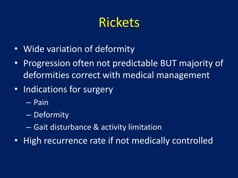
• Wide variation of deformity
• Progression often not predictable BUT majority of deformities correct with medical management
• Indications for surgery
– Pain
– Deformity
– Gait disturbance & activity limitation
• High recurrence rate if not medically controlled
RicketsManagement
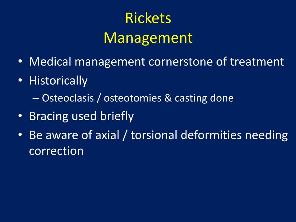
• Medical management cornerstone of treatment
• Historically
– Osteoclasis / osteotomies & casting done
• Bracing used briefly
• Be aware of axial / torsional deformities needing correction
RicketsManagement
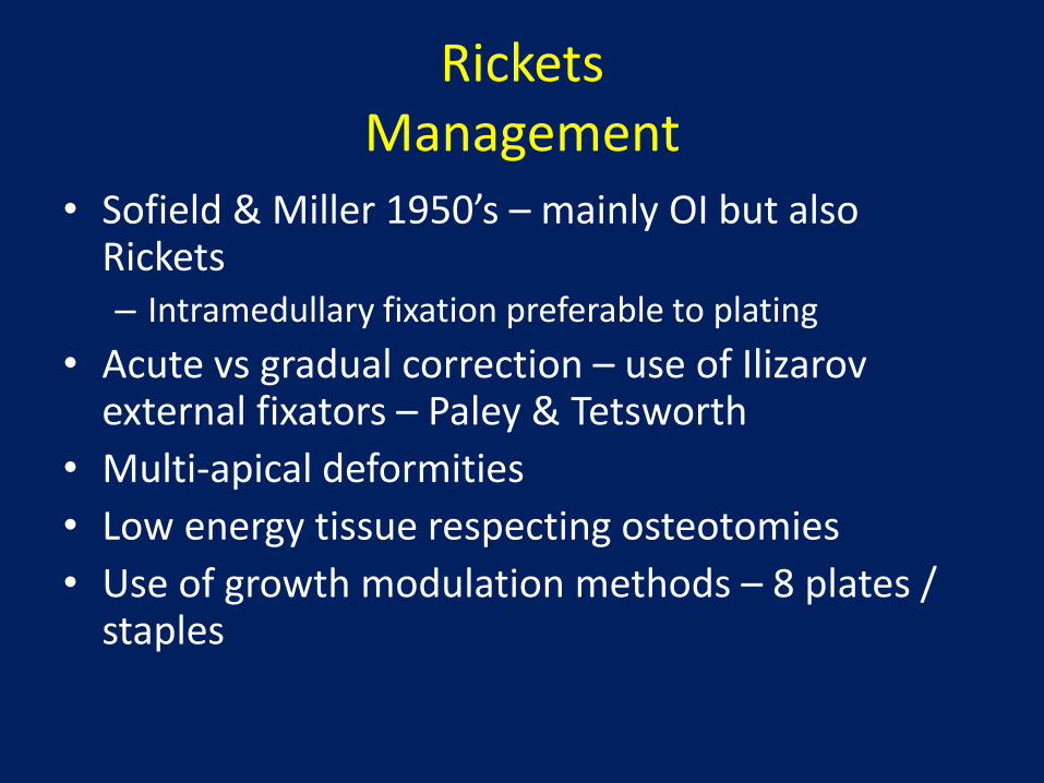
• Sofield & Miller 1950’s – mainly OI but also Rickets– Intramedullary fixation preferable to plating
• Acute vs gradual correction – use of Ilizarovexternal fixators – Paley & Tetsworth
• Multi-apical deformities
• Low energy tissue respecting osteotomies
• Use of growth modulation methods – 8 plates / staples
RicketsCase One
• Six year old female
• X-Linked hypo-phosphataemic Rickets
• Genu varum both legs
• ALP 589, dropped from 1000
• Bilateral lateral closing wedge osteotomiesfemurs
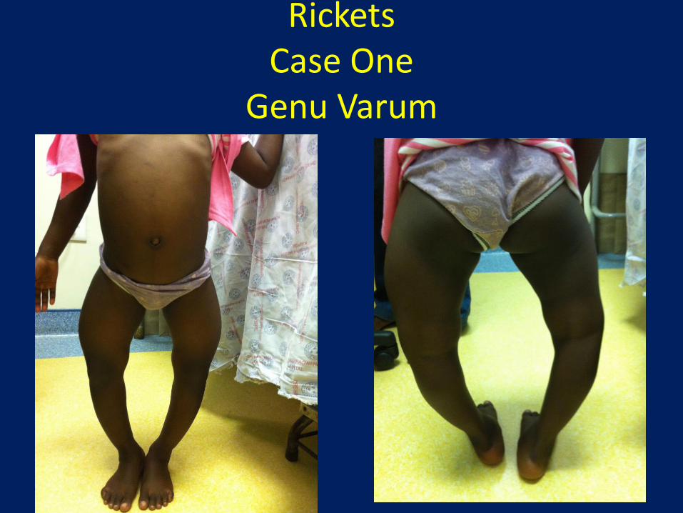
RicketsCase One
Genu Varum
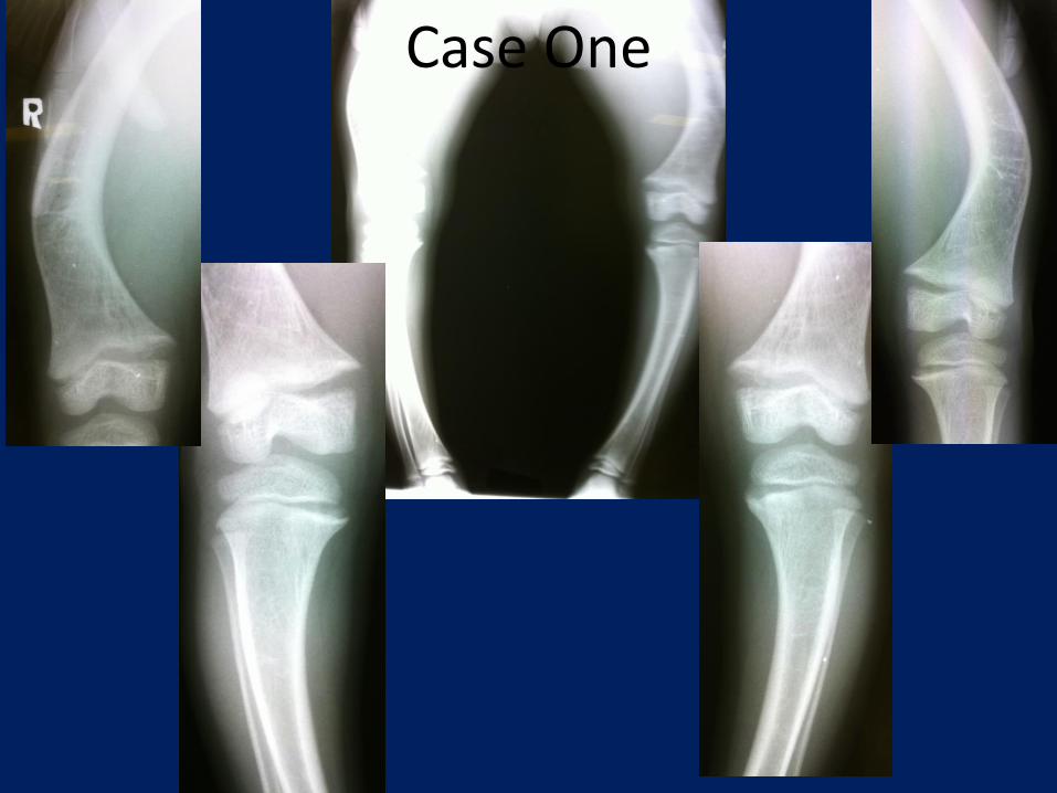
Case One
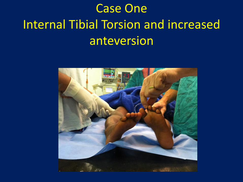
Case OneInternal Tibial Torsion and increased
anteversion

Case OneFemoral Osteotomy
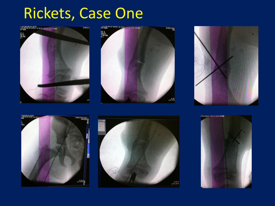
Rickets, Case One
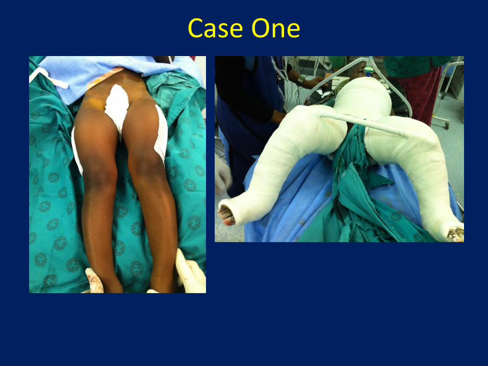
Case One
RicketsCase Two
• 10 year old boy
• 40 degree bilateral genu valgum
• 8 plates inserted bilateral femurs and tibiae
• Corrected and removed after 4 years
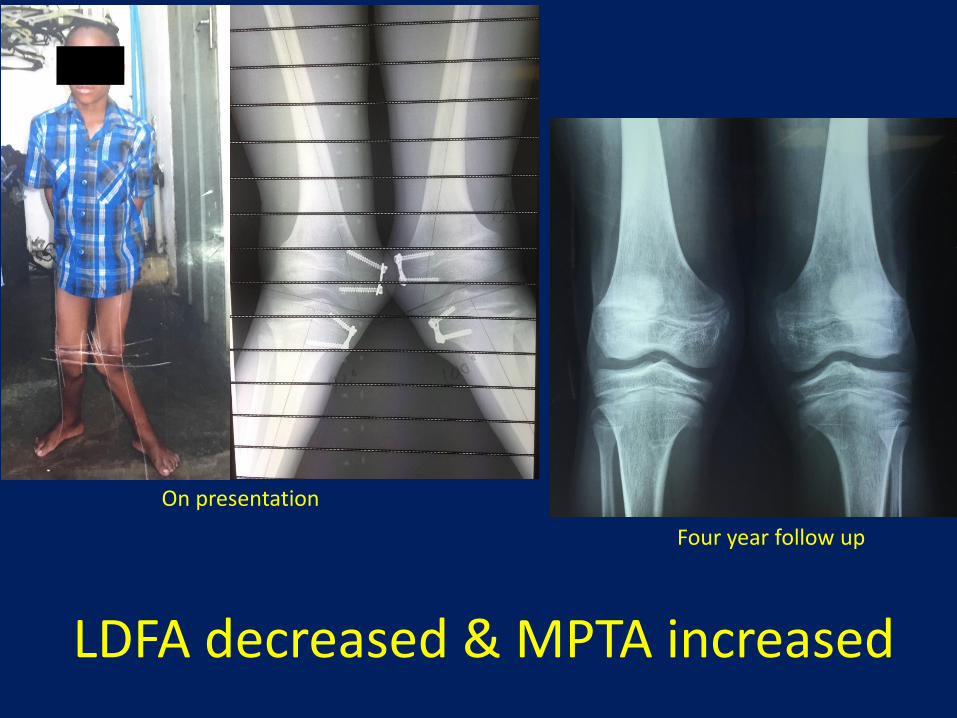
LDFA decreased & MPTA increased
On presentation
Four year follow up
Rickets & SCFE
• Slipped capital femoral epiphysis (SCFE)• More common with renal osteodystrophy• Slip through metaphyseal side of physis• Younger age• Always perform bilateral in situ pinning dt risk of
contralateral slip• AVN more commonly dt steroids than acute unstable
slip• Consider smooth pins to prevent coxa breva• Beware hardware cut-out and pin penetration dt soft
bone
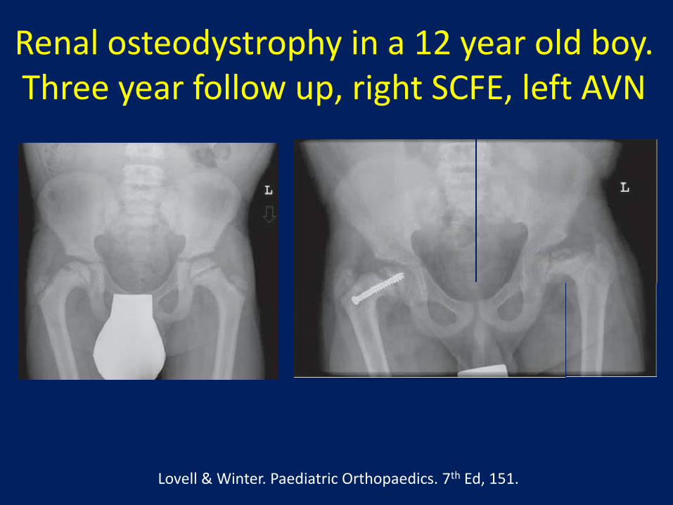
Renal osteodystrophy in a 12 year old boy. Three year follow up, right SCFE, left AVN
Lovell & Winter. Paediatric Orthopaedics. 7th Ed, 151.

Osteogenesis Imperfecta
• Osteogenesis imperfecta
– Brittle bone disease
– Genetic basis – spectrum of severity
• Variants – Bruck’s syndrome
– Deficiency or abnormality in collagen genes
– Deformities secondary to multiple fractures
Osteogenesis Imperfecta
• Looser – congenita vs tarda
• Sillence – 4 types
– Type 5 & 6 – Gloreaux et al
– Type 7 – Ward et al
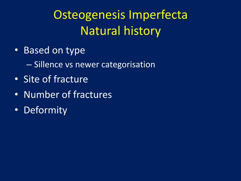
Osteogenesis ImperfectaNatural history
• Based on type
– Sillence vs newer categorisation
• Site of fracture
• Number of fractures
• Deformity
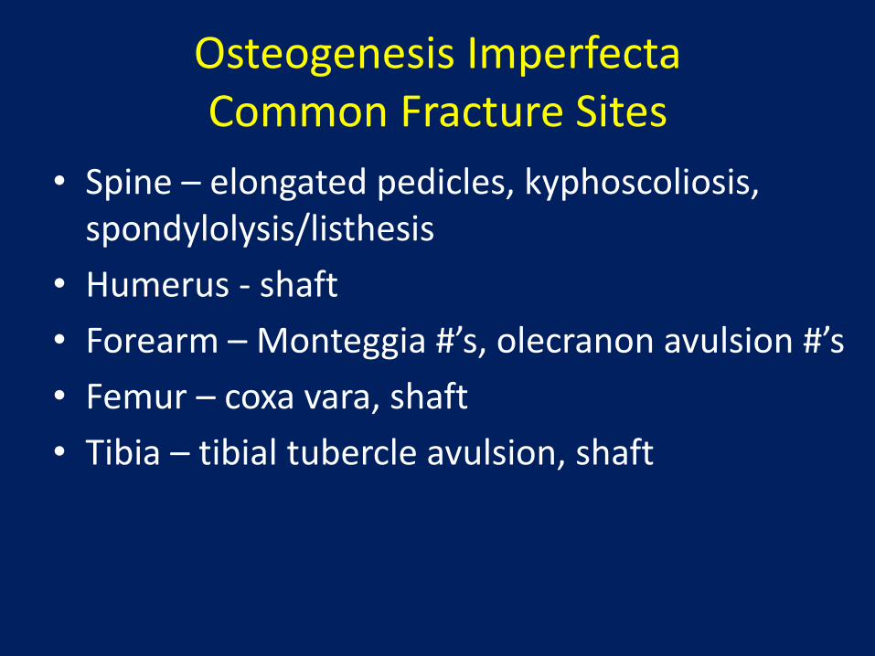
Osteogenesis ImperfectaCommon Fracture Sites
• Spine – elongated pedicles, kyphoscoliosis, spondylolysis/listhesis
• Humerus - shaft
• Forearm – Monteggia #’s, olecranon avulsion #’s
• Femur – coxa vara, shaft
• Tibia – tibial tubercle avulsion, shaft
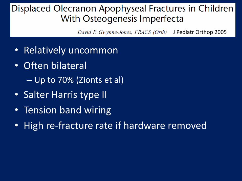
• Relatively uncommon
• Often bilateral
– Up to 70% (Zionts et al)
• Salter Harris type II
• Tension band wiring
• High re-fracture rate if hardware removed
J Pediatr Orthop 2005
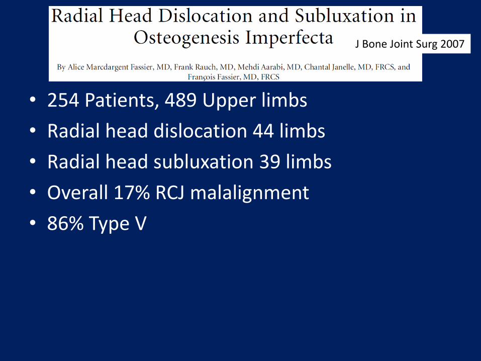
• 254 Patients, 489 Upper limbs
• Radial head dislocation 44 limbs
• Radial head subluxation 39 limbs
• Overall 17% RCJ malalignment
• 86% Type V
J Bone Joint Surg 2007
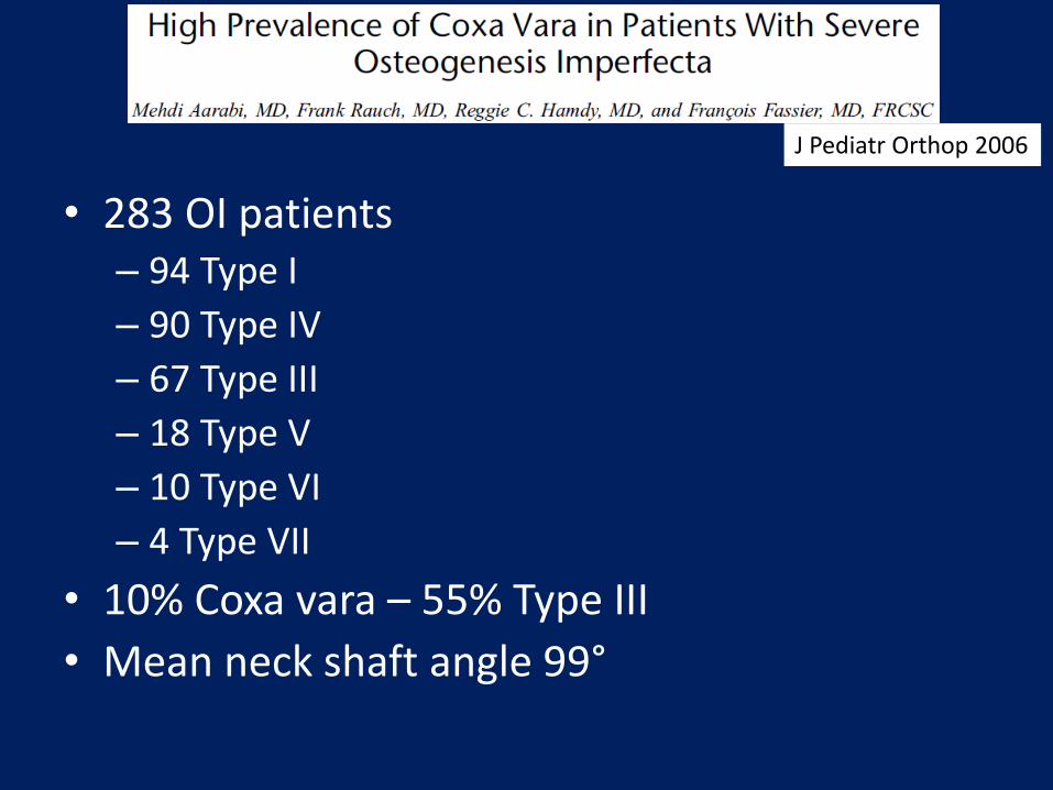
• 283 OI patients– 94 Type I
– 90 Type IV
– 67 Type III
– 18 Type V
– 10 Type VI
– 4 Type VII
• 10% Coxa vara – 55% Type III
• Mean neck shaft angle 99°
J Pediatr Orthop 2006
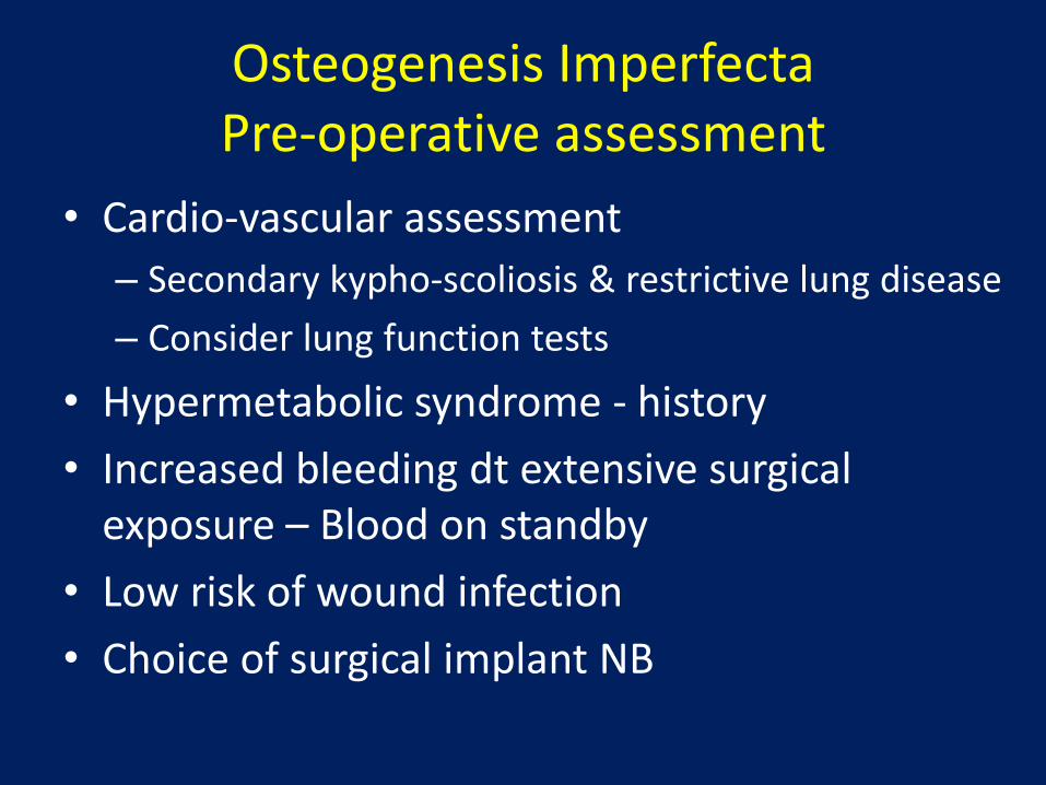
Osteogenesis ImperfectaPre-operative assessment
• Cardio-vascular assessment
– Secondary kypho-scoliosis & restrictive lung disease
– Consider lung function tests
• Hypermetabolic syndrome - history
• Increased bleeding dt extensive surgical exposure – Blood on standby
• Low risk of wound infection
• Choice of surgical implant NB
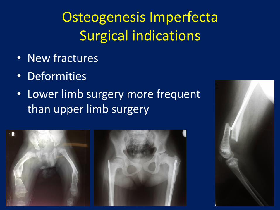
Osteogenesis ImperfectaSurgical indications
• New fractures
• Deformities
• Lower limb surgery more frequent than upper limb surgery
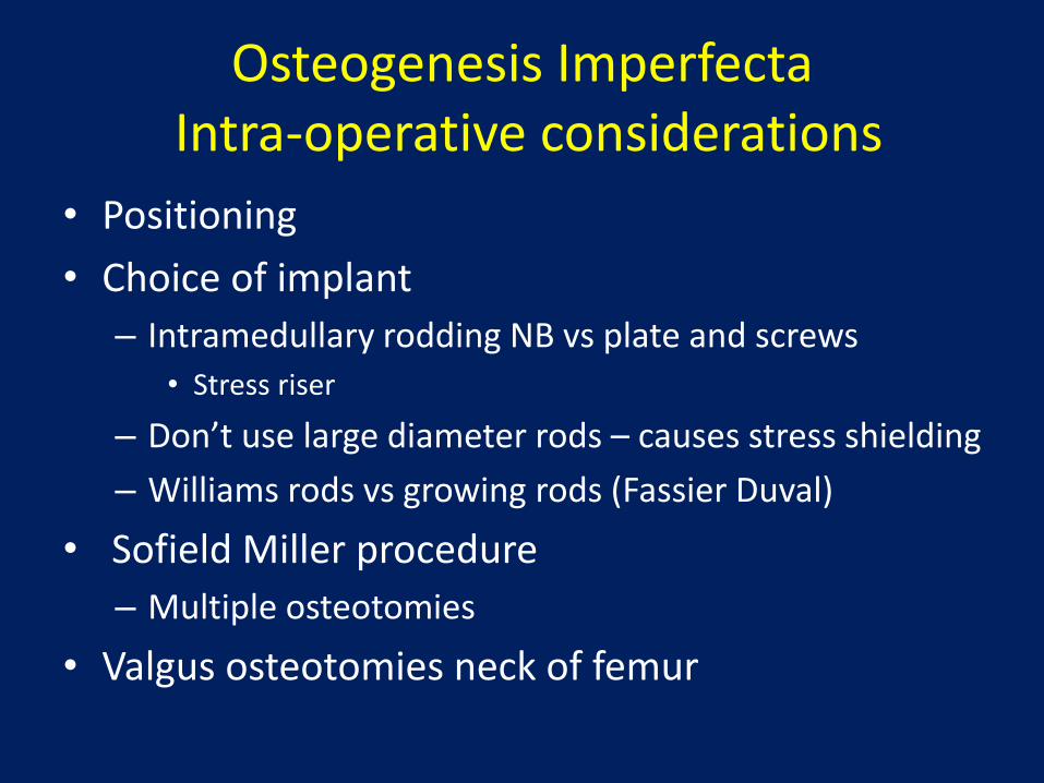
Osteogenesis ImperfectaIntra-operative considerations
• Positioning
• Choice of implant
– Intramedullary rodding NB vs plate and screws
• Stress riser
– Don’t use large diameter rods – causes stress shielding
– Williams rods vs growing rods (Fassier Duval)
• Sofield Miller procedure
– Multiple osteotomies
• Valgus osteotomies neck of femur
Osteogenesis ImperfectaOperative techniques
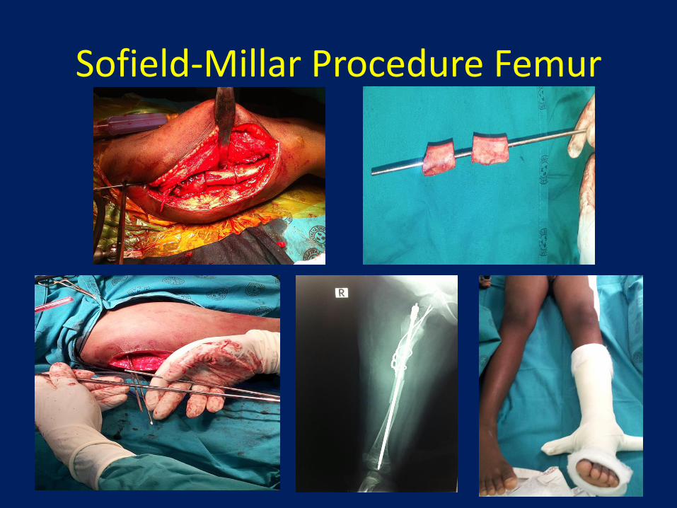
Sofield-Millar Procedure Femur
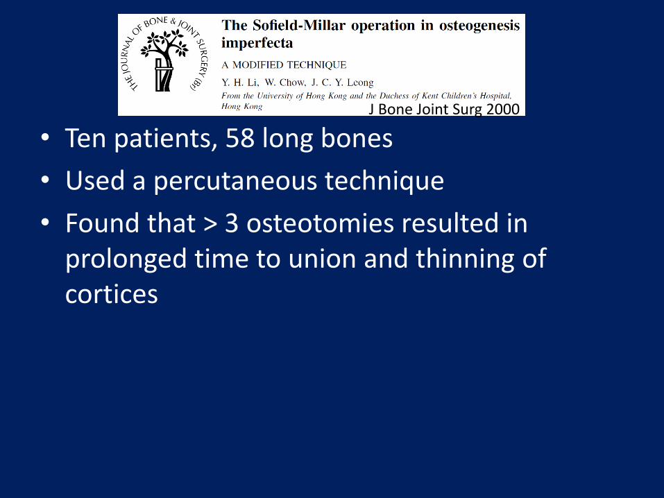
• Ten patients, 58 long bones
• Used a percutaneous technique
• Found that > 3 osteotomies resulted in prolonged time to union and thinning of cortices
J Bone Joint Surg 2000
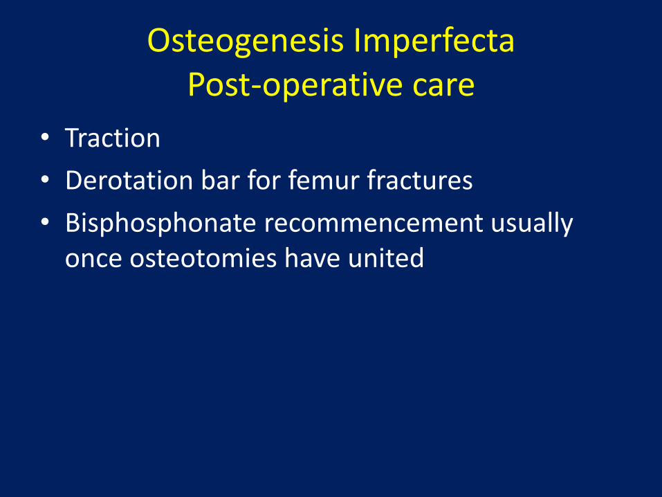
Osteogenesis ImperfectaPost-operative care
• Traction
• Derotation bar for femur fractures
• Bisphosphonate recommencement usually once osteotomies have united
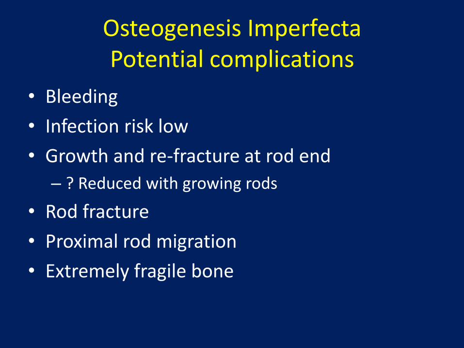
Osteogenesis ImperfectaPotential complications
• Bleeding
• Infection risk low
• Growth and re-fracture at rod end
– ? Reduced with growing rods
• Rod fracture
• Proximal rod migration
• Extremely fragile bone
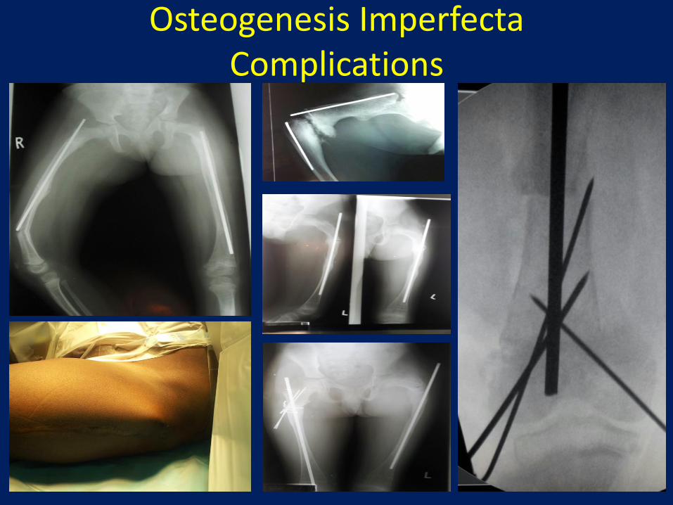
Osteogenesis ImperfectaComplications
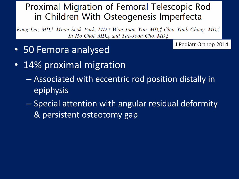
• 50 Femora analysed
• 14% proximal migration
– Associated with eccentric rod position distally in epiphysis
– Special attention with angular residual deformity & persistent osteotomy gap
J Pediatr Orthop 2014
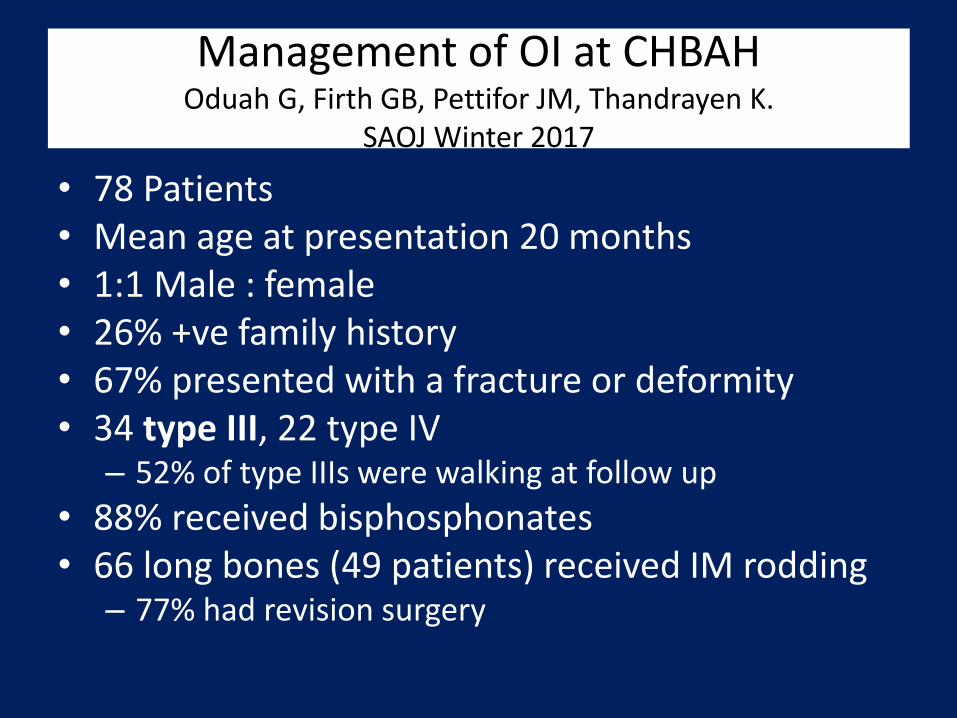
Management of OI at CHBAHOduah G, Firth GB, Pettifor JM, Thandrayen K.
SAOJ Winter 2017
• 78 Patients• Mean age at presentation 20 months• 1:1 Male : female• 26% +ve family history• 67% presented with a fracture or deformity• 34 type III, 22 type IV
– 52% of type IIIs were walking at follow up
• 88% received bisphosphonates• 66 long bones (49 patients) received IM rodding
– 77% had revision surgery
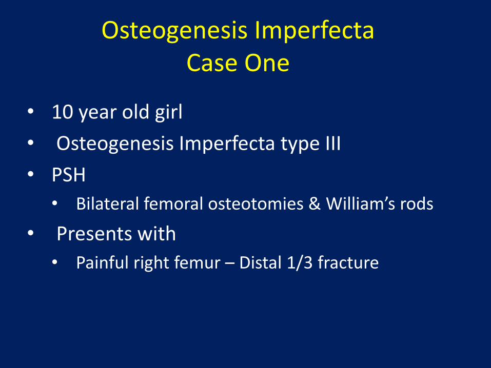
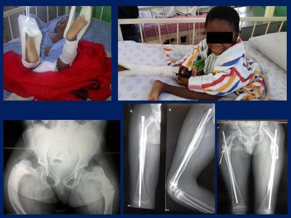
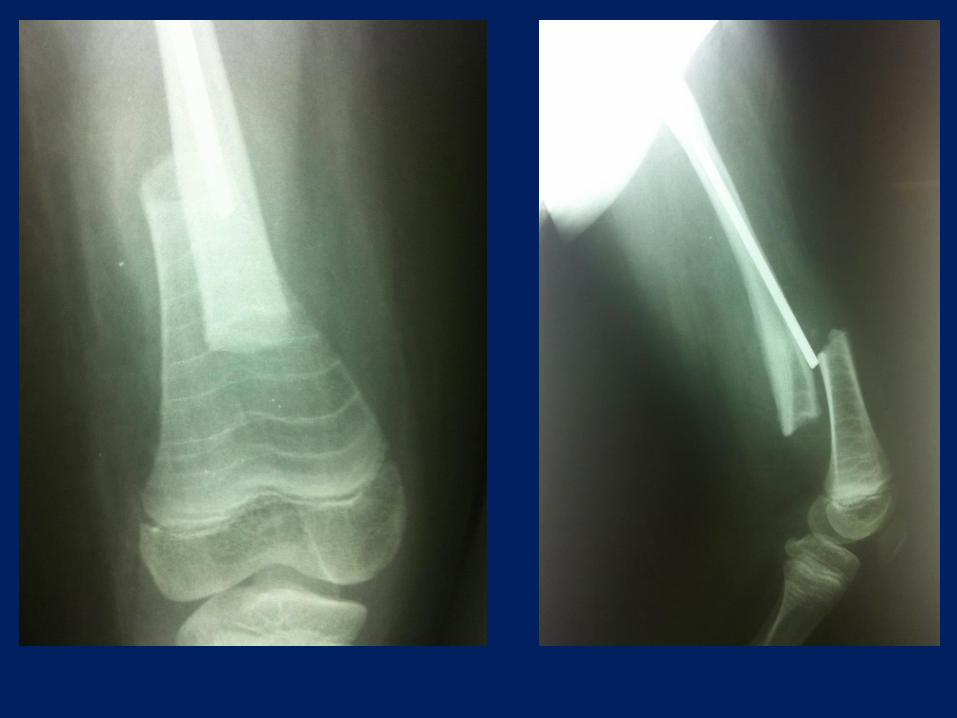
Osteogenesis ImperfectaCase One
• 10 year old girl
• Osteogenesis Imperfecta type III
• PSH
• Bilateral femoral osteotomies & William’s rods
• Presents with
• Painful right femur – Distal 1/3 fracture
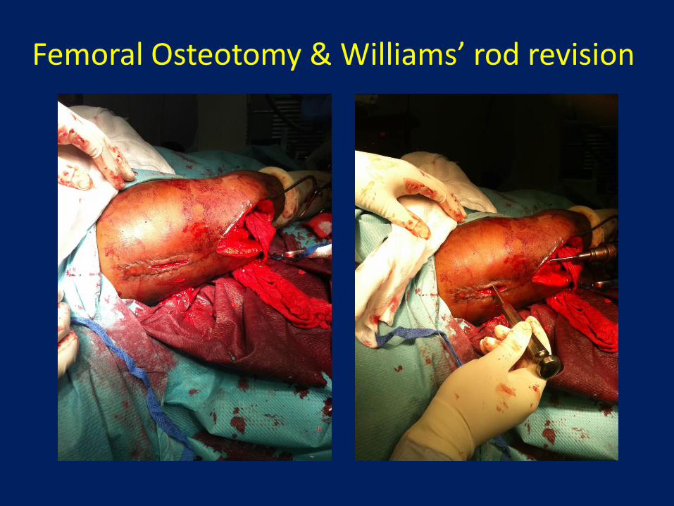
Femoral Osteotomy & Williams’ rod revision
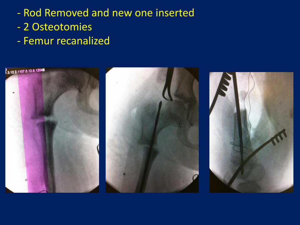
- Rod Removed and new one inserted- 2 Osteotomies- Femur recanalized
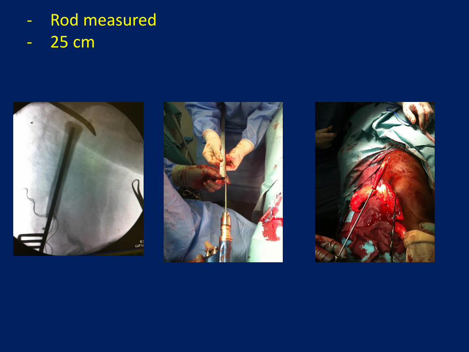
- Rod measured- 25 cm
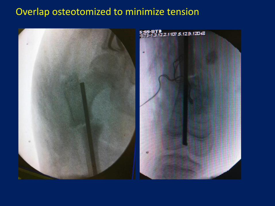
Overlap osteotomized to minimize tension
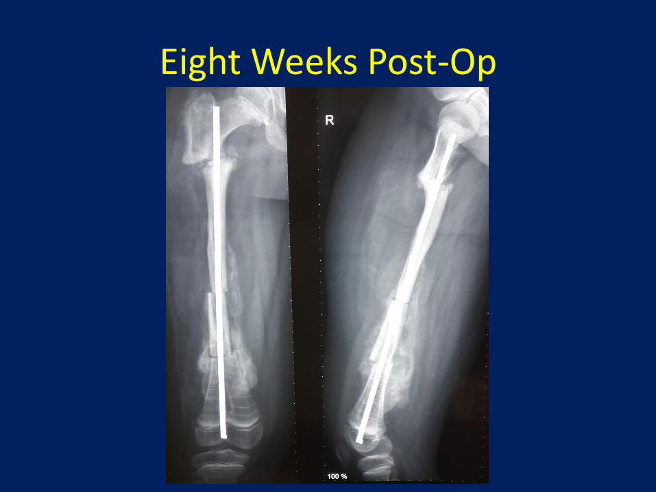
Eight Weeks Post-Op
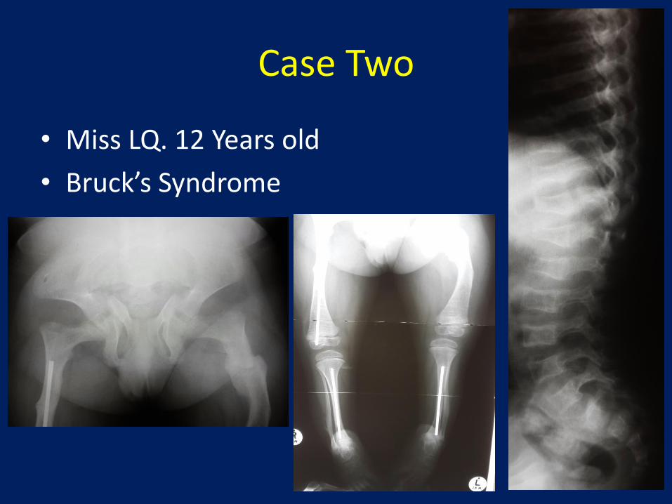
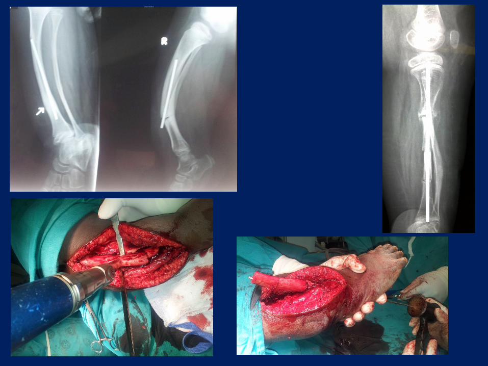
Case Two
• Miss LQ. 12 Years old
• Bruck’s Syndrome
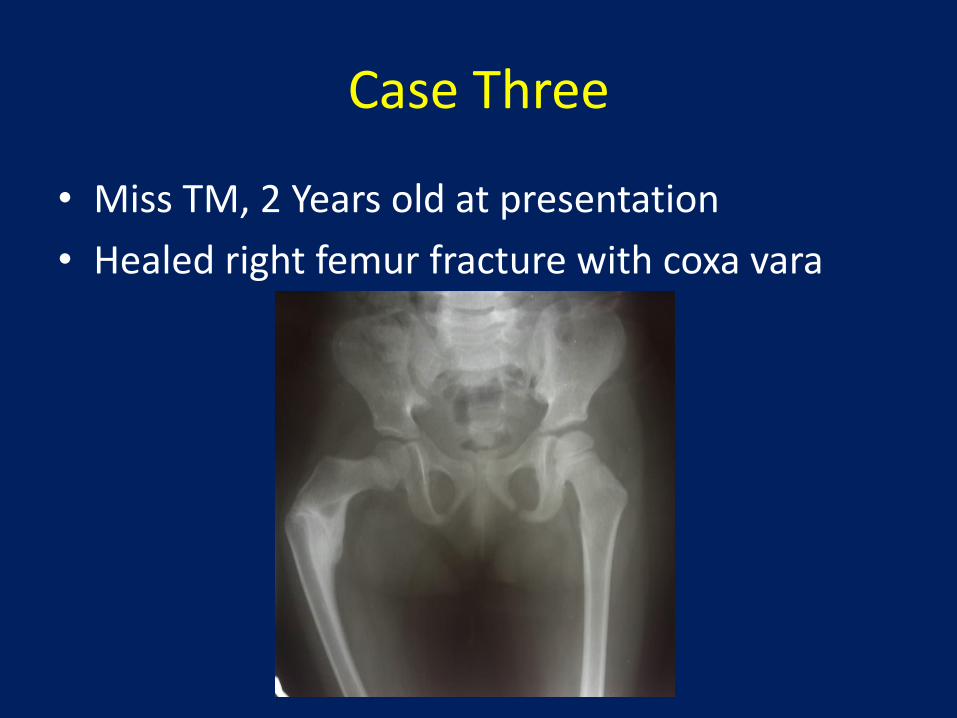
Case Three
• Miss TM, 2 Years old at presentation
• Healed right femur fracture with coxa vara
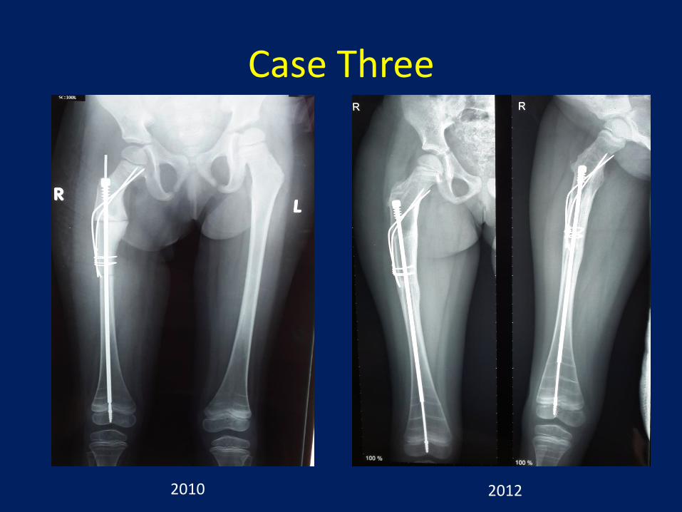
Case Three
2010 2012
Summary
• Surgical management of Rickets & OI beneficial
• Optimal medical management essential
• Multiple deformities make surgery complicated
• Intramedullary rodding standard
– Use of elongating rods inherent advantages
• Beware of high complication rate

Thank You