the transcriptional regulator cpsy is important for innate immune evasion...
TRANSCRIPT

The Transcriptional Regulator CpsY IsImportant for Innate Immune Evasion inStreptococcus pyogenes
Luis A. Vega,a Kayla M. Valdes,a Ganesh S. Sundar,a Ashton T. Belew,a
Emrul Islam,a Jacob Berge,a Patrick Curry,a Steven Chen,a Najib M. El-Sayed,a,b
Yoann Le Breton,a Kevin S. McIvera
Department of Cell Biology & Molecular Genetics and Maryland Pathogen Research Institutea and Center forBioinformatics and Computational Biology,b University of Maryland, College Park, Maryland, USA
ABSTRACT As an exclusively human pathogen, Streptococcus pyogenes (the group Astreptococcus [GAS]) has specifically adapted to evade host innate immunity andsurvive in multiple tissue niches, including blood. GAS can overcome the metabolicconstraints of the blood environment and expresses various immunomodulatory fac-tors necessary for survival and immune cell resistance. Here we present our investi-gation of one such factor, the predicted LysR family transcriptional regulator CpsY.The encoding gene, cpsY, was initially identified as being required for GAS survivalin a transposon-site hybridization (TraSH) screen in whole human blood. CpsY is ho-mologous with transcriptional regulators of Streptococcus mutans (MetR), Streptococ-cus iniae (CpsY), and Streptococcus agalactiae (MtaR) that regulate methionine trans-port, amino acid metabolism, resistance to neutrophil-mediated killing, and survivalin vivo. Our investigation indicated that CpsY is involved in GAS resistance to innateimmune cells of its human host. However, GAS CpsY does not manifest the in vitrophenotypes of its homologs in other streptococcal species. GAS CpsY appears toregulate a small set of genes that is markedly different from the regulons of its ho-mologs. The differential expression of these genes depends on the growth medium,and CpsY modestly influences their expression. The GAS CpsY regulon includesknown virulence factors (mntE, speB, spd, nga [spn], prtS [SpyCEP], and sse) and cellsurface-associated factors of GAS (emm1, mur1.2, sibA [cdhA], and M5005_Spy0500).Intriguingly, the loss of CpsY in GAS does not result in virulence defects in murinemodels of infection, suggesting that CpsY function in immune evasion is specific tothe human host.
KEYWORDS CpsY, LysR family regulators, human-specific virulence, immune evasion,transcriptional regulation
As an exclusively human pathogen, Streptococcus pyogenes, or group A streptococ-cus (GAS), has specifically adapted to evade host innate immunity and survive in
multiple tissue niches. GAS primarily colonizes the epithelia of the nasopharynx andskin, but a number of clinically important serotypes are capable of invading deep tissueand the bloodstream, resulting in pathologies with a high mortality rate (1, 2). Theinvasive phenotype in some of these strains has been linked to mutation of globaltranscriptional regulators, such as the CovRS/CsrRS two-component system (TCS),which regulates important virulence factors such as SpeB protease, streptokinase,streptodornase, and the hyaluronic acid capsule (3–5). Transcriptional regulators of GASare crucial to immune evasion, as observed for both stand-alone regulators like Mga (6,7) and TCSs like Ihk/Irr (8, 9) and CovRS/CsrRS (10). In the case of the latter TCSpathways, adaptation of these regulators following exposure of GAS to macrophagesenhanced survival of GAS during reinfection of immune cells (9). Thus, control of
Received 7 November 2016 Returned formodification 4 December 2016 Accepted 13December 2016
Accepted manuscript posted online 19December 2016
Citation Vega LA, Valdes KM, Sundar GS, BelewAT, Islam E, Berge J, Curry P, Chen S, El-SayedNM, Le Breton Y, McIver KS. 2017. Thetranscriptional regulator CpsY is important forinnate immune evasion in Streptococcuspyogenes. Infect Immun 85:e00925-16. https://doi.org/10.1128/IAI.00925-16.
Editor Nancy E. Freitag, University of Illinois atChicago
Copyright © 2017 American Society forMicrobiology. All Rights Reserved.
Address correspondence to Yoann Le Breton,[email protected], or Kevin S. McIver,[email protected].
BACTERIAL INFECTIONS
crossm
March 2017 Volume 85 Issue 3 e00925-16 iai.asm.org 1Infection and Immunity
on July 16, 2018 by guesthttp://iai.asm
.org/D
ownloaded from

virulence factor expression through transcriptional regulator modulation has significantconsequences for GAS-associated disease development.
Pathogenic streptococci exposed to host blood employ a variety of strategies tosurvive and propagate in this environment. The GAS cell surface specifically interactswith a high number of blood plasma proteins, and differences have been observedbetween the interaction profiles of invasive and noninvasive GAS strains (11). The hostcoagulation cascade functions during bacterial infection of the bloodstream to entrapbacteria within a fibrin clot for killing. However, GAS can circumvent this mechanism bysequestering host plasminogen on the bacterial cell surface, converting it into activeplasmin via streptokinase, and using it to degrade the fibrin network, thus liberatingGAS from clots (reviewed in reference 12). Investigation of isogenic invasive andnoninvasive GAS isolates found that expression of immunomodulatory factors (e.g., C5apeptidase, Gls24, and M protein) increased in response to growth in whole blood,particularly in invasive strains, and were necessary for survival in whole blood andneutrophil resistance (13).
GAS is also able to overcome the metabolic constraints imposed on it by the bloodenvironment. It possesses a metal transport operon (mtsABC) (14) as well as severaldedicated hemoprotein and iron acquisition systems (siaABC, shr, htsABC) (15–17) thatare controlled by multiple regulators (mtsR and perR; reviewed in reference 18) and arenecessary for survival in blood (19). Regulation of amino acid metabolism is required forboth growth and immune evasion. For example, arginine catabolism through CodY-mediated control of the arcABC operon (20) contributes to resistance to inducible nitricoxide synthase (iNOS)-mediated production of nitric oxide (21). Access to carbonsources directly impacts immune evasion, as recently demonstrated by loss of thefruB-encoded 1-phosphopfrucokinase adversely affecting GAS survival in human bloodand resistance to phagocytes (22). Thus, it is important to understand how GAS adaptsto specific host niches, metabolically and through immune evasion, to effectivelyaddress the rising incidence of invasive GAS strains (reviewed in reference 23).
In a transposon site hybridization (TraSH) forward genetic screen of M1T1 GAS 5448,cpsY was found to be required for survival in whole human blood (19). GAS CpsYdisplays 83% identity with MetR, a LysR family transcriptional regulator of Streptococcusmutans shown to activate transcription of genes involved in methionine biosynthesis(metE) and uptake (atmB) in a homocysteine-dependent manner (24). Homologs ofCpsY found in Streptococcus iniae and Streptococcus agalactiae (MtaR) have beencharacterized as virulence determinants that regulate methionine transport (25), aminoacid metabolism (26), and cell wall modifications necessary for resistance to neutrophil-mediated killing and survival in vivo (27–29). Thus, CpsY appears to play a crucial roleas a transcriptional regulator in the development of streptococcal invasive diseasethrough the promotion of immune cell resistance and host niche adaptation.
Here, we present evidence that the CpsY transcriptional regulator of GAS acts as aregulator of resistance to innate immune cells and survival in human blood in a mannerdistinct from those of other streptococcal pathogens. Loss of CpsY in GAS does notproduce the methionine-dependent in vitro growth defects or the in vivo survivalphenotypes observed with other streptococcal species in mouse models of infection.However, disruption of CpsY expression significantly impacts GAS resistance to humanphagocytic cells, indicating that CpsY function in immune evasion is specific to thehuman host. Transcriptome analysis revealed that CpsY influences the expression of asmall number of genes, both known and hypothetical, under our experimental condi-tions. The regulon includes genes encoding cell wall-modifying proteins and surfaceproteins, suggesting that CpsY-mediated resistance to host innate immunity mayrequire alterations to the GAS cell envelope.
RESULTSCpsY is required for GAS survival in whole human blood. The cpsY gene was
originally identified as necessary for GAS survival in heparinized whole human blood ina transposon mutagenesis (TraSH) screen (19). To further characterize the role of CpsY
Vega et al. Infection and Immunity
March 2017 Volume 85 Issue 3 e00925-16 iai.asm.org 2
on July 16, 2018 by guesthttp://iai.asm
.org/D
ownloaded from

in GAS virulence, we generated a stable insertional inactivation mutant, the (�)cpsYmutant, and a strain rescued for the mutation (the cpsYR strain) in the M1T1 5448background initially used in the blood screen (see Materials and Methods). The survivalin heparinized whole human blood of the mutant and its rescue strain was tested usingthe Lancefield blood bactericidal assay (19, 30). The (�)cpsY mutant had a significantlylower multiplication factor (MF) in heparinized whole blood than did wild-type 5448and the cpsYR strain (Fig. 1A), confirming the requirement of cpsY for GAS survival inblood. To assess whether the observed phenotype resulted from decreased resistanceto host immune cells found in whole blood or to the metabolic environment of blood,growth of 5448, the (�)cpsY mutant, and the cpsYR rescue strain in the presence ofhuman plasma was also examined. In plasma isolated from heparinized whole donorblood equilibrated 1:1 with tissue culture medium (RPMI 1640, 2.05 mM L-glutamine),no appreciable defects in growth were observed in the (�)cpsY or the cpsYR strainrelative to the 5448 strain (Fig. 1B).
Loss of CpsY does not adversely affect GAS growth in vitro. CpsY homologs inother streptococcal species have been shown to play a role in amino acid metabolism,impacting growth in various lab culture media and in human plasma. Finding nophenotype for growth in plasma, we examined whether the (�)cpsY strain exhibited anygrowth defects in commonly used media. Growth of (�)cpsY and its rescue strain (cpsYR)was comparable to that of the 5448 parent in rich medium (Todd-Hewitt mediumsupplemented with 0.2% yeast extract [THY]) (Fig. 2A). Likewise, in chemically definedmedium supplemented with 0.5% glucose (CDM) and in low-glucose, high-peptide Cmedium, the GAS (�)cpsY strain and its wild-type parent showed no differences ingrowth rates or yields (Fig. 2B and C). This was in contrast to the case with other
5448
(ι)cpsY
cpsY R
0
25
50
75
100
125
Mul
tiplic
atio
n Fa
ctor
(%
Rel
ativ
e to
544
8)
Whole Human Blood
*
A
0 60 120 180 240 300 360 420 480 540
104
105
106
107
Time (minutes)
CFU
/ml
RPMI+Plasma (50%)
5448
(ι)cpsY
cpsYR
B
FIG 1 CpsY is required for GAS survival in whole human blood. Growth of wild-type GAS 5448, anisogenic (�)cpsY mutant, and its rescue (cpsYR) strain were assayed in the human blood environment. (A)A Lancefield bactericidal assay was performed by monitoring growth in heparinized whole human blood.Wild-type GAS 5448 had a mean multiplication factor (MF) of 75.2 � 6.9 in the donor blood assayed. Dataare presented as percent growth in blood, corresponding to the MF of the mutant and rescue dividedby the MF of the wild type � 100. Data represent means � SEMs from four independent experiments.*, P � 0.05. (B) Growth of GAS was measured in RPMI 1640 supplemented with plasma (50%) obtainedfrom heparinized whole blood from human donors by plating aliquots at the indicated time points toquantify CFU. Results of a representative experiment out of three biological replicates are shown.
Role of CpsY in GAS Immune Evasion Infection and Immunity
March 2017 Volume 85 Issue 3 e00925-16 iai.asm.org 3
on July 16, 2018 by guesthttp://iai.asm
.org/D
ownloaded from

streptococci, for which disruption of cpsY led to significant growth phenotypes in thesemedia (25, 28). In S. agalactiae (GBS), growth defects for mutants of the CpsY homologMtaR were rescued by supplementing culture media with methionine. However, forGAS, addition of methionine to CDM had no effect on growth of either 5448 or the(�)cpsY mutant (see Fig. S1A in the supplemental material).
To further explore if the metabolic environment of blood contributed to the reducedsurvival phenotype observed for the (�)cpsY strain, we tested whether loss of CpsYimpacted carbohydrate metabolism using a bioMérieux API 50 CH assay strip that testsutilization of a subset of 49 carbohydrates (Fig. 2D). No significant changes in theutilization of different carbon sources were observed between 5448 and the (�)cpsY andcpsyR strains after either 24 h or 48 h of incubation. Thus, it appears that the reducedsurvival of the (�)cpsY mutant in whole human blood is not due to metabolic defects incarbon utilization.
Loss of CpsY reduces GAS resistance to opsonophagocytic killing. The reducedfitness of the M1T1 (�)cpsY mutant in whole human blood, combined with the absenceof growth defects in vitro, suggests that CpsY is involved in the evasion of cellularcomponents of host innate immunity (e.g., neutrophils and macrophages). To examinethis possibility, we assessed the viability of both 5448 and the (�)cpsY mutant in anopsonophagocytic killing assay (OKA). Bacteria were opsonized in plasma obtained
0 60 120 180 240 300 360 420 480 540
10
100
Time (Minutes)
Kle
tt Va
lue
(Log
)
THY
5448
cpsYR
(ι)cpsY
A
5448
cpsYR
(ι)cpsY
0 60 120 180 240 300 360 420 480 540 600 660 720
10
100
Time (Minutes)
Kle
tt Va
lue
(Log
)
CDM
5448
(ι)cpsY
B
0 60 120 180 240 300 360 420 480 540 600
10
100
Time (Minutes)
Kle
tt Va
lue
(Log
)
C MediumC
5448
(ι) c
psY
cpsY
R
5448
(ι)c
psY
cpsY
R
D-Galactose D-Glucose D-Fructose D-Mannose CH3- D-Glucopyranoside N-AcetylGlucosamine Salicin D-Cellobiose D-Maltose D-Lactose D-Sucrose D-Trehalose Starch Glycogen 24h 48h
D
FIG 2 CpsY is not required for growth of GAS in vitro. Growth curves for the (�)cpsY mutant and its rescue (cpsYR) strain in enriched THY medium (A), chemicallydefined medium (CDM) (B), and C medium (C) were measured by absorbance using a Klett-Summerson colorimeter and expressed in Klett units compared tothe value for wild-type 5448. Data are the results from three biological replicates. (D) Limited carbohydrate utilization profiles of 5448, the (�)cpsY mutant andits rescue (cpsYR), assayed using the API 50 CH assay. Results after incubation at 37°C for 24 h and 48 h are shown. Listed are selected carbon sources from apanel of 50 total carbohydrate sources, with complete utilization (white boxes), partial utilization (gray boxes), and no utilization (black boxes) based on thecolorimetric indicator dye shown for each.
Vega et al. Infection and Immunity
March 2017 Volume 85 Issue 3 e00925-16 iai.asm.org 4
on July 16, 2018 by guesthttp://iai.asm
.org/D
ownloaded from

from heparinized whole donor blood and presented to either cultured neutrophil-likedifferentiated HL60 cells or freshly isolated neutrophils or monocytes. The percentagesof viable GAS following incubation in the presence or absence of immune cells werecalculated by quantifying CFU recovered from wells with immune-cell-challengedbacteria versus bacteria in wells with OKA medium (i.e., RPMI plus 20% plasma) alone.
OKA data showed a reduction in the number of viable (�)cpsY mutant bacteria in thepresence of polymorphonuclear leukocytes (PMNs) (Fig. 3A and B) and monocytes anddifferentiated HL60 cells (Fig. 3C), suggesting that the loss of CpsY resulted in reducedresistance of GAS to opsonophagocytic killing. Inhibition of phagocytosis by cytocha-lasin D treatment and heat inactivation of plasma to inactivate opsonins producedsimilar levels of viable bacteria for 5448, the (�)cpsY mutant, and the cpsYR rescue strains(Fig. 3B and C, CytoD�HI Plasma). The decreased survival of the (�)cpsY mutant in thepresence of neutrophils likely stems from increased phagocytosis and/or reducedtolerance to intracellular antimicrobial mechanisms of the phagocytes. Additionally,reduced survival of the (�)cpsY mutant in the neutrophil killing assay was not related togrowth of the mutant in the assay medium, as multiplication factors were comparablefor all three strains in RPMI 1640 –2.05 mM L-glutamine plus plasma (Fig. S1B).
Absence of CpsY does not impact GAS virulence in mouse models of infection.Given the reduced resistance to innate immune cells, we examined whether the loss ofCpsY impacted GAS virulence in vivo using mouse models of soft tissue (subcutaneous
0 30 60 90 120 150104
105
106
107
Time (Minutes)
Viab
le B
acte
ria (C
FU/m
l)
5448 vs (ι)cpsY - PMNs
5448 PMNs
(ι)cpsY PMNs
5448 RPMI+Plasma
( )cpsY RPMI+Plasma
A
0
25
50
75
100
125
150
RPMI+ CytoD+ Plasma HI Plasma
% V
iabl
e B
acte
ria (M
OI=
0.1)
GAS + PMNs
***
5448
(ι)cpsY
cpsYR
B
5448
(ι)cpsY
cpsYR
C
0255075
100125150175
RPMI+ CytoD+ HL60s Plasma HI Plasma
Monocytes
% V
iabl
e B
acte
ria (M
OI=
0.1)
GAS + Monocytes/HL60s
** **
FIG 3 CpsY is required for GAS resistance to opsonophagocytic killing by human innate immune cells. Recovery of viable wild-type 5448(black) GAS following immune cell challenge was compared to those of the (�)cpsY mutant (red) and its cpsYR rescue (blue), with GASopsonized in either plasma or heat-inactivated plasma (HI Plasma) prior to exposure to untreated or cytochalasin D-treated (CytoD)immune cells. (A) Viable bacteria recovered from challenge by human neutrophils (PMNs). GAS (1 � 105 CFU/well) and neutrophils (1 �106 CFU/well) were incubated for 2 h at 37°C. Neutrophils were lysed at 30 min, 60 min, and 2 h, and the number of viable GAS wasenumerated as CFU recovered. Results of a representative experiment out of a minimum of three biological replicates are shown. (B) Viablebacteria recovered from challenge by human PMNs. GAS (1 � 105 CFU/well) and neutrophils (1 � 106 CFU/well) were incubated for 2 hat 37°C. Neutrophils were then lysed, and the percentage of viable GAS was determined and shown as survival relative to that of wild-type5448. (C) Viable bacteria recovered from challenge by monocytes and neutrophil-like HL60 cells. GAS (1 � 105 CFU/well) and eithermonocytes or differentiated HL60 cells (1 � 106 CFU/well) were incubated for 2 h at 37°C. Cells were lysed, and the percentage of viableGAS was determined relative to the survival of 5448. Data shown in panels B and C represent means � SEMs of the results from at leastthree biological replicates performed in triplicate, with P less than 0.001 (overall ANOVA). Individual groups were compared usingDunnett’s multiple-comparison test (***, P � 0.001; **, P � 0.01).
Role of CpsY in GAS Immune Evasion Infection and Immunity
March 2017 Volume 85 Issue 3 e00925-16 iai.asm.org 5
on July 16, 2018 by guesthttp://iai.asm
.org/D
ownloaded from

[s.c.]) and systemic (intraperitoneal [i.p.]) GAS infections. The (�)cpsY mutant showed nosignificant differences with respect to both 5448 and the cpsYR rescue strain in its abilityto cause disease in either model of infection (Fig. 4A and B). Though the lesion size forthe (�)cpsY mutant in the subcutaneous model of infection trended smaller than for5448 or the cpsYR rescue strain (data not shown), the differences were not statisticallysignificant. This indicates that CpsY does not significantly affect virulence in GAS mousemodels of infection. Interestingly, a modified Lancefield blood bactericidal assay em-ploying murine blood found that the (�)cpsY mutant did not exhibit the same growthdefect (Fig. 4C) as observed in whole human blood (Fig. 1A). These data suggest thatthe role of CpsY is specific to the evasion of human innate immune cells by GAS, whichis distinct from that observed for CpsY homologs in other streptococcal species.
GAS CpsY regulates a markedly different set of genes to other streptococcalspecies. Since CpsY activity in GAS appears to phenotypically differ from that ofhomologs in other streptococci, we hypothesized that it possessed a regulon differentfrom that in S. mutans, S. iniae, and S. agalactiae. RNA sequencing (RNA-seq) analysiswas performed on RNA isolated from either 5448 or (�)cpsY mutant cultures that weregrown to late logarithmic phase in either rich medium (THY) or RPMI 1640 plus 20%human donor plasma for 2 h to simulate OKA conditions. We identified distinct,medium-specific sets of transcripts, consisting of only 25 and 16 genes, respectively,that were differentially expressed in the (�)cpsY mutant relative to the 5448 parent(Table 1). Notably absent from the list of identified transcripts were genes regulated byCpsY homologs in other streptococci, such as the methionine transport operon atmBDE(metNPQ), the arginine transport operon artPQ, and the peptidoglycan modification-associated gene murA (Table 2). We confirmed these results by quantitative real-time
C
0 24 48 720
25
50
75
100
Time (hours)
% S
urvi
val
Mouse Intraperitoneal Infection
5448
(ι)cpsY
cpsYR
ns
A
0 24 48 72 96 120 144 1680
25
50
75
100
Time (hours)
% S
urvi
val
Mouse Subcutaneous Infection
ns
B
5448
(ι)cpsY
cpsYR
1
10
100
A B C
Mul
tiplic
atio
n Fa
ctor
Murine Whole Blood
5448
(ι)cpsY
FIG 4 CpsY does not impact GAS virulence in mouse models of infection. Virulence of the (�)cpsY mutant (red) and the cpsYR rescue strain(blue) was compared to that of wild-type 5448 (black) in the following mouse models. (A) Survival in a murine intraperitoneal (i.p.) modelof systemic infection. GAS (�2 � 108 CFU/mouse) bacteria were injected i.p. into mice (n � 10/strain) and monitored for morbidity overthe course of 72 h. Significance was determined using Kaplan-Meier survival analysis and the log rank test. (B) Survival in a murinesubcutaneous (s.c.) model of soft tissue infection. GAS (�2.8 � 108 CFU/mouse) were injected s.c. into mice (n � 10/strain) and monitoredfor morbidity over the course of 7 days. Significance was determined using Kaplan-Meier survival analysis and the log rank test. (C) Survivalin a modified Lancefield bactericidal assay using heparinized whole mouse blood. The data are presented as the multiplication factor (MF)of wild-type 5448 and the (�)cpsY mutant in whole mouse blood. Data represent GAS survival in blood obtained from multiple mice(n � 4 to 6) and are means � SEMs of the results of independent experiments.
Vega et al. Infection and Immunity
March 2017 Volume 85 Issue 3 e00925-16 iai.asm.org 6
on July 16, 2018 by guesthttp://iai.asm
.org/D
ownloaded from

PCR (qRT-PCR) analysis of artP, atmB (metN), and murA transcripts quantified in totalRNA isolated at various phases of growth from 5448 and (�)cpsY strains growing inenriched medium (THY). No changes in the relative expression of these genes wereobserved between the wild type and the (�)cpsY mutant at any of the time pointsexamined, with one exception (Fig. 5). Expression of artP showed a 2-fold increaseduring the late logarithmic phase of growth in the absence of CpsY (Fig. 5B).
Transcripts that were differentially expressed in OKA medium included severalsurface-associated factors of GAS (Table 1): the cell wall-anchored M protein (emm1),cell wall-modifying enzymes (mur1.2, sibA [cdhA], and M5005_Spy0500 [or simply
TABLE 1 Genes differentially expressed in the GAS (�)cpsY mutanta
Spy designation Annotation Gene name Log2 fold changeb
Differentially expressed in OKAmedium
VirulenceM5005_Spy0980 Manganese-specific efflux pump (43) mntE 1.630M5005_Spy1719 M protein emm1 �1.748
Cell wall modificationM5005_Spy0017 CHAP-domain-containing and chain-forming cell wall
hydrolase (80)sibA (cdhA) �1.318
M5005_Spy0500 N-Acetylmuramoyl-L-alanine amidase murA.2 1.011M5005_Spy0664 Autolysin mur1.2 �1.581
MetabolismM5005_Spy1297 Phosphor-2-dehydro-3-deoxyheptonate aldolase aroA2 1.295M5005_Spy1509 Pyruvate, phosphate dikinase 1.348
HypotheticalM5005_Spy0513 Hypothetical protein �1.021M5005_Spy0714 Hypothetical protein 1.454M5005_Spy0979 Hypothetical protein 2.181
Differentially expressed in THY (latelog phase)
VirulenceM5005_Spy0139 NAD glycohydrolase nga (spn) 1.292M5005_Spy0341 Lactocepin prtS (spyCEP) 1.280M5005_Spy1407 Esterase sse 1.281M5005_Spy1735 Streptococcal pyrogenic exotoxin B speB 5.178M5005_Spy1738 Phage-associated DNase spd 1.412
Fatty acid biosynthesis/degradationM5005_Spy0039 Bifunctional acetaldehyde-CoA/alcohol dehydrogenase adh2 (adhE) 2.285M5005_Spy0040 Alcohol dehydrogenase adhA (adhP) 1.431M5005_Spy1485 Acetyl-CoA carboxylase subunit beta accA �1.345M5005_Spy1487 3-Hydroxyacyl-acyl-carrier-protein dehydratase fabZ �1.337M5005_Spy1489 3-Oxoacyl-ACP synthase fabF �1.413M5005_Spy1490 3-Ketoacyl-ACP reductase fabG �1.522M5005_Spy1491 ACP S-malonyltransferase fabD �1.617M5005_Spy1492 Enoyl-ACP reductase fabK �1.666M5005_Spy1496 Enoyl-CoA hydratase phaB �1.342
Protein synthesis/processingM5005_Spy0694 ATP-dependent protease ATP-binding subunit clpL 1.229M5005_Spy1483 Seryl-tRNA synthetase serS 1.138M5005_Spy1732 Foldase PrsA prsA 2.326M5005_Spy1736 Molecular chaperone GroEL groEL 1.022
Energy productionM5005_Spy0132 V-type ATP synthase subunit B ntpB 1.478M5005_Spy0133 V-type ATP synthase subunit D ntpD 1.605
Carbohydrate metabolismM5005_Spy0834 Zn-dependent alcohol dehydrogenase 1.523
Transcription factorsM5005_Spy0440 Transcriptional regulator (81) rgg3 1.281M5005_Spy1277 Arginine repressor ArgR ahrC.2 (argR) 1.013
Signal transductionM5005_Spy1400 PTS system galactose-specific transporter subunit EIIB 1.986
aData for downregulated genes are shaded.bRNA-seq analysis results (P � 0.05).
Role of CpsY in GAS Immune Evasion Infection and Immunity
March 2017 Volume 85 Issue 3 e00925-16 iai.asm.org 7
on July 16, 2018 by guesthttp://iai.asm
.org/D
ownloaded from
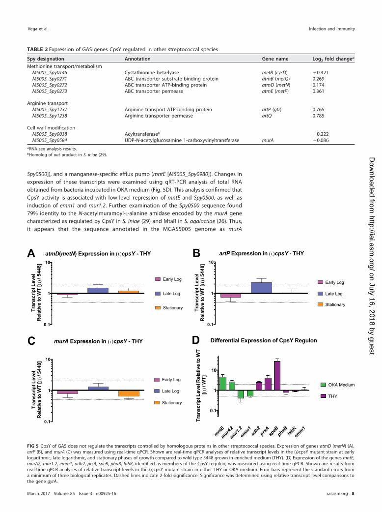
Spy0500]), and a manganese-specific efflux pump (mntE [M5005_Spy0980]). Changes inexpression of these transcripts were examined using qRT-PCR analysis of total RNAobtained from bacteria incubated in OKA medium (Fig. 5D). This analysis confirmed thatCpsY activity is associated with low-level repression of mntE and Spy0500, as well asinduction of emm1 and mur1.2. Further examination of the Spy0500 sequence found79% identity to the N-acetylmuramoyl-L-alanine amidase encoded by the murA genecharacterized as regulated by CpsY in S. iniae (29) and MtaR in S. agalactiae (26). Thus,it appears that the sequence annotated in the MGAS5005 genome as murA
TABLE 2 Expression of GAS genes CpsY regulated in other streptococcal species
Spy designation Annotation Gene name Log2 fold changea
Methionine transport/metabolismM5005_Spy0146 Cystathionine beta-lyase metB (cysD) �0.421M5005_Spy0271 ABC transporter substrate-binding protein atmB (metQ) 0.269M5005_Spy0272 ABC transporter ATP-binding protein atmD (metN) 0.174M5005_Spy0273 ABC transporter permease atmE (metP) 0.361
Arginine transportM5005_Spy1237 Arginine transport ATP-binding protein artP (gtr) 0.765M5005_Spy1238 Arginine transporter permease artQ 0.785
Cell wall modificationM5005_Spy0038 Acyltransferaseb �0.222M5005_Spy0584 UDP-N-acetylglucosamine 1-carboxyvinyltransferase murA �0.086
aRNA-seq analysis results.bHomolog of oat product in S. iniae (29).
atmD(metN) Expression in (ι)cpsY - THY
0.1
1
10
Tran
scrip
t Lev
el
Rel
ativ
e to
WT
[(ι) /
544
8]
Early Log
Late Log
Stationary
A
murA Expression in (ι)cpsY - THY
0.1
1
10
Tran
scrip
t Lev
el
Rel
ativ
e to
WT
[( ι) /
544
8]
Early Log
Late Log
Stationary
C D
artP Expression in (ι)cpsY - THY
0.1
1
10
Tran
scrip
t Lev
el
Rel
ativ
e to
WT
[( ι) /
544
8]
Early Log
Late Log
Stationary
B
mntE
murA2
mur1.2em
m1ad
h2prsA sp
eBphaB fab
Kem
m1
0.1
1
10
Tran
scrip
t Lev
el R
elat
ive
to W
T [(
ι) / W
T]
Differential Expression of CpsY Regulon
OKA Medium
THY
FIG 5 CpsY of GAS does not regulate the transcripts controlled by homologous proteins in other streptococcal species. Expression of genes atmD (metN) (A),artP (B), and murA (C) was measured using real-time qPCR. Shown are real-time qPCR analyses of relative transcript levels in the (�)cpsY mutant strain at earlylogarithmic, late logarithmic, and stationary phases of growth compared to wild type 5448 grown in enriched medium (THY). (D) Expression of the genes mntE,murA2, mur1.2, emm1, adh2, prsA, speB, phaB, fabK, identified as members of the CpsY regulon, was measured using real-time qPCR. Shown are results fromreal-time qPCR analyses of relative transcript levels in the (�)cpsY mutant strain in either THY or OKA medium. Error bars represent the standard errors froma minimum of three biological replicates. Dashed lines indicate 2-fold significance. Significance was determined using relative transcript level comparisons tothe gene gyrA.
Vega et al. Infection and Immunity
March 2017 Volume 85 Issue 3 e00925-16 iai.asm.org 8
on July 16, 2018 by guesthttp://iai.asm
.org/D
ownloaded from

(M5005_Spy0548) does not correspond to the homolog of the CpsY-regulated murAcharacterized in other streptococci. In light of this, we refer to M5005_Spy0500 asmurA2. Additional differentially expressed transcripts in OKA medium included pyru-vate metabolism proteins (aroA2 and M5005_Spy1509) and a set of short hypotheticalproteins. These results indicate that GAS CpsY regulates expression of a set of genesthat is markedly different from the ones its homologs in other streptococcal speciescontrol under the conditions we tested.
None of the transcripts identified in OKA medium, however, were differentiallyexpressed in the (�)cpsY mutant at late logarithmic growth in THY medium (Table 1). Incontrast, differentially expressed genes identified in THY include those of the fatty acidbiosynthetic pathway (accA, fabZFGDK, and phaB), genes for metabolic dehydrogenases(adhA [adhP] and adh2 [adhE]), genes involved in cell stress and protein processing(groEL, clpL, prsA, and serS), and genes for several known virulence factors (nga [spn], sse,spyCEP, speB, and spd). Changes in expression of several of these transcripts were alsoexamined using qRT-PCR analysis. Data from said analysis confirmed that in theabsence of CpsY activity, expression of adh2, prsA, and, to a large degree, speB(�30-fold change) is increased during late log growth in THY (Fig. 5D). Conversely,qRT-PCR analysis failed to detect significant changes in the expression of the phaB,fabK, and emm1 genes in the absence of CpsY activity. Overall, the qRT-PCR resultssupported the RNA-seq data, with a correlation coefficient (R2) calculated to be 0.82(n � 9 [Fig. S2]). Thus, it appears that the CpsY regulon of GAS comprises a smallnumber of genes of varied functions and is different from that of related streptococcalspecies, with the exception of artP and murA2 (M5005_Spy0500). Furthermore, GASCpsY influences expression of its regulon in a medium-dependent manner and, withthe exception of speB, the differential expression of CpsY regulon genes is modest(�4-fold).
Absence of CpsY does not influence GAS resistance to cell wall-targetingantimicrobials. CpsY regulates modifications of the bacterial cell wall in S. iniae thatare necessary for resistance to neutrophil-mediated killing, as indicated by the in-creased sensitivity to lysozyme and altered expression of a peptidoglycan amidase(murA) in a CpsY-lacking strain of this species (29). Our RNA-seq analysis indicates thatexpression of the murA homolog of S. iniae in GAS (Spy0500 [murA2]) is also influencedby GAS CpsY. We therefore examined whether the GAS (�)cpsY mutant also displayedaltered sensitivity to antimicrobials that attack different components of the bacterialcell wall. When grown in THY, the (�)cpsY mutant displayed a sensitivity to multiple cellwall-targeting compounds similar to that of wild-type 5448, including carbenicillin(minimum bactericidal concentration [MBC] � 0.15 �g ml�1), bacitracin (MBC � 0.5 �gml�1), nisin (MBC � 125 �g ml�1), and lysozyme (MBC � 2,350,000 U ml�1).
DISCUSSION
The cpsY gene was initially identified as being required for GAS survival in atransposon site hybridization (TraSH) screen in whole human blood (19). CpsY shareshomology (83% identity) with a LysR family transcriptional regulator of S. mutans(MetR), as well as to proteins in S. iniae (CpsY) and S. agalactiae (MtaR) that regulatemethionine transport (25), amino acid metabolism (26), and resistance to neutrophil-mediated killing and survival in vivo (27–29). This led us to hypothesize that CpsYfunctions as a transcriptional regulator of GAS metabolism required for survival inhuman host blood. Unlike with the CpsY homologs in S. iniae (28) and S. agalactiae (25),loss of CpsY function by insertional inactivation of the encoding gene (cpsY [Spy0701])did not affect growth of GAS in human plasma or rich medium (THY). CpsY was also notrequired for GAS growth in C medium or CDM, as seen for MtaR in S. agalactiae, nor didsupplementing media with methionine have any effect on the growth of either the(�)cpsY mutant or the parental 5448 GAS strain. Finally, we found no significantdifferences in utilization of various carbohydrate sources by the 5448 strain and (�)cpsYmutant. Together, these data suggest that the reduced fitness of a GAS mutant lackingCpsY in whole blood is not related to a broader metabolic defect.
Role of CpsY in GAS Immune Evasion Infection and Immunity
March 2017 Volume 85 Issue 3 e00925-16 iai.asm.org 9
on July 16, 2018 by guesthttp://iai.asm
.org/D
ownloaded from

We hypothesized that reduced survival of the (�)cpsY mutant in whole blood is dueto reduced resistance to immune cell activity, and/or decreased tolerance to cellularstresses encountered within these cells. Circulating neutrophils found abundantly inthe bloodstream employ multiple mechanisms to kill invading bacterial pathogens,including the production of antimicrobial peptides, neutrophil extracellular traps(NETs), and phagocytosis of the pathogen. Within the phagocytic cell, engulfed patho-gens must deal with production of reactive oxygen species, release of antibacterialgranule components, and acidification (31, 32). The production of virulence factors byGAS to resist these mechanisms of killing is well documented (33), yet their coordina-tion and regulation in different strain backgrounds is not fully understood (18). Whenwe tested the resistance to challenge by phagocytes in an opsonophagocytic killingassay (OKA), survival of the (�)cpsY mutant strain was significantly reduced across PMNs,monocytes, and neutrophil-like differentiated HL60 cells. Inhibiting phagocytosis andblocking opsonization by treating phagocytes with cytochalasin D and heat inactivatingthe plasma used in the assay rescued survival of the (�)cpsY mutant strain. This indicatesthat CpsY function is required for GAS to effectively resist opsonophagocytic killing byinnate immune cells. The fact that inhibition of phagocytosis rescues survival of the(�)cpsY mutant suggests that extracellular killing mechanisms of neutrophils such asNETs are likely not involved in the CpsY-associated phenotype, though we did notspecifically test for this. It appears that disruption of CpsY in GAS makes the pathogenmore susceptible to phagocytosis and/or less capable of intracellular growth/survivalwithin neutrophils. This is likely due to altered expression of a CpsY regulon that mayinclude antiphagocytic factors and/or proteins necessary for resisting cellular stresses tosustain bacterial growth within host immune cells.
The observed susceptibility to opsonophagocytic killing led us to postulate thatCpsY is required for virulence in an in vivo model of infection. We utilized both asubcutaneous (localized and disseminated) and an intraperitoneal (systemic) mousemodel of infection to test for changes in virulence resulting from the loss of CpsY. The(�)cpsY mutant strain did not show any decrease in virulence in either mouse modelcompared to both the 5448 wild-type parent and cpsYR rescue strain. We examinedwhether the (�)cpsY mutant strain also displayed reduced growth in whole mouseblood, and no differences were observed between the mutant and wild-type strains inthe mouse blood Lancefield assay. These results lead us to conclude that the role ofCpsY in resistance to opsonophagocytic killing involves a mechanism of the innateimmune cells of its human host that is absent from murine immune cells. Hostrestriction of GAS has been associated with differences in host innate immunity.Examination of Toll-like receptor (TLR) pathways of the innate immune response to GASfound that the TLR13 response in mice is specifically triggered by GAS rRNA and thatthe TLR13 molecule is absent in human macrophages, as is an equivalent route for GASRNA recognition (34). Furthermore, there are other instances of GAS regulators thatinfluence resistance to host immunity in a host-specific manner. FruR, the transcrip-tional repressor of the fruRBA operon encoding the fructose-specific EII of the GASphosphoenolpyruvate phosphotransferase system (PTS), was shown to mediate innateimmune evasion in human host blood but not in murine models of infection (22). Also,the human-specific antimicrobial cathelicidin LL-37 stimulates the CovRS/CsrRS two-component regulatory system-dependent expression of the capsule synthesis operon(hasABC), the interleukin 8 (IL-8) protease PrtS/ScpC, and the integrin-like/IgG proteaseMac/IdeS (35). Induction of these virulence factors by the LL-37 peptide throughCovRS/CsrRS resulted in a marked increase in GAS resistance to opsonophagocytickilling by human leukocytes.
Given its homology to transcriptional regulators of S. mutans (MetR), S. agalactiae(MtaR), and S. iniae, we also proposed that CpsY regulates expression of factors that, inthe absence of CpsY, are not adequately expressed and thus fail to sustain survival ofthe (�)cpsY strain in the presence of phagocytic immune cell challenge. We investigatedwhether there was any correspondence in the regulatory targets of GAS CpsY and itshomologs in other streptococci, despite the clear differences in phenotypes that we
Vega et al. Infection and Immunity
March 2017 Volume 85 Issue 3 e00925-16 iai.asm.org 10
on July 16, 2018 by guesthttp://iai.asm
.org/D
ownloaded from
observed. With the exception of a modest increase in artP expression during latelogarithmic growth in enriched medium (THY), both RNA-seq and qRT-PCR analysisrevealed that the transcript levels of known MetR/MtaR/CpsY regulon genes wereunchanged in the (�)cpsY GAS strain in all growth phases and under all mediumconditions tested. These results support our finding that GAS CpsY activity does notreproduce the phenotypes observed for its homologs in other streptococci.
Moreover, our results indicate that CpsY is a pleiotropic regulator of GAS thatinfluences expression of genes markedly different from those regulated by CpsYhomologs of related streptococcal species. RNA-seq analysis of cultures grown in THYand RPMI 1640 supplemented with human plasma (OKA medium) revealed two smalland distinct medium-associated sets of differentially expressed genes in the (�)cpsYmutant. Genes involved in modifying the cell surface of GAS dominate the OKAmedium-associated CpsY regulon. The reduced expression of genes encoding the cellwall-anchored M protein (emm1) and cell wall-modifying enzymes (mur1.2 and sibA[cdhA]) in the (�)cpsY mutant suggests that CpsY induces expression of these factorsduring phagocyte challenge. M protein is a known antiphagocytic factor of GAS (36–38)that is also involved in resistance to antimicrobial peptides (39). Likewise, modificationof the cell wall is important for immune cell evasion in multiple streptococcal species(8, 40–42). Cell wall-modifying enzymes have been linked to the expression of an-tiphagocytic factors in GAS. Eliminating D-alanine esterification of surface lipoteichoicacid resulted in reduced expression of emm transcripts in 5448 and a related covSmutant strain (8004) (41). The underlying mechanism was not described, but Cox et al.speculated that unidentified regulatory pathways may be responsible. Whether CpsY-mediated direct or indirect regulation of emm1, mur1.2, and sibA (cdhA) is part of amechanism linking cell wall modification and antiphagocytic factor expression in GASis being investigated.
The manganese-specific efflux pump encoded by mntE (Spy0980) is another knownimmune evasion factor of GAS (43) identified as differentially expressed in the absenceof CpsY. Increased transcript levels of mntE in the (�)cpsY mutant suggest that CpsYrepresses expression of the efflux pump. Turner et al. showed that deletion of mntEreduces GAS resistance to neutrophils and to oxidative stress (43). Their results indicatethat these phenotypes are due to loss of manganese homeostasis, which disruptsregulation of the peroxide-responsive repressor PerR and its respective regulon. How-ever, expression of PerR or its regulon was unaffected according to our RNA-seqanalysis (data not shown). Mn2�-dependent regulation of PerR is sensitive to ion fluxacross the bacterial membrane, and multiple targets of PerR regulation exhibit PerR-independent expression, so it is possible that our RNA-seq analysis failed to captureCpsY-related effects on mntE-dependent regulation of PerR.
RNA-seq analysis also identified increased transcript levels of Spy0500 in theabsence of CpsY. The M5005_Spy0500 gene (here called murA2) encodes a putativeN-acetylmuramoyl-L-alanine amidase that is 78% identical to the transcript identified byAllen and Neely as regulated by CpsY and annotated as murA in S. iniae (29). The geneannotated in M1T1 5448 as murA was not differentially expressed in the (�)cpsY mutant.In S. iniae, deletion of cpsY produces a strain (ΔcpsY) with increased expression of murA,a result that mirrors our observations on expression of the Spy0500 transcript in the(�)cpsY mutant. Loss of CpsY-dependent repression of murA in S. iniae reducedthe proportion of cross-linked muropeptide and altered the cell wall structure of thebacterium (29). However, overexpression of murA did not replicate the muropeptideprofile or the reduced neutrophil resistance phenotype observed in the ΔcpsY strain.This result in S. iniae and the variety of transcripts we have identified as differentiallyexpressed in the (�)cpsY strain suggest that CpsY-associated resistance to neutrophilscould be due to pleiotropic effects of CpsY.
In contrast to case with the OKA medium-associated CpsY regulon, an entirelydistinct set of transcripts was differentially expressed in the (�)cpsY mutant strain whenit was grown in rich medium (THY). Most relevant to immune cell resistance was theinduced set of known virulence factors of GAS (i.e., speB, spd, nga [spn], prtS [SpyCEP],
Role of CpsY in GAS Immune Evasion Infection and Immunity
March 2017 Volume 85 Issue 3 e00925-16 iai.asm.org 11
on July 16, 2018 by guesthttp://iai.asm
.org/D
ownloaded from

and sse). Expression of the SpeB cysteine protease promotes colonization and aids inimmune cell evasion (44–46), whereas downregulation of SpeB expression is linked toneutrophil-dependent selection of invasive GAS strains (47–49). Streptococcal nu-cleases like the one encoded by spd enhance GAS evasion of the innate immuneresponse (50) by degrading neutrophil extracellular traps (NETs) (51). The NAD-glycohydrolase encoded by nga (spn) promotes survival and escape from the phagoly-sosome of phagocytic cells (52, 53) and induces immune cell death (54). Coordinateregulation of these factors is crucial to resistance to host immunity, and our analysissuggests that CpsY does participate in this regulation during in vitro growth.
Another intriguing finding was the CpsY-associated differential expression of thefatty acid biosynthetic pathway. Recent studies have linked FabT-mediated regulationof fatty acid synthesis in GAS to virulence and intrahost genetic variation in invasivedisease (55). In the (�)cpsY mutant strain, the majority of the fabKDGFZ-accABCD operonwas repressed (Table 1), as was the enoyl coenzyme A (enoyl-CoA) hydratase-encodingphaB. Conversely, the acetaldehyde-CoA/alcohol dehydrogenases encoded by adhA(adhP) and adh2 (adhE) showed increased expression. Our qRT-PCR analysis confirmedthe differential expression of adh2 (adhE) in the (�)cpsY mutant, but not that of fabK andphaB. The greater differential expression of adh2 (adhE) than of fabK and phaB,indicated by the log2 fold change in expression obtained from RNA-seq analysis,suggests that this discrepancy could be due to the difference in sensitivity betweenRNA-seq and qRT-PCR analysis. The fabKDGFZ-accABCD operon and phaB constitute themajority of the unsaturated fatty acid biosynthesis pathway of GAS, whereas adhA(adhP) and adh2 (adhE) participate in fatty acid degradation. The potential influence ofCpsY on fatty acid biosynthesis merits further study.
Together, the results of our investigation indicate that CpsY has a role in GASresistance to the innate immune cells of its human host. Furthermore, GAS CpsYappears to regulate a small number of genes of varied functions whose expression CpsYinfluences fairly modestly. OKA medium represents the environment influencing theGAS transcriptome during immune cell challenge in our studies and thus we believemost accurately identifies the CpsY regulon relevant to resistance to opsonophagocytickilling. There was no overlap between the THY and OKA medium-associated regulons,and whether the CpsY regulon identified in THY reflects any CpsY-dependent generegulation relevant to GAS pathogenesis is an interesting question to explore. Ourinvestigation also indicates that the GAS CpsY regulon is markedly different fromthat of related streptococcal species, as the only overlap with the known MetR/MtaR/CpsY regulons of other streptococci consisted of just two genes (artP and murA2[Spy0500]) under specific medium and growth conditions. Additionally, the role of GASCpsY in resistance to immune cells is specific to human innate immune cells. The sourceof this specificity is unknown at this time and a research question we are activelypursuing.
Given that our data indicate that the CpsY regulon is medium dependent, CpsYpotentially influences expression of its regulon in response to signaling by either ametabolic or immunogenic molecule. The CovRS/CsrRS-mediated induction of GASvirulence factors by the LL-37 antimicrobial peptide is an example of the latter (35).LysR family transcriptional regulators like CpsY have a C-terminal cofactor-bindingdomain (reviewed in reference 56), which in various family members has been shownto bind metabolites like homocysteine in other streptococci (57) or signaling moleculeslike 4-hydroxy-2-heptylquinone in Pseudomonas aeruginosa (58) or to sense redoxchanges in Escherichia coli (59). Another possible source of specificity of the CpsYregulon for human immune cells is that the CpsY regulon is required for optimalresistance to immune factors exclusively present in human immune cells or absent fromimmune cells of murine models of infection. For example, murine neutrophils do notexpress defensins, and the murine immune response lacks several signaling factors likeCD4 on macrophages, Fc receptors like Fc�R1 (CD89) on neutrophils, and Fc�RIIA andFc�RIIC (60). Investigation of the source of specificity for immune cell opsonophago-
Vega et al. Infection and Immunity
March 2017 Volume 85 Issue 3 e00925-16 iai.asm.org 12
on July 16, 2018 by guesthttp://iai.asm
.org/D
ownloaded from

cytic killing resistance of the CpsY regulon and the mechanism of CpsY-mediated generegulation is an important avenue of research for understanding GAS pathogenesis.
MATERIALS AND METHODSBacterial strains and media. Streptococcus pyogenes (GAS) strain 5448 (61) is an M1T1 strain isolated
from an invasive infection. The genome of strain 5448 was used as a reference genome, and the genenomenclature used for this study corresponds to the MGAS5005 genome (62). GAS bacteria werecultured in either Todd-Hewitt medium supplemented with 0.2% yeast extract (THY), C medium, orchemically defined medium supplemented with 0.5% glucose (CDM). Escherichia coli strain DH5� (hsdR17recA1 gyrA endA1 relA1) was used as the host for plasmid constructions. All E. coli strains were grown inLuria-Bertani (LB) broth. Antibiotics were used at the following concentrations: spectinomycin (Sp) at 100�g ml�1 for both E. coli and GAS and kanamycin (Km) at 50 �g ml�1 for E. coli and 300 �g ml�1 for GAS.Growth of GAS was assayed by measuring absorbance using a Klett-Summerson colorimeter (A filter) andexpressed in Klett units. Alternatively, overnight cultures of GAS (10 ml) were grown in THY and adjustedto an optical density at 600 nm (OD600) of 0.2 in saline, and 50-�l aliquots were added to individual wellsof a 24-well plate (Corning/Costar) containing CDM with a given carbohydrate.
Construction of (�)cpsY and rescue strains. The (�)cpsY mutant strain and its rescue strain wereconstructed in the 5448 background using the pSinS/pHlpK mutagenesis system (63) for stable plasmidintegration into the GAS chromosome. A 345-nucleotide (nt) internal fragment of cpsY (M5005_Spy0701)was PCR amplified from GAS 5448 gDNA using the primers oIISpy0701F and oIISpy0701R (Table S1),digested with BamHI, and cloned into the pSinS suicide plasmid to obtain pSinS-CpsY. The pSinS-CpsYconstruct was introduced into pHlpK-containing GAS cells, and transformants were selected in thepresence of Km and Sp at a permissive temperature (30°C) to allow replication of the two plasmids.Chromosomal integration of the mutagenic plasmid was achieved by culturing the clone at a nonper-missive temperature (37°C) in the presence of Sp only. GAS clones that were Sp resistant and Kmsensitive were selected, and insertional inactivation of cpsY by chromosomal integration of thepSinS-CpsY construct was confirmed by PCR analysis and sequencing using the primers oSISpy0701V1and oSISpy0700V2 (Table S1). The rescue strain was obtained by reintroduction of the pHlpK plasmid intothe (�)cpsY mutant and two overnight passages at 30°C without antibiotic selection to allow excision ofthe integrated plasmid. Following passage, Sp- and Km-sensitive clones were selected for at 37°C andreconstitution of the cpsY wild-type allele was confirmed by PCR sequencing using primers oSISpy0701V1and oSISpy0700V2 (Table S1).
Bacterial growth and antimicrobial sensitivity assays. Growth and antimicrobial sensitivity assaysemployed mid-exponential-phase bacterial cultures (OD600 � 0.4) of either the wild type (5448) ormutant (�)cpsY or rescue (cpsYR) strains of GAS. Bacteria were washed twice in phosphate-buffered saline(PBS), normalized to 1 � 103 CFU ml�1, and incubated at 37°C in 5% CO2 unless otherwise indicated. Forthe assessment of GAS survival in human plasma, normalized bacterial cultures were used to inoculateplasma isolated from heparinized blood of volunteer donors. Inoculated plasma was suspended 1:1 inRPMI 1640 cell culture medium (HyClone; SH30027) supplemented with 2.05 mM L-glutamine andincubated in 48-well flat-bottom plates; at the desired time points, samples were taken for plating on THYagar to obtain viable CFU counts. For the assessment of the (�)cpsY strain response to methionine, CDMwas supplemented with 50 �g ml�1 of methionine.
Carbohydrate metabolic profiles were determined using API 50 CH strips (bioMérieux). Strains werecultured overnight on Trypticase soy agar with 5% sheep blood plates (TSA II; Becton DickinsonDiagnostic), resuspended in 1 ml of saline, and vortexed for 3 min. Strains were then diluted to a finalOD600 of 0.14 in 10 ml of API 50 CHL medium (bioMérieux) and cell suspension added to each of the 50cupules, representing one carbon source each, in addition to a negative control. Strains were incubatedcovered at 37°C in 5% CO2 for 48 h. Utilization scores were determined at 24 h and 48 h. A “�” was givenif the cupule changed from purple to yellow, indicating complete utilization. A “�/�” was given if therewas a partial color change, indicating partial utilization. A “�” was given if there was no color change atall, indicating no utilization.
GAS sensitivity to antimicrobial compounds was determined as previously described (29), with somemodifications. Briefly, working concentrations of the following antimicrobials (Sigma) were prepared inTHY broth: carbenicillin (3 �g ml�1), bacitracin (4 �g ml�1), and nisin (40 �g ml�1). Aliquots ofantimicrobial stock solutions (500 �l) were added to the last column of a 48-well flat-bottom plate andserially diluted 1:2 into 500 �l of THY broth containing normalized culture. For lysozyme treatments, wellswere prepared with increasing concentrations of chicken egg white lysozyme (46,900 U mg�1; Sigma),from 6.25 mg ml�1 to 50 mg ml�1 in a 500-�l total volume of THY broth. Plates were incubatedovernight, and samples were taken for plating on THY agar at 5 h and 16 h to determine viable CFUcounts. The minimum bactericidal concentration (MBC) was scored as the lowest antimicrobial concen-tration with no detectable colonies on THY agar after incubation for 5 h.
Bactericidal assays in whole blood. The ability of GAS strains to survive in heparinized wholehuman blood was tested using the Lancefield blood bactericidal assay as previously described (19, 30).Briefly, strains were grown to early mid-exponential phase (OD600 � 0.15) and serially diluted in saline.A 50-�l volume of a 10�4 dilution (ca. 50 to 200 CFU) was added to 500 �l of fresh whole human bloodand rotated for 3 h at 37°C. The multiplication factor (MF) was calculated by dividing CFU obtained fromblood challenge by initial CFU inoculated. Data are presented as percent growth in blood correspondingto the MF of the mutant divided by the MF of the wild type � 100.
The bactericidal assay was adapted to use whole mouse blood as follows: 8- to 10-week-old femaleCD-1 mice (Charles River Laboratories) were anesthetized with ketamine, and a blood volume of �500
Role of CpsY in GAS Immune Evasion Infection and Immunity
March 2017 Volume 85 Issue 3 e00925-16 iai.asm.org 13
on July 16, 2018 by guesthttp://iai.asm
.org/D
ownloaded from

�l was withdrawn by terminal cardiac puncture and the blood immediately heparinized. A 30-�l volumeof GAS diluted mid-exponential-phase culture (ca. 30 to 150 CFU) was added to 300 �l of fresh wholemouse blood and incubated as indicated above, and the MF was calculated. For the MF of each strainin murine blood obtained from multiple mice (n � 4 to 6), the MF was normalized to the MF of thewild-type strain (5448). All animal experiments were approved by the Institutional Animal Care and UseCommittee (IACUC) at the University of Maryland, College Park (protocol R-16-05). Results of bactericidalassays were tested for significance by unpaired t test comparing the (�)cpsY mutant or cpsYR rescue strainto the wild-type 5448 strain.
Cell culture. Neutrophils (polymorphonuclear leukocytes [PMNs]) and monocytes were isolated fromheparinized blood of nonimmune volunteer donors using Polymorphprep (Axis-Shield) and Ficoll PaquePLUS (GE Healthcare), respectively, as per the manufacturer’s instructions. Contaminating red blood cellswere removed by treatment with red blood cell lysis solution (Epicentre) and washing with Dulbecco’smodified PBS (HyClone). Isolated PMNs and monocytes were both maintained in RPMI 1640 cell culturemedium (HyClone) supplemented with 2.05 mM L-glutamine and 20% plasma from donor blood for theduration of the assay.
HL60 cells (Sigma) were maintained as indicated in the UAB-GBS-OPA protocol of Nahm and Burton(version E.02 [64]). Briefly, HL60 cells line were cultured in RPMI 1640 medium supplemented with 2.05mM L-glutamine and 10% FBS (HyClone). Low-passage HL60 cells were differentiated into neutrophil-likecells for opsonophagocytic killing assays by supplementing culture medium with 1 �M all-trans retinoicacid (ATRA) in dimethyl sulfoxide (DMSO; 3-mg ml�1 stock solution). PMNs, monocytes, and HL60 cellswere maintained at 37°C in 5% CO2.
OKAs. Isolated PMNs or monocytes or differentiated HL60 cells were seeded at a density of 106 ml�1
in 24-well plates with RPMI 1640 medium supplemented with 2.05 mM L-glutamine. Wild-type GAS 5448and mutant (�)cpsY strains from overnight cultures were diluted into fresh THY and grown to mid-logphase (OD600 � 0.4). For opsonophagocytic killing assays (OKAs), bacteria were opsonized prior tophagocyte challenge by suspension in donor plasma for 30 min at 37°C. GAS organisms were then addedby centrifugation (500 � g for 5 min in a Sorvall/Heraeus 6445 rotor) to seeded phagocytes to the desiredmultiplicity of infection (MOI; an MOI of 0.1 was used unless otherwise indicated) at a final volume of 1ml of RPMI 1640 medium (HyClone)–2.05 mM L-glutamine plus 20% donor plasma. Phagocyte-challengedbacteria were incubated at 37°C in 5% CO2 for 30 min or 2 h. For nonphagocytic killing assays,opsonophagocytosis of GAS was prevented by the addition of cytochalasin D (10 �g ml�1) to PMNs,monocytes, or HL60 cells 10 min prior to bacterial challenge, as well as resuspension of GAS inheat-inactivated donor plasma as required prior to addition to seeded phagocytes. GAS organisms werealso incubated in RPMI 1640 –2.05 mM L-glutamine plus 20% donor plasma in the absence of PMNs,monocytes, or differentiated HL60 cells for the purpose of survival comparison.
Following incubation, viable GAS were harvested by collecting well supernatants as well as neutro-phils. Phagocytes were immediately lysed by resuspension in sterile H2O and the intracellular contentspelleted and recombined with corresponding well supernatants for plating on THY agar to obtain totalviable bacterial counts after overnight incubation at 37°C in 5% CO2. Resistance of GAS to opsonophago-cytic killing was assessed by comparing CFU obtained from plating of viable bacteria isolated from killingassays to CFU obtained from GAS incubation in cell culture media in the absence of phagocytes [(CFUobtained in PMNs/CFU obtained in media) � 100]. The percentages of viable bacteria recoveredpresented are normalized to that of wild-type 5448 bacteria, set to 100%, under the condition tested.Data presented are the results of at least 3 biological replicates, each performed in triplicate, and resultswere tested for significance by one-way analysis of variance (ANOVA) with Dunnett’s multiple-comparison test for comparison to the wild-type strain (5448).
In vivo mouse models of GAS infection. All animal experiments were approved by the IACUC at theUniversity of Maryland, College Park (protocol R-16-05). An overnight GAS culture was used to inoculate80 ml of THY and incubated statically at 37°C to late-logarithmic phase (Klett value, 90 to 100). A bacterialsuspension in saline of approximately 4 � 109 CFU ml�1, as determined by microscope counts andverified by plating for viable colonies, was used to infect 5- to 6-week-old female CD-1 mice (Charles RiverLaboratories). For the systemic infection model, the mice were injected intraperitoneally (i.p.) with 100�l of the bacterial suspension in saline (4 � 108 CFU/mouse). Mice were monitored three times daily overa period not exceeding 72 h for morbidity. For the subcutaneous infection model, the mice wereanesthetized with ketamine, fur was removed from an �3-cm2 area of the haunch with Nair (CarterProducts), and 100 �l of a cell suspension in saline (3 � 108 CFU/mouse) was injected under the skin.Mice were monitored twice daily for 7 days and were euthanized by CO2 asphyxiation upon signs ofmorbidity. Lesions were recorded photographically at 24, 48, and 72 h postinfection and measured usingImageJ software. Lesion size data were analyzed using GraphPad Prism (GraphPad Software) and testedfor significance using an unpaired two-tailed t test with 99% confidence. Survival data were assessed byKaplan-Meier survival analysis and tested for significance by log rank test. Data shown represent valuesfor 10 mice for each strain.
RNA isolation and qRT-PCR analysis. Quantitative real-time PCR (qRT-PCR) was performed asfollows. Total RNA was isolated from GAS grown in enriched medium (THY) to early and mid- andlate-logarithmic phase or to mid-logarithmic phase in THY (Klett value, 55 to 65), resuspended in RPMI1640 –2.05 mM L-glutamine plus 20% plasma isolated from heparinized whole blood from human donorsand incubated for 2 h as described above. RNA was isolated using the Direct-zol RNA MiniPrep kit (ZymoResearch) with a modified procedure to improve GAS cell disruption, as described previously (22). Briefly,cells were resuspended in 700 �l of TRIzol supplemented with ca. 300 mg of acid-washed glass beads(Sigma) and disrupted by vortexing for 5 min. Beads were collected by brief centrifugation, and the cell
Vega et al. Infection and Immunity
March 2017 Volume 85 Issue 3 e00925-16 iai.asm.org 14
on July 16, 2018 by guesthttp://iai.asm
.org/D
ownloaded from
lysate was used for RNA purification as recommended by the manufacturer. Total RNA (5 �g) wassubjected to DNase I treatment with the Turbo DNase-free kit (Life Technologies) to avoid gDNAcontamination. Subsequently, 25 ng of DNase-treated total RNA was added to SYBR green master mix(Applied Biosystems) with 6.5 �l of each gene-specific real-time primer from a 20 nM stock (Table S1)using the one-step protocol on a Light Cycler 480 (Roche). Real-time primers were designed using theinteractive tool Primer3 (http://biotools.umassmed.edu/bioapps/primer3_www.cgi). The levels shownrepresent ratios of the experimental/wild-type levels relative to gyrA transcripts as the internal control.Standard error was calculated from three biological replicates, and differences over 2-fold in expressionwere considered significant.
RNA-seq and data analysis. For RNA sequencing (RNA-seq), bacteria were grown in enrichedmedium (THY) either to late logarithmic phase (Klett value, 85 to 95) or to mid-logarithmic phase (Klettvalue, 55 to 65), resuspended in RPMI 1640 –2.05 mM L-glutamine plus 20% plasma isolated from humanblood donors and incubated for 2 h as described above. Following growth, cells were resuspended 1:2in RNAprotect bacterial reagent (Qiagen) and incubated at room temperature for 5 min to stabilize RNA.Total RNA was extracted using the Direct-zol RNA miniprep kit (Zymo Research) as described above. RNAsamples were treated with the Turbo DNase-free kit (Life Technologies) to avoid genomic DNA (gDNA)contamination. A total of 5 �g of DNase-treated RNA was subjected to rRNA removal using the Ribo-ZeroMagnetic kit (Epicentre) for Gram-positive bacteria, and rRNA-depleted RNA was then purified with anRNAClean XP kit (Agencourt). Sample quality was assessed using a Bioanalyzer 2100 (Agilent), and samplequantity was determined using a NanoDrop 8000 spectrophotometer (Thermo Scientific).
RNA-seq directional libraries were generated using the ScriptSeq v2 RNA-seq library preparation kit(Illumina) according to the manufacturer’s recommendations. Briefly, 45 ng of rRNA-depleted RNA wasfragmented and used for reverse transcription with random primers containing a 5=-tagging sequence.The 5=-tagged cDNA was then modified at its 3= end by a terminal-tagging reaction to generatedi-tagged, single-stranded cDNA that was then purified using the AMPure system (Agencourt). Thepurified di-tagged cDNA was used as a template to generate second-strand cDNA containing Illuminaadaptor sequences, to incorporate index barcodes, and to amplify the library by limited-cycle PCR. Theresulting RNA-seq libraries were purified using the AMPure system (Agencourt), and RNA-seq libraryquality was verified as described above. Rapid-run 100-bp single-read DNA sequencing was thenperformed at the Institute for Bioscience and Biotechnology Research (IBBR) Sequencing Facility at theUniversity of Maryland, College Park, using the Illumina HiSeq 1500 platform. Data were generated in thestandard Sanger FastQ format.
Read quality was measured using FastQC (65), filtered and trimmed using trimmomatic (66), andmapped against the GAS 5448 genome at the National Center for Biotechnology Information (accessionnumber CP008776) using bowtie (67), bowtie2 (68), and tophat (69), with options to allow one mismatchand randomly map multihit reads. The resulting alignments were converted to sorted BAM alignments(70) and counted (71) by coding and intergenic region. Initial visualizations of the sequencing mappingwere performed using the Integrative Genomics Viewer (IGV) (72). Differential expression analyses wereperformed following size factor and quantile normalization of read counts, and batch effect estimationwas taken into account by including date in the Limma (73) statistical model. The resulting metrics ofexpression were visualized using circos (74) as well as tested for ontology enrichment using KEGG (75),goseq (76), clusterProfiler (77), GOstats (78), and topGO (79). Correlation coefficients for RNA-seq weredetermined by plotting the log2 value of the array on the x axis to the log2 value of the quantitativereal-time PCR on the y axis. Linear regression was used to determine the line of best fit, and the resultingR2 value was calculated, which represented the fitness of the data.
Accession number(s). Raw reads were deposited with the Sequence Read Archive (SRA) at theNational Center for Biotechnology Information (Bioproject PJRNA351857).
SUPPLEMENTAL MATERIAL
Supplemental material for this article may be found at https://doi.org/10.1128/IAI.00925-16.
TEXT S1, PDF file, 2.9 MB.
REFERENCES1. Stetzner ZW, Li D, Feng W, Liu M, Liu G, Wiley J, Lei B. 2015. Serotype M3
and M28 group A streptococci have distinct capacities to evade neutro-phil and TNF-alpha responses and to invade soft tissues. PLoS One10:e0129417. https://doi.org/10.1371/journal.pone.0129417.
2. Reglinski M, Sriskandan S. 2014. The contribution of group A strepto-coccal virulence determinants to the pathogenesis of sepsis. Virulence5:127–136. https://doi.org/10.4161/viru.26400.
3. Kansal RG, Datta V, Aziz RK, Abdeltawab NF, Rowe S, Kotb M. 2010. Dissec-tion of the molecular basis for hypervirulence of an in vivo-selected phe-notype of the widely disseminated M1T1 strain of group A Streptococcusbacteria. J Infect Dis 201:855–865. https://doi.org/10.1086/651019.
4. Mayfield JA, Liang Z, Agrahari G, Lee SW, Donahue DL, Ploplis VA,Castellino FJ. 2014. Mutations in the control of virulence sensor genefrom Streptococcus pyogenes after infection in mice lead to clonal bac-
terial variants with altered gene regulatory activity and virulence. PLoSOne 9:e100698. https://doi.org/10.1371/journal.pone.0100698.
5. Graham MR, Smoot LM, Migliaccio CA, Virtaneva K, Sturdevant DE,Porcella SF, Federle MJ, Adams GJ, Scott JR, Musser JM. 2002. Virulencecontrol in group A Streptococcus by a two-component gene regulatorysystem: global expression profiling and in vivo infection modeling. ProcNatl Acad Sci U S A 99:13855–13860. https://doi.org/10.1073/pnas.202353699.
6. Nordenfelt P, Grinstein S, Bjorck L, Tapper H. 2012. V-ATPase-mediatedphagosomal acidification is impaired by Streptococcus pyogenes throughMga-regulated surface proteins. Microbes Infect 14:1319 –1329. https://doi.org/10.1016/j.micinf.2012.08.005.
7. Liu G, Feng W, Li D, Liu M, Nelson DC, Lei B. 2015. The Mga regulon butnot deoxyribonuclease Sda1 of invasive M1T1 group A Streptococcus
Role of CpsY in GAS Immune Evasion Infection and Immunity
March 2017 Volume 85 Issue 3 e00925-16 iai.asm.org 15
on July 16, 2018 by guesthttp://iai.asm
.org/D
ownloaded from

contributes to in vivo selection of CovRS mutations and resistance toinnate immune killing mechanisms. Infect Immun 83:4293– 4303. https://doi.org/10.1128/IAI.00857-15.
8. Voyich JM, Sturdevant DE, Braughton KR, Kobayashi SD, Lei B, VirtanevaK, Dorward DW, Musser JM, DeLeo FR. 2003. Genome-wide protectiveresponse used by group A Streptococcus to evade destruction by hu-man polymorphonuclear leukocytes. Proc Natl Acad Sci U S A 100:1996 –2001. https://doi.org/10.1073/pnas.0337370100.
9. Hertzén E, Johansson L, Kansal R, Hecht A, Dahesh S, Janos M, Nizet V,Kotb M, Norrby-Teglund A. 2012. Intracellular Streptococcus pyogenes inhuman macrophages display an altered gene expression profile. PLoSOne 7:e35218. https://doi.org/10.1371/journal.pone.0035218.
10. Agrahari G, Liang Z, Mayfield JA, Balsara RD, Ploplis VA, Castellino FJ.2013. Complement-mediated opsonization of invasive group A Strepto-coccus pyogenes strain AP53 is regulated by the bacterial two-component cluster of virulence responder/sensor (CovRS) system. J BiolChem 288:27494 –27504. https://doi.org/10.1074/jbc.M113.494864.
11. Sjöholm K, Karlsson C, Linder A, Malmstrom J. 2014. A comprehensiveanalysis of the Streptococcus pyogenes and human plasma protein inter-action network. Mol Biosyst 10:1698 –1708. https://doi.org/10.1039/C3MB70555B.
12. Loof TG, Deicke C, Medina E. 2014. The role of coagulation/fibrinolysisduring Streptococcus pyogenes infection. Front Cell Infect Microbiol4:128.
13. Tsatsaronis JA, Hollands A, Cole JN, Maamary PG, Gillen CM, Ben ZakourNL, Kotb M, Nizet V, Beatson SA, Walker MJ, Sanderson-Smith ML. 2013.Streptococcal collagen-like protein A and general stress protein 24 areimmunomodulating virulence factors of group A Streptococcus. FASEB J27:2633–2643. https://doi.org/10.1096/fj.12-226662.
14. Janulczyk R, Pallon J, Bjorck L. 1999. Identification and characterizationof a Streptococcus pyogenes ABC transporter with multiple specificity formetal cations. Mol Microbiol 34:596 – 606. https://doi.org/10.1046/j.1365-2958.1999.01626.x.
15. Bates CS, Montanez GE, Woods CR, Vincent RM, Eichenbaum Z. 2003.Identification and characterization of a Streptococcus pyogenes operoninvolved in binding of hemoproteins and acquisition of iron. InfectImmun 71:1042–1055. https://doi.org/10.1128/IAI.71.3.1042-1055.2003.
16. Dahesh S, Nizet V, Cole JN. 2012. Study of streptococcal hemoproteinreceptor (Shr) in iron acquisition and virulence of M1T1 group A strep-tococcus. Virulence 3:566 –575. https://doi.org/10.4161/viru.21933.
17. Lei B, Liu M, Voyich JM, Prater CI, Kala SV, DeLeo FR, Musser JM. 2003.Identification and characterization of HtsA, a second heme-bindingprotein made by Streptococcus pyogenes. Infect Immun 71:5962–5969.https://doi.org/10.1128/IAI.71.10.5962-5969.2003.
18. Vega LA, Malke H, McIver KS. 2016. Virulence-related transcriptionalregulators of Streptococcus pyogenes, p 270 –303. In Ferretti JJ, StevensDL, Fischetti VA (ed), Streptococcus pyogenes: basic biology to clinicalmanifestations. University of Oklahoma Health Sciences Center, Okla-homa City, OK.
19. Le Breton Y, Mistry P, Valdes KM, Quigley J, Kumar N, Tettelin H, McIverKS. 2013. Genome-wide identification of genes required for fitness ofgroup A Streptococcus in human blood. Infect Immun 81:862– 875.https://doi.org/10.1128/IAI.00837-12.
20. Malke H, McShan WM, Ferretti JJ. 2009. Integration of metabolic andvirulence pathways in serological group A and C streptococci: nutritionalstatus meets virulence, p 12–25. In Manger K, Klöcking H-P (ed), Sym-biosen—wissenschaftliche Wechselwirkungen zu gegenseitigem Vorteil,vol 39. Sonderschriften, Erfurt, Germany.
21. Cusumano ZT, Watson ME, Jr, Caparon MG. 2014. Streptococcus pyogenesarginine and citrulline catabolism promotes infection and modulatesinnate immunity. Infect Immun 82:233–242. https://doi.org/10.1128/IAI.00916-13.
22. Valdes KM, Sundar GS, Vega LA, Belew AT, Islam E, Binet R, El-Sayed NM,Le Breton Y, McIver KS. 2016. The fruRBA operon is necessary for groupA streptococcal growth in fructose and for resistance to neutrophilkilling during growth in whole human blood. Infect Immun 84:1016 –1031. https://doi.org/10.1128/IAI.01296-15.
23. Efstratiou A, Lamagni T. 2016. Epidemiology of Streptococcus pyogenes,p 465– 477. In Ferretti JJ, Stevens DL, Fischetti VA (ed), Streptococcuspyogenes: basic biology to clinical manifestations. University of Okla-homa Health Sciences Center, Oklahoma City, OK.
24. Sperandio B, Gautier C, McGovern S, Ehrlich DS, Renault P, Martin-Verstraete I, Guedon E. 2007. Control of methionine synthesis and
uptake by MetR and homocysteine in Streptococcus mutans. J Bacteriol189:7032–7044. https://doi.org/10.1128/JB.00703-07.
25. Shelver D, Rajagopal L, Harris TO, Rubens CE. 2003. MtaR, a regulator ofmethionine transport, is critical for survival of group B Streptococcus invivo. J Bacteriol 185:6592– 6599. https://doi.org/10.1128/JB.185.22.6592-6599.2003.
26. Bryan JD, Liles R, Cvek U, Trutschl M, Shelver D. 2008. Global transcrip-tional profiling reveals Streptococcus agalactiae genes controlled by theMtaR transcription factor. BMC Genomics 9:607. https://doi.org/10.1186/1471-2164-9-607.
27. Koskiniemi S, Sellin M, Norgren M. 1998. Identification of two genes,cpsX and cpsY, with putative regulatory function on capsule expressionin group B streptococci. FEMS Immunol Med Microbiol 21:159 –168.https://doi.org/10.1111/j.1574-695X.1998.tb01162.x.
28. Allen JP, Neely MN. 2011. The Streptococcus iniae transcriptional regu-lator CpsY is required for protection from neutrophil-mediated killingand proper growth in vitro. Infect Immun 79:4638 – 4648. https://doi.org/10.1128/IAI.05567-11.
29. Allen JP, Neely MN. 2012. CpsY influences Streptococcus iniae cell walladaptations important for neutrophil intracellular survival. Infect Immun80:1707–1715. https://doi.org/10.1128/IAI.00027-12.
30. Lancefield RC. 1957. Differentiation of group A streptococci with acommon R antigen into three serological types, with special reference tothe bactericidal test. J Exp Med 106:525–544. https://doi.org/10.1084/jem.106.4.525.
31. Okumura CY, Nizet V. 18 June 2014. Subterfuge and sabotage: evasionof host innate defenses by invasive gram-positive bacterial pathogens.Annu Rev Microbiol https://doi.org/10.1146/annurev-micro-092412-155711.
32. Kruger P, Saffarzadeh M, Weber AN, Rieber N, Radsak M, von Bernuth H,Benarafa C, Roos D, Skokowa J, Hartl D. 2015. Neutrophils: between hostdefence, immune modulation, and tissue injury. PLoS Pathog 11:e1004651. https://doi.org/10.1371/journal.ppat.1004651.
33. Kwinn LA, Nizet V. 2007. How group A Streptococcus circumvents hostphagocyte defenses. Future Microbiol 2:75– 84. https://doi.org/10.2217/17460913.2.1.75.
34. Fieber C, Janos M, Koestler T, Gratz N, Li XD, Castiglia V, Aberle M, SauertM, Wegner M, Alexopoulou L, Kirschning CJ, Chen ZJ, von Haeseler A,Kovarik P. 2015. Innate immune response to Streptococcus pyogenesdepends on the combined activation of TLR13 and TLR2. PLoS One10:e0119727. https://doi.org/10.1371/journal.pone.0119727.
35. Gryllos I, Tran-Winkler HJ, Cheng MF, Chung H, Bolcome R, III, Lu W,Lehrer RI, Wessels MR. 2008. Induction of group A Streptococcus viru-lence by a human antimicrobial peptide. Proc Natl Acad Sci U S A105:16755–16760. https://doi.org/10.1073/pnas.0803815105.
36. Whitnack E, Beachey EH. 1982. Antiopsonic activity of fibrinogen boundto M protein on the surface of group A streptococci. J Clin Invest69:1042–1045. https://doi.org/10.1172/JCI110508.
37. Horstmann RD, Sievertsen HJ, Knobloch J, Fischetti VA. 1988. Antiphago-cytic activity of streptococcal M protein: selective binding of comple-ment control protein factor H. Proc Natl Acad Sci U S A 85:1657–1661.
38. Horstmann RD, Sievertsen HJ, Leippe M, Fischetti VA. 1992. Role offibrinogen in complement inhibition by streptococcal M protein. InfectImmun 60:5036 –5041.
39. LaRock CN, Dohrmann S, Todd J, Corriden R, Olson J, Johannssen T,Lepenies B, Gallo RL, Ghosh P, Nizet V. 2015. Group A streptococcal M1protein sequesters cathelicidin to evade innate immune killing. Cell HostMicrobe 18:471– 477. https://doi.org/10.1016/j.chom.2015.09.004.
40. Ramos-Sevillano E, Urzainqui A, Campuzano S, Moscoso M, Gonzalez-Camacho F, Domenech M, Rodriguez de Cordoba S, Sanchez-Madrid F,Brown JS, Garcia E, Yuste J. 2015. Pleiotropic effects of cell wall amidaseLytA on Streptococcus pneumoniae sensitivity to the host immune re-sponse. Infect Immun 83:591– 603. https://doi.org/10.1128/IAI.02811-14.
41. Cox KH, Ruiz-Bustos E, Courtney HS, Dale JB, Pence MA, Nizet V, Aziz RK,Gerling I, Price SM, Hasty DL. 2009. Inactivation of DltA modulatesvirulence factor expression in Streptococcus pyogenes. PLoS One 4:e5366.https://doi.org/10.1371/journal.pone.0005366.
42. Lecours MP, Gottschalk M, Houde M, Lemire P, Fittipaldi N, Segura M.2011. Critical role for Streptococcus suis cell wall modifications andsuilysin in resistance to complement-dependent killing by dendriticcells. J Infect Dis 204:919 –929. https://doi.org/10.1093/infdis/jir415.
43. Turner AG, Ong CL, Gillen CM, Davies MR, West NP, McEwan AG, WalkerMJ. 2015. Manganese homeostasis in group A Streptococcus is critical
Vega et al. Infection and Immunity
March 2017 Volume 85 Issue 3 e00925-16 iai.asm.org 16
on July 16, 2018 by guesthttp://iai.asm
.org/D
ownloaded from

for resistance to oxidative stress and virulence. mBio 6:e00278-15.https://doi.org/10.1128/mBio.00278-15.
44. Lukomski S, Burns EH, Jr, Wyde PR, Podbielski A, Rurangirwa J, Moore-Poveda DK, Musser JM. 1998. Genetic inactivation of an extracellularcysteine protease (SpeB) expressed by Streptococcus pyogenes decreasesresistance to phagocytosis and dissemination to organs. Infect Immun66:771–776.
45. Terao Y, Mori Y, Yamaguchi M, Shimizu Y, Ooe K, Hamada S, KawabataS. 2008. Group A streptococcal cysteine protease degrades C3 (C3b) andcontributes to evasion of innate immunity. J Biol Chem 283:6253– 6260.https://doi.org/10.1074/jbc.M704821200.
46. Hung CH, Tsao N, Zeng YF, Lu SL, Chuan CN, Lin YS, Wu JJ, Kuo CF. 2012.Synergistic effects of streptolysin S and streptococcal pyrogenic exo-toxin B on the mouse model of group A streptococcal infection. MedMicrobiol Immunol 201:357–369. https://doi.org/10.1007/s00430-012-0241-6.
47. Kansal RG, McGeer A, Low DE, Norrby-Teglund A, Kotb M. 2000. Inverserelation between disease severity and expression of the streptococcalcysteine protease, SpeB, among clonal M1T1 isolates recovered frominvasive group A streptococcal infection cases. Infect Immun 68:6362– 6369. https://doi.org/10.1128/IAI.68.11.6362-6369.2000.
48. Cole JN, McArthur JD, McKay FC, Sanderson-Smith ML, Cork AJ, RansonM, Rohde M, Itzek A, Sun H, Ginsburg D, Kotb M, Nizet V, Chhatwal GS,Walker MJ. 2006. Trigger for group A streptococcal M1T1 invasive dis-ease. FASEB J 20:1745–1747. https://doi.org/10.1096/fj.06-5804fje.
49. Li J, Liu G, Feng W, Zhou Y, Liu M, Wiley JA, Lei B. 2014. Neutrophilsselect hypervirulent CovRS mutants of M1T1 group A Streptococcusduring subcutaneous infection of mice. Infect Immun 82:1579 –1590.https://doi.org/10.1128/IAI.01458-13.
50. Sumby P, Barbian KD, Gardner DJ, Whitney AR, Welty DM, Long RD,Bailey JR, Parnell MJ, Hoe NP, Adams GG, Deleo FR, Musser JM. 2005.Extracellular deoxyribonuclease made by group A Streptococcus assistspathogenesis by enhancing evasion of the innate immune response.Proc Natl Acad Sci U S A 102:1679 –1684. https://doi.org/10.1073/pnas.0406641102.
51. Buchanan JT, Simpson AJ, Aziz RK, Liu GY, Kristian SA, Kotb M, FeramiscoJ, Nizet V. 2006. DNase expression allows the pathogen group A Strep-tococcus to escape killing in neutrophil extracellular traps. Curr Biol16:396 – 400. https://doi.org/10.1016/j.cub.2005.12.039.
52. Bastiat-Sempe B, Love JF, Lomayesva N, Wessels MR. 2014. StreptolysinO and NAD-glycohydrolase prevent phagolysosome acidification andpromote group A Streptococcus survival in macrophages. mBio5:e01690-14. https://doi.org/10.1128/mBio.01690-14.
53. Sharma O, O’Seaghdha M, Velarde JJ, Wessels MR. 2016. NAD�-glycohydrolase promotes intracellular survival of group A Streptococcus.PLoS Pathog 12:e1005468. https://doi.org/10.1371/journal.ppat.1005468.
54. Chandrasekaran S, Caparon MG. 2016. The NADase-negative variant ofthe Streptococcus pyogenes toxin NAD(�) glycohydrolase inducesJNK1-mediated programmed cellular necrosis. mBio 7:e02215-15.https://doi.org/10.1128/mBio.02215-15.
55. Eraso J, Olsen RJ, Beres SB, Kachroo P, Porter AR, Nasser W, Bernard PE,DeLeo F, Musser JM. 18 November 2016. Genomic landscape of intrahostvariation in group A Streptococcus: repeated and abundant mutationalinactivation of the fabT gene encoding a regulator of fatty acid synthe-sis. Infect Immun https://doi.org/10.1128/IAI.00608-16.
56. Maddocks SE, Oyston PC. 2008. Structure and function of the LysR-typetranscriptional regulator (LTTR) family proteins. Microbiology 154:3609 –3623. https://doi.org/10.1099/mic.0.2008/022772-0.
57. Kovaleva GY, Gelfand MS. 2007. Transcriptional regulation of the methi-onine and cysteine transport and metabolism in streptococci. FEMSMicrobiol Lett 276:207–215. https://doi.org/10.1111/j.1574-6968.2007.00934.x.
58. Cao H, Krishnan G, Goumnerov B, Tsongalis J, Tompkins R, Rahme LG.2001. A quorum sensing-associated virulence gene of Pseudomonasaeruginosa encodes a LysR-like transcription regulator with a uniqueself-regulatory mechanism. Proc Natl Acad Sci U S A 98:14613–14618.https://doi.org/10.1073/pnas.251465298.
59. Farr SB, Kogoma T. 1991. Oxidative stress responses in Escherichia coliand Salmonella typhimurium. Microbiol Rev 55:561–585.
60. Mestas J, Hughes CC. 2004. Of mice and not men: differences betweenmouse and human immunology. J Immunol 172:2731–2738. https://doi.org/10.4049/jimmunol.172.5.2731.
61. Chatellier S, Ihendyane N, Kansal RG, Khambaty F, Basma H, Norrby-
Teglund A, Low DE, McGeer A, Kotb M. 2000. Genetic relatedness andsuperantigen expression in group A Streptococcus serotype M1 isolatesfrom patients with severe and nonsevere invasive diseases. Infect Im-mun 68:3523–3534. https://doi.org/10.1128/IAI.68.6.3523-3534.2000.
62. Sumby P, Porcella SF, Madrigal AG, Barbian KD, Virtaneva K, Ricklefs SM,Sturdevant DE, Graham MR, Vuopio-Varkila J, Hoe NP, Musser JM. 2005.Evolutionary origin and emergence of a highly successful clone ofserotype M1 group A Streptococcus involved multiple horizontal genetransfer events. J Infect Dis 192:771–782. https://doi.org/10.1086/432514.
63. Le Breton Y, Belew AT, Valdes KM, Islam E, Curry P, Tettelin H, Shirtliff ME,El-Sayed NM, McIver KS. 2015. Essential genes in the core genome of thehuman pathogen Streptococcus pyogenes. Sci Rep 5:9838. https://doi.org/10.1038/srep09838.
64. Nahm MH, Burton RL. December 2014. Protocol for multiplexedopsonophagocytic killing assay (UAB-MOPA) for antibodies againstStreptococcus pneumoniae, version E.02. University of Alabama, Bir-mingham, AL.
65. Andrews S. 2010. FASTQC. A quality control tool for high throughputsequence data. Babraham Bioinformatics, Cambridge, United Kingdom.http://www.bioinformatics.babraham.ac.uk/projects/fastqc.
66. Bolger AM, Lohse M, Usadel B. 2014. Trimmomatic: a flexible trimmer forIllumina sequence data. Bioinformatics 30:2114 –2120. https://doi.org/10.1093/bioinformatics/btu170.
67. Langmead B. 2010. Aligning short sequencing reads with Bowtie. CurrProtoc Bioinformatics Chapter 11:Unit 11.7.
68. Langmead B, Salzberg SL. 2012. Fast gapped-read alignment with Bow-tie 2. Nat Methods 9:357–359. https://doi.org/10.1038/nmeth.1923.
69. Trapnell C, Roberts A, Goff L, Pertea G, Kim D, Kelley DR, Pimentel H,Salzberg SL, Rinn JL, Pachter L. 2012. Differential gene and transcriptexpression analysis of RNA-seq experiments with TopHat and Cufflinks.Nat Protoc 7:562–578. https://doi.org/10.1038/nprot.2012.016.
70. Li H, Handsaker B, Wysoker A, Fennell T, Ruan J, Homer N, Marth G,Abecasis G, Durbin R. 2009. The Sequence Alignment/Map format andSAMtools. Bioinformatics 25:2078 –2079. https://doi.org/10.1093/bioinformatics/btp352.
71. Anders S, Pyl PT, Huber W. 2015. HTSeq—a Python framework to workwith high-throughput sequencing data. Bioinformatics 31:166 –169.https://doi.org/10.1093/bioinformatics/btu638.
72. Robinson JT, Thorvaldsdóttir H, Winckler W, Guttman M, Lander ES, GetzG, Mesirov JP. 2011. Integrative genomics viewer. Nat Biotechnol 29:24 –26. https://doi.org/10.1038/nbt.1754.
73. Ritchie ME, Phipson B, Wu D, Hu Y, Law CW, Shi W, Smyth GK. 2015.limma powers differential expression analyses for RNA-sequencing andmicroarray studies. Nucleic Acids Res 43:e47. https://doi.org/10.1093/nar/gkv007.
74. Krzywinski M, Schein J, Birol I, Connors J, Gascoyne R, Horsman D, JonesSJ, Marra MA. 2009. Circos: an information aesthetic for comparativegenomics. Genome Res 19:1639 –1645. https://doi.org/10.1101/gr.092759.109.
75. Ogata H, Goto S, Sato K, Fujibuchi W, Bono H, Kanehisa M. 1999. KEGG:Kyoto Encyclopedia of Genes and Genomes. Nucleic Acids Res 27:29 –34.https://doi.org/10.1093/nar/27.1.29.
76. Young MD, Wakefield MJ, Smyth GK, Oshlack A. 2010. Gene ontologyanalysis for RNA-seq: accounting for selection bias. Genome Biol 11:R14.https://doi.org/10.1186/gb-2010-11-2-r14.
77. Yu G, Wang LG, Han Y, He QY. 2012. clusterProfiler: an R package forcomparing biological themes among gene clusters. OMICS 16:284 –287.https://doi.org/10.1089/omi.2011.0118.
78. Beissbarth T, Speed TP. 2004. GOstat: find statistically overrepresentedgene ontologies within a group of genes. Bioinformatics 20:1464 –1465.https://doi.org/10.1093/bioinformatics/bth088.
79. Alexa A, Rahnenfuhrer J, Lengauer T. 2006. Improved scoring of func-tional groups from gene expression data by decorrelating GO graphstructure. Bioinformatics 22:1600 –1607. https://doi.org/10.1093/bioinformatics/btl140.
80. Pancholi V, Boel G, Jin H. 2010. Streptococcus pyogenes Ser/Thr kinase-regulated cell wall hydrolase is a cell division plane-recognizing andchain-forming virulence factor. J Biol Chem 285:30861–30874. https://doi.org/10.1074/jbc.M110.153825.
81. Chang JC, LaSarre B, Jimenez JC, Aggarwal C, Federle MJ. 2011. Twogroup A streptococcal peptide pheromones act through opposing Rggregulators to control biofilm development. PLoS Pathog 7:e1002190.https://doi.org/10.1371/journal.ppat.1002190.
Role of CpsY in GAS Immune Evasion Infection and Immunity
March 2017 Volume 85 Issue 3 e00925-16 iai.asm.org 17
on July 16, 2018 by guesthttp://iai.asm
.org/D
ownloaded from