the tyrosine kinase csk associates with flt3 and c-kit...
TRANSCRIPT
LUND UNIVERSITY
PO Box 117221 00 Lund+46 46-222 00 00
The tyrosine kinase CSK associates with FLT3 and c-Kit receptors and regulatesdownstream signaling.
Kazi, Julhash U.; Vaapil, Marica; Agarwal, Shruti; Bracco, Enrico; Påhlman, Sven;Rönnstrand, LarsPublished in:Cellular Signalling
DOI:10.1016/j.cellsig.2013.05.016
2013
Link to publication
Citation for published version (APA):Kazi, J. U., Vaapil, M., Agarwal, S., Bracco, E., Påhlman, S., & Rönnstrand, L. (2013). The tyrosine kinase CSKassociates with FLT3 and c-Kit receptors and regulates downstream signaling. Cellular Signalling, 25(9), 1852-1860. https://doi.org/10.1016/j.cellsig.2013.05.016
General rightsUnless other specific re-use rights are stated the following general rights apply:Copyright and moral rights for the publications made accessible in the public portal are retained by the authorsand/or other copyright owners and it is a condition of accessing publications that users recognise and abide by thelegal requirements associated with these rights. • Users may download and print one copy of any publication from the public portal for the purpose of private studyor research. • You may not further distribute the material or use it for any profit-making activity or commercial gain • You may freely distribute the URL identifying the publication in the public portal
Read more about Creative commons licenses: https://creativecommons.org/licenses/Take down policyIf you believe that this document breaches copyright please contact us providing details, and we will removeaccess to the work immediately and investigate your claim.
1
The tyrosine kinase CSK associates with FLT3 and c-Kit receptors and regulates downstream
signaling
Julhash U. Kazi1, Marica Vaapil2, Shruti Agarwal1, Enrico Bracco3, Sven Påhlman2 and Lars
Rönnstrand1*
1Experimental Clinical Chemistry, Department of Laboratory Medicine, Lund University, Wallenberg
Laboratory, Skåne University Hospital, 20502 Malmö, Sweden
2Center for Molecular Pathology, Department of Laboratory Medicine, Lund University, Wallenberg
Laboratory, Skåne University Hospital, 20502 Malmö, Sweden
3Department of Oncology, University of Turin, Turin, Italy
*Corresponding author:
Lars Rönnstrand
Experimental Clinical Chemistry
Department of Laboratory Medicine
Lund University
Wallenberg Laboratory
Skåne University Hospital
20502 Malmö, Sweden,
Tel.: +46 40 33 72 22
Fax: +46 40 33 11 04
E-mail: [email protected]
2
Abstract
Type III receptor tyrosine kinases (RTKs), FLT3 and c-Kit play important roles in a variety of
cellular processes. A number of SH2-domain containing proteins interact with FLT3 and c-Kit and
regulate downstream signaling. The SH2-domain containing non-receptor protein tyrosine kinase
CSK is mainly studied in context of regulating Src family kinases. Here we present an addition role of
this kinase in RTK signaling. We show that CSK interacts with FLT3 and c-Kit in a phosphorylation
dependent manner. This interaction is facilitated through the SH2-domain of CSK. Under basal
conditions CSK is mainly localized throughout the cytosolic compartment but upon ligand stimulation
it is recruited to the inner side of cell membrane. CSK association did not alter receptor ubiquitination
or phosphorylation but disrupted downstream signaling. Selective depletion of CSK using siRNA, or
inhibition with CSK inhibitor, led to increased phosphorylation of Akt and Erk, but not p38, upon
FLT3 ligand (FL) stimulation. Stem cell factor (SCF)-mediated Akt and Erk activation was also
elevated by CSK inhibition. However, siRNA mediated CSK knockdown increased SCF stimulated
Akt phosphorylation but decreased Erk phosphorylation. CSK depletion also significantly increased
both FL- and SCF-induced SHC, Gab2 and SHP2 phosphorylation. Furthermore, CSK depletion
contributed to oncogenic FLT3- and c-Kit-mediated cell proliferation, but not to cell survival. Thus,
the results indicate that CSK association with type III RTKs, FLT3 and c-Kit can have differential
impact on receptor downstream signaling.
Highlights
CSK associates with type III receptor tyrosine kinases
CSK SH2 domain and receptor activation are mandatory for this association
CSK differentially regulates FLT3 and c-Kit downstream signaling
CSK contributes in control of oncogenic FLT3 and c-Kit mediated cell proliferation
Keywords
FLT3, c-Kit, CSK.

3
1. Introduction
The protein tyrosine kinase is one of the largest groups of protein kinases in mammalian
genome encoding by more than 90 different genes [1, 2]. This group is further divided into 20 families
of receptor tyrosine kinases (RTKs) and 10 families of non-receptor tyrosine kinases (NRTKs). The
type III receptor tyrosine kinase (also known as PDGFR family) is a family of RTKs consisting of
five RTKs including CSF1R, FLT3, c-Kit, PDGFR and PDGFR. Members of this family of
proteins play important roles in a number of physiological conditions and many of them are frequently
mutated in variety of cancers [3-6]. While RTKs need ligand binding to the extracellular domains for
dimerization and activation, oncogenic mutations allows RTKs to escape from the ligand dependency
for activation [4].
Receptor activation triggers a plethora of downstream signaling pathways which are tightly
regulated by a number of different interacting proteins. For example, association of Grb10 or SLAP
with FLT3 enhances downstream signaling while SOCS2, SOCS6 or LNK negatively regulates
signaling of the same receptor [7-11]. The c-Kit signaling has also been reported to be regulated by
various interacting proteins such as Grb2, SHP2 , Cbl, Crk etc. (for review see [3]). A number of
interacting proteins modulate receptor signaling by altering its stability while others help the receptor
to transduce information by recruiting signaling proteins.
The Src family tyrosine kinases (SFKs) are a major group of non-receptor tyrosine kinases
abundantly expressed in variety of cells. This family is comprised of eight members including Src,
Yes, Fyn, Fgr, Lyn, Hck, Lck and Blk. The C-terminal Src kinase (CSK; also known as C-terminal
Src kinase) and CSK homologous kinase (CHK) are well-known negative regulators of SFKs which
phosphorylate SFKs at the C-terminal tyrosine residue corresponding to Y530 in the human sequence
of Src. This residue along with CSK recognition sequence is highly conserved within the SFKs [12].
Phosphorylation of the C-terminal tyrosine residue allows SFKs to fold in an inactive conformation.
Thus, loss of CSK function results in an aberrant activation of SFKs as well as survival pathways. In
this report we show that CSK associates with various type III receptor tyrosine kinases and
differentially regulates receptor signaling.
4
2. Materials and Methods
2.1. Plasmids
The pcDNA3-FLT3-WT, pcDNA3-FLT3-ITD, pcDNA3-c-Kit-WT, pcDNA3-c-Kit-D816V,
pMSCV-c-Kit-WT, pMSCV-c-Kit-D816V and pcDNA3-PDGFRβ plasmids were described
elsewhere [13-15]. The pMSCV-FLT3-WT and pMSCV-FLT3-ITD plasmids were a kind gift from
Dr. D. Gary Gilliland. We generated a modified pcDNA3 vector (pcFLAG) by inserting double strand
oligo containing M-BamHI-XhoI-FLAG-Stop. We used pcFLAG vector to sub-clone wild-type
human CSK using BamHI and XhoI cloning sites. The CSK-R107E mutant was generated by site
directed mutagenesis using the QuikChange mutagenesis XL kit (Stratagene, La Jolla, CA). The CSK-
ΔSH3 (73 to 450 amino acids) and CSK-SH3-SH2 (2 to 190 amino acids) mutants were generated by
sub-cloning respective parts of human CSK in pcFLAG vector.
2.2. Antibodies
Rabbit polyclonal anti-FLT3, anti-c-Kit, anti-PDGFRβ and anti-Erk antibodies were
described previously [13-15]. Mouse anti-FLAG and anti-β-Actin antibodies were purchased from
Sigma. Phycoerythrin (PE)-conjugated anti-FLT3 and anti-c-Kit antibodies were from BD
Biosciences. Rabbit polyclonal anti-phospho-Gab2, anti-phospho-SHP2 and Alexa Fluor 647
conjugated anti-FLAG antibodies were from Cell signaling. Rabbit polyclonal anti-CSK, anti-
phospho-Erk1/2, anti-SHP2 and goat polyclonal anti-Akt antibodies were from Santa Cruz
Biotechnology. Mouse anti-mono-ubiquitin antibody was form Covance Research Products. General
phosphotyrosine antibody 4G10 was from Millipore. Rabbit polyclonal anti-Akt antibody was form
Epitomics. Mouse monoclonal anti-phospho-p38 and anti-p38 antibodies, and rabbit anti-SHC
antibody were from BD Transduction Laboratories.
2.3. Chemical reagents, ligands and siRNAs
The transfection reagents jetPEI and Lipofectamine 2000 were from Polyplus-transfection SA
and Invitrogen respectively. Cycloheximide and MG132 were from Sigma. Chloroquine diphosphate

5
was from Sigma. FLT3 ligand (FL), stem cell factor (SCF), and PDGF-BB were purchased from
Prospec Tany.
2.4. Cell culture
The COS-1 cells were cultured in Dulbecco's modified Eagle's medium (DMEM)
supplemented with 10% fetal bovine serum (FBS). Ba/F3 cells were cultured in RPMI 1640 medium
supplemented with 10% heat inactivated FBS and 10 ng/ml recombinant murine interleukin-3 (IL-3).
Since IL3-withdrawal interferes functional redundancy of oncogenic FLT3 and c-Kit signaling [16],
we used IL-3 containing medium for the culture of all transfected Ba/F3 cells.
2.5. Transient transfection
The JetPEI transfection reagent (Polyplus Transfection) was used for transient transfections.
Cells were seeded in the evening for next morning transfections. For co-transfections, 5 µg of total
plasmids (2 µg receptor plasmid, 3 µg CSK plasmid) and 15 µl of JetPEI were used. Transfection was
carried out in a 10 cm petri-dish with 5 ml of complete medium. Six hours after transfection cells
washed with PBS for three times and cultured in serum free medium for 18 hours before stimulation
and/or lysis.
2.6. Retroviral transduction
Retroviral transfection procedure was described previously [8, 9]. In brief, packaging
EcoPack cells were transfected with respective plasmids using Lipofectamine 2000 (Life
Technologies) as per the manufacturers’ protocol. Six hours after transfection medium was replaced
with fresh complete growth medium. Cells were then grown for 48 hours to produce viral particles.
Then Ba/F3 cells were cultured in medium containing viral particles for 24 hours and were grown for
an additional 48 hours in complete Ba/F3 medium before starting puromycin selection.
2.7. Electroporation of siRNA
6
For siRNA transfection to Ba/F3 cells, we used 4D-Nucleofector system from Lonza. Cells
were washed with PBS before transfection. Five million cells were used to transfect 6 µg of siRNA in
100 µl of SG solution (Lonza) using CM150 program.
2.8. Immunoprecipitation, SDS-PAGE and Western blotting
Ba/F3 cells were kept in IL-3 and serum-free medium for 4 hours before stimulation. After
stimulation cells were washed with ice-cold PBS. Cells were then lysed in 1% Triton X100 on ice.
Lysates were processed for immunoprecipitation, SDS-PAGE and Western blotting [8].
2.9. Subcellular localization:
Cells transfected with respective CSK and FLT3/c-Kit plasmids were fixed using 4% para-
formaldehyde. Cells were then permeabilized with 0.5% Triton X100 and blocked with 5% goat
serum. Cells were stained with fluorophore-conjugated anti-FLAG and anti-FLT3/anti-c-KIT
antibodies. Nuclei were stained with DAPI and subcellular localization was visualized with a Carl
Zeiss LSM 710 Laser Scanning Microscope.
2.10. Receptor Ubiquitination
Cells were serum starved for 4 hours and then incubated with MG132 and chloroquine
diphosphate for 30 minutes. Cells were then stimulated with FL or SCF for the indicated period of
time in presence of MG132 and chloroquine diphosphate and processed for lysis. Cell lysates were
immunoprecipitated with an anti-FLT3 or anti-c-Kit antibody followed by Western blotting analysis.
2.11. Cell proliferation
Transfected Ba/F3 cells were washed three times with PBS to remove IL-3 and seeded in 96-well
plates (10,000 cells/well). Cells were then incubated with or without 100 ng/ml FL or SCF for 46
hours. Then10 μl of PrestoBlue ((Molecular Probes) was added to each well, followed by 2 h of
incubation. Absorbance at 570 nm and 600 nm was measured using a 96-well plate reader and cell
viability was calculated according to the manufacturer's protocol.

7
2.12. Cell survival
The annexin V/7-aminoactinomycin D kit (Pharmingen) was used to measure cell survival. Double-
negative (annexin V/7-aminoactinomycin D) cells were counted using BD FACSArray (BD
Biosciences) for cell survival.
3. Results
3.1. CSK associates with FLT3 and c-Kit in a phosphorylation dependent manner
Besides association with SFKs, CSK interacts with several receptors including insulin
receptor (IR), insulin-like growth factor-I receptor (IGF-IR) [17] and T cell antigen receptor (TCR)
zeta [18]. Phosphorylation on receptors’ tyrosine residues was essential for interaction. Type III
receptors also become phosphorylated on multiple tyrosine residues upon ligand stimulation. Thus we
tested whether type III receptor tyrosine kinases also interact with CSK. We used transient
transfection to detect interactions. COS-1 cells were transfected with FLAG-tagged CSK and FLT3,
c-Kit or PDGFRβ constructs. We observed that CSK associates with FLT3 (Fig. 1A), c-Kit (Fig. 1B)
and PDGFRβ (Fig. 1C). This association was dependent on ligand stimulation and oncogenic mutants
of FLT3 (FLT3-ITD) and c-Kit (c-Kit-D816V) displayed constitutive association with CSK (Fig. 1A-
B). These data indicate that CSK interacts with receptor tyrosine kinases FLT3, c-Kit and PDGFRβ,
and that receptor phosphorylation is required for association. Although, we demonstrated interaction
between CSK and multiple type III RTKs by overexpressing both proteins, we failed to detect an
endogenous interactions probably due to the poor specificity of the antibodies used.
3.2. CSK-SH2 domain is sufficient for the interaction with FLT3 and c-Kit
The IGF-IR and IR interact with CSK through the CSK-SH2 domain [17]. Since we observed
that receptor phosphorylation is required for association of CSK with FLT3 and c-Kit we
hypothesized that CSK associates with FLT3 and c-Kit through the CSK SH2 domain. To verify our
hypothesis we generated multiple CSK mutants (Fig. 1D). The CSK-R107E mutant lacks a critical
arginine residue in the phosphotyrosine binding pocket of the SH2 domain which is replaced by

8
negatively charged glutamic acid. This mutation abrogates the ability to the SH2 domain to interact
with phosphotyrosine residues. Indeed, we observed that neither FLT3 (Fig. 1E, 3rd panel) nor c-Kit
(Fig. 1F, 3rd panel) was able to associate with CSK-R107E mutant indicating that a functional CSK-
SH2 domain is required for interaction. Conversely, either the SH3 domain deletion mutant (CSK-
ΔSH3) or a kinase domain deletion mutant (CSK-SH3-SH2) associated fairly with both FLT3 (Fig.
1E) and c-Kit (Fig. 1F). Thus we assert that CSK interacts with receptor tyrosine kinases through its
SH2 domain and that the SH3 domain is not required for interaction.
3.3. CSK is recruited to the cell membrane in response to ligand stimulation
A previous study showed that CHK translocates from the cytosol to the membrane vicinity
upon heregulin stimulation and associates with the ERBB2 receptor [19]. CSK displays considerable
structural homology as well as functional similarity to CHK. Thus it is of relevance to check whether
CSK also localizes to the cytoplasmic side of the cell membrane in response to FL and SCF. We
stained CSK-FLAG and FLT3-WT or c-Kit-WT transfected cells with fluorophore-conjugated anti-
FLAG and anti-FLT3 or anti-c-Kit antibodies. We observed that under basal conditions CSK is
predominantly localized within the cytosolic compartment, but upon ligand stimulation it is recruited
to the plasma membrane (Fig. 2A and 2B). Similar localization patterns were displayed by the CSK-
ΔSH3 (Fig. 2C and 2D) and CSK-SH3-SH2 (Fig. 2E and 2F) mutants. On the contrary, the non-
functional SH2-domain mutant (CSK-R107E) did not show a membrane translocation (Fig. 2G and
2H). Furthermore, colocalization in between receptors and CSK was significantly increased in all
functional SH2 domain containing CSK mutants (Fig. 2I and 2J). These results suggest that a
functional SH2 domain is required for the interaction with the receptor and thus for the localization of
CSK to the inner side of the plasma membrane.
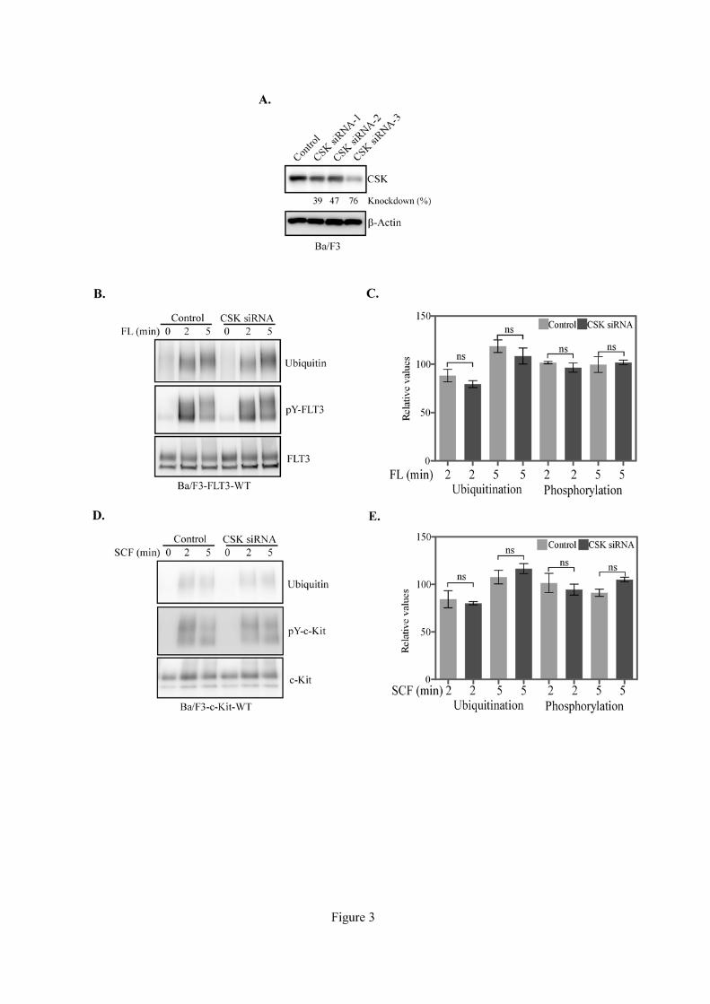
3.4. CSK depletion did not alter neither receptor ubiquitination nor phosphorylation
Several receptor interacting proteins alter receptor stability and/or phosphorylation. For
instance, SOCS6 and SLAP accelerate receptor degradation by increasing ubiquitination without
interfering with phosphorylation [8, 10], while LNK associates with FLT3 and decreases FLT3

9
phosphorylation [7]. To test whether CSK disrupts receptor stability or phosphorylation, we generated
stably transfected Ba/F3 cells by transfecting either FLT3 or c-Kit. Ba/F3 cells express a decent level
of endogenous CSK. To study the effects of CSK on FLT3 and c-Kit receptors, we attempted to
silence CSK by means of CSK specific siRNA achieving up to 76% of knockdown of endogenous
CSK (Fig. 3A). CSK depletion did not significantly affect neither FLT3 ubiquitination nor
phosphorylation (Fig. 3B and 3C). Similarly to FLT3, ubiquitination or phosphorylation of c-Kit was
also not altered by CSK siRNA (Fig. 3D and 3E).
3.5. CSK negatively regulates ligand-induced Akt phosphorylation
CSK regulates integrin-mediated signaling through Akt activation [20] and overexpression of
CHK potentiates Akt phosphorylation in breast cancer MCF-7 cell line [21]. Furthermore, CSK
overexpression in PC12 cells leads to increased Akt phosphorylation in response to nerve growth
factor [22]. These observations suggest that CSK and its homologous kinase differentially regulate
Akt activation in different biological contexts, although both proteins exhibit similar function in SFKs
regulation. Thus we tested whether CSK plays a role in FL- or SCF-mediated Akt activation. Ba/F3-
FLT3-WT or Ba/F3-c-Kit-WT cells were transfected with control siRNA or CSK siRNA. Two days
after transfections cells were serum-starved for four hours before ligand stimulation. We observed that
CSK depletion led to an increase in Akt activation in both Ba/F3-FLT3-WT (Fig. 4A) and Ba/F3-c-
Kit-WT (Fig. 4B) cells suggesting that CSK acts as a negative regulator of Akt signaling downstream
to type III receptor tyrosine kinases. In addition, a selective CSK inhibitor (ASN2324598) [23, 24]
also augmented Akt phosphorylation in response to both FL (Fig. 4C) or SCF (Fig. 4D) stimulation
indicating that functional CSK is required to maintain basal Akt phosphorylation.
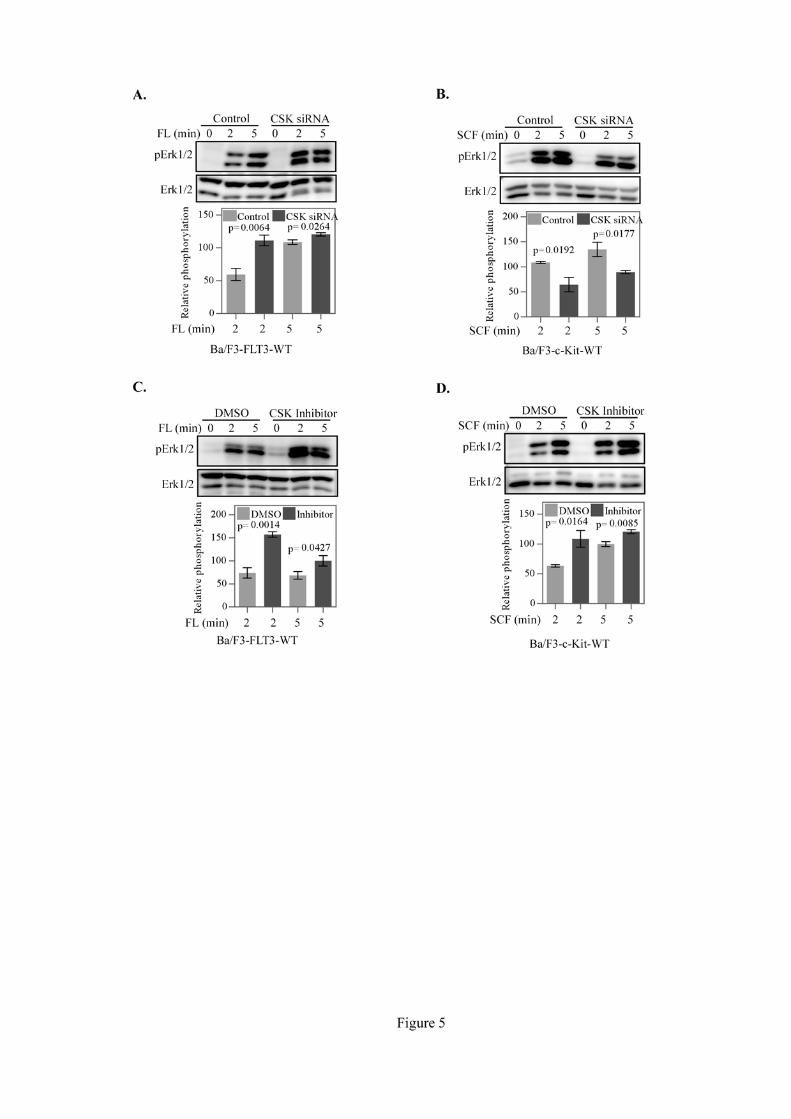
3.6. CSK differentially regulates FL- and SCF-induced Erk1/2 phosphorylation
Type III receptor tyrosine kinases transduce mitogenic signaling through activation of the Erk
mitogen-activated protein kinase [3-5]. Overexpression of CSK decreases Erk1/2 phosphorylation in
response to different stimuli [20, 22]. We observed that while CSK knockdown enhanced Erk1/2
phosphorylation in response to FL stimulation (Fig. 5A), it decreased Erk1/2 phosphorylation in

10
response to SCF (Fig. 5B). The ASN2324598-mediated CSK inhibition led to a prolonged Erk1/2
activation in response to both FL (Fig. 5C) and SCF (Fig. 5D). On the whole, these results indicate
that CSK differentially regulates MAPK signaling downstream of FLT3 and c-Kit receptors and that
intact CSK is required for c-Kit-mediated Erk1/2 signaling.
3.7. CSK depletion has no significant effect on p38 activation
Since both FL and SCF can activate p38, we then tested whether CSK also controls ligand-
stimulated p38 phosphorylation. CSK depletion slightly decreased FL-stimulated p38 phosphorylation
at the 2 minute time point but slightly increased at the 5 minute time point (Fig. 6A). Similar to FL,
SCF induced p38 activation was slightly attenuated in CSK knockdown cells at the 2 minute time
point (Fig. 6B). Although knockdown of CSK led to a slight deregulation of p38 phosphorylation
induced by either receptor, differences were not statistically significant.
3.8. CSK depletion increases SHC, Gab2 and SHP2 tyrosine phosphorylation
Activation of Akt and Erk1/2 by receptor tyrosine kinases FLT3 and c-Kit is mediated
through activation of various signaling proteins such as SHP2, SHC and Gab2 [3, 4]. Therefore we
tested which intermediate regulators are involved in transducing signals from FLT3 and c-Kit
receptors through CSK. Ba/F3-FLT3-WT and Ba/F3-c-Kit-WT cells were transfected with control
and CSK siRNA. Two days after transfection cells were serum-starved for four hours followed by two
minutes and five minutes ligand stimulation. We observed that phosphorylation on tyrosine residues
of SHC was increased in CSK siRNA transfected cells in response to both FL (Fig. 7A) and SCF (Fig.
7B). Using phospho-specific antibodies against phosphotyrosine residues in Gab2 we showed that
both FL (Fig. 7C) and SCF (Fig. 7D) stimulation enhanced Gab2 phosphorylation in cells where CSK
expression had been knocked down. Similarly to SHC and Gab2, we also observed an increase in
SHP2 phosphorylation following knockdown of CSK in Ba/F3-FLT3-WT (Fig. 7E) and Ba/F3-c-Kit-
WT (Fig. 7F) cells upon respective ligand stimulation.

11
3.9. CSK depletion increases oncogenic FLT3- and c-Kit-mediated cell proliferation but not cell
survival
Since we observed that CSK differentially regulates Akt and Erk1/2 signaling in response to
the FL and SCF stimulation, we investigated whether CSK depletion resulted in differential biological
outcomes. We tested cell proliferation and survival using constitutively active mutants of FLT3 and c-
Kit which are known to induce IL-3-independent Ba/F3 cell survival [16]. We observed that CSK
depletion resulted in an increased proliferation of Ba/F3-FLT3-ITD (Fig. 8A) and Ba/F3-c-Kit-
D816V cells (Fig. 8B), while cell survival remained unchanged (Fig. 8C and 8D).
4. Discussion
The C-terminal Src kinase (CSK) contributes to regulation of SFKs activity. Thus, loss of
CSK function mediates aberrant SFK signaling leading to abnormal cell growth. Apart from its
catalytic activity CSK is known to interact with variety of signaling proteins. CSK associates with
TCR complex through CSK-SH2 domain and this interaction brings CSK closer to SFKs resulting in
negative regulation of TCR signaling [18]. In this study, we demonstrate that CSK associates with
multiple type III receptor tyrosine kinases. Moreover, we showed that a functional CSK SH2 domain
is necessary for interactions to the activated receptors. Furthermore, our results indicate that ligand
activation triggers CSK membrane localization and that CSK regulates receptor downstream signaling
eventually leading to receptors-mediated cell proliferation.
Both CSK and CHK deficient mice have been generated. Although CHK deficient mice
survived and were apparently healthy [25], CSK deficient mice died due to defects in the neural
tissues [26, 27] suggesting that CSK plays additional roles in embryogenesis other than regulation of
SFKs. Differential subcellular localization in response to different physiological stimulant might
regulate downstream signaling differentially. Many kinases display differential localization patterns
and thereby differentially regulate downstream signaling [28-31]. CSK probably also elicit different
signaling whether it is cytosolic or membrane-localized. While cytosolic CSK interacts with adapters
or scaffold proteins [12], membrane localized CSK associates with receptors and transduces or blocks
receptor signaling.

12
CSK is predominantly present in cytosol due to lack of a transmembrane domain or fatty-acyl
modifications, but its substrate SFKs are mainly anchored inner surface of the cell membrane.
Association of CSK with ligand-stimulated receptors brings CSK in close proximity to the cell
membrane where it phosphorylates SFKs [12]. Since, both FLT3 and c-Kit transduce signal through
SFKs [4], CSK association with the receptors might have negative impact on receptor downstream
signaling. Elevated SHC phosphorylation in CSK depleted cells further supports this hypothesis.
SFKs induce tyrosine phosphorylation of several signaling proteins including Gab2 and SHP2
resulting in activation of downstream serine/threonine kinases Akt and Erk1/2 [4]. Our data also
suggests that loss of CSK function contributes to increased Gab2 and SHP2 tyrosine phosphorylation
as well as to increased Akt serine phosphorylation. Since activation of the Akt pathway facilitates cell
survival and proliferation through transcriptional activation of cell cycle activators and
phosphorylation-dependent inactivation of cell cycle repressors [4, 32], it is anticipated that depletion
of CSK will further accelerate oncogenic receptor-mediated cell proliferation.
The observation that CSK depletion or inhibition elevated FL-induced Erk1/2
phosphorylation could also be explained by the fact that loss of CSK function reduces control of SFKs
activation. However, while CSK inhibition potentiated SCF-induced Erk1/2 phosphorylation which is
in line with the FLT3 downstream signaling, siRNA-mediated CSK depletion decreased SCF-
mediated phosphorylation of same proteins. This observation cannot be explained by an influence on
SFKs regulation and it is of interest how CSK association to the c-Kit induces prolonged Erk1/2
phosphorylation. One possible explanation could be adaptor activity. The presence of both SH3 and
SH2 domains helps many proteins to link multiple signaling proteins [33]. Thus, CSK might link c-
Kit with other signaling proteins resulting in activation of Erk1/2 which is independent of the
regulations of SFKs.
5. Conclusions
In this study we demonstrate that CSK associates with multiple type III receptor tyrosine
kinases including FLT3 and c-Kit. While this association is mediated through CSK SH2 domain and
tyrosine phosphorylated receptors, SH3 domain is not required for the interaction. Association of CSK
13
with receptors increases the membrane localization of CSK resulting in extended SFKs regulation,
which further controls of downstream PI3K-Akt pathway as well as cell proliferation. Although
FLT3-CSK association negatively regulates Erk1/2 signaling, c-Kit-CSK interaction enables
prolonged Erk1/2 activation. Thus our results suggest a novel role of CSK in downstream signaling of
type III receptor tyrosine kinases.
Acknowledgements
This research was funded by the Swedish Cancer Society, the Swedish Children’s Cancer
Organization, the Swedish Research Council and ALF governmental clinical grant.
Competing interests
The authors declare no conflict of interest.
Authors' contributions
JUK conceived, designed and performed the experiments, analyzed the data and wrote the
manuscript. MV and SP contributed in the localization experiments. SA contributed in the cell
proliferation and survival assays. EB conceived the experiments and wrote the manuscript. LR
conceived and designed the experiments, discussed data and wrote the manuscript. All authors read
and approved the final manuscript.
References
[1] Kabir NN, Kazi JU, Genetics and molecular biology. 2011;34:587-591.
[2] Kazi JU, Kabir NN, Soh JW, Gene. 2008;410:147-153.
[3] Lennartsson J, Ronnstrand L, Physiological reviews. 2012;92:1619-1649.
[4] Masson K, Ronnstrand L, Cellular signalling. 2009;21:1717-1726.
[5] Heldin CH, Ostman A, Ronnstrand L, Biochimica et biophysica acta. 1998;1378:F79-113.
[6] Kabir NN, Ronnstrand L, Kazi JU, Medical oncology. 2013;30:462.

14
[7] Lin DC, Yin T, Koren-Michowitz M, Ding LW, Gueller S, Gery S, Tabayashi T, Bergholz U, Kazi
JU, Ronnstrand L, Stocking C, Koeffler HP, Blood. 2012;120:3310-3317.
[8] Kazi JU, Sun J, Phung B, Zadjali F, Flores-Morales A, Ronnstrand L, The Journal of biological
chemistry. 2012;287:36509-36517.
[9] Kazi JU, Ronnstrand L, Molecular oncology. 2012;DOI: 10.1016/j.molonc.2012.11.003.
[10] Kazi JU, Ronnstrand L, PloS one. 2012;7:e53509.
[11] Kazi JU, Ronnstrand L, Molecular oncology. 2013;DOI: 10.1016/j.molonc.2013.02.020.
[12] Okada M, International journal of biological sciences. 2012;8:1385-1397.
[13] Heiss E, Masson K, Sundberg C, Pedersen M, Sun J, Bengtsson S, Ronnstrand L, Blood.
2006;108:1542-1550.
[14] Sun J, Pedersen M, Ronnstrand L, The Journal of biological chemistry. 2009;284:11039-11047.
[15] Blume-Jensen P, Claesson-Welsh L, Siegbahn A, Zsebo KM, Westermark B, Heldin CH, The
EMBO journal. 1991;10:4121-4128.
[16] Kazi JU, Sun J, Ronnstrand L, Experimental Hematology. 2013; DOI:
10.1016/j.exphem.2013.03.005.
[17] Arbet-Engels C, Tartare-Deckert S, Eckhart W, The Journal of biological chemistry.
1999;274:5422-5428.
[18] Catipovic B, Lijecnicki vjesnik. 1996;118:264-271.
[19] Zrihan-Licht S, Deng B, Yarden Y, McShan G, Keydar I, Avraham H, The Journal of biological
chemistry. 1998;273:4065-4072.
[20] Gu J, Nada S, Okada M, Sekiguchi K, Biochemical and biophysical research communications.
2003;303:973-977.
[21] Zagozdzon R, Bougeret C, Fu Y, Avraham HK, International journal of oncology. 2002;21:1347-
1352.
[22] Dey N, Howell BW, De PK, Durden DL, Experimental cell research. 2005;307:1-14.
[23] Kunte DP, Wali RK, Koetsier JL, Hart J, Kostjukova MN, Kilimnik AY, Pyatkin IG, Strelnikova
SR, Roy HK, FEBS letters. 2005;579:3497-3502.

15
[24] Sirvent A, Benistant C, Pannequin J, Veracini L, Simon V, Bourgaux JF, Hollande F, Cruzalegui
F, Roche S, Oncogene. 2010;29:1303-1315.
[25] Hamaguchi I, Yamaguchi N, Suda J, Iwama A, Hirao A, Hashiyama M, Aizawa S, Suda T,
Biochemical and biophysical research communications. 1996;224:172-179.
[26] Nada S, Yagi T, Takeda H, Tokunaga T, Nakagawa H, Ikawa Y, Okada M, Aizawa S, Cell.
1993;73:1125-1135.
[27] Imamoto A, Soriano P, Cell. 1993;73:1117-1124.
[28] Medina M, Wandosell F, International journal of Alzheimer's disease. 2011;2011:479249.
[29] Kazi JU, Frontiers in Biology. 2011; 6:328-336
[30] Kazi JU, Soh JW, Molecules and cells. 2008;26:462-467.
[31] Kazi JU, Soh JW, Biochemical and biophysical research communications. 2007;364:231-237.
[32] Franke TF, Hornik CP, Segev L, Shostak GA, Sugimoto C, Oncogene. 2003;22:8983-8998.
[33] Koch CA, Anderson D, Moran MF, Ellis C, Pawson T, Science. 1991;252:668-674.
Figure legends
Figure 1. CSK associates with phosphorylated FLT3 and c-Kit receptors through the CSK-SH2
domain
Plasmids encoding wild-type CSK and either of FLT3-WT, FLT3-ITD (A), c-Kit-WT, c-Kit-
D816V (B) or PDGFR(C)were transfected into COS-1 cells. Six hours after transfections cells were
serum-starved for 18 hours and then stimulated with 100 ng/ml with either FL, SCF or PDGF-BB,
respectively, followed by lysis. Cell lysates were immunoprecipitated with anti-FLAG antibody and
then subjected to Western blotting analysis. (D) Schematic representation of CSK mutants generated
for this study. Plasmids expressing different mutants of CSK and FLT3-WT (E) or c-Kit-WT (F) were
transfected in COS-1 cells and analyzed as described above.
Figure 2. Ligand stimulation increases CSK membrane abundance

16
(A-H) Cells were transfected with plasmids encoding FLAG tagged-CSK mutants and FLT3
or c-Kit. Six hours after transfection cells were serum starved for 18 hours and then stimulated or not
with the respective ligand for 10 minutes before fixing. Fixed cells were permeabilized and stained
with fluorophore-conjugated anti-FLAG and anti-FLT3/anti-c-Kit antibodies. Nuclei were stained
with DAPI and subcellular localization was visualized with a Carl Zeiss LSM 710 Laser Scanning
Microscope. (I, J) Colocalization was measured using CoLocalizer Pro 2.7.1.
Figure 3. CSK depletion did not alter receptor ubiquitination or phosphorylation:
(A) Ba/F3 cells were transfected with control siRNA or three different CSK siRNAs. Two
days after transfection cells were lysed and cell lysates were subjected to Western blotting analysis.
(B) Ba/F3-FLT3-WT cells were transfected with control siRNA or CSK siRNA. Two days after
transfection cells were serum-starved for four hours before stimulation. Cells were then lysed and
subjected to immunoprecipitation and Western blotting analysis. (C) Signal intensities from multiple
blots of experiment "B" were quantified using Multi-Gauge software. (D) Ba/F3-c-Kit-WT cells were
transfected with control siRNA or CSK siRNA. Two days after transfection cells were serum starved
for four hours before stimulation. Cells were then lysed and subjected to immunoprecipitation and
Western blotting analysis. (E) Signal intensities from multiple blots of experiment "D" were
quantified using Multi-Gauge software. Data were analyzed with GraphPad Prism 5.0. ns, not
significant.
Figure 4. CSK depletion increases Akt phosphorylation
Ba/F3-FLT3-WT (A) or Ba/F3-c-Kit-WT (B) cells were transfected with control siRNA or
CSK siRNA. Two days after transfection cells were serum-starved for four hours before stimulation
with the respective ligand. Ba/F3-FLT3-WT (C) or Ba/F3-c-kit-WT (D) cells were serum-starved for
four hours and then treated with 10 µM CSK inhibitor for 1 hour before ligand stimulation. Cells were
then lysed and lysates were subjected to Western blotting analysis. Signal intensities from multiple
blots of experiment were quantified using Multi-Gauge software and analyzed with GraphPad Prism
5.0.

17
Figure 5. CSK depletion differentially regulates Erk1/2 phosphorylation:
Ba/F3-FLT3-WT (A) or Ba/F3-c-Kit-WT (B) cells were transfected with scramble control
siRNA or CSK siRNA. Two days after transfection cells were serum-starved for four hours before
stimulation. Ba/F3-FLT3-WT (C) or Ba/F3-c-Kit-WT (D) cells were serum-starved for four hours and
then treated with 10 µM CSK inhibitor for 1 hour before stimulation. Cells were then lysed and
lysates were subjected to Western blotting analysis with phospho-Erk and Erk antibodies. Signal
intensities from multiple blots of experiment were quantified using Multi-Gauge software and
analyzed with GraphPad Prism 5.0.
Figure 6. CSK depletion did not affect p38 phosphorylation
Ba/F3-FLT3-WT (A) or Ba/F3-c-Kit-WT (B) cells were transfected with control siRNA or
CSK siRNA. Two days after transfection cells were serum-starved for four hours before stimulation
with FL and SCF, respectively. Cells were then lysed and whole cell lysates were subjected to
Western blotting analysis with phosphor-p38 and p38 antibodies. Signal intensities from multiple
blots of experiment were quantified using Multi-Gauge software and analyzed with GraphPad Prism
5.0
Figure 7. CSK depletion increases SHC, Gab2 and SHP2 phosphorylation:
Ba/F3-FLT3-WT or Ba/F3-c-Kit-WT cells were transfected with control siRNA or CSK
siRNA. Two days after transfection cells were serum-starved for four hours before stimulation. Cells
were then lysed and subjected to immunoprecipitation and Western blotting analysis. Signal
intensities from multiple blots of experiment were quantified using Multi-Gauge software and
analyzed with GraphPad Prism 5.0. (A and B) Cell lysates were immunoprecipitated using anti-SHC
antibody and membranes were probed with 4G10 and anti-SHC antibodies. Whole cell lysates were
used for analysis and membranes were probed with (C and D) phospho-Gab2 and Gab2, and (E and F)
phospho-SHP2 and SHP2.

18
Figure 8. CSK depletion increases cell proliferation but not cell survival:
Ba/F3-FLT3-ITD (A) or Ba/F3-c-Kit-D816V (B) cells were transfected with control siRNA
or CSK siRNA and then cells were seeded in a 96-well plate. Cells were cultured with or without FL
or SCF, respectively, for 46 hours followed by PrestoBlue cell proliferation assay. Ba/F3-FLT3-ITD
cells (C) or Ba/F3-c-Kit-D816V cells (D) were transfected with control siRNA or CSK siRNA and
then seeded in a 12-well plate. Cells were cultured with or without FL or SCF, respectively, for 48
hours followed by Annexin V/7AAD apoptosis assay.