the university of jordan faculty of nursing introduction
TRANSCRIPT

Dr. Ahmad Aqel
2021
The University of JordanFaculty of Nursing
Introduction to Adult Health Nursing

Definition: Complete or incomplete disruption in the
continuity of bone structure
▪ occur when the bone is subjected to stress greater than it
can absorb.
Causes:
Consequences of bone fracture
Dr. Ahmad Aqel 2
▪ Direct blows, ▪ Crushing forces
▪ Sudden twisting motions ▪ Extreme muscle contractions.
▪ Edema, Hemorrhage, Joint dislocations , Rupture
tendons , Nerves injury, Blood vessels damage.

Types of Fractures
Dr. Ahmad Aqel 3
▪ Complete fracture
▪ break across the entire cross-section
of the bone, frequently displaced.
▪ Incomplete fracture
▪ break through only part of the cross-
section of the bone (eg, green- stick)
▪ Comminuted fracture
▪ fracture that produces several fragments
▪ A closed fracture: does not cause a
break in the skin.

Types of Fractures
Dr. Ahmad Aqel 4
Open fracture: skin wound extends to the
fractured bone
Intra-articular fracture
• Extends into the joint surface of a bone.
• Difficult to seen on the x-ray because cartilage
is non-radiopaque. MRI or arthroscopy will
identify the fracture
• Lead to posttraumatic arthritis
Management: immobilize the joint with a splint or
cast, no weight bearing

Dr. Ahmad Aqel 5
A fragment of bone has been pulled away
bone has splintered into fragments
compressed bone (Seen in vertebral fracture)
Avulsion Comminuted Compression
Fractures
Depressed
fragments are driven inward (seen in fractures of skull and facial bones)

Impacted
Dr. Ahmad Aqel 6
bone fragment is driven into another bone fragment
Greenstick
• one side of a bone is broken and the other side is bent
Fractures
• Occurs due to diseased bone without trauma or fall
Pathologic Epiphyseal
• A fracture through the epiphysis

Dr. Ahmad Aqel 7
• A fracture and damage to skin or mucous membranes (called compound fracture)
Oblique
• A fracture at an angle across the bone
Transverse
• straight fracture across the bone shaft
Open fracture
Fractures

Dr. Ahmad Aqel 8
Simple fracture Stress Spiral
A fracture that remains contained, with no disruption of the skin integrity
Results from repeated loading of bone and muscle
Twists around the shaft of the bone
Fractures

Signs and Symptoms of Fracture
Dr. Ahmad Aqel 9
1. Acute pain:
2. Loss of function
3. Deformity
4. Shortening of the extremity
5. Crepitus
6. Localized edema and ecchymosis.
Testing for crepitus can produce further tissue damage

Emergency Management
Dr. Ahmad Aqel 10
▪ Immobilize the body part before moving pt.
▪ Immobilize the Joints proximal & distal to the fracture.
▪ Immobilize the lower extremities by
bandaging the legs together, with the
unaffected extremity.
▪ Bandage the arm to the chest,
place forearm in a sling.
1) Immobilization:

Emergency Management
Dr. Ahmad Aqel 11
2. Assess for peripheral pulse & nerve function distal to the
injury before and after splinting.
3. Cover the open wound with a sterile dressing.
4. Do not reduce the fracture
In the emergency department
– Complete evaluation.
– Remove the clothes gently first from the uninjured side
– The fractured extremity is moved gently

Medical Management
Dr. Ahmad Aqel 12
Fracture reduction
▪ Refers to restoration of the fracture fragments to anatomic alignment and positioning.
▪ Types of reduction
1. closed reduction (manipulation and manual traction)
2. open reduction ( surgical approach)
Reduces a fracture ASAP to prevent loss of elasticity from
the tissues through infiltration by edema or hemorrhage

Medical Management
Dr. Ahmad Aqel 13
Closed Reduction
– Bringing the bone into anatomic alignment
through manual traction.
– Hold the extremity in the aligned position while
the physician applies a cast, splint.
– Minor analgesia may be used
– Obtain X-rays to verify that the bone fragments
are correctly aligned

Medical Management
Dr. Ahmad Aqel 14
Open Reduction
by surgery, the
fracture fragments
aligned.
• Internal fixation
by pins, wires,
screws, plates,
nails

Dr. Ahmad Aqel 15
• Immobilization by internal or external fixation.
• Methods of external fixation include bandages, casts,
splints, continuous traction, and external fixators.
Medical Management
Immobilization

Dr. Ahmad Aqel 16
▪ Elevate extremity and apply ice to reduce edema
▪ Monitor neurovascular status
▪ Change position, and pain relief
▪ Teach pt Isometric and muscle-setting exercises : to
minimize atrophy and to promote circulation.
▪ How to use assistive devices (crutches, walkers).
▪ Patient teaching: self-care, medication, complications.
▪ Modify the home environment as needed
▪ Reassurance: alleviate restlessness and anxiety
Nursing Management With Closed Fractures

Nursing Management / Open Fractures
Dr. Ahmad Aqel 17
Risk of open fracture: osteomyelitis, tetanus, gas gangrene
1. Administer IV antibiotics, T. toxoid, Wound irrigation and
debridement, wound swab for C&S as ordered
2. Surgical external fixation of fractures carries a risk of
infection. (caring of wires, scrows)
3. Elevate extremity to minimize edema.
4. Monitor Temperature and signs of infection (tenderness,
pain, redness, swelling, local warmth, elevated
temperature, and purulent drainage).
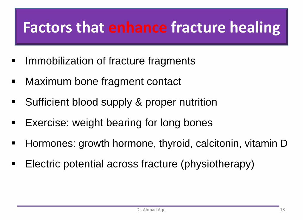
Factors that enhance fracture healing
Dr. Ahmad Aqel 18
▪ Immobilization of fracture fragments
▪ Maximum bone fragment contact
▪ Sufficient blood supply & proper nutrition
▪ Exercise: weight bearing for long bones
▪ Hormones: growth hormone, thyroid, calcitonin, vitamin D
▪ Electric potential across fracture (physiotherapy)

Factors that inhibit fracture healing
Dr. Ahmad Aqel 19
▪ Extensive local trauma & Bone loss
▪ Weight bearing prior to approval
▪ Mal-alignment of the fracture fragments
▪ Inadequate immobilization
▪ Space or tissue between bone fragments
▪ Infection
▪ Localized malignancy
▪ Age (elderly persons heal more slowly)
▪ Corticosteroids (inhibit the repair rate)

Early complications Delayed complications
Dr. Ahmad Aqel
• Shock
• Fat embolism
• Compartment syndrome
• Deep vein thrombosis
• Pulmonary embolism
• Delayed union, Malunion,
Nonunion
• Reaction to internal
fixation devices
• Complex regional pain
syndrome
• Heterotopic ossification
20
Complications of fracture

Dr. Ahmad Aqel 21
• Hypovolemic shock
– resulting from severe hemorrhage
– More common with pelvic fractures & displaced
femoral fracture.
Treatment for shock
– stabilizing the fracture, proper immobilization,
– restoring blood volume and circulation,
Complications of fracture

Dr. Ahmad Aqel 22
Fat Embolism
▪ More common in: fracture long bones or pelvic,
▪ Clinical manifestations: The first manifestations are
(hypoxia and tachypnea). petechial rash, CP, crackles,
wheezes, cough, thick white sputum, tachycardia.
▪ Neurological symptoms: (headache, mild agitation to
delirium & coma).
▪ sudden restlessness, irritability, or confusion occurs post
fracture are indications for immediate ABG studies.
Complications of fracture

Dr. Ahmad Aqel 23
▪ Immediate immobilization of fractures
– early fixation, minimal manipulation,
– maintenance of fluid and electrolyte balance.
▪ Prompt initiation of respiratory support
– High-flow oxygen, mechanical ventilation with PEEP
– Corticosteroids & Vasopressor medications
– Accurate I&O
▪ Acute pulmonary edema and ARDS are the most
common causes of death from Fat embolism.
Prevention and Management of fat emboli

Dr. Ahmad Aqel 24
Compartment Syndrome
▪ A sudden decrease in blood flow to the tissues distal to injury site that results in ischemic necrosis
▪ C/O: deep, throbbing pain, increase despite opioids
▪ Hallmark sign: pain intensifies with passive ROM
▪ pain caused by tight muscle fascia , constrictive cast, edema or hemorrhage from the fracture site
▪ Most common on the lower leg, forearm is also at risk.
▪ Permanent lose occurs if the anoxic situation continues for longer than 4 hours.
Complications of fracture

Dr. Ahmad Aqel 25
Assess:
▪ Pain, pallor, paresthesia, paralysis,pulselessness.
▪ Cyanotic nail beds suggest venous congestion.
▪ Pallor and cool & prolonged capillary refill suggest
diminished arterial perfusion.
▪ Use Doppler ultrasonography to verify a pulse .
Nursing management of compartment syndrome

Safety Nursing Alert
Dr. Ahmad Aqel 26
Management
▪ Maintaining the extremity at the
heart level (not above heart
▪ Opening and bivalving the cast or
opening the splint
▪ A fasciotomy (excision of the fascia)
to relieve the constrictive muscle
▪ Moist, sterile saline dressings, A
vacuum dressing may be used to
remove fluids
Compartment syndrome

Dr. Ahmad Aqel 27
Delayed union: healing not occur within the expected time
Nonunion: failure of the ends of a fractured bone to unite
▪ treated with internal fixation, bone grafting, electrical
bone stimulation.
Malunion: the healing in a mal aligned position
Factors contributing to nonunion and mal-union
▪ Infection, Interposition of tissue between the bone ends,
Inadequate immobilization & manipulation, excessive
space between bone fragments, impaired blood supply.
Delayed Complications of fracture

Delayed complication of fracture
Dr. Ahmad Aqel 28
.
The bone graft may be an
Autograft (from the patient, frequently from the iliac crest
Allograft (from a donor)
Bone grafting complications include:
graft infection, fracture of the graft, nonunion
Delayed Complications of fracture
❖ The electrical stimulation enhances
mineral deposition and bone formation
that promotes bone growth. Bone healing stimulator applied to the arm

Nursing Management.
Dr. Ahmad Aqel 29
• Emotional support
• Encourages adherence to the treatment regimen.
• Patient with a bone graft:
– pain management and monitor for complications.
– education concerning bone graft, immobilization, non–
weight-bearing exercises, wound care, signs of infection,
and follow-up
• Patient using bone stimulation devices
– education regarding immobilization, weight-bearing
restrictions, and correct daily use of the stimulator
Nursing management

Dr. Ahmad Aqel 30
Complex Regional Pain Syndrome
▪ severe burning pain, local edema, hyperesthesia, stiffness,
skin discoloration,
▪ Prevention: elevation, immobilization, early effective pain relief
avoids using the affected extremity for BP & venipuncture
Avascular Necrosis of Bone
▪ bone loses blood supply and dies.
Heterotopic ossification:
▪ Abnormal formation of bone, near bones or in muscle.
Delayed Complications of fracture

Which term refers to the failure of fragments of a fractured bone to heal together?
a) Malunion
b) Subluxation
c) Dislocation
d) Nonunion.
Dr. Ahmad Aqel 31