the us2 gene product of herpes simplex virus 2 is a membrane
TRANSCRIPT

The Us2 Gene Product of Herpes Simplex Virus 2 Is a Membrane-Associated Ubiquitin-Interacting Protein
Ming-Hsi Kang, Bibhuti B. Roy, Renée L. Finnen, Valerie Le Sage, Susan M. Johnston, Hui Zhang, Bruce W. Banfield
Department of Biomedical and Molecular Sciences, Queen’s University, Kingston, Canada
The Us2 gene encodes a tegument protein that is conserved in most members of the Alphaherpesvirinae. Previous studies on thepseudorabies virus (PRV) Us2 ortholog indicated that it is prenylated, associates with membranes, and spatially regulates theenzymatic activity of the MAP (mitogen-activated protein) kinase ERK (extracellular signal-related kinase) through direct bind-ing and sequestration of ERK at the cytoplasmic face of the plasma membrane. Here we present an analysis of the herpes simplexvirus 2 (HSV-2) Us2 ortholog and demonstrate that, like PRV Us2, HSV-2 Us2 is a virion component and that, unlike PRV Us2, itdoes not interact with ERK in yeast two-hybrid assays. HSV-2 Us2 lacks prenylation signals and other canonical membrane-tar-geting motifs yet is tightly associated with detergent-insoluble membranes and localizes predominantly to recycling endosomes.Experiments to identify cellular proteins that facilitate HSV-2 Us2 membrane association were inconclusive; however, thesestudies led to the identification of HSV-2 Us2 as a ubiquitin-interacting protein, providing new insight into the functions ofHSV-2 Us2.
The Alphaherpesvirinae are a large group of viruses that includethe common human pathogens herpes simplex virus 1 (HSV-
1), HSV-2, and varicella-zoster virus, as well as numerous impor-tant veterinary viruses, including the swine pathogen pseudora-bies virus (PRV) (1). All herpesvirus virions have a commonstructure (2): an icosahedral nucleocapsid containing a lineardouble-stranded DNA genome, surrounded by a lipid envelopeembedded with numerous glycoprotein spikes. Between the nu-cleocapsid and the envelope lies a proteinaceous compartmentcalled the tegument.
The initial stages of herpesvirus assembly take place in the nu-cleus, where newly replicated virus genomes are packaged intopreformed capsids. Although some controversy exists, strong bio-chemical and genetic evidence indicates that DNA-containingcapsids gain access to the cytoplasm by first acquiring a primaryenvelope at the inner nuclear membrane by budding into the pe-rinuclear space (3). Perinuclear virions are subsequently de-envel-oped through fusion of the primary virion envelope with the outernuclear membrane, thereby releasing the capsid into the cyto-plasm (4). The tegument is formed through the recruitment oftegument proteins to capsid components, through interactionsbetween tegument proteins, and through interactions betweentegument proteins and the cytoplasmic tails of membrane pro-teins destined for the envelopes of mature virions. The mostwidely accepted model of herpesvirus egress posits that the virionacquires its final envelope through the budding of capsid-tegu-ment complexes into membranes derived from the trans-Golginetwork (TGN), or possibly late endosomes (LE), in a processreferred to as secondary envelopment (5–8). The TGN- or LE-derived vesicles containing infectious enveloped virus then trafficto and fuse with the plasma membrane (PM) of the cell, releasingvirus into the extracellular environment. Little is known about themolecular and cellular biology of the trafficking of these virus-containing vesicles to the cell surface. A general assumption hasbeen that these vesicles follow a default, constitutive, secretorypathway to reach the cell surface. However, evidence supportingthe idea that this process is regulated by both viral and cellularcomponents is accumulating.
Rémillard-Labrosse and colleagues have reported that the cel-lular serine-threonine kinase protein kinase D (PKD) facilitatesthe transport of TGN-derived vesicles containing HSV-1 virionsto the PM (9). This finding strongly supports the notion that thetransport of virions from the site of secondary envelopment tothe cell surface is regulated. Previous studies have indicated that thealphaherpesvirus tegument protein Us2 is involved in the releaseof enveloped viruses from infected cells. The Us2 gene is found inthe genomes of many alphaherpesviruses, including those of hu-man, equine, canine, bovine, avian, and porcine origin (10–16).Although it is highly conserved, Us2 has been found to be nones-sential for virus replication in many cultured cells, and aside fromthe ability of PRV Us2 to regulate ERK (extracellular signal-re-lated kinase) activity spatially, no specific function has been as-signed to the protein (17, 18, 19, 20). Wagenaar and colleagueshave reported that mature enveloped virions accumulate insidecytoplasmic vesicles in primary cultures of porcine nasal mucosaepithelial cells infected with PRV mutants with deletions of Us2(21), and previous work from our laboratory has demonstratedthat PRV Us2-null mutants accumulate infectious virus insidecells (22). Possible interpretations of these findings include thehypothesis that Us2 activity is required for the efficient fusion ofvirion-containing vesicles with the PM or, alternatively, that Us2functions in the transport of the vesicles to the cell periphery. Insupport of the latter hypothesis, Kramer and colleagues recentlyidentified PRV Us2 as part of a complex containing the tail-an-chored type II viral membrane protein Us9 and the microtubule
Received 11 April 2013 Accepted 15 June 2013
Published ahead of print 19 June 2013
Address correspondence to Bruce W. Banfield, [email protected].
M.-H.K. and B.B.R. contributed equally to this article.
Supplemental material for this article may be found at http://dx.doi.org/10.1128/JVI.00994-13.
Copyright © 2013, American Society for Microbiology. All Rights Reserved.
doi:10.1128/JVI.00994-13
9590 jvi.asm.org Journal of Virology p. 9590–9603 September 2013 Volume 87 Number 17
Dow
nloa
ded
from
http
s://j
ourn
als.
asm
.org
/jour
nal/j
vi o
n 07
Jan
uary
202
2 by
46.
70.2
29.6
1.

motor KIF1A, which are required for axonal sorting and efficientanterograde transport of virion-containing vesicles within axons(23).
In this study, we sought to characterize the Us2 ortholog en-coded by HSV-2. The results indicate that although HSV-2 Us2lacks canonical membrane-targeting motifs, a large fraction ofHSV-2 Us2 is found in detergent-resistant membranes and local-izes to recycling endosomes and the PM. We also discovered thatHSV-2 Us2 interacts with ubiquitin-conjugated proteins, suggest-ing a role for Us2 in the trafficking of endosomal membranes inHSV-2-infected cells.
MATERIALS AND METHODSCells and viruses. Vero and 293T cells were maintained in Dulbecco’smodified Eagle’s medium (DMEM) supplemented with 10% fetal calfserum (FCS) in a 5% CO2 environment. HSV-1 (17�) and HSV-2(HG52) were generously provided by A. Dolan and D. McGeoch, formerlyof the MRC Virology Unit, Institute of Virology, Glasgow, Scotland.HSV-1 and HSV-2 strains were propagated and titers determined on Verocells.
The recombinant HSV-2 strain encoding Flag-tagged Us2 was con-structed by a two-step Red-mediated mutagenesis procedure (24) utiliz-ing the HSV-2 (186) bacterial artificial chromosome (BAC) pYEbac373 inEscherichia coli strain GS1783 (25). GS1783 was a kind gift of G. Smith,Northwestern University. Primers 5=-GTGAGTGAAGATCGGACCACGGGCCTAATATACCGACATGGATTACAAGGATGACGACGATAAG-3= and 5=-CGTTGGTCTAGGAGGGTAACCACACTTACAACAACAACGCCCTTATCGTCGTCATCCTTGTAATCCATGTCCAACCAATTAACCAATTCTGATTAG-3= were used to amplify a PCR productfrom pEP-Kan-S2, a kind gift of N. Osterrieder, Freie Universität Berlin,which was then used to introduce a Flag epitope onto the N terminus ofHSV-2 Us2. An HSV-2 Us2-null virus was constructed using primers 5=-ACAGGCGAGTGAGTGAAGATCGGACCACGGGCCTAATATACCGACATGGGCTGATGAGTTAGGATGACGACGATAAGTAGGG-3= and5=-TCGTTGGTCTAGGAGGGTAACCACACTTACAACTCATCAGCCCATGTCGGTATATTAGGCCAACCAATTAACCAATTCTGATTAG-3=. The repaired HSV-2 Us2-null strain was constructed usingprimers 5=-ACAGGCGAGTGAGTGAAGATCGGACCACGGGCCTAATATACCGACATGGGCGTTGTTGTTAGGATGACGACGATAAGTAGGG-3= and 5=-TCGTTGGTCTAGGAGGGTAACCACACTTACAACAACAACGCCCATGTCGGTATATTAGGCCAACCAATTAACCAATTCTGATTAG-3=. Using an HSV-1 (F) BAC, pYEbac102 (26) (kindlyprovided by Y. Kawaguchi, The University of Tokyo), an HSV-1 recom-binant carrying a Flag-tagged Us2 gene was constructed using primers5=-GTGCCCCAAATCGGACACGGGCCTGTAATATACCAACATGGATTACAAGGATGACGACGATAAG-3= and 5=-TCTGGTCAAGGAGGGTCATTACGTTGACGACAACAACGCCCTTATCGTCGTCATCCTTGTAATCCATGTTCAACCAATTAACCAATTCTGATTAG-3=.
Virus reconstitution. To produce HSV-2 (186) and HSV-1 (F) strainsthat lacked the BAC DNA sequences, we cotransfected wild-type (WT) orrecombinant pYEbac373 or pYEbac102 and the NLS (nuclear localizationsignal)-Cre-expressing plasmid pOG231 (27) into Vero cells. Briefly, Verocells were trypsinized and were resuspended in DMEM–10% FBS (fetalbovine serum) containing 10 mM BES [N,N-bis(2-hydroxyethyl)-2-ami-noethanesulfonic acid] (pH 7.2) to a concentration of 4 � 107 cells/ml.pYEbac373, or pYEbac102, and pOG231 (1 �g each) were added to a250-�l cell suspension, which was then transferred to an electroporationcuvette (gap, 0.4 cm; Fisher Scientific, Ottawa, Ontario, Canada). Electro-poration was carried out at settings of 210 V, 950 �F, and 200 � by usinga BTX ECM 630 electroporator. Cells and DNA were immediately platedonto 100-mm dishes, and the infection was allowed to proceed for as longas 3 days. Supernatants were collected, and the WT or recombinant HSVstrains were plaque purified twice on Vero cells.
Immunological reagents. Monoclonal antibodies against HSV-2 gG,gD, ICP5, ICP8, and ICP27 were purchased from Virusys (Taneytown,MD) and were used for Western blotting at the following dilutions: anti-gG, 1:5,000; anti-gD, 1:10,000; anti-ICP5, 1:3,000; anti-ICP8, 1:4,000; an-ti-ICP27, 1:1,000. An anti-GFP monoclonal antibody (Clontech, Moun-tain View, CA) was used for Western blotting at a dilution of 1:1,000;horseradish peroxidase (HRP)-conjugated goat anti-mouse IgG and rab-bit anti-rat IgG (Sigma, St. Louis, MO) were used for Western blotting atdilutions of 1:10,000 and 1:80,000, respectively. Alexa Fluor 488-conju-gated donkey anti-rat IgG, Alexa Fluor 568-conjugated donkey anti-ratIgG, and Alexa Fluor 568-conjugated donkey anti-mouse IgG (Invitro-gen, Burlington, Ontario, Canada) were used for indirect immunofluo-rescence microscopy at a dilution of 1:500. The GM1 ganglioside wasdetected by Western blotting by use of biotinylated cholera toxin B sub-unit (Sigma, St. Louis, MO) diluted to 1 �g/ml, followed by incubationwith an HRP-streptavidin conjugate (Invitrogen, Burlington, Ontario,Canada) diluted at 1:1,000. An antibody against mono- and polyubiquitin(FK2) was purchased from Enzo Life Sciences (Burlington, Ontario,Canada).
Insoluble GST (glutathione S-transferase)-Us2 fusion proteins de-rived from HSV-1 and HSV-2 were isolated from IPTG (isopropyl-�-D-thiogalactopyranoside)-induced E. coli Rosetta (DE3) using the B-Perbacterial protein extraction reagent (Pierce, Rockford, IL). The partiallypurified proteins were electrophoresed on preparative 10% SDS-PAGEgels. Slabs of polyacrylamide containing the GST-Us2 fusion proteinswere excised from the preparative gels and were sent to Cedarlane Labo-ratories (Burlington, Ontario, Canada) for the production of polyclonalantisera in Wistar rats. The resulting antisera were used for Western blot-ting at a dilution of 1:500 and for indirect immunofluorescence micros-copy at a dilution of 1:200.
Construction of Us2 expression plasmids. To construct the HSV-2Us2 expression plasmid pHSV2Us2, the full-length HSV-2 Us2 DNA se-quence flanked by EcoRI sites was obtained by PCR amplification usingforward primer 5=-CCGAATTCATGGCCTGTCGTAAGTTCTGTGG-3=and reverse primer 5=-CCGAATTCTCACTTAGGGTGAAATAGCG-3=from the pATHSV-2HindIIIL template (a kind gift from A. Dolan and D.McGeoch), which contains a HindIII fragment encompassing the major-ity of the unique short region of the HSV-2 genome (10). The amplifiedfragment was subcloned into the pCR-Blunt II-TOPO vector (Invitrogen,Burlington, Ontario, Canada) by using the manufacturer’s protocols. Allplasmids utilizing PCR in their construction were sequenced to ensurethat no spurious mutations had been introduced in the process. An EcoRIfragment encompassing the amplified sequences was excised from thisintermediate vector, purified, and then ligated to a similarly digestedpCINeo vector (Promega, Madison, WI).
To fuse the red fluorescent protein monomeric Cherry (mCherry)(28) to the N terminus of HSV-2 Us2, the mCherry gene was amplified byPCR with forward primer 5=-CGCGCTAGCATGGTGAGCAAGGGCGAGG-3= and reverse primer 5=-CGCCTCGAGTCTTGTACAGCTCGTCCATGC-3= from the pRSETb mCherry template, a kind gift from R. Tsien,University of California at San Diego, and the amplified fragment wascloned directly into the pCR-Blunt II TOPO vector. An NheI/XhoI frag-ment containing the amplified sequences was excised from this interme-diate vector, purified, and ligated to similarly digested pHSV2Us2.
A plasmid encoding Flag-tagged HSV-2 Us2 was constructed by am-plifying the HSV-2 Us2 gene using forward primer 5=-ATCGATATCGATGGGCGTTGTTGTTGTAAGTGTGG-3= and reverse primer 5=-GTAGTCGACTTAGAGGTTGGTGATTGGATAGC-3=, and the amplifiedfragment was cloned directly into the pCR-Blunt II TOPO vector. Next,an EcoRV/SalI fragment containing the amplified sequences was excisedfrom this intermediate vector, purified, and ligated to similarly digestedpFlag-CMV-2 (Sigma, St. Louis, MO). A yeast expression plasmid encod-ing HSV-2 Us2 fused to GAL4-DBD was constructed by amplifying theHSV-2 Us2 gene using forward primer 5=-GATCGAATTCATGGGCGTTGTTGTTGTAAGTGTGG-3= and reverse primer 5=-GTAGTCGACTTA
HSV-2 Us2 Is a Ubiquitin-Interacting Membrane Protein
September 2013 Volume 87 Number 17 jvi.asm.org 9591
Dow
nloa
ded
from
http
s://j
ourn
als.
asm
.org
/jour
nal/j
vi o
n 07
Jan
uary
202
2 by
46.
70.2
29.6
1.

GAGGTTGGTGATTGGATAGC-3=, and the amplified fragment wascloned directly into the pCR-Blunt II TOPO vector. Next, an EcoRI/SalIfragment containing the amplified sequences was excised from this inter-mediate vector, purified, and ligated to similarly digested pGBKT7 (Clon-tech, Mountain View, CA).
Analysis of Us2 expression kinetics. Confluent monolayers of Verocells growing in 6-well dishes were infected with HSV-2 at a multiplicity ofinfection (MOI) of 10. At 0, 2, 4, and 6 h postinfection, the medium wasremoved, and the cells were washed three times with phosphate-bufferedsaline (PBS). Cells were scraped into 200 �l of lysis buffer (10 mM Tris[pH 7.4], 150 mM NaCl, 1% NP-40, 1% Na-deoxycholate) containingprotease inhibitors (Roche, Laval, Quebec, Canada) and were transferredto a 1.5-ml microcentrifuge tube. Lysates were kept on ice for 30 min withintermittent mixing and were then centrifuged at 10,000 � g for 5 min.For Western blot analysis, 10 to 20 �l of the cellular extract was mixedwith SDS-PAGE sample buffer, heated to 100°C for 5 min, electropho-resed through 12.5% SDS-PAGE gels, and analyzed by Western blottingwith a polyclonal antiserum to HSV-2 Us2 and monoclonal antibodies togG, ICP5, or ICP8.
PAA treatment. Confluent monolayers of Vero cells growing in 6-wellplates were used for experiments with phosphonoacetic acid (PAA). At 1h prior to infection, the cell culture medium was replaced with DMEM–10% FCS either with or without 200 �g/ml PAA (Sigma, St. Louis, MO).Cells were infected with HSV-2 at an MOI of 10 and were incubated in thepresence or absence of 200 �g/ml PAA for 6 h. Cell extracts were preparedas described above and were electrophoresed through a 12.5% SDS-PAGEgel. Western blotting was performed with a polyclonal antiserum to Us2or a monoclonal antibody to gG, ICP5, or ICP8.
Virion purification. Virions were purified essentially as described pre-viously (29). Briefly, three confluent 150-mm dishes of Vero cells wereinfected with HSV-2 at an MOI of 10. At 16 h postinfection, the mediumwas collected and was centrifuged three times in succession at 600 � g topellet detached cells and cellular debris. The clarified supernatant waslayered onto a 30% sucrose (wt/vol, in PBS) cushion and was centrifugedin a Beckman SW28 rotor at 23,000 rpm for 3 h. The pelleted virions wereresuspended in 1 ml of PBS, layered onto a 4-ml 30% sucrose (wt/vol, inPBS) cushion, and centrifuged in a Beckman SW55 Ti rotor at 28,000 rpmfor 90 min. Pelleted virions were resuspended in 200 �l of PBS, snap-frozen in liquid nitrogen in 100-�l aliquots, and stored at �80°C.
Protease treatment of virions. Isolated virions were treated with 10�g of proteinase K (PK) (Fisher Scientific, Ottawa, Ontario, Canada) perml in either the presence or the absence of 1% NP-40 (Igepal; Sigma, St.Louis, MO). After incubation for 60 min at room temperature, phenyl-methylsulfonyl fluoride (Sigma, St. Louis, MO) was added to each sampleto a final concentration of 2 mM to inhibit further proteolysis. Sampleswere immediately separated on 10% SDS-PAGE gels, and proteins weretransferred to membranes for Western blot analysis.
Transfections. 293T cells were transfected for the purpose of prepar-ing cellular extracts by using the calcium phosphate coprecipitationmethod (30). Vero cells were transfected for the purpose of microscopicanalyses by using FuGene 6 (Roche, Laval, Quebec, Canada) according tothe manufacturer’s instructions.
Indirect immunofluorescence microscopy. Cells for microscopicanalyses were grown either on glass coverslips or on glass-bottom dishes(MatTek, Ashland, MA). Cells were then transfected or infected as de-scribed above. In some experiments, cells were incubated with 3 �g/mlbrefeldin A (BFA) (Epicentre Biotechnologies, Madison, WI) or with0.3% ethanol (vehicle). Cells were fixed in 4% paraformaldehyde–PBS for10 min at room temperature. Fixed cells were washed 3 times with PBScontaining 1% BSA (bovine serum albumin) (PBS-BSA) and were perme-abilized for 3 min at room temperature with PBS-BSA containing 0.1%Triton X-100 (TX-100). Cells were then washed 3 times with PBS andonce with PBS-BSA, and 100 �l of primary antiserum diluted appropri-ately in PBS-BSA was applied for 45 min at room temperature. Cells werewashed 3 times with PBS-BSA, and 100 �l of an Alexa Fluor-conjugated
secondary antibody diluted appropriately in PBS-BSA was applied for 30min at room temperature. Cells were then washed 3 times with PBS-BSA.To visualize nuclei, cells were incubated with Hoechst 33342 (Sigma, St.Louis, MO) diluted to 0.5 �g/ml in PBS for 7 min at room temperature.Stained cells on coverslips were washed 3 times in PBS and were mountedin PBS containing 50% (vol/vol) glycerol on glass slides; stained cells inglass-bottom dishes were washed 3 times in PBS and were stored underPBS-BSA. Images were captured by using an Olympus FV1000 laser scan-ning confocal microscope with a 60� objective (numerical aperture[NA], 1.42) and a digital zoom factor of 2 to 4 and were analyzed byFluoView CS3 software. Composites of representative images were pre-pared using Adobe Photoshop CS3 software.
Live-cell imaging. Vero cells growing on glass-bottom dishes weretransfected with an mCherry–HSV-2 Us2 expression plasmid. At 24 hafter transfection, the medium was replaced with DMEM lacking phenolred and containing 10% FCS, and the dish was mounted on a confocalmicroscope contained within a humidified 37°C, 5% CO2 environment.Images were captured every 2.2 s by using an Olympus FV1000 laserscanning confocal microscope with a 60� objective (numerical aperture[NA], 1.42) and FluoView CS3 software. Composite images of the timecourse were prepared using Adobe Photoshop CS3 software.
Transferrin uptake assays. Vero cells growing on glass coverslips weretransfected with an mCherry–HSV-2 Us2 expression plasmid or were in-fected with HSV-2 at an MOI of 1. At 24 h posttransfection or 8 h postin-fection, cells were incubated with Alexa Fluor 647-conjugated transferrin(Invitrogen, Burlington, Ontario, Canada) for 30 min at 4°C in DMEM.The cells were then washed; fresh medium was added; and the cells weretransferred to 37°C for the indicated times. Samples were taken at differ-ent time points, fixed, and, in the case of HSV-2-infected cells, stained forUs2. Images were then collected by confocal microscopy. Measurement ofthe colocalization of Us2-labeled vesicles and fluorescent transferrin wasfacilitated by FluoView CS3 software. Briefly, regions of interest (ROIs)containing individual Us2-labeled vesicles were selected, and the averagefluorescence intensity of the Us2 and transferrin signals was measured. AnROI was scored positive for transferrin if the average fluorescence inten-sity of the ROI was twice that of the background fluorescence in the trans-ferrin channel. Background fluorescence was defined as the averaged flu-orescence intensity of 5 ROIs per field that contained no discernibletransferrin-labeled puncta.
Membrane flotation assays. Methods for the analysis of membrane-associated proteins by sucrose density gradient flotation were used essen-tially as described by Brignati and colleagues (31), with the followingmodifications. Three subconfluent 150-mm-diameter dishes of 293T cellswere cotransfected with HSV-2 Us2 and EGFP (enhanced green fluores-cent protein) expression plasmids. Cells were washed and were harvestedusing trypsin at 48 h posttransfection. Cells were pelleted at 220 � g for 7min at 4°C and were washed once with cold PBS and once with homoge-nization buffer (0.25 M sucrose, 10 mM KCl, 10 mM Tris [pH 7.4], 1.5mM MgCl2). Cells were then suspended by gentle vortexing in 0.5 ml ofhomogenization buffer supplemented with a protease inhibitor cocktail(Roche, Laval, Quebec, Canada). Following a 30-min incubation on ice,cells were passed repeatedly through a 26-gauge syringe needle in order tohomogenize the cells while leaving the nuclei intact. Cell homogenateswere centrifuged at 600 � g for 10 min at 4°C to remove unbroken cellsand nuclei. Then 0.3 ml of the postnuclear supernatant (PNS) was mixedwith 2.7 ml of 85% (wt/vol) sucrose in NTE buffer (100 mM NaCl, 10 mMTris [pH 7.4], 1 mM EDTA) (final sucrose concentration, 71.5%) and wasplaced in the bottom of an SW41 centrifuge tube. Next, 6 ml of 65%sucrose in NTE buffer, followed by 3 ml of 10% sucrose in NTE buffer, waslayered onto the PNS. This sucrose step gradient was centrifuged at100,000 � g for 18 h at 4°C in a Beckman SW41 rotor. Twelve 1-mlfractions were collected from the top of the tube. Fraction 13 representedthe material that pelleted at the bottom of the tube. Aliquots (500 �l) offractions 1 to 12 were diluted with 500 �l of NTE buffer plus proteaseinhibitor cocktail, placed in an MLA-130 centrifuge tube, and centrifuged
Kang et al.
9592 jvi.asm.org Journal of Virology
Dow
nloa
ded
from
http
s://j
ourn
als.
asm
.org
/jour
nal/j
vi o
n 07
Jan
uary
202
2 by
46.
70.2
29.6
1.

for 15 min at 100,000 rpm in a Beckman MLA-130 rotor to pellet themembranes. Supernatants were discarded, and pelleted material was re-suspended in 60 �l of PBS; 60 �l of 2� SDS-PAGE sample buffer wasadded, and the samples were boiled. Fraction 13 was boiled in 120 �l of1� SDS-PAGE sample buffer.
For further analysis, the membrane-associated material at the inter-face of the 10%– 65% sucrose gradient was collected, and aliquots weretreated with either 1 M NaCl, 0.2 M Na2CO3 (pH 11), 1% Triton X-100, or0.01% digitonin for 1 h at 4°C and were subsequently centrifuged for 15min at 100,000 rpm in a Beckman MLA-130 rotor to pellet the mem-branes. Supernatants were collected in a new tube, and pelleted materialwas resuspended in 30 �l of SDS-PAGE sample buffer. Proteins in thesupernatants were precipitated with trichloroacetic acid (TCA), pelleted,washed three times with cold acetone, dried, and resuspended in SDS-PAGE sample buffer.
Isolation of detergent-insoluble membranes. Detergent-insolublemembranes were isolated by flotation using OptiPrep (Sigma, St. Louis,MO) as described by Lyman and colleagues (32). Briefly, Vero cells cul-tured in 150-mm dishes were infected with HSV-2 at a multiplicity ofinfection of 5. At 12 h postinfection (hpi), cells were washed with coldDMEM followed by cold PBS. Cells were lysed with 1 ml of lysis bufferconsisting of 1% TX-100 in TNE buffer (25 mM Tris-HCl [pH 6.8], 150mM NaCl, 5 mM EDTA) with a protease inhibitor cocktail (Roche, Laval,Quebec, Canada). The lysate was homogenized by passage through an18-gauge needle and was mixed with 2 ml of ice-cold 60% OptiPrep. Themixture was placed at the bottom of a Beckman SW41 ultracentrifugetube and was overlaid sequentially with 5 ml of ice-cold 30% OptiPrep inTNE buffer and 4 ml of ice-cold 5% OptiPrep in TNE buffer. Sampleswere centrifuged at 200,000 � g and 4°C for 20 h. Twelve 1-ml fractionswere collected from the top to the bottom of the tube. Proteins and gan-gliosides in fractions 3 through 11 were precipitated with TCA and wereanalyzed by Western blotting.
Coimmunoprecipitation. To identify HSV-2 Us2-interacting part-ners, a plasmid expressing Flag-HSV-2 Us2 was transfected into 293T cellsgrown in 150-mm dishes. At 24 h posttransfection, cells were collected topurify membrane fractions by using the membrane flotation assay de-scribed above. Membranes were incubated with an anti-Flag M2 affinitygel overnight at 4°C. Anti-Flag beads were washed three times with Tris-buffered saline (TBS). Precipitated protein complexes were subjected toSDS-PAGE and were stained using SimplyBlue SafeStain according to themanufacturer’s instructions (Invitrogen, Burlington, Ontario, Canada).Bands were excised and were sent for LC (liquid chromatography)–MS-MS (tandem mass spectrometry) analysis to the University of VictoriaGenome BC Protein Centre.
To determine whether HSV-2 Us2 could interact with ubiquitin-con-jugated proteins in infected cells, Vero cells growing in 150-mm disheswere infected with HSV-2 WT strain 186, an HSV-2 Us2-null strain, or anHSV-2 Us2 repair strain at an MOI of 5. At 6 hpi, cells were harvested inlysis buffer and were centrifuged at 14,000 � g for 20 min at 4°C. Lysateswere incubated with an anti-Flag M2 affinity gel overnight at 4°C. Anti-Flag beads were washed three times with TBS. Precipitated protein com-plexes were subjected to Western blotting. To determine whether HSV-2Us2 could be pulled down by ubiquitin-conjugated proteins, lysates wereincubated with an antibody against mono- and polyubiquitin (FK2; EnzoLife Sciences, Burlington, Ontario, Canada) overnight at 4°C. Protein Gagarose was added to the antigen/antibody complexes, and samples werenutated at 4°C for 2 h. Protein G agarose was pelleted, washed three timeswith TBS, resuspended in 1� SDS-PAGE sample buffer, boiled for 5 min,and analyzed by Western blotting.
Ubiquitin interaction assay. To determine whether HSV-2 Us2 inter-acts with ubiquitin, Vero cells grown in 150-mm dishes were infected withHSV-2 at an MOI of 5 for 6 h. Cells were collected in lysis buffer and werecentrifuged at 14,000 � g for 20 min at 4°C. Ubiquitin-conjugated beads(Boston Biochem, Cambridge, MA) were washed twice with 25 mMHEPES, pH 7.5, before use. Lysates were incubated with monoubiquitin-
conjugated beads or with K48-linked or K63-linked tetraubiquitin-con-jugated beads overnight at 4°C. Beads were washed three times with washbuffer (10 mM Tris [pH 7.4], 100 mM NaCl, 0.1% NP-40), resuspendedwith 1� SDS-PAGE sample buffer, boiled for 5 min, and analyzed byWestern blotting.
To determine the ability of Us2 to interact with ubiquitin in the ab-sence of other viral proteins, a plasmid expressing Flag-tagged HSV-2 Us2was transfected into 293T cells growing on 150-mm dishes. At 24 h post-transfection, cells were harvested in 800 �l of lysis buffer per dish and werecentrifuged at 14,000 rpm for 15 min. Supernatants were incubated withan anti-Flag M2 affinity gel (Sigma, St. Louis, MO) overnight at 4°C. Theimmune complexes were washed three times with TBS and were thensubjected to competitive elution by using a 3�Flag peptide (Sigma, St.Louis, MO) according to the manufacturer’s instructions. Eluates fromtwo dishes were pooled and were incubated with monoubiquitin-conju-gated beads overnight at 4°C. Beads were washed three times with washbuffer, resuspended in 1� SDS-PAGE sample buffer, boiled for 5 min,and analyzed by Western blotting.
RESULTSExpression of HSV-2 Us2. To enable the detection of HSV-2 Us2,a rat polyclonal antiserum was raised against a GST-Us2 fusionprotein. This antiserum detected a protein of the predicted mo-lecular mass for HSV-2 Us2 (�33 kDa) in 293T cells transfectedwith an HSV-2 Us2 or mCherry-fused HSV-2 Us2 (�60-kDa)expression construct (Fig. 1A), as well as in extracts prepared fromVero cells infected with HSV-2 (HG52) (Fig. 1B). This antiserumwas unable to detect HSV-1 Us2 (Fig. 1B). Consistent with thefindings of Jiang and colleagues, working with HSV-2 strain 186(20), we found that Us2 encoded by strain HG52 was first detectedin cell lysates by Western blotting by 4 h after infection (Fig. 1C).The viral proteins gG and ICP5 were also detected by 4 h afterinfection, and the ICP8 protein was detected by 2 h postinfection.Herpesvirus genes are grouped into four kinetic classes: immedi-ate early, early, late, and leaky late. Late gene expression is depen-dent on viral DNA replication, whereas leaky-late genes are ex-pressed poorly if viral DNA synthesis is inhibited (2). As was seenwith ICP5 and gG, expression of HSV-2 Us2 was barely detectablein the presence of the viral DNA synthesis inhibitor PAA, whereasthe expression of the early gene product ICP8 was unaffected(Fig. 1C). These findings are consistent with the notion thatHSV-2 Us2 is expressed with late or leaky-late kinetics, like theequine herpesvirus-1 (EHV-1) Us2 protein (18) but in contrast tothe PRV Us2 protein, which is expressed with early kinetics (33).To determine whether HSV-2 Us2 was packaged into virions, wepurified extracellular virions from infected-cell supernatants. Pu-rified virions and cell lysates were probed for the presence of Us2and the nonstructural virus protein ICP27 by Western blotting(Fig. 1D). While ICP27 was readily detected in the infected-celllysate, it was not detected in the virion preparation. Us2 was de-tected in both the virion preparation and the infected-cell lysate.These data suggest that Us2 protein is a component of HSV-2particles. To determine whether Us2 in the virion preparation wascontained within the virion envelope, as would be expected for ategument protein, a protease protection assay was performed (Fig.1E). Whereas the ectodomain of the virion transmembrane pro-tein gD was susceptible to protease digestion in the absence ofdetergent (NP-40), Us2 was resistant to protease in the absence ofNP-40 but not in its presence, indicating that Us2 is protected bythe virion envelope.
Interactions of HSV-2 Us2 with ERK. Previous studies have
HSV-2 Us2 Is a Ubiquitin-Interacting Membrane Protein
September 2013 Volume 87 Number 17 jvi.asm.org 9593
Dow
nloa
ded
from
http
s://j
ourn
als.
asm
.org
/jour
nal/j
vi o
n 07
Jan
uary
202
2 by
46.
70.2
29.6
1.

indicated that PRV Us2 binds specifically to ERK and recruits it tocellular membranes (22, 34). To determine whether this propertyis shared with HSV-2 Us2, we analyzed the interaction of HSV-2Us2 with ERK in a yeast two-hybrid assay. As expected, PRV Us2interacted strongly with ERK2, whereas even weak interactionsbetween HSV-2 Us2 and ERK1 or ERK2 could not be detected inthese assays (data not shown). These data suggest that HSV-2interacts with a different subset of proteins than PRV Us2.
HSV-2 Us2 is tightly associated with cellular membranes. Intransfected Vero cells, HSV-2 Us2 localized to punctate cytoplas-
mic structures, to small vesicles that often appeared associatedwith the PM, and to the PM itself (Fig. 2A). In infected Vero cells,HSV-2 Us2 localized primarily to cytoplasmic vesicles at 6 hpostinfection and could be detected at the PM by 10 h postinfec-tion (Fig. 2Bi and ii). At these times, a fraction of Us2 was alsolocalized diffusely throughout the cell. At 18 h postinfection,HSV-2 Us2 was found predominantly at the PM, in cytoplasmicvesicles, and also localized diffusely throughout the cytoplasm(Fig. 2Biii). Because HSV-2 Us2 lacks any obvious signals or mo-tifs predicated to target it to membranes, we investigated the na-ture of Us2 membrane association further.
A membrane flotation assay was performed to determinewhether HSV-2 Us2 was capable of physically associating withmembranes. 293T cells were cotransfected with HSV-2 Us2 andEGFP expression plasmids, and membrane fractions were isolatedas described in Materials and Methods. As expected, EGFP was notenriched in the membrane-containing fractions (Fig. 3B). In con-trast, Us2 floated with cellular membranes (Fig. 3A, fractions 3and 4), in agreement with the idea that Us2 is membrane associ-ated.
Analysis of the amino acid sequence of HSV-2 Us2 indicatesthat it lacks a canonical N-terminal signal sequence, has no puta-tive membrane-spanning domain or signals for lipid modifica-
FIG 1 Analysis of HSV-2 Us2 expression and virion localization. (A) A poly-clonal antiserum from rats immunized with GST–HSV-2 Us2 specifically de-tects HSV-2 Us2. Equal volumes of cellular extracts prepared from 293T cellseither mock transfected or transfected with a plasmid encoding HSV-2 Us2 ormCherry–HSV-2 Us2 were analyzed by Western blotting. Note the shift in themobility of the band detected in cells transfected with the plasmid encodingthe mCherry–HSV-2 Us2 fusion protein. (B) The anti-HSV-2 Us2 polyclonalantiserum reacts with HSV-2 Us2 but not with HSV-1 Us2. Equal volumes ofcellular extracts prepared from uninfected, HSV-1 17�-infected, or HSV-2HG52-infected Vero cells were analyzed by Western blotting. (Top) The gelwas probed with a rat polyclonal anti-HSV-2 Us2 antiserum. (Bottom) Theinfection control gel was probed with anti-HSV-2 ICP5, which also cross-reacts with ICP5 from HSV-1. (C) Kinetics of Us2 synthesis in HSV-2-infectedcells. At the indicated times postinfection, the expression of Us2, ICP8, ICP5,and gG in cell lysates was analyzed by Western blotting. Additionally, theexpression of Us2, ICP8, ICP5, and gG in the presence of the viral DNA syn-thesis inhibitor PAA was analyzed at 6 h postinfection. The asterisk at the topindicates the position of a nonspecific cross-reacting band that serves as aninternal loading control. (D) Us2 is incorporated into HSV-2 virions. At 16 hafter the infection of Vero cells with HSV-2 HG52, virions were purified fromthe cell medium and were analyzed alongside HSV-2-infected and mock-in-fected cell lysates for the presence of ICP27 and Us2 by Western blotting.Molecular size markers (in kilodaltons) are indicated on the left. (E) Proteaseprotection assay. (Left) Purified virions were treated with either PBS (�),NP-40, PK (Prot K), or both NP-40 and PK and were then analyzed for thepresence of the virion transmembrane protein gD and Us2. Note that gD isdegraded by PK in the absence of NP-40, whereas Us2 remains intact. (Right)Migration of gD and Us2 from an infected-cell lysate.
FIG 2 Subcellular localization of HSV-2 Us2. (A) HSV-2 Us2 localizes tomembranes in transfected Vero cells. Shown is a representative image of Verocells transfected with a plasmid encoding HSV-2 Us2 and stained with a poly-clonal antiserum specific for HSV-2 Us2 (green) at 24 h posttransfection. Nu-clei were detected using the DNA stain Hoechst 33342. (B) Kinetics of HSV-2Us2 expression and localization in infected Vero cells. Shown are representa-tive images of Vero cells infected with HSV-2. Cells were fixed at 6 h (i), 10 h(ii), or 18 h (iii) after infection and were stained for Us2 (green) or the HSV-2nuclear protein ICP8 (red). Nuclei were stained with Hoechst 33342 (blue).Images of stained cells were captured by confocal microscopy.
Kang et al.
9594 jvi.asm.org Journal of Virology
Dow
nloa
ded
from
http
s://j
ourn
als.
asm
.org
/jour
nal/j
vi o
n 07
Jan
uary
202
2 by
46.
70.2
29.6
1.

tions, and has an isoelectric point of 8.44. These features, com-bined with the data indicating that Us2 is incorporated into thevirion tegument (18, 20, 33), suggested that HSV-2 Us2 is a pe-ripheral membrane protein that associates with the cytoplasmicfaces of membranes, possibly through electrostatic interactions. Itwas expected that, if Us2 associated with membranes throughelectrostatic interactions, treatment of membranes with 1 M NaClor 0.2 M Na2CO3 (pH 11) would strip Us2 from the membranes(35, 36). The data shown in Fig. 3C and D indicate that treatmentwith these agents did not liberate Us2 from the membrane frac-tion. Treatment of Us2-containing membrane fractions with thecholesterol-sequestering agent digitonin (37) also failed to releaseUs2 from the membranes, suggesting that Us2 does not associatewith membranes by binding cholesterol (Fig. 3E). In contrast,treatment of membranes with 1% Triton X-100, a nonionic deter-gent, solubilized a portion of Us2 (Fig. 3D). However, the findingthat a significant portion of Us2 remained in the pelleted mem-branes after Triton X-100 exposure suggested that a fraction ofUs2 associates with detergent-resistant membranes (38).
To explore this idea further, detergent-resistant membranes
were isolated from HSV-2-infected cells and were probed for thepresence of Us2, the lipid raft-associated molecules caveolin-1 andganglioside GM1, and the non-raft-associated membrane proteintransferrin receptor (Fig. 4). These experiments indicate that invirus-infected cells, the bulk of Us2 associates with detergent-re-sistant membranes and cofractionates with caveolin-1 and GM1.
Us2 localization after BFA treatment. The data presented sofar suggest that Us2 is associated with detergent-insoluble mem-branes and localizes predominantly to the PM and cytoplasmicvesicles. To determine whether Us2-containing cytoplasmic vesi-cles represent components of the biosynthetic secretory pathway,cells were exposed to the fungal metabolite BFA, which preventsanterograde trafficking from the endoplasmic reticulum (ER)through the Golgi apparatus, with the net result that Golgi com-ponents are relocalized to the ER (39–41). As expected, the Golgiapparatus was reversibly disrupted by BFA treatment, as indicatedby the staining of cells for the Golgi apparatus-resident proteingiantin (Fig. 5A, B, and C). In contrast to that of giantin, thelocalization of Us2 appeared to be unaffected by BFA treatment;however, under these conditions, a fraction of Us2 appeared to
FIG 3 HSV-2 Us2 physically associates with membranes. (A and B) 293T cells were cotransfected with an HSV-2 Us2 and EGFP expression plasmid. At 24 h aftertransfection, membrane fractions were isolated by membrane flotation on discontinuous sucrose step gradients as described in Materials and Methods. Fractionsfrom the top (10% sucrose) to the bottom (71.5% sucrose) of the step gradient were analyzed by Western blotting for the presence of Us2 (A) or EGFP as anegative control (B). (C through E) Membrane fractions were treated with either 1 M NaCl (C), 0.2 M Na2CO3 or 1% Triton X-100 (D), or 0.01% digitonin (E)for 1 h at 4°C prior to the pelleting of membranes by centrifugation. Pelleted membranes and protein from supernatant fractions that had been concentrated byTCA precipitation were analyzed for Us2 contents by Western blotting.
HSV-2 Us2 Is a Ubiquitin-Interacting Membrane Protein
September 2013 Volume 87 Number 17 jvi.asm.org 9595
Dow
nloa
ded
from
http
s://j
ourn
als.
asm
.org
/jour
nal/j
vi o
n 07
Jan
uary
202
2 by
46.
70.2
29.6
1.

colocalize with giantin in what might represent cytoplasmic Golgiremnants (42) (Fig. 5B). To determine whether Us2 localization tothe PM was dependent on ER/Golgi trafficking, we treated cellswith BFA immediately after the transfection of cells with a Us2expression plasmid (Fig. 5D). While the architecture of the Golgiapparatus was perturbed, as expected, Us2 localization to the PMwas not affected by extended BFA treatment. These findings raisethe possibility that Us2 does not travel through the biosyntheticsecretory pathway en route to the PM.
Us2-containing vesicles share the properties of recycling en-dosomes. Imaging of Us2 dynamics in live Vero cells transientlytransfected with mCherry–HSV-2 Us2 expression constructs re-vealed that Us2-containing cytoplasmic vesicles are highly dy-namic, frequently forming long, filamentous tubules, a hallmarkof recycling endosomal compartments (Fig. 6; see also movie S1 inthe supplemental material) (43, 44). These results prompted us toexamine the association of Us2 with endosomes more closely.
Upon binding to its cell surface receptor, diferric transferrin isinternalized into clathrin-coated vesicles and is delivered to anearly endosomal compartment. Two Fe3� atoms are released fromtransferrin in the acidic environment of the early endosome, andthe resultant apotransferrin, still bound to its receptor, traffics toan endocytic recycling compartment, where it is sorted back to thePM (44). This movement through the endocytic pathway can bemonitored using fluorescently conjugated diferric transferrin.Vero cells were transfected with an mCherry–HSV-2 Us2 expres-sion plasmid. At 24 h posttransfection, cells were incubated withAlexa Fluor 647-conjugated transferrin on ice for 30 min. Un-bound transferrin was washed away, and the cells were transferredto 37°C for 0, 5, 12, or 30 min before fixation and analysis byconfocal microscopy (Fig. 7). After 5 min at 37°C, the majority ofthe transferrin localized near the cell surface, and 15% of HSV-2Us2-containing vesicles colocalized with transferrin (Fig. 7A and
D). By 12 min after the shift to 37°C, the majority of the transferrinhad entered the cells, and 49% of Us2-containing vesicles colocal-ized with transferrin (Fig. 7B and D). At 30 min after incubation at37°C, more-extensive colocalization of transferrin and Us2 was
FIG 5 Effect of BFA treatment on Us2 localization. (A to C) Twenty-fourhours after transfection with an HSV-2 Us2 expression plasmid, Vero cellseither were treated with vehicle (0.3% ethanol) (A) or with 3 �g/ml BFA (B)for 1 h or were treated with BFA for 1 h, washed, and cultured for an additional2 h in a medium lacking BFA (C). Cells were fixed and immunostained for theGolgi apparatus-resident protein giantin (green) and Us2 (red). Nuclei weredetected using the DNA stain Hoechst 33342 (blue). (D) Alternatively, cellswere treated with 3 �g/ml BFA 6 h after transfection and were maintained inBFA for 24 h prior to fixing and staining. Images of stained cells were capturedby confocal microscopy. Representative images are shown.
FIG 6 Dynamics of Us2-containing vesicles. Vero cells were transfected witha plasmid encoding mCherry–HSV-2 Us2. At 24 h after transfection, live cellswere imaged over 134 s at a rate of 2.2 frames per second by using a confocalmicroscope. Static images demonstrating the dynamics of Us2 in a represen-tative cell are shown. The signal is mCherry fluorescence. Arrows highlight anexample of a Us2-containing vesicle emitting an extended membrane tubule.The full data set is provided as movie S1 in the supplemental material. DIC,differential inference contrast.
FIG 4 HSV-2 Us2 fractionates with detergent-insoluble membranes. Verocells infected with HSV-2 were harvested at 12 h after infection, and detergent-insoluble membranes were isolated by flotation through an OptiPrep stepgradient. Gradient fractions 3 through 11 were analyzed by Western blottingfor Us2, caveolin-1 (Cav1), ganglioside GM1, or the transferrin receptor (TfR).Detergent-insoluble membranes float to the interface between the 5% and30% OptiPrep portions of the step gradient, which is collected in fraction 5.The migration positions of molecular size markers (in kilodaltons) are shownon the left of each blot.
Kang et al.
9596 jvi.asm.org Journal of Virology
Dow
nloa
ded
from
http
s://j
ourn
als.
asm
.org
/jour
nal/j
vi o
n 07
Jan
uary
202
2 by
46.
70.2
29.6
1.

observed: 64% of Us2-containing vesicles colocalized with trans-ferrin (Fig. 7C and D). To examine the association of Us2 with theendocytic pathway in the context of virus infection, the kinetics oftransferrin association with Us2-containing vesicles was analyzedat 8 h after HSV-2 infection of Vero cells (Fig. 7E). After 5 min at37°C, 11% of Us2-containing vesicles colocalized with transferrin,compared to 26% and 40% at 12 and 30 min after the temperatureshift, respectively. The kinetics of transferrin-Us2 colocalizationin transfected and infected cells are consistent with the localiza-tion of Us2 to an endocytic recycling compartment (45, 46).
To identify Us2-containing endosomal membranes more pre-cisely, cells were transfected with plasmids encoding either theearly endosomal marker Rab5-GFP, the late endosomal markerRab7-GFP, or the recycling endosomal marker Rab11-GFP, alongwith a Us2 expression plasmid. The cells were then examined by
confocal microscopy, and colocalization was quantified (Fig. 8).In agreement with the transferrin trafficking data shown above,25% of Us2-containing vesicles colocalized with Rab5-GFP, 10%with Rab7-GFP, and 63% with Rab11-GFP (Fig. 8D). Taken to-gether, these findings strongly suggest that Us2 localizes predom-inantly to recycling endosomes.
HSV-2 Us2 binds to ubiquitinated proteins. In an attempt toidentify cellular proteins that facilitate Us2 interactions withmembranes, a Flag–HSV-2 Us2 expression plasmid was trans-fected into 293T cells. At 24 h posttransfection, cells were col-lected, and membranes were purified by membrane flotation asdescribed above for Fig. 3A. Purified membranes were incubatedwith an anti-Flag affinity gel to immunoprecipitate protein com-plexes. Protein species specifically binding to Flag-Us2 were ex-cised from SDS-PAGE gels and were identified by LC–MS-MS
FIG 7 A subset of Us2 colocalizes with endosomal vesicles. (A to C) Vero cells transfected with an mCherry–HSV-2 Us2 expression plasmid were incubated withAlexa Fluor 647-conjugated transferrin for 30 min at 4°C, washed, and shifted to 37°C for 5 min (A), 12 min (B), or 30 min (C) prior to fixation. The transferrinsignal is shown in green. The Us2 signal is red. Nuclei were detected using the DNA stain Hoechst 33342 (blue). Representative images are shown. (D)Quantification of transferrin colocalization with Us2 puncta in cells transfected with mCherry–HSV-2 Us2. The data are presented as percentages of Us2-containing puncta that are also positive for transferrin. A total of 12 fields of cells and 298 Us2 puncta were analyzed. Error bars represent the standard errors ofthe means observed for differences between different fields of cells. (E) Quantification of transferrin colocalization with Us2 puncta in HSV-2-infected cells ateach time point. The data are presented as percentages of Us2-containing puncta that are also positive for transferrin. Us2 puncta were identified by indirectimmunofluorescence confocal microscopy using a polyclonal antiserum against Us2 and an Alexa Fluor 588-conjugated secondary antibody. A total of 30 fieldsof cells and 938 Us2 puncta were analyzed. Error bars represent the standard errors of the means observed for differences between different fields of cells at eachtime point.
HSV-2 Us2 Is a Ubiquitin-Interacting Membrane Protein
September 2013 Volume 87 Number 17 jvi.asm.org 9597
Dow
nloa
ded
from
http
s://j
ourn
als.
asm
.org
/jour
nal/j
vi o
n 07
Jan
uary
202
2 by
46.
70.2
29.6
1.

analysis. While a number of proteins were identified in this anal-ysis, including the membrane proteins erlin-1, erlin-2, and cal-nexin, these proved to have nonspecific interactions withHSV-2 Us2 (data not shown). Strikingly, a large proportion ofpeptides identified in our LC–MS-MS analysis were derivedfrom ubiquitin.
To investigate the interaction of Us2 with ubiquitin in infectedcells, Vero cells were infected with either WT HSV-2, HSV-2�Us2, or HSV-2 with Flag-tagged Us2. Cell lysates were preparedat 6 hpi and were incubated with an anti-Flag affinity gel. Precip-itated protein complexes were subjected to Western blotting andwere probed with monoclonal antibody FK2, which is reactiveagainst mono- and polyubiquitinated proteins (Fig. 9A). The re-sults showed that Flag-tagged Us2 could specifically pull downubiquitin-conjugated proteins. Similar results were obtained forlysates of cells infected with a recombinant HSV-1 strain with
Flag-tagged Us2 (Fig. 9B). Despite the observation that Flag-tagged HSV-1 Us2 was not detected at high levels in infected-celllysates, immunoaffinity purification of HSV-1 Us2 clearly dem-onstrated the copurification of ubiquitin-modified proteins.
HSV-2 Us2 is coprecipitated by ubiquitin-conjugated pro-teins. To confirm the interaction between HSV-2 Us2 and ubiq-uitinated proteins, Vero cells were either mock infected or in-fected with WT HSV-2 or HSV-2 �Us2 for 6 h. Cell lysates wereincubated overnight with antibody FK2, which is reactive againstmono- and polyubiquitin. Antigen/antibody complexes were pre-cipitated using protein G agarose and were analyzed by Westernblotting (Fig. 9C). The results show that Us2 could be pulled downusing antibody FK2, confirming the interaction between HSV-2Us2 and ubiquitin-conjugated proteins. As a specificity control,viral proteins, including the major capsid protein ICP5, thetegument protein UL21, the regulatory protein ICP27, and glyco-
FIG 8 Us2 colocalizes predominantly with Rab11, a marker of recycling endosomes. (A to C) Vero cells were cotransfected with plasmids encoding HSV-2 Us2and either Rab5-GFP (A), Rab7-GFP (B), or Rab11-GFP (C). Plasmids encoding Rab5-, Rab7-, and Rab11-GFP fusion proteins were a kind gift from C. Parish,Cornell University. Cells were fixed at 24 h posttransfection and were stained for Us2. Images of stained cells were captured by confocal microscopy. Rab signalsare EGFP fluorescence and are displayed in green. The Us2 signal is red. Nuclei were detected using the DNA stain Hoechst 33342 (blue). Arrowheads in panelC highlight the colocalization of Us2 and Rab11 signals. Representative images are shown. (D) Quantification of the colocalization of Us2-labeled puncta withRab5, Rab7, and Rab11. The data are presented as percentages of Us2-containing puncta that are also positive for Rab5, Rab7, or Rab11. A total of 12 fields of cellsand 249 Us2 puncta were analyzed. The analysis was performed in a manner similar to that for the quantification of Us2-transferrin colocalization, for whichresults are shown in Fig. 7D and E. Error bars represent the standard errors of the means observed for differences between different fields of cells for each Rabprotein analyzed.
Kang et al.
9598 jvi.asm.org Journal of Virology
Dow
nloa
ded
from
http
s://j
ourn
als.
asm
.org
/jour
nal/j
vi o
n 07
Jan
uary
202
2 by
46.
70.2
29.6
1.
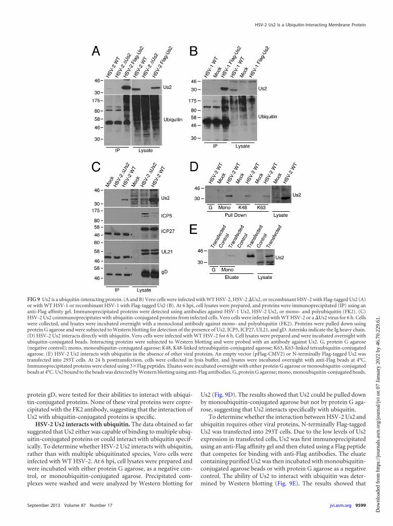
protein gD, were tested for their abilities to interact with ubiqui-tin-conjugated proteins. None of these viral proteins were copre-cipitated with the FK2 antibody, suggesting that the interaction ofUs2 with ubiquitin-conjugated proteins is specific.
HSV-2 Us2 interacts with ubiquitin. The data obtained so farsuggested that Us2 either was capable of binding to multiple ubiq-uitin-conjugated proteins or could interact with ubiquitin specif-ically. To determine whether HSV-2 Us2 interacts with ubiquitin,rather than with multiple ubiquitinated species, Vero cells wereinfected with WT HSV-2. At 6 hpi, cell lysates were prepared andwere incubated with either protein G agarose, as a negative con-trol, or monoubiquitin-conjugated agarose. Precipitated com-plexes were washed and were analyzed by Western blotting for
Us2 (Fig. 9D). The results showed that Us2 could be pulled downby monoubiquitin-conjugated agarose but not by protein G aga-rose, suggesting that Us2 interacts specifically with ubiquitin.
To determine whether the interaction between HSV-2 Us2 andubiquitin requires other viral proteins, N-terminally Flag-taggedUs2 was transfected into 293T cells. Due to the low levels of Us2expression in transfected cells, Us2 was first immunoprecipitatedusing an anti-Flag affinity gel and then eluted using a Flag peptidethat competes for binding with anti-Flag antibodies. The eluatecontaining purified Us2 was then incubated with monoubiquitin-conjugated agarose beads or with protein G agarose as a negativecontrol. The ability of Us2 to interact with ubiquitin was deter-mined by Western blotting (Fig. 9E). The results showed that
FIG 9 Us2 is a ubiquitin-interacting protein. (A and B) Vero cells were infected with WT HSV-2, HSV-2 �Us2, or recombinant HSV-2 with Flag-tagged Us2 (A)or with WT HSV-1 or recombinant HSV-1 with Flag-tagged Us2 (B). At 6 hpi, cell lysates were prepared, and proteins were immunoprecipitated (IP) using ananti-Flag affinity gel. Immunoprecipitated proteins were detected using antibodies against HSV-1 Us2, HSV-2 Us2, or mono- and polyubiquitin (FK2). (C)HSV-2 Us2 coimmunoprecipitates with ubiquitin-conjugated proteins from infected cells. Vero cells were infected with WT HSV-2 or a �Us2 virus for 6 h. Cellswere collected, and lysates were incubated overnight with a monoclonal antibody against mono- and polyubiquitin (FK2). Proteins were pulled down usingprotein G agarose and were subjected to Western blotting for detection of the presence of Us2, ICP5, ICP27, UL21, and gD. Asterisks indicate the Ig heavy chain.(D) HSV-2 Us2 interacts directly with ubiquitin. Vero cells were infected with WT HSV-2 for 6 h. Cell lysates were prepared and were incubated overnight withubiquitin-conjugated beads. Interacting proteins were subjected to Western blotting and were probed with an antibody against Us2. G, protein G agarose(negative control); mono, monoubiquitin-conjugated agarose; K48, K48-linked tetraubiquitin-conjugated agarose; K63, K63-linked tetraubiquitin-conjugatedagarose. (E) HSV-2 Us2 interacts with ubiquitin in the absence of other viral proteins. An empty vector (pFlag-CMV2) or N-terminally Flag-tagged Us2 wastransfected into 293T cells. At 24 h posttransfection, cells were collected in lysis buffer, and lysates were incubated overnight with anti-Flag beads at 4°C.Immunoprecipitated proteins were eluted using 3�Flag peptides. Eluates were incubated overnight with either protein G agarose or monoubiquitin-conjugatedbeads at 4°C. Us2 bound to the beads was detected by Western blotting using anti-Flag antibodies. G, protein G agarose; mono, monoubiquitin-conjugated beads.
HSV-2 Us2 Is a Ubiquitin-Interacting Membrane Protein
September 2013 Volume 87 Number 17 jvi.asm.org 9599
Dow
nloa
ded
from
http
s://j
ourn
als.
asm
.org
/jour
nal/j
vi o
n 07
Jan
uary
202
2 by
46.
70.2
29.6
1.

HSV-2 Us2 interacted with ubiquitin purified from transfectedcells, suggesting that the interaction does not require other viralproteins.
Ubiquitin binding proteins can interact either with ubiquitinmolecules or with the linkages between conjugated ubiquitin mol-ecules. The preference of binding toward a specific ubiquitin link-age is associated with the function of ubiquitin binding proteins.We hypothesized that if HSV-2 Us2 was involved in the protea-somal degradation pathway, it would interact with K48-linkedpolyubiquitin chains with higher affinity. Alternatively, if Us2 wasinvolved in endocytosis or lysosomal degradation, it might be pre-dicted to interact preferentially with K63-linked polyubiquitinchains. To study the Us2-ubiquitin interaction in more detail,HSV-2-infected cell lysates were incubated with either K48-linkedor K63-linked tetraubiquitin-conjugated agarose, and Us2 inter-actions were determined by Western blotting (Fig. 9D). The re-sults showed that HSV-2 Us2 interacted with both K48-linkedtetraubiquitin and K63-linked tetraubiquitin, with similar effi-ciencies.
DISCUSSION
Jiang and coworkers reported previously that HSV-2 Us2 was avirus structural component that was expressed with late kinetics(20). The data presented here are consistent with those findings(Fig. 1). However, the previous study suggested that HSV-2 Us2localized primarily to discrete granules within the nucleus in in-fected cells and diffusely throughout the cytoplasm of transfectedcells (20). Our findings are in stark contrast to those results, sincewe have demonstrated that Us2 localizes predominantly to recy-cling endosomes and to the PM. Differences in specificity betweenthe anti-HSV-2 Us2 antisera reported here and that described byJiang and colleagues may be responsible for this discrepancy; how-ever, it is noteworthy that the localizations of Flag-tagged Us2,mCherry-Us2, and nonfused WT Us2 were indistinguishable inour studies (data not shown).
Although HSV-2 Us2 lacks canonical membrane associationmotifs, membrane flotation experiments revealed that it physi-cally associates with membranes (Fig. 3A). Previous reports haveshown that the EHV-1 Us2 protein localized to the PM despitelacking a classical N-terminal signal sequence, a putative trans-membrane domain, or signals for lipid modifications (18). Like-wise, PRV Us2 also associates with the PM; however, PRV Us2 PMlocalization can be explained, in part, by a prenylation motif thatdirects the addition of a farnesyl moiety to the C terminus of theprotein (33). Interestingly, PRV Us2 mutants that cannot be pre-nylated maintain the ability to associate with vesicular mem-branes, indicating that additional membrane-targeting informa-tion is contained within Us2 (34). How then does HSV-2 Us2associate with membranes? Based on the subcellular localizationof Us2 and its ability to be incorporated into the virion tegument,we and others (18) hypothesized that Us2 is a peripheral mem-brane protein that associates with the cytoplasmic face of themembrane. Accordingly, we predicted that treatment with 1 MNaCl or 0.2 M Na2CO3 (pH 11) would strip HSV-2 Us2 frommembranes, as has been observed with other herpesvirus tegu-ment proteins that associate with membranes (31). Surprisingly,treatment of purified membrane fractions with these agents failedto release Us2 from membranes (Fig. 3C and D). Additionally,exposure of membranes to 0.01% digitonin failed to liberate Us2from the membrane, suggesting that Us2 does not associate with
membranes via a direct interaction with cholesterol (Fig. 3E). Col-lectively, these findings suggest that a portion of Us2 may be in-serted into the membrane. It is unlikely that HSV-2 Us2 is a mem-brane-spanning protein, because, as has been reported for PRVUs2 (33), in the absence of detergent, virion-associated HSV-2Us2 is protected from proteolytic digestion (Fig. 1E). A conservedstretch of hydrophobic amino acids between residues 121 and 137of HSV-2 Us2 may be involved in anchoring Us2 to membranes.
Us2 membrane localization was largely restricted to endocyticvesicles and the PM. Most notably, Us2 did not colocalize with theGolgi apparatus or the ER, nor was its localization sensitive toBFA. At late times postinfection, Us2 was also observed to localizediffusely throughout the cytoplasm (Fig. 2B), suggesting that itssynthesis was not occurring on ER-associated ribosomes. Takentogether, these findings suggest that Us2 is not a component of thebiosynthetic secretory pathway and that its association with mem-branes may occur posttranslationally. Us2 demonstrated substan-tial colocalization with Rab11, a marker of recycling endosomes(Fig. 8C and D). The finding that Us2 was associated with deter-gent-insoluble membranes (Fig. 4) is also consistent with itslocalization in recycling endosomes. Endocytic recycling compart-ments are rich in cholesterol, a major component of detergent-insoluble membranes, and it has been proposed that detergent-insoluble membranes play a role in the sorting of proteins withinthe recycling endosome (47, 48). Rab11 functions both in exocyticmembrane traffic from the TGN to the PM (49) and in endocytictrafficking from early endosomes to the TGN (50). Consistentwith these activities, imaging of Us2 in living cells suggested thatUs2 is highly dynamic, and vesicles bearing Us2 exhibited rapidexchange between other Us2-containing vesicles and the PM.Moreover, the elaboration of tubular membrane projections fromUs2 vesicles (Fig. 6; see also movie S1 in the supplemental mate-rial) and the kinetics of colocalization of Us2 with internalizedtransferrin (Fig. 7) suggested that Us2 localized to compartmentsthat recycle membrane lipids and/or membrane proteins (43, 44).A recent ultrastructural study demonstrated that HSV-1 capsidsare enveloped at the membranes of endocytic tubules in primaryhuman fibroblasts and that Rab5 and Rab11, which are requiredfor the formation of these endocytic tubules, are required for effi-cient virus production (51). The localization of Us2 to Rab11-positive membranes is consistent with the idea that Us2 is incor-porated into nascent virions at endocytic tubules.
A number of enveloped viruses utilize Rab11-containingmembranes for their assembly and egress (52). After their synthe-sis in the nucleus, the viral ribonucleoprotein complexes (vRNPs)of influenza virus A are exported into the cytoplasm and musttraffic to the PM for incorporation into budding virions. Thetransport of these vRNPs from a perinuclear compartment tothe PM is dependent on Rab11. It has been hypothesized that thevRNPs are tethered to the cytoplasmic faces of Rab11-containingvesicles en route to the PM (53, 54). A similar mechanism has beenproposed to explain the trafficking of Sendai virus vRNPs fromtheir site of synthesis in the cytoplasm to the PM (55). Andes virus,a hantavirus that acquires its envelope by budding into Golgi ap-paratus-derived membranes, relies on Rab11/Rab8-bearing vesi-cles for the transport of vesicles containing enveloped virionsfrom the Golgi apparatus to the cell surface (56). Since Rab11vesicle cargos are directed to the apical surfaces of polarized cells,it has been suggested that the engagement of these RNA viruseswith Rab11 vesicles promotes the budding and/or release of virus
Kang et al.
9600 jvi.asm.org Journal of Virology
Dow
nloa
ded
from
http
s://j
ourn
als.
asm
.org
/jour
nal/j
vi o
n 07
Jan
uary
202
2 by
46.
70.2
29.6
1.

from the apical surfaces of polarized cells (52). Because alphaher-pesviruses, such as HSV-2, infect a variety of polarized cell typesduring natural infections, including mucosal epithelial cells andneurons of the peripheral nervous system, it may be that Us2interactions with Rab11 vesicles regulate the trafficking of virionsor subvirion components to apical surfaces.
An unresolved question is how Us2 achieves specificity in itsmembrane association. A number of motifs, typically found intransmembrane proteins, have been identified that can facilitatethe localization of proteins to endocytic membranes and the TGN(57). These include the tyrosine-based YXXØ (where Y is tyrosine,X is any amino acid, and is any bulky hydrophobic amino acid)motif and dileucine motifs that interact with the mu subunits ofthe clathrin adapters AP-1 and AP-2, which are involved in select-ing cargos from the TGN and PM, respectively. Acidic clusters ofamino acids, which additionally contain serine or threonine resi-dues capable of being phosphorylated, have also been shown tolocalize proteins to the TGN. Notable alphaherpesvirus proteinsthat contain TGN-targeting acidic clusters include varicella-zostervirus glycoprotein gE and the HSV-1 tegument protein VP22 (58,59). HSV-2 Us2 does not contain an acidic cluster motif; however,it does contain both YXXØ and dileucine motifs. The positions ofthese motifs in the Us2 sequence are not widely conserved amongUs2 orthologs, so it is not clear if they are functional. It may be thatthe specificity of Us2 membrane localization is imparted throughinteraction with one or more cellular proteins.
To identify membrane-associated interacting partners ofHSV-2 Us2, Flag-Us2 was transfected into mammalian cells, andprotein complexes were immunoprecipitated and identified byLC–MS-MS analysis. Although membrane proteins identified inthis analysis that also localize to detergent-resistant membranes(erlin-1 and erlin-2 [60]) were confirmed to be false positives,conspicuous amounts of ubiquitin were present in the precipi-tated protein complexes, suggesting that HSV-2 Us2 might inter-act with ubiquitin. The interaction between HSV-2 Us2 and ubiq-uitin was further confirmed by reciprocal immunoprecipitationsand ubiquitin binding assays using Us2 derived from both infect-ed- and transfected-cell lysates (Fig. 9). Together, these resultsindicate that Us2 interacts specifically with ubiquitin and can doso in the absence of other viral proteins; however, the possibilityremains that the interaction between Us2 and ubiquitin is indirectand is mediated by a cellular ubiquitin-binding molecule thatbridges the interaction. It has been estimated that more than 150cellular proteins contain one or more ubiquitin binding domains(UBDs), and more than 20 distinct UBDs have been identifiedthus far (61). HSV-2 Us2 does not appear to contain any previ-ously recognized UBD.
Ubiquitin and ubiquitination play important roles in virtuallyevery cellular process. The role of ubiquitination in the endocyticpathway is a subject of intense investigation (62). Mono- or poly-ubiquitination of cell surface transmembrane proteins, usually bya K63 linkage, is sufficient to promote their rapid internalization.Enzymatic removal of ubiquitin by deubiquitinases can direct theinternalized proteins back to the PM via recycling endosomes,while failure to remove the ubiquitin moiety from the internalizedprotein promotes interaction with ESCRT (endosomal sortingcomplex required for transport) components, incorporation intomultivesicular bodies (MVBs), and, ultimately, degradation uponfusion of the MVB with lysosomes. A number of alphaherpesvirusglycoproteins undergo endocytosis from the PM, and some, but
not all, bear well-defined motifs that promote their internalization(63). Additionally, at least two HSV-1 membrane proteins, Us9and gB, are modified by ubiquitination (64, 65). Calistri and col-leagues have suggested that K63 ubiquitination of gB promotesthe trafficking of gB to modified MVB membranes that couldserve as sites of final virion envelopment (65). HSV-1 Us9 is atail-anchored type II membrane protein that lacks lysine residues,to which ubiquitin molecules are most frequently attached (64, 66,67). HSV-1 Us9 is nevertheless conjugated to ubiquitin (64), pre-sumably through the addition of a cysteine to the N terminus ofthe protein (68), as is seen in the modification of E2 ubiquitin-conjugating enzymes by E1 ubiquitin-activating enzymes (69), orby a nonconventional mechanism, such as that mediated by selectE3 ligases on serine and threonine residues (70). The conse-quences of HSV-1 Us9 ubiquitination are not presently under-stood, nor is it known whether Us9 orthologs from other alpha-herpesviruses are modified by ubiquitin. It is intriguing, however,that PRV Us2 was identified in a complex containing Us9 and themicrotubule motor KIF1A, both of which are required for axonalsorting and efficient anterograde transport of virion-containingvesicles within neurons (23). These findings raise the possibilitythat Us2 interacts with, and modulates the activity of, the Us9/KIF1A vesicular sorting complex via its interaction with ubiquitin.
Defining the precise role of Us2 in the regulation and traffick-ing of ubiquitin-modified proteins requires further study. Theimportance of ubiquitin modifications to herpesvirus infections isunderscored by the conservation of proteins containing E3 ubiq-uitin ligase activity or deubiquitinase activity throughout the Her-pesviridae (71–73). While the functions of these activities are notfully understood at present, future investigation of the role ofUs2-ubiquitin interactions during herpesvirus infections prom-ises to advance our understanding of key virus-cell interactionsand reveal new targets for antiviral intervention strategies for thisgroup of important human and veterinary pathogens.
ACKNOWLEDGMENTS
This work was supported by Canadian Institutes of Health Research op-erating grant 93804, Natural Sciences and Engineering Council of CanadaDiscovery Grant 418719, Canada Foundation for Innovation award16389, and an award from the Violet E. Powell Research Fund toB.W.B. M.-H.K. was supported in part by R. Samuel McLaughlin andRobert John Wilson fellowships from Queen’s University and by an On-tario Graduate Scholarship.
We thank R. Tsien (UCSD/HHMI), C. Parish (Cornell University),and N. Osterrieder (Freie Universität Berlin) for providing plasmids, A.Dolan and D. McGeoch (MRC Virology Unit, University of Glasgow) forproviding plasmids and viruses, and G. Smith (Northwestern University)and Y. Kawaguchi (The University of Tokyo) for providing bacterialstrains used in this study. Special thanks to Sina Kachoei for initial char-acterization of mCherry–HSV-2 Us2 fusion constructs. We acknowledgemembers of the Banfield laboratory for helpful comments on the manu-script and M. Lyman for helpful discussions.
REFERENCES1. Ben-Porat T, Kaplan AS. 1985. Molecular biology of pseudorabies virus,
p 105–173. In Roizman B (ed), The herpesviruses. Plenum Press, NewYork, NY.
2. Roizman B, Knipe DM. 2001. Herpes simplex viruses and their replica-tion, p 2399 –2459. In Knipe DM, Howley PM, Griffin DE, Lamb RA,Martin MA, Roizman B, Straus SE (ed), Fields virology, 4th ed. LippincottWilliams & Wilkins, Philadelphia, PA.
3. Mettenleiter TC, Klupp BG, Granzow H. 2009. Herpesvirus assembly: anupdate. Virus Res. 143:222–234.
HSV-2 Us2 Is a Ubiquitin-Interacting Membrane Protein
September 2013 Volume 87 Number 17 jvi.asm.org 9601
Dow
nloa
ded
from
http
s://j
ourn
als.
asm
.org
/jour
nal/j
vi o
n 07
Jan
uary
202
2 by
46.
70.2
29.6
1.

4. Wisner TW, Wright CC, Kato A, Kawaguchi Y, Mou F, Baines JD,Roller RJ, Johnson DC. 2009. Herpesvirus gB-induced fusion betweenthe virion envelope and outer nuclear membrane during virus egress isregulated by the viral US3 kinase. J. Virol. 83:3115–3126.
5. Jones F, Grose C. 1988. Role of cytoplasmic vacuoles in varicella-zostervirus glycoprotein trafficking and virion envelopment. J. Virol. 62:2701–2711.
6. Turcotte S, Letellier J, Lippe R. 2005. Herpes simplex virus type 1 capsidstransit by the trans-Golgi network, where viral glycoproteins accumulateindependently of capsid egress. J. Virol. 79:8847– 8860.
7. Whealy ME, Card JP, Meade RP, Robbins AK, Enquist LW. 1991. Effectof brefeldin A on alphaherpesvirus membrane protein glycosylation andvirus egress. J. Virol. 65:1066 –1081.
8. Johnson DC, Baines JD. 2011. Herpesviruses remodel host membranesfor virus egress. Nat. Rev. Microbiol. 9:382–394.
9. Rémillard-Labrosse G, Mihai C, Duron J, Guay G, Lippe R. 2009.Protein kinase D-dependent trafficking of the large herpes simplex virustype 1 capsids from the TGN to plasma membrane. Traffic 10:1074 –1083.
10. Dolan A, Jamieson FE, Cunningham C, Barnett BC, McGeoch DJ. 1998.The genome sequence of herpes simplex virus type 2. J. Virol. 72:2010 –2021.
11. Haanes EJ, Tomlinson CC. 1998. Genomic organization of the canineherpesvirus US region. Virus Res. 53:151–162.
12. Leung-Tack P, Audonnet JC, Riviere M. 1994. The complete DNAsequence and the genetic organization of the short unique region (US) ofthe bovine herpesvirus type 1 (ST strain). Virology 199:409 – 421.
13. McGeoch DJ, Dolan A, Donald S, Brauer DH. 1986. Complete DNAsequence of the short repeat region in the genome of herpes simplex virustype 1. Nucleic Acids Res. 14:1727–1745.
14. Telford EA, Watson MS, McBride K, Davison AJ. 1992. The DNAsequence of equine herpesvirus-1. Virology 189:304 –316.
15. van Zijl M, van der Gulden H, de Wind N, Gielkens A, Berns A. 1990.Identification of two genes in the unique short region of pseudorabiesvirus; comparison with herpes simplex virus and varicella-zoster virus. J.Gen. Virol. 71(Part 8):1747–1755.
16. Sakaguchi M, Urakawa T, Hirayama Y, Miki N, Yamamoto M, Hirai K.1992. Sequence determination and genetic content of an 8.9-kb restrictionfragment in the short unique region and the internal inverted repeat ofMarek’s disease virus type 1 DNA. Virus Genes 6:365–378.
17. Longnecker R, Roizman B. 1987. Clustering of genes dispensable forgrowth in culture in the S component of the HSV-1 genome. Science236:573–576.
18. Meindl A, Osterrieder N. 1999. The equine herpesvirus 1 Us2 homologencodes a nonessential membrane-associated virion component. J. Virol.73:3430 –3437.
19. Weber PC, Levine M, Glorioso JC. 1987. Rapid identification of nones-sential genes of herpes simplex virus type 1 by Tn5 mutagenesis. Science236:576 –579.
20. Jiang YM, Yamada H, Goshima F, Daikoku T, Oshima S, Wada K,Nishiyama Y. 1998. Characterization of the herpes simplex virus type 2(HSV-2) US2 gene product and a US2-deficient HSV-2 mutant. J. Gen.Virol. 79(Part 11):2777–2784.
21. Wagenaar F, Pol JM, Peeters B, Gielkens AL, de Wind N, Kimman TG.1995. The US3-encoded protein kinase from pseudorabies virus affectsegress of virions from the nucleus. J. Gen. Virol. 76(Part 7):1851–1859.
22. Lyman MG, Randall JA, Calton CM, Banfield BW. 2006. Localization ofERK/MAP kinase is regulated by the alphaherpesvirus tegument proteinUs2. J. Virol. 80:7159 –7168.
23. Kramer T, Greco TM, Taylor MP, Ambrosini AE, Cristea IM, EnquistLW. 2012. Kinesin-3 mediates axonal sorting and directional transport ofalphaherpesvirus particles in neurons. Cell Host Microbe 12:806 – 814.
24. Tischer BK, Smith GA, Osterrieder N. 2010. En passant mutagenesis: atwo step markerless red recombination system. Methods Mol. Biol. 634:421– 430.
25. Le Sage V, Jung M, Alter JD, Wills EG, Johnston SM, Kawaguchi Y,Baines JD, Banfield BW. 2013. The herpes simplex virus 2 UL21 proteinis essential for virus propagation. J. Virol. 87:5904 –5915.
26. Tanaka M, Kagawa H, Yamanashi Y, Sata T, Kawaguchi Y. 2003.Construction of an excisable bacterial artificial chromosome containing afull-length infectious clone of herpes simplex virus type 1: viruses recon-stituted from the clone exhibit wild-type properties in vitro and in vivo. J.Virol. 77:1382–1391.
27. O’Gorman S, Fox DT, Wahl GM. 1991. Recombinase-mediated gene
activation and site-specific integration in mammalian cells. Science 251:1351–1355.
28. Shaner NC, Campbell RE, Steinbach PA, Giepmans BN, Palmer AE,Tsien RY. 2004. Improved monomeric red, orange and yellow fluorescentproteins derived from Discosoma sp. red fluorescent protein. Nat. Biotech-nol. 22:1567–1572.
29. Lyman MG, Demmin GL, Banfield BW. 2003. The attenuated pseudo-rabies virus strain Bartha fails to package the tegument proteins Us3 andVP22. J. Virol. 77:1403–1414.
30. Graham FL, van der Eb AJ. 1973. A new technique for the assay ofinfectivity of human adenovirus 5 DNA. Virology 52:456 – 467.
31. Brignati MJ, Loomis JS, Wills JW, Courtney RJ. 2003. Membraneassociation of VP22, a herpes simplex virus type 1 tegument protein. J.Virol. 77:4888 – 4898.
32. Lyman MG, Curanovic D, Enquist LW. 2008. Targeting of pseudorabiesvirus structural proteins to axons requires association of the viral Us9protein with lipid rafts. PLoS Pathog. 4:e1000065. doi:10.1371/journal.ppat.1000065.
33. Clase AC, Lyman MG, del Rio T, Randall JA, Calton CM, Enquist LW,Banfield BW. 2003. The pseudorabies virus Us2 protein, a virion tegu-ment component, is prenylated in infected cells. J. Virol. 77:12285–12298.
34. Kang MH, Banfield BW. 2010. Pseudorabies virus tegument protein Us2recruits the mitogen-activated protein kinase extracellular-regulated ki-nase (ERK) to membranes through interaction with the ERK commondocking domain. J. Virol. 84:8398 – 8408.
35. Subramaniam VN, bin Mohd Yusoff AR, Wong SH, Lim GB, Chew M,Hong W. 1992. Biochemical fractionation and characterization of pro-teins from Golgi-enriched membranes. J. Biol. Chem. 267:12016 –12021.
36. Cockrell AS, Muggeridge MI. 1998. Herpes simplex virus 2 UL45 is a typeII membrane protein. J. Virol. 72:4430 – 4433.
37. Harder T, Kellner R, Parton RG, Gruenberg J. 1997. Specific release ofmembrane-bound annexin II and cortical cytoskeletal elements by se-questration of membrane cholesterol. Mol. Biol. Cell 8:533–545.
38. Hooper NM. 1999. Detergent-insoluble glycosphingolipid/cholesterol-rich membrane domains, lipid rafts and caveolae. Mol. Membr. Biol. 16:145–156.
39. Doms RW, Russ G, Yewdell JW. 1989. Brefeldin A redistributes residentand itinerant Golgi proteins to the endoplasmic reticulum. J. Cell Biol.109:61–72.
40. Lippincott-Schwartz J, Yuan LC, Bonifacino JS, Klausner RD. 1989.Rapid redistribution of Golgi proteins into the ER in cells treated withbrefeldin A: evidence for membrane cycling from Golgi to ER. Cell 56:801– 813.
41. Misumi Y, Miki K, Takatsuki A, Tamura G, Ikehara Y. 1986. Novelblockade by brefeldin A of intracellular transport of secretory proteins incultured rat hepatocytes. J. Biol. Chem. 261:11398 –11403.
42. Ulmer JB, Palade GE. 1991. Effects of brefeldin A on the Golgi complex,endoplasmic reticulum and viral envelope glycoproteins in murine eryth-roleukemia cells. Eur. J. Cell Biol. 54:38 –54.
43. Bonifacino JS, Rojas R. 2006. Retrograde transport from endosomes tothe trans-Golgi network. Nat. Rev. Mol. Cell Biol. 7:568 –579.
44. Maxfield FR, McGraw TE. 2004. Endocytic recycling. Nat. Rev. Mol. CellBiol. 5:121–132.
45. Ghosh RN, Maxfield FR. 1995. Evidence for nonvectorial, retrogradetransferrin trafficking in the early endosomes of HEp2 cells. J. Cell Biol.128:549 –561.
46. Sönnichsen B, De Renzis S, Nielsen E, Rietdorf J, Zerial M. 2000.Distinct membrane domains on endosomes in the recycling pathway vi-sualized by multicolor imaging of Rab4, Rab5, and Rab11. J. Cell Biol.149:901–914.
47. Hao M, Mukherjee S, Sun Y, Maxfield FR. 2004. Effects of cholesteroldepletion and increased lipid unsaturation on the properties of endocyticmembranes. J. Biol. Chem. 279:14171–14178.
48. Mayor S, Sabharanjak S, Maxfield FR. 1998. Cholesterol-dependentretention of GPI-anchored proteins in endosomes. EMBO J. 17:4626 –4638.
49. Chen W, Feng Y, Chen D, Wandinger-Ness A. 1998. Rab11 is requiredfor trans-Golgi network-to-plasma membrane transport and a preferen-tial target for GDP dissociation inhibitor. Mol. Biol. Cell 9:3241–3257.
50. Wilcke M, Johannes L, Galli T, Mayau V, Goud B, Salamero J. 2000.Rab11 regulates the compartmentalization of early endosomes requiredfor efficient transport from early endosomes to the trans-Golgi network. J.Cell Biol. 151:1207–1220.
Kang et al.
9602 jvi.asm.org Journal of Virology
Dow
nloa
ded
from
http
s://j
ourn
als.
asm
.org
/jour
nal/j
vi o
n 07
Jan
uary
202
2 by
46.
70.2
29.6
1.

51. Hollinshead M, Johns HL, Sayers CL, Gonzalez-Lopez C, Smith GL,Elliott G. 2012. Endocytic tubules regulated by Rab GTPases 5 and 11 areused for envelopment of herpes simplex virus. EMBO J. 31:4204 – 4220.
52. Bruce EA, Stuart A, McCaffrey MW, Digard P. 2012. Role of the Rab11pathway in negative-strand virus assembly. Biochem. Soc. Trans. 40:1409 –1415.
53. Amorim MJ, Bruce EA, Read EK, Foeglein A, Mahen R, Stuart AD, DigardP. 2011. A Rab11- and microtubule-dependent mechanism for cytoplasmictransport of influenza A virus viral RNA. J. Virol. 85:4143–4156.
54. Eisfeld AJ, Kawakami E, Watanabe T, Neumann G, Kawaoka Y. 2011.RAB11A is essential for transport of the influenza virus genome to theplasma membrane. J. Virol. 85:6117– 6126.
55. Chambers R, Takimoto T. 2010. Trafficking of Sendai virus nucleocap-sids is mediated by intracellular vesicles. PLoS One 5:e10994. doi:10.1371/journal.pone.0010994.
56. Rowe RK, Suszko JW, Pekosz A. 2008. Roles for the recycling endosome,Rab8, and Rab11 in hantavirus release from epithelial cells. Virology 382:239 –249.
57. Bonifacino JS, Traub LM. 2003. Signals for sorting of transmembraneproteins to endosomes and lysosomes. Annu. Rev. Biochem. 72:395– 447.
58. Zhu Z, Hao Y, Gershon MD, Ambron RT, Gershon AA. 1996. Targetingof glycoprotein I (gE) of varicella-zoster virus to the trans-Golgi networkby an AYRV sequence and an acidic amino acid-rich patch in the cytosolicdomain of the molecule. J. Virol. 70:6563– 6575.
59. O’Regan KJ, Brignati MJ, Murphy MA, Bucks MA, Courtney RJ. 2010.Virion incorporation of the herpes simplex virus type 1 tegument proteinVP22 is facilitated by trans-Golgi network localization and is independentof interaction with glycoprotein E. Virology 405:176 –192.
60. Browman DT, Resek ME, Zajchowski LD, Robbins SM. 2006. Erlin-1and erlin-2 are novel members of the prohibitin family of proteins thatdefine lipid-raft-like domains of the ER. J. Cell Sci. 119:3149 –3160.
61. Dikic I, Wakatsuki S, Walters KJ. 2009. Ubiquitin-binding domains—from structures to functions. Nat. Rev. Mol. Cell Biol. 10:659 – 671.
62. Clague MJ, Liu H, Urbe S. 2012. Governance of endocytic trafficking andsignaling by reversible ubiquitylation. Dev. Cell 23:457– 467.
63. Brideau AD, Enquist LW, Tirabassi RS. 2000. The role of virion mem-brane protein endocytosis in the herpesvirus life cycle. J. Clin. Virol. 17:69 – 82.
64. Brandimarti R, Roizman B. 1997. Us9, a stable lysine-less herpes simplexvirus 1 protein, is ubiquitinated before packaging into virions and associ-ates with proteasomes. Proc. Natl. Acad. Sci. U. S. A. 94:13973–13978.
65. Calistri A, Sette P, Salata C, Cancellotti E, Forghieri C, Comin A,Gottlinger H, Campadelli-Fiume G, Palu G, Parolin C. 2007. Intracel-lular trafficking and maturation of herpes simplex virus type 1 gB andvirus egress require functional biogenesis of multivesicular bodies. J. Vi-rol. 81:11468 –11478.
66. Brideau AD, Banfield BW, Enquist LW. 1998. The Us9 gene product ofpseudorabies virus, an alphaherpesvirus, is a phosphorylated, tail-anchored type II membrane protein. J. Virol. 72:4560 – 4570.
67. Lyman MG, Kemp CD, Taylor MP, Enquist LW. 2009. Comparison ofthe pseudorabies virus Us9 protein with homologs from other veterinaryand human alphaherpesviruses. J. Virol. 83:6978 – 6986.
68. Ciechanover A, Ben-Saadon R. 2004. N-terminal ubiquitination: moreprotein substrates join in. Trends Cell Biol. 14:103–106.
69. Pickart CM, Eddins MJ. 2004. Ubiquitin: structures, functions, mecha-nisms. Biochim. Biophys. Acta 1695:55–72.
70. Shimizu Y, Okuda-Shimizu Y, Hendershot LM. 2010. Ubiquitylation ofan ERAD substrate occurs on multiple types of amino acids. Mol. Cell40:917–926.
71. Everett RD, Boutell C, McNair C, Grant L, Orr A. 2010. Comparison ofthe biological and biochemical activities of several members of the alpha-herpesvirus ICP0 family of proteins. J. Virol. 84:3476 –3487.
72. Ohmura-Hoshino M, Goto E, Matsuki Y, Aoki M, Mito M, Uematsu M,Hotta H, Ishido S. 2006. A novel family of membrane-bound E3 ubiqui-tin ligases. J. Biochem. 140:147–154.
73. Kattenhorn LM, Korbel GA, Kessler BM, Spooner E, Ploegh HL. 2005.A deubiquitinating enzyme encoded by HSV-1 belongs to a family ofcysteine proteases that is conserved across the family Herpesviridae. Mol.Cell 19:547–557.
HSV-2 Us2 Is a Ubiquitin-Interacting Membrane Protein
September 2013 Volume 87 Number 17 jvi.asm.org 9603
Dow
nloa
ded
from
http
s://j
ourn
als.
asm
.org
/jour
nal/j
vi o
n 07
Jan
uary
202
2 by
46.
70.2
29.6
1.