the use of an inactivated vaccine in farmed nile
TRANSCRIPT

THE USE OF AN INACTIVATED VACCINE IN FARMED
NILE CROCODILES (CROCODYLUS NILOTICUS) FOR
THE CONTROL OF MYCOPLASMA CROCODYLI
INFECTION
By
Miemie Grobler
Submitted in partial fulfilment of the requirements for the degree of
Magister Scientiae (Veterinary Tropical Diseases)
In the
Department of Veterinary Tropical Diseases
Faculty of Veterinary Science
University of Pretoria, South Africa.
October 2012
Supervisor: Professor Moritz van Vuuren
Co-supervisor: Mr Johan Gouws
©© UUnniivveerrssiittyy ooff PPrreettoorriiaa

University of Pretoria - M Grobler (2012)
ii
DEDICATION
“Not to us, Lord, not to us but to your name be the glory, because of your love and faithfulness.”
Psalm 115:1

University of Pretoria - M Grobler (2012)
iii
ACKNOWLEDGEMENTS
I would like to express my sincere gratitude to the following persons and institutions:
My supervisor, Professor Moritz van Vuuren, for his guidance and support during this project and his
patience with the preparation of this manuscript.
My co-supervisor, Mr Johan Gouws, for his assistance with the laboratory assays.
Dr Chris Foggin and his staff from the Wildlife Veterinary Unit, Harare, Zimbabwe for their assistance
with the collection and processing of samples.
The managers and staff of Nuanetsi Ranch for their hospitality during my visits and their financial and
time investment in the project.
Professor Geoff Fosgate for his help with the statistical analysis of the results.
The Institute of Tropical Medicine, Antwerpen, Belgium, and the Department of Veterinary Tropical
Disease for a bursary to support this project.
My current and previous colleagues, in particular Ms Joubert and Drs. Maartens, Pienaar, Pulker and
Smit, for their support, help and guidance.
My family and friends for their love, understanding and encouragement.
University of Pretoria - M Grobler (2012)
iv
TABLE OF CONTENTS
Dedication ............................................................................................................................................... ii
Acknowledgements .................................................................................................................................iii
Table of Contents ................................................................................................................................... iv
List of Tables ..........................................................................................................................................vii
List of Figures ........................................................................................................................................ viii
Summary ................................................................................................................................................. 1
Chapter 1: Introduction ............................................................................................................................ 3
Chapter 2: Literature review .................................................................................................................... 5
M. crocodyli ......................................................................................................................................... 5
History of Mycoplasma outbreaks ................................................................................................... 5
Disease caused by M. crocodyli ...................................................................................................... 5
Comparison between M. crocodyli and other pathogenic Mycoplasma spp ....................................... 6
Mycoplasma spp infections of the respiratory tract ......................................................................... 7
Mycoplasma spp infections causing arthritis ................................................................................... 7
Mycoplasma infections of other reptiles .......................................................................................... 7
Crocodile husbandry and mycoplasmosis ........................................................................................ 10
General aspects of crocodile farming ........................................................................................... 10
Crocodilian stress factors .............................................................................................................. 11
Impact of stress on crocodilians .................................................................................................... 11
Pathogenesis of mycoplasmosis ....................................................................................................... 12
Intracellular location of Mycoplasma spp ...................................................................................... 12
Adhesion to host cells and antigenic variation .............................................................................. 13
Interaction between Mycoplasma spp and the host immune system ........................................... 14
Reptilian immune system .................................................................................................................. 14
Adaptive immunity ......................................................................................................................... 15
Control of mycoplasmosis ................................................................................................................. 16
Vaccination .................................................................................................................................... 16
Serodiagnosis of mycoplasmosis ...................................................................................................... 18
Latex agglutination test ................................................................................................................. 18
Metabolic inhibition assay ............................................................................................................. 19
University of Pretoria - M Grobler (2012)
v
Chapter 3: Experimental design and methods ...................................................................................... 20
Facilities and experimental animals .................................................................................................. 20
Study subjects ............................................................................................................................... 20
Housing conditions ........................................................................................................................ 20
Nutrition, feeding and watering ..................................................................................................... 20
Daily care of animals ..................................................................................................................... 21
Handling and restraint of crocodiles .............................................................................................. 21
Farm layout ................................................................................................................................... 21
Cleaning and disinfection .............................................................................................................. 22
Biosecurity ..................................................................................................................................... 22
Vaccine production ............................................................................................................................ 22
Mycoplasma-strain included in experimental vaccine ................................................................... 22
Vaccine production and formulation .............................................................................................. 23
Quality control of vaccine .............................................................................................................. 23
Safety testing of the autogenous vaccine ......................................................................................... 23
Efficacy testing of the autogenous vaccine ....................................................................................... 23
Serum collection for serological testing ............................................................................................ 25
Sampling points for serum collection ............................................................................................ 25
Procedure for serum collection ..................................................................................................... 26
Procedure for serum processing and transport to laboratories ..................................................... 26
Latex slide agglutination test ............................................................................................................. 27
Mycoplasma strains and growth conditions .................................................................................. 27
Coating of microspheres ............................................................................................................... 27
Procedure for latex slide agglutination .......................................................................................... 27
Growth/metabolism inhibition assay ................................................................................................. 28
Mycoplasma strain and culture ..................................................................................................... 28
Mycoplasma medium .................................................................................................................... 28
Procedure for MI test ..................................................................................................................... 28
Statistical evaluation of results .......................................................................................................... 28
Evaluating sero-conversion ........................................................................................................... 29
Evaluating the association between serological status, disease status and vaccination status
during disease outbreak ................................................................................................................ 29
University of Pretoria - M Grobler (2012)
vi
Evaluating the diagnostic performance of the latex agglutination test .............................................. 29
Chapter 4: Results ................................................................................................................................ 30
Safety trial ......................................................................................................................................... 30
Efficacy trial ....................................................................................................................................... 30
Latex agglutination assays ............................................................................................................ 30
Growth/metabolic inhibition assays ............................................................................................... 31
Results from outbreak, October 2011 ............................................................................................... 32
Pathology ...................................................................................................................................... 32
Serology ........................................................................................................................................ 33
Statistical evaluation of results .......................................................................................................... 34
Evaluating the relationship between vaccination and seroconversion.......................................... 34
Evaluating the relationship between vaccination status, serological status and development of
clinical disease during a disease outbreak ................................................................................... 35
Evaluating the diagnostic performance of the latex agglutination test .............................................. 36
Chapter 5: Discussion ........................................................................................................................... 37
Vaccine safety ................................................................................................................................... 37
Vaccine efficacy ................................................................................................................................ 37
Immunogenicity ............................................................................................................................. 37
Vaccine efficacy during a disease outbreak .................................................................................. 39
Performance of the latex agglutination assay ................................................................................... 44
Chapter 6: Conclusion ........................................................................................................................... 46
Reference List ....................................................................................................................................... 47

University of Pretoria - M Grobler (2012)
vii
LIST OF TABLES
Table
number Table title
Page
number
Table 3.1 2x2 contingency table for determining diagnostic performance 21
Table 4.1 Summary of LAT results 31
Table 4.2 Summary of MI test results 31
Table 4.3 Summary of outbreak results - LAT 33
Table 4.4 Summary of outbreak results - MI test 34
Table 4.5 Relationship between LAT results and MI test results 36
University of Pretoria - M Grobler (2012)
viii
LIST OF FIGURES
Figure
number
Figure title Page
number
Figure 3.1 Layout of crocodile unit of Nuanetsi Ranch, Mwenezi, Zimbabwe 21
Figure 3.2 Schematic representation of trial 24
Figure 3.3 Vaccination of crocodiles. 25
Figure 3.4 Crocodile tail indicating the healed lesion after removal of one scute from the
right
25
Figure 3.5 Collecting blood from a crocodile. 26
Figure 4.1 Layout of a typical LAT plate. 30
Figure 4.2 Two examples of positive reactions 30
Figure 4.3 An example of a negative reaction. 30
Figure 4.4 Acute arthritis. 32
Figure 4.5 Subacute arthritis. 32
Figure 4.6 Chronic arthritis. 33
Figure 4.7 Typical skin lesions on the feet and over the sternum 33
Figure 4.8 Percentage seropositive animals at different times after vaccination 34
Figure 4.9 Percentage of crocodiles in each LAT titre range 35
Figure 4.10 Percentage of crocodiles in each MI assay titre range 36

University of Pretoria - M Grobler (2012)
1
SUMMARY
THE USE OF AN INACTIVATED VACCINE IN FARMED NILE CROCODILES (CROCODYLUS
NILOTICUS) FOR THE CONTROL OF MYCOPLASMA CROCODYLI INFECTION
Since the first report of Mycoplasma-associated polyarthritis in farmed Nile crocodiles in 1995, the
disease has spread across Zimbabwe and South Africa and has resulted in significant economic
losses on infected farms. Due to poor response to antimicrobial treatment and frequent relapses, the
use of an autogenous vaccine to manage disease outbreaks was evaluated. Two previous trials had
been performed with a similar vaccine and the results suggested that the vaccine could be effective in
alleviating disease, although the numbers of animals were limited in both. This trial aimed to evaluate
an inactivated, alum-adjuvanted M. crocodyli whole-cell vaccine in a large group of yearling crocodiles
under field conditions on a farm in Zimbabwe where repeated M. crocodyli outbreaks have been
reported.
The safety of the vaccine was assessed by administrating the vaccine intraperitoneally to a subset of
crocodiles. No adverse clinical reactions were observed in any of these crocodiles.
A group of two thousand two hundred crocodiles received two intramuscular vaccinations four weeks
apart in the autumn of 2011, while another group of two thousand two hundred crocodiles served as
unvaccinated controls. Serum was collected from a subset of the vaccinated and unvaccinated
crocodiles at different time-points before and after vaccination to evaluate the humoral response to
vaccination. Latex slide agglutination tests (LAT) were performed on all samples and positive samples
were titrated with the latex slide agglutination test and metabolism inhibition assay.
A low percentage of sera were positive with serological tests done prior to vaccination, suggesting
either circulating Mycoplasma or maternal immunity. Statistically significant increase in sero-positivity
was detected with LAT four weeks after primary vaccination, although the titre remained low. Six
weeks after the booster vaccination the percentage seropositive vaccinated crocodiles had decreased
and there were no statistically significant difference between the percentage seropositive vaccinated
and unvaccinated crocodiles.
A significant outbreak of Mycoplasma-like polyarthritis was encountered 6 months after vaccination, in
October 2011. Both vaccinated and unvaccinated crocodiles were affected. Serum samples from
different subsets of crocodiles were collected and evaluated similar to the vaccine trial. The results
indicated that a similar rate of sero-positivity was present in all crocodiles, irrespective of vaccination-
or disease status.
Sera collected during this trial was used to evaluate the performance of the latex slide agglutination
assay compared to the metabolism inhibition assay (“Gold standard” assay), as the performance of
the LAT had not been evaluated previously. The calculated diagnostic sensitivity was 72%, diagnostic

University of Pretoria - M Grobler (2012)
2
specificity was 32%, the predictive value of the positive test was 36% while the predictive value of the
negative test was 69%.
This trial indicated that the autogenous, inactivated, alum-adjuvanted, whole-cell vaccine against M.
crocodyli was not able to protect farmed Nile crocodiles on an infected farm against clinical
Mycoplasma-associated polyarthritis. It was also found that the latex slide agglutination assay could
be useful as a robust, pen-side assay to evaluate exposure to M. crocodyli, although other assays,
such as PCR, bacterial culture or growth inhibition assays, has to be performed to confirm the
presence of disease.
University of Pretoria - M Grobler (2012)
3
CHAPTER 1: INTRODUCTION
Farming of crocodilians is primarily concerned with the production of crocodile skins for luxury leather
markets. Although this is a fluctuating market, it is estimated that between one and two million
crocodilian skins are internationally traded annually (Caldwell 2012). The brown caiman (Caiman
crocodilus fuscus) is the “top-seller” and accounts for around half of exported skins, followed by the
American alligator (Alligatoris mississippiensis) and the Nile crocodile (Crocodylus niloticus) (Caldwell
2012). Crocodile meat is also internationally traded but is regarded as a by-product of skin production
(Caldwell 2010).
Zimbabwe is the largest producer of Nile crocodile skins with approximately half the annual CITES-
reported Nile crocodile skins exported from that country. Commercial trade in crocodile skins has
been a key driving factor in the rescue of the species in Zimbabwe, because of the economic value
that is currently attached to this species that was previously classified as vermin and hunted almost to
extinction (Revel 1995, Caldwell 2010). Since the first crocodile farm was established in the mid-
1960s, the production of skins has progressively shifted from wild-harvested skins to captive-bred
skins (Caldwell 2010). With a proportion of bred crocodiles re-introduced into the wild, the
Zimbabwean wild population (as well as the wild populations in most of Southern African countries) is
currently listed under Appendix II of CITES (Ferguson 2010).
Crocodile farming, particularly in Zimbabwe, also has the benefit of creating employment and socio-
economic improvement in this economically challenged country. Nuanetsi crocodile ranch, Mwenezi,
Zimbabwe is a good example of such a project. Despite the political controversy surrounding the farm
(Scoones et al. 2012), more than 2000 employment opportunities have been created (Riley 2010).
Recurrent epidemics of polyarthritis and paralysis were reported on Nuanetzi in 2010, affecting up to
40% of rearing stock as well as breeding stock. Financial losses of around $1 million were
experienced, severely hampering the economic sustainability of the operation. During August 2010,
South African researchers and crocodile experts were approached to assist with the investigation and
management of these outbreaks. Mycoplasma crocodyli was isolated from arthritic lesions in affected
crocodiles and confirmed as the causative agent.
Mycoplasmosis in farmed Nile crocodiles is clinically characterized by polyarthritis of the appendicular
and axial skeleton. Crocodiles consequently become lame, paretic and paralytic, fail to feed and
starve to death. Paralysis is the most common sign reported by commercial farmers. Overgrowth of
normal commensal bacteria and fungi on the skin of paralysed crocodiles result in the development of
ulcers and scars, which further reduces hide value, thereby worsening the economic effects of the
disease.
In response to the mentioned outbreak, the managers of Nuanetsi Ranch and the scientists and
veterinarians involved, decided to develop an experimental, autogenous vaccine against the cultured
University of Pretoria - M Grobler (2012)
4
organism. This route was decided on because the current method of control on commercial farms
relies on the application of antimicrobial therapy, which is costly and did not provide the expected
clinical improvement during outbreaks. A similar vaccine had been developed and tested on two
previous occasions in Zimbabwe (Mohan et al. 1997, Mohan et al. 2001), and, although some of the
results were promising, the vaccine had not previously been tested in a large population in the face of
a disease outbreak.
The primary objectives of the study were therefore, to test and demonstrate the safety and efficacy of
an experimental, inactivated, alum-adjuvanted Mycoplasma crocodyli whole-cell vaccine in a large
group of yearling crocodiles under field conditions on a farm in Zimbabwe where repeated M.
crocodyli outbreaks have been reported. A secondary objective arose from the serological tests,
namely to evaluate the diagnostic performance of a latex slide agglutination assay which had been
developed by the same researchers.
University of Pretoria - M Grobler (2012)
5
CHAPTER 2: LITERATURE REVIEW
In this section, the relevant literature on M. crocodyli will be considered and important aspects
highlighted. As mentioned in the previous chapter, vaccination against M. crocodyli has been
evaluated on two previous occasions. Because M. crocodyli is a relatively recently described
pathogen, aspects of other Mycoplasma species will also be included where applicable. As
Mycoplasma infections in most species are encountered in intensive housing and production setups,
relevant management and stress factors will be considered. Reptile immunology is also of
importance, as vaccination and sero-diagnostics cannot be contemplated without understanding the
host immune response to a pathogen. Lastly, vaccination and sero-diagnosis (the focus of this study)
will be reported on.
M. crocodyli
History of Mycoplasma-outbreaks
The first published outbreak of Mycoplasma associated disease in crocodilians was reported in
Zimbabwe in 1995 (Mohan et al. 1995). Since this first outbreak, the disease has reportedly spread
across the country, with approximately 35% of commercial farms affected by 2001. It has also been
diagnosed in South Africa, where it is reported to be widespread (Huchzermeyer & Picard 2004), the
Canary Islands and Israel (Huchzermeyer et al. 1997). A similar disease has also been reported in
alligators in the USA, but with a dramatically higher mortality rate than described for M. crocodyli
outbreaks (Clippenger et al. 2000).
The first outbreak affected only young crocodiles (1-3 years), and low morbidity and mortality were
reported (Mohan et al. 1995). Low morbidity and mortality have also been reported for South African
outbreaks thus far (F.W. Huchzermeyer, unpublished results, 2011). A significantly higher morbidity
and mortality rate was however, encountered during a second published outbreak in Zimbabwe
(Mohan et al. 2001). More than 2500 crocodiles were affected and morbidity peaked at 50% while
mortality was estimated at over 20% (Mohan et al. 2001). It was suggested that this outbreak was
triggered by translocation stress (Mohan et al. 2001).
Disease caused by M. crocodyli
Soon after the first outbreak, Mycoplasma crocodyli was named, classified and described as a new
Mycoplasma species (Kirchhoff et al. 1997). M. crocodyli, as with other Mycoplasmas, lacks true cell
walls and has a typical fried-egg appearance on solid medium, but grows relatively well in artificial
medium (Kirchhoff et al. 1997). Glucose and mannose are both fermented, and cholesterol or serum
is required for growth (Kirchhoff et al. 1997). It is one of the few Mycoplasma spp that fulfils Koch’s
postulates for disease causation (Kirchhoff et al. 1997). M. crocodyli has a peculiar preferred
temperature range for in vitro growth as optimal growth is described at 37 ˚C a temperature which
could be lethal for its host (Kirchhoff et al. 1997, Huchzermeyer 2002), while it would be expected that
a pathogen of an exothermic host would prefer temperatures closer to the host’s natural temperature
range (Razin 2006). It is unknown whether this temperature preference holds true for in vivo
University of Pretoria - M Grobler (2012)
6
conditions. Mycoplasma crocodyli has a low G+C content (27.6%) which is characteristic for
Mycoplasmas (Kirchhoff et al. 1997).
The complete genome sequence of M. crocodyli has recently been reported but, although at least five
potential virulence factors have been identified, their role and significance are still unclear, particularly
as no acknowledged adhesion factors have been identified (Brown et al. 2011).
Polyarthritis is the best described clinical and pathological sign associated with M. crocodyli (Mohan
et al. 1995). Clinical signs of polyarthritis include progressive weakness, ranging from stiffness to
complete immobility (Mohan et al. 1995). Both the appendicular and axial skeletons are affected and
joints display marked swelling, although this may be difficult to appreciate ante-mortally in the spinal
column (Mohan et al. 1995). Different stages of exudative polyarthritis are encountered at necropsy,
ranging from turbid mucous containing Mycoplasma spp in acute and subacute cases, to yellow,
inspissated exudate in chronic cases (Mohan et al. 1995). Histopathological changes include
inflammatory oedema of the surrounding tissue, necrosis of the superficial layers of the synovial
membrane, and fibrin deposition, lymphocytic infiltration and fibrosis of the joint capsule (Mohan et al.
1995). Joint fluid and heart blood are good samples for the culture of the organism.
Apart from polyarthritis, the organism also triggers pneumonia, histopathologically characterised by
consolidation and oedema of affected areas, with a white blood cell (particularly polymorphonuclear
cells and mononuclear cells) and erythrocyte infiltration (Mohan et al. 1995). Although the respiratory
involvement of M. crocodyli is less often recognized, the respiratory tract is a common predilection
site for Mycoplasma spp. colonization in many hosts and is the likely site for host invasion.
Comparison between M. crocodyli and other pathogenic Mycoplasma spp
Mycoplasma spp are some of the most widespread parasites of living organisms, and have been
found in association with mammals, reptiles, birds, fish, arthropods and plants (Razin 1998). Over 200
species have been identified to date (Chazel et al. 2010), and it has been suggested that this is but a
fraction of existing species (Razin & Hayflick 2010). Although only a small proportion of these are
regarded as pathogenic, a range of conditions of animals and humans is associated with Mycoplasma
spp. These include respiratory disease, mastitis, keratoconjunctivitis, arthritis and synovitis, as well as
reproductive disorders and infectious anaemia. Little is known about Mycoplasma spp of crocodiles
and vaccination against reptilian mycoplasmosis in general. This section will briefly mention
pathogenic Mycoplasma-infections to which crocodile Mycoplasma can be related (primarily
respiratory and joint complications) before considering mycoplasmosis of other reptiles. The
successes and failures of vaccines (particularly inactivated vaccines) and sero-diagnostic tests
against some of these mycoplasmas will be considered later in this chapter.
University of Pretoria - M Grobler (2012)
7
Mycoplasma spp infections of the respiratory tract
Colonization and infection of the respiratory tract is the best-described Mycoplasma spp pathology
and two disease syndromes can be differentiated. The first is characterized by subacute to chronic
interstitial pneumonia and/or bronchopneumonia, with or without non-specific upper respiratory
disease. In this instance, infection with Mycoplasma spp. seldom results in fulminant disease by itself.
It increases the hosts’ susceptibility to other pulmonary pathogens, particularly bacteria, resulting in
bacterial bronchopneumonia, which often masks the Mycoplasma infection (Ley 2006, Caswell &
Archambault 2008, Sibila et al. 2009). Well-known Mycoplasma spp associated with this syndrome
include M. pneumoniae (humans), M. gallisepticum (poultry), M. hyopneumoniae (swine), M. bovis
(cattle) and M. ovipneumoniae (sheep).
Contagious pleuro-pneumonia is the second important respiratory complication associated with
Mycoplasma spp colonization of the respiratory tract. Contagious bovine pleuro-pneumonia (CBPP),
caused by M. mycoides subsp. mycoides Small colony, and contagious caprine pleuro-pneumoniae
(CCPP), caused by M. carpicolum subsp. capripneumoniae, are examples of this condition. Both are
classified as diseases of major economic importance by the OIE (World Organisation for Animal
Health), with CBPP being recognized as the most important transboundary disease of cattle in Africa
(Nicholas & Churchward 2012, Thiaucourt et al. 2012). CBPP and CCPP differ from other respiratory
mycoplasmoses, as it can cause fatal disease by itself with prominent involvement of the pleural
membranes and pleural effusion (Thiaucourt 2004, Nicholas et al. 2008). Macroscopic sequestra are
often encountered in recovered chronically infected animals (Thiaucourt 2004), and serve as the
source of infection for other hosts.
Although some fatal cases of M. crocodyli disease have been described (Mohan et al. 2001), the
general pulmonary pathology is more consistent with what is described for M. bovis, M. gallisepticum
and M hyopneumoniae, i.e. chronic pulmonary infection/colonization and inflammation.
Mycoplasma spp infections causing arthritis
Polyarthritis is often the main lesion associated with M. crocodyli. It is believed to result from the
systemic spread of the organism, which has a tropism for serous membranes, such as pleura,
pericardium and synovial membranes. Well-described Mycoplasma-associated arthritis agents include
M. synoviae (poultry), M. hyosynoviae (swine), M. bovis (cattle), M. agalaciae (small ruminants) and
M. arthritidis (rodents). The pathological lesions described for M. crocodyli are similar to the joint
pathology described for most of the mentioned diseases (Hagedorn-Olsen et al. 1999, Kleven 2006,
Nicholas et al. 2008, Hewicker-Trautwein et al. 2009).
Mycoplasma infections of other reptiles
As mentioned above, Mycoplasma spp parasites have also been identified in reptiles other than
crocodiles. A summary of identified Mycoplasma spp, their host range and associated disease
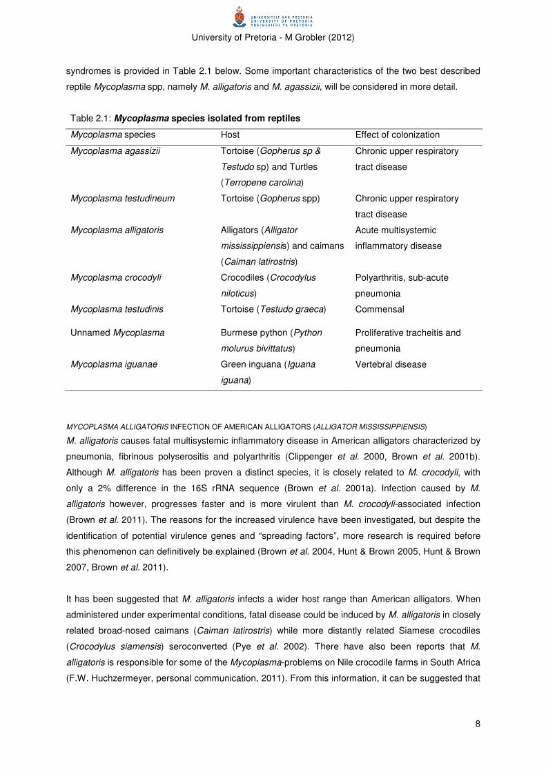
University of Pretoria - M Grobler (2012)
8
syndromes is provided in Table 2.1 below. Some important characteristics of the two best described
reptile Mycoplasma spp, namely M. alligatoris and M. agassizii, will be considered in more detail.
Table 2.1: Mycoplasma species isolated from reptiles
Mycoplasma species Host Effect of colonization
Mycoplasma agassizii Tortoise (Gopherus sp &
Testudo sp) and Turtles
(Terropene carolina)
Chronic upper respiratory
tract disease
Mycoplasma testudineum Tortoise (Gopherus spp) Chronic upper respiratory
tract disease
Mycoplasma alligatoris Alligators (Alligator
mississippiensis) and caimans
(Caiman latirostris)
Acute multisystemic
inflammatory disease
Mycoplasma crocodyli Crocodiles (Crocodylus
niloticus)
Polyarthritis, sub-acute
pneumonia
Mycoplasma testudinis Tortoise (Testudo graeca) Commensal
Unnamed Mycoplasma Burmese python (Python
molurus bivittatus)
Proliferative tracheitis and
pneumonia
Mycoplasma iguanae Green inguana (Iguana
iguana)
Vertebral disease
MYCOPLASMA ALLIGATORIS INFECTION OF AMERICAN ALLIGATORS (ALLIGATOR MISSISSIPPIENSIS)
M. alligatoris causes fatal multisystemic inflammatory disease in American alligators characterized by
pneumonia, fibrinous polyserositis and polyarthritis (Clippenger et al. 2000, Brown et al. 2001b).
Although M. alligatoris has been proven a distinct species, it is closely related to M. crocodyli, with
only a 2% difference in the 16S rRNA sequence (Brown et al. 2001a). Infection caused by M.
alligatoris however, progresses faster and is more virulent than M. crocodyli-associated infection
(Brown et al. 2011). The reasons for the increased virulence have been investigated, but despite the
identification of potential virulence genes and “spreading factors”, more research is required before
this phenomenon can definitively be explained (Brown et al. 2004, Hunt & Brown 2005, Hunt & Brown
2007, Brown et al. 2011).
It has been suggested that M. alligatoris infects a wider host range than American alligators. When
administered under experimental conditions, fatal disease could be induced by M. alligatoris in closely
related broad-nosed caimans (Caiman latirostris) while more distantly related Siamese crocodiles
(Crocodylus siamensis) seroconverted (Pye et al. 2002). There have also been reports that M.
alligatoris is responsible for some of the Mycoplasma-problems on Nile crocodile farms in South Africa
(F.W. Huchzermeyer, personal communication, 2011). From this information, it can be suggested that
University of Pretoria - M Grobler (2012)
9
the epidemiological investigation of M. crocodyli, particularly with regards to reservoir hosts, might
require sampling and testing of other reptile species as well.
MYCOPLASMOSIS IN TORTOISES
Upper respiratory tract disease (URTD) is a disease syndrome of various tortoise species (including
gopher tortoise (Gopherus polyphemus) and desert tortoise (Gopherus agasizzii)), currently ascribed
to Mycoplasma agassizii and M. testiduneum infection (Brown et al. 1994, Brown et al. 1999,
Wendland et al. 2005). Other microorganisms may also be involved (Sandmeier et al. 2009). The
main clinical signs are chronic rhinitis and conjunctivitis, resulting in serous to mucopurulent nasal and
ocular discharge with partial or complete occlusion of one or both nares (Jacobson et al. 1991,
Lederle et al. 1997, Homer et al. 1998, McLaughlin et al. 2000, Christopher et al. 2003, Wendland et
al. 2005). Systemic effects are non-specific and include generalized cachexia and lymphocytic
infiltration (Wendland et al. 2005). A fatal outcome of disease is associated with secondary infections
and/or nutritional and metabolic disturbances (Jacobson et al. 1991, Homer et al. 1998, McLaughlin et
al. 2000).
Mycoplasmosis of tortoises do not resemble mycoplasmosis of crocodiles, but some aspects of the
sero-diagnostics will be described as it emphasizes the importance of critical analysis of serological
tests and their results in general, but also specifically for reptile mycoplasmosis.
Because URTD was implicated as a causative factor for a decline in desert tortoise populations in the
1980s and 1990s, a conservation plan was formulated according to which all ELISA positive or
suspect -positive tortoises, destined for translocation, should be euthanized (Brown et al. 1994,
Schumacher et al. 1997, Homer et al. 1998, Seigel et al. 2003, Sandmeier et al. 2009). The plan
aimed to limit/prevent the spread of the disease to other tortoise populations and was based on the
positive correlation described between clinical signs and ELISA seropositivity (Schumacher et al.
1993, Schumacher et al. 1997).
This strategy was recently challenged, primarily because of the demonstration of natural antibodies to
M. agassizii in desert tortoises but also due to problems with the ELISA (Hunter et al. 2008,
Sandmeier et al. 2009). Natural antibodies (described in more detail under the section on reptile
immunology) is of note as the titres, which are influenced by individual variation and not disease
exposure, could be high enough to be recorded as positive (Sandmeier et al. 2009). Unexposed
animals could thus be classified as seropositive, and handled accordingly. Problems with the ELISA
include the absence of a gold standard assay, monoclonal antispecies conjugate against only one
type of chelonian immunoglobulin light chain and variability in ELISA cut-off values (Sandmeier et al.
2009).The prescribed management strategy regarding a positive ELISA result from an individual
tortoise as an indication of a shedder of the organism, is also problematic. This is in contrast to the
more common interpretation that a single sero-positive ELISA only indicates current infection if
persistent infection have been proven for the specific disease.
University of Pretoria - M Grobler (2012)
10
Apart from demonstrating the potential pitfalls of serodiagnostics, the problems also emphasize how
the current gaps in our understanding of the reptilian immune system complicate the interpretation of
diagnostic assays that were developed for mammalian hosts.
Crocodile husbandry and mycoplasmosis
A clear pattern for husbandry practices, associated with mycoplasmosis can also be identified for
crocodiles, namely that it is persistently associated with intensive production systems (Bradbury
2005). This is similar to the recurring pattern of host tissue tropism described above. Several
characteristics of these systems enhance the general likelihood of infection, including close contact
between animals (particularly for environmentally sensitive pathogens such as Mycoplasma spp and
viruses), frequent addition of immunologically naïve animals, and increased host stress (social stress
due to high stocking densities, metabolic stress due to abnormal feeding practices, environmental
stress due to temperature extremes etc.) (Nicholas & Ayling 2003). For various pathogens (including
M. gallisepticum (Bradbury 2005)), it has been shown that the interaction of various environmental
factors with the organism, results in the potentiation of a previously imperceptible disease into one of
economic importance. Therefore, when considering the epidemiology and control strategies of
Mycoplasma spp, it is unavoidable to consider the environmental factors involved in the disease, and
particularly those factors that could influence the host immune response.
General aspects of crocodile farming
Crocodiles are farmed for their skins, which are used in the production of luxury leather goods. Most
crocodile farms consist of various operations, including a hatchery, rearing facilities, abattoir, feed
mixing and storage, and a breeding colony. The typical production processes include seasonal
collection of eggs from the breeders, followed by artificial incubation and the raising of the young from
hatching until a suitable size has been reached. Skins “harvesting” is performed in accordance to the
size preference of the leather industry.
Rearing can be performed indoors or outdoors. Pens are lined with concrete and typically include
pond and dry areas. Pen walls should be constructed high enough to prevent escape of crocodiles.
On some farms, shade and/or heating may be provided. An ideal stocking density for pens has been
suggested by Huchzermeyer (2003) but it is unlikely that this guideline is followed on all farms
because of the expense of the construction of concrete pens.
Various feeding strategies are followed. Many farms include animal carcasses in feed, at least to
breeder crocodiles. Some farms may feed predominantly animal protein (carcasses) while others feed
a formulated ration, which include carbohydrates, fats, minerals and vitamins. Feeding intervals vary
from farm to farm; although it is suggested that gastric emptying takes 36 hours in crocodiles, many
farms perform daily feeding (Huchzermeyer 2003).
University of Pretoria - M Grobler (2012)
11
Cleaning of pens, with high pressure hoses, should be performed at least daily and it is suggested
that a detergent is applied on a weekly basis. This is due to the high levels of bacteria present in
crocodilian faeces and the build-up of a layer of fat in the pens, due to leached-out and undigested fat
(Huchzermeyer 2003).
Guidelines for keeping crocodiles have been published (Huchzermeyer 2003, Peuker et al. 2005) and
the importance of keeping crocodiles stress- and disease-free and well-nourished has been stated,
but not all the guidelines are necessarily followed and several stress factors have been identified. The
most important of these factors are discussed below, followed by a brief discussion on the influence of
stress on the host.
Crocodilian stress factors
Environmental temperature is one of the most important stress factors for poikilothermic animals such
as reptiles. Overheating and chilling could both cause stress in crocodiles. Under commercial
crocodile housing conditions in temperate regions both could result as different thermal environments
are seldom provided (Huchzermeyer et al. 2002). Concrete crocodile housing often does not provide
shade or shelter, and ponds are relatively shallow, not protected from the sun and do not have a
constant inflow of water. Therefore, crocodiles cannot make use of thermal gradients to maintain ideal
body temperature because these are not available, and the animals are particularly vulnerable to
environmental temperature fluctuations.
A second common stressor is the handling of crocodiles. As could be expected for a non-
domesticated species, capture and restraint are stressful as it neutralises the crocodile’s natural flight
instinct (Huchzermeyer 2003). Scientific studies on various methods of restraint detected a significant
increase in various blood parameters, including corticosterone, in estuarine crocodiles (Crocodylus
pososus) following manual restraint (Franklin et al. 2003). Capture and restraint are performed for
various reasons on crocodile farms, including the movement of animals, measurement and skin
inspection and teeth trimming.
Overstocking is another relatively common stressor of captive animals. Stress results as overstocking
prevents crocodiles from moving away from other individuals when threatened, which is a natural
instinct (Huchzermeyer 2003). The positive link between stocking density and plasma corticosteroid
levels in captive alligators has been confirmed experimentally (Elsey et al. 1990).
Impact of stress on crocodilians
Environmental stress factors are discussed above and, together with other management factors, such
as abnormal social groups and procedures such as electro-stunning (Cash et al. 1997, Huchzermeyer
2003, Morgan & Tromberg 2007), it can cause acute and chronic stress. Chronic stress is of particular
importance as glucocorticoids have a significant effect on the host immune system (Dickens et al.
2010).
University of Pretoria - M Grobler (2012)
12
As for most species, long term increased glucocorticoid levels are immunosuppressive to reptiles
(Saad et al. 1986, Huchzermeyer 2003). Suppression of macrophages, T-lymphocytes and plasma
cells have been reported in various reptilian species (Saad et al. 1984, Saad et al. 1986, Mondal &
Rai 2002, Hareramadas et al. 2004), although the molecular regulation has not been detailed
(Verbrugghe et al. 2011).
A second important consequence of stressful events to crocodiles is the disruption of the gut mucosal
barrier, which results in the translocation of gut commensal bacteria into the systemic circulation,
resulting in septicaemia (Huchzermeyer 2003). Under normal circumstances, these organisms will be
removed by the host immune system, but if host immunity is impaired (as a result of stress), systemic
invasion and pathology could result (Huchzermeyer 2003).
In conclusion, it can be stated that, in contrast to the common misconception that reptiles are quite
tolerant to abnormal conditions (Case et al. 2005), it is clear that stress is also experienced and can
have a profound influence on the health and welfare of reptiles. Several common husbandry practices
could lead to stress and subsequent immune suppression. Immune suppression not only influences
the ability of an animal to eliminate a potential pathogenic infection, but also influences the immune
response to vaccination.
The following section will deal with the environmental factors that play a role in the outcome of
infection, followed by an examination of some of the mechanisms employed by the pathogen
(Mycoplasma spp) in the development of disease.
Pathogenesis of mycoplasmosis
Mycoplasma spp have an exceptionally wide host range and tropism for a variety of tissues.
Mycoplasma spp have been described as perfect parasites as the majority occur as commensals in
their hosts without causing any harm (Razin & Hayflick 2010). The reasons for and the pathogenesis
of disease caused by pathogenic Mycoplasma spp are still under investigation, although molecular
studies on these organisms have illuminated some interesting facts, including the importance of the
interaction between the organisms and host cells (i.e. adhesion to host cells and/or intracellular
location of some Mycoplasma spp), the expression of surface-antigen variation, and the modulation of
the host immune system by Mycoplasma spp (Razin 2006). All these aspects could play a role in the
host immune reaction to the pathogen and , the efficacy of the host immune reaction in eliminating the
microorganism and, therefore, in the efficacy of vaccination as a control strategy. Recent discoveries
in these areas are thus reviewed in this section.
Intracellular location of Mycoplasma spp
Since the report of the intracellular location of M. penetrans (Lo et al. 1993), the invasion of host cells
by Mycoplasmas has been reported for various pathogens including M. pneumoniae (Yovlavich et al.
University of Pretoria - M Grobler (2012)
13
2004), M. fermentans, M. genitalium (Rottem 2003) and M. gallisepticum (Winner et al. 2000). The
mechanism/s by which these organisms gain entry is not well understood, but from what is known, it
seems that various species make use of different approaches (Rottem 2003).
The most important consequence of the intracellular location of the Mycoplasma is that this location
will protect the organism from the host immune response and, even if only temporarily for a specific
individual cell, will enhance the survival of the population and the chronicity of the infection (Razin
2006).
Adhesion to host cells and antigenic variation
It is accepted that the vast majority of Mycoplasma spp are extracellular pathogens that adhere tightly
and persistently to host cells, particularly mucous epithelial linings despite the intracellular penetration
that has been described for some (Razin 2006). Adhesion is recognized as a prerequisite for host
colonization and infection (Razin 2006).
Mycoplasmal adhesins (membrane proteins and lipoproteins) are recognized as key role players in
adhesion, although it is suspected that the process is multifactorial and involves accessory membrane
proteins (Razin 2006). Adhesins, because of their position on the interface between the host and
organism, and the cardinal role of adhesion in host colonization and infection, are also major targets
of the host immune response (Citti et al. 2010).
Mycoplasmas cause chronic infections in immune-competent hosts, even in the face of an adaptive
immune response, despite the fact that it would be expected that Mycoplasmas, with their reduced
genomes, lack of sophisticated genetic machinery to evade the host immune system, and lacking a
rigid cell wall would be removed from the host relatively easily (Razin 2006). The discovery of phase
and antigenic variation has provided an explanation for this discrepancy.
Antigen and phase variation refer to the genetic events, which lead to phenotypic changes in the
structure and composition of adhesins and other major surface antigens (Citti et al. 2010). These
events are reversible, mutation-based and result in a phenotypic heterogeneous population in which
certain cells are capable of surviving despite environmental challenges, particularly the host immune
response (Razin 1998, Citti et al. 2010). Since the first description of phase variation in 1990
(Rosengarten & Wise 1990), substantial research effort has gone into the investigation of antigenic
variation in various pathogenic Mycoplasma spp (Citti et al. 2010). It has become clear that the
presence of antigenic variation is wide spread among Mycoplasma spp but has evolved
independently in different species (Razin 2006). The described mechanisms include mechanisms for
ON/OFF switching of genes or combinations of genes (phase variation), variation in the size of
antigens (size variation) (based on repeating certain regions for a variable number of times) as well as
domain shuffling, all which occur at a relatively high frequency (Citti et al. 2010).
University of Pretoria - M Grobler (2012)
14
A variation in surface antigens is an important feature in the persistence of Mycoplasma spp in the
face of the host immune reaction as it presents the immune system with a constantly varying array of
antigens. This has major implications for the development of vaccines, as vaccines will have to mimic
this variation in order to stimulate complete protection against the pathogen (Citti et al. 2010).
Interaction between Mycoplasma spp and the host immune system
A complex interaction between Mycoplasma spp and their hosts has been described, as would be
expected for a pathogen with sophisticated machinery to evade the host immune system. The host
employs various specific protective mechanisms to eliminate the organism, including the production of
systemic and local immunoglobulins of various classes, the stimulation of cell-mediated immune
reactions and opsonisation and phagocytosis of invading cells (Razin 1998). Mycoplasmas on the
other hand, have various mechanisms of resisting the host immune reaction (including the antigenic
variation described above) and have been shown to not only supress and/or modulate the host
immune response (Muneta et al. 2008), but also play a role in development and exacerbation of
lesions caused by Mycoplasmas (Razin 1998, Rottem 2003, Razin 2006).
A major implication of this complex interaction for the control of mycoplasmosis, particularly for
vaccination, is that the stimulated immune response that is meant to protect the host against the
disease, could in fact enhance disease severity. This has been described in some Mycoplasma spp
vaccine trials where vaccinated animals developed more severe clinical and pathological signs
(Bryson et al. 2002, Maunsell et al. 2009). Thus, the evaluation and characterization of both the
immune-stimulatory and immune-pathological features of Mycoplasma spp seem obligatory in the
development of effective disease management strategies.
Both previous sections have referred to the role of host immunity in the outcome of infection,
particularly for Mycoplasma spp. This system is examined in the next section.
Reptilian immune system
There are still lacunas in our understanding of the reptilian immune system. Since the 1980s,
significant research effort has gone into mammalian (particularly human) and avian immunology,
while the interest in reptilian immunology has waned (Origgi 2007). This vacuum is particularly evident
when species-specific knowledge is sought. General references on reptile immunology are present in
the literature (see Origgi 2007 and Zimmerman et al. 2010) and therefore, this discussion has been
shortened to focus on the adaptive immune system. It was felt that this system is important as (1) the
two key characteristics of adaptive immunity (namely specificity to antigens and immunological
memory) are fundamental in the practice of vaccination and (2) serology is based on the detection of
immunoglobulins (humoral immune factors) in peripheral circulation.

University of Pretoria - M Grobler (2012)
15
Adaptive immunity
Immunoglobulins (antibodies) (Ig) form the humoral arm of the adaptive immune system while cell-
mediated immunity constitutes the cellular arm. The key components of cell-mediated immunity are
cytotoxic T-lymphocytes (and their helper-T lymphocytes) and the focus is intracellular antigens, while
the main components of humoral immunity are Ig secreted by activated B-lymphocytes (called plasma
cells). It has been shown that both B- and T-cells are present in reptiles (Coe 1972, Coe et al. 1976,
Cuchens & Clem 1979, El Deeb 1990, Work et al. 2000, Burnham et al. 2005), although the mode of
interaction between T- and B-cells needs clarification (Zimmerman et al. 2010).
Immunoglobulins have also been reported for a variety of reptiles (Coe 1972, Coe et al. 1976, Warr et
al. 1995, Work et al. 2000, Origgi 2001). While five classes have been described in mammals (IgM,
IgG, IgA, IgD and IgE) and four in birds (IgY, IgM, IgA and IgD), it has been demonstrated that most
reptiles produce IgM and IgY, with evidence of IgD and IgA in some species (Zimmerman et al. 2010).
Current research suggests that crocodilians have only IgM and IgY-like immunoglobulins (Origgi
2007).
Natural antibodies (Nabs)) are also encountered (Longenecker & Mosmann 1980), but their role in
reptile immunity has not been defined. These have been described in many different taxa from sharks
to humans (Adelman et al. 2004). IgM-, IgA- and IgG-like Nabs have been described, although IgM
seems to be the predominant isotype (Boes et al. 1998). They differ from “traditional” antibodies in
that they are released spontaneously in the absence of specific antigen stimulation from B-cells that
have a low antigen affinity but are polyreactive (Ochsenbein & Zinkernagel 2000). In essence, they
function as part of the innate immune system (although they are produced by B-cells) by non-
specifically targeting broad categories of antigens, such as bacteria and viruses (Boes et al. 1998,
Ochsenbein & Zinkernagel 2000, Madsen et al. 2006). Nabs are often dismissed as non-specific
background signals when serological assays are performed (Madsen et al. 2006). It is possible
however, that these antibodies form an important part of the reptile immune system (Madsen et al.
2006, Zimmerman et al. 2010).
Significant differences in the kinetics of the antibody response and the timing of class switching in
reptiles, as opposed to mammals, have been documented (Origgi 2007, Zimmerman et al. 2010). It is
proposed that IgM only peaks 6 weeks after exposure and may still be detectable more than 20
weeks after an insult (Zimmerman et al. 2010). Furthermore, although it is suggested that antibody
response after a second exposure is faster, it is stated that the isotype of the secondary response has
not been determined (Zimmerman et al. 2010). These factors could have major implications for the
development of serological assays aimed at the detection of certain antibody isotypes.
Immunological memory and antigen specificity are critical in the development of successful vaccines
with class switching, somatic hypermutation and affinity maturation of immunoglobulins constituting
the three cornerstones of increased antigen specificity (Origgi 2007). However, literature on these
University of Pretoria - M Grobler (2012)
16
characteristics in reptiles is contradictory, with some authors reporting negative results (Grey 1963,
Turchin & Hsu 1996, Hsu 1998), while others report positive results (Coe 1972, Coe et al. 1976,
Brown 2002). References to an anamnestic response suggest that immunological memory should be
present in reptiles (Work et al. 2000, Huchzermeyer 2003).
In conclusion, it can be stated that there is a definite need for further research in reptile immunology,
particularly in different classes, and that vaccination regimens and serological techniques deduced
from mammalian and avian medicine should be interpreted with caution.
Control of mycoplasmosis
In general, mycoplasmosis control can be divided into three important sectors, namely vaccination,
medication and keeping disease-free animals (Desrosiers 2001, Ley 2006, Caswell & Archambault
2008, Kleven 2008, Nicholas et al. 2008). These are generally not mutually exclusive and are used in
combination as required.
Farming with Mycoplasma spp-free stock is economically advantageous as the cost of treatment and
prevention is circumvented. It requires a strict biosecurity program however, effective surveillance
program, knowledge of the epidemiology of the disease and, usually, Mycoplasma spp -free stock to
start (Kleven 2008). At this stage too little is known about the epidemiology, particularly disease
reservoirs and vertical transmission, to formulate an evidence-based eradication strategy for crocodile
mycoplasmosis. Investigation of the epidemiological characteristics, requires, among other things,
diagnostic tools to monitor pathogen exposure, host reaction to the pathogen, pathogen shedding by
an infected host etc. PCR and serological assays are commonly used diagnostic tools, and the
serological assays used in this trial are discussed in more detail in the last section.
Medication, including parenteral treatment of diseased crocodiles and/or in-feed treatment, have been
performed during crocodile mycoplasmosis outbreaks, but treatment failures (Mohan et al 2002),
reports on antimicrobial resistance (Ayling et al. 2000, Reinhardt et al. 2002, Rosenbusch et al. 2005,
Antunes et al. 2007) and high costs eliminates this as long term control strategy.
Vaccination against mycoplasmosis is widely used in commercial pig, poultry and cattle production
systems, particularly in multi-age set-ups because it often is the only viable long-term option. Both
inactivated and live-attenuated vaccines have been tested and are currently in use (Nicholas et al.
2009). The focus of the following discussion will be on inactivated vaccines as this is the type of
vaccine dealt with in this study.
Vaccination
Vaccines to control animal mycoplasmosis had been in use even before the class Mollicutes was
isolated or described (Nicholas et al. 2009). The first vaccination regimens against CBPP involved the
insertion of infectious lung material subcutaneously in the base of the tail or the bridge of the nose of
University of Pretoria - M Grobler (2012)
17
cattle and, although the animal was reportedly protected against subsequent disease insults, resulted
in severe adverse reactions such as loss of the tail or development of a horn-like bony outgrowth
(Blancou 1996). This “vaccine” was neither inactivated nor attenuated and emphasizes the
importance of vaccine safety.
Inactivated vaccines have been the first type of vaccine to be evaluated for most Mycoplasma
infections because of the inherent safety thereof. Inactivated vaccines currently in use for major
Mycoplasma spp infections include M. hyopneumoniae in swine, M. gallisepticum in poultry, M.
pneumoniae in humans, M. capricolum capripneumoniae in goats and M. agalactiae in small
ruminants (Nicholas et al. 2009). Most, if not all, of these are composed of inactivated, whole-cell
adjuvanted vaccines, which are prescribed for either subcutaneous or intramuscular administration, at
least twice before exposure to the pathogen, with periodic booster vaccinations suggested for some
(Nicholas et al. 2009).
Despite the encouraging results obtained with inactivated vaccines, particularly concerning production
parameters in poultry, swine and bovines (M. gallisepticum: Hildebrand et al. 1983, Glisson & Kleven
1984, Yoder & Hopkins 1984, Glisson & Kleven 1985, Yoder et al. 1985, Karaca & Lam 1987,
Yogahashi et al. 1987, Barbour & Newman 1990, Elfaki et al. 1993, Nakamura et al. 1995) (M.
hyopneumoniae: Maes et al. 1998, Maes et al. 1999, Thacker et al. 2000, Dawson et al. 2002,
Siugzdaite & Garlaite 2002, Baccaro et al. 2006, Siugdiate et al. 2006, Maes et al. 2008) (M. bovis:
Chima et al. 1980, Howard et al. 1987, Nicholas et al. 2002, Cho et al. 2008, Maunsell et al. 2009,
Soehnlan et al. 2011), these vaccines fail to prevent host colonization/infection, the establishment of a
carrier state, the spread of the organism, or cure of previously infected animals (Yoder & Hopkins
1984, Kleven 1985, Talkington & Kleven 1985, Yoder et al. 1985, Khan et al. 1986, Maes et al. 1998,
Thacker et al. 1998, Ley 2006, Meyns et al. 2006, Kleven 2008, Villareal et al. 2011). It has been
suggested that the systemic immunity that is induced is effective in minimizing the systemic effects of
host infection, which influences parameters such as average daily gain, feed conversion ratio, egg
production etc. (Razin 2006).
Fewer studies have been performed on the efficacy of inactivated vaccines against arthritic
mycoplasmosis, but in general, favourable responses to vaccination have been reported (Chima et al.
1980, Washburn & Weaver 1997, Nicholas et al. 2002)
Unfortunately, disappointing results have been reported with inactivated vaccines against various
mycoplasmas, the best described being M. pneumoniae in humans (Linchevski et al. 2009). Strain
variation and in vivo antigen variation (discussed above) are two of the inherent characteristics of
Mycoplasma spp that could complicate the use of inactivated vaccines. Furthermore, there have been
reports suggesting that host immunity may exacerbate pathology (Poumarat et al. 1999 cited by
Nicholas et al. 2008b, Bryson et al. 2002).
University of Pretoria - M Grobler (2012)
18
In summary, inactivated vaccines have been successfully used in the control of the negative effects of
mycoplasmosis in some species, but various constraints have been reported. It is therefore difficult to
predict or extrapolate the efficacy of an inactivated vaccine to a novel host and parasite.
Serodiagnosis of mycoplasmosis
Serological assays are often used to test animals for exposure to infectious agents, and include many
of the common laboratory procedures such as the enzyme-linked immunosorbent assay (ELISA),
agglutination, precipitation, neutralisation etc. Serological assays are preferred as they are often less
time consuming and costly, and can be performed on live animals. Serology is also used to study
disease epidemiology (Dawo & Mohan 2007) and to evaluate the efficacy of vaccination, particularly if
protective antibody titres have been determined. Very few serological assays have been developed
for infectious diseases of reptiles. A major constraint for these tests is the requirement for reptile-
specific diagnostic reagents, which are not commercially available (Jacobson & Origgi 2002).
For the diagnosis of crocodile mycoplasmosis, two serological assays, indirect ELISA and
immunoblotting, have been developed (Dawo & Mohan 2007, Dawo & Mohan 2008). Unfortunately,
neither of these tests is commercially available and therefore a locally developed latex agglutination
test, and a growth inhibition assay were used in this trial.
Latex agglutination test
The latex agglutination test (LAT) is based on the observation of visible clumps, which form when
cross-linking of antigen (attached to latex beads) by antibody (in test serum) results in the formation of
visible aggregates (Gella et al. 1991, Stanley 2002). The use of coloured latex particles facilitate the
observation of aggregates (Stanley 2002).
LAT is used as a screening test as it is simple, inexpensive, fast to perform, does not require
sophisticated equipment and can, therefore, be used as a pen-side test (Rurangirwa et al. 1987, Gella
et al. 1991, Nicholas et al. 2000, Gasparyan 2002, Stanley 2002).
Unfortunately, LAT has several weaknesses, including inconsistencies in endpoint readouts, cross-
reaction with other antigens and variations in test performance due to batch variation (Gella et al.
1991, Stanley 2002). It has also been found that the main antibody detected by agglutination is IgM
(Karppelin et al. 1993, Rastawicki et al. 2002, Kleven 2006), as this pentameric antibody is more
effective in cross-linking several particles, thus forming larger clumps, which are more readily
identified (Stanley 2002).
Agglutination tests have been described for many Mycoplasma spp infections and, despite the
acknowledged constraints, are currently used in determining exposure to M. gallisepticum and M.
synoviae (rapid serum plate agglutination assay), and M. capricolum subsp. capripneumoniae (latex
agglutination) (OIE 2008a, OIE 2008b).

University of Pretoria - M Grobler (2012)
19
Metabolic inhibition assay
Growth inhibition (GI) assay is the preferred serological technique for the characterization of a new
Mollicute species, and the metabolic inhibition (MI) assay is a modification of this assay (Whitcom et
al. 1995, Brown et al. 2007). GI assay is based on the principle that antibody specific to the
Mycoplasma species will inhibit the in vitro growth thereof (Taylor-Robinson et al. 1965). The MI
assay, on the other hand, make use of the principle that certain Mycoplasma species metabolize
glucose (resulting in lowering of the pH of the medium), and evaluate the metabolism (and
consequently the Mycoplasma growth) by including a pH indicator in the growth medium (Taylor-
Robinson et al. 1965). Although the GI assay is the generally recommended assay and the MI assay
is suggested as alternative only for species that do not grow easily (Whitcom et al. 1995, Brown et al.
2007), it has been found that there is a close relation between growth inhibiting antibody and
metabolism inhibiting antibody (Purcell et al. 1967)
Growth inhibition assays are highly specific for the Mycoplasma species and used to differentiate
species (Black 1973, Whitcom et al. 1995, Brown et al. 2007). Unfortunately, these assays are
laborious and difficult to perform, and media contamination could obscure results.
University of Pretoria - M Grobler (2012)
20
CHAPTER 3: EXPERIMENTAL DESIGN AND METHODS
Facilities and experimental animals
Study subjects
A group of approximately four thousand four hundred yearling farmed Nile crocodiles (Crocodylus
niloticus) (22-24 months-of-age) was identified as the study subjects. The crocodiles were all part of
the rearing stock of the Crocodile Unit of Nuanetsi Ranch, Mwenezi, Zimbabwe. These animals were
bred in captivity at Nuanetsi Ranch.
The yearling crocodiles used in this study were all housed in House 6 (See farm layout and housing
conditions below) during the period of vaccination. Of this group, two thousand two hundred
crocodiles (housed in eight of the thirty yearling pens in house 6) were vaccinated with the
experimental vaccine and were regarded as the experimental group. The remainder of the yearling
crocodiles in this specific house was left unvaccinated and served as the control group.
All study subjects were moved into grower pens (See farm layout and housing conditions below)
during June 2011. Each of the two pens in the grower houses were stocked with approximately one
thousand one hundred vaccinated and one thousand one hundred unvaccinated (control) crocodiles.
Housing conditions
House 6 is one of four yearling houses. These are all divided into thirty smaller pens, arranged in six
rows of five pens each, with walkways between rows 1 and 2, 3 and 4, 5 and 6 (fig 3.2). Each pen
contains between 200 and 250 crocodiles, at a stocking density of 9 crocodiles per square metre.
Each pen has two water ponds, each approximately 30 cm deep; approximately 50% of the floor area
of the pen is occupied by the water ponds. Feed is provided on the concrete between the two water
ponds. The entire pen is lined by concrete and pens are separated by a 50cm-high concrete wall.
Shade cloth is used to cover ponds and these are opened and closed based on weather conditions.
The grower houses used for this experiment (pens 9B and 11B) are two of the grower crocodile pens
in the unit. Each grower house consists of two pens (A and B), each with the capacity to house two
thousand one hundred to two thousand two hundred grower crocodiles at a stocking density of 1
crocodile per square metre. Each pen has three water ponds, each approximately 50 cm deep and
approximately 50% of the pen floor area is occupied by water ponds. Similar to the yearling pens,
grower pens are also lined with concrete and pens are separated by a 100-120cm high concrete wall,
but no shade is provided in these pens.
Nutrition, Feeding and watering
Feeding of yearling and grower crocodiles is done once daily. The diet is primarily meat based, but
trace minerals, limiting amino-acids, carbohydrates and lipids are also included. The amount fed per
day is determined by the amount of feed consumed. Breeding stock is fed once a week on a meat-
based diet.
University of Pretoria - M Grobler (2012)
21
No formal quality control inspection is performed on feed ingredients on arrival.
The water used on the farm is pumped from the nearby Runde River. There is no formal quality
control performed on the water; and no chemical or physical water treatment is done on water before
using it in the Crocodile Unit.
Daily care of animals
The daily care of the crocodiles was performed by the personnel of Nuanetsi Ranch. As this trial was
performed to evaluate the efficacy of the vaccine under field conditions, the farm-personnel was
asked to treat experimental animals exactly the same as all other crocodiles.
Handling and restraint of crocodiles
All handling and restrain of crocodiles were performed by the personnel of Nuanetsi Ranch.
Crocodiles were manually restrained for application of vaccine during the safety testing (see below)
and for the application of the primary vaccination.
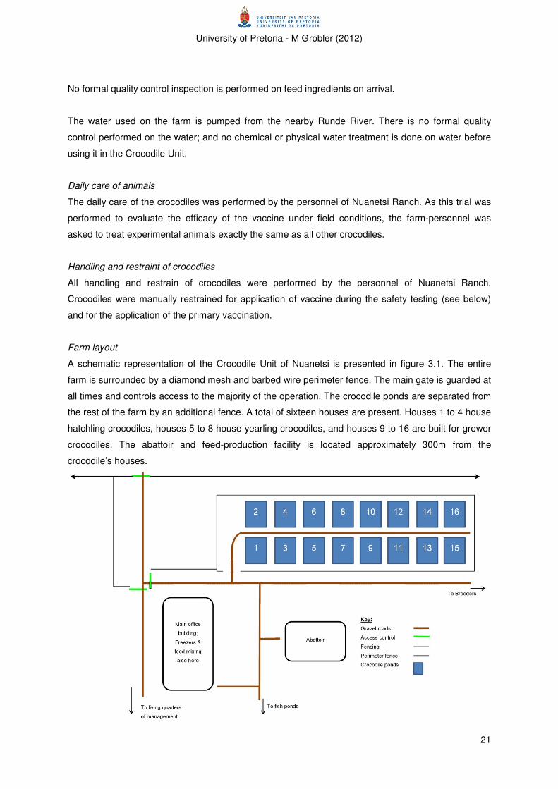
Farm layout
A schematic representation of the Crocodile Unit of Nuanetsi is presented in figure 3.1. The entire
farm is surrounded by a diamond mesh and barbed wire perimeter fence. The main gate is guarded at
all times and controls access to the majority of the operation. The crocodile ponds are separated from
the rest of the farm by an additional fence. A total of sixteen houses are present. Houses 1 to 4 house
hatchling crocodiles, houses 5 to 8 house yearling crocodiles, and houses 9 to 16 are built for grower
crocodiles. The abattoir and feed-production facility is located approximately 300m from the
crocodile’s houses.

University of Pretoria - M Grobler (2012)
22
Fig. 3.1 Layout of crocodile unit of Nuanetsi Ranch, Mwenezi, Zimbabwe
Cleaning and disinfection
In the yearling crocodile pens, every pen is cleaned daily; this includes the removal of excess feed
and faeces from the concrete surfaces and draining of the ponds.
In the grower crocodile pens, one of the three ponds is drained per day; the drained pond is then left
empty for the day – the reasoning behind this being that the ultraviolet light from the sun will supply a
sterilising effect on the pond. Practically this would equate into a single pond being empty for one day
and filled for two days in every three-day cycle.
The farm employs an all-in-all-out system for an entire pen between batches of crocodiles being
moved from the yearling to the grower pens, or from the hatchling to the yearling pens. After removal
of all crocodiles, the pens are cleaned from all organic material, washed with Chlor-clean (Guest
Medical Limited) and sprayed with Vircon® S (DuPont Chemical Solutions Enterprise). These
chemicals are registered disinfectants. The active ingredients present in Chlor-clean is troclose
sodium (decomposing to chlorine (Cl2), hypochlorous acid (HClO) and cyanuric acid ((HOCN)3) on
contact with moisture) and in Vircon® S potassium peroxomonosulfate (KHSO5 – an oxidising agent),
sodium dodecylbenzene sulphonare (C12H25C6H4SO3Na – a surfactant) and sulfamic acid (H3NSO3).
The pen is left empty for a minimum of 10 days after cleaning, before new crocodiles are moved in.
Due to circumstances, such as an increase in the numbers of crocodiles, it is not always practically to
follow this cleaning regime. The two grower pens in which the study crocodiles were kept were
however, subjected to this cleaning regime.
Biosecurity
Access control to the farm as well as to the crocodile pens is practised. Personnel and visitors are
expected to step into a footbath before entering crocodile pens. Dead crocodiles are removed from
pens on a daily basis. Pens are cleaned daily as described above. Separate cleaning equipment for
different pens are supplied but not necessarily used. Feed transport crates are shared between pens.
Natural vermin control by cats is practised. On a previous occasion yearling crocodiles had been
bought from other crocodile farms and co-mingled with Nuanetsi crocodiles without practicing
quarantine before introduction; this was followed by the first outbreak of suspected mycoplasmosis.
Vaccine production
Mycoplasma-strain included in experimental vaccine
The isolate used for the preparation of the vaccine was cultured from the joint fluid of a sick crocodile
(crocodile no. 2), which was euthanized during a visit to Nuanetsi Ranch during August 2010. It was
identified as M. crocodyli by means of growth inhibition of mono-specific antisera and an indirect
fluorescent antibody test.

University of Pretoria - M Grobler (2012)
23
Vaccine production and formulation
The isolate was cultured in a modified Hayflick’s broth (Hayflick 1965) and inactivated with 0.4%
formalin for 3 days at 37oC. The antigen titre was adjusted to a minimum of 108 cfu/ml. Aluminium
hydroxide (at a concentration of 33%) was added as adjuvant. The final product was bottled in 100ml
vaccine vials and supplied ready-for-use to the farmer.
Quality control of vaccine
After formulation, the vaccine was tested for sterility by DESIGN BIOLOGIX, Pretoria, South Africa.
Safety testing of the autogenous vaccine
To evaluate the safety of the autogenous vaccine, it was administered intraperitoneally to a subset of
twenty crocodiles one month before commencement of the vaccine immunogenicity phase. Of these
crocodiles, five were given a single dose of 2 ml of the vaccine, five received a double dose, and ten
were sham vaccinated controls and received 2ml sterile water intraperitoneally. The crocodiles were
permanently marked by removal of the scutes from the left side of the tail; for the single vaccine dose
a single scute was removed from the left side, for the double vaccine dose two scutes were removed
from the left side and for the control animals three scutes were removed from the left side.
Crocodiles were evaluated daily for signs of disease, mainly lethargy and anorexia, and recorded by
personnel from Nuanetsi Ranch.
Efficacy testing of the autogenous vaccine
To evaluate the efficacy of the experimental vaccine, two thousand two hundred yearling crocodiles
were vaccinated with the experimental vaccine. The other two thousand two hundred yearling
crocodiles were kept as unvaccinated control animals. A schematic representation of the trial is
provided in Figure 3.2 below.

University of Pretoria - M Grobler (2012)
24
Fig. 3.2: Schematic representation of trial
The experimental vaccine was administered intramuscularly in the deep muscles of the tail (either the
caudal femoral muscle (ventral to the transverse process of the vertebrae) or the m longissimus
caudalis (dorsal to the transverse process of the vertebrae) (Richardson et al. 2002, Huchzermeyer
2003). A dose of 1 ml vaccine per animal was administered with a 20G needle. The needle was
changed after every ten animals, or sooner if it became damaged.
Control group: 2200 crocodiles
Experimental group: 2200 crocodiles
Blood collection: 50
Safety testing: 20 crocodiles
Safety trial
Control group: 2200 crocodiles
Experimental group: 2200 crocodiles; First inoculation
First vaccination
Experimental group: 2200 crocodiles; Second inoculation
Control group: 2200 crocodiles
Blood collection: 50
Second vaccination
Control group: 2200 crocodiles
Experimental group: 2200 crocodiles
Blood collection: 50
Blood collection: 50
6 weeks post- vaccination
1100 Control
crocodiles
+
1100 Vaccinated
crocodiles
1100 Control
crocodiles
+
1100 Vaccinated
crocodiles
Pens 9 B and 11 B
Total: 4400 crocodiles House 6
Clinical monitoring for disease
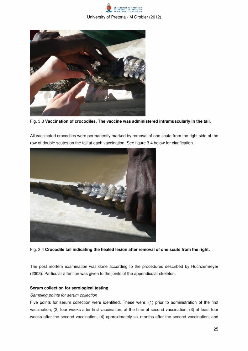
University of Pretoria - M Grobler (2012)
25
Fig. 3.3 Vaccination of crocodiles. The vaccine was administered intramuscularly in the tail.
All vaccinated crocodiles were permanently marked by removal of one scute from the right side of the
row of double scutes on the tail at each vaccination. See figure 3.4 below for clarification.
Fig. 3.4 Crocodile tail indicating the healed lesion after removal of one scute from the right.
The post mortem examination was done according to the procedures described by Huchzermeyer
(2003). Particular attention was given to the joints of the appendicular skeleton.
Serum collection for serological testing
Sampling points for serum collection
Five points for serum collection were identified. These were: (1) prior to administration of the first
vaccination, (2) four weeks after first vaccination, at the time of second vaccination, (3) at least four
weeks after the second vaccination, (4) approximately six months after the second vaccination, and

University of Pretoria - M Grobler (2012)
26
(5) during and/or after an outbreak of disease. Samples from the experimental group were taken at all
five points, while samples from the control group were only taken at points 3, 4 and 5. Approximately
fifty samples were collected from each group at sampling points 1, 2 and 3.
Sampling points 4 and 5 represents sampling during a disease outbreak. Samples at sample point 4
were collected from approximately 50 vaccinated animals with clinical disease. Samples at sample
point 5 were collected from approximately 50 vaccinated animals without clinical disease, 50
unvaccinated animals with clinical disease and 50 unvaccinated animals without clinical disease.
Procedure for serum collection
All samples were collected from electrically stunned crocodiles except for the samples collected pre-
vaccination which were collected from crocodiles restrained manually.
The samples were collected from the dorsal coccygeal vein, as previously described by
Huchzermeyer (2003). In short, the procedure entails identification of the correct area, careful
insertion of the hypodermic needle through the intervertebral ligament, and slow application of suction
to the syringe to draw the blood into the syringe. The sample size ranged from 5 to 10 ml of blood.
Samples were collected with a 20G to 21G 1.5” hypodermic needle, and a 5 or 10ml single-use
syringe. Each animal was sampled with a separate needle and syringe. The collected blood was
transferred to a serum tube (SG-vac), where the blood was allowed to clot.
Fig. 3.5 Collecting blood from a crocodile.
Procedure for serum processing and transport to laboratories
After removal of the serum from the blood clot by the personnel from the Wildlife Veterinary Unit
laboratory, Harare, Zimbabwe, the samples were stored at -18˚C until transport to the DVTD
bacteriology laboratory.

University of Pretoria - M Grobler (2012)
27
Latex slide agglutination test
Latex slide agglutination was performed as a screening test to detect sero-positive samples. Serial
two-fold dilutions were made of all sero-positive samples to determine the titre of the samples. The
reciprocal of the last dilution where agglutination could be observed, was taken as the titre.
Mycoplasma strains and growth conditions
The isolate (seed material) used for the preparation of the latex agglutination assay, was the same
strain used to prepare the experimental vaccine
A culture of the organism was prepared in modified Hayflick’s broth (Hayflick 1965) at 37˚C. The
culture was transferred every second day to fresh broth, with inocula of approximately 10%, at least
three times, in order to prepare a final volume of approximately 4 litres.
Coating of Microspheres
A similar procedure to that published by Senthilkumar et al. (2008) for canine leptospirosis was used
with slight modifications. The procedure is summarized below.
To prepare the culture for adsorption to the latex microspheres, the organism was inactivated by the
addition of 0.4% formalin and incubation at 37˚C for five days. Cells were concentrated by high speed
centrifugation, at 15000rpm at 4˚C for thirty minutes, washed twice in phosphate buffered saline (PBS
pH 7.2) and re-suspended as 10ml per 1 litre Mycoplasma culture. The suspension was dialysed
against PBS pH 7.2 for 24 hours at 4˚C, and re-suspended to 100ml per 1 litre Mycoplasma culture, in
PBS to which 0.1% sodium azide had been added.
A 10% suspension of latex beads (0.80 µm, SIGMA-ALDRICH) was added to the suspended
Mycoplasma culture at a ratio of 1 part latex beads to 9 parts Mycoplasma culture. The suspension
was gently stirred for 6 hours at 37˚C, after which it was centrifuged at 8000 rpm at 4˚C for three
minutes. The resultant pellet was re-suspended in PBS pH 7.2 with 0.1% sodium azide, at a dilution
of 10ml PBS pH 7.2 per 10ml latex-antigen suspension. The coated latex beads were stored at 4˚C
until use.
Procedure for latex slide agglutination
The methodology was adapted from Senthilkumar et al. (2008), and is similar to the LATs published
for other Mycoplasma spp. (Morton 1966, Slavik & Switzer 1979, March et al. 2000, March et al.
2003). In short, the LAT was performed by pipetting 20µl of serum and 20µl of antigen onto a glass
slide (approximately 22 samples were tested per run on a 150 x 210 mm glass slide, and positive and
negative controls were included on each run)(all reagents at room temperature). The reagents were
gently mixed and hand-rocked for approximately 2-3 minutes at room temperature, after which the
test was read. If fine granular clumps formed in the mixture, a positive reaction was recorded, and if
the suspension remained homogenously pale blue, a negative reaction was recorded. For all positive

University of Pretoria - M Grobler (2012)
28
sera, the procedure was repeated with serial two fold serum dilutions to determine antibody titre. The
titre of the serum was recorded as the reciprocal of the last dilution where a positive reaction could be
distinguished.
Growth/metabolism inhibition assay
The metabolism inhibition assay (MI) was performed (a) to determine the titre of the sera which tested
positive on LAT and (b) as a gold standard assay to evaluate the performance of the latex
agglutination assay. The method employed was similar to that originally described by Taylor-
Robinson et al. (1966), with some modification, and is briefly described below.
Mycoplasma strain and culture
For the MI test, the same strain as for the latex agglutination test which is described above was used.
It was grown in modified Hayflick’s medium (Hayflick 1965) at 37°C, and the culture was transferred to
fresh broth every second day, with inocula of approximately 10% each time. The final transferral was
performed just before performance of the MI test.
Mycoplasma medium
The organism was grown in modified Hayflick’s medium (Hayflick 1965) with the addition of phenol
red (phenolsulfonephthalein) (as pH indicator).
Procedure for MI test
The tests were performed on disposable, plastic 96-well microplates (NUNC 96F Untreated straight
with lid, Thermo Fisher Scientific). All procedures were performed in a laminar flow cabinet to
minimize contamination. Serial two-fold serum dilutions were prepared with Mycoplasma medium, to a
volume of 100µl. 100µl of the prepared Mycoplasma culture was then added to each well, to obtain a
final serum two-fold dilution series from 1:2 to 1:256. The microplates were sealed with adhesive, see-
through plastic and incubated at 37°C for 48-54 hours. The reactions were read when obvious colour
changes (pink to yellow) could be observed. A positive result was read when the colour remained pink
(i.e. metabolism of glucose was inhibited and the pH remained constant) while a negative result was
read if the colour changed from pink to yellow (i.e. metabolism of glucose and a decrease in pH
occurred). The titre of the serum was read as the reciprocal of the last dilution where glucose
metabolism was inhibited (i.e. no colour change could be observed). The remaining Mycoplasma
broth was also incubated as control to determine the sterility of the broth.
Statistical evaluation of results
Data was captured in Microsoft Excel 2010 spreadsheets and analyses were carried out with the aid
of MINITAB Statistical software (Release 13.32) and Microsoft Excel spreadsheets.
University of Pretoria - M Grobler (2012)
29
Evaluating sero-conversion
The strength of association between vaccination and serological status was evaluated. The
proportions of sero-positive crocodiles before vaccination, 4 weeks after primary vaccination and 6
weeks after booster vaccination were compared using a Pearson’s chi-square test.
A significance level of α = 0.05 was used for all comparisons
Evaluating the association between serological status, disease status and vaccination status during
disease outbreak
The strength of association between the ranked titre, disease status and vaccination status of
crocodiles at the time of the outbreak was compared using stepwise regression. Association was
assessed at a significance level of α = 0.05.
Evaluating the diagnostic performance of the latex agglutination test
The diagnostic performance of the latex agglutination test had not been evaluated previously for a
large sample size and it was therefore, calculated from the data generated in this study. As MI tests
were not performed for all sera on which LAT was performed, only the subset of sera on which both
tests had been performed for all 4 sampling points were used to calculate this data. The sensitivity,
specificity, positive and negative predictive values were calculated according to standard formulae
(Thrusfield 2005), and are provided below:
Table 3.1 2x2 contingency table for determining diagnostic performance
Gold standard positive Gold standard negative Total
Test positive a b a+b
Test negative c d c+d
Total a+c b+d a+b+c+d
• Sensitivity =
• Specificity =
• Positive predictive value =
• Negative predictive value =
• Accuracy =
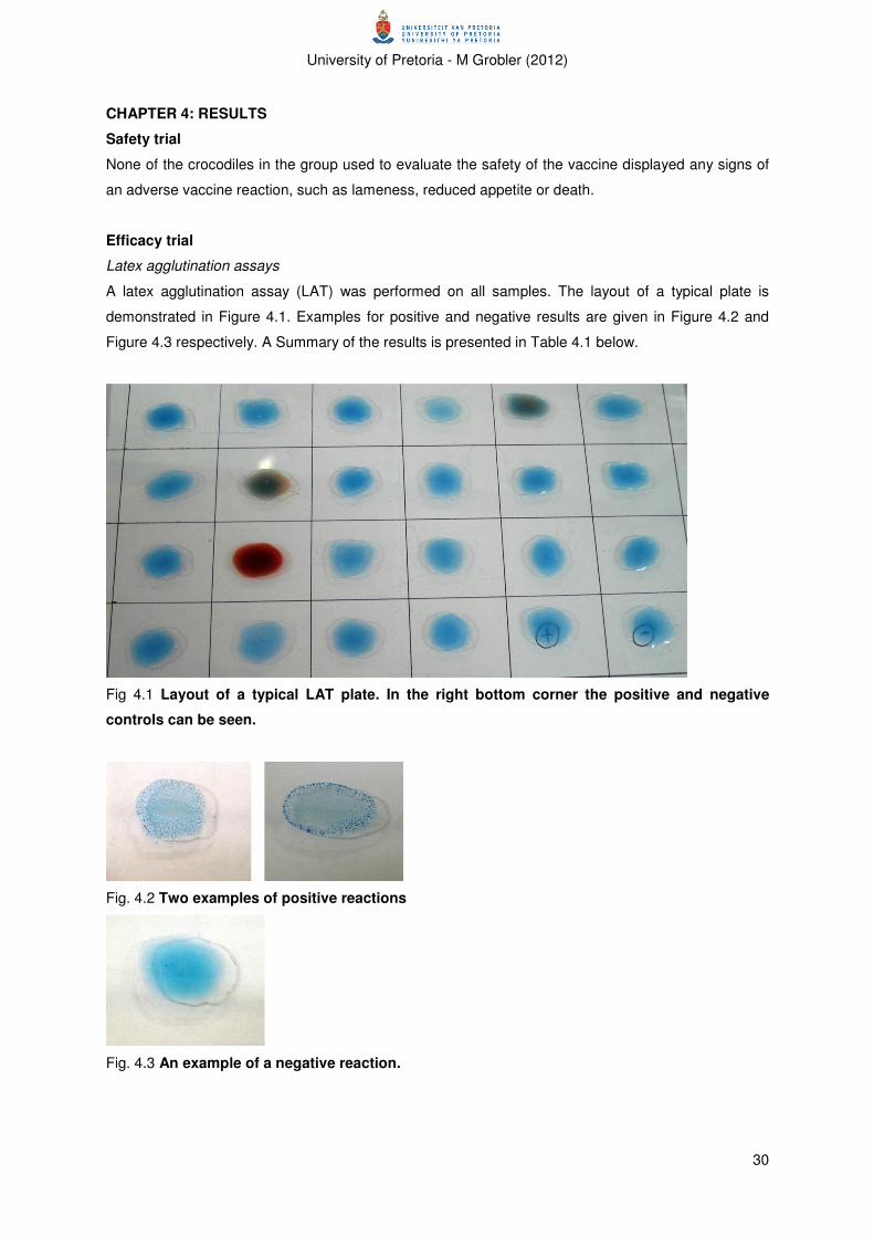
University of Pretoria - M Grobler (2012)
30
CHAPTER 4: RESULTS
Safety trial
None of the crocodiles in the group used to evaluate the safety of the vaccine displayed any signs of
an adverse vaccine reaction, such as lameness, reduced appetite or death.
Efficacy trial
Latex agglutination assays
A latex agglutination assay (LAT) was performed on all samples. The layout of a typical plate is
demonstrated in Figure 4.1. Examples for positive and negative results are given in Figure 4.2 and
Figure 4.3 respectively. A Summary of the results is presented in Table 4.1 below.
Fig 4.1 Layout of a typical LAT plate. In the right bottom corner the positive and negative
controls can be seen.
Fig. 4.2 Two examples of positive reactions
Fig. 4.3 An example of a negative reaction.

University of Pretoria - M Grobler (2012)
31
Table 4.1 Summary of LAT results
Pre-
vaccination
4 weeks
post
primary
vaccination
6 weeks
post
booster
vaccination
6 weeks
post booster
vaccination
Vaccination status of animals N/A Vaccinated Vaccinated Unvaccinated
Number of seropositive animals 4 24 12 9
Number of seronegative animals 67 26 38 40
Total number of animals 71 50 50 49
% Seropositive animals: 5.63% 48.00% 24.00% 20.41%
95% Confidence interval for seropositive
animals
1.56-
13.80%
33.66 -
62.58%
13.06-
38.17% 8.76-32.02%
From this data it can be seen that:
• almost 6% of the tested animals were seropositive before the vaccine was administered;
• 48% of vaccinated animals were seropositive 4 weeks after the first vaccination;
• 24% of vaccinated animals were seropositive 6 weeks after the booster vaccination;
• 20% of unvaccinated animals were seropositive 6 weeks after the booster vaccination.
Growth/metabolic inhibition assays
A summary of the results of the metabolic inhibition assays are presented in Table 4.2 below.
Table 4.2 Summary of MI test results
Pre-
vaccination
4 weeks
post
primary
vaccination
6 weeks post booster
vaccination
Total number of animals 50 23 17 10
Vaccination status of animals Unvaccinated Vaccinated Unvaccinated Vaccinated
Number of seropositive animals 5 0 14 8
% Seropositive animals: 10.00% 0.00% 82.35% 80.00%
In summary this data shows that:
• 10% of crocodiles were seropositive with the MI test before vaccination, although the titres
were low;
• None of the vaccinated crocodiles were seropositive with the MI test 4 weeks after the first
vaccination;
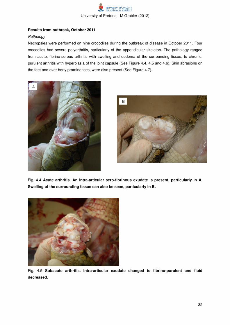
University of Pretoria - M Grobler (2012)
32
Results from outbreak, October 2011
Pathology
Necropsies were performed on nine crocodiles during the outbreak of disease in October 2011. Four
crocodiles had severe polyarthritis, particularly of the appendicular skeleton. The pathology ranged
from acute, fibrino-serous arthritis with swelling and oedema of the surrounding tissue, to chronic,
purulent arthritis with hyperplasia of the joint capsule (See Figure 4.4, 4.5 and 4.6). Skin abrasions on
the feet and over bony prominences, were also present (See Figure 4.7).
Fig. 4.4 Acute arthritis. An intra-articular sero-fibrinous exudate is present, particularly in A.
Swelling of the surrounding tissue can also be seen, particularly in B.
Fig. 4.5 Subacute arthritis. Intra-articular exudate changed to fibrino-purulent and fluid
decreased.
A
B

University of Pretoria - M Grobler (2012)
33
Fig. 4.6 Chronic arthritis. Purulent intra-articular exudate is present.
Fig. 4.7 Typical skin lesions on the feet (A) and over the sternum (B).
Serology
The results of LAT and MI assays on serum collected during the disease outbreak are summarized in
Table 4.3 and 4.4 below.
Table 4.3 Summary of Outbreak results-LAT
Vaccination status of animals Vaccinated Unvaccinated Vaccinated Unvaccinated
Disease status of animals Diseased Diseased Healthy Healthy
Total number of animals 41 48 50 53
Number of seropositive animals 29 41 42 40
% Seropositive animals: 70.73% 85.42% 84.00% 75.47%
95% Confidence interval for seropositive
animals
54.46-
83.87% 72.24-93.93%
70.88-
92.83% 61.72-86.24%
% Seronegative animals: 29.27% 14.58% 16.00% 24.53%
A B
A B
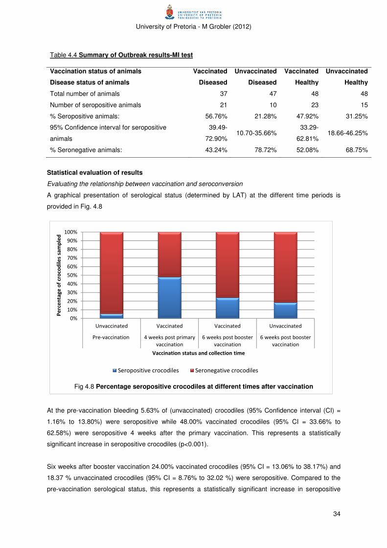
University of Pretoria - M Grobler (2012)
34
Table 4.4 Summary of Outbreak results-MI test
Vaccination status of animals Vaccinated Unvaccinated Vaccinated Unvaccinated
Disease status of animals Diseased Diseased Healthy Healthy
Total number of animals 37 47 48 48
Number of seropositive animals 21 10 23 15
% Seropositive animals: 56.76% 21.28% 47.92% 31.25%
95% Confidence interval for seropositive
animals
39.49-
72.90% 10.70-35.66%
33.29-
62.81% 18.66-46.25%
% Seronegative animals: 43.24% 78.72% 52.08% 68.75%
Statistical evaluation of results
Evaluating the relationship between vaccination and seroconversion
A graphical presentation of serological status (determined by LAT) at the different time periods is
provided in Fig. 4.8
At the pre-vaccination bleeding 5.63% of (unvaccinated) crocodiles (95% Confidence interval (CI) =
1.16% to 13.80%) were seropositive while 48.00% vaccinated crocodiles (95% CI = 33.66% to
62.58%) were seropositive 4 weeks after the primary vaccination. This represents a statistically
significant increase in seropositive crocodiles (p<0.001).
Six weeks after booster vaccination 24.00% vaccinated crocodiles (95% CI = 13.06% to 38.17%) and
18.37 % unvaccinated crocodiles (95% CI = 8.76% to 32.02 %) were seropositive. Compared to the
pre-vaccination serological status, this represents a statistically significant increase in seropositive
0%
10%
20%
30%
40%
50%
60%
70%
80%
90%
100%
Unvaccinated Vaccinated Vaccinated Unvaccinated
Pre-vaccination 4 weeks post primary
vaccination
6 weeks post booster
vaccination
6 weeks post booster
vaccination
Pe
rce
nta
ge
of
cro
cod
ile
s sa
mp
led
Vaccination status and collection time
Seropositive crocodiles Seronegative crocodiles
Fig 4.8 Percentage seropositive crocodiles at different times after vaccination

University of Pretoria - M Grobler (2012)
35
crocodiles for both groups (p<0.05). However, when comparing the vaccinated and unvaccinated
groups with each other, there is no statistically significant difference in seropositive animals (p>0.05).
Furthermore, when comparing the proportion of seropositive vaccinated crocodiles 6 weeks after the
booster vaccination there is a statistically significant decrease compared to the proportion of
seropositive vaccinated crocodiles 4 weeks after the primary vaccination (p<0.05).
Evaluating the relationship between vaccination status, serological status and development of clinical
disease during a disease outbreak
A graphical presentation of the percentage of crocodiles represented in each serum dilution tested
with the LAT and MI assay is presented in figures 4.9 and 4.10 respectively.
From the graph in Fig. 4.9 it can be said that there is no clear pattern in the titre of sampled crocodile
groups, regardless of the vaccination and/or disease state. This was confirmed by performing a
stepwise regression to assess the dependence of the ranked LAT titre to disease status, vaccination
status and/or the interaction between these variables (p>0.05, r2<5).
0.00%
5.00%
10.00%
15.00%
20.00%
25.00%
30.00%
35.00%
40.00%
<2 2 4 8 16 32 64 128 256
Pe
rce
nta
ge
of
cro
co
dil
es
pe
r g
rou
p
eva
lua
ted
LAT Titre
Sick Vaccinated Sick Unvaccinated Not sick Vaccinated Not sick Unvaccinated
Fig 4.9 Percentage crocodiles in each LAT titre range
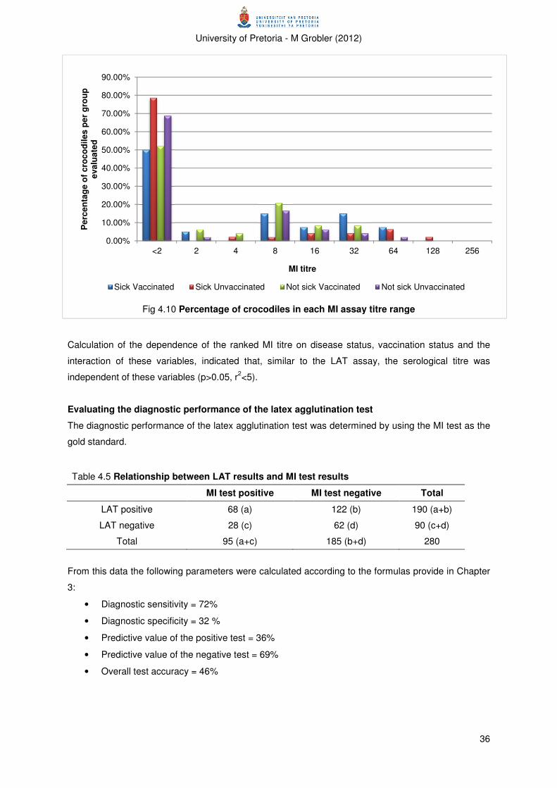
University of Pretoria - M Grobler (2012)
36
Calculation of the dependence of the ranked MI titre on disease status, vaccination status and the
interaction of these variables, indicated that, similar to the LAT assay, the serological titre was
independent of these variables (p>0.05, r2<5).
Evaluating the diagnostic performance of the latex agglutination test
The diagnostic performance of the latex agglutination test was determined by using the MI test as the
gold standard.
Table 4.5 Relationship between LAT results and MI test results
MI test positive MI test negative Total
LAT positive 68 (a) 122 (b) 190 (a+b)
LAT negative 28 (c) 62 (d) 90 (c+d)
Total 95 (a+c) 185 (b+d) 280
From this data the following parameters were calculated according to the formulas provide in Chapter
3:
• Diagnostic sensitivity = 72%
• Diagnostic specificity = 32 %
• Predictive value of the positive test = 36%
• Predictive value of the negative test = 69%
• Overall test accuracy = 46%
0.00%
10.00%
20.00%
30.00%
40.00%
50.00%
60.00%
70.00%
80.00%
90.00%
<2 2 4 8 16 32 64 128 256
Pe
rce
nta
ge
of
cro
co
dil
es
pe
r g
rou
p
eva
lua
ted
MI titre
Sick Vaccinated Sick Unvaccinated Not sick Vaccinated Not sick Unvaccinated
Fig 4.10 Percentage of crocodiles in each MI assay titre range

University of Pretoria - M Grobler (2012)
37
CHAPTER 5: DISCUSSION
The aim of this study was to determine whether protective immunity could be stimulated in farmed
Nile crocodiles by the administration of an inactivated Mycoplasma crocodyli vaccine. The in vivo
safety and immunogenicity as well as the efficacy in the face of a natural outbreak were evaluated.
Vaccine safety
The clinical data indicated that the vaccine did not cause any systemic adverse reactions and no
increase in mortalities was reported for crocodiles in the study group. It is therefore concluded that the
vaccine was safe for use in crocodiles.
To evaluate the safety of the vaccine, it was decided to inoculate the crocodiles intraperitoneally
rather than intra-muscularly. The reasons for this are as follows: Firstly, the serosal surface of the
peritoneum should be more sensitive to the presence of an irritant substance, and therefore more
likely to result in notable adverse vaccine reactions, than the muscle. Secondly, if any live infectious
Mycoplasma organisms were present in the vaccine, clinical disease was more likely to result when
given intraperitoneally, as this was the most successful method of reproducing clinical mycoplasmosis
(Mohan et al.1995).
Pathological and histopathological evaluation of the injection sites were not performed because the
study subjects were part of the stock of a commercial crocodile farm and not available for sacrifice.
When evaluating the safety of such a vaccine for commercial production, safety assessment by
pathological and histopathological examination of the injection site as well as pathological
examination of animals to evaluate systemic effect of vaccination, is recommended (EMEA 2001).
Vaccine efficacy
Immunogenicity
The first step in evaluating the efficacy of the vaccine was to determine if the vaccine could induce a
humoral immune response. Several conclusions can be drawn from the serological data, presented in
tables 4.1 and 4.2. Firstly, both tests (LAT and MI test) detected a small percentage of crocodiles
sero-positive before the onset of vaccination, although no individual animal was positive on both
assays. Secondly, there was a significant increase in the proportion of LAT seropositive crocodiles
four weeks after primary vaccination but none of the crocodiles that were positive on LAT, at this time
point, were also positive with the MI test. Furthermore, there was also an increase in LAT seropositive
crocodiles when comparing pre-vaccination samples to samples collected six weeks after the booster
vaccination. However, there was actually a significant decrease in the proportion of LAT seropositive
crocodiles since the previous collection (four weeks after primary vaccination). In contrast, the
proportion of LAT seropositive crocodiles that also tested positive with the MI assay increased after
booster vaccination compared to either of the previous samplings.

University of Pretoria - M Grobler (2012)
38
Unfortunately, interpreting these results is obscured by the fact that only the pre-vaccination samples
were evaluated with both assays. As set out by the study protocol, only LAT positive sera were meant
to be evaluated with the MI test, which was the procedure followed for the samples collected after
primary and booster vaccinations. A second serious constraint was that, although vaccinated
crocodiles were explicitly marked and distinguishable from unvaccinated crocodiles, individuals could
not be discerned, resulting in the inability to trace the serological and/or clinical response of individual
crocodiles. Despite these constraints, it was felt that some hypotheses for the mentioned trends can
be formulated, and these are discussed below.
The presence of seropositive samples before onset of the vaccination trial and in unvaccinated
crocodiles during the trial is of interest. There are a few possible explanations for this finding. Firstly, it
could indicate that the organism was present and circulating through the population during the study,
and, therefore, that the organism is persistently present on the farm. This epidemiological situation
seems feasible if one considers the information available for poultry and pig mycoplasmosis on multi-
age production units (Desrosier 2001, Nicholas 2004, Ley 2006). If this were the case, our knowledge
of the management of mycoplasmosis in the mentioned species would indicate that the use of an
inactivated vaccine in such a situation could be beneficial to reduce losses but is unlikely to eliminate
the problems (Desrosiers 2001, Ley 2006).
A second probable explanation for positive pre-vaccination samples is non-specific cross-reaction of
serological assays. Non-specific reactions are well-described for the rapid serum plate agglutination
assay used for M. gallisepticum (Ahmad et al. 1988, Avakian & Kleven 1990, Ross et al. 1990, Ben
Abdelmoumen & Roy 1995). It would be ideal to evaluate the analytical specificity of a newly
developed assay to quantify the occurrence of false positives due to cross-reactions. However,
because a wide variety of, seemingly, unrelated antigens could be involved, as clearly demonstrated
for the M. gallisepticum assay, determining analytical specificity is often only a theoretical concept.
Running serological assays in series, is one of the methods employed to improve the specificity of a
test.
The role of natural antibodies in pre-vaccination seropositive assays also requires clarification. As
mentioned in chapter 2, it has been reported that natural antibodies to Mycoplasmas occur in tortoises
and that these could play a complicating role in tortoise Mycoplasma serology (Hunter et al. 2008,
Sandmeier et al. 2009). A similar situation in crocodiles should be considered, particularly because
this could result in the erroneous diagnosis of a Mycoplasma-infected animal/group/farm, while the
specific individual could simply have detectable “innate” immunity to Mycoplasma-like antigens.
These uncertainties make interpretation of the rest of the data-trends even more complicated. It was
decided that, in order to draw conclusions, regard all seropositive cases will be viewed as having a
true immune response to the administered antigen.
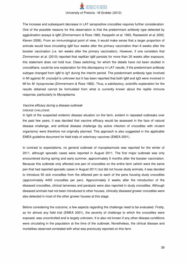
University of Pretoria - M Grobler (2012)
39
The increase and subsequent decrease in LAT seropositive crocodiles requires further consideration.
One of the possible reasons for this observation is that the predominant antibody type detected by
agglutination assays is IgM (Zimmermann & Ross 1982, Karppelin et al. 1993, Rastawicki et al. 2002,
Kleven 2006). From an immunological point of view, it would make sense that a larger proportion of
animals would have circulating IgM four weeks after the primary vaccination than 6 weeks after the
booster vaccination (i.e. ten weeks after the primary vaccination). However, if one considers that
Zimmerman et al. (2010) reported that reptilian IgM persists for more than 20 weeks after exposure,
this statement does not hold true. Class switching, for which the details have not been studied in
crocodilians, could be one explanation for this discrepancy in LAT results, if the predominant antibody
subtype changed from IgM to IgY during the interim period. The predominant antibody type involved
in MI against M. crocodyli is unknown but it has been reported that both IgM and IgG were involved in
MI for M. hyosynoviae (Zimmermann & Ross 1982). Thus, a satisfactory, scientific explanation for the
results obtained cannot be formulated from what is currently known about the reptile immune
response, particularly to Mycoplasma.
Vaccine efficacy during a disease outbreak
DISEASE CHALLENGE
In light of the suspected endemic disease situation on the farm, evident in repeated outbreaks over
the past few years, it was decided that vaccine efficacy would be assessed in the face of natural
disease challenge, and artificial disease challenge (by active infection of crocodiles with virulent
organisms) were therefore not originally planned. This approach is also suggested in the applicable
EMEA guideline document for field trials of veterinary vaccines (EMEA 2001).
In contrast to expectations, no general outbreak of mycoplasmosis was reported for the winter of
2011, although sporadic cases were reported in August 2011. The first major outbreak was only
encountered during spring and early summer, approximately 5 months after the booster vaccination.
Because this outbreak only affected one pen of crocodiles on the entire farm (which were the same
pen that had reported sporadic cases in August 2011) but did not house study animals, it was decided
to introduce 50 sick crocodiles from the affected pen to each of the pens housing study crocodiles
(approximately 4400 crocodiles per pen). Approximately 2 weeks after the introduction of the
diseased crocodiles, clinical lameness and paralysis were also reported in study crocodiles. Although
diseased animals had not been introduced to other houses, clinically diseased grower crocodiles were
also detected in most of the other grower houses at this stage.
Before considering the outcome, a few aspects regarding the challenge need to be evaluated. Firstly,
as for almost any field trial (EMEA 2001), the severity of challenge to which the crocodiles were
exposed, was uncontrolled and is largely unknown. It is also not known if any other disease conditions
were circulating in the population at the time of the outbreak. Nonetheless, the clinical disease and
mortalities observed correlated with what was previously reported on this farm.
University of Pretoria - M Grobler (2012)
40
Secondly, the number of diseased crocodiles introduced into each pen was chosen arbitrarily and was
mainly based on what was practical to perform. The main motivation behind this was the lack of
information to make use of a more sophisticated method, as very little is known about the
epidemiology of crocodile mycoplasmosis and the basic reproductive number (R0) is therefore
unknown.
A third complication related to disease introduction and study design, was the co-mingling of
vaccinated and unvaccinated crocodiles. It has been stated that in a population consisting of both
vaccinated and unvaccinated animals, the unvaccinated group amplifies the pathogen until an
infectious pathogen load is reached which overwhelms the pre-induced immunity in the vaccinated
group (Dohoo & Montgomery 1996, Maunsell et al. 2009). This was identified as a potential problem
before the onset of the trial and it was suggested that close monitoring of the study crocodiles is
performed in order to determine if a difference in the susceptibility to disease and the outcome of
disease related to vaccination could be discerned. Unfortunately, clinical monitoring of study animals
turned out to be more difficult than anticipated (see below).
Furthermore, the timing of this outbreak is of significance. In contrast to outbreaks in previous years,
which usually occurred during cooler winter months (May to July/August), this outbreak occurred
during hot weather. It was also not linked to the movement of crocodiles from yearling houses to
grower houses, which was identified as a possible stressful event precipitating previous outbreaks of
mycoplasmosis. The reason/s for these differences is unknown. Despite the uncertainties in the
serological data (described above), it could theoretically be possible that vaccination stimulated
partial, though short-term immunity and protected the group of grower crocodiles for a limited period.
However, considering the complete lack of disease in the entire population, it seems more likely that
disease challenge was either absent or that the correct combination of predisposing factors were
absent.
Finally, the described time from introduction of infected crocodiles to disease in susceptible crocodiles
correlates well with the described incubation time for swine and poultry mycoplasmosis (Desrosiers
2001, Ley 2006).
CLINICAL AND PATHOLOGICAL MONITORING
Because the study was performed on a Zimbabwean crocodile farm where the disease is endemic, it
was not possible for the principal investigator to be present on the farm for the duration of the
outbreak. Hence clinical observation, including the number of diseased animals, number of deaths,
response to treatment and environmental monitoring, were left to the ranch manager. Due to the size
of the operation and the magnitude of managerial tasks, the manager was unfortunately unable to
complete the predetermined records.

University of Pretoria - M Grobler (2012)
41
During the outbreak, all clinically diseased crocodiles (according to the ranch managers’ and workers’
discretion, which usually entailed complete paralysis) from the entire operation were moved to the
originally infected pen, in order to control the disease challenge to the rest of the operation.
Unfortunately this resulted in over 4000 crocodiles being housed in this pen at the time of the principal
investigator’s visit to the farm, which made it impossible to identify and count diseased, vaccinated
crocodiles. The determination of the extent of the outbreak in diseased crocodiles was further
complicated by the slaughter of diseased crocodiles, already in progress at the time of the principal
investigator’s visit to the farm. Therefore, it was not possible to determine how many vaccinated
crocodiles had already been slaughtered.
All the pens housing grower crocodiles were significantly overstocked at the time of the outbreak
(building of additional pens was already in progress). The farm had also experienced very hot weather
during the period preceding the outbreak. Both these factors could have significantly stressed the
crocodiles, and increased their susceptibility to disease (Huchzermeyer 2003).
Logistics prevented performance of pathological examinations on study crocodiles throughout the trial
period. However, necropsies were performed on diseased crocodiles during the time of the major
disease outbreak in October 2011. A subset of diseased crocodiles and mortalities were examined,
the number limited by time constrains. Five clinical cases (all severely affected and completely lame)
and four mortalities (from the previous night) were examined. As stated, only four of these animals,
two of the clinical cases and two of the mortalities, displayed clear signs of polyarthritis. Of the
remaining crocodiles, the two remaining mortalities were too autolysed to make a diagnosis of cause
of death and the three clinical cases displayed non-specific signs of disease, including generalized
congestion which could indicate septicaemia.
Although the clinical signs and pathology were consistent with that expected for crocodile
mycoplasmosis, culture was not performed to confirm the aetiology of this outbreak.
Despite all the mentioned technical difficulties, the managers of the crocodile farm felt that the vaccine
did not provide protection against the disease in the face of an outbreak. The overall picture from the
serological data, correlated with this conclusion.
SEROLOGICAL MONITORING
A significant part of the evaluation of vaccine efficacy was based on serological monitoring, because
of the mentioned difficulties encountered with clinical and pathological monitoring. However, these are
not without limitations, which will be discussed in this section.
The serological results of samples collected during the outbreak are presented in Tables 4.3 and 4.4,
and Figures 4.9 and 4.10. As indicated, no correlation was found between vaccination status, disease
status and serological status. Some explanations for these disappointing results, which indicate failure
University of Pretoria - M Grobler (2012)
42
of the vaccine, will be discussed. (As the same serological assays were employed, the limitations
mentioned under “Immunogenicity” and “Performance of the latex agglutination assay” need to be
considered but will not be repeated.)
The presence of antigen variation is the first and, probably, most important reason to consider for the
failure of this Mycoplasma vaccine. As discussed in chapter 2, it is well recognized that many (if not
all) pathogenic Mycoplasma species make use of variable immuno-dominant surface proteins and this
has been presented as one of the primary reasons for the persistence of Mycoplasmas in the face of
a specific host immune response (Bercina et al. 1994, Levisohn & Kleven 2000, Citti et al. 2010).
Therefore, if the host is unable to eliminate the organism or to develop lasting immunity after a natural
infection, it seems unlikely that an artificial method of immune-protection, mimicking natural infection,
would provide complete, long-term protection (Razin 2006).
Inactivated vaccines face two additional limitations related to antigen variation. Firstly, it is likely that
the surface antigens present in inactivated vaccines are fixed as the organism is grown and
inactivated in vitro. Therefore, because the host immune system has only been primed with limited
surface protein variation, it is even more likely that the invading Mycoplasma could simply express a
different surface antigen and circumvent the pre-existing immune-protection. Secondly, it is unknown
if the surface antigens expressed in vitro are representative of that expressed in vivo. The possibility
of different proteins expressed in different milieu (which is well described for poultry mycoplasmosis)
requires further investigation.
In addition to the above-mentioned disadvantages of inactivated vaccines, the composition of the
vaccine also needs to be considered when apparent vaccine failure is reported. Inactivated vaccines
are composed of two major components, namely the antigen and the adjuvant. There are various
problems which could be encountered with the antigen, but, high antigen yield and good antigenicity
is “the most important characteristics” (OIE 2008a). The general recommendation for poultry
mycoplasmosis and CBPP is a titre above 108 CFU/ml (OIE 2008a, OIE 2008b). Although the titre of
the antigen prepared for this vaccine was above this level (109 CFU/ml), the inherent immunogenicity
of the M. crocodyli is unknown, and it is therefore difficult to extrapolate. Investigation of the
inactivation process may be warranted as denaturation of proteins at the high levels of formalin and
prolonged incubation period could have influenced the immunogenicity of the vaccine.
The role of the aluminium hydroxide adjuvant used in the experimental vaccine should also be
considered. Aluminium salts have a long history of inclusion as vaccine adjuvants; in 1926 aluminium
potassium sulphate was the first recorded vaccine adjuvant (Garçon et al. 2011). Since then
aluminium salts, particularly aluminium hydroxide and aluminium phosphate, commonly called alums,
have become the most commonly used vaccine adjuvants in both human and veterinary vaccines
(Gupta 1998, Bowersock & Martin 1999, Aguilar & Rodríguez 2007). The success of alum adjuvants
are primarily linked to their safety record (Bowersock & Martin 1999, Singh & O’Hagan 2003, Reed et
University of Pretoria - M Grobler (2012)
43
al. 2008, Garçon et al. 2011). Other advantages are the low cost (Bowersock & Martin 1999) and
simple formulation, making it suitable for large scale production (Reed et al. 2008). However, it is also
well known that alum does not stimulate cell-mediated immune responses, specifically Type I helper
T-lymphocytes and Cytotoxic T-lymphocytes (Bowersock & Martin 1999, Aguilar & Rodríguez 2007,
Reed et al. 2008, Garçon et al. 2011). This response is of critical importance in host-protection
against intracellular pathogens, such as viruses. Other problems with alums include granulomas
reported at the injection site (Reed et al. 2008) particularly if the vaccine is administered intradermally
or subcutaneously (Aguilar & Rodríguez 2007) and loss of potency when vaccines are inadvertently
frozen (Reed et al. 2008). Although good results are reported for inactivated M. hyopneumoniae
(swine enzootic pneumonia) in alum adjuvant, results with human and poultry inactivated alum-
adjuvanted Mycoplasma vaccines are quite disappointing (Linchevski et al. 2009, and better results
are reported in poultry where mineral-oil adjuvants are used (Panigraphy et al. 1981). It may be useful
to consider alternative vaccine adjuvants in future.
Given the mentioned restrictions and possible flaws of inactivated Mycoplasma vaccines, it seems
questionable how any of these could have beneficial results, even though they are extensively used
for certain diseases. It has to be considered that (as indicated in chapter 2) improved production and
a reduction in the severity of clinical disease, rather than prevention of clinical disease in the face of
virulent pathogen challenge, is the main advantage reported for most of the successful inactivated
vaccines (Razin 2006). These advantages are linked, in general, to the presence of systemic
immunity to Mycoplasma, which is more readily stimulated by administration of parenteral vaccines, in
contrast to the local response, required to protect against host colonization etc., stimulated by
administration of live vaccines to mucosal surfaces (Razin 2006). Therefore, it should be emphasized
that, based on our current knowledge, a combination of live and inactivated vaccines are required to
counteract all the effects of Mycoplasma infection.
Apart from problems inherent to the vaccine itself, one has to consider the method of vaccine
evaluation. A major pitfall for the use of serology to monitor the level of protection, particularly for
mycoplasmosis, is because it has been well documented that, at least in poultry, the levels of
circulating antibody to Mycoplasma does not necessarily correlate with host protection (Lam & Lin
1984, Lin & Kleven 1984, Talkington & Kleven 1985, Whithear et al. 1990). It is possible that this
discrepancy is due to the importance of cell-mediated immunity and/or also linked with surface
antigen variation. Nonetheless, circulating antibody does play a role in resistance, with faster
clearance of the organism and less severe tracheal lesions associated with the presence of antibody
(Yagihashi & Tajima 1986, Elfaki et al. 1992, Yagihashi et al. 1992, Avakian & Ley 1993), and it is
more practical to evaluate in vitro. Therefore, the use of serological assays are warranted, but these
limitations need to be considered.

University of Pretoria - M Grobler (2012)
44
In conclusion, it can be stated that the performance of the vaccine in the face of a virulent disease
outbreak was disappointing but there are several characteristics of the organism and disease in
question, which could be responsible for the lack in protection.
Performance of the latex agglutination assay
The four performance characteristics evaluated were the sensitivity, specificity, and positive and
negative predictive value of the latex agglutination test. The results are summarized in chapter 3.
Unfortunately, the measured characteristics revealed disappointingly poor performance of the LAT,
which need to be further analysed before conclusions and recommendations can be made.
Before analysing the performance of an assay, it is critical to reconsider the intended usage of the
assay (Greenhalgh 1997, Banoo et al. 2010, OIE 2010). In this case, the LAT was initially developed
as pen-side screening assay to be used by farmers/veterinarians in rural areas to confirm a diagnosis
of Mycoplasma-associated arthritis. The ease and simplicity make it ideal for a pen-side assay but the
poor performance characteristics require attention before it can be recommended.
The performance characteristics of a new assay are often compared to the performance of existing
assays. Two other serological assays have been developed for crocodile mycoplasmosis, namely an
indirect ELISA (Dawo & Mohan 2007) and a Western blot assay (Dawo and Mohan 2008), and both
reported moderately-high sensitivity (above 80%) and very high specificity (100%). The LAT performs
poorly in comparison to these assays. However, it has to be stated that the performance of these
assays were evaluated with very small sample sizes and the “gold standard” in these cases were the
presence or absence of clinical disease, which is not ideal.
Several technical difficulties need to be acknowledged. The evaluation of the performance of the latex
agglutination assay was not included in the initial study plan, and resulted because the absence
thereof was recognized as a constraint for interpretation of the serological results of the vaccination
trial. Hence, important recommended elements were absent including identification of a suitable study
population and study subjects, determination of the ideal sample size and reference test or tests
(Banoo et al. 2010).
Indeed, the performance of the metabolic inhibition assay, used as the gold standard, has not been
evaluated. Because it is prescribed as serological assay to differentiate Mycoplasma species, it is
accepted that this assay is highly specific (Black 1973, Whitcom et al. 1995, Brown et al. 2007).
However, the sensitivity of this type of assay has been questioned (Lin & Kass 1974). This was,
however, the only other assay available.
Despite the mentioned problems and their potential complicating effects, the poor assay performance
requires attention. Although poor specificity is not uncommon for a Mycoplasma agglutination assay,
University of Pretoria - M Grobler (2012)
45
the reported sensitivity is unusually poor – a sensitivity of 100% is reported for some of the serum
plate agglutination assays used for M. gallisepticum in poultry (Ahmad et al. 1988).
Firstly, because of the uncertainties existing in our current understanding of reptile immunology, non-
specific reactions due to natural antibodies or other unknown innate factors have to be considered
(see immunogenicity above for further clarification). It will only be possible to reduce such
confounding factors when they have been identified.
Secondly, the mentioned surface antigen variation has to be considered. Many assays, including
agglutination assays and ELISA’s, make use of pre-expressed antigens fixed on a solid phase.
Therefore, if the host antibody response that is measured is predominantly aimed against a different
antigen phenotype than used in the assay, it is very likely that false negative results could be
reported. There is a distinct possibility that this complication could have an influence on the
serological results during the disease outbreak (although it would not affect the serological results
evaluated in the immunogenicity stage as the vaccine and the assay was prepared form the same
isolate and culture conditions) as the identity of the strain/s causing the October 2011 outbreak, and
there similarity to the strain used for preparation of the assays, is unknown.
In addition to these problems with the antigen, the possibility of severe denaturation also needs to be
considered because of the, relatively, harsh inactivation conditions used in preparation of the antigen.
Although the need thereof, from a biosafety point of view, can be appreciated, the possibility of the
significant structural derangements needs to be considered, which could also contribute to the poor
sensitivity recorded.
Another possible reason for the poor performance of the LAT is that optimization of assay conditions
has not been performed. In particular, the optimal concentration of antigen and dilution of antiserum
has not been determined. Although not commonly specified, it has been stated that a pro-zone effect,
similar to that described for ELISA’s, can occur in agglutination assays (Stanley 2002). In this zone,
optimal agglutination would be inhibited by an overabundance of antibody. Dilution of the serum
(antibody) would result in an increase in visible agglutination, and improvement of the tests’
performance.
In conclusion, although the latex agglutination assay was less complex and time consuming to
perform and did not require any advanced equipment, the poor performance reported in this trial
indicates that further sophistication of the assay is warranted before it can be recommended for
routine use.

University of Pretoria - M Grobler (2012)
46
CHAPTER 6: CONCLUSION
The serological analysis and outcome of the disease challenge indicated that the vaccine did not
stimulate protective immunity. Evaluation of vaccine efficacy was however, hampered by a multitude
of complications and limitations. Various reasons for the poor performance of the vaccine should be
considered. These include aspects of Mycoplasma pathogenicity, including surface antigen variation
and the interaction between the pathogen and the host immune system, exposure of animals to the
pathogen prior to vaccination, vaccine formulation and overwhelming infection. The parameters used
to evaluate vaccine efficacy also need consideration as various constraints have been described for
Mycoplasma sero-monitoring and, from reports on mycoplasmosis in other species, inactivated
vaccines are more reliable in reducing disease severity than to prevent disease. The predisposing
factors surrounding the outbreak of mycoplasmosis also need investigation, as these could play an
important role in breakdown of immune-protection.
The latex agglutination assay performed poorly in comparison to other serological assays for
crocodile Mycoplasma. Various reasons for the poor performance can be suggested, including the
unique host and our lack of knowledge on its immune system, lack of assay optimization and the
inherent constraints of the assay. It is suggested that this assay should not be used as diagnostic
assay without confirming the results with another assay, such as culture.
The trial emphasized the need for further research in a variety of areas. Firstly, the knowledge of
reptile immunology, and specifically crocodile immunology, is deficient in comparison to mammalian
and even avian immunology. This makes the development and evaluation of applied procedures,
such as vaccination trials and sero-diagnostics, very challenging. Secondly, the epidemiology of and
predisposing factors to crocodile mycoplasmosis require urgent attention. Without better knowledge
on the source of infection, transmission, possible disease reservoirs etc. development of a
scientifically based control strategy is virtually impossible. Related to this, is the necessity for reliable
and fast assays, such as PCR, to evaluate host infection with the pathogen. Furthermore, research on
the characteristics of Mycoplasma crocodyli is required in order to determine the presence of, extent
and influence of surface antigen variation and the host-parasite interaction on the disease outcome.

University of Pretoria - M Grobler (2012)
47
REFERENCE LIST
1. ADELMAN, M.K., SCHLUTER, S.F. & MARCHALONIS, J.J., 2004. The natural antibody
repertoire of sharks and humans recognizes the potential universe of antigens. Protein
Journal, 23:103–118.
2. AGUILAR, J.C. & RODRÍGUEZ, E.G., 2007. Vaccine adjuvants revisited. Vaccine, 25: 3752-
3762.
3. AHMAD, I., KLEVEN, S.H., AVAKIAN, A.P. & GLISSON, J.R., 1988. Sensitivity and specificity
of Mycoplasma gallisepticum agglutination antigens prepared from medium with artificial
liposomes substituting for serum. Avian diseases, 32:519-526.
4. ANTUNES, N.R., TAVÍO, M.M., ASSUNÇÃO, P., ROSALES, R.S., AQUILI, V., DE LA FÉ, C.
& POVEDA, J.B., 2007. In vitro susceptibilities of field isolates of Mycoplasma mycoides
subsp. mycoides large colony type to 15 antimicrobials. Veterinary microbiology, 119:72-75.
5. AUCOUTURIER, J., DUPUIS, L. & GANNE, V., 2001. Adjuvants designed for veterinary and
human vaccines. Vaccine, 19: 266-2672
6. AVAKIAN, A.P. & LEY, D.H., 1993. Inhibition of Mycoplasma gallisepticum growth and
attachment to chick tracheal rings by antibodies to a 64-kilodalton membrane protein of M.
gallisepticum. Avian Disease, 37: 706-714.
7. AVAKIAN, A.P., KLEVEN, S.H. & GLISSON, J.R., 1988. Evaluation of the specificity and
sensitivity of two commercial enzyme-linked immunosorbent assay kits, the serum plate
agglutination test, and the hemagglutination-inhibition test for antibodies formed in response
to Mycoplasma gallisepticum. Avian diseases, 32:262-272.
8. AYLING, R.D., BAKER, S.E., NICHOLAS, R.A.J., PEEK, M.L. & SIMON, A.J., 2000.
Comparison of in vitro activity of danofloxacin, florfenicol, oxytetracycline, spectinomycin and
tilmicosin against Mycoplasma mycoides subspecies mycoides small colony type. Veterinary
record, 146:243-246.
9. BACCARO, M.R., HIROSE, F., UMEHARA, O., CONÇALVES, L.C.B., DOTO, D.S., PAIXÃO,
SHINYA, L.T. & MORENO, A.M., 2006. Comparative efficacy of two single-dose bacterins in
the control of Mycoplasma hyopneumoniae in swine raised under commercial conditions in
Brazil. The veterinary journal, 172:526-531.
10. BANOO, S., BELL, D., BOSSUYT, P., HERRING, A., MABEY, D., POOLE, F., SMITH, P.G.,
SRIRAM, N., WONGSRICHANALAI, C., LINKE, R., O’BRIEN, R., PERKINS, M.,
CUNNINGHAM, J., MATSOSO, P., NATHANSON, C.M., OLLIARO, P., PENNING, R.W. &
RAMSEY, A., 2010. Evaluation of diagnostic tests for infectious disease: general principles.
Nature reviews Microbiology, S17-S29.
11. BARBOUR, E.K. & NEWMAN, J.A., 1990. Preliminary data on efficacy of Mycoplasma
gallisepticum vaccines containing different adjuvants in laying hens. Veterinary immunology
and immunopathology, 26:115-123.
12. BEN ABDELMOUMEN, B. & ROY, R.S., 1995. Antigenic relatedness between seven avian
Mycoplasma species as revealed by western blot analysis. Avian Diseases, 39:250-62.

University of Pretoria - M Grobler (2012)
48
13. BERČINA D., KLEVEN, S.H., ELFAKI, M.G., SNOJ, A., DOVČ, P., DORRER, D. & RUSS, I.,
1994. Variable expression of epitopes on the surface of Mycoplasma gallisepticum
demonstrated with monoclonal antibodies. Avian pathology, 23:19-36.
14. BLACK, F.T., 1973. Modifications of the growth inhibition test and its application to human T-
Mycoplasmas. Applied and environmental microbiology, 25:528-533.
15. BLANCOU, J., 1996. Early methods of surveillance and control for contagious bovine
pleuropneumonia in Africa. Revue Scientifique et Technique (Office International des
Epizooties), 15:1263–1282.
16. BOES, M., 2000. Role of natural and immune IgM antibodies in immune responses. Molecular
Immunology, 37:1141–1149.
17. BRADBURY, J.M., 2005. Poultry Mycoplasmas: sophisticated pathogens in simple guise.
British Poultry Science, 46:125-136 NICHOLAS, R.A.J. & AYLING, R.D., 2003. Mycoplasma
bovis: disease, diagnosis and control. Research in Veterinary Science, 74:105-112
18. BROWN, D. R., 2002. Mycoplasmosis and immunity of fish and reptiles. Frontiers in
Bioscience, 7: 1338-1346.
19. BROWN, D.R., FARLEY, J.M., ZACHER, L. A., CARLTON, J.M., CLIPPENGER, T.L., &
TULLY, M.B., 2001a. Mycoplasma alligatoris sp. nov. from American alligators. International
Journal of Systematic and Evolutionary Microbiology, 51:419-424.
20. BROWN, D.R., FARMERIE, W.G., MAY, M., BENDERS, G.A., DURKIN, A.S., HLAVINKA, K.,
HOSTETLER, J., JACKSON, J., JOHNSON, J., MILLER, R.H., PARALANOV, V.,
RADULANE, D., SZCZYPINSKI, B. & GLASS, J.I., 2011. Genome sequences of Mycoplasma
alligatoris A21JP2T and Mycoplasma crocodyli MP 145T�. Journal of Bacteriology, 193: 2892-
2893.
21. BROWN, D.R., NOGUEIRA, M.F., SCHOEB, T.R., VLIET, K.A., BENNETT, R.A., PYE, G.W.
& JACOBSON, E.R., 2001b. Pathology of experimental mycoplasmosis in American
Alligators. Journal of Wildlife Diseases, 37:671-679.
22. BROWN, D.R., SCHUMACHER, I.M., NOGUEIRA, M.F., RICHEY, L.J., ZACHER, L.A.,
SCHOEB, T.R., VLIET, K.A., BENNETT, R.A., JACOBSON, E.R. & BROWN, M.B., 2001c.
Detection of antibodies to a pathogenic Mycoplasma in American Alligators (Alligator
mississippiensis), Broad-Nosed Caimans (Caiman latirostris), and Siamese Crocodiles
(Crocodylus siamensis). Journal of clinical microbiology, 39: 285-292
23. BROWN, D.R., WHITCOMB, R.F. & BRADBURY, J.M., 2007. Revised minimal standards for
description of new species of the class Mollicutes (division Tenericutes). International journal
of systematic and evolutionary microbiology, 57:2703-2719.
24. BROWN, D.R., ZACHER, L.A., & FARMERIE W.G., 2004. Spreading factors of Mycoplasma
alligatoris, a flesh-eating Mycoplasma. Journal of Bacteriology, 186: 3922-3927
25. BROWN, M.B., MCLAUGHLIN, G.A., KLEIN, P.A., CRENSHAW, B.C., SCHUMACHER,
O.M., BROWN, D.R., & JACOBSON, E.R., 1999. Upper respiratory tract disease in gopher
tortoise is caused by Mycoplasma aggassizii. Journal of Clinical Microbiology, 37: 2262-2269.

University of Pretoria - M Grobler (2012)
49
26. BROWN, M.B., SCHUMACHER, I.M., KLEIN, P.A., HARRIS, K., CORRELL, T. &
JACOBSON, E.R., 1994. Mycoplamsa agassizii causes upper respiratory tract disease in
desert tortoise. Infection and Immunity, 62:458-4586.
27. BRYSON, T.D.G., BALL, H.J., FOSTER, F. & BRICE, N., 2002. Enhanced severity of induced
Mycoplasma bovis pneumonia in calves following immunisation with different antigenic
extracts. Research in veterinary science, 72 (Supplement 1):19.
28. CALDWELL, J., 2010. Global trade in crocodilian meat. UNEP-WCMC, Cambridge.
29. CALDWELL, J., 2012. World trade in crocodilian skins 2008-2010. UNEP-WCMC,
Cambridge.
30. CASE, B.C., LEWBART, G.A. & DOERR, P.D., 2005. The physiological and behavioural
impacts of and preference for an enriched environment in the eastern box turtle (Terrpene
carolina carolina). Applied animal behaviour science, 92:353-365.
31. CASH, W.B., HOLBERTON, R.L. & KNIGHT, S.S., 1997. Corticosterone secretion in
response to capture and handling in free-living red-eared slider turtles. General and
comparative endocrinology. 108: 427-433.
32. CASWELL, J.F. & ARCHAMBAULT, M., 2008. Mycoplasma bovis in cattle. Animal Health
Research Reviews, 8:161–186.
33. CHAZEL, M., TARDY, F., LE GRAND, D., CALAVAS, D. & POUMARAT, F., 2010.
Mycoplasmoses of ruminants in France: recent data from the national surveillance network.
BMC Veterinary Research, 6:32-39
34. CHIMA, J.C., WILKIE, B.N., RUHNKE, H.L., TRUSCOTT, R.B. & CURTIS, R.A., 1980.
Immunoprophylaxis of experimental Mycoplasma bovis arthritis in calves. Protective efficacy
of live organisms and formalinized vaccines. Veterinary microbiology, 5:113-122.
35. CHO, Y.S., LEE, H.S., LIM, S-K., JOO, Y-S., KIM, J.M. & KIM J. H., 2008. Safety and efficacy
testing of a novel multivalent bovine bacterial respiratory vaccine composed of five bacterins
and two immunogens. Journal of Veterinary Medical science, 70:959-964.
36. CHRISTOPHER, M.M., BERRY, K.H., HENEN, B.T. & NAGY, K.A., 2003. Clinical disease
and laboratory abnormalities in free-ranging tortoises in California (1990–1995). Journal of
Wildlife Diseases, 39: 35–56
37. CITTI, C., KIM, M.F. & WISE, K.S., 1997. Elongated versions of VLP surface lipoproteins
protect Mycoplasma hyorhinis escape variants from growth-inhibiting host antibodies.
Infection and immunity, 65:1773-1785.
38. CITTI, C., NOUVEL, L-X. & BARANOWSKI, E., 2010. Phase and antigenic variation in
Mycoplasmas. Future microbiology, 5:1073-1085.
39. CLIPPENGER. T.L., BENNETT, R.A., JOHNSON, C.M., VLIET, K.A., DEEM, S.L., OROS, J.,
JACOBSON, E.R., SCHUMACHER, I.M., BROWN, D.R. & BROWN, M.D., 2000. Morbidity
and mortality associated with a new Mycoplasma species from captive American alligators
(Alligator mississippiensis). Journal of Zoo and Wildlife Medicine, 31:303-314.
40. COE, J.E., 1972. Immune response in the turtle (Chrysemys picta). Immunology, 23:45-52.

University of Pretoria - M Grobler (2012)
50
41. COE, J.E., LEONG, D., PORTIS, J.L. & THOMAS, L.A., 1976. Immune response in the garter
snake (Thamnophis ordinoides). Immunology, 31:417-424.
42. CUCHENS, M.A. & CLEM, L.W., 1979. Phylogeny of lymphocyte heterogeneity. IV. Evidence
for T-like and B-like cells in reptiles. Developmental and Comparative Immunology, 3:465-
475.
43. DAWO, F. AND MOHAN, K., 2007, Development and application of an indirect ELISA test for
the detection of antibodies to Mycoplasma crocodyli infection in crocodiles (Crocodylus
niloticus), Veterinary Microbiology, 119: 283-289.
44. DAWO, F. AND MOHAN, K., 2008, Use of immunoblotting to detect antibodies to
Mycoplasma crocodyli infection in the sera of crocodiles (Crocodylus niloticus), The
Veterinary Journal, 175: 279-281
45. DAWSON, A., HARVEY, R.E., THEVASAGAYAM, S.J., SHERINGTON, J. & PETERS, A.R.,
2002. Studies of the field efficacy and safety of a single-dose Mycoplasma hyopneumoniae
vaccine for pigs. Veterinary record, 151:535-538.
46. DESROSIERS, R., 2001. A review of some aspects of the epidemiology, diagnosis and
control of Mycoplasma hyopneumoniae infections. Journal of Swine Health and production,
9:233-237.
47. DICKINSON, V.M., SCHUMACHER, I.M., JARCHOW, J.L., DUCK, T. & SCHWALBE, C.R.,
2005. Mycoplasmosis in free-ranging desert tortoises in Utah and Arizona. Journal of Wildlife
Diseases, 41:839–842.
48. DOHOO, I. R. & MONTGOMERY, M. E., 1996. A field trial to evaluate a Mycoplasma
hyopneumoniae vaccine: Effects on lung lesions and growth rate in swine Canadian
Veterinary Journal 37:299-302.
49. EL DEEB, S.O. & SAAD, A.H.M., 1990. Ontogenic maturation of the immune system in
reptiles. Developmental and Comparative Immunology, 14:151-159.
50. ELFAKI, M.G., KLEVEN, S.H., JIN, L.H. & RAGLAND, W.L., 1992. Protection against
airsacculitis with sequential systemic and local immunization of chickens using killed
Mycoplasma gallisepticum bacterin with iota carrageenan adjuvant. Vaccine, 11:311-317.
51. ELSEY, R.M., JOANEN, R., MCNEASE, L. & LANCE, V., 1990. Stress and plasma
corticosterone levels in the American Alligator – relationships with stocking density and
nesting success. Comparative Biochemistry and Physiology, Part A, 95:55-63.
52. EMEA, 2001. Note for guidance. Field trials with veterinary vaccines. London: The European
agency for the evaluation of medicinal products.
53. FERGUSSON, R.A., 2010. Nile Crocodile Crocodylus niloticus. in Crocodiles. Status Survey
and Conservation Action Plan. 3rd Ed, edited by S.C. Manolis and C. Stevenson. Crocodile
Specialist Group: Darwin, 84-89.
54. FRANKLIN, C.E., DAVIS, B.M., PEUCKER, S.K.J., STEPHENSON, H., MAYER, R.,
WHITTIER, J., LEVER, J. & GRIGG, G.C., 2003. Comparison of stress induced by manual
restraint and immobilisation in estuarine crocodile, Crocodylus porosus. Journal of
experimental zoology, 298A:86-92.

University of Pretoria - M Grobler (2012)
51
55. GARÇON, N., LEROUX-ROELS, G. & CHENG, W-F., 2011. Vaccine adjuvants. Perspectives
in vaccinology, 1: 89-113.
56. GASPARYAN, V.K., 2002. Preparation and sensitization of polystyrene latex beads by some
antigens and antibodies. Factors affecting sensitivity and specificity of latex agglutination
tests. Journal of immunoassay and immunochemistry, 23:399-406.
57. GELLA, F.J., SERRA, J. & GENER, J., 1991. Latex agglutination procedures in
immunodiagnosis. Pure and Applied Chemistry, 63:1131-1134
58. GLISSON, J.R. & KLEVEN, S.H., 1984. Mycoplasma gallisepticum vaccination: effects on
egg transmission and egg production. Avian diseases, 28:406-415.
59. GLISSON, J.R. & KLEVEN, S.H., 1985. Mycoplasma gallisepticum vaccination: further
studies on egg transmission and egg production. Avian diseases, 29:408-415.
60. GLISSON, J.R., DAWE, J.F. & KLEVEN, S.H., 1984. The effect of oil-emulsion vaccines on
the occurrence of nonspecific plate agglutination reaction for Mycoplasma gallisepticum and
M. synoviae. Avian diseases, 28:397-405.
61. GREENHALGH, T., 1997. How to read a paper: papers that report diagnostic and screening
tests. British Medical Journal, 315: S40-S43
62. GREY, H.M., 1963. Phylogeny of the immune response. Studies on some physical, chemical
and serological characteristics of antibody produced in the turtle. Journal of Immunology,
91:819.
63. GUPTA, R.K., 1998. Aluminum compounds as vaccine adjuvants. Advanced drug delivery
reviews, 32: 155-172.
64. HAGEDORN-OLSEN, T., BASSE, A., JENSEN, T.K. & NIELSEN, N.C., 1999. Gross and
histopathological findings in synovial membranes of pigs with experimentally induced
Mycoplasma hyosynoviae arthritis. APMIS, 107:201-210.
65. HARERAMADAS, B. & RAI, U., 2005. Mechanism of androgen-induced thymic atrophy in the
wall lizard, Hemidactylus flaviviridis: An in vitro study. General and comparative
endocrinology, 144:10-19.
66. HAYFLICK, L. 1965. Tissue culture and Mycoplasmas. Texas Reports on Biology and
Medicine, 23:285-303.
67. HEWICKER-TRAUTWEIN, M., FELDMANN, M., KEHLER, W., SCHMIDT, R., THIEDE, S.,
SEELIGER, F., WOHLSEIN, P., BALL., H.J., BUCHENAU, I., SPERGSER, J. &
ROSENGARTEN, R., 2002. Outbreak of pneumonia and arthritis in beef calves associated
with Mycoplasma bovis and Mycoplasma californicum. Veterinary record, 151:699-702.
68. HOMER, B.L., BERRY, K.H., BROWN, M.B., ELLIS, G. & JACOBSON, E.R., 1998. Pathology
of diseases in wild desert tortoises from California. Journal of Wildlife diseases, 34: 508-523
69. HOWARD, C.J., STOTT, E.J., THOMAS, L.H., GOURLAY, R.N. & TAYLOR, G., 1987.
Protection against respiratory diseases in calves induced by vaccines containing respiratory
syncytial virus, parainfluenza type 3 virus, Mycoplasma bovis and M. dispar. The Veterinary
Record. 121:372-376.

University of Pretoria - M Grobler (2012)
52
70. HUCHZERMEYER, F. & PICARD, J., 2004. Outbreaks of mycoplasmosis in farmed Nile
Crocodiles in South Africa. Proceedings of the 17th working meeting of the crocodile specialist
group, Darwin, 2004:350-352
71. HUCHZERMEYER, F. W., 2003, Crocodiles: Biology, Husbandry and diseases, Wallingford:
CABI Publishing.
72. HUCHZERMEYER, F.W., 1997. Public health risks of ostrich and crocodile meat. Revue
Scientifique et Technique de l’Office International des Epizooties,16.(2), 599-604
73. HUCHZERMEYER, F.W., 2000. Mycobacterial infections in farmed and captive crocodiles In:
Crocodiles. Proceedings of the 15th Working Meeting of the Crocodile Specialist Group IUCN
– The World Concervation Union, Gland, Switzerland, pp. 109-112
74. HUCHZERMEYER, F.W., 2002. Diseases of farmed crocodiles and ostriches. Revue
Scientifique et Technique de l’Office International des Epizooties, 21:265-276.
75. HUNT, M.E. & BROWN, D.R., 2005. Mycoplasma alligatoris infection promotes CD95 (FasR)
expression and apoptosis of primary cardiac fibroblasts. Clinical and diagnostic laboratory
immunology, 12: 1370-1377.
76. HUNT, M.E. & BROWN, D.R., 2007. Role of sialidase in Mycoplasma alligatoris-induced
pulmonary fibroblast apoptosis. Veterinary Microbiology, 121: 73-82.
77. HUNTER, K.W., DUPRÉ, S.A., SHARP, T., SANDMEIER, F.C. & TRACY, C.R., 2008.
Western blot can distinguish natural and acquired antibodies to Mycoplasma agassizii in the
desert tortoise (Gopherus agassizii). Journal of microbiological methods, 75: 464-471.
78. JACOBSON, E.R. & ORIGGI, F.C., 2002. Use of serology in reptile medicine. Seminars in
Avian and exotic pet medicine, 11:33-45.
79. JACOBSON, E.R., GASKIN, J.M., BROWN, M.B., HARRIS, R.K., GRADINER, C.H.,
LAPOINTE, J.L., 1991. Chronic upper respiratory-tract disease of free-ranging tortoises
(Xerobates agassizii). Journal of Wildlife diseases, 27:296-316
80. KARACA, K. & LAM, K.M., 1987. Efficacy of commercial Mycoplasma gallisepticum bacterin
(MG-Bac®) in preventing air-sac lesion in chickens. Avian diseases, 31:202-203.
81. KARPPELIN, M., HAKKARAINEN, K., KLEEMOLA, M. & MIETTINEN, A., 1993. Comparison
of three serological methods for diagnosing Mycoplasma pneumoniae infection. Journal of
clinical pathology, 46:1120-1123.
82. KHAN, M.I., MCMARTIN, D.A., YAMAMOTO, R. & ORTMAYER, H.B., 1986. Observations on
commercial layers vaccinated with Mycoplasma gallisepticum (MG) bacterin on a multiple-age
site endemically infected with MG. Avian diseases, 30:309-312.
83. KIRCHHOFF, H., MOHAN, K., SCHMIDT, R., RUNGE, M. R., BROWN, D.R., BROWN, M.B.,
FOGGIN, C.M., MUVAVARIWA, P., LEHMANN, H. & FLOSSDORF, J., 1997. Mycoplasma
crocodyli sp. nov., a new species from crocodiles. International Journal of Systematic
Bacteriology, 47: 742–746.
84. KLEVEN, S.H., 1985. Tracheal populations of Mycoplasma gallisepticum after challenge of
bacterin-vaccinated chickens. Avian diseases, 29:1012-1017.
University of Pretoria - M Grobler (2012)
53
85. KLEVEN, S.H., 2006. Mycoplasma synoviae infection, in Diseases of Poultry 11th Ed, edited
by Y.M. Saif. Ames: Iowa state press: 756-766.
86. KLEVEN, S.H., 2008. Control of avian Mycoplasma-infections in commercial poultry. Avian
diseases, 52:367-374
87. KLEVEN, S.H., MORROW, C.J. & WHITHEAR, K.G., 1988. Comparison of Mycoplasma
gallisepticum strain by hemagglutination-inhibition and restriction endonuclease analysis.
Avian diseases, 32:731-741.
88. LAM, K.M. & LIN, W., 1984. Resistance of chickens immunized against Mycoplasma
gallisepticum is mediated by bursal dependent lymphoid cells. Veterinary microbiology, 9:509-
514.
89. LEDERLE, P.E., RAUTENSTRAUCH, K.R., RAKESTRAW, D.L., ZANDER, K.Z., & BOONE,
J.L., 1997. Upper respiratory tract disease and mycoplasmosis in desert tortoises from
Nevada. Journal of Wildlife Diseases, 33: 759–765
90. LEVISOHN, S. & KLEVEN, S.H., 2000. Avian mycoplasmosis (Mycoplasma gallisepticum).
Revue Scientifique et Technique de l’Office International des Epizooties, 19:425—42.
91. LEY, D.H., 2006. Mycoplasma gallisepticum infection, in Diseases of Poultry 11th Ed, edited
by Y.M. Saif. Ames: Iowa State Press:722-744.
92. LIN, J-S. & KASS, E.H., 1974. Serological reactions of Mycoplasma hominis: differences
among Mycoplasmacidal, metabolic inhibition, and growth agglutination tests. Infection and
immunity, 10:535-540.
93. LIN, M.Y. & KLEVEN, S.H., 1984. Transferred humoral immunity in chickens to Mycoplasma
gallisepticum. Avian diseases, 28:79-87.
94. LINCHEVSKI, I., KLMENET, E. & NIR-PAZ, R., 2009. Mycoplasma pneumoniae vaccine
protective efficacy and adverse reactions – systemic review and meta-analysis. Vaccine,
27:2437-2446.
95. LO, S.C., HAYES, M.M., KOTANI, H., PIERCE, P.F., WEAR, D.J., NEWTON, P.B., TULLY,
J.G., & SHIH, J.W., 1993. Adhesion onto and invasion into mammalian cells by Mycoplasma
penetrans: a newly isolated Mycoplasma from patients with AIDS. Modern Pathology, 6:276–
280.
96. LONGENECKER, B.M. & MOSMAN, T.R., 1980. “Natural” antibodies to chickens MHC
antigens are present in mice, rats, humans, alligators and allogeneic chickens.
Immunogenetics, 11:293-302.
97. MADSEN, T., UJVARI, B., NANDAKUMAR, K.S., HASSELQUIST, D. & HOLMDAHL, R.,
2007. Do “infectious” prey select high levels of natural antibodies in tropical pythons?
Evolution and Ecology, 21: 271-279.
98. MAES, D., DELUYKER, H., VERDONCK, M., CASTRYCK, F., MIRY, C., LEIN, A., VRIJENS,
B. & DE KRUIF, A., 1998. The effect of vaccination against Mycoplasma hyopneumoniae in
pigs herds with a continuous production system. Journal of Veterinary Medicine Series B,
45:495-505.
University of Pretoria - M Grobler (2012)
54
99. MAES, D., DELUYKER, H., VERDONCK, M., CASTRYCK, F., MIRY, C., VRIJENS, B.,
VERBEKE, W., VIAENE, J. & DE KRUIF, A., 1999. Effect of vaccination against Mycoplasma
hyopneumoniae in pig herds with an all-in/all-out production system. Vaccine, 17:1024-1034.
100. MAES, D., SEGALES, J., MEYNS, T., SIBILA, M., PIETER, M. & HAESEBROUCK,
F., 2008. Control of Mycoplasma hyopneumoniae in pigs. Veterinary microbiology, 126:297-
309.
101. MARCH, J.B., GAMMACK, C. & NICHOLAS, R., 2000. Rapid detection of Contagious
caprine pleuropneumonia using a Mycoplasma capricolum subsp. capripneumoniae capsular
polysaccharide-specific antigen detection latex agglutination test. Journal of clinical
microbiology, 38:4152-4159.
102. MARCH, J.B., KERR, K. & LEMA, B., 2003. Rapid detection of contagious bovine
pleuropneumonia by a Mycoplasma mycoides subsp. mycoides SC capsular polysaccharide-
specific antigen detection latex agglutination test. Clinical and diagnostic laboratory
immunology, 10:223-240.
103. MAUNSELL, F.P., DONOVAN, G.A., RISCO, C. & BROWN, M.B., 2009. Field
evaluation of a Mycoplasma bovis bacterin in young dairy calves. Vaccine, 27: 2781-2788.
104. MCLAUGHLIN, G.S., JACOBSON, E.R., BROWN, D.R., MCKENNA C.E.,
SCHUMACHER, I.M., ADAMS, P., 2000. Pathology of upper respiratory tract disease of
Gopher Tortoises in Florida. Journal of Wildlife Disease, 36: 272-283
105. MOHAN, K., FOGGIN, C.M., DZIVA, F. & MUVAVARIWA, P., 2001. Vaccination to
control an outbreak of Mycoplasma crocodyli infection. Onderstepoort Journal of Veterinary
Research, 68: 49-50.
106. MOHAN, K., FOGGIN, C.M., MUVAVARIRIWA, P. & HONYWILL, J., 1997.
Vaccination of farmed crocodiles (Crocodylus niloticus) against Mycoplasma crocodyli
infection. Veterinary Record, 141:476
107. MOHAN, K., FOGGIN, C.M., MUVAVARIWA, P., HONYWILL, J. & PAWANDIWA, A.,
1995. Mycoplasma-associated polyarthritis in farmed crocodiles (Crocodylus niloticus) in
Zimbabwe. Onderstepoort Journal of Veterinary Research, 62: 45–49.
108. MONDAL, S. & RAI, U., 1999. Sexual dimorphism in phagocytic activity of Wall
lizard’s splenic macrophages and its control by sex steroids. General and comparative
endocrinology, 116:291-298.
109. MORGAN, K.N. & TROMBERG, C.T., 2007. Sources of stress in captivity. Applied
animal behaviour studies, 102: 262-302.
110. MORTON, H.E., 1966. Mycoplasma-latex agglutination reaction. Journal of
bacteriology, 92:1196-1205.
111. MUNETA, Y., PANICKER, I.S., KANCI, A., CRAICK, D., NOORMAHAMMADI, A.H.,
BEAN, A., BROWNING, G.F. & MARKHAM, P.F., 2008. Development and immunogenicity of
recombinant Mycoplasma gallisepticum vaccine strain ts-11 expressing chicken IFN-ɣ.
Vaccine, 26:5449-5454.

University of Pretoria - M Grobler (2012)
55
112. NAKAMURA, T., HOSHI, S., NAGASAWA, Y. & UEDA, S., 1995. The effect of route
of inoculation on protection by killed vaccines in chickens. Avian diseases, 39:507-513.
113. NICHOLAS, R. & CHURCHWARD, C., 2012. Contagious Caprine Pleuropneumonia:
New Aspects of an old disease. Transboundary and emerging diseases, 59:189-196.
114. NICHOLAS, R., AYLING, R. & MCAULIFFE, L., 2008. Mycoplasma Diseases of
Ruminants. Commonwealth Agricultural Bureaux International, Wallingford, UK.
115. NICHOLAS, R., BASHIRUDDIN, J., AYLING, R. & MILES, R., 2000. Contagious
bovine pleuropneumonia: a review of recent developments. Veterinary Bulletin, 70:827-838.
116. NICHOLAS, R.A.J. & AYLING, R.D., 2003. Mycoplasma bovis: disease, diagnosis
and control. Research in Veterinary Science, 74:105-112.
117. NICHOLAS, R.A.J., 2004. Recent developments in the diagnosis and control of
Mycoplasma infections in cattle, In: Proceedings of the 23rd
World Buiatrics Congress,
Canada, July 11-16, 2004.
118. NICHOLAS, R.A.J., AYLING, R.D. & MCAULIFFE, L., 2009. Vaccines for
Mycoplasma diseases in animals and man. Journal of comparative pathology, 140:85-96.
119. NICHOLAS, R.A.J., AYLING, R.D. & STIPKOVITS, L.P., 2002. An experimental
vaccine for calf pneumonia caused by Mycoplasma bovis: clinical, serological and
pathological findings. Vaccine, 20:3569-3575.
120. OCHSENBEIN, A.F. & ZINKERNAGEL, R.M., 2000. Natural antibodies and
complement link innate and acquired immunity. Immunology Today, 21: 624–630.
121. OIE, 2008a. Chapter 2.3.5. Avian Mycoplasmosis (Mycoplasma gallisepticum, M.
synoviae), in Manual of diagnostic tests and vaccines for terrestrial animals OIE: 482-496.
122. OIE, 2008b. Chapter 2.7.6. Contagious caprine pleuropneumonia, in Manual of
diagnostic tests and vaccines for terrestrial animals OIE:1000-1012.
123. OIE, 2010. Chapter 1.1.4/5. Principles and methods of validation of diagnostic assays
for infectious diseases, in Manual of diagnostic tests and vaccines for terrestrial animals
OIE:1-18.
124. ORIGGI, F.C., 2007. Reptile immunology. in Infectious diseases and pathology of
reptiles, edited by E. R. Jacobson. Boca Raton: CRC Press: 131-166.
125. PYE, G.W., BROWN, D.R., NOGUEIRA, M.F., VLIET, K.A., SCHOEB, T.R.,
JACOBSON, E.R. & BENNETT, R.A., 2001. Experimental inoculation of broad-nosed
caimans (Caiman latirostris) and Siamese crocodiles (Crocodylus siamensis) with
Mycoplasma alligatoris. Journal of zoo and wildlife medicine, 32:196-201.
126. RASTAWICKI, W., JAGIELSKI, M., KALUZEWSKI, S. & GIERCZYÑSKI, R., 2002.
Evaluation of latex agglutination test for detection of Immunoglobulin M (IgM) and IgG
antibodies to Mycoplasma pneumoniae. European Journal of clinical microbiology and
infectious diseases, 21:417-418.
127. RAZIN, S. & HAYFLICK, L., 2010. Highlights of Mycoplasma research – An historical
perspective. Biologicals, 38: 183-190.

University of Pretoria - M Grobler (2012)
56
128. RAZIN, S., 2006. The Genus Mycoplasma and related genera (Class Mollicutes).
Prokaryotes, 4:836-904.
129. RAZIN, S., YOGEV, D. & NAOT, Y., 1998. Molecular biology and pathogenicity of
Mycoplasmas. Microbiology and molecular biology reviews, 62: 1094-1156.
130. REED, S.G., BERTHOLET, S., COLER, R.N. & FRIEDE, M., 2008. New horizons in
adjuvants for vaccine development. Trends in immunology, 30: 23-32.
131. REINHARDT, A.K., KEMPH, I., KOBISCH, M., GAUTIER-BOUCHARDON, A.V.,
2002. Fluoroquinolone resistance in Mycoplasma gallisepticum: DNA gyrase as primary target
of enrofloxacin and impact of mutations in topoisomerases on resistance level. Journal of
antimicrobial chemotherapy, 50:589-592.
132. REVOL, B., 1995. Crocodile farming and conservation, the example of Zimbabwe.
Biodiversity and conservation, 4:299-305.
133. RICHARDSON, K.C., WEBB, G.J.W. & MANOLIS, S.C., 2002. Crocodiles: inside out:
a guide to the crocodilians and their functional morphology. Sidney: Surrey Beatty & Sons.
134. RILEY, C., 2010. Zimbabwe Bio Energy sets the record straight regarding Nuanetsi
Ranch. http://pressportal.co.za
135. ROSENBUSCH, R.F., KINYON, J.M., APLEY, M., FUNK, N.D., SMITH, S. &
HOFFMAN, L.J., 2005. In vitro antimicrobial inhibition profiles of Mycoplasma bovis isolates
recovered from various regions of the United States from 2002 to 2003. Journal of veterinary
diagnostic investigation, 17:436-441.
136. ROSENGARTEN, R. & WISE, K.S., 1990. Phenotypic switching in Mycoplasmas:
phase variation of diverse surface lipoproteins. Science, 247:315-318.
137. ROSS, T., SLAVIK, M., BAYYARI, G. & SKEELES, J., 1990. Elimination of
Mycoplasma plate agglutination cross-reaction in sera from chickens inoculated with
infectious bursal disease viruses. Avian disease, 34:663-667.
138. RASTAWICKI, W., JAGIELSKI, M., KALUZEWSKI, S. & GIERCZYÑSKI, R., 2002.
Evaluation of a latex agglutination test for detection of immunoglobulin M (IgM) and IgG
antibodies to Mycoplasma pneumoniae. European Journal of Clinical Microbiology and
Infectious Diseases, 21:417-418.
139. ROTTEM, S., 2003. Interaction of Mycoplasmas with host cells. Physiological
reviews, 83:417-432.
140. RURANGIRWA, F.R., MCGUIRE, T.C., KIBOR, A. & CHEMA, S., 1987. A latex
agglutination test for field diagnosis of contagious caprine pleuropneumonia. Veterinary
Record, 29:191-193.
141. SAAD, A.H., EL RIDI, R., EL DEEB, S. & SOLIMAN, M.A.W., 1986. Effect of
hydrocortisone on immune system of the lizard, Chalcides ocellatus III. Effect on cellular and
humoral immune responses. Developmental and comparative immunology, 10:235-245.
142. SAAD, A.H., EL RIDI, R., ZADA, S. & BADIR, N., 1984. Effect of hydrocortisone on
immune system of the lizard, Chalcides ocellatus II. Differential action on T and B
lymphocytes. Developmental and comparative immunology, 8:835-844.
University of Pretoria - M Grobler (2012)
57
143. SANDMEIER, F.C., TRACY, C.R., DUPRÉ, S. & HUNTER, K., 2009. Upper
respiratory tract disease (URTD) as a threat to desert tortoise populations: A reevaluation.
Biological
144. SCHUMACHER, I.M., BROWN, M.B., JACOBSON, E.R., COLLINS, B.R. & KLEIN,
P.A., 1993. Detection of antibodies to a pathogenic Mycoplasma in desert tortoises
(Gopherus agassizii) with Upper respiratory tract disease. Journal of clinical microbiology,
31:1454-1460.
145. SCHUMACHER, I.M., HARDENBROOK, D.B., BROWN, M.B., JACOBSON, E.R. &
KLEIN, P.A., 1997. Relationship between clinical signs of upper respiratory tract disease and
antibodies to Mycoplasma agassizii in desert tortoises from Nevada. Journal of Wildlife
disease, 33: 261-266.
146. SCOONES, I., CHAUMBA, J., MAVEDZENGE, B. & WOLMER, W., 2012. The new
politics of Zimbabwe’s lowveld: Struggles over land at the margins. African Affairs, 00/00:1-
24, doi:10.1093/afraf/ads057.
147. SENTHILKUMAR, T.M.A., SUBATHRA, M., PHIL, M., RAMADASS, P. &
RAMASWAMY, V., 2008. Rapid serodiagnosis of leptospirosis by latex agglutination test and
flow-through assay. Indian Journal of Medical microbiology, 26:45-49.
148. SIBILA, M., PIETERS, M., MOLITOR, T., MAES, D., HAESEBROUCK, F. &
SEGALÉS, J., 2009. Current perspectives on the diagnosis and epidemiology of Mycoplasma
hyopneumoniae infection. The Veterinary Journal, 181: 221-231.
149. SIEGEL, R.A., SMITH, R.B. & SEIGEL, N.A., 2003. Swine flu or 1918 pandemic?
Upper respiratory tract disease and sudden mortality of Gopher Tortoises on a protected
habitat in Florida. Journal of Herpetology, 37: 137-144
150. SINGH, M. & O’HAGAN, D., 2003. Recent advances in veterinary vaccine adjuvants.
International Journal for parasitology, 33: 469-478.
151. ŠIUGŽDAITE, J. & GARLAITE, K., 2002. Effect of vaccination against Mycoplasma
hyopneumoniae in a pig herd from birth to slaughter. Acta Veterinaria Brno, 71:549-553.
152. ŠIUGŽDAITE, J., JUKNA, Č., JUKNA, V., ŽILINSKAS, H. & GARLAITE, K., 2006. A
field trial to study the efficacy of Respirasure® One vaccine against pigs Mycoplasmal
pneumonia. Acta Vaterinaria (Beograd), 56:333-341.
153. SLAVIK, M.F. & SWITZER, W.P., 1979. Adaptation of a latex agglutination tube test
for diagnosis of Mycoplasma hyopneumoniae swine pneumonia. Veterinary microbiology,
4:157-168.
154. SOEHNLEN, M.K., AYDIN, A., LENGERICH, E.J., HOUSER, B.A., FENTON, G.D.,
LYSCZEK, H.R., BURNS, C.M., BYLER, L.I., HATTLE, A.L., WOLFGANG, D.R. & JAYARAO,
B.M., 2011. Blinded, controlled field trial of two commercially available Mycoplasma bovis
bacterin vaccines in veal calves. Vaccine, 29:5347-5354.
155. STANLEY ,J., 2002. Essentials of immunology and serology. Albany: Delmar
Thomson Learning.
University of Pretoria - M Grobler (2012)
58
156. TALKINGTON, F.D. & KLEVEN, S.H., 1985. Evaluation of protection against
colonization of chicken trachea following administration of Mycoplasma gallisepticum bacterin.
Avian diseases, 29:998-1003.
157. TAYLOR-ROBINSON, D., PURCELL, R.H., WONG, D.C. & CHANOCK, R.M., 1966.
A colour test for the measurement of antibody to certain Mycoplasma species based upon the
inhibition of acid production. The journal of hygiene, 64:91-104.
158. THACKER, E.L., THACKER, B.J., BOETTCHER, T.B. & JAYAPPA, H., 1998.
Comparison of antibody production, lymphocyte stimulation and protection induced by four
commercial Mycoplasma hyopneumoniae bacterins. Swine health and production, 6:107-112.
159. THACKER, E.L., THACKER, B.J., KUHN, M., HAWKINS, P.A. & WATERS, W.R.,
2000. Evaluation of local and systemic immune responses induced by intramuscular injection
of a Mycoplasma hyopneumoniae bacterin to pigs. American Journal of Veterinary Research,
61:1384-1389.
160. THIAUCOURT, F., VAN DER LUGT, J.J. & PROVOST, A., 2004. Contagious bovine
pleuropneumonia, in Infectious diseases of South Africa, edited by J.A.W. Coetzer & Tustin.
Cape Town: Oxford University Press, 3:2045-2059.
161. THRUSFIELD, M.V., 2005. Veterinary epidemiology. Iowa: Blackwell Publishers.
162. VERBRUGGHE, E., BOYEN, F., GAASTRA, W., BEKHUIS, L., LEYMAN, B., VAN
PARYS, A., HAESEBROUCK, F. & PASMANS, F., 2011. The complex interplay between
stress and bacterial infections in animals. Veterinary Microbiology, Article in Press.
163. VILLAREAL, I., MEYNS, T., DEWULF, J., VRANCKX, K., CALUS, D., PASMANS, F.,
HAESEBROUCK, F. & MAES, D., 2011. The effect of vaccination on the transmission of
Mycoplasma hyopneumoniae in pigs under field conditions. The Veterinary Journal, 188:48-
52.
164. WARR, G.W., MAGOR, K.E. & HIGGINS, D.A., 1995. IgY: clues to the origins of
modern antibodies. Immunology Today, 16:392-398.
165. WASHBURN, L.R. & WEAVER, E.J., 1997. Protection of rats against Mycoplasma
arthritidis-induced arthritis by active and passive immunizations with two surface antigens.
Clinical and diagnostic laboratory immunology, 4:321-327.
166. WENDLAND, L. R., BROWN, D.R., KLEINK, P.A. & BROWN, M.B., 2006. Upper
respiratory tract disease (Mycoplasmosis) in tortoises. In: Reptile medicine and surgery,
edited by R. Madler. Missourri: Elsevier Saunders: 931-938.
167. WHITCOMB, R.F., TULLY, J.G., BOVÉ, J.M., BRADBURY, J.M., CHRISTIANSEN,
G., KAHANE, I., KIRKPATRICK, B.C., LAIGRET, F., LEACH, R.H., NEIMARK, H.C.,
POLLACK, J.D., RAZIN, S., SEARS, B.B. & TAYLOR-ROBINSON, D., 1995. Revised
minimum standards for description of new species of the class Mollicutes (division
Tenericutes). International journal of systematic bacteriology, 45:605-612.
168. WHITHEAR, K.G., SOERIPTO, K., HARRIGAN, E., & GHIOCAS, E., 1990.
Immunogenicity of a temperature sensitive mutant Mycoplasma gallisepticum vaccine.
Australian Veterinary Journal, 67:168—74.
University of Pretoria - M Grobler (2012)
59
169. WINNER, F., ROSENGARTEN, R. & CITTI, C., 2000. In vitro cell invasion of
Mycoplasma gallisepticum. Infection and immunity, 68:4238-4244.
170. WORK, T.M., BALAZS, G.H., RAMEYER, R.A., CHANG, S.P. & BERESTECKY, J.,
2000. Assessing humoral and cell-mediated immune response in Hawaiian green turtles,
Chelonia mydas. Veterinary Immunology and Immunopathology, 74:179-194.
171. YAGIHASHI, T. & TAJIMA, M., 1986. Antibody responses in sera and respiratory
secretions from chickens infected with Mycoplasma gallisepticum. Avian diseases, 30:543-
550.
172. YAGIHASHI, T., NUNOYA, T., SANNAI, S. & TAJIMA, M., 1992. Comparison of
immunity induced with a Mycoplasma gallisepticum bacterin between high- and low-
responder lines of chickens. Avian Diseases, 36:125-33.
173. YAVLOVICH, A., TARSHIS, M. & ROTTEM, S., 2004. Internalization and intracellular
survival of Mycoplasma pneumoniae by non-phagocytic cells. FEMS microbiology letters,
233:241-246.
174. YODER, H.W. & HOPKINS, S.R., 1985. Efficacy of experimental inactivated
Mycoplasma gallisepticum oil-emulsion bacterin in egg-layer chickens. Avian diseases,
29:322-334.
175. YODER, H.W., HOPKINS, S.R. & MITCHELL, B.W., 1984. Evaluation of inactivated
Mycoplasma gallisepticum oil-emulsion bacterins for protection against airsacculitis in
broilers. Avian diseases, 28:224-234.
176. ZIMMERMAN, L.M., VOGEL, L. A. & BOWDEN, R. M., 2010. Commentary
Understanding the vertebrate immune system: insights from the reptilian perspective. Journal
of Experimental Biology, 213: 661-672.