the use of enamel matrix protein in the treatment of iocaiized
TRANSCRIPT
Periodontics
The use of enamel matrix protein in the treatment ofiocaiized aggressive periodontitis: A case reportHernán Bonta, DDSVFernando Llambes, DDS /̂Antonio J. Moretti, DDS,Hiru Mathur, DDS, MSVOtis J. Bouwsma, MS, PhD,
The previously named localized juvenile periodontitis (LJP), recently reclassified as localized aggressiveperiodcntitis (LAP) seen in young people, is a rare condition characterized by severe periodchtal destruc-tion arotjnd firsf molars and incisors in individuals with little or ho accumulation of visible plaque and/orcalculus. Treatment of this condition has tradificnally included periodonfal surgery and adjunctive antibiotictherapy. Even though several pericdontal regeneration techniques in these patients have been reported,there have been no reports of the use of the enamel matrix proteins in the treatment of intrabony defectscaused by this disease. This report describes the case of a 15-year-old patient who was diagnosed ashaving LAP and the resulting treatment and outcome. The treatment consisted of systemic antibiotic ther-apy and periodcntal surgical procedures combined with enamel matrix protein application.The 6-monthclinical probing and attachment level measurements and radiographie findings are reported.(Quintessence Int 2003:34:247-252)
Key words: attachment level, enamel matrix proteins, intrabony defect, localized aggressive periodontitis,periodontal regeneration, probing depth
The literature has described rapid periodontal at-tachment loss in young adults. The initial designa-
tion was periodontosis and was introduced byGottlieb in 1923,' It was believed that this disease wasatrophie in nature. The term juvenile periodontitis wasintroduced in 1969,^ and was further clarified byBaer,̂ who defined the disease as a rapid loss of alveo-lar bone in the permanent dentition of adolescentswith minimal clinical signs of inflammation. Twoforms have been described; a generalized form (seenin late teens) and a locahzed form (seen around pu-berty) affecting mainly first molars and incisors.'^During the most recent Ititernational Workshop for
'Prívale Practice, Buenos Aires, Argentina.
^Private Practice, Valencia, Spain.
'Assistant Professor, Department of Endodontics and Peiiodontics,University ot Texas Healtti Science Center at Houston Dental Branch,Hojslor, Texas.
'Ciirncai Assistant Professor, Department of Endodontics and Periodontics,University of Texas htealtfi Science Center at Houston Dental Branch,iHouston, Texas.
=Associate Professor and Graduate Piogram Director in Penodonlics,Department of Endodontics and Peiiodonlics, University of Texas HealthScience Cenler at Hojston Dental Branch, Houston, Texas.
Reprint requests: Dr Otis J. Bouwsma, Department of Endodonlics andPeriodontics, University of Texas Heaith Science Centei at Houston DentalBranch, Room 375A, S516 M.O. Anderson Boulevard, Houston, Texas77030. E-mail: OtJs.J.Boü[email protected],edu
the Classification of Periodontal Disease andConditions,^ patiicipants agreed to discard classifica-tion terminologies that were age dependent. Therefore,the terms Iocaiized and generalized juvenile periodon-titis (LJP, GJP) have been renamed as localized andgeneralized aggressive periodontitis (LAP, GAP).
The prevalence of LAP varies according to the di-agnostic criteria, geographies, and the diverse data-base of the different studies.'-^ The largest epidemio-logic study performed in the United States reported anoverall prevalence of 0.53% between the ages of 14and 17 years'"; however, blacks had a higher preva-lence than whites (2.64''/o versus 0.17%), Amongblacks, males were 2.9 times more likely to have LAPthan females. Several factors appear to be involved inthe etiology and pathogenesis of this condition.Newman et al" and Newman and Socransky'^ carriedout investigations on the specific microbial compo-nents present in LAP. Actinobacillus actinomycetem-comitans (Aa) and Capnocytophaga are the mostcommon pathogens identified.'^"'' The high virulenceand pathogenic potential of Aa is due to the produc-tion of leukotoxins, endotoxins, chemotactic inhibi-tion factors, and collagenase. In addition, host re-sponse plays a major role in the pathogenesis of thiscondition, Immunologie deficiencies have been de-tected in many cases in which lymphocyte activity,'^function of polymorphonuclear leukocytes," antibody
Quintessence International 247

Bonta et al
TABLE 1 Probing depth and attactimetit levelmeasurements (mm) at baseline and 6 motithsposttreatment and resultant probitig depthreduction and clinical attachment gain
Tooth/site
16
MesiobticcaiMesiopaiatal
36
MesiobuccalMesiolingual
Probing depth (PD)
Baseline
9
6
10
6
6 mo
4
3
3
3
PDreduction
5
3
7
3
Clinical attachmentlevel (CAL)
Baseline 6 mo
8 35 2
9 25 2
CALgam
5
3
7
3
production,'* and complement activation'^ are af-fected, A genetic component is likely to be present inLAP since some studies have shown an increasedprevalence among certain family groups.^"
The elimination of Aa is orte of the main goals oftreatment. This can lead to the arrest of the diseaseand allow an environment in which healing can occur.Continued suppression of Aa is apparently critical forposttreatment periodontal stability. Nonsurgical andsurgical therapy with or without adjunctive use of an-tibiotics has shown reasonable success in controllingattachment loss. '̂-^* A number of approaches havebeen suggested for restoration of the lost periodon-tium. A wide variety of materials, including bonegrafts, bave been used in conjunction witb surgicalprocedures witb tbe goal of acbieving periodontal re-generation wben treating LAP."-̂ ^ However, tbere hasbeen little or no evidence regarding tbe type of perio-dontal attacbment obtained after tbese procedures inLAP patients. Enamel matrix proteins (Emdogain,Biora) bave been sbown to produce periodontal re-generation in some cases.^^'^' Human histology hasdemonstrated tbat tbe formation of new bone, cemen-tum, and periodontal ligament can be ohtalned follow-ing the use of this material, '̂•^•' This report describesibe clinical and radiographie outcomes following useof enamel matrix proteins in the treatment of localizedaggressive periodontitis in a 15-year-old patient.
CASE DESCRIPTION AND RESULTS
A 15-year-old black male was referred to thePeriodontics Postgraduate Clinical Service at TbeUniversity of Texas Health Science Center HoustonDental Branch, for periodontal diagnosis and treat-ment. The patient was diagnosed as having untreated
LAP. Medical, dental, and family histories were notclinically significant
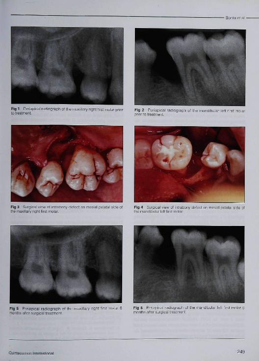
A complete clinical and radiographie examinationrevealed several small carious lesions. The initiaiO'Leary plaque control record^' was 30%. In general,gingival tissues presented with a pink color, firm tex-ture, and adequate contour. Probing depth (PD) mea-surements ranged from 1 to 10 mm, with the most se-vere sites being present around tbe maxillary rigbtfirst molar (tooth 16[3]) and the mandibular left firstmolar (tootb 36[19]), Clinical attacbment level (CAL)ranged from 0 to 9 mm, and again tbe greatest losswas found at teetb 16 and 56 (Table 1}. Bleeding onprobing was localized at the mesiobuccal andmesiopaiatal surface of tooth 16 and tbe mesiolingualsurface of tooth 36. No furcation involvement or in-creased mobility was detected. Radiographically,mesial vertical bony defects were associated witb bothteeth 16 and 36 (Figs 1 and 2), Initial tberapy in-cluded oral hygiene Instruction and scaling and rootplaning. Probing deptbs following initial therapy werevirtually uncbanged. Following consultation with thepatient and bis parents, it was decided tbat tbe patientwould benefit from reduction in probing depths. Toachieve this goal, periodontal surgeries were suggestedon teetb 16 and 36, and the patient and bis familyagreed. Full tbickness, mucoperiosteal flaps were ele-vated in both areas, the defects were debrided, andtbe roots were scaled and root planed. A large two-walled circumferential bony defect of 5 mm in depth(from the hone crest to tbe most apical portion of thedefect) was found on the mesial sitrface of tooth 16(Fig 3). A large two/three-walled bony defect of 6 tnmin depth (from tbe bone crest to the most apical por-tion oí the defect) was detected on the mesial surfaceof tooth 36 (Fig 4). Root surfaces were conditionedwith 24% ethylenediaminetetraacetic acid (EDTA) ata neutral pH (PrefGel, Biora) for 2 minutes. After irri-gation of the sites with sterile saline to remove EDTA,a gel containing enamel matrix proteins (Emdogain,Biora) was applied to the root surfaces. The flaps thenwere repositioned and single, interrupted Vicryl 4/0(Ethicon) sutures were placed, which resulted in pri-mary closure. Postoperative care included 100 mgdoxycycline hyclate per day for 3 weeks (200 mg onlyon the first day)'«-̂ * and a 0.12% chlorhexidine diglu-conate mouthrinse twice daily for 6 weeks. The su-tures were removed 14 days after surgery, and profes-sional supragingival plaque control was scheduledevery 2 weeks during the first 6 months followingsurgery,5«-'"' Healing was uneventful. There was amarked improvement in botb clinical and radio-graphic parameters at 6-month postsurgery examina-tion, (Table 1, Figs 5 and 6),
248
• Bonta et al •
Fig 1 Periapical radiograptí of the maxillary right first molar prior Fig 2 Periapical radiograph of the mandibular ieft tirst moiarprior to treatment.
Fig 3 Surgical view of intrabony defect on mesiai paiatal side ot Fig 4 Surgicai view of intrabony defect on mesial palatai side ofthe maxillary right first molar. the mandibular lefL first molar.
Fig 5 Periapical radiograph of the maxillary right firsi mo[ar 6months after surgical treatment.
Fig 6 Periapioai radiograph of the mandibuiai ieft first moiar 6months after surgical treatment
Quintessence International 249
• Bonta et al •
DISCUSSION
Localized aggressive periodontitis (previously called lo-calized juvenile periodontitis) has been described as aform of periodontai disease seen in young people char-acterized by severe periodontai destruction with littleor no visible accumulation of plaque or calculus,^""Several treatment strategies have been implemented inthe past to control the progression of this conditionand to regenerate the destroyed periodontium.Investigators have recommended surgical therapy inconjunction with systemic antimicrobial therapy.^'Clinical success has been reported with the use of bonegrafts with or without antimicrobial therapy.̂ '-̂ "̂̂ *
It has been suggested that the most clinically stableresult occurs when Aa is eliminated.^' Scaling and rootplaning alone has been found to be unsuccessful in theeradication of Aa from the periodontai pocket, whichhas been suggested to be due to the tissue penetrationby b acted a, •'̂ ••'̂ Most investigators have used antimi-crobial therapy to assist in complete removal of thesebacteria. While Lindhe and Liljenberg^' used tetracy-cline (1 g/day for 14 days) in conjunction with surgery,Mandell and Socransky^' found success with doxycy-cline (200 mg during the first day, followed by 100mg/day for 14 days) in conjunction with surgery,Novak et al'''' reported effective initial control of earlyLAP with systemic tetracycline therapy (1 g/day) for 6weeks combined with supragingival plaque control. In1991, Novak et al*' reported long-term (1 to 4 years}clinical and radiographie improvement in four subjectsfollowing previously mentioned treatment regimen.
The use of enamel matrix proteins in these patientsrepresents a new approach to the goal of regeneratinglost periodontai tissues. The enamel matrix proteinproduct is composed principally of amelogenin and re-lated proteins that are derived from porcine toothbuds,'" These enamel matrix proteins may be impor-tant in the development of acellular cementum, peri-odontai ligament, and bone,'"'^
There have been several reports of clinically suc-cessful usage of enamel matrix proteins in treating in-trabony defects.'" The use of enamel matrix proteins orguided tissue regeneration techniques to treat intra-bony defects has been shown to yield similar re-sults,* '̂̂ ^ Froum et aH* found the percentage of bonefill during re-entry after the use of enamel matrix pro-teins to be superior to that obtained with open-flapdebridement, Heden et a V in a series of cases, re-ported that 87% of sites treated with enamel matrixproteins exhibited probing attachment gain of morethan 2 mm. In 2000, Hcden^' found a mean pocketdepth reduction of 4,7 mm and mean attachment levelgain of 4.2 mm 12 months after treating intrabony de-fects with enamel matrix proteins.
The current study is the first known report in theliterature describing the use of these proteins in intra-bony defects in patients with localized aggressive peri-odontitis. The patient in this case report presentedwith several typical features associated with LAP,Clinically, the patient presented with deep intrabonydefects at the first molars (teeth 16 and 36}, Aftertreatment of these defects with enamel matrix proteins,the authors found an average 5-mm probing depth re-duction and an approximately 5-mm gain in probingattachment level, which is comparable to the resuitsreported in the literature.̂ '-^^ Although the studies arenot directly comparable, since the patients in previousstudies were diagnosed with chronic periodontitis, theresults of this case report suggest that patients havingLAP may heal in a similar fashion as patients havingchronic periodontitis. It should be stressed that thisparticular patient was placed in a very strict plaque-control program with periodontai maintenance visitsevery 2 weeks for the initial 6 months after periodon-tai surgeries. This maintenance program is not sub-stantially different from the maintenance program uti-lized in the clinicai trials for the enamel matrix proteinproduct,'" The treatment of intrabony defects withenamel matrix proteins in patients with LAP repre-sents a reasonable alternative to otber periodontai re-generative therapies. Small and narrow, three-walleddefects respond very favorably to surgical therapy."The vertical bony defects in this case were large andtherefore less favorable to respond positively.
CONCLUSION
It can be concluded that the use of enamel matrix pro-teins, as an alternative periodontai regenerative mater-ial, was found to be successful in the treatment of onecase of LAP, Clinical and radiographie parameters hadimproved significantly at 6 months after the use ofenamel matrix proteins. Additional case reports andclinical trial data will be needed to further substantiatethe benefits of using enamel matrix proteins as aperiodontai regenerative material in LAP patients.
REFERENCES
1, Gottlieb G, The diffuse atrophy of the alveolar process [inGerman], Z Stomat 1923;21:195,
2, Butler JH. A familial pattern of juvenile periodontitis (perio-dontosis), J Periodontol 1969;40:n5-n8,
3, Baer PN, The case for pcriodontosis as a clinical entity, [Periodontol 1971;42:516-520,
4, Tenenbaum B, Karshan M, Ziskin D, Nahoum H, Clinicaland microscopic study of the gingivae in periodontosis, JAm Dent Assoc 19S0;40:302-314,
250Voiume 34, Number 4, 2fin,T

5. Burmeister JA, Best AM, Palcanis KG, Caine FA, RanneyRR. Localized juvenile periodontitis and generalized severeperiodontitis; Clinical findings, I Clin Periodontol 1984 11181-192.
6. Artnitage GC. Development of a classification system forperiodontal diseases and conditions, Ann Periodontol 19994;l-6.
7. Neely AL. Prevalence of juvenile periodontitis in a circum-pubertal population. I Clin Periodontol 1992;19:367-372,
8. Melvin WL, Sandifer ]B, Gray fL. The prevalence and sexratio of juvenile periodontitis in a young racially mixed pop-ulation. I Periodontol 1991;62:330-334.
9. Cogen RB, Wright IT, Täte AL. Destructive periodontal dis-ease in healthy children, ] Periodontol 1992 ;63;761-765,
10. Loe H, Brown LI. Early onset periodontitis in the UnitedStates of America. J Periodontol 1991;62:608-616.
11. Newman MG, Socransky SS, Savltt ED, Propas DA,Crawford A. Studies of the microbiology of periodontosis. \Periodontol 1976;47:373-379.
12. Newman MG, Socransky SS. Predominant cultivable micro-biota in periodontosis. I Periodontal Res 1977;12:120-128.
13. Tanner AC, Haffer C, Bratthall GT, Visconti RA, SocranskySS. A study of the bacteria associated with advancing peri-odontitis in man. I Clin Periodontol 1979;6:278-307
14. Eisenmann AC, Eisenmann R, Sousa O, Slots I. Micro-biological study of localized juvenile periodontitis in Pana-ma. J Periodontol 1983;54:712-713,
15. Zambón ], Christersson L, Slots I, Actinobaciilus actino-mycetemcomitans in human periodontal disease prevalencein patient groups and distribution of biotypes and serotypeswithin families. I Periodontol 1983;54;707-711,
16. Lehner T, Wilton IM, Ivanyi L, Manson JD. Immunologicalaspects of juvenile periodontitis (periodontosis). I Perio-dontal Res 1974 ;9:261-272.
17. Cianciola L, Genco R, Patters M, McKenna J, van Oss CI.Defective polymorphonuclear leukocyte function in ahuman periodontal disease. Nature 1977 ;265:445-447
18. Waldrop TC, Mackler BF, Schur P, Killoy W. Immunologiestudy of human periodontosis (juvenile periodontitis]. JPeriodontol 1981Í52:8-15.
19. Schenkein H, Cianciola L, Genco R, Complement activa-tion in gingival pocket fluid from patients with periodonto-sis and severe periodontitis [abstract 581]. ] Dent Res 1976;55:B207.
20. Saxén L, Nevanlinna HR. Autosomal recessive inheritanceof juvenile periodontitis; Test of a hypothesis. Clin Genet1984;25:332-335.
21. Slots N, Rosling BG. Suppression of period on topathic mi-croflora in localized juvenile periodontitis by systemic tetra-cycline. ] Clin Periodontol 1983; 10:465-486.
22. Komman KS, Robertson PB. Clinical and microbiologicalevaluation of therapy for juvenile periodontitis, J Perio-dontol 1985;56:443-446.
23. Lindhe J, Liljenbei^ B. Treatment of localized juvenile peri-odontitis. Results after 5 years. J Clin Periodontol 1984;11:399-410.
24. Wennstrom A, Wennström I, Lindbe J. Healing followingsurgical and non surgical treatment of juvenile periodontitis,A 5-year longitudinal study. J Clin Periodontol 1986;13:869-882.
25. DeMarco T, Scaletta L. The use of autogenous hip marrowin the treatment of juvenile periodontosis: A case report. ]Poriodontol 1970;41:683-684.
26. Evian CI, Amsterdam M, Rosenberg ES. Juvenile periodon-titis-Heaiing following therapy to control inflammation andtraumatic etiologic components of the disease. J ClinPeriodontol 1982,9:1-21.
27 Yukna RA, Sepe WW. Clinical evaluation of localized peri-odontosis defects treated with freeze-dried bone allograftscombined with local and systemic tetracyclines. Int JPeriodontics Restorative Dent 1982;2(5):8-2L
28. Mabry T, Yulina RA, Sepe WW. Freeze-died bone allograftscombined with tetracycline in the treatment of juvenile peri-odontitis, I Periodontol 1985;56:74-81.
29. Zetterström 0, Andersson C, Eriksson L, et al. Clinicalsafety of enamel matrix derivative (EMDOGAIN) in thetreatment of periodontal defects. J Clin Periodontol1997;24:697-704,
30. Hammarstrom L, Enamel matrix, cementum developmentand regeneration. I Clin Periodontol 1997;24:658-668.
31. Hammarstrom L, HeijI L, Gestrelius S. Periodontal regener-ation in a buccal dehiscence model in monkeys after appli-cation of enamel matrix proteins. I Clin Periodontol 1997;24:669-677.
32. Heijl L. Periodontal regeneration with enamel matrix deriv-arive in one human experimental defect. A case report. JClin Periodontol 1997 ;24:693-696,
33. Mellonig IT. Enamel matrix derivative for periodontal re-constructive surgery: Technique and clinical and histologiecase report. Int | Periodontics Restorative Dent 1999;19:9-19.
34. Yukna RA, Mellonig IT. Histologie evaluation of periodon-tal healing in humans following regenerative therapy withenamel matrix derivative. A 10-case series. J Periodontol2000;71:752-759.
35. O'Leary Tj, Drake RB, Nayfor JE. The plaque controlrecord, j Periodontol 1972;43:38.
36. Mandel! RL, Tripodi LS, Savitt E, Goodson IM, SocranskySS, The effect of treatment on Actinobaciilus actinomyce-temcomitans in localized juvenile periodontitis. JPeriodontol 1986;57:94-99.
37. Mandell RL, Socransky SS. Microbiological and clinical ef-fects of surgery plus do^cycline on juvenile periodontitis. JPeriodontol 1988;59:373-379,
38. Saxén L, Asikainen S, Kanervo A, Kary K, Jousimies-SomerH. The long-term efficacy of systemic doxy cycline medica-tion in the treatment of Actinobaciilus actinomycetemcomi-tans associated periodontitis. Arch Oral Biol 1990;35:227S-229S.
39. Nyman S, Rosling B, Lindhe I. Effect of professional tootbcleaning on healing after periodontal surgery. J ClinPeriodontol 1975;2:80-86,
40. Rosling B, Nyman S, Lindhe |, Jem B. The healing potentialof the periodontal tissues following different techniques ofperiodontai surgery in plaque free dentitions. I ClinPeriodontol 1976; 3:23 3-2 50.
41. Liljenberg B, Lindhe J. Juveniie periodontitis. Some micro-biological, h istopathological and clinical characteristics. JClin Periodontol 1980;7:48-61.
42. Carranza FA, Saglie R, Newman MG, Valentin PL.Scanning and transmission electron microscopic study oftissue-invading microorganisms in localized juvenile perio-dontitis. J Periodontol 1983;54:598-617
Quintessence I nier national 251
Bonta et ai
43. Christersson LA, Albini B, Zambón J), Slots ), Genco RJ,Demonstration of Actinobacillus actinomycctemconiitans inlocalized juvenile periodontitis [abstract 2561. J Dent Res1983;62:198.
44. Novak MJ, Poison AM, Adair SM. Tetracycline ttierapy inpatients with early juvenile periodontitis, | Periodontol1988;59:366-372,
45. Novak MJ, Stamatelakys C, Adair SM, Resolution of earlylesions of juvenile periodontitis with tetracytline therapyalone: Long-term observations of 4 cases, J Periodontol1991;62:628-633.
46. Gestrelius S, Andersson C, LidstrÖm D, Hammarstrom L,Somerman M, In vitro studies on periodontal ligament cellsand enamel matrix derivative. J Clin Feriodontol t997;24:685-692,
47. Heij) L, Heden G, Svärdström G, Östgren A. Enamel matrixderivative (EMDOGAIN) in th« treatment of intrabony pe-riodontal defects. J Clin Periodontol 1997;24:705-714.
48. Frouni S), Weinberg MA, Rosenberg E, Tarnow D. A com-parative study utilizing open flap debridement with andwithout enamel matrbt derivative in the treatment of perio-dontal intrabony defects: A 12-month re-entry study. JPeriodontol 2001;72:25-34.
49. Pontoriero R, Wennström J, Lindhe J The use of barriermembranes and en ame i matrix proteins in the treatment ofangular bone defects, A prospective controlled clinicalstudy, J Clin Periodontol 1999;26:833-840.
50. Heden G, Wermström J, Lindhe J. Periodontal tissue alter-ations following Emdogain treatment of periodontal siteswith angular bone defects. A series of case reports. J ClinPeriodontol 1999;26:855-860.
51. Heden G. A case report study of 72 consecutive emdogain-treated intrabony periodontal defects: Clinical and radio-graphic findings after one year. Int J PeriodonticsRestorative Dent 2000j20:127-139.
52. Scuiean A, Donos N, Blaes A, Lauermann M, Reich E,Brecx M, Comparison of enamel matrix proteins and bioab-sorbable membranes in the treatment of intrabony perio-dontal defects. A split-motith study, J Periodontol 1999;70:255-262.
53. Garrett S. Periodontal regeneration around natural teeth.Ann Periodontol 1996;l:621-666.
Dental Materials In Vivo:Aging and Related PhenomenaEdited by George EUades, Theodore Eliades,William Brantley, and David Watts
This comprehensivereview brings to-gether research bybiomateriais expertsin various fields ofdentistry, includingorai and maxillofa-cial surgery, ortho-dontics, periodon-tics, prosthodontics,and restorative den-tistry. Through thepresentation of evi-
dence derived exclusivelyfrom in vivo studies, the mechanisms governingthe aging of materials placed in the oral cavityare clarified and selective aspects of the in vivoperformance of materials demonstrated.
296 pp (softcover): 183 illus (37 color);ISBN 0-86715-399-7; US $110
' Biomaterial Surface Alterations FoliowingExposure to Biologic Fluid
' Aging of Casting Alloys Used in Prosthodonticsand Restorative Dentistry
' Ceramic Behavior Under Different Envi ron menta Iand Loading Conditions
' Characteristics of Retrieved Implants'Alterations of Dental Amalgam'Aging of Glass-lonomer Cements'Degradation Mechanisms of Dental ResinComposites
' Disintegration of Orthodontic Appliances'Characteristics of Used Orthodontic Brackets'Orthodontic Utilities and Auxiliaries'Endodontic Instruments and Materials'Stainless Steel Oral and Maxillofacial SurgicalImplants
' Leaching of iVletallic Ions from Plates and ScrewsUsed in Jaw Fracture Fixation
'Sutures in the Oral Cavity• Bioactive Glass Bone-Grafting iViaterials
To OrderCal! toll free 800-621-0387
or Fax 630-Ó82-3288Website www.quintpub.com
1 E-mail [email protected]
252 Volume 34, Number 4. 2003
C|uinlcwence _ _ . ^
book/ Quintessence Publishing Co, me