the use of fragments of thin veneers as a restorative therapy for anterior teeth disharmony a case...
TRANSCRIPT
7/23/2019 The Use of Fragments of Thin Veneers as a Restorative Therapy for Anterior Teeth Disharmony a Case Report Wit…
http://slidepdf.com/reader/full/the-use-of-fragments-of-thin-veneers-as-a-restorative-therapy-for-anterior 1/5
Ricardo Coelho Okida et al
416JAYPEE
CASE REPORT
The Use of Fragments of Thin Veneers as a RestorativeTherapy for Anterior Teeth Disharmony: A Case Reportwith 3 Years of Follow-up
Ricardo Coelho Okida, Aljomar José Vechiato Filho, Valentim Adelino Ricardo Barão, Daniela Micheline dos SantosMarcelo Coelho Goiato
10.5005/jp-journals-10024-1160
ABSTRACT
Aim: The present case report described the use of contact lens
of thin porcelain veneers as a restorative therapy to solve anterior
teeth disharmony.
Background: Fragments of thin veneers are minimally invasive
restorations with little or no dental preparation and present
thickness ranging from 0.2 to 0.5 mm. They are used in case of
diastema closure, small changes of teeth, color and restoration
of teeth with small fractures.
Case report: A 25-year-old man was admitted at a dental clinic
complaining about the diastema presence on the upper anterior teeth. Patient was referred to an orthodontic treatment in order to
provide better distribution of the diastemas and harmonious
proportion of the teeth. Afterwards, contact lens of thin porcelain
veneers were fabricated on the six upper anterior teeth.
Conclusion: Based on the outcomes of this clinical report, we
considered the use of fragments of thin veneers as a successful
treatment option after 3 years of follow-up.
Clinical significance: The fragments of thin veneers have been
established to be an interesting alternative to esthetically restore
the anterior teeth with minimal invasiveness. However, since it
is a new treatment modality, longitudinal studies are necessary
to understand the material’s behavior.
Keywords: Porcelain, Dental contact lens, Diastema, Oral
rehabilitation, Dental esthetic, Clinical case.
How to cite this article: Okida RC, Filho AJV, Barão VAR, dos
Santos DM, Goiato MC. The Use of Fragments of Thin Veneers
as a Restorative Therapy for Anterior Teeth Disharmony: A Case
Report with 3 Years of Follow-up. J Contemp Dent Pract
2012;13(3):416-420.
Source of support: Nil
Conflict of interest: None declared
BACKGROUND
The advent of new restorative materials and new
technologies in oral rehabilitation during the past 30 years
improved the restorative dentistry field. There are a variety
of approaches to treat the different cases of shape, position,
alignment, symmetry, proportion, surface texture and color
of anterior teeth in our daily practices.1 Thus, the clinician
should choose more conservative treatments, i.e. with
greater preservation of healthy dental structure. The
porcelain veneers have been used as a successful approach
to solve esthetic problems in anterior region.
The use of fragments of thin veneers also known as
contact lens is an innovative technique in the restorativeand esthetic dentistry fields. These restorations are
minimally invasive with reduced or no dental preparation,
which is their principal advantage,2-5 present 0.2 to 0.5 mm
thickness1,5 and can be used to redraw the smile, increase
the teeth size, change teeth format and reduce the diastema.6
Nevertheless, it is imperative for the patient to have good
oral hygiene and to take care when chewing hard foods.4
Therefore, the aim of this report was to describe step-
by-step the clinical process of the use of contact lens of
thin porcelain veneers as a therapy to solve esthetic
disharmony in the anterior teeth.
CASE REPORT
A 25-year-old man was admitted at the Aracatuba Dental
School, São Paulo State University, Brazil complaining
about the diastema presence and color alteration on the upper
anterior teeth (Fig. 1). Diagnostic impressions were made
using stock trays with irreversible hydrocolloid (Hydrogum,
Zhermack SpA Rovigo, Italy) then poured in dental stone
(Gesso-Rio, Orlando Antonio Bussioli-ME, Rio Claro, SP,
Brazil). Several treatment options (direct composite veneers, porcelain veneers, contact lens of thin porcelain veneers)
were discussed with the patient in relation to life expectancy,

7/23/2019 The Use of Fragments of Thin Veneers as a Restorative Therapy for Anterior Teeth Disharmony a Case Report Wit…
http://slidepdf.com/reader/full/the-use-of-fragments-of-thin-veneers-as-a-restorative-therapy-for-anterior 2/5
The Use of Fragments of Thin Veneers as a Restorative Therapy for Anterior Teeth Disharmony: A Case Report
The Journal of Contemporary Dental Practice, May-June 2012;13(3):416-420 417
JCDP
Fig. 1: Initial clinical aspect
Fig. 2: Fixed retainer installed
Figs 3A to C: Clinical aspects of the prepared teeth: (A) Frontal view; (B) right lateral view; (C) left lateral view
invasiveness, esthetic results, clinical time and cost. The
patient chose rehabilitation with contact lens of thin porcelain
veneers. The driving forces toward patient’s decision were
that contact lens of thin porcelain veneers provide a durable,
stain-resistant surface and conserve the dental structure.
After deciding the treatment approach, patient wasreferred to an orthodontic treatment in order to provide better
distribution of the diastemas, harmonious proportion of the
teeth, and to balance the midline (Fig. 2). After 1 year of
bracketed orthodontic treatment, a removable retainer was
manufacture in order to maintain the distributed spaces
among the anterior teeth.
Patient underwent a home bleaching teeth with 16%
carbamide peroxide (Whiteness perfect, FGM, Joinville, SC,
Brasil) during 2 weeks. After that, very conservative teeth
preparation was carried out in the six anterior upper teeth.
The mesial and distal interproximal areas of the central andlateral incisors and canines were slightly reduced. The facial
surfaces were slightly shaped and a gentle subgingival
chamfer margin was created. The corners of the prepared
teeth were slightly rounded. Preparations involved no more
than 0.2 mm of tooth reduction so, that the veneers would
be very thin and healthy teeth structure preserved (Figs 3A
to C).
Impressions of the prepared teeth were made using vinyl
polysiloxane impression material (Aquasil ULV, Dentsply,
A
B C
7/23/2019 The Use of Fragments of Thin Veneers as a Restorative Therapy for Anterior Teeth Disharmony a Case Report Wit…
http://slidepdf.com/reader/full/the-use-of-fragments-of-thin-veneers-as-a-restorative-therapy-for-anterior 3/5
Ricardo Coelho Okida et al
418JAYPEE
Caulk Milford, DE, USA). Size # 0 gingival retraction cord
(Ultrapak, Ultradent Products, Inc., South Jordan, UT, USA)
was used to expose the cervical margins of the preparations.
The patient was provided provisional veneers fabricated
with direct composite resin (Filtek Z250, 3M Espe, St Paul,
Minnesota, USA) using a mock-up. They were slightly bonded with the same resin material after spot-etching the
prepared teeth. On the master cast, the six contact lens of
thin porcelain veneers were fabricated using the shade of a
Vita B1 ceramic (IPS Emax, Ivoclar Vivadent, Barueri, SP)
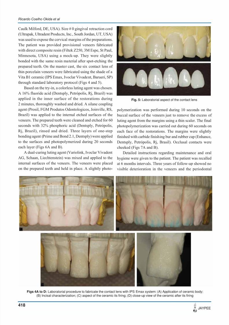
through standard laboratory protocol (Figs 4 and 5).
Based on the try-in, a colorless luting agent was chosen.
A 16% fluoride acid (Dentsply, Petrópolis, Rj, Brazil) was
applied in the inner surface of the restorations during
2 minutes, thoroughly washed and dried. A silane coupling
agent (Prosil, FGM Produtos Odontologicos, Joinville, RS,
Brazil) was applied to the internal etched surfaces of theveneers. The prepared teeth were cleaned and etched for 60
seconds with 32% phosphoric acid (Dentsply, Petrópolis,
Rj, Brazil), rinsed and dried. Three layers of one-step
bonding agent (Prime and Bond 2.1, Dentsply) were applied
to the surfaces and photopolymerized during 20 seconds
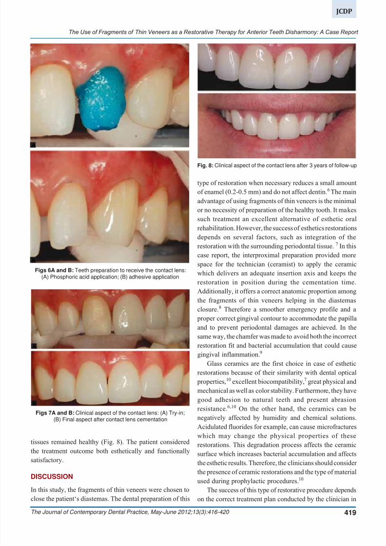
each layer (Figs 6A and B).
A dual-curing luting agent (Variolink, Ivoclar Vivadent
AG, Schaan, Liechtenstein) was mixed and applied to the
internal surfaces of the veneers. The veneers were placed
on the prepared teeth and held in place. A slightly photo-
Figs 4A to D: Laboratorial procedure to fabricate the contact lens with IPS Emax system: (A) Application of ceramic body;(B) Incisal characterization; (C) aspect of the ceramic its firing; (D) close-up view of the ceramic after its firing
Fig. 5: Laboratorial aspect of the contact lens
polymerization was performed during 10 seconds on the
buccal surface of the veneers just to remove the excess of luting agent from the margins using a thin scaler. The final
photopolymerization was carried out during 60 seconds on
each face of the restorations. The margins were slightly
finished with carbide finishing bur and rubber cup (Enhance,
Dentsply, Petrópolis, Rj, Brasil). Occlusal contacts were
cheeked (Figs 7A and B).
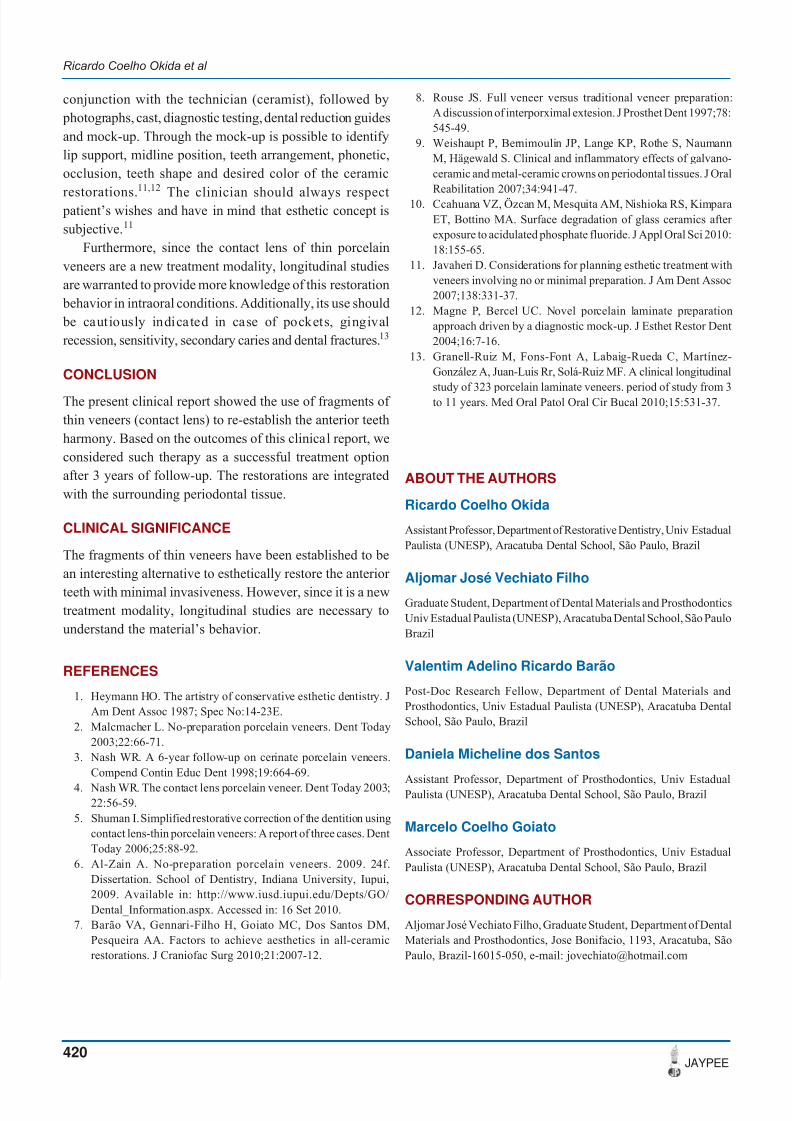
Detailed instructions regarding maintenance and oral
hygiene were given to the patient. The patient was recalled
at 6 months intervals. Three years of follow-up showed no
visible deterioration in the veneers and the periodontal
A B
C D
7/23/2019 The Use of Fragments of Thin Veneers as a Restorative Therapy for Anterior Teeth Disharmony a Case Report Wit…
http://slidepdf.com/reader/full/the-use-of-fragments-of-thin-veneers-as-a-restorative-therapy-for-anterior 4/5
The Use of Fragments of Thin Veneers as a Restorative Therapy for Anterior Teeth Disharmony: A Case Report
The Journal of Contemporary Dental Practice, May-June 2012;13(3):416-420 419
JCDP
Figs 6A and B: Teeth preparation to receive the contact lens:(A) Phosphoric acid application; (B) adhesive application
Figs 7A and B: Clinical aspect of the contact lens: (A) Try-in;(B) Final aspect after contact lens cementation
tissues remained healthy (Fig. 8). The patient considered
the treatment outcome both esthetically and functionally
satisfactory.
DISCUSSION
In this study, the fragments of thin veneers were chosen to
close the patient‘s diastemas. The dental preparation of this
type of restoration when necessary reduces a small amount
of enamel (0.2-0.5 mm) and do not affect dentin.6 The mainadvantage of using fragments of thin veneers is the minimal
or no necessity of preparation of the healthy tooth. It makes
such treatment an excellent alternative of esthetic oral
rehabilitation. However, the success of esthetics restorations
depends on several factors, such as integration of the
restoration with the surrounding periodontal tissue. 7 In this
case report, the interproximal preparation provided more
space for the technician (ceramist) to apply the ceramic
which delivers an adequate insertion axis and keeps the
restoration in position during the cementation time.
Additionally, it offers a correct anatomic proportion among
the fragments of thin veneers helping in the diastemas
closure.8 Therefore a smoother emergency profile and a
proper correct gingival contour to accommodate the papilla
and to prevent periodontal damages are achieved. In the
same way, the chamfer was made to avoid both the incorrect
restoration fit and bacterial accumulation that could cause
gingival inflammation.9
Glass ceramics are the first choice in case of esthetic
restorations because of their similarity with dental optical
properties,10
excellent biocompatibility,7
great physical andmechanical as well as color stability. Furthermore, they have
good adhesion to natural teeth and present abrasion
resistance.6,10 On the other hand, the ceramics can be
negatively affected by humidity and chemical solutions.
Acidulated fluorides for example, can cause microfractures
which may change the physical properties of these
restorations. This degradation process affects the ceramic
surface which increases bacterial accumulation and affects
the esthetic results. Therefore, the clinicians should consider
the presence of ceramic restorations and the type of material
used during prophylactic procedures.10
The success of this type of restorative procedure depends
on the correct treatment plan conducted by the clinician in
Fig. 8: Clinical aspect of the contact lens after 3 years of follow-up
A
B
A
B
7/23/2019 The Use of Fragments of Thin Veneers as a Restorative Therapy for Anterior Teeth Disharmony a Case Report Wit…
http://slidepdf.com/reader/full/the-use-of-fragments-of-thin-veneers-as-a-restorative-therapy-for-anterior 5/5
Ricardo Coelho Okida et al
420JAYPEE
conjunction with the technician (ceramist), followed by
photographs, cast, diagnostic testing, dental reduction guides
and mock-up. Through the mock-up is possible to identify
lip support, midline position, teeth arrangement, phonetic,
occlusion, teeth shape and desired color of the ceramic
restorations.11,12
The clinician should always respect patient’s wishes and have in mind that esthetic concept is
subjective.11
Furthermore, since the contact lens of thin porcelain
veneers are a new treatment modality, longitudinal studies
are warranted to provide more knowledge of this restoration
behavior in intraoral conditions. Additionally, its use should
be cautiously indicated in case of pockets, gingival
recession, sensitivity, secondary caries and dental fractures.13
CONCLUSION
The present clinical report showed the use of fragments of
thin veneers (contact lens) to re-establish the anterior teeth
harmony. Based on the outcomes of this clinical report, we
considered such therapy as a successful treatment option
after 3 years of follow-up. The restorations are integrated
with the surrounding periodontal tissue.
CLINICAL SIGNIFICANCE
The fragments of thin veneers have been established to be
an interesting alternative to esthetically restore the anterior
teeth with minimal invasiveness. However, since it is a newtreatment modality, longitudinal studies are necessary to
understand the material’s behavior.
REFERENCES
1. Heymann HO. The artistry of conservative esthetic dentistry. JAm Dent Assoc 1987; Spec No:14-23E.
2. Malcmacher L. No-preparation porcelain veneers. Dent Today2003;22:66-71.
3. Nash WR. A 6-year follow-up on cerinate porcelain veneers.Compend Contin Educ Dent 1998;19:664-69.
4. Nash WR. The contact lens porcelain veneer. Dent Today 2003;22:56-59.
5. Shuman I. Simplified restorative correction of the dentition usingcontact lens-thin porcelain veneers: A report of three cases. DentToday 2006;25:88-92.
6. Al-Zain A. No-preparation porcelain veneers. 2009. 24f.Dissertation. School of Dentistry, Indiana University, Iupui,2009. Available in: http://www.iusd.iupui.edu/Depts/GO/Dental_Information.aspx. Accessed in: 16 Set 2010.
7. Barão VA, Gennari-Filho H, Goiato MC, Dos Santos DM,Pesqueira AA. Factors to achieve aesthetics in all-ceramicrestorations. J Craniofac Surg 2010;21:2007-12.
8. Rouse JS. Full veneer versus traditional veneer preparation:A discussion of interporximal extesion. J Prosthet Dent 1997;78:545-49.
9. Weishaupt P, Bernimoulin JP, Lange KP, Rothe S, NaumannM, Hägewald S. Clinical and inflammatory effects of galvano-ceramic and metal-ceramic crowns on periodontal tissues. J OralReabilitation 2007;34:941-47.
10. Ccahuana VZ, Özcan M, Mesquita AM, Nishioka RS, KimparaET, Bottino MA. Surface degradation of glass ceramics after exposure to acidulated phosphate fluoride. J Appl Oral Sci 2010:18:155-65.
11. Javaheri D. Considerations for planning esthetic treatment withveneers involving no or minimal preparation. J Am Dent Assoc2007;138:331-37.
12. Magne P, Bercel UC. Novel porcelain laminate preparationapproach driven by a diagnostic mock-up. J Esthet Restor Dent2004;16:7-16.
13. Granell-Ruiz M, Fons-Font A, Labaig-Rueda C, Martínez-González A, Juan-Luis Rr, Solá-Ruiz MF. A clinical longitudinal
study of 323 porcelain laminate veneers. period of study from 3to 11 years. Med Oral Patol Oral Cir Bucal 2010;15:531-37.
ABOUT THE AUTHORS
Ricardo Coelho Okida
Assistant Professor, Department of Restorative Dentistry, Univ EstadualPaulista (UNESP), Aracatuba Dental School, São Paulo, Brazil
Aljomar José Vechiato Filho
Graduate Student, Department of Dental Materials and ProsthodonticsUniv Estadual Paulista (UNESP), Aracatuba Dental School, São PauloBrazil
Valentim Adelino Ricardo Barão
Post-Doc Research Fellow, Department of Dental Materials andProsthodontics, Univ Estadual Paulista (UNESP), Aracatuba DentalSchool, São Paulo, Brazil
Daniela Micheline dos Santos
Assistant Professor, Department of Prosthodontics, Univ EstadualPaulista (UNESP), Aracatuba Dental School, São Paulo, Brazil
Marcelo Coelho Goiato
Associate Professor, Department of Prosthodontics, Univ EstadualPaulista (UNESP), Aracatuba Dental School, São Paulo, Brazil
CORRESPONDING AUTHOR
Aljomar José Vechiato Filho, Graduate Student, Department of DentalMaterials and Prosthodontics, Jose Bonifacio, 1193, Aracatuba, SãoPaulo, Brazil-16015-050, e-mail: [email protected]