the use of morphological characteristics and texture
TRANSCRIPT
tsqeastendpmt4td
cs1catc9cddami
y
TIUIA
mUI
The Use of Morphological Characteristicsand Texture Analysis in the Identification ofTissue Composition in Prostatic Neoplasia
JAMES DIAMOND, PHD, NEIL H. ANDERSON, MD,PETER H. BARTELS, PHD, FIAC, MD,RODOLFO MONTIRONI, MD, FRCPATH, AND
PETER W. HAMILTON, PHDsdgcotssptpc1
P
Quantitative examination of prostate histology offers clues inhe diagnostic classification of lesions and in the prediction of re-ponse to treatment and prognosis. To facilitate the collection ofuantitative data, the development of machine vision systems is nec-ssary. This study explored the use of imaging for identifying tissuebnormalities in prostate histology. Medium-power histologicalcenes were recorded from whole-mount radical prostatectomy sec-ions at � 40 objective magnification and assessed by a pathologist asxhibiting stroma, normal tissue (nonneoplastic epithelial compo-ent), or prostatic carcinoma (PCa). A machine vision system waseveloped that divided the scenes into subregions of 100 � 100ixels and subjected each to image-processing techniques. Analysis oforphological characteristics allowed the identification of normal
issue. Analysis of image texture demonstrated that Haralick featurewas the most suitable for discriminating stroma from PCa. Using
hese morphological and texture measurements, it was possible to
efine a classification scheme for each subregion. The machine visionwsmddpcan
omthooartid
sssvtsimdoi:10.1016/j.humpath.2004.05.010
1121
ystem is designed to integrate these classification rules and generateigital maps of tissue composition from the classification of subre-ions; 79.3% of subregions were correctly classified. Establishedlassification rates have demonstrated the validity of the methodol-gy on small scenes; a logical extension was to apply the methodologyo whole slide images via scanning technology. The machine visionystem is capable of classifying these images. The machine visionystem developed in this project facilitates the exploration of mor-hological and texture characteristics in quantifying tissue composi-
ion. It also illustrates the potential of quantitative methods torovide highly discriminatory information in the automated identifi-ation of prostatic lesions using computer vision. HUM PATHOL 35:121-1131. © 2004 Elsevier Inc. All rights reserved.
Key words: histopathology, imaging, prostate, texture analysis.Abbreviations: H4, Haralick feature 4; PCa, prostatic carcinoma;
IN, prostatic intraepithelial neoplasia.
Prostate cancer is set to become the most commonancer in men within the next 3 years.1 New figureshow that the incidence has been rising steadily since971, and if trends continue, it will overtake lung can-er before 2006. Around 22,000 cases of prostate cancerre diagnosed in the UK each year. New statistics showhat deaths from prostate cancer have gradually de-lined since the early 1990s, but mortality is still high;500 men die from the disease each year in the UK.1 Aontinuing challenge to the medical community is toevelop successful strategies for treatment and earlyiagnosis of prostate cancer. It has been suggested thatutomated machine vision systems would form an ele-ent of this overall diagnostic strategy by providing
mproved accuracy and reproducibility of diagnosis.This attractive concept has been around for many
ears now, but it has been limited to cytopathology,
From the Biomedical Imaging and Informatics Research Group,he Queen’s University of Belfast, Grosvenor Road, Belfast, Northern
reland, UK; The Royal Hospital Trust, Belfast, Northern Ireland,K; Optical Sciences Center, University of Arizona, Tucson, AZ; and
nstitute of Pathological Anatomy and Histopathology, University ofncona, Ancona, Italy. Accepted for publication May 6, 2004.
Address correspondence and reprint requests to Dr. James Dia-ond, Biomedical Imaging and Informatics Group, The Queen’sniversity of Belfast, Grosvenor Road, Belfast, BT12 6BL, Northern
reland, UK.0046-8177/$—see front matter© 2004 Elsevier Inc. All rights reserved.
here progress has been made in developing cervicalcreening devices.2,3 In histopathology, the develop-ent of automated systems has been lagging, essentially
ue to the complexity of imagery. Today, however, withevelopments in machine vision and intelligent imagerocessing systems combined with advancements inomputer hardware, the analysis of histopathologic im-ges for the purpose of objective disease classification isow possible.4-9
The adoption of state-of-the-art quantitative meth-dologies by pathologists requires constant and activeotivation. Baak10 has suggested that the absence of
his motivation by pathologists is a dominant factorindering modernization of pathology. The objectivef the present study was to investigate emerging meth-dologies in quantifying diagnostic pathology with theim of increasing reproducibility and predictive accu-acy in diagnosis. Baak10 also suggested that thesehemes should not be “merely academically interest-ng,” but should be required if the concept of objectiveiagnosis is to be embraced.
Central to this study is the generation of a repre-entative image set conveying the varying tissue compo-itions observed in prostate histology. Images were pre-elected by a pathologist (N.A. and R.M.) as reflectingariation in stroma, normal tissue (nonneoplastic epi-helial component), or prostatic carcinoma (PCa). Thetudy assessed the use of quantitative methods in defin-ng the criteria required for implementing an auto-
ated system capable of the unsupervised interpreta-

tg
tiiisttcn
MT
rlIvati
I
ioag
H
swpS(rs2lh
HUMAN PATHOLOGY Volume 35, No. 9 (September 2004)
ion of histological scenes into the aforementionedroups on an objective basis.
Contextually, texture plays an important role inhe perception of scenes and is used mainly to achievemage segmentation.11 It has been used widely in thenterpretation of prostatic ultrasound images12,13 andn nuclear classification.14-16 Previously it has beenhown that texture analysis also provides an effectiveool in tissue classification.17-19 It has been suggestedhat texture analysis combined with image morphologyan provide an effective tool for identifying tissue ab-ormalities in prostate histology.
ATERIALS AND METHODSissue
Twelve cases (4 training and 8 test) from whole- mountadical prostatectomy sections that had been fixed in forma-in embedded in paraffin, were retrieved from files at Thenstitute of Pathological Anatomy and Histopathology, Uni-ersity of Ancona, Italy. A set of 5-�m-thick sections were cutnd stained with hematoxylin and eosin and assessed by pa-hologists (N.A. and R.M.) as showing regions of the follow-ng:
1. Stroma: Fibroelastic tissue containing randomly ori-entated smooth muscle bundles that act as a frame-
FIGURE 1. Histolog
work to support the prostatic architecture, morpho- f
1122
logically forming a homogeneous texture withindefined subregions
2. Normal tissue: Prostatic tissue with increased amountsof smooth muscle, glandular, and/or stromal compo-nents
3. PCa: Prostatic adenocarcinomas, histologically diverseand having more than one characteristic composition.
mplementation
Image analysis was implemented using the Zeiss KS400maging system (Carl Zeiss, Oberkochen, Germany). A meth-dology was designed to integrate image processing, texturenalysis (after Haralick20), and classification rules for theeneration of digital tissue composition maps.
istological Scene Establishment
Twelve histological scenes were constructed by creating aeamless mosaic of component images (Fig 1). These imagesere captured at � 40 objective magnification using an Olym-us BH2 microscope (Olympus, Lake Success, NY) and aony DX930P CCD camera (512 � 512 pixel, 8-bit, grayscale)Sony, Ridge Park, NJ) and overlapped by 10% to allow foregistration. The system was calibrated by adjusting the lightource for an empty field until the mean field pixel value was55 (white). Images often exhibit shading variation due toighting irregularities, vignetting by the optical system, andeterogeneous sensitivity in the camera system or lens arti-
scene description.
icalact. These effects were minimized by applying a shading
cmtfaj
S
d2lscoo
C
smgpase
S
m
qsustsitesatwAcmpcm
RC
bslfia
FD rma
TISSUE COMPOSITION IN PROSTATIC NEOPLASIA (Diamond et al)
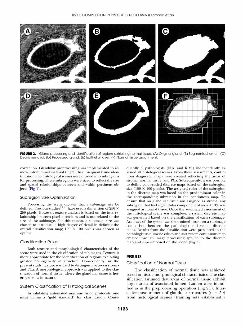
orrection. Glandular preprocessing was implemented to re-ove intraluminal material (Fig 2). In subsequent tissue iden-
ification, the histological scenes were divided into subregionsor processing. These subregions were sized to reflect the sizend spatial relationships between and within pertinent ob-ects (Fig 1).
ubregion Size Optimization
Processing the scene dictates that a subimage size beefined. Previous studies17,21 have used a dimension of 256 �56 pixels. However, texture analysis is based on the interre-ationship between pixel intensities and is not related to theize of the subimage. For this reason, a subimage size washosen to introduce a high degree of detail in defining theverall classification map; 100 � 100 pixels was chosen asptimal.
lassification Rules
Both texture and morphological characteristics of thecene were used in the classification of subimages. Texture isore appropriate for the identification of regions exhibiting
reater homogeneity in structure. Consequently, in theresent study, texture was used to distinguish between stromand PCa. A morphological approach was applied to the clas-ification of normal tissue, where the glandular tissue is het-rogeneous in nature.
ystem Classification of Histological Scenes
In validating automated machine vision protocols, one
IGURE 2. Gland processing and identification of regions exhebris removal. (D) Processed gland. (E) Epithelial layer. (F) No
ust define a “gold standard” for classification. Conse- f
1123
uently, 2 pathologists (N.A. and R.M.) independently as-essed all histological scenes. From these assessments, contin-ous diagnostic maps were created reflecting the areas oftroma, normal tissue, and PCa. Subsequently, it was possibleo define color-coded discrete maps based on the subregionize (100 � 100 pixels). The assigned color of the subregionn the discrete map was based on the predominant color inhe corresponding subregion in the continuous map. Tonsure that no glandular tissue was assigned as stroma, anyubregion that had a glandular component of area �10% wasssigned as normal tissue. Once the automated assessment ofhe histological scene was complete, a system discrete mapas generated based on the classification of each subimage.ccuracy of the system was determined based on a subimageomparison between the pathologist and system discreteaps. Results from the classification were presented to the
athologist as numeric values and as a system continuous mapreated through image processing applied to the discreteap and superimposed on the scene (Fig 3).
ESULTSlassification of Normal Tissue
The classification of normal tissue was achievedased on tissue morphological characteristics. The clas-ification assumed that areas of normal tissue exhibitarger areas of associated lumen. Lumen were identi-ed as in the preprocessing operation (Fig 2C). Inter-ctive measurement of glandular structures (n � 50)
g normal tissue. (A) Original gland. (B) Segmented lumen. (C)l tissue assignment.
ibitin
rom histological scenes (training set) established a

FIGURE 3. The identification and classification of tissue composition.

mosesn
C
HitPtsFe
atftsac
A
hfisnd
TISSUE COMPOSITION IN PROSTATIC NEOPLASIA (Diamond et al)
ean epithelial region width of 50 pixels. The lumenbjects identified were dilated by 50 pixels. Subsequentubtraction of the original lumen object defines thepithelial layer (Fig 2E). All subimages that containome part of this object (and lumen) are classified asormal tissue and are highlighted in Figure 2F.
lassification of Stroma and PCa
Investigation of Haralick features suggested thataralick 4 (H4) was the most suitable for discriminat-
ng between stroma and PCa. Figure 4 shows the spec-rum of change of H4 observed for areas of stroma andCa over the histological scenes (training set). A dis-inct change in image tissue composition betweentroma (H4 � 8.0) and PCa (H4 � 8.0) can be seen.igure 4 also highlights the variation in H4 values for all
FIGURE 4. Threshold boundary for
xample subimages in the training set (nstroma � 1000 p
1125
nd nPCa � 1000). A discrimination threshold was es-ablished at H4 � 8.0 from examination of subimagesrom both stroma and PCa regions (training set). Thishreshold was set closer to the mean value of H4 fortroma, to minimize the potential for misclassifyingreas showing PCa as stroma and to reflect the visualhange observed in Figure 4.
nalysis of Histological Scenes
In testing the system, tissue composition from 8istological scenes (test set) was automatically identi-ed based on the aforementioned criteria. Overall clas-ification rates for the system in discriminating stroma,ormal tissue, or PCa are given in Table 1. Selectiveetailed classifications are detailed in the following
discrimination of stroma and PCa.
thearagraphs and shown in Figures 5, 6, and 7.
S
tabmPhpfirfts
S
irBPgmr
S
isdt
S
iimfinfiaws
S
tetegt
D
lpasrma
tttsmwamssoa
miamdsoin
12345678S
otal s
HUMAN PATHOLOGY Volume 35, No. 9 (September 2004)
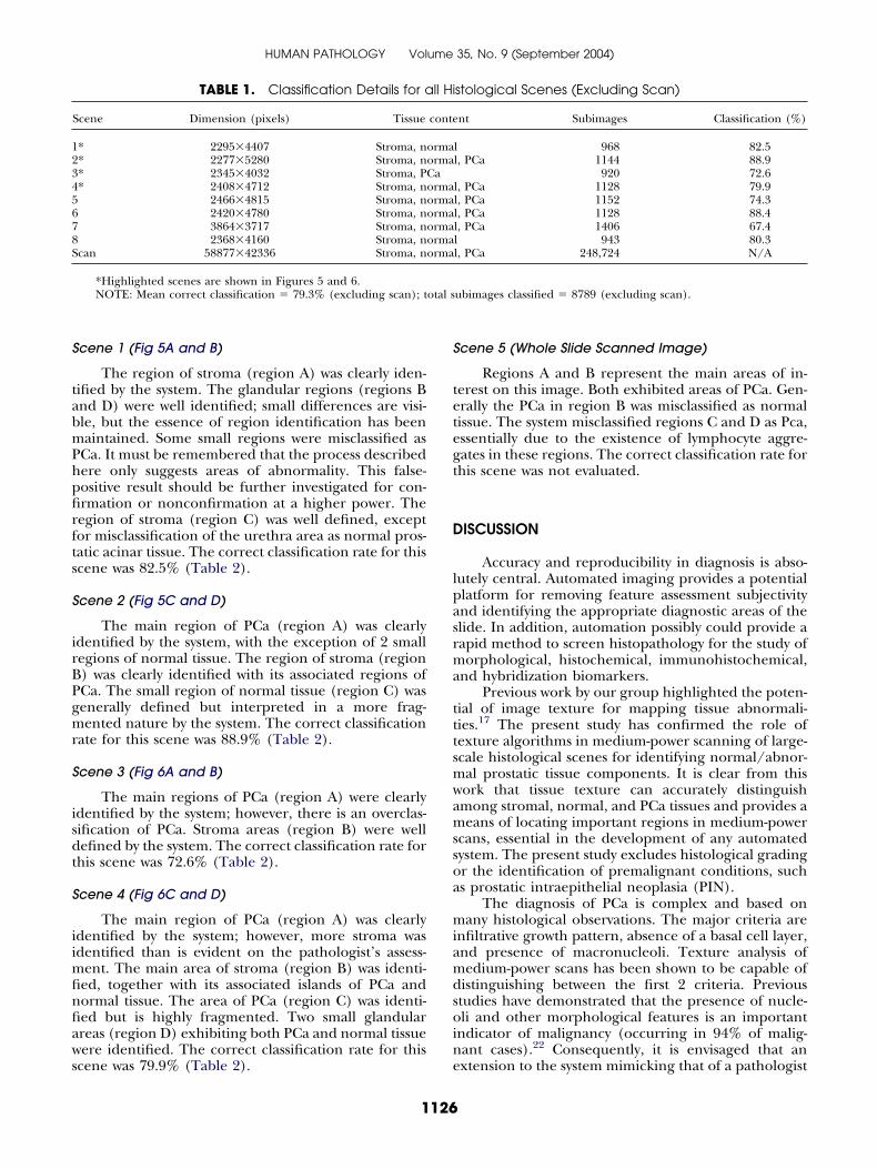
cene 1 (Fig 5A and B)
The region of stroma (region A) was clearly iden-ified by the system. The glandular regions (regions Bnd D) were well identified; small differences are visi-le, but the essence of region identification has beenaintained. Some small regions were misclassified as
Ca. It must be remembered that the process describedere only suggests areas of abnormality. This false-ositive result should be further investigated for con-rmation or nonconfirmation at a higher power. Theegion of stroma (region C) was well defined, exceptor misclassification of the urethra area as normal pros-atic acinar tissue. The correct classification rate for thiscene was 82.5% (Table 2).
cene 2 (Fig 5C and D)
The main region of PCa (region A) was clearlydentified by the system, with the exception of 2 smallegions of normal tissue. The region of stroma (region) was clearly identified with its associated regions ofCa. The small region of normal tissue (region C) wasenerally defined but interpreted in a more frag-ented nature by the system. The correct classification
ate for this scene was 88.9% (Table 2).
cene 3 (Fig 6A and B)
The main regions of PCa (region A) were clearlydentified by the system; however, there is an overclas-ification of PCa. Stroma areas (region B) were wellefined by the system. The correct classification rate forhis scene was 72.6% (Table 2).
cene 4 (Fig 6C and D)
The main region of PCa (region A) was clearlydentified by the system; however, more stroma wasdentified than is evident on the pathologist’s assess-
ent. The main area of stroma (region B) was identi-ed, together with its associated islands of PCa andormal tissue. The area of PCa (region C) was identi-ed but is highly fragmented. Two small glandularreas (region D) exhibiting both PCa and normal tissueere identified. The correct classification rate for this
TABLE 1. Classification Details for a
Scene Dimension (pixels) Tissue
* 2295�4407 Stroma, n* 2277�5280 Stroma, n* 2345�4032 Stroma, P* 2408�4712 Stroma, n
2466�4815 Stroma, n2420�4780 Stroma, n3864�3717 Stroma, n2368�4160 Stroma, n
can 58877�42336 Stroma, n
*Highlighted scenes are shown in Figures 5 and 6.NOTE: Mean correct classification � 79.3% (excluding scan); t
cene was 79.9% (Table 2). e
1126
cene 5 (Whole Slide Scanned Image)
Regions A and B represent the main areas of in-erest on this image. Both exhibited areas of PCa. Gen-rally the PCa in region B was misclassified as normalissue. The system misclassified regions C and D as Pca,ssentially due to the existence of lymphocyte aggre-ates in these regions. The correct classification rate forhis scene was not evaluated.
ISCUSSION
Accuracy and reproducibility in diagnosis is abso-utely central. Automated imaging provides a potentiallatform for removing feature assessment subjectivitynd identifying the appropriate diagnostic areas of thelide. In addition, automation possibly could provide aapid method to screen histopathology for the study oforphological, histochemical, immunohistochemical,
nd hybridization biomarkers.Previous work by our group highlighted the poten-
ial of image texture for mapping tissue abnormali-ies.17 The present study has confirmed the role ofexture algorithms in medium-power scanning of large-cale histological scenes for identifying normal/abnor-al prostatic tissue components. It is clear from thisork that tissue texture can accurately distinguishmong stromal, normal, and PCa tissues and provides aeans of locating important regions in medium-power
cans, essential in the development of any automatedystem. The present study excludes histological gradingr the identification of premalignant conditions, suchs prostatic intraepithelial neoplasia (PIN).
The diagnosis of PCa is complex and based onany histological observations. The major criteria are
nfiltrative growth pattern, absence of a basal cell layer,nd presence of macronucleoli. Texture analysis ofedium-power scans has been shown to be capable of
istinguishing between the first 2 criteria. Previoustudies have demonstrated that the presence of nucle-li and other morphological features is an important
ndicator of malignancy (occurring in 94% of malig-ant cases).22 Consequently, it is envisaged that an
stological Scenes (Excluding Scan)
nt Subimages Classification (%)
l 968 82.5l, PCa 1144 88.9
920 72.6l, PCa 1128 79.9l, PCa 1152 74.3l, PCa 1128 88.4l, PCa 1406 67.4l 943 80.3l, PCa 248,724 N/A
ubimages classified � 8789 (excluding scan).
ll Hi
conte
ormaormaCaormaormaormaormaormaorma
xtension to the system mimicking that of a pathologist

FabpmCsf
TISSUE COMPOSITION IN PROSTATIC NEOPLASIA (Diamond et al)
IGURE 5. Histological assessment (scenes 1nd 2). White lines enclose normal tissue;lack lines enclose PCa. (A) Conventionalathologist assessment for scene 1. (B) Auto-ated system assessment for scene 1. (C)onventional pathologist assessment for
cene 2. (D) Automated system assessmentor scene 2.
1127

HUMAN PATHOLOGY Volume 35, No. 9 (September 2004)
FIGURE 6. Histological assessment(scenes 3 and 4). White lines enclosenormal tissue; black lines enclose PCa.(A) Conventional pathologist assess-ment for scene 3. (B) Automated sys-tem assessment for scene 3. (C) Con-ventional pathologist assessment forscene 4. (D) Automated system assess-ment for scene 4.

FIGURE 7. Whole slide processing. (A) Whole slide scan. (B) Whole slide abnormality map.
wo
ecgdTsmpsf
tnsuantattdvtiswtpCtd
tsog
amrimqctazsdmjna(h4sep
t
S
S
S
S
HUMAN PATHOLOGY Volume 35, No. 9 (September 2004)
ould be beneficial, whereby confirmation of all areasf PCa would be examined at higher power.
Clearly, the classification of PIN should be consid-red in an extension to this investigation. The classifi-ation PIN is complicated by its segregation into low-rade/high-grade forms with its varying compositionescribed as tufting, micropapillary, cribriform, or flat.exture analysis will be unable to classify these compo-
itions well, and a combined technique with glandularorphology will be needed. Texture analysis may also
rovide an approach for histological grading. This isuggested in Figure 4, which shows a spectrum of dif-erentiation with respect to H4.
Many established methods exist for the classifica-ion of textured images. Unfortunately, most tech-iques assume that the textures are uniformly pre-ented and captured in the same orientation. This is annrealistic assumption for pathological sections. Forpplications such as the current system, texture analysiseeds to be invariant to orientation. Genuine orienta-
ion invariance is extremely difficult to obtain. Inchieving pseudoinvariance, texture features withinhis system are averaged over the four principal direc-ions (0, 45, 90, and 135 degrees), thus minimizingirection effects. In all texture-based applications, in-ariance in the staining of sections and in the illumina-ion when acquiring imagery is also important. It ismpossible to ensure total consistency in the staining ofections, and knowledge of whether the slides usedere batch-processed is not available. However, given
hese limitations, functionality was built into the imagereprocessing technique to minimize these effects.onsideration of the classification results has shown
hat the variations in classification rate are essentiallyue to image complexity.
Image analysis technology is a powerful resourcehat can be exploited to provide objective decisionupport for diagnostic pathology. Research and devel-pment of automated systems in pathology has been
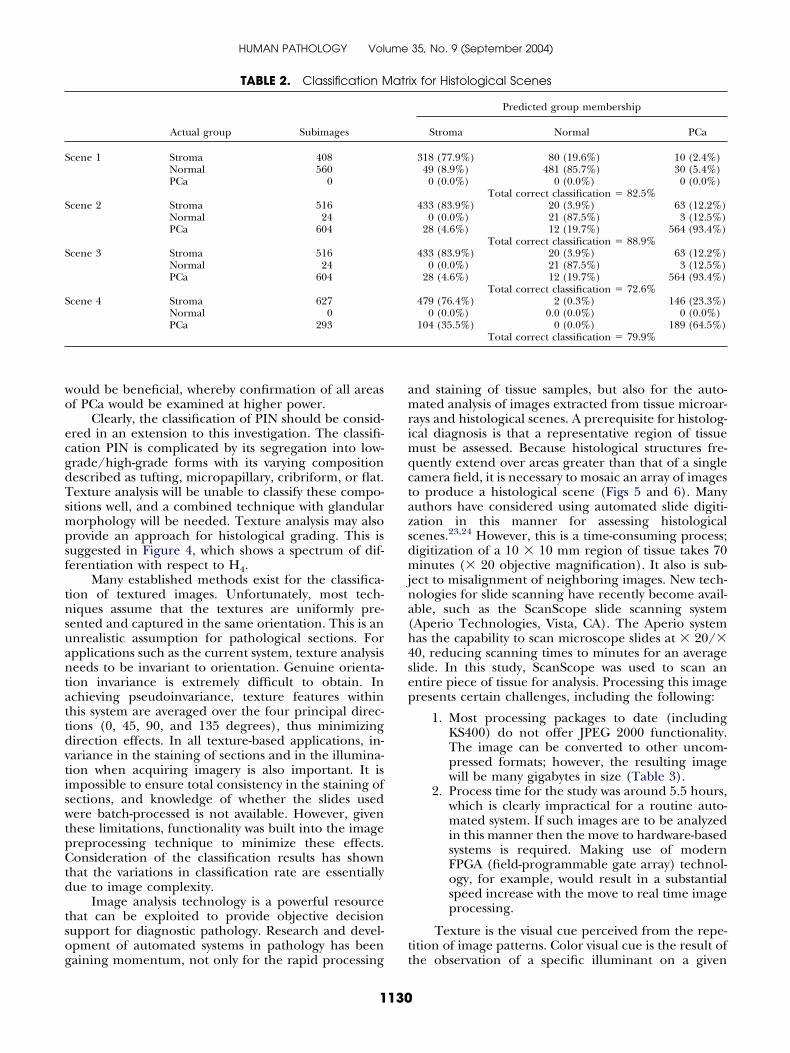
TABLE 2. Classification M
Actual group Subimages
cene 1 Stroma 408Normal 560PCa 0
cene 2 Stroma 516Normal 24PCa 604
cene 3 Stroma 516Normal 24PCa 604
cene 4 Stroma 627Normal 0PCa 293
aining momentum, not only for the rapid processing t
1130
nd staining of tissue samples, but also for the auto-ated analysis of images extracted from tissue microar-
ays and histological scenes. A prerequisite for histolog-cal diagnosis is that a representative region of tissue
ust be assessed. Because histological structures fre-uently extend over areas greater than that of a singleamera field, it is necessary to mosaic an array of imageso produce a histological scene (Figs 5 and 6). Manyuthors have considered using automated slide digiti-ation in this manner for assessing histologicalcenes.23,24 However, this is a time-consuming process;igitization of a 10 � 10 mm region of tissue takes 70inutes (� 20 objective magnification). It also is sub-
ect to misalignment of neighboring images. New tech-ologies for slide scanning have recently become avail-ble, such as the ScanScope slide scanning systemAperio Technologies, Vista, CA). The Aperio systemas the capability to scan microscope slides at � 20/�0, reducing scanning times to minutes for an averagelide. In this study, ScanScope was used to scan anntire piece of tissue for analysis. Processing this imageresents certain challenges, including the following:
1. Most processing packages to date (includingKS400) do not offer JPEG 2000 functionality.The image can be converted to other uncom-pressed formats; however, the resulting imagewill be many gigabytes in size (Table 3).
2. Process time for the study was around 5.5 hours,which is clearly impractical for a routine auto-mated system. If such images are to be analyzedin this manner then the move to hardware-basedsystems is required. Making use of modernFPGA (field-programmable gate array) technol-ogy, for example, would result in a substantialspeed increase with the move to real time imageprocessing.
Texture is the visual cue perceived from the repe-ition of image patterns. Color visual cue is the result of
ix for Histological Scenes
Predicted group membership
Stroma Normal PCa
318 (77.9%) 80 (19.6%) 10 (2.4%)49 (8.9%) 481 (85.7%) 30 (5.4%)0 (0.0%) 0 (0.0%) 0 (0.0%)
Total correct classification � 82.5%433 (83.9%) 20 (3.9%) 63 (12.2%)
0 (0.0%) 21 (87.5%) 3 (12.5%)28 (4.6%) 12 (19.7%) 564 (93.4%)
Total correct classification � 88.9%433 (83.9%) 20 (3.9%) 63 (12.2%)
0 (0.0%) 21 (87.5%) 3 (12.5%)28 (4.6%) 12 (19.7%) 564 (93.4%)
Total correct classification � 72.6%479 (76.4%) 2 (0.3%) 146 (23.3%)
0 (0.0%) 0.0 (0.0%) 0 (0.0%)104 (35.5%) 0 (0.0%) 189 (64.5%)
Total correct classification � 79.9%
atr
he observation of a specific illuminant on a given

sabiPtcacd
icbtbPcithlah
wircawo
R
cr
r1
m
sH
sA
bP
t1
sp
me
p2
1
tG
sU
fQ
mQ
mc
lt
ht
tC
i
tC
fi
i
tg
TTCCCCIIIFSS
TISSUE COMPOSITION IN PROSTATIC NEOPLASIA (Diamond et al)
urface using 3 different types of sensors (red, green,nd blue [RGB]). Because images are obtained in 24-it format from the scanner, the possibility for extend-
ng texture analysis into the RGB domain is possible.rocessing each color band separately or deriving tex-ural information from the luminance plane along withhrominance features has been shown to increase theccuracy of the classification process. Consequently,olor information may play a role in improving theiscriminative power of machine vision systems.
In conclusion, the diagnosis of PCa is complex ands based on many histological observations. The majorriteria are infiltrative growth pattern, absence of aasal cell layer, and presence of macronucleoli. Tex-ure analysis of medium-power scans has been shown toe capable of distinguishing between the first 2 criteria.revious studies have shown that the presence of nu-leoli and other morphological features is an importantndicator of malignancy.22 Consequently, it is obvioushat full automation will require the identification ofigh-power clues for diagnostic confirmation. However,
ow-power scans provide the means of identifying keyreas of the tissue section that require scrutiny atigher power.
Advances in automated imaging in histopathologyill continue, and tissue texture is likely to have an
mportant role. Future work will require close collabo-ation between pathologists, software engineers, andomputer scientists. This not only will enhance ourbility to rapidly analyze patient material in an objectiveay, but also will possibly identify new reliable markersf diagnosis and prognosis.
EFERENCES
1. The Institute of Cancer Research: Prostate cancer set to be-ome the most common men’s cancer within three years [presselease]. May 27, 2002
TABLE 3. Scanning Details for Histological Slide
Description Value
issue (X dimension) 27 mmissue (Y dimension) 19 mmompression JPEG2000olor depth 24 bitompression quality 30ompression ratio 16.38
mage width 58,877 pixelsmage height 42,336 pixelsmage size 7,477,850,016 bytesile size 456,416,687 bytescanning resolution 54000 pixels/inchcanning Speed 40 mm2/minute
1131
2. O’Leary TJ, Tellado M, Bruckner SB, et al: PAPNET-assistedescreening of cervical smears: Cost and accuracy compared with00% manual rescreening strategy. JAMA 279:235-237, 1998
3. Patten SF, Lee JS, Nelson AC: NeoPath AutoPap 300 auto-ated Pap screener system. Acta Cytol 40:45-52, 1996
4. Thompson D, Bartels PH, Bartels H, et al: Knowledge-basedegmentation of colorectal histologic imagery. Anal Quant Cytolistol 15:236-246, 1993
5. Hamilton PW, Thompson D, Sloan J, et al: Knowledge-guidedegmentation and morphometric analysis of colorectal dysplasia.nal Quant Cytol Histol 17:172-182, 1995
6. Bartels PH, Thompson D, Bartels H, et al: Machine vision–ased histometry of premalignant and malignant prostatic lesions.athol Res Pract 191:935-944, 1995
7. Thompson D, Bartels PH, Bartels H, et al: Image segmenta-ion of cribriform gland tissue. Anal Quant Cytol Histol 17:314-322,995
8. Anderson NH, Hamilton PW, Bartels PH, et al: Computerizedcene segmentation for the discrimination of architectural features inroliferative lesions of the breast. J Pathol 181:374-380, 1997
9. Keenan S, Diamond J, McCluggage WG, et al: An automatedachine vision system for the histological grading of cervical intra-
pithelial neoplasia (CIN). J Pathol 192:351-362, 200010. Baak JPA: The framework of pathology: Good laboratory
ractice by quantitative and molecular methods. J Pathol 198:277-83, 2002
11. Julesz B: Texture and visual perception. Sci Am 212:38-48,965
12. Richard WD, Keen CG: Automated texture-based segmenta-ion of ultrasound images of the prostate. Comput Med Imagingraph 20:131-140, 1996
13. Basset O, Sun Z, Mestas JL, et al: Texture analysis of ultra-onic images of the prostate by means of co-occurrence matrices.ltrason Imaging 15:218-237, 1993
14. Christen R, Xiao J, Ninimo C, et al: Chromatin textureeatures in hematoxylin and eosin–stained prostate tissue. Analuant Cytol Histol 15:383-388, 1993
15. Bartels PH, Montironi R, Hamilton PW, et al: Nuclear chro-atin texture in prostatic lesions: 1. PIN and adenocarcinoma. Analuant Cytol Histol 20:389-396, 1998
16. Bartels PH, Montironi R, Hamilton PW, et al: Nuclear chro-atin texture in prostatic lesions: 2. PIN and malignancy-associated
hanges. Anal Quant Cytol Histol 20:397-406, 199817. Hamilton PW, Bartels PH, Thompson D, et al: Automated
ocation of dysplastic fields in colorectal histology using image tex-ure analysis. J Pathol 182:68-75, 1997
18. Hamilton PW, Bartels PH, Montironi R, et al: Automatedistometry in quantitative prostate pathology. Anal Quant Cytol His-
ol 20:443-460, 199819. Bartels PH, Bartels H, Montironi R, et al: Machine vision in
he detection of prostate lesions in histologic sections. Anal Quantytol Histol 20:358-364, 1998
20. Haralick RM, Shanmugsm K, Dinstein I: Texture features formage classification. IEEE Trans Syst Man Cybern 3:610-621, 1973
21. Bartels PH, Bartels H, Montroni R, et al: Machine vision inhe detection of prostate lesions in histologic sections. Anal Quantytol Histol 20:358-364, 1998
22. Varma M, Lee MW, Tamboli P, et al: Morphologic criteriaor the diagnosis of prostatic adenocarcinoma in needle biopsy spec-mens. Arch Pathol Lab Med 126:554-561, 2002
23. Thompson D, Richards D, Bartels H, et al: Multimegapixelmages in histopathology. Anal Quant Cytol Histol 23:169-177, 2001
24. Leong FJW-M, McGee JO’D: Automated complete slide digi-ization: A medium for simultaneous viewing by multiple patholo-ists. J Pathol 195:508-514, 2002