the value of cdx2 and cytokeratins 7 and 20 expression in
TRANSCRIPT

RESEARCH Open Access
The value of CDX2 and cytokeratins 7 and 20expression in differentiating colorectaladenocarcinomas from extraintestinalgastrointestinal adenocarcinomas: cytokeratin 7-/20+ phenotype is more specific than CDX2antibodyReyhan Bayrak, Hacer Haltas* and Sibel Yenidunya
Abstract
Background/Objective: Metastatic adenocarcinoma from an unknown primary site is a common clinical problem.Determining the cytokeratin (CK) 7/CK20 pattern of tumors is one of the most helpful procedures for this purposesince the CK7-/CK20+ pattern is typical of colorectal adenocarcinomas. CDX2, a critical nuclear transcription factorfor intestinal development, is expressed in intestinal epithelium and adenocarcinomas. In the present study, wecompared the sensitivity and specificity of CDX2 expression and the CK7-/CK20+ phenotype in differentiatingcolorectal adenocarcinomas from pancreatic and gastric adenocarcinomas.
Methods: CK7/CK20 staining pattern and CDX2 expression were evaluated in 118 cases of colorectal, 59 cases ofgastric, and 32 cases of pancreatic adenocarcinomas. The sensitivity, specificity, and positive and negativepredictive values of the CK7-/CK20+ phenotype and of CDX2 expression were analyzed.
Results: The CK7-/CK20+ immunophenotype was expressed by 75 of 118 (64%) colorectal and 3 of 59 (5%) gastrictumors and was not observed in any pancreatic adenocarcinomas. The CK7+/CK20+ immunophenotype wasexpressed in 24/118 (20%) of colon, 28/59 (48%) of gastric and 7/32 (22%) of pancreatic adenocarcinomas. The CK7+/CK20- expression pattern was observed in only 2% (2 of 118) of colorectal carcinomas. CDX2 was expressed in114 of 118 (97%) colorectal, 36 of 59 (61%) gastric, and 5 of 32(16%) pancreatic adenocarcinomas. There was nosignificant association between CDX2 expression and tumor differentiation in colorectal carcinomas. In gastriccarcinomas, CDX2 expression was more common in intestinal type tumors than in diffuse type carcinomas. TheCK7-/CK20+ phenotype showed a specificity of 96.7% in predicting colorectal adenocarcinomas, which wassuperior to that of CDX2 expression. CDX2 expression at both cut-off levels (> 5% and > 50%) had a highersensitivity (96.6% and 78%) than the CK phenotype.
Conclusions: Both the CK7-/CK20+ phenotype and expression of the antibody CDX2 are highly specific andsensitive markers of colorectal origin. CDX2 expression should be a useful adjunct for the diagnosis of intestinaladenocarcinomas, particularly when better established markers such as CK7 and CK20 yield equivocal results. TheCK7-/CK20+ phenotype is superior in its specificity and positive predictive value and might be preferred.
Virtual slides: The virtual slide(s) for this article can be found here:http://www.diagnosticpathology.diagnomx.eu/vs/4851011866354821
Keywords: gastrointestinal adenocarcinomas, CK7, CK20, CDX2, immunohistochemistry
* Correspondence: [email protected] of Pathology, Fatih University Hospital, Ankara, Turkey
Bayrak et al. Diagnostic Pathology 2012, 7:9http://www.diagnosticpathology.org/content/7/1/9
© 2012 Bayrak et al; licensee BioMed Central Ltd. This is an Open Access article distributed under the terms of the Creative CommonsAttribution License (http://creativecommons.org/licenses/by/2.0), which permits unrestricted use, distribution, and reproduction inany medium, provided the original work is properly cited.

BackgroundMetastatic adenocarcinoma from an unknown primarysite is a common clinical problem that leads to extensiveand costly clinical and radiological examinations, some-times with discouraging results [1,2]. It is often impor-tant to determine the site of origin of a metastaticcarcinoma of unknown primary site, particularly becausethis may affect the choice of the treatment. A more pre-cise diagnosis leads to more effective treatment, substan-tially improving the overall outcome [3]. Determinationof the primary site may take several steps. Clinical fea-tures, such as age, sex, and site of metastases may give afirst indication. The histological assessment is often veryhelpful, but may not differentiate adequately betweenvarious primary tumors. Immunohistochemistry is themost common adjunctive method used in the analysis ofthe patient with cancer of unknown primary site [4,5].Cytokeratins (CKs) represent the epithelial class of inter-
mediate-sized filaments of the cytoskeleton. There are 20subtypes of cytokeratin (CK) intermediate filaments.These have different molecular weights and demonstratedifferential expression in various cell types and tumors [6].Among the most useful cytokeratins are CK7 and CK20[7]. CK7 is found in many ductal and glandular epithelia,including lung, breast, ovary, and endometrium [8,9].CK20 is expressed in the gastrointestinal (GI) epithelium,urothelium, and Merkel cells [10]. The combined expres-sion patterns of CK7 and CK20 have been extensively stu-died in various primary and metastatic carcinomas[5,7,11-17]. CK20 is expressed alone in the majority ofintestinal adenocarcinoma and in Merkel cell carcinomaswhereas CK7 is present without CK20 in most breast, lungand ovarian adenocarcinoma, and with CK20 in urothelial,pancreatic and gastric carcinomas. The CK7-/CK20+expression pattern is known to be highly characteristic ofcolorectal carcinomas [11,12,17-19], however, not all col-orectal carcinomas show the CK7-/CK20+ expression pat-tern. Occasionally colorectal carcinomas may showsignificant CK7 expression and conversely, expression ofCK20 may be seen in a variety of non-colorectal adenocar-cinomas such as urothelial, gastric and pancreatobiliarytract carcinomas [20-24]. For this reason, there is contin-ued interest in the development of new and more specificmarkers of intestinal differentiation and CDX2 appears tobe such a marker.CDX2 is a caudal-type homeobox gene, encoding a
transcription factor that plays an important role in pro-liferation and differentiation of intestinal epithelial cells[25]. The protein (CDX2) is normally expressedthroughout embryonic and postnatal life within nucleiof intestinal epithelial cells from the proximal duode-num to the distal rectum [26,27]. Previous studiesshowed that CDX2 is expressed in normal and
neoplastic intestinal epithelial cells with a relatively highsensitivity and specificity and that it can be used as animmunohistochemical marker for neoplasms of intest-inal origin [28-32]. However, CDX2 expression was alsofound in gastric carcinoma, and other carcinomas withintestinal-type morphology [33-36].In the present study, we examined the expression pro-
files of CK7, CK20, and CDX2 immunohistochemicalmarkers in primary colorectal, gastric and pancreaticadenocarcinomas in consideration of the potentialapplicability of these markers in the clinical context ofmetastatic adenocarcinomas. We also evaluated the sen-sitivity, specificity, positive predictive value, and negativepredictive value of CDX2 expression and CK7-/CK20+immunophenotype to differentiate colorectal adenocarci-nomas from pancreatic and gastric adenocarcinomas.
Materials and methodsCase selection and tissue samplesOne hundred eighteen colorectal, 59 gastric and 32 pan-creatic adenocarcinoma resection specimens wereretrieved from the archival files of the Department ofPathology, Fatih University Medical School, between Jan-uary 2006 and December 2009. Pathological findings,including histological type, histological differentiation,depth of invasion, and lymph node status, were gatheredfrom hematoxylin and eosin stained sections. All caseswere reviewed to confirm the diagnosis. The grade andhistological type of colorectal and pancreatic adenocarci-nomas were determined according to criteria of theWorld Health Organization (WHO) Classification ofTumors [37]. Well and moderately differentiated tumorswere grouped together as low-grade tumors and werecompared with high-grade tumors, which includedpoorly differentiated and undifferentiated tumors, andsignet ring cell carcinomas. Histological typing of gastriccarcinomas was made according to Lauren classification[38]. Adenocarcinomas of intestinal type, which werewell or moderately differentiated, were recorded as lowgrade tumors, whereas the poorly differentiated intestinaltype adenocarcinomas and the diffuse type adenocarcino-mas were recorded as high grade tumors. Postoperativepathological staging was performed according to theAmerican Joint Committee on Cancer (AJCC) TNM sta-ging system [39]. One paraffin block with the maximumamount of tumor and proper fixation was selected fromeach case for immunohistochemical studies. This studywas approved by Ethics Committee of Fatih UniversityHospital (09.23.2010/B 302 FTH 0200000)
Immunohistochemical AnalysisFour μm-thick sections were cut from blocks of paraffinembedded tissue, deparaffinized, and rehydrated as
Bayrak et al. Diagnostic Pathology 2012, 7:9http://www.diagnosticpathology.org/content/7/1/9
Page 2 of 11

usual. To reduce non-specific background staining dueto endogenous peroxidase, slides were incubated inHydrogen Peroxide Block for 15 min. Before immunos-taining, antigen retrieval was performed by incubatingthe slides for 15 min with pepsin (LabVision; catalog no.AP-9007) at a concentration of 1mg/ml for CK20. Slideswere microwaved in 10 mM of citric acid at pH6.0 for
20 min for CK7 and CDX2. The slides were incubatedfor 60 min with primary antibodies to CK7 (clone OV-TL 12/30, LabVision/NeoMarkers; 1:50), CK20 (cloneKs20.8, Dako; 1:50) and CDX2 (clone AMT 28, Novo-Castra; 1:50) at room temperature. The Standard avidin-biotin-peroxidase complex (ABC) technique was per-formed using the LabVision Secondary Detection Kit(UltraVision Detection System Anti-polyvalent, HRP).AEC was used as chromogen. All slides were counterstained with Mayer’s hematoxylin.
Microscopic EvaluationFor CDX2, only nuclear staining was considered posi-tive. Cytoplasmic positivity was infrequently encoun-tered, and was considered an artifact. Positiveimmunostaining for CK7 and CK20 was identified in thecytoplasm, cell membrane, or both. The percentage ofpositive cells was scored in a semiquantitative methodaccording to the following scheme: 0 (less than 5% oftumor cells); 1+ (positive signal of any intensity in 5-25% of tumor cells); 2+ (26-50% of tumor cells); 3+ (51-75% of tumor cells); and 4+ (greater than 75% of tumorcells). Furthermore, staining in less than 50% of thetumor cells was considered focal, and staining in morethan 50% of the tumor cells was considered diffuse posi-tivity. In general, cases showing 3+ and 4+ staining alsohad strong intense staining, so intensity was not used indetermination of the final reactivity score. Normal colo-nic mucosal tissue was used as a CK20 and CDX2-posi-tive control, and normal pancreatic tissue was used as aCK7-positive control. For negative control samples, theprimary antibody was omitted for each run.
Statistical analysisc2and Fisher exact tests were used to compare the dif-ferences in percentages of positive results betweengroups. SPSS 13.0 for Windows was used for all statisti-cal analyses. The sensitivity, specificity, and positive andnegative predictive values of the CK7-/CK20+ phenotypeand of CDX2 expression were counted.
ResultsTables 1 and 2 show the percentage of cases thatstained with CDX2, CK7, and CK20 in colorectal adeno-carcinomas, gastric adenocarcinomas and pancreaticadenocarcinomas.
CK7 and CK20CK7 expression was detected in 22% (26/118) of color-ectal, in 80% (47/59) of gastric, and in 97% (31/32) ofpancreatic adenocarcinomas. CK20 reactivity was foundin 84% (99/118) of colorectal, in 53% (31/59) of gastric,and in 22% (7/32) of pancreatic adenocarcinomas. TheCK7-/CK20+ immunophenotype was expressed by 75 of118 (64%) colorectal and 3 of 59 (5%) gastric tumorsand was not observed in any pancreatic adenocarcino-mas (c2 = 79.992; p < 0.001). The CK7+/CK20+ immu-nophenotype was expressed in 24/118 (20%) of colon,28/59 (48%) of gastric and 7/32 (22%) of pancreatic ade-nocarcinomas, which was not helpful in the differentialdiagnosis. However, among the CK20 positive cases,CK20 reactivity was diffuse (more than 50% of cellswere positive) in the majority of colorectal carcinomasin 64% (63/99) of the cases and mainly focal ( < 50% ofcells were positive) in gastric and pancreatic adenocarci-nomas in 71% (22/31) and 100% (7/7) of cases respec-tively (c2 = 19.509; p < 0.001) (Figure 1). Conversely,among the CK7 positive cases, CK7 reactivity was dif-fuse in the majority of gastric and pancreatic adenocar-cinomas in 74% (35/47) and 94% (29/31) of casesrespectively, and this reactivity was focal in 54% (14/26)of colorectal carcinomas (c2 = 16.228;p < 0.001) (Figure
Table 1 Distribution of CK7, CK20 and CDX2 staining with percentages of positive cells in primary colorectal, gastricand pancreatic adenocarcinomas
Negative Positive Total
0 1+ 2+ 3+ 4+
Colorectal adenocarcinoma CK7 92 (78%) 8 (7%) 6 (5%) 5 (4%) 7 (6%) 118
CK20 19 (16%) 20 (17%) 16 (13%) 22 (19%) 41 (35%) 118
CDX2 4 (3%) 7 (6%) 15 (13%) 21 (18%) 71 (60%) 118
Gastric adenocarcinoma CK7 12 (20%) 5 (9%) 7 (12%) 6 (10%) 29 (49%) 59
CK20 28 (47%) 16 (27%) 6 (10%) 8 (14%) 1 (2%) 59
CDX2 23 (39%) 13 (22%) 9 (15%) 10 (17%) 4 (7%) 59
Pancreatic adenocarcinoma CK7 1 (3%) 1 (3%) 1 (3%) 9 (28%) 20 (63%) 32
CK20 25 (78%) 6 (20%) 1 (3%) 0 (0%) 0 (0%) 32
CDX2 27 (85%) 3 (9%) 2 (6%) 0 (0%) 0 (0%) 32
Bayrak et al. Diagnostic Pathology 2012, 7:9http://www.diagnosticpathology.org/content/7/1/9
Page 3 of 11

2). The CK7+/CK20- expression pattern was observed inonly 2% (2 of118) of colorectal carcinomas, although itwas expressed in 32% (19/59) of gastric and 75% (24/32)of pancreatic adenocarcinomas (c2 = 85.607; p < 0.001).In our study, 17(14%) colorectal, 9 (15%) gastric, andonly 1 (3%) pancreatic adenocarcinomas showed aCK7-/CK20- immunophenotype.CK7 and CK20 expression were compared with the
clinicopathological characteristics of the tumors (Table
3). No association between CK7 expression and anatomi-cal location of carcinomas, tumor type, stage, and gradewas found. No association was observed among CK20expression and tumor type, tumor stage (pT), or nodalstatus. Among the colorectal tumors, CK20 positivity wasmore common in rectal carcinomas than in nonrectalcolon carcinomas (89% versus 70%, c2 = 6.839; p = 0.009)and in low grade carcinomas than in high grade carcino-mas (91% versus 55%, c2 = 17,247; p < 0.001).
Table 2 CK7/20 phenotype and CDX2 expression in our studied groups
Colorectal AdenoCa(n = 118)
Gastric AdenoCa(n = 59)
Pancreatic AdenoCa(n = 32)
CDX2+ CDX2- CDX2+ CDX2- CDX2+ CDX2-
CK7-/CK20+ 74 (62%) 1 (1%) 3 (5%) 0 (0%) 0 (0%) 0 (0%)
CK7+/CK20+ 22 (19%) 2 (2%) 21 (36%) 7 (12%) 2 (6%) 5 (16%)
CK7+/CK20- 2 (2%) 0 (0%) 8 (14%) 11 (19%) 3 (9%) 21 (66%)
CK7-/CK20- 16 (14%) 1 (1%) 4 (7%) 5 (9%) 0 (0%) 1 (3%)
A
A B
C
Figure 1 CK20 staining in colorectal, gastric, and pancreatic adenocarcinomas. (A) Colorectal adenocarcinoma displayed diffuse and strongimmunoreactivity in the cytoplasm of cancer cells. (B) Gastric and (C) pancreatic adenocarcinomas exhibited focal cytoplasmic staining for CK20.Original magnification × 100.
Bayrak et al. Diagnostic Pathology 2012, 7:9http://www.diagnosticpathology.org/content/7/1/9
Page 4 of 11

CDX2CDX2 was expressed in 114 of 118 (97%) colorectal, 36of 59 (61%) gastric, and 5 of 32 (16%) pancreatic adeno-carcinomas (c2 = 93.576; p < 0.001). In positive cases,the immunoreactivity was predominantly nuclear withoccasional faint cytoplasmic staining. The majority ofcases (92/114, 81%) demonstrated strong and diffuseimmunostaining in more than 50% of cells in colorectaltumors. Among the CDX2 positive gastric carcinomas(36/59), reactivity was focal in 22 cases (22/36, 61%).Among the 32 cases of pancreatic adenocarcinoma, only5 cases were focally positive for CDX2 (c2 = 33.462; p <0.001) (Figure 3).CDX2 expression was also compared with the clinico-
pathological characteristics of the tumors (Table 3). Ingastric carcinomas CDX2 expression was more commonin intestinal type tumors than in diffuse type carcinomas(77% versus 45%, c2 = 6.284; p = 0.012). There was nosignificant association between CDX2 expression and
tumor differentiation in colorectal carcinomas (98% oflow grade tumors and 91% of high grade tumors werepositive for CDX2) (Figure 4). Conversely, among gastriccarcinomas CDX2 positivity was more common in lowgrade carcinomas than in high grade carcinomas (80%versus 51%, c2 = 4.584; p = 0.032). No association wasobserved among CDX2 expression and anatomical loca-tion of carcinomas, tumor stage (pT), or nodal status.
Comparison of CK7/20 staining pattern and CDX2expressionThe CK7-/CK20+/CDX2+ phenotype was highest,accounting for 63% (74/118) of colorectal adenocarcino-mas. In gastric and pancreatic adenocarcinomas, CK7+/CK20+/CDX2+ (21/59, 36%) and CK7+/CK20-/CDX2- (21/32, 66%) were the most common patternsrespectively. In CK7+/CK20+ tumors, CDX2 expressionwas observed in 22 of 24 (92%) colorectal, 21 of 28(75%) gastric, and 2 of 7 (29%) pancreatic carcinomas.
A
B
C
Figure 2 CK7 immunostaining in colonic, gastric, and pancreatic tumors. (A) Gastric and (B) pancreatic carcinomas showed diffuse andstrong positive CK7 immunostaining. (C) Colonic adenocarcinoma displayed focal cytoplasmic immunoreactivity for CK7. (A)-(B), originalmagnification × 100; (C), original magnification × 200.
Bayrak et al. Diagnostic Pathology 2012, 7:9http://www.diagnosticpathology.org/content/7/1/9
Page 5 of 11

This reactivity was diffuse in majority of colorectal car-cinomas in 68% (15/22) of the cases and mainly focal ingastric and pancreatic adenocarcinomas in 57% (12/21)and 100% (2/2) of cases respectively (c2 = 5.979; p =0.051). Among the CK7-/CK20- colorectal tumorsCDX2 was positive in 16 of 17 (94%) cases.We also evaluated the sensitivity, specificity, positive
predictive value, and negative predictive value ofCDX2 expression and CK7-/CK20+ immunophenotypeto differentiate colorectal adenocarcinoma from pan-creatic and gastric adenocarcinomas (Table 4). Deter-mining the CK7/CK20 phenotype proved to be morespecific in differentiating colorectal adenocarcinomafrom pancreatic and gastric adenocarcinomas (specifi-city 96.7%) than the expression of CDX2 was. TheCK7-/CK20+ phenotype had a superior positive predic-tive value (96.2%) in these circumstances. CDX2expression at both cut-off levels (> 5% and > 50%) hada higher sensitivity (96.6% and 78%) and higher nega-tive predictive value (92.6% and 74.8%) than the CKphenotype. The specificity of CDX2 expression did notreach the level of specificity of CK7/CK20 phenotypeat a > 50% level, either (84.6%).
DiscussionThe diagnosis of the metastatic carcinoma of unknownorigin can be very difficult. The determination of theprimary site of the metastasis is a challenge to bothoncologists and pathologists, having potentially impor-tant clinical and therapeutic consequences [1-3]. In thesetting of carcinomas of unknown primary, clinicopatho-logical correlation and a panel of standard immunos-tains help define the primary site, and direct appropriatetreatment [4,5].Cytokeratins are group of approximately 20 proteins
that consist of a type of intermediate filament and aredifferentially expressed in epithelia of various sites. The
cytokeratins most often used are CK7 and CK20 [7-10].CK7 is found in the glandular epithelium and epithelialtumors of lung, ovary, endometrium and breast, but isnot found in GI epithelium. Conversely, CK20 isexpressed principally in the normal glands and epithelialtumors of the GI tract, urothelium, and Merkel cells.The cytokeratin 7/20 profile of a particular tumor hasproved to be a useful aid in differential diagnosis of car-cinomas, since primary and metastatic tumors tend toretain the cytokeratin profiles of the epithelium fromwhich they arise [13]. In his review article, Tot summar-ized the results of 29 studies containing more than 3500reported cases of adenocarcinomas stained with CK20and CK7. This review stated that metastatic colorectal,gastric and pancreatic adenocarcinomas have similarCK7 and CK20 staining ratios as their respective pri-mary tumors. Only gastric adenocarcinomas showed sta-tistically significant differences in CK20 expression whenthe primary and metastatic locations were compared[13].Normal epithelium of the small bowel, appendix and
colorectum, and adenocarcinomas from these sites, arealmost consistently CK7-/CK20+, helping to distinguishthese adenocarcinomas from adenocarcinomas of manyother primary sites [9-15]. The CK7-/CK20+ patternwas identified in 65% to 95% of the colorectal adenocar-cinomas in different series [11,12,20-23,31]. On theother hand approximately one third of gastric and lessthan 10% of pancreatic adenocarcinomas also show thispattern [11,12,23]. The CK7-/CK20+ immunophenotypewas expressed by 75 of 118 (64%) colorectal and 3 of 59(5%) gastric tumors and was not observed in any pan-creatic adenocarcinomas in the present study. Therefore,it has been presumed that CK7 is not typically expressedby colonic epithelial tumors. Interestingly, severalreports have described CK7 expression in colorectaladenocarcinoma, with expression ranging from 5% to
Table 3 Expression of CK7, CK20, and CDX2 in cancer tissues by histopathological characteristics
CK7+ CK20+ CDX2+
n % n % n %
Colorectal adenocarcinoma (n = 118)
Low grade (n = 96) 22 22.9 87 90.6 * 94 97.9
High grade (n = 22) 4 18.2 12 54.5 * 20 90.9
Rectal (n = 85) 18 21.2 76 89.4§ 83 97.6
Nonrectal (n = 33) 8 24.2 23 69.7§ 31 93.9
Gastric adenocarcinoma (n = 59)
Intestinal type (n = 30) 21 70.0 16 53.3 23 76.7 #
Diffuse type (n = 29) 26 89.7 15 51.7 13 44.8 #
Pancreatic adenocarcinoma (n = 32)
Low grade (n = 30) 30 100.0 7 23.3 5 16.7
High grade (n = 2) 1 50.0 0 0.0 0 0.0
* p < 0.001 § p = 0.009 # p = 0.012
Bayrak et al. Diagnostic Pathology 2012, 7:9http://www.diagnosticpathology.org/content/7/1/9
Page 6 of 11
74% [11,12,22,23,31]. The reasons for this discrepancyare unclear. However, this may be the result of differ-ences in the studied population or the interpretive cri-teria that was used. In our study, CK7 expression wasdetected in 22% (26/118) of colorectal adenocarcinomas.In comparison with the CK7-/CK20+ immunoprofile,
the CK7+/CK20+ immunoprofile is commonly present
A B
C
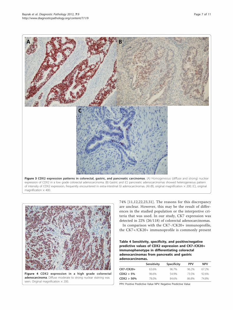
Figure 3 CDX2 expression patterns in colorectal, gastric, and pancreatic carcinomas. (A) Homogeneous (diffuse and strong) nuclearexpression of CDX2 in a low grade colorectal adenocarcinoma. (B) Gastric and (C) pancreatic adenocarcinomas showed heterogeneous patternof intensity of CDX2 expression, frequently encountered in extra-intestinal GI adenocarcinomas. (A)-(B), original magnification × 200; (C), originalmagnification × 400.
Figure 4 CDX2 expression in a high grade colorectaladenocarcinoma. Diffuse moderate to strong nuclear staining wasseen. Original magnification × 200.
Table 4 Sensitivity, specificity, and positive/negativepredictive values of CDX2 expression and CK7-/CK20+immunophenotype in differentiating colorectaladenocarcinomas from pancreatic and gastricadenocarcinomas.
Sensitivity Specificity PPV NPV
CK7-/CK20+ 63.6% 96.7% 96.2% 67.2%
CDX2 > 5% 96.6% 54.9% 73.5% 92.6%
CDX2 > 50% 78.0% 84.6% 86.8% 74.8%
PPV: Positive Predictive Value NPV: Negative Predictive Value
Bayrak et al. Diagnostic Pathology 2012, 7:9http://www.diagnosticpathology.org/content/7/1/9
Page 7 of 11

in urothelial carcinomas, gastric carcinomas and tumorsof the pancreatobiliary tract [11,12,15]. Gastric adeno-carcinomas are the most heterogeneous subgroup ofcarcinomas with respect to their CK7/CK20 immuno-phenotype. While most gastric adenocarcinomas areCK20+, they may or may not be CK7+ [11,12,23]. Theresults of CK7/CK20 immunohistochemistry for cholan-giocarcinomas, gall bladder carcinoma and pancreaticductal adenocarcinoma are conflicting. While all studieshave found CK7 immunopositivity in these tumours,many studies have found the majority are CK20-[40,41], while others have found the majority to beCK20+ [11,12]. In the present study the largest propor-tion of gastric carcinomas was of the CK7+/CK20+ phe-notype (48%), and a substantial proportion was of theCK7+/CK20- phenotype (32%). CK7+/CK20- immuno-profile was the most common pattern, accounting for75% of pancreatic adenocarcinomas. The CK7+/CK20+immunophenotype was expressed in 20% of colon, 48%of gastric and 22% of pancreatic adenocarcinomas,which was not helpful in the differential diagnosis. How-ever, CK20 reactivity was diffuse (more than 50% of cellswere positive) in the majority of colorectal carcinomacases and mainly focal ( < 50% ofcells were positive) ingastric and pancreatic adenocarcinomas as in previousstudies [22,23,31,41].Since, occasional colorectal carcinomas may show sig-
nificant CK7 expression and conversely, expression ofCK20 may be seen in a variety of non-colorectal adeno-carcinomas, there is interest in the development of newand more specific markers of intestinal differentiation.Human CDX2 protein is a member of the homeoboxgenes that encodes an intestine-specific transcriptionfactor. This protein regulates intestinal development andis expressed in the nuclei of epithelial cells throughoutthe intestinal tract in embryonic and postnatal life[25-27]. Expression of CDX2 mRNA has been shown tobe highly restricted to intestinal epithelium [42]. Thesensitivity and specificity of antibodies to CDX2 proteinas a marker of colonic adenocarcinoma has beenrecently evaluated in various studies with reported sensi-tivity and specificity of greater than 90% [28-33]. Wer-ling et al [28] examined CDX2 expression across 476samples of human tumors and concluded that it is anexcellent marker of adenocarcinomas arising in the GItract, particularly the duodenum and colon. Theseauthors reported that high levels (> 75% positive cells)of CDX2 expression were found almost exclusively inadenocarcinomas of the colorectum, and intermediatelevels (26%-75% positive cells) of immunostaining werefound in many adenocarcinomas arising elsewhere inthe GI tract. They also demonstrated that primary andmetastatic colorectal carcinomas showed remarkablysimilar scoring patterns. All primary and 25 of 26
metastatic colonic adenocarcinomas showed high levelsof CDX2 expression (2+ or 3+) in this study. In anotherstudy, Kaimaktchiev et al [32] observed a greater than80% concordance for CDX2 expression in the analysisof matched primary and lymph node metastases. Inaddition, all 17 colorectal metastases examined by wholesections were CDX2 positive in this study. Using tissuemicroarrays, Moskaluk et al [29] analyzed CDX2 stain-ing in 745 samples of human cancer and arrived at simi-lar conclusions. Barbareschi et al [30] compared CDX2expression in primary and metastatic tumors found inthe lung and concluded that this marker is highly selec-tive for tumors originating from the colon and rectum,but also stains metastases from the stomach and ovary.In our study, CDX2 was expressed in 114 of 118 (97%)colorectal, 36 of 59 (61%) gastric, and 5 of 32(16%) pan-creatic adenocarcinomas. The majority of cases (92/114,81%) demonstrated strong and diffuse immunostainingin more than 50% of cells in colorectal tumors. Amongthe CDX2 positive gastric carcinomas (36/59), reactivitywas focal in 22 cases (22/36, 61%). Among the 32 casesof pancreatic adenocarcinoma, only 5 cases were focallypositive for CDX2.Among colorectal adenocarcinomas, the relationship
between tumor grade and CDX2 staining has been con-troversial. Hinoi et al [43] demonstrated that a rare sub-set of poorly differentiated colonic carcinomas termedlarge cell minimally differentiated carcinoma or medul-lary carcinoma are characterized by microsatelliteinstability and loss of CDX2 expression. Kaimaktchiev etal [32] recently studied tissue microarray samples of1109 colorectal adenocarcinomas and found a lack ofCDX2 reactivity in 14 (28%) of 50 poorly differentiatedtumors. They concluded that CDX2 expressiondecreases with tumor differentiation. Other series, how-ever, failed to find a strong correlation between CDX2expression and the level of differentiation in colorectaladenocarcinomas. In the study of Werling et al [28], 74of 75 colonic carcinomas showed high levels of CDX2expression (2+ or 3+). Although several high-gradetumors showed scores of 2+ (26%-75% positive cells)compared with scores of 3+ (> 75% positive cells) thatwere observed in all well-differentiated carcinomas, theauthors concluded that the expression of CDX2 did notappear to correlate with the level of tumor differentia-tion. Saad et al [31] also showed that CDX2 expressionwas not influenced by tumor grade. In this study, therewas no significant association between CDX2 expressionand tumor differentiation in colorectal carcinomas (98%of low grade tumors and 91% of high grade tumorswere positive for CDX2). Our semiquantitative scoringsystem did not, however, take into account the intensityof immunostaining, but focused exclusively on the frac-tion of cells positively immunostained. It is likely that
Bayrak et al. Diagnostic Pathology 2012, 7:9http://www.diagnosticpathology.org/content/7/1/9
Page 8 of 11

other methods of assessing absolute levels of CDX2expression might show differences related to tumordifferentiation.CDX2 represents the latest in a series of transcription
factors that have found important applications in diag-nostic surgical pathology as highly specific and sensitivemarkers of specific cell and tumor types. Nuclear tran-scription factors have several distinct advantages overcytoplasmic ‘’differentiation’’ markers: (1) transcriptionfactors generally yield an ‘’all or none’’ signal, with mostof the positive cases containing positive signal in > 90%of the tumor cell population; (2) given the nuclear loca-lization of the signal, it is much less likely to be con-fused with biotin or other sources of false positivecytoplasmic signals; and (3) lack of association betweenthe levels of expression of nuclear transcription factorsand the state of differentiation of the tumor [28]. Forexample, in the study described here, 114 of 118 casesof colonic adenocarcinoma were CDX2-positive, inde-pendent of tumor grade.Expression of CDX2 in tumors other than colorectal
carcinoma has been previously reported[28,29,32-35,40,41]. CDX2 expression has been docu-mented in gastric adenocarcinoma by several differentgroups [28,32,43-46]. Werling et al [28] reported scoresof 2+ (26%-75% positive cells) and 3+ (> 75% positivecells) positivity in 17 (70%) of 24 cases. These authorsalso reported that no association between any histologi-cal subtypes within pancreatic or gastric tumors andCDX2 expression could be discerned. In the study ofKaimaktchiev et al [32], CDX2 staining was observed ingastric adenocarcinomas (16 of 71), more commonly inthe intestinal-type than in the diffuse-type (28.9 vs11.5%). Our results are entirely consistent with thesestudies in that CDX2 staining was observed in 61% ofgastric adenocarcinomas and significantly favored in theintestinal-type tumors over the diffuse variants (77%versus 45%). Park et al [44] reported that, CDX2 expres-sion was decreased in early gastric cancers, when com-pared with dysplasia, and was even more reduced inadvanced cancers. Similarly, Kim et al [45] reported les-ser CDX2 expression in early gastric cancers comparedto advanced tumors. Liu et al [46] also showed thatCDX2 expression is progressively decreased in gastricintestinal metaplasia, dysplasia, and cancer. We didn’tfind any association between CDX2 expression andstage of gastric adenocarcinomas. As for CDX2 expres-sion in pancreatic ductal adenocarcinomas, thereappears to be somewhat less agreement in the literature.Werling et al [28] reported scores of 2+ (26%-75% posi-tive cells) and 3+ (> 75% positive cells) positivity in 7(32%) of 22 cases, Moskaluk et al [29] found 1+ ( <25% positive cells) expression in 8 (33%) of 24 cases,and in the series of Chu et al [35], CDX2 reacted with
10 (22%) of 46 cases. In contrast, Kaimaktchiev et al[32] found that only 3 of 70 cases were positive for thismarker. In the present study, among the 32 cases ofpancreatic adenocarcinomas, only 5 cases were focallypositive for CDX2.Based on the studies mentioned above CDX2 expression
alone does not reliably distinguish between colorectal ade-nocarcinomas and adenocarcinomas arising elsewhere inthe GI tract, particularly pancreatobiliary and gastric ade-nocarcinomas, although the sensitivityof CDX2 for color-ectal cancer is significantly higher than for these lattertumors. Qualitatively, focal and weak CDX2 expression ina given tumor favors extra-intestinal origin whereas uni-form intense expression favors intestinal origin. In com-parison with the CK7-/CK20+ immunoprofile Tot [47]found that CK7-/CK20+ expression pattern was more spe-cific for colonic adenocarcinoma metastases than CDX2alone (98.7% vs 90%), but less sensitive (79.5% vs. 84%).We also evaluated the sensitivity, specificity, positive pre-dictive value, and negative predictive value of CDX2expression and CK7-/CK20+ immunophenotype to differ-entiate colorectal adenocarcinoma from pancreatic andgastric adenocarcinomas. Determining the CK7/CK20phenotype proved to be more specific in differentiatingcolorectal adenocarcinoma from pancreatic and gastricadenocarcinomas (specificity 96.7%) than the expression ofCDX2 was. The CK7-/CK20+ phenotype had a superiorpositive predictive value (96.2%) in these circumstances.CDX2 expression at both cut-off levels (> 5% and > 50%)had a higher sensitivity (96.6% and 78%) and higher nega-tive predictive value (92.6% and 74.8%) than the CK phe-notype. The specificity of CDX2 expression did not reachthe level of specificity of CK7/CK20 phenotype at a > 50%level, either (84.6%).
ConclusionsBoth the CK7-/CK20+ phenotype and expression of theantibody CDX2 are highly specific and sensitive markersof colorectal origin. CDX2 expression should be a usefuladjunct for the diagnosis of intestinal adenocarcinomas,especially those with CK7+/CK20+ or CK7-/CK20- pro-files. The CK7-/CK20+ immunophenotype is more spe-cific in differentiating colorectal adenocarcinomas frompancreatic and gastric adenocarcinomas than CDX2expression. The CK7-/CK20+ phenotype is superior inits specificity and positive predictive value and might bepreferred.
Authors’ contributionsRB carried out the data collection, the pathological andimmunohistochemical evaluation and interpretation, drafting and wrote thefinal manuscript; SY and HH participated in pathological andimmunohistochemical evaluation and interpretation. All authors read andapproved the final manuscript.
Bayrak et al. Diagnostic Pathology 2012, 7:9http://www.diagnosticpathology.org/content/7/1/9
Page 9 of 11

Competing interestsThe authors declare that they have no competing interests.
Received: 15 November 2011 Accepted: 23 January 2012Published: 23 January 2012
References1. Varadhachary GR: Carcinoma of unknown primary origin. Gastrointest
Cancer Res 2007, 1:229-35.2. Varadhachary GR, Abbruzzese JL, Lenzi R: Diagnostic strategies for
unknown primary cancer. Cancer 2004, 100:1776-85.3. Pavlidis N, Briasoulis E, Hainsworth J, Greco FA: Diagnostic and therapeutic
management of cancer of an unknown primary. Eur J Cancer 2003,39:1990-2005.
4. Pecciarini L, Cangi MG, Doglioni C: Identifying the primary sites ofmetastatic carcinoma: the increasing role of immunohistochemistry. CurrDiag Pathol 2001, 7:168-175.
5. Park SY, Kim BH, Kim JH, Lee S, Kang GH: Panels of immunohistochemicalmarkers help determine primary sites of metastatic adenocarcinoma.Arch Pathol Lab Med 2007, 131:1561-1567.
6. Moll R, Franke WW, Schiller DL, Geiger B, Krepler R: The catalog of humancytokeratins: Patterns of expression in normal epithelia, tumors andcultured cells. Cell 1982, 31:11-24.
7. Campbell F, Herrington CS: Application of cytokeratin 7 and 20immunohistochemistry to diagnostic pathology. Current DiagnosticPathology 2001, , 7: 113-122.
8. Van Niekerk CC, Jap PH, Ramaekers FC, Van de Molengraft F, Poels LG:Immunohistochemical demonstration of keratin7 in routinely fixedparafin embedded human tissues. J Pathol 1991, 165:145-52.
9. Ramaekers C, Van Niekerk C, Poels L, Schaafsma E, Huijsmans A, Robben H,Schaart G, Vooijs P: Use of monoclonal antibodies to keratin7 indifferential diagnosis of adenocarcinomas. Am J Pathol 1990, , 136:641-655.
10. Moll R, Lowe A, Laufer J, Franke WW: Cytokeratin 20 in humancarcinomas.A new histodiagnostic marker detected by monoclonal antibodies. Am JPathol 1992, 140:427-447.
11. Wang NP, Zee S, Zarbo RJ, Bacchi CE, Gown AM: Coordinate expression ofcytokeratins 7 and 20 defines unique subsets of carcinomas. ApplImmunohistochem Mol Morphol 1995, 3:99-107.
12. Chu P, Wu E, Weiss LM: Cytokeratin 7 and cytokeratin 20 expression inepithelial neoplasms: a survey of 435 cases. Mod Pathol 2000, , 13:962-972.
13. Tot T: Cytokeratins 20 and 7 as biomarkers: usefulness in discriminatingprimary from metastatic adenocarcinoma. Eur J Cancer 2002, , 38:758-763.
14. Lagendijk JH, Mullink H, Van Diest PJ, Meijer GA, Meijer CJ: Tracing theorigin of adenocarcinomas with unknown primary usingimmunohistochemistry: Differential diagnosis between colonic andovarian carcinomas as primary sites. Hum Pathol 1998, 29:491-497.
15. Tot T: Adenocarcinomas metastatic to the liver. The value of cytokeratins20 and 7 in the search for unknown primary tumors. Cancer 1999,85:171-177.
16. Wauters CC, Smedts F, Gerrits LG, Bosman FT, Ramaekers FC: Keratins 7and 20 as diagnostic markers of carcinomas metastatic to the ovary.Hum Pathol 1995, 26:852-855.
17. Loy TS, Calauce RD, Keeney GL: Cytokeratin immunostaining indifferentiating primary ovarian carcinoma from metastatic colonicadenocarcinoma. Mod Pathol 1996, 9:1040-1044.
18. Loy TS, Calaluce RD: Utility of cytokeratin immunostaining in separatingpulmonary adenocarcinomas from colonic adenocarcinomas. Am J ClinPathol 1994, 102:764-767.
19. Ikeda S, Fujimori M, Shibata S, Okajima M, Ishizaki Y, Kurihara T, Miyata Y,Iseki M, Shimizu Y, Tokumoto N, Ozaki S, Asahara T: Combinedimmunohistochemistry of beta-catenin, cytokeratin 7 andcytokeratin 20is useful in discriminating primary lung from metastatic colorectalcarcinoma. BMC Cancer 2006, 6:31-36.
20. Bayrak R, Yenidünya S, Haltas H: Cytokeratin 7 and cytokeratin 20expression in colorectal adenocarcinomas. Pathol Res Pract 2011,207:156-60.
21. Hernandez BY, Frierson HF, Moskaluk CA, Li YJ, Clegg L, Cote TR,McCusker ME, Hankey BF, Edwards BK, Goodman MT: CK20 and CK7
protein expression in colorectal carcinoma: demonstration of the utilityof a population-based tissue microarray. Hum Pathol 2005, 36:275-281.
22. Zhang PJ, Shah M, Spiegel GW, Brooks JJ: Cytokeratin 7 immunoreactivityin rectal adenocarcinomas. Appl Immunohistochem Mol Morphol 2003,11:306-310.
23. Park SY, Kim HS, Hong EK, Kim WH: Expression of cytokeratins 7 and 20 inprimary carcinomas of the stomach and colorectum andt heirvalue inthe differential diagnosis of metastatic carcinomas to the ovary. HumPathol 2002, 33:1078-1085.
24. Rullier A, Le Bail B, Fawaz R, Blanc JF, Saric J, Bioulac-Sage P: Cytokeratin 7and 20 expression in cholangiocarcinomas varies along the biliary tractbut stil differs from that in colorectal carcinomas metastasis. Am J SurgPathol 2000, 24:870-876.
25. Drummond F, Putt W, Fox M, Edwards YH: Cloning and chromosomeassignment of the human CDX2 gene. Ann Hum Genet 1997, , 61:393-400.
26. Walters JR, Howard A, Rumble HE, Prathalingam SR, Shaw-Smith CJ,Legon S: Differences in expression of homeobox transcription factors inproximal and distal human small intestine. Gastroenterology 1997,113:472-477.
27. Silberg DG, Swain GP, Suh ER, Traber PG: Cdx1 and Cdx2 expressionduring intestinal development. Gastroenterology 2000, 119:961-971.
28. Werling RW, Yaziji H, Bacchi CE, Gown AM: CDX2, a highly sensitive andspecific marker of adenocarcinomas of intestinal origin. Animmunohistochemical survey of 476 primary and metastatic carcinomas.Am J SurgPathol 2003, 27:303-310.
29. Moskaluk CA, Zhang H, Powell SM, Cerilli LA, Hampton GM, Frierson HF:Cdx2 protein expression in normal and malignant human tissues: animmunohistochemical survey using tissue microarrays. Mod Pathol 2003,16:913-919.
30. Barbareschi M, Murer B, Colby TV, Chilosi M, Macri E, Loda M, Doglioni C:CDX2 homeobox gene expression is a reliable marker of colorectaladenocarcinoma metastases to the lungs. Am J SurgPathol 2003, , 27:141-149.
31. Saad RS, Silverman JF, Khalifa MA, Rowsell C: CDX2, cytokeratins 7 and 20immunoreactivity in rectal adenocarcinoma. Appl Immunohistochem MolMorphol 2009, , 17: 196-201.
32. Kaimaktchiev V, Terracciano L, Tornillo L, Spichtin H, Stoios D, Bundi M,Korcheva V, Mirlacher M, Loda M, Sauter G, Corless CL: The homeoboxintestinal differentiation factor CDX2 is selectively expressed ingastrointestinal adenocarcinomas. Mod Pathol 2004, 17:1392-9.
33. De Lott LB, Morrison C, Suster S, Cohn DE, Frankel WL: CDX2 is a usefulmarker of intestinal-type differentiation: a tissue microarray-based studyof 629 tumors from various sites. Arch Pathol Lab Med 2005, 129:1100-5.
34. Almeida R, Silva E, Santos-Silva F, Silberg DG, Wang J, De Bolós C, David L:Expression of intestinal specific transcription factors, CDX1 and CDX2 inintestinal metaplasia and gastric carcinomas. J Pathol 2003, , 199: 36-40.
35. Chu PG, Schwarz RE, Lau SK, Yen Y, Weiss LM: Immunohistochemicalstaining in the diagnosis of pancreatobiliary and ampulla of Vateradenocarcinoma: application of CDX2, CK17, MUC1, and MUC2. Am JSurgPathol 2005, , 29: 359-67.
36. Ortiz-Rey JA, Alvarez C, San Miguel P, Iglesias B, Antón I: Expression ofCDX2, cytokeratins 7 and 20 in sinonasal intestinal typeadenocarcinoma. Appl Immunohistochem Mol Morphol 2005, 13:142-146.
37. World Health Organization Classification of Tumours: Pathology andGenetics of Tumours of theDigestive System. Edited by: Hamilton SR,Aaltonen LA. Lyon, France: IARC Press; 2000:.
38. Lauren P: The two histological main types of gastric carcinoma: Diffuseand so-called intestinal type carcinoma. Acta Pathol Microbiol Scand 1965,64:31-45.
39. AJCC Cancer Staging Manual. Edited by: Edge SB, Byrd DR, Carducci MA,Compton CC. New York, NY: Springer; , 7 2009:.
40. Alexander J, Krishnamurthy S, Kovacs D, Dayal Y: Cytokeratin profile ofextrahepatic pancreaticobiliary epithelia and their carcinomas:diagnostic application. Appl Immunohistochem Mol Morphol 1997,5:216-222.
41. Duval JV, Savas L, Banner BF: Expression of cytokeratins 7 and 20 incarcinomas of the extrahepatic biliary tract, pancreas, and gallbladder.Arch Pathol Lab Med 2000, 124:1196-1200.
42. Mallo GV, Rechreche H, Frigerio JM, Rocha D, Zweibaum A, Lacasa M,Jordan BR, Dusetti NJ, Dagorn JC, Iovanna JL: Molecular cloning,
Bayrak et al. Diagnostic Pathology 2012, 7:9http://www.diagnosticpathology.org/content/7/1/9
Page 10 of 11

sequencing and expression of the mRNA encoding human Cdx1 andCdx2 homeobox. Downregulation of Cdx1 and Cdx2 mRNA expressionduring colorectal carcinogenesis. Int J Cancer 1997, 74:35-44.
43. Hinoi T, Tani M, Lucas PC, Caca K, Dunn RL, Macri E, Loda M, Appelman HD,Cho KR, Fearon ER: Loss of CDX2 expression and microsatellite instabilityare prominent features of large cell minimally differentiated carcinomasof the colon. Am J Pathol 2001, , 159: 2239-2248.
44. Park do Y, Srivastava A, Kim GH, Mino-Kenudson M, Deshpande V,Zukerberg LR, Song GA, Lauwers GY: CDX2 expression in the intestinal-type gastric epithelial neoplasia: frequency and significance. Mod Pathol2010, , 23: 54-61.
45. Ha Kim G, Am Song G, Youn Park D, Han Lee S, Hyun Lee D, Oh Kim T, JaeJo H, Heo J, Hwan Kang D, Cho M: CDX2 expression is increased ingastric cancers with less invasiveness and intestinal mucin phenotype.Scand J Gastroenterol 2006, , 41: 880-886.
46. Liu Q, Teh M, Ito K, Shah N, Ito Y, Yeoh KG: CDX2 expression isprogressively decreased in human gastric intestinal metaplasia, dysplasiaand cancer. Mod Pathol 2007, 20:1286-1297.
47. Tot T: Identifying colorectal metastases in liver biopsies: the novel CDX2antibody is less specific than the cytokeratin 20+/7- phenotype. Med SciMonit 2004, 10:BR139-143.
doi:10.1186/1746-1596-7-9Cite this article as: Bayrak et al.: The value of CDX2 and cytokeratins 7and 20 expression in differentiating colorectal adenocarcinomas fromextraintestinal gastrointestinal adenocarcinomas: cytokeratin 7-/20+phenotype is more specific than CDX2 antibody. Diagnostic Pathology2012 7:9.
Submit your next manuscript to BioMed Centraland take full advantage of:
• Convenient online submission
• Thorough peer review
• No space constraints or color figure charges
• Immediate publication on acceptance
• Inclusion in PubMed, CAS, Scopus and Google Scholar
• Research which is freely available for redistribution
Submit your manuscript at www.biomedcentral.com/submit
Bayrak et al. Diagnostic Pathology 2012, 7:9http://www.diagnosticpathology.org/content/7/1/9
Page 11 of 11