the vascular territories in the cerebellum and brainstem · cerebellar (aica), and superior...
TRANSCRIPT
Mario Savoiardo 1
Maurizio Bracchi Angelo Passerini
Anna Visciani
Received March 24, 1986; accepted after revision June 16, 1986.
Presented at the Congress of the European Society of Neuroradiology, Amsterdam, September 1985, and at the Congress of the Italian Association of Neuroradiology, Alghero, Italy, October 1985, where it was awarded the Professor Guido Lombardi prize.
1 All authors: Department of Neuroradiology, Istituto Neurologico "C. Besta," Via Celoria 11 , 20133 Milano, Italy. Address reprint requests to M. Savoiardo.
AJNR 8:199-209, March/April 1987 0195-6108/87/0802-0199 © American Society of Neuroradiology
The Vascular Territories in the Cerebellum and Brainstem: CT and MR Study
199
More than 100 CT and 15 MR studies of infarcts in the cerebellum and brainstem were reviewed to define the most typical distribution of infarcts in the different vascular territories. Posterior inferior cerebellar artery and anterior inferior cerebellar artery territories are variable in size and are in a sort of equilibrium with each other. The posterior inferior cerebellar artery territory in transverse sections reveals a characteristic posterior crescent caused by its cranial posterior extension. The anterior inferior cerebellar artery territory may be limited to the lateral inferior pontine and floccular regions but usually extends over the whole petrosal surface of the cerebellum up to the lateral angle. Superior cerebellar artery territory is the most extensive territory and includes the largest part of the deep white matter. Infarcts in a single-branch distribution, vermian or hemispheric, have a characteristic sagittal or oblique orientation. Watershed cerebellar infarcts can also be recognized. In the brainstem, paramedian, lateral, and dorsal penetrating arteries have characteristic distributions at the medullary, pontine, and mesencephalic levels. With MR, lateral medullary infarcts can be demonstrated. Paramedian penetrating arteries are paired, and symmetric and small infarcts at medullary and pontine levels are sharply delimited on the midline. At the mesencephalic level, infarcts in this distribution usually involve all the arteries originating from the tip of the basilar artery and from the precommunicating segment of the posterior cerebral arteries, resulting in a central mesencephalic infarct with bilateral upward extension in the thalami. The different vascular territories in the cerebellum and in the brainstem are illustrated in schematic drawings in transverse, coronal, and sagittal planes. Knowledge of the vascular territories gained by the multiplanar capabilities of MR, and knowledge of the CT patterns of enhancement and evolution, will improve recognition and definition of infarcts.
The distribution of the vascular territories in the supratentorial compartment of the head is well known: This knowledge has been obtained through correlations between the distribution of infarcts as seen on CT, angiographic, anatomic, and pathologic studies. Knowledge of the vascular territories in the posterior fossa is more limited. There are several reasons for this: (1) infarcts in the posterior fossa are less common, accounting for only 5% of total intracranial infarcts in a review of the first 500 demonstrated by CT [1]; (2) bone-related artifacts sometimes prevent good demonstration of lesions in the inferior part of posterior fossa; and (3) the variations in the course of the arteries of the vertebrobasilar system have probably contributed to the idea that definition of vascular territories in the posterior fossa is nearly impossible.
Few articles have addressed the separation of the various vascular territories involved in infarcts of the cerebellum [2-4] . Most articles dealing with posterior fossa infarcts have only discussed the problems of mass effect and resulting hydrocephalus [5-7] .
Our purpose was to define on CT scans and MR images the most typical distribution of infarcts in the different vascular territories of the cerebellum and brainstem. Some characteristic aspects of the evolution of the infarct densities and
200 SAVOIARDO ET AL. AJNR:8, March/April 1987
of contrast enhancement will be shown; however, we will focus mainly on the patterns of distribution of the infarcted areas, which are best observed in the late phase of necrosis, and cystic transformation of the areas of softening.
Materials and Methods
More than 100 CT and 15 MR images showing an infarct in the posterior fossa were reviewed. The diagnosis of infarction was made on the basis of the radiologic aspects and evolution and on the basis of clinical presentation and follow-up examinations. Available angiographic studies were also reviewed. The CT studies included all the cases of posterior fossa infarcts observed at our institute and several cases studied at two other clinics where two of us are consultants . Because the population is mixed and the different CT units have different capacities for demonstrating lesions in the lower brainstem and cerebellum, no attempt was made to analyze the relative frequency of infarcts in the various territories of the cerebellum and brainstem. All MR studies were performed on Philips resistive equipment operating at 0.15 T, with 10-mm slices.
The distribution of the infarcts was analyzed on the grounds of the known anatomic distribution of the major vessels of the posterior fossa; that is , posterior inferior cerebellar (PICA), anterior inferior cerebellar (AICA), and superior cerebellar (SCA) arteries. The distribution of infarcts was also compared with the microangiograms, which demonstrate the penetrating branches of these arteries and of the basilar artery, as reported by Salamon and Huang [8, 9).
Results
A few general concepts regarding the CT evolution of density changes and of the contrast enhancement described for supratentorial infarcts are also valid for infarcts in the posterior fossa [10]. The density in the infarcted area varies according to its evolution: the infarcts may be unrecognizable for the first 24-48 hr; the density then diminishes and allows recognition of the infarcted area in this first phase of ischemic edema (about 1 week). The swelling in this period may create diagnostic problems if clinical history of acute stroke is not available. The mass effect of edema may mimic a neoplastic lesion, and a few infarct cases were referred to our institute with the diagnosis of tumor. In large infarcts in the territories of the SCA and PICA, which are the largest and the most commonly involved areas in the cerebellum, the hypodensity in the acute edematous phase may easily extend across the midline (Fig . 1 A) . However, the ischemic edema is limited to the infarcted area and does not extend along the white matter as vasogenic edema does in tumors. That the hypodensity of an infarct is limited to the involved hemisphere and to the ipsilateral part of the vermis is perfectly demonstrated by its evolution on CT. With the disappearance of the edema associated with the infarct, the sharp delimitation on the midline becomes evident and is best recognized in the late, permanent phase of necrosis (Figs. 1 C and 1 D). This corresponds on the angiograms to a return to the midline of the vermian branches of the PICA and SCA. It must be remembered that both the vermian branches of the SCA and PICA are usually paired and symmetric on the superior and inferior surfaces of the cerebellum. Occasional anastomoses at the level of the peritonsillar branches are sometimes demonstrated on angiography, and even all superior vermian branches may orig-
inate from one SCA [9] ; but the blood supply usually is strictly ipsilateral on both the superior and the inferior vermis.
In the second phase of the evolution of the infarcts, that of migration of macrophages and of proliferation of capillaries with a poor blood-brain barrier (in about the second and third week after stroke), contrast enhancement may help in recognizing the infarct if the "fogging effect" partially or totally cancels the hypodensity. Enhancement, however, may be present for a longer period and even in the early phase of edema. What is important to observe is the pattern of enhancement, which is cortical as in the supratentorial infarcts. The difference is that in the cerebellum the cortex is arranged in parallel folia, not in tortuous convolutions. The parallel stripes of enhancement, therefore, are typical of cerebellar infarcts and help in the differential diagnosis when edema and mass effect are still present. If the mass effect were associated with a tumor, a nodular or anular enhancement would be expected, not a cortical , regularly parallel enhancement (Fig. 1 B).
In MR studies, infarcts were found to have low signal on inversion-recovery (IR) and in T1-weighted spin-echo (SE) images. On T2-weighted SE images, a high signal was found that sometimes better demonstrated the full extent of the lesion (see Fig. 7C). Because of our limited experience, we cannot make any statement about the evolution of signals in infarcted areas. In the only patient who underwent two studies, a small infarct in the distal territory of the PICA was recognized as a high-signal area on T2-weighted SE images 24 hr after stroke; the lesion appeared smaller and less evident at follow-up 13 days later. Therefore, MR demonstration of infarcts was used, together with CT results, to demonstrate the morphology or distribution of the infarcts; this was done separately for the different vascular territories.
Posterior Inferior Cerebellar Artery (PICA)
It is known from anatomy and angiography that there are frequent variations in the origin and course of the PICA, which is in a sort of equilibrium with the AICA. The larger the territory of the PICA, the smaller that of the AICA, and vice versa. The variations are mostly in the region of the lateral angle of the cerebellar hemisphere.
The most typical territory of the PICA encompasses the whole inferior posterior surface of the cerebellar hemisphere and the ipsilateral part of the inferior vermis. The posterior limit of the area of the PICA is at the level of the great horizontal fissure , or just slightly above it in the superior semilunar lobule, at the posterolateral margin of the cerebellum that divides the inferior, OCCipital surface from the superior, tentorial surface.
The region at the lateral angle of the cerebellum is usually the watershed area betwen the PICA and AICA; however, when the AICA is prominent and has a long ascending branch to the great horizontal fissure [11], the most lateral part of the posterior surface of the hemisphere around the great horizontal fissure becomes the territory of the AICA.
The penetrating branches of the PICA reach only the inferior part of the deep white matter, which is mostly the territory of the SCA. On CT of PICA territory infarcts, therefore, we usually observe a low density involving the whole inferior part
AJNR:8, March/April1987 TERRITORIES OF CEREBELLUM AND BRAINSTEM 201
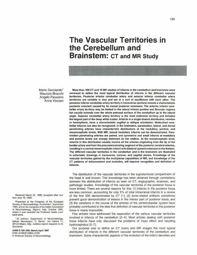
Fig. 1.-Evolution of infarct in distribution of superior cerebellar artery. Ataxia of 2 weeks' duration worsened markedly 24 hr before first CTstudy.
A, Low density only in first infarcted area and mass effect.
B, 10 days later. Cortical enhancement in recent infarct. Folia of cerebellum are separated by edema.
C and D, Late, permanent phase of necrosis, 2 months after stroke. Low density in all infarcted areas with sharp delimitation on midline.
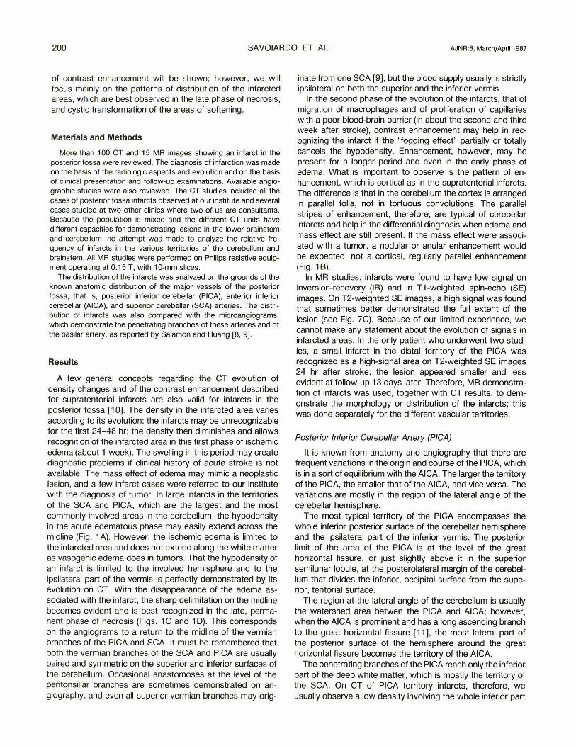
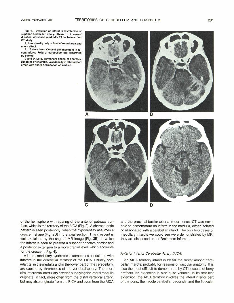
of the hemisphere with sparing of the anterior petrosal surface, which is the territory of the AICA (Fig. 2). A characteristic pattern is seen posteriorly, when the hypodensity assumes a crescent shape (Fig. 2D) in the axial section. This crescent is well explained by the sagittal MR image (Fig. 3B), in which the infarct is seen to present a superior concave border and a posterior extension to a more cranial level, which accounts for the crescent (Fig. 4).
A lateral medullary syndrome is sometimes associated with infarcts in the cerebellar territory of the PICA. Usually both infarcts, in the medulla and in the lower part of the cerebellum, are caused by thrombosis of the vertebral artery: The short circumferential medullary arteries supplying the lateral medulla originate, in fact, more often from the distal vertebral artery, but may also originate from the PICA and even from the AICA
and the proximal basilar artery. In our series, CT was never able to demonstrate an infarct in the medulla, either isolated or associated with a cerebellar infarct. The only two cases of medullary infarcts we could see were demonstrated by MR; they are discussed under Brainstem Infarcts.
Anterior Inferior Cerebellar Artery (AICA)
An AICA territory infarct is by far the rarest among cerebellar infarcts, probably for reasons of vascular anatomy. It is also the most difficult to demonstrate by CT because of bony artifacts. Its extension is also quite variable. In its smallest extension , the AICA territory involves the lateral inferior part of the pons, the middle cerebellar peduncle, and the floccular
202
A B
c o
A B
SAVOIARDO ET AL AJNR:8. March/April 1987
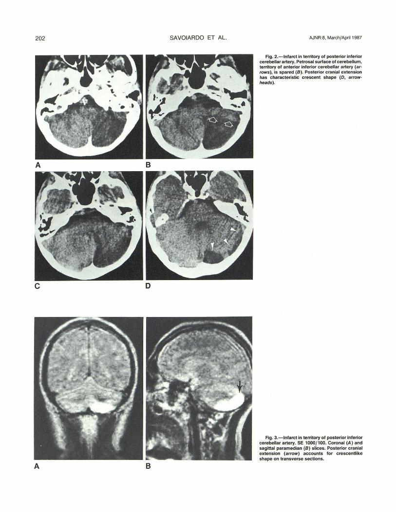
Fig. 2.-lnfarct in territory of posterior inferior cerebellar artery. Petrosal surface of cerebellum, territory of anterior inferior cerebellar artery (ar· rows), is spared (8). Posterior cranial extension has characteristic crescent shape (D, arrow· heads).
Fig. 3.-lnfarct in territory of posterior inferior cerebellar artery. SE 1000/100. Coronal (A) and sagittal paramedian (8) slices. Posterior cranial extension (arrow) accounts for crescentlike shape on transverse sections.
AJNR:8, March/April 1987
0 0 ~
a
c
e
TERRITORIES OF CEREBELLUM AND BRAINSTEM
PICA d AICA
C SCA
WSCA b
a
. ....... '" ~. .. ....
t • • • .. .. .. .. .. .. .. .. .. ~ .. .. .. .. .. .. ~ .. . . . .. .. . .. ..
I f
e
b
d
f
203
0 PPA
0 LPA
DPA
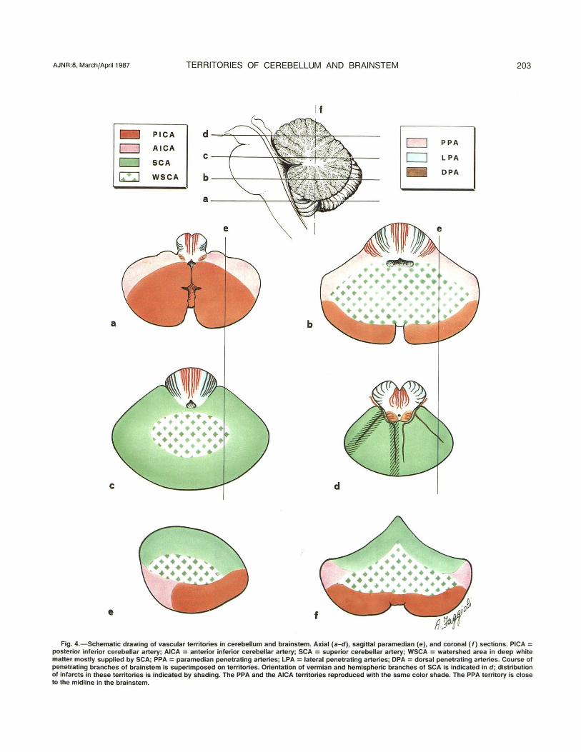
Fig. 4.-Schematic drawing of vascular territories in cerebellum and brainstem. Axial (a-d) , sagittal paramedian (e), and coronal (f) sections. PICA = posterior inferior cerebellar artery; AICA = anterior inferior cerebellar artery; SCA = superior cerebellar artery; WSCA = watershed area in deep white matter mostly supplied by SCA; PPA = paramedian penetrating arteries; LPA = lateral penetrating arteries; DPA = dorsal penetrating arteries. Course of penetrating branches of brainstem is superimposed on territories. Orientation of vermian and hemispheric branches of SCA is indicated in d ; distribution of infarcts in these territories is indicated by shading. The PPA and the AICA territories reproduced with the same color shade. The PPA territory is close to the midline in the brainstem.
204
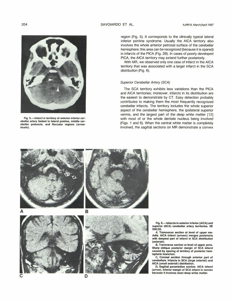
Fig. S.-Infarct in territory of anterior inferior cerebellar artery limited to lateral pontine, middle cerebellar peduncle, and floccular regions (arrow· heads).
B
SAVOIARDO ET AL. AJNR:8, March/April 1987
region (Fig. 5). It corresponds to the clinically typical lateral inferior pontine syndrome. Usually the AICA territory also involves the whole anterior petrosal surface of the cerebellar hemisphere; this area can be recognized (because it is spared) in infarcts of the PICA (Fig. 28). In cases of poorly developed PICA, the AICA territory may extend further posteriorly.
With MR, we observed only one case of infarct in the AICA territory that was associated with a larger infarct in the SCA distribution (Fig. 6).
Superior Cerebellar Artery (SCA)
The SCA territory exhibits less variations than the PICA and AICA territories; moreover, infarcts in its distribution are the easiest to demonstrate by CT. Easy detection probably contributes to making them the most frequently recognized cerebellar infarcts. The territory includes the whole superior aspect of the cerebellar hemisphere, the ipsilateral superior vermis, and the largest part of the deep white matter [12] with most of or the whole dentate nucleus being involved (Figs. 1 and 6). When the central white matter is completely involved, the sagittal sections on MR demonstrate a convex
Fig. S.-Infarcts in anterior inferior (AICA) and superior (SCA) cerebellar artery territories. SE 500/50.
A, Transverse section at level of upper medulla. AICA infarct (arrows) merges posteriorly with deepest part of infarct in SCA distribution (asterisk).
B, Transverse section at level of upper pons. Sharp oblique posterior margin of SCA infarct caused by sparing of territory of posterior hemispheric branches.
C, Coronal section through anterior part of cerebellum. Infarcts in SCA (large asterisk) and AICA (small asterisk) distribution.
0, Sagittal paramedian section. AICA infarct (arrow). Inferior margin of SCA infarct is convex because it involves most deep white matter.
AJNR:8, March/April 1987 TERRITORIES OF CEREBELLUM AND BRAINSTEM 205
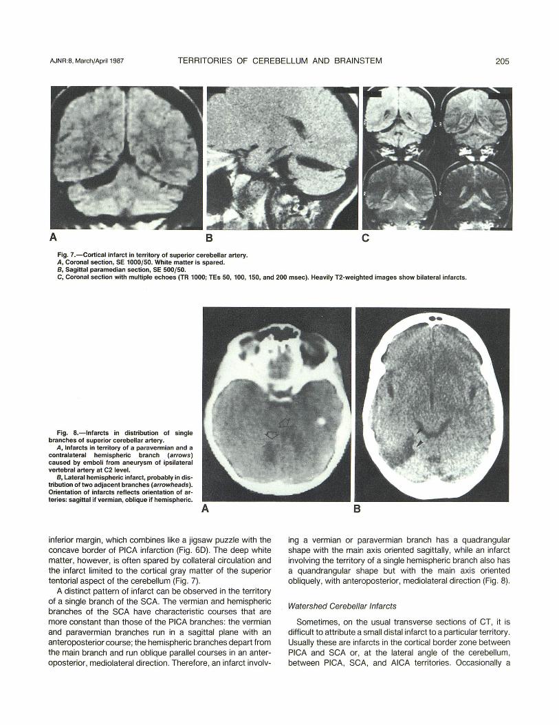
A 8 Fig. 7.-Cortical infarct in territory of superior cerebellar artery. A, Coronal section, SE 1000/50. White matter is spared. B, Sagittal paramedian section, SE 500/50.
c
C, Coronal section with multiple echoes (TR 1000; TEs 50, 100, 150, and 200 msec). Heavily T2-weighted images show bilateral infarcts.
Fig. a.-Infarcts in distribution of single branches of superior cerebellar artery.
A, Infarcts in territory of a paravermian and a contralateral hemispheric branch (arrows) caused by emboli from aneurysm of ipsilateral vertebral artery at C2 level.
B, Lateral hemispheric infarct, probably in distribution of two adjacent branches (arrowheads). Orientation of infarcts reflects orientation of arteries: sagittal if vermian, oblique if hemispheric.
inferior margin, which combines like a jigsaw puzzle with the concave border of PICA infarction (Fig . 60). The deep white matter, however, is often spared by collateral circulation and the infarct limited to the cortical gray matter of the superior tentorial aspect of the cerebellum (Fig . 7).
A distinct pattern of infarct can be observed in the territory of a single branch of the SCA. The vermian and hemispheric branches of the SCA have characteristic courses that are more constant than those of the PICA branches: the vermian and paravermian branches run in a sagittal plane with an anteroposterior course; the hemispheric branches depart from the main branch and run oblique parallel courses in an anteroposterior, mediolateral direction. Therefore, an infarct involv-
ing a vermian or paravermian branch has a quadrangular shape with the main axis oriented sagittally, while an infarct involving the territory of a single hemispheric branch also has a quandrangular shape but with the main axis oriented obliquely, with anteroposterior, mediolateral direction (Fig . 8).
Watershed Cerebellar Infarcts
Sometimes, on the usual transverse sections of CT, it is difficult to attribute a small distal infarct to a particular territory. Usually these are infarcts in the cortical border zone between PICA and SCA or, at the lateral angle of the cerebellum, between PICA, SCA, and AICA territories. Occasionally a
206 SAVOIARDO ET AL. AJNR:8, March/April 1987
small cortical watershed infarct has to be differentiated from an enlarged great horizontal fissure [13]. Localization on the border zone is obviously easier with MR, because of its multiplanar capabilities, or with CT reformatted images (Fig . 9). Watershed cerebellar infarcts were observed more often in this cortical border-zone area in the superior semilunar lobule.
Infarcts in the deep white matter of the cerebellar hemispheres, rarely seen, must also be considered watershed infarcts (Fig . 10). This area, in fact, is the border zone between the penetrating branches of the PICA, SCA, and AICA. Being a border zone, and therefore the site of this characteristic type of infarction, this area is often spared in cortical infarcts because of the better possibility of collaterals . In fact , if the most common areas of infarct in the PICA, SCA, and AICA distributions are combined as in a jigsaw puzzle, this deep area often is not involved and needs to be added to complete the puzzle (Fig . 4).
Brainstem Infarcts
Patients with large brainstem infarcts often do not even reach the hospital. Most of the cases observed in our series presented small infarcts. Their distribution , however, is interesting because it indicates the distribution of the penetrating branches that supply the brainstem.
The penetrating branches can be divided into paramedian , lateral , and dorsal, depending on the place of entry and on the area they supply.
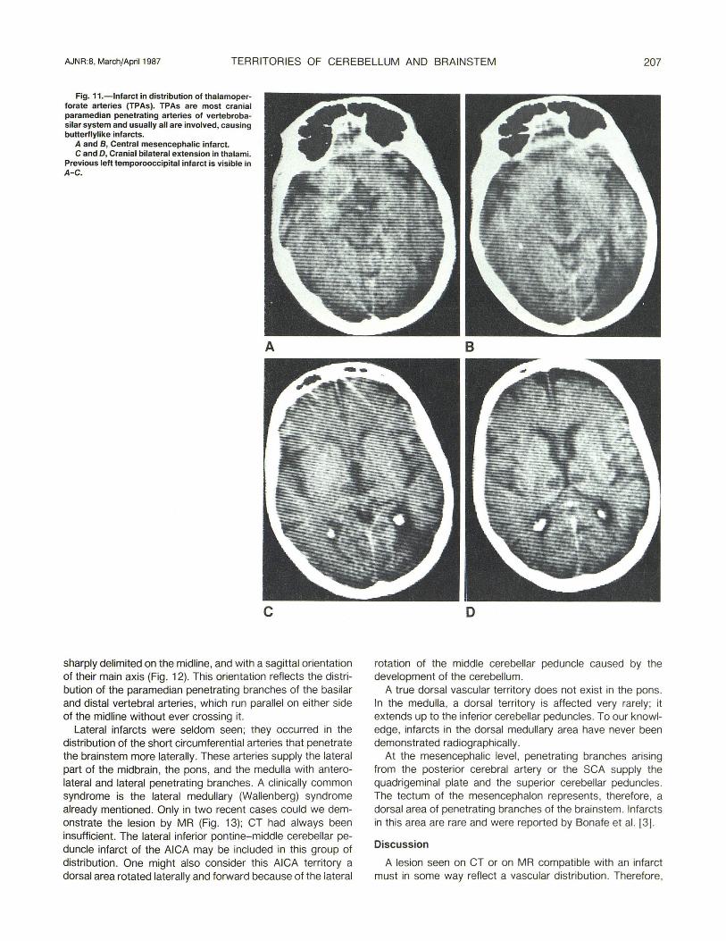
Only at the mesencephalic level were midline infarcts often seen . They result from occlusion of the thalamoperforate arteries (TPAs), which originate from the tip of the basilar artery and from the adjacent interpeduncular precommunicating segments of the posterior cerebral arteries. In addition to the median and paramedian central area of the midbrain, infarcts in the TPA distribution involve the medial portion of
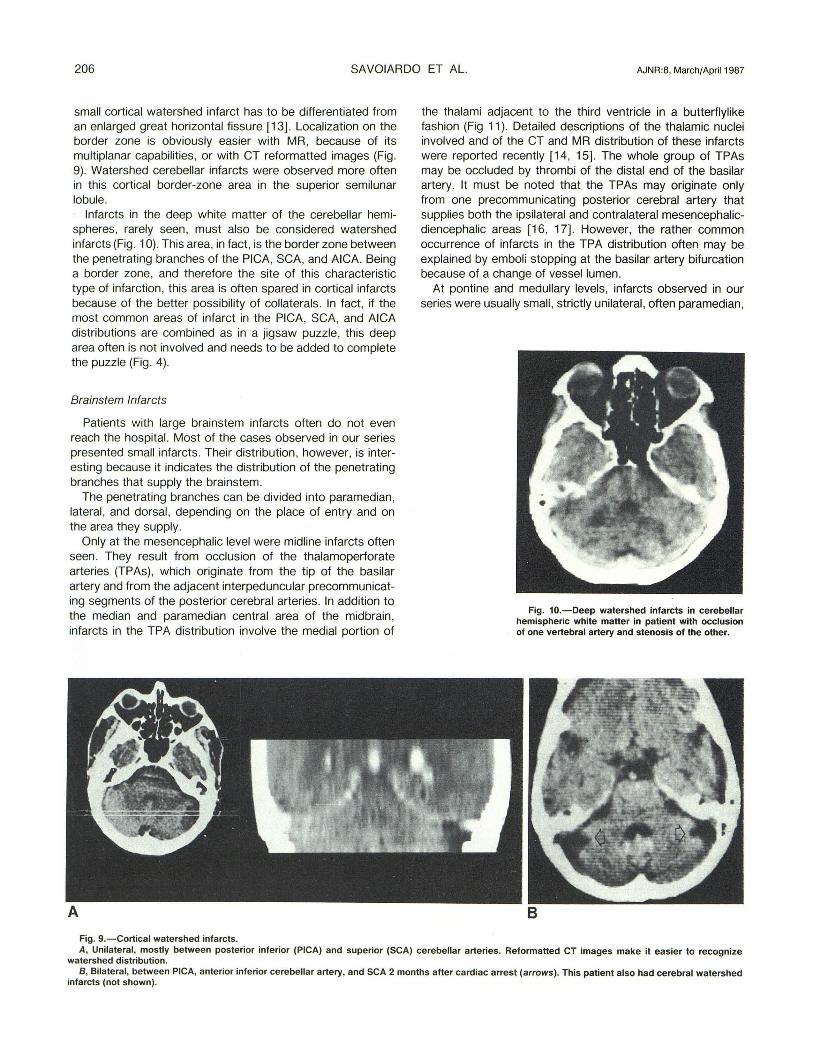
Fig. g.-Cortical watershed infarcts.
the thalami adjacent to the third ventricle in a butterflylike fashion (Fig 11). Detailed descriptions of the thalamic nuclei involved and of the CT and MR distribution of these infarcts were reported recently [14, 15). The whole group of TPAs may be occluded by thrombi of the distal end of the basilar artery. It must be noted that the TPAs may originate only from one precommunicating posterior cerebral artery that supplies both the ipsilateral and contralateral mesencephalicdiencephalic areas [16, 17). However, the rather common occurrence of infarcts in the TPA distribution often may be explained by emboli stopping at the basilar artery bifurcation because of a change of vessel lumen.
At pontine and medullary levels, infarcts observed in our series were usually small , strictly unilateral , often paramedian,
Fig. 10.- Deep watershed infarcts in cerebellar hemispheric white matter in patient with occlusion of one vertebral artery and stenosis of the other.
A, Unilateral, mostly between posterior inferior (PICA) and superior (SCA) cerebellar arteries. Reformatted CT images make it easier to recognize watershed distribution.
B, Bilateral, between PICA, anterior inferior cerebellar artery, and SCA 2 months after cardiac arrest (arrows) . This patient also had cerebral watershed infarcts (not shown).
AJNR:8, March/April 1987 TERRITORIES OF CEREBELLUM AND BRAINSTEM 207
Fig. 11.-lnfarct in distribution of thalamoperforate arteries (TPAs). TPAs are most cranial paramedian penetrating arteries of vertebrobasilar system and usually all are involved, causing butterflylike infarcts.
A and B, Central mesencephalic infarct. C and D, Cranial bilateral extension in thalami.
Previous left temporooccipital infarct is visible in A-C.
A
c
sharply delimited on the midline, and with a sagittal orientation of their main axis (Fig. 12). This orientation reflects the distribution of the paramedian penetrating branches of the basilar and distal vertebral arteries, which run parallel on either side of the midline without ever crossing it.
Lateral infarcts were seldom seen; they occurred in the distribution of the short circumferential arteries that penetrate the brain stem more laterally. These arteries supply the lateral part of the midbrain, the pons, and the medulla with anterolateral and lateral penetrating branches. A clinically common syndrome is the lateral medullary (Wallenberg) syndrome already mentioned. Only in two recent cases could we demonstrate the lesion by MR (Fig. 13); CT had always been insufficient. The lateral inferior pontine-middle cerebellar peduncle infarct of the AICA may be included in this group of distribution. One might also consider this AICA territory a dorsal area rotated laterally and forward because of the lateral
B
o
rotation of the middle cerebellar peduncle caused by the development of the cerebellum.
A true dorsal vascular territory does not exist in the pons. In the medulla, a dorsal territory is affected very rarely; it extends up to the inferior cerebellar peduncles. To our knowledge, infarcts in the dorsal medullary area have never been demonstrated radiographically.
At the mesencephalic level, penetrating branches arising from the posterior cerebral artery or the SCA supply the quadrigeminal plate and the superior cerebellar peduncles. The tectum of the mesencephalon represents , therefore, a dorsal area of penetrating branches of the brainstem. Infarcts in this area are rare and were reported by Bonafe et al. [3] .
Discussion
A lesion seen on CT or on MR compatible with an infarct must in some way reflect a vascular distribution. Therefore,
208 SAVOIARDO ET AL. AJNR:8, March/April 1987
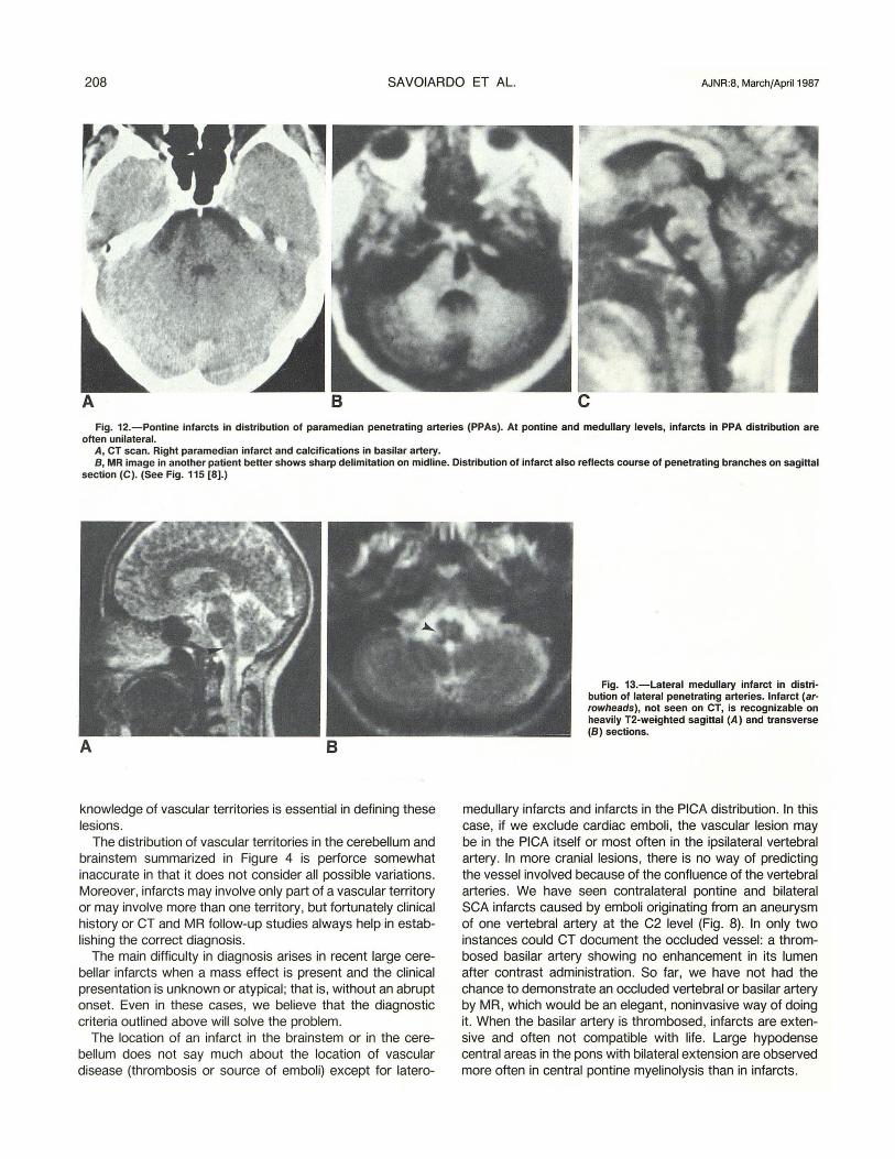
Fig. 12.-Pontine infarcts in distribution of paramedian penetrating arteries (PPAs). At pontine and medullary levels, infarcts in PPA distribution are often unilateral.
A, CT scan. Right paramedian infarct and calcifications in basilar artery. B, MR image in another patient better shows sharp delimitation on midline. Distribution of infarct also reflects course of penetrating branches on sagittal
section (e). (See Fig. 115 [8).)
knowledge of vascular territories is essential in defining these lesions.
The distribution of vascular territories in the cerebellum and brainstem summarized in Figure 4 is perforce somewhat inaccurate in that it does not consider all possible variations. Moreover, infarcts may involve only part of a vascular territory or may involve more than one territory, but fortunately clinical history or CT and MR follow-up studies always help in establishing the correct diagnosis.
The main difficulty in diagnosis arises in recent large cerebellar infarcts when a mass effect is present and the clinical presentation is unknown or atypical ; that is, without an abrupt onset. Even in these cases, we believe that the diagnostic criteria outlined above will solve the problem.
The location of an infarct in the brain stem or in the cerebellum does not say much about the location of vascular disease (thrombosis or source of emboli) except for latero-
Fig. 13.-Lateral medullary infarct in distribution of lateral penetrating arteries. Infarct (arrowheads), not seen on CT, is recognizable on heavily T2-weighted sagittal (A) and transverse (B) sections.
medullary infarcts and infarcts in the PICA distribution. In this case, if we exclude cardiac emboli, the vascular lesion may be in the PICA itself or most often in the ipsilateral vertebral artery. In more cranial lesions, there is no way of predicting the vessel involved because of the confluence of the vertebral arteries. We have seen contralateral pontine and bilateral SCA infarcts caused by emboli originating from an aneurysm of one vertebral artery at the C2 level (Fig. 8). In only two instances could CT document the occluded vessel: a thrombosed basilar artery showing no enhancement in its lumen after contrast administration. So far, we have not had the chance to demonstrate an occluded vertebral or basilar artery by MR, which would be an elegant, noninvasive way of doing it. When the basilar artery is thrombosed, infarcts are extensive and often not compatible with life. Large hypodense central areas in the pons with bilateral extension are observed more often in central pontine myelinolysis than in infarcts.
AJNR :8, March/April 1987 TERRITORIES OF CEREBELLUM AND BRAINSTEM 209
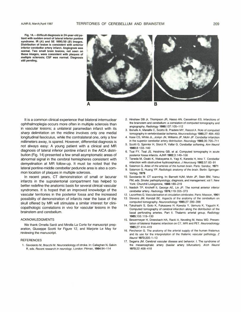
Fig. 14.-Diflicult diagnosis in 24-year-old patient with sudden onset of lateral inferior pontine syndrome. IR (A) and SE 1000/50 (8) images. Distribution of lesion is consistent with anterior inferior cerebellar artery infarct. Angiogram was normal. Two small brain lesions, not seen on these images, were consistent with plaques of multiple sclerosis; CSF was normal. Diagnosis still pending.
It is a common clinical experience that bilateral internuclear ophthalmoplegia occurs more often in multiple sclerosis than in vascular lesions; a unilateral paramedian infarct with its sharp delimitation on the midline involves only one medial longitudinal fasciculus , while the contralateral one, only a few millimeters away, is spared. However, differential diagnosis is not always easy: A young patient with a clinical and MR diagnosis of lateral inferior pontine infarct in the AICA distribution (Fig . 14) presented a few small asymptomatic areas of abnormal signal in the cerebral hemispheres consistent with demyelination at MR follow-up. It must be noted that the lateral pontine-middle cerebellar peduncle area is also a common location of plaques in multiple sclerosis.
In recent years, CT demonstration of small or lacunar infarcts in the supratentorial compartment has helped to better redefine the anatomic basis for several clinical vascular syndromes. It is hoped that an improved knowledge of the vascular territories in the posterior fossa and the increased possibility of demonstration of infarcts near the base of the skull offered by MR will stimulate a similar interest for clinicopathologic correlations in vivo for vascular lesions in the brainstem and cerebellum.
ACKNOWLEDGMENTS
We thank Omelia Sardi and Mirella La Corte for manuscript preparation, Giuseppe Scotti for Figure 12, and Marjorie Le May for reviewing the manuscript.
REFERENCES
1. Savoiardo M, Bracchi M. Neuroradiology of stroke. In: Callaghan N, Galvin R, eds. Recent research in neurology. London: Pitman, 1984:94-114
2. Hinshaw DB Jr, Thompson JR, Hasso AN, Casselman ES. Infarctions of the brain stem and cerebellum: a correlation of computed tomography and angiography. Radiology 1980;137 :105-112
3. Bonafe A, Manelfe C, Scot1o B, Pradere MY, Rascol A. Role of computed tomography in vertebrobasilar ischemia. Neuroradiology 1985;27 :484- 493
4. Kase CS, White JL, Joslyn IN, Williams JP, Mohr JP. Cerebellar infarction in the superior cerebellar artery distribution. Neurology 1985;35 :705-711
5. Scot1i G, Spinnler H, Sterzi R, Vallar G. Cerebellar softening. Ann Neurol 1980;8: 133- 140
6. Tsai FY, Teal JS, Heishima GB, et al. Computed tomography in acute posterior fossa infarcts. AJNR 1982;3 : 149-1 56
7. Taneda M, Ozaki K, Wakayama A, Vagi K, Kaneda H, Irino T. Cerebellar infarction with obstructive hydrocephalus. J Neurosurg 1982;57 :83-91
8. Salamon G. Atlas of the arteries of the human brain . Paris: Sandoz, 1971 9. Salamon G, Huang YP. Radiologic anatomy of the brain . Berlin: Springer
Verlag, 1976 10. Savoiardo M. CT scanning. In: Barnett HJM, Mohr JP, Stein BM, Yatsu
FM, eds. Stroke: pathophysiology, diagnosis, and management , vol1 . New York: Churchill Livingstone, 1986:189-219
11. Naidich TP, Kricheff II , George AE, Lin JP. The normal anterior inferior cerebellar artery. Radiology 1976;119 :355-373
12. Lazorthes G. Vascularisation et circulation cerebrales . Paris: Masson, 1961 13. Stevens JM, Kendall BE. Aspects of the anatomy of the cerebellum on
computed tomography. Neuroradiology 1985;27 :390- 398 14. Takahashi S, Goto K, Fukasawa H, Kawata Y, Uemura K, Yaguchi K.
Computed tomography of cerebral infarction along the distribution of the basal perforating arteries. Part II. Thalamic arterial group. Radiology 1985;155 : 119- 130
15. Bewermeyer H, Dreesbach HA, Rackl A, Neveling M, Heiss WD. Presen· tation of bilateral thalamiC infarction on CT, MRI and PET. Neuroradiology 1985;27 :414-419
16. Percheron G. The anatomy of the arterial supply of the human thalamus and its use for the interpretation of the thalamic vascular pathology. Z Neuro/1973;205 : 1-13
17. Segarra JM. Cerebral vascular disease and behavior. I. The syndrome of the mesencephalic artery (basilar artery bifurcation). Arch Neurol 1970;22: 408- 418