the wrist: common injuries and...
TRANSCRIPT
Prim Care Clin Office Pract
32 (2005) 35–70
The Wrist: Common Injuries andManagement
Katrina Parmelee-Peters, MD, Scott W. Eathorne, MD*Providence Athletic Medicine, Providence Medical Center-Providence Park,
47601 Grand River Avenue, Suite A101, Novi, MI 48374, USA
Wrist injuries are common in athletes. They may result from a single,traumatic force or as a result of repetitive-loading activity. Complex wristand hand anatomy can make diagnosis of wrist injuries an challenging task.A good understanding of wrist anatomy, as discussed elsewhere in this issuein ‘‘The Wrist: Clinical Anatomy and Physical Examination—an Update’’by Eathorne, and an awareness of the common presentations of sport-specific injuries will facilitate accurate diagnosis. Labeling an athlete’s injuryas a ‘‘wrist sprain’’ without a specific diagnosis may allow a competitiveathlete to continue to play through the pain without proper treatment andexacerbate an injury. Management of a wrist injury in the athlete requiresthat the physician balance the athlete’s objective to return to sport promptlywith treatment that allows healing and prevents long-term complicationsof an injury. It is important for the primary care physician to have anawareness of the broad range of injuries that occur in the athlete’s wrist, tobe familiar with appropriate conservative management, and to referappropriately.
Epidemiology
Child and adolescent athletes suffer relatively more wrist injuries thanadult athletes. Three percent to 9% of all athletic injuries involve the handand wrist [1]. This number is as high as 14% in high school football [2], and46 to 87% of gymnasts suffer wrist injuries or have chronic wrist pain [3,4].Injuries of the wrist can be divided into acute traumatic injuries and overuseinjuries.
* Corresponding author.
E-mail address: [email protected] (S.W. Eathorne).
0095-4543/05/$ - see front matter � 2005 Elsevier Inc. All rights reserved.
doi:10.1016/j.pop.2004.11.015 primarycare.theclinics.com

36 PARMELEE-PETERS & EATHORNE
Acute wrist fractures are common injuries among athletes. In a study offootball players aged 9 to 15, 35% of injuries were to the upper extremities,and most were distal radius fractures [5]. Distal radius metaphyseal andphyseal fractures are common in skating, football, basketball, andsnowboarding. The scaphoid is the most commonly injured carpal bone,accounting for 70% of carpal fractures [2]. An athlete falling on anoutstretched hand with the wrist dorsiflexed is a common mechanism. Thetriquetral bone is the second most commonly injured carpal bone. Ittypically occurs from a fall on a wrist in ulnar deviation. Pisiform fracturesoccur due to a direct blow, such as from a pitched ball.
Stress fractures occur in athletes whose sport requires repetitive motioninvolving wrist compression or twisting. Sports such as gymnastics andweightlifting place large repetitive compressive forces across the wrist. Distalradius physis stress syndrome, avascular necrosis of the capitate, and stressfracture of the scaphoid have been reported in these athletes [4,6].Reportedly, up to 87% of elite gymnasts sustain distal radial physealinjuries [3]. Hook of hamate fractures have been seen in baseball, golf, andtennis players from the repetitive stress of bat, club, or racquet, respectively[7]. Repetitive stress is also thought to be a cause of avascular necrosis of thelunate or Kienbock’s disease.
Soft-tissue injuries may either be due to acute trauma or overuse. Overusesyndromes such as deQuervain’s tenosynovitis, extensor carpi ulnaristendonitis, and sprains of pisotriquetral ligament [8] are associated withthrowing and racquet sports. Dislocation of the distal radioulnar joint(DRUJ), midcarpal instability, and triangular fibrocartilage complex(TFCC) tears can occur due to a traumatic fall, or due to repetitive twistingmotion as seen in gymnasts. Carpal dislocation typically requires significantforce, such as a collision in football or a fall from a height in cheerleading.
General approach to wrist injuries
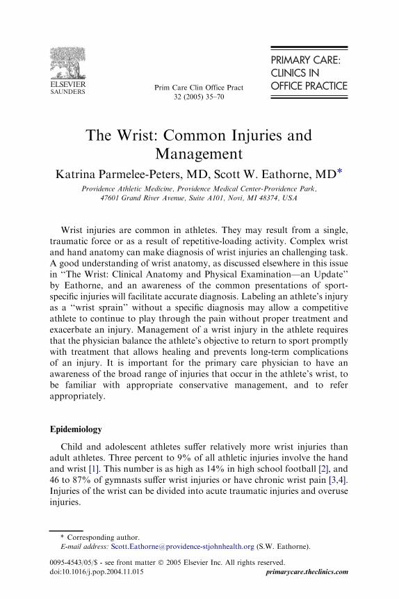
To begin, be familiar with the most common wrist injuries in activepeople and the common sport-specific injuries. Obtain a careful historyregarding the athlete’s wrist complaint, including how the complaint isrelated to activity and rest. It is important to have good clinical knowledgeof the functional anatomy of the wrist in order to maximize the informationgathered on examination. The evaluation of wrist complaints requires atleast two radiographic views of the wrist (Fig. 1). An oblique view, inaddition to a posterior-anterior (PA) and true lateral, is useful in identifyingfractures. Special radiographic views of the wrist are also useful and will beaddressed further in the discussion of specific injuries (Table 1). Severalthings are important to consider as treatment is initiated for a wrist injuryincluding: the athlete�s sport, his or her desire�s regarding return to play, andthe impact of injury management on the athlete�s future participation in his
37WRIST INJURIES
or her sport. The primary care physician should understand that manyinjuries have a poor outcome if unrecognized. If the diagnosis is not clear,the athlete’s wrist can be protected with a splint and referred for additionalevaluation.
Management of all wrist injuries should include rehabilitation of musclesweakened and motion lost by pain, inflammation, and immobilization.Rehabilitation should proceed through five goal-oriented phases. Rehabili-tation goals include: (1) decreasing pain and minimizing inflammation andedema; (2) increasing pain-free range of motion; (3) strengthening andimproving general condition; (4) increasing sport-specific skill, coordination,and flexibility; and (5) return to sport with prevention of injury, which mayinclude use of protective equipment. Physical therapists and athletic trainerscan play a key role the safe and expeditious return to play of the athlete.
Tendonopathies
DeQuervain’s tenosynovitis
DeQuervain’s tenosynovitis is the most common tendonopathy of thewrist in athletes [9]. DeQuervain’s tenosynovitis is inflammation of thetenosynovium of the first dorsal compartment tendons, the abductor pollicislongus (APL) and extensor pollicis brevis (EPB). These tendons courseunder the extensor retinaculum in a groove along the radial styloid process.
Fig. 1. Routine wrist radiographs. PA view (A) and lateral view (B).
38 PARMELEE-PETERS & EATHORNE
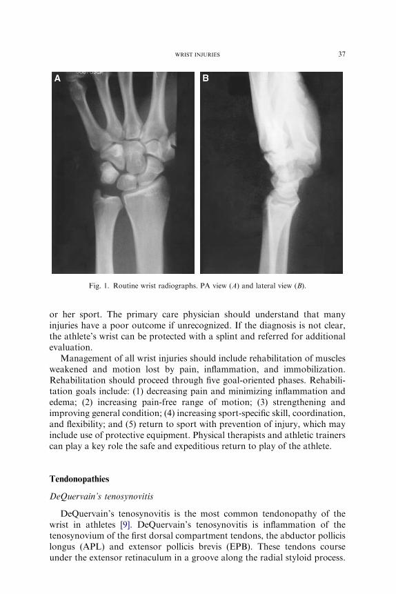
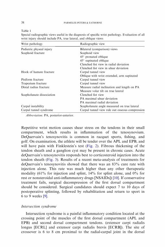
Repetitive wrist motion causes shear stress on the tendons in their smallcompartment, which results in inflammation of the tenosynovium.DeQuervain’s tenosynovitis is common in racquet sports, fishing, andgolf. On examination, the athlete will be tender over the APL and EPB, andwill have pain with Finklestein’s test (Fig. 2). Fibrous thickening of thetendon sheath and a ganglion cyst may be present in chronic cases. AcutedeQuervain’s tenosynovitis responds best to corticosteroid injection into thetendon sheath (Fig. 3). Results of a recent meta-analysis of treatments fordeQuervain’s tenosynovitis showed that there was an 83% cure rate withinjection alone. This rate was much higher than any other therapeuticmodality (61% for injection and splint, 14% for splint alone, and 0% forrest or nonsteroidal anti-inflammatory drugs [NSAIDs]) [10]. If conservativetreatment fails, surgical decompression of the first dorsal compartmentshould be considered. Surgical candidates should expect 7 to 10 days ofpostoperative splinting, followed by rehabilitation and return to sport in6 to 9 weeks [9].
Intersection syndrome
Intersection syndrome is a painful inflammatory condition located at thecrossing point of the muscles of the first dorsal compartment (APL andEPB) and second dorsal compartment tendons. (extensor carpi radialislongus [ECRL] and extensor carpi radialis brevis [ECRB]). The site ofcrossover is 6 to 8 cm proximal to the radial-carpal joint in the dorsal
Table 1
Special radiographic views useful in the diagnosis of specific wrist pathology. Evaluation of all
wrist injury should include PA, true lateral, and oblique views
Wrist pathology Radiographic view
Pediatric physeal injury Bilateral (comparison) views
Scaphoid fracture Scaphoid view
45� pronated oblique
45� supinated oblique
Clenched fist view in radial deviation
Clenched fist view in ulnar deviation
Hook of hamate fracture Carpal tunnel view
Oblique with wrist extended, arm supinated
Pisiform fracture Carpal tunnel view
Trapezium fracture Carpal tunnel view
Distal radius fracture Measure radial inclination and length on PA
Measure volar tilt on true lateral
Scapholunate dissociation Clenched fist view
PA maximal ulnar deviation
PA maximal radial deviation
Carpal instability Scapholunate angle measured on true lateral
Carpal tunnel syndrome Carpal tunnel view rule out osseous compression
Abbreviation: PA, posterior-anterior.
39WRIST INJURIES
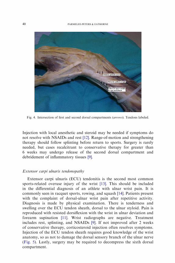
forearm (Fig. 4). This site is tender and may be swollen. There is oftena palpable crepitus at the intersection with moving the wrist through flexionand extension, leading to the name ‘‘squeakers syndrome.’’ It is seen inathletes who play sports requiring forceful, repetitive wrist flexion andextension (rowing, weight lifting, gymnastics, and racquet sports). Thepathophysiology is still unclear, but it is thought to be a tenosynovitis of thesecond dorsal compartment tendons, or inflammation of an adventitialbursa between the APL and ECRB due to friction at the intersection. Thissyndrome responds well to conservative treatment of rest, local icing, andNSAIDs, with a gradual return to sports. Splinting with a thumb spicasplint in 15� of wrist extension for 2 weeks is helpful to rest the muscles [11].
Fig. 2. Demonstration of Finkelstein’s test. The forearm is stabilized with one hand while
gently forcing the wrist into ulnar deviation.
Fig. 3. First dorsal wrist extensor compartment of cadaveric model. Injection site for
deQuervain’s tenosynovitis is demonstrated.
40 PARMELEE-PETERS & EATHORNE
Injection with local anesthetic and steroid may be needed if symptoms donot resolve with NSAIDs and rest [12]. Range-of-motion and strengtheningtherapy should follow splinting before return to sports. Surgery is rarelyneeded, but cases recalcitrant to conservative therapy for greater than6 weeks may undergo release of the second dorsal compartment anddebridement of inflammatory tissues [9].
Extensor carpi ulnaris tendonopathy
Extensor carpi ulnaris (ECU) tendonitis is the second most commonsports-related overuse injury of the wrist [13]. This should be includedin the differential diagnosis of an athlete with ulnar wrist pain. It iscommonly seen in racquet sports, rowing, and squash [14]. Patients presentwith the complaint of dorsal-ulnar wrist pain after repetitive activity.Diagnosis is made by physical examination. There is tenderness andswelling over the ECU tendon sheath, dorsal to the ulnar styloid. Pain isreproduced with resisted dorsiflexion with the wrist in ulnar deviation andforearm supination [11]. Wrist radiographs are negative. Treatmentincludes rest, splinting, and NSAIDs [9]. If not improved after 2 weeksof conservative therapy, corticosteroid injection often resolves symptoms.Injection of the ECU tendon sheath requires good knowledge of the wristanatomy, so as not to damage the dorsal sensory branch of the ulnar nerve(Fig. 5). Lastly, surgery may be required to decompress the sixth dorsalcompartment.
Fig. 4. Intersection of first and second dorsal compartments (arrows). Tendons labeled.

41WRIST INJURIES
ECU subluxation is a less common injury, but is important to consider inthe differential diagnosis of chronic ulnar wrist pain. ECU subluxationoccurs with forceful supination, palmar flexion, and ulnar deviation of thewrist [15]. This injury is seen in tennis players hitting a low forehand, or inthe trailing hand of a baseball player at the end of a swing [14]. It may alsooccur after a fall on an outstretched hand (FOOSH) [15,16]. Patientstypically complain of dorsal ulnar wrist pain and ‘‘snapping’’ that areaggravated by forearm rotation [15]. Tenderness and swelling are elicitedover the ECU in the area of the ulnar head. Marked pronation or supinationmay reproduce the ‘‘ping’’ as the tendon subluxes out of its groove. Wristradiographs are normal. A 6-week period of immobilization in a long armcast may be tried [9]; however, in several case reports [15,16] thisconservative therapy has not been successful. For symptomatic patients,surgical repair of the ruptured tendon subsheath is recommended [17,18],followed by 4 to 6 weeks of immobilization, with return to sport anticipated8 to 10 weeks following surgery [9].
Fig. 5. Relative anatomy of extensor carpi ulnaris and the dorsal sensory branch of the ulnar
nerve. A, artery; N, nerve. (From McCue FC, Bruce JF, Koman JD. The wrist in the adult. In:
DeLee JC, Drez D, editors. Orthopedic sports medicine: principles and practice. 2nd edition.
Philadelphia: WB Saunders; 2003. p. 1343; with permission.)
42 PARMELEE-PETERS & EATHORNE
Flexor carpi ulnaris tendinopathy
Flexor carpi ulnaris (FCU) tendonitis presents with palmar-ulnar sidewrist pain, and is seen in racquet sport athletes. Examination revealstenderness along the FCU and pain with wrist flexion. Dorsal wrist splintingwith 25� of flexion for 1 to 2 weeks and a short course of NSAIDs typicallyresolve symptoms [14]. Corticosteroid injection is considered for recalcitrantcases. Excision of the pisiform and Z-plasty lengthening of the FCU hasbeen described for chronic cases [14].
Flexor carpi radialis tendinopathy
Flexor carpi radialis (FCR) tendonitis presents with pain in the palmar-radial wrist with repetitive wrist flexion. Tenderness is over the FCR at itsinsertion on the base of the second and third metacarpals, and pain isreproducible with resisted wrist flexion. As with FCU tendonitis, treatmentis rest with brief splinting, NSAIDs, and stretching, and if symptoms areprolonged, surgical release may be indicated.
Distal radioulnar joint and triangular fibrocartilage complex
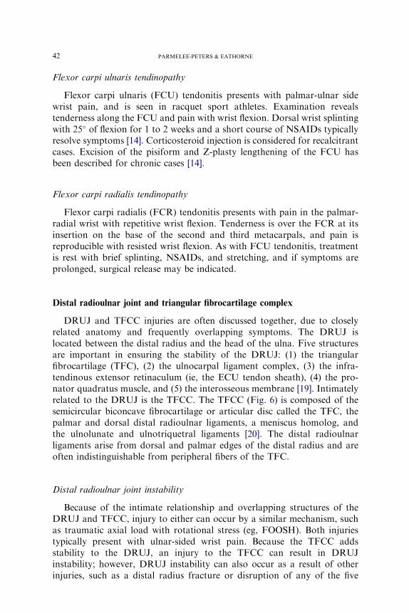
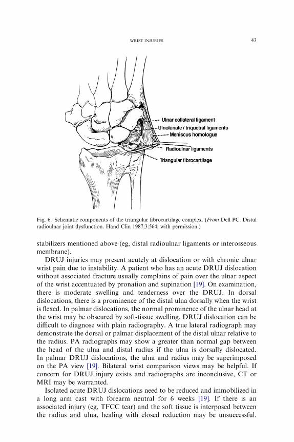
DRUJ and TFCC injuries are often discussed together, due to closelyrelated anatomy and frequently overlapping symptoms. The DRUJ islocated between the distal radius and the head of the ulna. Five structuresare important in ensuring the stability of the DRUJ: (1) the triangularfibrocartilage (TFC), (2) the ulnocarpal ligament complex, (3) the infra-tendinous extensor retinaculum (ie, the ECU tendon sheath), (4) the pro-nator quadratus muscle, and (5) the interosseous membrane [19]. Intimatelyrelated to the DRUJ is the TFCC. The TFCC (Fig. 6) is composed of thesemicircular biconcave fibrocartilage or articular disc called the TFC, thepalmar and dorsal distal radioulnar ligaments, a meniscus homolog, andthe ulnolunate and ulnotriquetral ligaments [20]. The distal radioulnarligaments arise from dorsal and palmar edges of the distal radius and areoften indistinguishable from peripheral fibers of the TFC.
Distal radioulnar joint instability
Because of the intimate relationship and overlapping structures of theDRUJ and TFCC, injury to either can occur by a similar mechanism, suchas traumatic axial load with rotational stress (eg, FOOSH). Both injuriestypically present with ulnar-sided wrist pain. Because the TFCC addsstability to the DRUJ, an injury to the TFCC can result in DRUJinstability; however, DRUJ instability can also occur as a result of otherinjuries, such as a distal radius fracture or disruption of any of the five
43WRIST INJURIES
stabilizers mentioned above (eg, distal radioulnar ligaments or interosseousmembrane).
DRUJ injuries may present acutely at dislocation or with chronic ulnarwrist pain due to instability. A patient who has an acute DRUJ dislocationwithout associated fracture usually complains of pain over the ulnar aspectof the wrist accentuated by pronation and supination [19]. On examination,there is moderate swelling and tenderness over the DRUJ. In dorsaldislocations, there is a prominence of the distal ulna dorsally when the wristis flexed. In palmar dislocations, the normal prominence of the ulnar head atthe wrist may be obscured by soft-tissue swelling. DRUJ dislocation can bedifficult to diagnose with plain radiography. A true lateral radiograph maydemonstrate the dorsal or palmar displacement of the distal ulnar relative tothe radius. PA radiographs may show a greater than normal gap betweenthe head of the ulna and distal radius if the ulna is dorsally dislocated.In palmar DRUJ dislocations, the ulna and radius may be superimposedon the PA view [19]. Bilateral wrist comparison views may be helpful. Ifconcern for DRUJ injury exists and radiographs are inconclusive, CT orMRI may be warranted.
Isolated acute DRUJ dislocations need to be reduced and immobilized ina long arm cast with forearm neutral for 6 weeks [19]. If there is anassociated injury (eg, TFCC tear) and the soft tissue is interposed betweenthe radius and ulna, healing with closed reduction may be unsuccessful.
Fig. 6. Schematic components of the triangular fibrocartilage complex. (From Dell PC. Distal
radioulnar joint dysfunction. Hand Clin 1987;3:564; with permission.)
44 PARMELEE-PETERS & EATHORNE
DRUJ dislocations with associated fractures are generally not amenable tononoperative management [19].
DRUJ subluxation is a cause of chronic ulnar wrist pain. The ulnar headis prominent in pronation as it rides onto the dorsal lip of the radius.Supination may then be restricted, often followed by a distinct snap duringforearm rotation [19]. The ‘‘piano key sign’’ indicates distal radioulnar jointinstability [1], which allows subluxation of the ulna on the radius. This signis elicited by having the patient place both palms on the examining table andforcefully press downward. There is exaggerated dorsal-palmar translationof the distal radius compared with the opposite side. Alternatively, this signcan be elicited by depressing the ulnar head while supporting the forearm inpronation; the ulnar head springs back like a piano key, indicating laxityof the DRUJ [21]. Patients who have chronic subluxation may receivetemporary relief with a distal forearm splint that exerts a relocating force onthe ulnar head. Definitive treatment for the symptomatic athlete, however, istypically surgical [19].
Triangular fibrocartilage complex injury
The TFCC is a cartilaginous and ligamentous structure important in thestabilization of the distal radial ulnar joint (as mentioned above). Thearticular disc of the TFCC separates the ulna and the proximal carpal row,and carries about 20% of the axial load from wrist to forearm [22]. There isa relative lack of blood supply to the central portion of the TFCC, leadingto poor healing of tears [23]. Injuries to the TFCC occur with repetitiveulnar loading (eg, bench press, racquet sports) or acute traumatic axial loadwith rotational stress (eg, FOOSH). Most injuries to the TFCC havea component of hyperextension of the wrist and rotational load. Ulnar-sidedwrist pain made worse with ulnar deviation, wrist extension, or heavy use isthe common complaint of an athlete who has a TFCC injury. TFCC injuriesare more commonly seen in such sports as gymnastics, hockey, racquetsports, boxing, and pole vaulting [24].
The TFCC is palpated in the hollow between the pisiform, FCU, andulnar styloid. It is most easily palpated with the wrist in pronation. Injury tothe TFCC is indicated by tenderness on palpation of the TFCC, with orwithout distal radioulnar joint instability. TFCC compression by forcedulnar deviation and axial compression with repeated flexion and extensionwill impact the ulnar styloid and TFCC. This will result in pain or clickingif the TFCC is involved [3]. The ‘‘press test’’ reproduces the patient’s painwhen the patient lifts herself out of a chair while bearing weight on theextended wrists [25]. The ‘‘supination lift test’’ has also been described forlocalized tear to the peripheral, dorsal TFCC. With this test, pain isreproduced when the patient attempts to lift the examination table with thepalm flat on the underside of the table [26] This forces a load across the
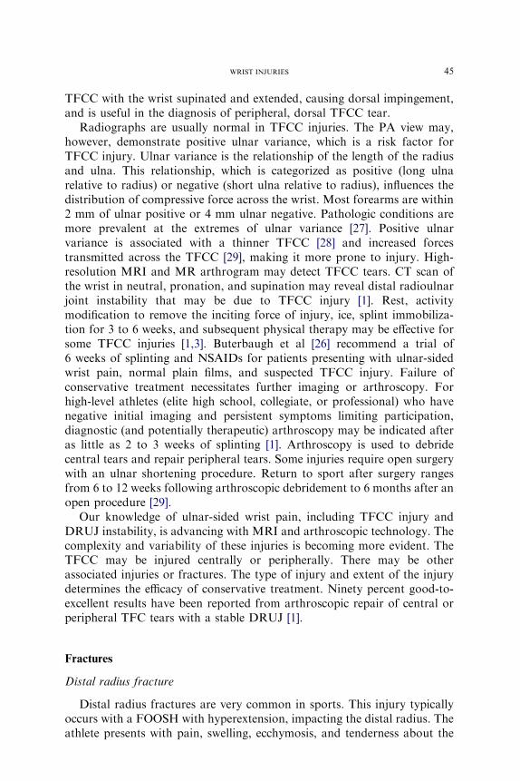
45WRIST INJURIES
TFCC with the wrist supinated and extended, causing dorsal impingement,and is useful in the diagnosis of peripheral, dorsal TFCC tear.
Radiographs are usually normal in TFCC injuries. The PA view may,however, demonstrate positive ulnar variance, which is a risk factor forTFCC injury. Ulnar variance is the relationship of the length of the radiusand ulna. This relationship, which is categorized as positive (long ulnarelative to radius) or negative (short ulna relative to radius), influences thedistribution of compressive force across the wrist. Most forearms are within2 mm of ulnar positive or 4 mm ulnar negative. Pathologic conditions aremore prevalent at the extremes of ulnar variance [27]. Positive ulnarvariance is associated with a thinner TFCC [28] and increased forcestransmitted across the TFCC [29], making it more prone to injury. High-resolution MRI and MR arthrogram may detect TFCC tears. CT scan ofthe wrist in neutral, pronation, and supination may reveal distal radioulnarjoint instability that may be due to TFCC injury [1]. Rest, activitymodification to remove the inciting force of injury, ice, splint immobiliza-tion for 3 to 6 weeks, and subsequent physical therapy may be effective forsome TFCC injuries [1,3]. Buterbaugh et al [26] recommend a trial of6 weeks of splinting and NSAIDs for patients presenting with ulnar-sidedwrist pain, normal plain films, and suspected TFCC injury. Failure ofconservative treatment necessitates further imaging or arthroscopy. Forhigh-level athletes (elite high school, collegiate, or professional) who havenegative initial imaging and persistent symptoms limiting participation,diagnostic (and potentially therapeutic) arthroscopy may be indicated afteras little as 2 to 3 weeks of splinting [1]. Arthroscopy is used to debridecentral tears and repair peripheral tears. Some injuries require open surgerywith an ulnar shortening procedure. Return to sport after surgery rangesfrom 6 to 12 weeks following arthroscopic debridement to 6 months after anopen procedure [29].
Our knowledge of ulnar-sided wrist pain, including TFCC injury andDRUJ instability, is advancing with MRI and arthroscopic technology. Thecomplexity and variability of these injuries is becoming more evident. TheTFCC may be injured centrally or peripherally. There may be otherassociated injuries or fractures. The type of injury and extent of the injurydetermines the efficacy of conservative treatment. Ninety percent good-to-excellent results have been reported from arthroscopic repair of central orperipheral TFC tears with a stable DRUJ [1].
Fractures
Distal radius fracture
Distal radius fractures are very common in sports. This injury typicallyoccurs with a FOOSH with hyperextension, impacting the distal radius. Theathlete presents with pain, swelling, ecchymosis, and tenderness about the
46 PARMELEE-PETERS & EATHORNE
wrist. Initial radiographs should include PA, lateral, and oblique views ofthe wrist. The examiner needs to determine the type of distal radial fractureand assess displacement, shortening, and intra-articular involvement. Thegoal of treatment is to correct and maintain radial inclination, palmar tilt,length, and congruity of the distal radial articulations (carpal and ulnar).
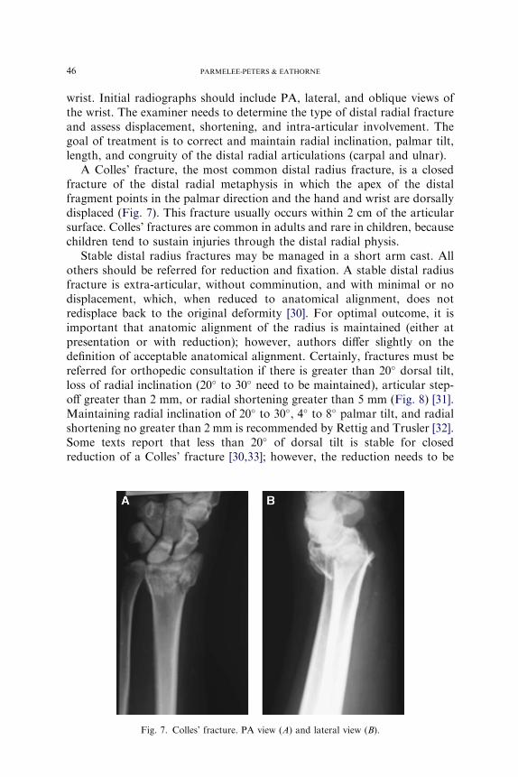
A Colles’ fracture, the most common distal radius fracture, is a closedfracture of the distal radial metaphysis in which the apex of the distalfragment points in the palmar direction and the hand and wrist are dorsallydisplaced (Fig. 7). This fracture usually occurs within 2 cm of the articularsurface. Colles’ fractures are common in adults and rare in children, becausechildren tend to sustain injuries through the distal radial physis.
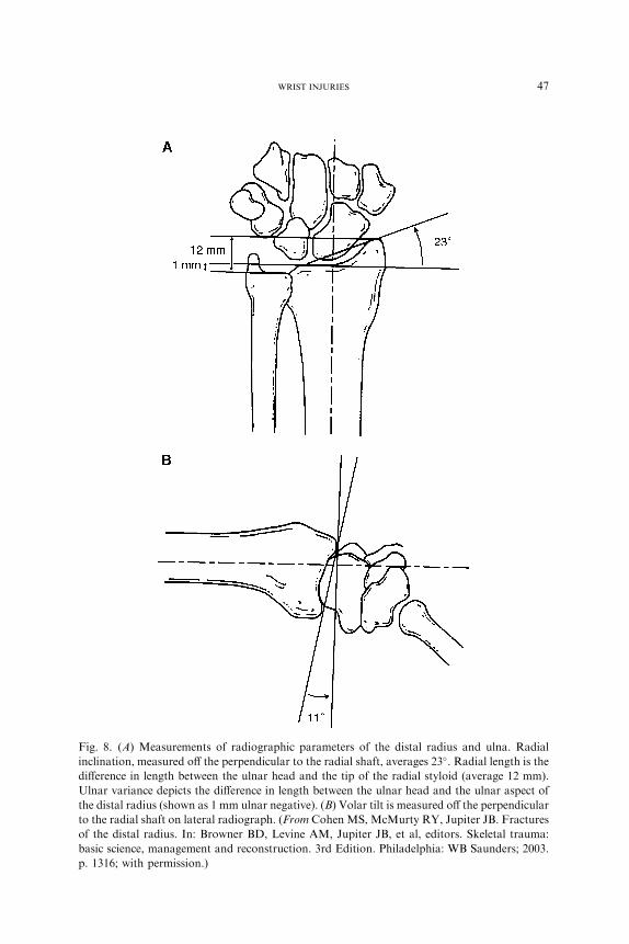
Stable distal radius fractures may be managed in a short arm cast. Allothers should be referred for reduction and fixation. A stable distal radiusfracture is extra-articular, without comminution, and with minimal or nodisplacement, which, when reduced to anatomical alignment, does notredisplace back to the original deformity [30]. For optimal outcome, it isimportant that anatomic alignment of the radius is maintained (either atpresentation or with reduction); however, authors differ slightly on thedefinition of acceptable anatomical alignment. Certainly, fractures must bereferred for orthopedic consultation if there is greater than 20� dorsal tilt,loss of radial inclination (20� to 30� need to be maintained), articular step-off greater than 2 mm, or radial shortening greater than 5 mm (Fig. 8) [31].Maintaining radial inclination of 20� to 30�, 4� to 8� palmar tilt, and radialshortening no greater than 2 mm is recommended by Rettig and Trusler [32].Some texts report that less than 20� of dorsal tilt is stable for closedreduction of a Colles’ fracture [30,33]; however, the reduction needs to be
Fig. 7. Colles’ fracture. PA view (A) and lateral view (B).
47WRIST INJURIES
Fig. 8. (A) Measurements of radiographic parameters of the distal radius and ulna. Radial
inclination, measured off the perpendicular to the radial shaft, averages 23�. Radial length is the
difference in length between the ulnar head and the tip of the radial styloid (average 12 mm).
Ulnar variance depicts the difference in length between the ulnar head and the ulnar aspect of
the distal radius (shown as 1 mm ulnar negative). (B) Volar tilt is measured off the perpendicular
to the radial shaft on lateral radiograph. (From Cohen MS, McMurty RY, Jupiter JB. Fractures
of the distal radius. In: Browner BD, Levine AM, Jupiter JB, et al, editors. Skeletal trauma:
basic science, management and reconstruction. 3rd Edition. Philadelphia: WB Saunders; 2003.
p. 1316; with permission.)

48 PARMELEE-PETERS & EATHORNE
close to anatomic alignment. Laboratory studies demonstrate that alterationof palmar inclination by 20� or more can cause dorsal shift in the scaphoidand lunate, leading to decreased range of motion and high pressure areas onthe distal radius [34]. In an individual who normally has 11� of palmartilt, the maximum acceptable alteration in palmar inclination is 9� of dorsaltilt. Clinical studies also demonstrate that patients who have excessivedorsal tilt are more likely to have poor outcome. McQueen and Jaspers [35]reported on 30 patients who had a Colles’ fracture at 4 years follow-up.Patients who had as little as 10� dorsal tilt were much more likely to havepain, stiffness, weakness, and poor function.
Fractures may ‘‘settle’’ or displace in the cast. If healing occurs witha displaced fracture fragment, wrist range of motion will be compromised. Adistal radius fracture that is considered stable is managed with a short armcast, but must be followed with weekly radiographs for at least 3 weeks toensure that the fracture does not displace in the cast. If cast immobilizationis not able to maintain less than 10� of dorsal radial inclination and less than5 mm radial shortening, internal fixation is recommended [30].
Some surgeons are electing to manage even traditionally stable distalradius fractures with internal fixation. The reason seems to be twofold. Thecloser to anatomical alignment the fracture is maintained, particularly inpalmar tilt, the better the outcome. Also, ‘‘stable’’ fractures may displacewith cast immobilization, termed ‘‘secondary instability,’’ and requireinternal fixation. In a prospective radiological study performed on 170Colles’ fractures that were treated with closed reduction and castimmobilization [36], 29 fractures displaced, requiring further reductionand external fixation. Seventeen additional fractures suffered malunion,with significant increase in radial angulation and decrease in radius length.
Common distal radius fractures in children include torus, greenstick, andphyseal fractures. A torus fracture occurs when the tough periosteum, whileremaining intact, buckles circumferentially at the fracture site. If one side ofthe periosteum buckles but the other side breaks, it is called a ‘‘greenstick’’fracture. Physeal injuries are typically classified radiographically using theSalter-Harris classification. Type I fracture is a disruption of the physis.Type II is a fracture through the physis extending obliquely through themetaphysis. Type III is an intra-articular fracture through the epiphysis thatextends across the physis to the periphery. Type IV fractures cross theepiphysis, physis, and metaphysis. Type V fractures are compression injuriesof the physis, typically diagnosed retrospectively due to growth disturbance.Type III and IV fractures are also at risk of growth disturbance, andfrequently require surgical fixation. Stable distal radius fractures (eg, torusfracture, Salter I or II fractures) may be treated in a short arm cast for 4 to6 weeks, followed by protective splinting and rehabilitation [31]. Aprotective splint should be used upon return to sports for at least 2 weeks.Intra-articular, comminuted, angulated, or shortened fractures, or thosethat demonstrate loss of radial inclination, may require operative treatment
49WRIST INJURIES
and should be managed by an orthopedist. Salter-Harris III–V injuriesrequire orthopedic consultation.
A related injury, a stress injury to the distal radius physis, has beenreported in high-level gymnasts. This stress fracture should be suspected inthe athlete who presents with dorsal wrist pain made worse by stressloading, such as vaulting or hand-walking. There is no history of acutetrauma or loss of motion, and examination reveals tenderness over the distalradial epiphysis. Radiographs may be normal or may demonstrate wideningor haziness of the epiphysis [37]. Treatment is immobilization, followed bywrist range-of-motion and strengthening rehabilitation. Noncompliance orinappropriate treatment places the athlete at risk for growth disturbance ofthe distal radius.
Scaphoid fracture
Clinicians must have a high index of suspicion for scaphoid fracture whenpresented with the complaint of radial wrist pain in any contact-sportathlete. The scaphoid bone is unique for two reasons. First, it spans both theproximal and distal carpal row, making an intact scaphoid imperative forcarpal stability. Second, the scaphoid relies on an interosseous blood supplyfrom branches of the radial artery that enter the scaphoid distal to themiddle third and provide the sole blood supply to the proximal pole [38].Therefore, fractures through the proximal third disrupt the blood supplyand are prone to osteonecrosis and nonunion.
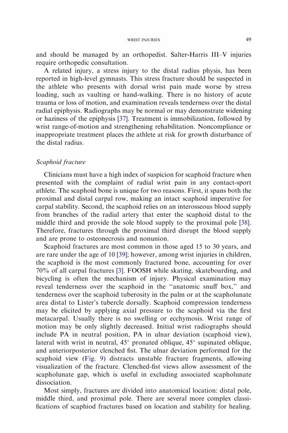
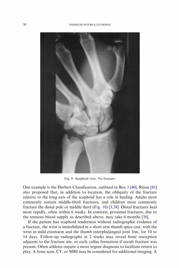
Scaphoid fractures are most common in those aged 15 to 30 years, andare rare under the age of 10 [39]; however, among wrist injuries in children,the scaphoid is the most commonly fractured bone, accounting for over70% of all carpal fractures [3]. FOOSH while skating, skateboarding, andbicycling is often the mechanism of injury. Physical examination mayreveal tenderness over the scaphoid in the ‘‘anatomic snuff box,’’ andtenderness over the scaphoid tuberosity in the palm or at the scapholunatearea distal to Lister’s tubercle dorsally. Scaphoid compression tendernessmay be elicited by applying axial pressure to the scaphoid via the firstmetacarpal. Usually there is no swelling or ecchymosis. Wrist range ofmotion may be only slightly decreased. Initial wrist radiographs shouldinclude PA in neutral position, PA in ulnar deviation (scaphoid view),lateral with wrist in neutral, 45� pronated oblique, 45� supinated oblique,and anteriorposterior clenched fist. The ulnar deviation performed for thescaphoid view (Fig. 9) distracts unstable fracture fragments, allowingvisualization of the fracture. Clenched-fist views allow assessment of thescapholunate gap, which is useful in excluding associated scapholunatedissociation.
Most simply, fractures are divided into anatomical location: distal pole,middle third, and proximal pole. There are several more complex classi-fications of scaphiod fractures based on location and stability for healing.
50 PARMELEE-PETERS & EATHORNE
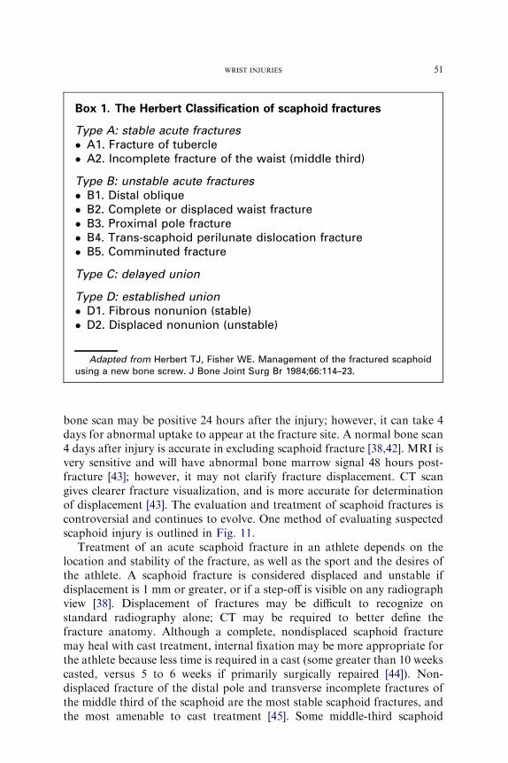
One example is the Herbert Classification, outlined in Box 1 [40]. Russe [41]also proposed that, in addition to location, the obliquity of the fracturerelative to the long axis of the scaphoid has a role in healing. Adults mostcommonly sustain middle-third fractures, and children most commonlyfracture the distal pole or middle third (Fig. 10) [3,38]. Distal fractures healmost rapidly, often within 6 weeks. In contrast, proximal fractures, due tothe tenuous blood supply as described above, may take 6 months [38].
If the patient has scaphoid tenderness without radiographic evidence ofa fracture, the wrist is immobilized in a short arm thumb spica cast, with thewrist in mild extension and the thumb interphalangeal joint free, for 10 to14 days. Follow-up radiographs at 2 weeks may reveal bone resorptionadjacent to the fracture site, or early callus formation if occult fracture waspresent. Often athletes require a more urgent diagnosis to facilitate return toplay. A bone scan, CT, or MRI may be considered for additional imaging. A
Fig. 9. Scaphoid view. No fracture.
51WRIST INJURIES
bone scan may be positive 24 hours after the injury; however, it can take 4days for abnormal uptake to appear at the fracture site. A normal bone scan4 days after injury is accurate in excluding scaphoid fracture [38,42]. MRI isvery sensitive and will have abnormal bone marrow signal 48 hours post-fracture [43]; however, it may not clarify fracture displacement. CT scangives clearer fracture visualization, and is more accurate for determinationof displacement [43]. The evaluation and treatment of scaphoid fractures iscontroversial and continues to evolve. One method of evaluating suspectedscaphoid injury is outlined in Fig. 11.
Treatment of an acute scaphoid fracture in an athlete depends on thelocation and stability of the fracture, as well as the sport and the desires ofthe athlete. A scaphoid fracture is considered displaced and unstable ifdisplacement is 1 mm or greater, or if a step-off is visible on any radiographview [38]. Displacement of fractures may be difficult to recognize onstandard radiography alone; CT may be required to better define thefracture anatomy. Although a complete, nondisplaced scaphoid fracturemay heal with cast treatment, internal fixation may be more appropriate forthe athlete because less time is required in a cast (some greater than 10 weekscasted, versus 5 to 6 weeks if primarily surgically repaired [44]). Non-displaced fracture of the distal pole and transverse incomplete fractures ofthe middle third of the scaphoid are the most stable scaphoid fractures, andthe most amenable to cast treatment [45]. Some middle-third scaphoid
Box 1. The Herbert Classification of scaphoid fractures
Type A: stable acute fractures� A1. Fracture of tubercle� A2. Incomplete fracture of the waist (middle third)
Type B: unstable acute fractures� B1. Distal oblique� B2. Complete or displaced waist fracture� B3. Proximal pole fracture� B4. Trans-scaphoid perilunate dislocation fracture� B5. Comminuted fracture
Type C: delayed union
Type D: established union� D1. Fibrous nonunion (stable)� D2. Displaced nonunion (unstable)
Adapted from Herbert TJ, Fisher WE. Management of the fractured scaphoidusing a new bone screw. J Bone Joint Surg Br 1984;66:114–23.

52 PARMELEE-PETERS & EATHORNE
fractures, particularly vertical oblique fractures, are less stable, take greaterthan 12 weeks to heal, and have a higher rate of nonunion. These areprimarily fixed by some surgeons [45]. Displaced fractures and proximalpole fractures, which have a greater risk of nonunion and malunion (seebelow), should be referred for operative treatment Fig. 12.
A nondisplaced distal scaphoid fracture or incomplete fracture may beimmobilized in a short-arm thumb spica cast for 4 to 8 weeks, with follow-up visits and radiograph every 2 weeks until radiographic union. Healingtime is typically 6 to 8 weeks. Nondisplaced middle-third fractures aretreated with long arm cast for 3 to 4 weeks, followed by a short arm cast foranother 6 to 8 weeks [1]. Healing takes 9 to 12 weeks, with a minimum of3 months out of sport. Ninety to 100% of transverse, nondisplaced, middle-third fractures will heal with casting if treatment is started within 3 weeks of
Fig. 10. Scaphoid fracture.

53WRIST INJURIES
injury [46]. Delay in immobilization beyond 3 weeks from fracture hasa higher incidence of nonunion, and should be referred to an orthopedicsurgeon [47]. For some sports, such as football and soccer, a playing castmay be used after the initial 4 weeks of casting; however, one study noteda higher nonunion rate (39%), ultimately requiring surgery, with playingcasts, compared with a rate of 15% with traditional casting and no sportsparticipation [1].
Open reduction and internal fixation has become standard for proximalpole fractures, and is required for unstable fractures. It is also becomingmore accepted to surgically repair minimally displaced or nondisplacedmiddle-third fractures, particularly for earlier return to sport, when a playingcast is not an option [44,48]. Inoue and Shionoya [44] compared casttreatment of nondisplaced middle-third scaphoid fractures with internalfixation in laborers, and noted return to work in an average of 10.2 weeks inthe cast group and 5.8 weeks in the internal fixation group, with nearly100% union in both groups. Another study [41] compared the effectivenessof immediate open reduction and internal fixation with the Herbert screwversus nonoperative treatment with a playing cast, in an athletic population.Return to sport was earlier in the cast-treated group (4.3 weeks) than inthe surgical fixation group (8.0 weeks); however, a subsequent study [49]
Suspected Scaphoid Fracture
X-Ray
Fracture No Fracture Seen
CT Scan Cast 2 Weeks
Displaced Fracture Nondisplaced Fracture Exam +Xray +
Exam +Xray -
ImmediateDiagnosisNecessary
ORIFCast vs Fixation*
Cast vs Fixation
Increased Uptakeat Scaphoid
Negative
NoScaphoidFracture
NondisplacedFracture (Cast)
Bone Scan Τ
* Specialist’s preference. Τ 4 days post-injury, if bone scan is negative there is no scaphoid fracture.§ Within 48 hr post-injury, MRI demonstrates fracture.
Bone Scan
MRI§
vs
Fig. 11. Algorithm for one method of suspected scaphoid fracture management. ORIF, open
reduction, internal fixation.

54 PARMELEE-PETERS & EATHORNE
demonstrated that return to sport averaged 5.8 weeks for acute midthirdscaphoid fractures. Both treatment methods yield union rates comparablewith those in other studies. The athletes in this study did not have increaserisk of nonunion secondary to participation in sports. A playing cast is anacceptable option for a stable fracture after an initial 4 weeks ofimmobilization. Internal fixation of an acute scaphoid fracture allows safeand early return to sports between 5 to 6 weeks [1,38,44,49], when a playingcast is not an acceptable option and when an athlete accepts the risks ofsurgery.
To summarize, patients who have proximal, displaced, angulated, orcomplex scaphoid fractures (scaphoid fracture associated with distal radiusfracture, open fracture, or perilunate fracture dislocation), or those whohave delayed diagnosis or nonunion should be referred for surgery.
Fig. 12. Scaphoid malunion.

55WRIST INJURIES
Consider referral for any athlete or manual laborer, because many surgeonsoffer percutaneous-screw fixation techniques to these patients in order todecrease the time of cast immobilization [1,38,44,49]. Following prolongedimmobilization, referral to a physical therapist will help regain motion andstrength. Return to sport after healing and rehabilitation should include useof a protective rigid splint for 3 months.
Kienbock’s disease
Acute isolated fracture of the lunate is rare, because it is well enclosed inthe large lunate fossa of the distal radius; however, avascular necrosis of thelunate, Kienbock�s disease, is seen in the young adult population. Itgenerally affects the dominant wrist and is unilateral [3]. This lesion presentswith dorsal wrist pain and swelling, decreased grip strength, and decreasedrange of motion, particularly in extension [50]. Typically, there is noapparent history of trauma. Pain may be produced in the lunate region byan axial strike at the distal end of the extended third digit. Plain radiographsare useful in making the diagnosis of Kienbock’s disease (Fig. 13). Theprecise etiology of Kienbock’s disease is unknown, but several causes havebeen proposed. Vascular compromise from repetitive trauma (in such sportsas handball, volleyball, golf, gymnastics, tennis, and martial arts) is thoughtto cause microfractures and excessive stress on the microscopic architecture[3,50]. Individuals who have increased risk are those who have ulnarnegative variance. The shortened ulna is thought to increase shear forceacross the lunate and cause vascular insufficiency.
The natural history of Kienbock’s disease is progressive sclerosis,fragmentation, and arthrosis [51]. Kienbock’s disease advances through
Fig. 13. Kienbock’s disease. MRI demonstrates avascularity of the lunate.

56 PARMELEE-PETERS & EATHORNE
four radiographic stages: (1) normal on plain radiographs, but the lunate isabnormal on bone scan, consistent with microfracture; (2) lunate sclerosiswithout collapse; (3) lunate fragmentation and collapse; and (4) perilunatearthritic changes. Functional disability may not always be progressive.Several studies suggest that some patients who have Kienbock’s disease canhave minimal pain and dysfunction for years with no treatment orintermittent immobilization [52,53]. Others report disappointing resultswith conservative treatment: worsening pain, limited motion, and pro-gressive arthrosis on radiographs [54]. It is difficult to know which patientswill have delayed dysfunction and which will have rapid progression.Conservative treatment will not change the natural history of the disease,but may allow the patient to recover from an acute flare of arthriticsynovitis. Surgery is the only definitive treatment for Kienbock’s disease.With more advanced disease, there are fewer surgical options [51]. Earlysurgical management involves unloading the lunate, which may requirechanging the length of the ulna or radius. Bone grafting and revascular-ization are newer techniques being investigated [51]. Once the disease hasprogressed to arthrosis, surgical options tend to be limited to wrist fusion.Individuals who have Kienbock’s disease being managed conservativelyneed to be re-evaluated periodically for pain and dysfunction. They shouldbe made aware of the disease progression and treatment options. Earlyorthopedic referral is ideal.
Hamate fractures
Hamate fractures occur in the body or hook. Both fractures present withill-defined pain in the ulnar aspect of the wrist, and may have associatedulnar paresthesias. Most commonly, such injuries present late, due tochronic pain, rather than acutely at time of injury. Palmar palpation of thehook produces tenderness if a hook fracture is present. Body fractures aretender dorsally. Hook of the hamate fractures are most common in sportsinvolving gripping a stick, bat, or racquet. Injury occurs by sportingequipment repeatedly compressing the hook of the hamate, causing fracture,or by acute trauma [50]. Routine wrist radiographs with the addition ofa carpal tunnel view are helpful to demonstrate a hook fracture (Fig. 14).Flexor digitorum function of the ring and little fingers, as well as ulnar nervefunction, should be assessed, because both structures are in close proximityto the hamate and can be injured with a fracture [38]. Cast immobilizationmay permit healing of acute hook fractures; however, nonunion is common,occurring in up to 46% of cases [7,55]. In the athlete who has an acute hookfracture, excision of the fracture fragment is the treatment of choice. Thissuccessfully resolves symptoms and allows return to sport 6 to 10 weeksafter surgery [1,31,55]. Nonunions are also treated with excision of the hook.Isolated hamate body fractures are rare. Nondisplaced body fractures may

57WRIST INJURIES
be treated with short-arm cast immobilization for 4 to 6 weeks. Displacedfractures require open reduction, internal fixation (ORIF).
Pisiform fractures
The pisiform is a sesamoid bone contained within the flexor carpi radialistendon. Pisiform fractures are uncommon and, as with other carpal bones,tend to occur with a FOOSH injury. Examination reveals tenderness overthe pisiform. If routine wrist films do not reveal the fracture (Fig. 15),
Fig. 14. Carpal tunnel view. Hook of hamate intact (arrow).
Fig. 15. Pisiform fracture (arrows) demonstrated on lateral view.
58 PARMELEE-PETERS & EATHORNE
a carpal tunnel view may be useful. Most fractures heal with 3 to 6 weeks ofshort arm cast immobilization; however, if nonunion occurs, the pisiformcan be excised [50].
Other carpal fractures
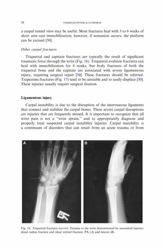
Triquetral and capitate fractures are typically the result of significanttraumatic force through the wrist (Fig. 16). Triquetral avulsion fractures canheal with immobilization for 4 weeks, but body fractures of both thetriquetral bone and the capitate are associated with severe ligamentousinjury, requiring surgical repair [50]. These fractures should be referred.Trapezium fractures (Fig. 17) tend to be unstable and to easily displace [50].These injuries usually require surgical fixation.
Ligamentous injury
Carpal instability is due to the disruption of the interosseous ligamentsthat connect and stabilize the carpal bones. These severe carpal disruptionsare injuries that are frequently missed. It is important to recognize that allwrist pain is not a ‘‘wrist sprain,’’ and to appropriately diagnose andproperly treat suspected carpal instability injuries. Carpal instability isa continuum of disorders that can result from an acute trauma or from
Fig. 16. Triquetral fractures (arrow). Trauma to the wrist demonstrated by associated injuries:
distal radius fracture and ulnar styloid fracture. PA (A) and lateral (B).

59WRIST INJURIES
repetitive injury. Multiple ligaments may be torn in an acute traumaticevent, causing complete lunate dislocation. Alternatively, minor trauma orrepetitive events may disrupt individual ligaments, which if left untreatedwill progress along the continuum of instability disorders, resulting in post-traumatic arthritis with pain and dysfunction [56]. Clinical suspicion ofthese injuries and arming oneself with appropriate diagnostic skills willfacilitate early diagnosis and appropriate treatment.
Scapholunate injuries result from a fall on a hand in which the wrist isextended and ulnar deviated. There typically is swelling, decreased range ofmotion, and dorsal wrist tenderness in the area of the scaphoid and lunate.A positive scaphoid shift test or Watson’s sign (see anatomy section of thearticle on wrist anatomy and examination by Eathorne elsewhere in thisissue) indicates a scapholunate tear. A positive scaphoid shift testreproduces pain and elicits a ‘‘pop’’ when the distal pole of the scaphoidis loaded dorsally and the wrist is moved from ulnar to radial deviation.
Scapholunate dissociation is diagnosed radiographically as a widening([3 mm) between the scaphoid and lunate [33]. This may be difficult to seeon the standard PA view, depending on the degree of ligamentous injury(Fig. 18). Stress views are helpful for diagnosis, because they elicita widening between the carpals if there is a ligament injury. Useful stressviews include clenched-fist view (an PA view of the wrist with a clenchedfist), PA maximal radial deviation, and PA maximal ulnar deviation. Leftuntreated, the scapholunate dissociation widens, the capitate shifts downinto the gap, and the carpal rows collapse. This is known as a SLAC(scapholunate advanced collapse) wrist [57]. If this is not evident on plainradiographs, MRI may demonstrate a complete scapholunate tear. MRI isunreliable for the diagnosis of incomplete tears. Arthroscopy has become
Fig. 17. Trapezium fracture.

60 PARMELEE-PETERS & EATHORNE
the standard method of diagnosing intercarpal ligament injuries [1]. Smalltears in the scapholunate ligament may not result in carpal instability, butare a common cause of chronic wrist pain associated with synovitis organglion cyst formation. Large tears of the scapholunate ligament causeinstability (ie, scapholunate dissociation). Any scapholunate ligament tearthat demonstrates dissociation or instability on imaging requires surgicalrepair [11,58]. Small, partial tears that do not demonstrate static separationof the ligaments on imaging may respond to a 3-month trial of conservativetherapy [11]. Conservative therapy includes activity to a pain-free level andsplint immobilization, followed by rehabilitation and progressive exercise.The decision to proceed with arthroscopy and the length of conservativetherapy depend largely on the athlete. A high-level athlete whose sportcannot tolerate wrist immobilization and who may miss an entire seasonmay proceed more quickly to arthroscopy.
Lunotriquetral tears also result from a FOOSH, but are much lesscommon than scapholunate tears. Lunotriquetral tears present with ulnar-sided wrist pain, weakness, and possibly clicking. There is tenderness overthe area of the lunotriquetral ligament. Dorsal pressure over the pisiformand palmar force on the lunate may produce a painful click. Plain radio-graphs are usually normal. Clinically correlated arthrography, with radio-carpal and midcarpal injection, can be useful for diagnosis. Bone scan may
Fig. 18. Scapholunate dissociation. Greater than 3 mm widening between the scaphoid and
lunate on PA view (A) is abnormal, indicating ligamentous injury. Scaphoid view (B)
accentuates widening.

61WRIST INJURIES
be obtained to rule out an occult fracture. MRI may be of benefit to rule outother ulnar-sided pathology, such as TFCC injury, but is not yet reliablysensitive for diagnosis of lunotriquetral ligament injury [1,59]. Lunotrique-tral injuries are treated with immobilization and NSAIDs. Immobilizationresults in lunotriquetral ligament healing in 80% of cases [60]. If symptomspersist following 4 to 6 of immobilization, the patient should be referred fordiagnostic and potentially therapeutic arthroscopy [1,32]. Because mostlunotriquetral tears do not cause instability, they usually do not lead toprogressive arthrosis.
In addition to stress radiographs mentioned above, careful evaluation ofthe true lateral view is important in the evaluation of carpal instability. Onthe normal true lateral view there is colinearity of radius, lunate, andcapitate. These three bones should be lined up, appearing as stacked cups. Ifthe lunate is grossly dislocated, it rotates so the concavity tips palmarly,termed the ‘‘spilled teacup sign’’ (Fig. 19A) [61]. Perilunate dislocation isalso the result of gross ligament disruption. The lunate and radius are inproper alignment, but the remaining carpal bones are dislocated dorsally(Fig. 19B). Measurement of the scapholunate angle on the lateral film willaid assessment of scapholunate instability, which often presents with subtleradiographic findings. To measure this scapholunate angle (Fig. 20), drawa line through the long axis of the scaphoid on the true lateral and a secondline through the center of the concavity of the lunate and radial shaft.
Fig. 19. (A) Lunate dislocation is demonstrated by the spilled teacup sign. The lunate is
dislocated and tipped volarly. (B) Perilunate dislocation.

62 PARMELEE-PETERS & EATHORNE
Measure the dorsal angle created by these lines. Normal scapholunate angleis 30� to 60�. An angle of less than 30� indicates that the lunate is volarly(palmarly) angulated, indicating a volar intercalated segment instability(VISI). Greater than 60� dorsal angulation of the lunate indicates dorsalintercalated segment instability (DISI). Knowledge of wrist ligamentousanatomy assists one in understanding these injuries. First, dorsal and ventralrefer to the position of the lunate. Second, the scapholunate ligament isattached to the volar (palmar) aspect of the lunate, and the lunotriquetralligament is attached to the dorsal aspect of the lunate. A rupture of thescapholunate ligament, which is volarly attached, allows DISI. A rupture ofthe lunotriquetral ligament, which is dorsally attached, may allow VISI;however, although scapholunate tears frequently cause DISI, lunotriquetraltears rarely cause VISI, due to other stabilizing ligaments. If there issignificant force, several ligaments may rupture, leading to perilunatedislocation. The capitate is displaced dorsally on the lunate on true lateralradiograph. If the lunate is dislocated, it is the displaced volarly into thecarpal tunnel, resulting in the spilled teacup sign. Both injuries requiresurgical repair.
Due to the potentially poor outcome of carpal instability, whenevera physician suspects a carpal sprain and is unable to exclude significantligament injury, referral is recommended. The patient should be splinted andseen in consultation within a few days.
Fig. 20. Measurement of the scapholunate angle.
63WRIST INJURIES
Wrist ganglia
Ganglia are common about the wrist. Thick, clear, fluid leaks througha tear (degenerative or traumatic) in the joint capsule or tendon sheath toform a ganglion cyst. Common locations include the dorsum of the wristdirectly over the scapholunate joint, and the volar radial aspect. Dorsalwrist ganglia arise from the scapholunate joint, and volar ganglia typicallyarise from the radiocarpal joint, scaphotrapezial joint [62], or FCU tendon.Ganglion cysts may or may not be painful. Typically there is no history ofwrist trauma. If pain or limitation of function mandates, the ganglion can beaspirated or excised. In a recent study of children who had asymptomaticwrist ganglia, 79% resolved spontaneously within 1 year [63]. Therefore,observation, particularly in children, is preferred. In a recent prospectivestudy of different treatment methods in adults (aspiration versus excision)for volar wrist ganglia, similar recurrence rates were demonstrated [64]. At5-year follow up, recurrence rates of a volar wrist ganglion were 42% afterexcision and 47% after aspiration. Fifty-one percent of untreated gangliahad disappeared spontaneously.
Dorsal wrist ganglia also tend to recur after aspiration. One studyreported a cure rate of 13% after single aspiration [65], whereas anotherstudy reported an 85% cure rate if three or more aspirations were performed[66]. Recurrence rates are unchanged by cortisone injection followingaspiration [67]. Recurrence rate of dorsal ganglia after cyst excision isapproximately 13% to 40%; however, with careful excision of the cyst’sstalk, recurrence rates decrease to 4% [68].
Occult dorsal wrist ganglia can produce chronic wrist pain, which may beconstant or activity related [62]. Localized dorsal wrist tenderness,maximum aggravation of pain during wrist flexion, decreased range ofmotion, and weak grip on examination may indicate an occult dorsal wristganglion. Initial examination and imaging should exclude scapholunateinstability. MRI may help identify an occult ganglion and differentiate itfrom a scapholunate ligament tear. Conservative therapy of occult dorsalwrist ganglia, including corticosteroid injection followed by immobilizationin a cock-up wrist splint for 7 to 10 days, may be tried initially [62].Definitive diagnosis and treatment is surgical exploration and excision.
Compressive neuropathies
Upper extremity compression neuropathies are relatively uncommon inathletes, particularly at the wrist; however, direct contusion of the tissueoverlying peripheral nerves or repetitive activity causing tissue swelling cancause neuropathic symptoms. Neuropraxia is the type of nerve lesion mostcommonly seen in athletes. This is a conduction block along the nerve,typically from compression or impingement, with nerve elements intact.
64 PARMELEE-PETERS & EATHORNE
Compression of radial, median, distal posterior interosseous, and ulnarnerves at the wrist has been seen in athletes. It is important to consider thecervical spine and elbow as other sites of impingement. With early diagnosis,rest, splinting, and activity modification often lead to symptom resolutionand return to sport.
Median nerve
Carpal tunnel syndrome (CTS) is the most common neuropathy seen inathletes. It is common in cyclists, gymnasts, throwing athletes, wheelchairathletes, and those who participate in sports that require gripping [69]. Themedian nerve is entrapped as it passes through the nonyielding carpal tunnel(see the anatomy section of the article on wrist anatomy and examination byEathorne elsewhere in this issue). A history of paresthesias affecting theradial three and one-half digits and nighttime pain are typical. Phalen’s testis often positive, and Tinel’s sign, thought to be less sensitive, may also bepresent. Electromyography (EMG) and nerve conduction studies (NCS) arehelpful in assessing ongoing denervation. It has been generalized thatapproximately 80% of patients who have carpal tunnel syndrome initiallyrespond to conservative treatment; however, symptoms recur in 80% ofthese patients after 1 year [70]. One effective, noninvasive, short-termtherapy is night wrist splinting. A study [71] found that among patients whohad improvement with night splint use for at least 6 weeks, 31% still hadsymptom improvement at 12 months.
A recent systematic review [72] of randomized, controlled trials ofconservative treatments for CTS found that NSAIDs, diuretics, andpyridoxine are no more effective than placebo in relieving the symptomsof carpal tunnel syndrome. Both oral corticosteroid therapy and localcorticosteroid injection have been shown to be effective in short-termtherapy (2 to 4 weeks) for electrophysiologically confirmed carpal tunnelsyndrome [72–75]; however, the optimal oral corticosteroid dosage andduration of treatment remains to be determined [73]. Additionally, at 8 to12 weeks post-treatment, symptom improvement was seen only with localcorticosteroid injection [75]. One double-blind, placebo-controlled trialfound that 77% of patients given a local corticosteroid injection hadsignificant symptom improvement, compared with 20% in the control group[76]. Yet another study [77] demonstrated that only 11% of patients whohad initial improvement with corticosteroid injection had relief of symptomsat 12 months post-injection.
Surgical treatment has significantly better long-term outcomes thanconservative therapy [78]. DeStefano et al [79] reported that patients whohad CTS and who underwent surgical release were six times more likely thanthose treated nonoperatively to have resolution of symptoms. Carpal tunnelrelease surgery should be considered in patients who have prolongedsymptoms that do not respond to conservative measures, and who have

65WRIST INJURIES
progressive slowing of nerve conduction. Those patients who respond toconservative therapy tend to show improvement of symptoms within 8 to12 weeks [80]. Most patients who will respond to steroid injection do so by4 weeks post-injection [72–75]. Patients who have severe nerve entrapment,as evidenced by nerve conduction studies, thenar atrophy, sensory loss, ormotor weakness, should referred for surgical release of the flexorretinaculum. A 7.0-millisecond or greater delay of the median nerve distallatency represents severe compression of the median nerve [81]. To preventpermanent nerve damage, these individuals should be referred for surgerywithout delay.
Ulnar nerve
‘‘Cyclist’s palsy’’ is an ulnar neuropathy caused by compression of therelatively superficial distal ulnar nerve, and it can be caused by wristposition during prolonged bicycling. It has also been reported in racquetsports and weight lifting, and in hockey goaltenders [82]. Ulnar nervecompression at Guyon’s canal (see the anatomy section of the article onwrist anatomy and examination by Eathorne elsewhere in this issue) usuallypresents with pain and paresthesias of the small finger and the ulnar half ofthe ring finger. Symptoms depend on the location of compression relative toGuyon’s canal (ie, ulnar tunnel). Sensation and motor function of the ulnarnerve should be assessed. If radiographs, including carpal tunnel views,exclude bony pathology, then rest, splinting, and NSAIDs can be initiated.Upon return to sport, padding over the palmar-ulnar aspect of the wristmay prevent recurrence. Replacing handlebar padding or cycling gloves andhaving body weight properly distributed on the handlebars by appropriatebike fitting may remedy a cyclist’s problem. Patients who do not respond toconservative treatment may require additional studies (eg, MRI, EMG) torule out space-occupying lesions and evidence of denervation.
Distal posterior interosseous nerve syndrome
Athletes such as weight lifters and gymnasts may present with complaintsof pain with wrist extension. If there is no evidence of mass or carpalinstability, distal posterior interosseous nerve syndrome should besuspected. This purely sensory nerve can be entrapped where it passesover the distal radius and enters the wrist capsule, due to fibrosis that canoccur with repetitive, forceful wrist extension [83]. Splinting and cortico-steroid injection are the initial treatments. Surgery may be required forsymptomatic relief.
Radial nerve
Wartenberg’s syndrome is radial nerve compression in the forearm. Thenerve runs subcutaneous between the brachioradialis and extensor carpi

66 PARMELEE-PETERS & EATHORNE
radialis longus, and is subject to irritation by wristbands and gloves [69].Patients present with pain and decreased sensation over the dorsoradialhand, dorsal thumb, and index finger. There is no motor loss. Conservativetreatment of activity modification is generally effective.
Summary
Primary care physicians not only have an important role in the diagnosisand initial treatment of wrist injuries, but also play a key role in theeducation of families about prevention. Children and adolescents are oftencompetitive in sports throughout the year. Periods of rest can be importantin prevention of overuse injuries in the very active, developing athlete.Protective gear such as wrist guards, used during activities such as inlineskating and snowboarding, has been shown to prevent acute injuries thatoften require surgery or lead to prolonged disability [84,85].
A primary care physician will often be the first health care providerto assess most wrist complaints. The intent of this article is to familiarizethe primary care physician with the most common wrist injuries in activepeople, and to demonstrate that many injuries can have poor outcomesif unrecognized. It is important to have good clinical knowledge of thefunctional anatomy of the wrist in order to maximize the informationgathered on examination and to narrow one’s differential diagnosis. Theathlete’s sport and desires regarding return to play, and the impact of thetiming of injury management on his or her further participation in sport areimportant to consider. A highly active person may be referred to a mus-culoskeletal specialist for advanced testing or surgical repair earlier in theevaluation of certain injuries than a less active one. Armed with good clini-cal knowledge of anatomy and an understanding of common wrist injuries,primary care physicians can successfully manage many wrist complaints.
Acknowledgments
The authors thank Jean-Paul Guiboux, MD of Michigan Hand & Wristand Thomas Hall, MD, Providence Hospital and Medical CentersDepartment of Radiology for their assistance with radiographs.
References
[1] Rettig AC. Athletic injuries of the wrist and hand, part 1: traumatic injuries of the wrist. Am
J Sports Med 2003;31(6):1038–48.
[2] Rettig AC, Patel DV. Epidemiology of elbow, forearm, and wrist injuries in the athlete. Clin
Sports Med 1995;14(2):289–97.
[3] Manusov EG. Hand and wrist injuries. In: Birrer RB, Griesemer BA, CatalettoMB, editors.
Pediatric sports medicine for primary care. 1st edition. Phiadelphia: Lippincott Williams &
Wilkins; 2002. p. 367–84.
67WRIST INJURIES
[4] De Smet L, Claessens A, Lefevre J, et al. Gymnast wrist: an epidemiological survey of ulnar
variance and stress changes of the radial physis in elite female gymnasts. Am J Sports Med
1994;22(6):846–50.
[5] Roser LA, Clawson DK. Football injuries in the very young athlete. Clin Orthop 1970;69:
219–23.
[6] Dobyns JH, Gabel GT. Gymnast’s wrist. Hand Clin 1990;6(3):493–505.
[7] Stark HH, Jobe FW, Boyes JH, et al. Fracture of the hook of the hamate in athletes. J Bone
Joint Surg Am 1977;59(5):575–82.
[8] Mirabello SC, Loeb PE, Andrews JR. The wrist: field evaluation and treatment. Clin Sports
Med 1992;11(1):1–25.
[9] Rettig AC. Wrist and hand overuse syndromes. Clin Sports Med 2001;20(3):591–611.
[10] Richie CA 3rd, Briner WW Jr. Corticosteroid injection for treatment of de Quervain’s
tenosynovitis: a pooled quantitative literature evaluation. J Am Board Fam Pract 2003;
16(2):102–6.
[11] Nunley JA, Goets T. Injuries to the soft tissues of the wrist. In: Garrett WE, Speer KP,
KirkendallDT, editors. Principles and practices of orthopedic sportsmedicine. Philadelphia:
Lippincott Williams & Wilkins; 2000. p. 273–87.
[12] Grundberg AB, Reagan DS. Pathologic anatomy of the forearm: intersection syndrome.
J Hand Surg [Am] 1985;10(2):299–302.
[13] Wood MB, Dobyns JH. Sports-related extraarticular wrist syndromes. Clin Orthop 1986;
202:93–102.
[14] Kiefhaber TR, Stern PJ. Upper extremity tendinitis and overuse syndromes in the athletes.
Clin Sports Med 1992;11(1):39–55.
[15] Eckhardt WA, Palmer AK. Recurrent dislocation of extensor carpi ulnaris tendon. J Hand
Surg [Am] 1981;6(6):629–31.
[16] Chun S, Palmer AK. Chronic ulnar wrist pain secondary to partial rupture of the extensor
carpi ulnaris tendon. J Hand Surg [Am] 1987;12(6):1032–5.
[17] Inoue G, Tamura Y. Recurrent dislocation of the extensor carpi ulnaris tendon. Br J Sports
Med 1998;32(2):172–4.
[18] Burkhart SS,WoodMB, Linscheid RL. Posttraumatic recurrent subluxation of the extensor
carpi ulnaris tendon. J Hand Surg [Am] 1982;7(1):1–3.
[19] Garcia-Elias M, Dobyns JH. Dorsal and palmar dislocations of the distal radioulnar joint.
In: Cooney WP, Linscheid RL, Dobyns JH, editors. The wrist: diagnosis and operative
treatment. 1st edition. St. Louis (MO): Mosby; 1998. p. 758–72.
[20] Palmer AK. Triangular fibrocartilage complex lesions: a classification. J Hand Surg [Am]
1989;14(4):594–606.
[21] Cooney WP, Bishop AT, Linscheid RL. Physical examination of the wrist. In: Cooney WP,
LinscheidRL,Dobyns JH, editors. The wrist: diagnosis and operative treatment. 1st edition.
St. Louis (MO): Mosby; 1998. p. 236–61.
[22] Cooney WP. Tears of the triangular fibrocartilage of the wrist. In: Cooney WP, Linscheid
RL, Dobyns JH, editors. The wrist: diagnosis and operative treatment. 1st edition. St. Louis
(MO): Mosby; 1998. p. 710–42.
[23] BednarMS,Arnoczky SP,WeilandAJ. Themicrovasculature of the triangular fibrocartilage
complex: its clinical significance. J Hand Surg [Am] 1991;16(6):1101–5.
[24] Palmer AK, Werner FW. Triangular fibrocartilage complex of the wrist: anatomy and
function. J Hand Surg [Am] 1981;6(2):153–62.
[25] Lester B, Halbrecht J, Levy IM, et al. ‘‘Press test’’ for office diagnosis of triangular
fibrocartilage complex tears of the wrist. Ann Plast Surg 1995;35(1):41–5.
[26] Buterbaugh GA, Brown TR, Horn PC. Ulnar-sided wrist pain in athletes. Clin Sports Med
1998;17(3):567–83.
[27] Linscheid RL. Biomechanics of the distal radioulnar joint. Clin Orthop 1992;275:46–55.
[28] Palmer AK, Glisson RR, Werner FW. Relationship between ulnar variance and triangular
fibrocartilage complex thickness. J Hand Surg [Am] 1984;9(5):681–2.

68 PARMELEE-PETERS & EATHORNE
[29] NagleDJ. Triangular fibrocartilage complex tears in the athlete. Clin SportsMed 2001;20(1):
155–66.
[30] Cooney WP. Fractures of the distal radius. In: Cooney WP, Linscheid RL, Dobyns JH,
editors. The wrist: diagnosis and operative treatment. 1st edition. St. Louis (MO): Mosby;
1998. p. 310–55.
[31] Morgan WJ, Slowman LS. Acute hand and wrist injuries in athletes: evaluation and
management. J Am Acad Orthop Surg 2001;9(6):389–400.
[32] Rettig AC, Trusler ML. Athletic injuries of the hand and wrist. In: Arendt EA, editor.
Orthopedic knowledge update: sports medicine 2. 1st edition. Rosemont (IL): American
Academy of Orthopedic Surgeons; 1999. p. 249–70.
[33] Eiff PM,Hatch RL, CalmbachWL. Carpal fractures. In: Fracture management for primary
care. 2nd edition. Philadelphia: WB Saunders; 2003. p. 96–115.
[34] PogueDJ, Viegas SF, PattersonRM, et al. Effects of distal radius fracturemalunion on wrist
joint mechanics. J Hand Surg [Am] 1990;15(5):721–7.
[35] McQueenM, Caspers J. Colles fracture: does the anatomical result affect the final function?
J Bone Joint Surg Br 1988;70(4):649–51.
[36] Abbaszadegan H, von Sivers K, Jonsson U. Late displacement of Colles’ fractures. Int
Orthop 1988;12(3):197–9.
[37] Mastey RD, Weiss AP, Akelman E. Primary care of hand and wrist athletic injuries. Clin
Sports Med 1997;16(4):705–24.
[38] Caputo AE, Goldner RD. Fractures of the carpal bones. In: Garrett WE, Speer KP,
KirkendallDT, editors. Principles and practices of orthopedic sportsmedicine. Philadelphia:
Lippincott Williams & Wilkins; 2000. p. 257–72.
[39] Rettig AC, Ryan R, Shelbourne KD, et al. Metacarpal fractures in the athlete. Am J Sports
Med 1989;17(4):567–72.
[40] Herbert TJ, Fisher WE. Management of the fractured scaphoid using a new bone screw.
J Bone Joint Surg Br 1984;66:114–23.
[41] Russe O. Fracture of the carpal navicular: diagnosis, non-operative treatment and operative
treatment. J Bone Joint Surg Am 1960;42:759–68.
[42] Coupland DB. Determining the presence of scaphoid fracture with a day 4 bone scan. Clin J
Sport Med 1996;6(2):137.
[43] Ring D, Jupiter JB, Herndon JH. Acute fractures of the scaphoid. J Am Acad Orthop Surg
2000;8(4):225–31.
[44] Inoue G, Shionoya K. Herbert screw fixation by limited access for acute fractures of the
scaphoid. J Bone Joint Surg Br 1997;79(3):418–21.
[45] LinscheidRL,Weber ER. Scaphoid fractures and nonunion. In: CooneyWP, LinscheidRL,
Dobyns JH, editors. The wrist: diagnosis and operative treatment. 1st edition. St. Louis
(MO): Mosby; 1998. p. 385–430.
[46] Cooney WP 3rd, Dobyns JH, Linscheid RL. Nonunion of the scaphoid: analysis of the
results from bone grafting. J Hand Surg [Am] 1980;5(4):343–54.
[47] Langhoff O, Andersen JL. Consequences of late immobilization of scaphoid fractures.
J Hand Surg [Br] 1988;13(1):77–9.
[48] Rettig AC, Weidenbener EJ, Gloyeske R. Alternative management of midthird scaphoid
fractures in the athlete. Am J Sports Med 1994;22(5):711–4.
[49] Rettig AC, Kollias SC. Internal fixation of acute stable scaphoid fractures in the athlete. Am
J Sports Med 1996;24(2):182–6.
[50] Geissler WB. Carpal fractures in athletes. Clin Sports Med 2001;20(1):167–88.
[51] Palmer AK, Benoit MY. Lunate fractures: Kienbock’s disease. In: Cooney WP, Linscheid
RL, Dobyns JH, editors. The wrist: diagnosis and operative treatment. 1st edition. St. Louis
(MO): Mosby; 1998. p. 431–73.
[52] Kristensen SS, Thomassen E, Christensen F. Kienbock’s disease: late results by non-surgical
treatment. J Hand Surg [Br] 1986;11(3):422–5.
69WRIST INJURIES
[53] Beckenbaugh RD, Shives TC, Dobyns JH, et al. Kienbock’s disease: the natural history of
Kienbock’s diease and consideration of lunate fractures. Clin Orthop 1980;149:98–106.
[54] Almquist EE. Kienbock’s disease. Hand Clin 1987;3(1):141–8.
[55] Parker RD, BerkowitzMS, BrahmsMA, et al. Hook of the hamate fractures in athletes. Am
J Sports Med 1986;14(6):517–23.
[56] Herzberg G, Comtet JJ, Linshceid RL, et al. Perilunate dislocations and fracture-
dislocations: a multicenter study. J Hand Surg [Am] 1993;18(5):768–79.
[57] Anderson I, Read JW, Steinweg J. The hand and wrist. In: Atlas of imaging in sports
medicine. 1st edition. Sydney: McGraw-Hill Professional; 1999. p. 17–73.
[58] Taleisnik J, Linscheid RL. Scapholunate instability. In: CooneyWP, Linscheid RL, Dobyns
JH, editors. The wrist: diagnosis and operative treatment. 1st edition. St. Louis (MO):
Mosby; 1998. p. 501–26.
[59] BishopAT,ReaganDS. Lunotriquetral sprains. In: CooneyWP, LinscheidRL,Dobyns JH,
editors. The wrist: diagnosis and operative treatment. 1st edition. St. Louis (MO): Mosby;
1998. p. 527–49.
[60] Cohen MS. Ligamentous injuries of the wrist in the athlete. Clin Sports Med 1998;17(3):
533–52.
[61] Kozin SH. Perilunate injuries: diagnosis and treatment. J Am Acad Orthop Surg 1998;6(2):
114–20.
[62] HalikisMN,Taleisnik J. Soft-tissue injuries of thewrist. Clin SportsMed 1996;15(2):235–59.
[63] Wang AA, Hutchinson DT. Longitudinal observation of pediatric hand and wrist ganglia.
J Hand Surg [Am] 2001;26(4):599–602.
[64] Dias J, Buch K. Palmar wrist ganglion: does intervention improve outcome? A prospective
study of the natural history andpatient-reported treatment outcomes. JHandSurg [Br] 2003;
28(2):172–6.
[65] Richman JA, GelbermanRH, EngberWD, et al. Ganglions of the wrist and digits: results of
treatment by aspiration and cyst wall puncture. J Hand Surg [Am] 1987;12(6):1041–3.
[66] Zubowicz VN, Ishii CH. Management of ganglion cysts of the hand by simple aspiration.
J Hand Surg [Am] 1987;12(4):618–20.
[67] Varley GW, Needoff M, Davis TR, et al. Conservative management of wrist ganglia:
aspiration versus steroid infiltration. J Hand Surg [Br] 1997;22(5):636–7.
[68] Thornburg LE. Ganglions of the hand and wrist. J Am Acad Orthop Surg 1999;7(4):
231–8.
[69] Izzi J, Dennison D, Noerdlinger M, et al. Nerve injuries of the elbow, wrist, and hand in
athletes. Clin Sports Med 2001;20(1):203–17.
[70] Kanaan N, Sawaya RA. Carpal tunnel syndrome: modern diagnostic and management
techniques. Br J Gen Pract 2001;51(465):311–4.
[71] GerritsenAA,Korthals-de Bos IB, Laboyrie PM, et al. Splinting for carpal tunnel syndrome:
prognostic indicators of success. J Neurol Neurosurg Psychiatry 2003;74(9):1342–4.
[72] Gerritsen AA, de Krom MC, Struijs MA, et al. Conservative treatment options for carpal
tunnel syndrome: a systematic review of randomised controlled trials. J Neurol 2002;249(3):
272–80.
[73] Chang MH, Chiang HT, Lee SS, et al. Oral drug of choice in carpal tunnel syndrome.
Neurology 1998;51(2):390–3.
[74] Marshall S, Tardif G, Ashworth N. Local corticosteroid injection for carpal tunnel
syndrome. Cochrane Database Syst Rev 2002;(4):CD001554.
[75] Wong SM, Hui AC, Tang A, et al. Local vs systemic corticosteroids in the treatment of
carpal tunnel syndrome. Neurology 2001;56(11):1565–7.
[76] Dammers JW, Veering MM, Vermeulen M. Injection with methylprednisolone proximal to
the carpal tunnel: randomised double blind trial. BMJ 1999;319(7214):884–6.
[77] Gonzalez MH, Bylak J. Steroid injection and splinting in the treatment of carpal tunnel
syndrome. Orthopedics 2001;24(5):479–81.

70 PARMELEE-PETERS & EATHORNE
[78] Gerritsen AA, de Vet HC, Scholten RJ, et al. Splinting vs surgery in the treatment of carpal
tunnel syndrome: a randomized controlled trial. JAMA 2002;288(10):1245–51.
[79] DeStefano F, NordstromDL, Vierkant RA. Long-term symptom outcomes of carpal tunnel
syndrome and its treatment. J Hand Surg [Am] 1997;22(2):200–10.
[80] Pianka G, Hershman EB. Neurovascular injuries. In: Nicholas JA, Hershman EB, editors.
The upper extremity in sports medicine. 2nd edition. St. Louis (MO): Mosby; 1995.
p. 665–93.
[81] Chow JCY. Carpal tunnel syndrome. In: Watson HK, Weinzweig J, editors. The wrist. 1st
edition. Philadelphia: Lippincott Williams & Wilkins; 2001. p. 107–22.
[82] Planchier KD, Peterson RK, Steichen JB. Compressive neuropathies and tendinopathies in
the athletic elbow and wrist. Clin Sports Med 1996;15(2):331–71.
[83] Carr D, Davis P. Distal posterior interosseous nerve syndrome. J Hand Surg [Am] 1985;
10(6):873–8.
[84] Schieber RA, Branche-Dorsey CM, Ryan GW, et al. Risk factors for injuries from in-line
skating and the effectiveness of safety gear. N Engl J Med 1996;335(22):1630–5.
[85] O’Neill DF. Wrist injuries in guarded versus unguarded first time snowboarders. Clin
Orthop 2003;409:91–5.