theoretical study of the anti-ncp molecular mechanism of
TRANSCRIPT
doi.org/10.26434/chemrxiv.12016236.v1
Theoretical Study of the anti-NCP Molecular Mechanism of TraditionalChinese Medicine Lianhua-Qingwen Formula (LQF)Chenghao Ye, Meina Gao, Wangqiang Lin, Kunqian Yu, Peng Li, Guanghui Chen
Submitted date: 21/03/2020 • Posted date: 23/03/2020Licence: CC BY-NC-ND 4.0Citation information: Ye, Chenghao; Gao, Meina; Lin, Wangqiang; Yu, Kunqian; Li, Peng; Chen, Guanghui(2020): Theoretical Study of the anti-NCP Molecular Mechanism of Traditional Chinese MedicineLianhua-Qingwen Formula (LQF). ChemRxiv. Preprint. https://doi.org/10.26434/chemrxiv.12016236.v1
Due to the good clinical efficacy in treating Novel Coronavirus Pneumonia (NCP) resulted from SARS-CoV-2,as the traditional Chinese medicine(TCM) prescription, Lianhua Qingwen Formula (LQF) was composed intothe Diagnosis and Treatment Programs of 2019 New CoronavirusPneumonia (from fourth to seventh editions) formulated by the National Health Commission of China. Aimingto prevent and treat viral influenza, LQF was patented from 2003 in China, and passed the Phase II clinicaltrial by FDA in the United States in 2015. However, the molecular mechanism of LQF anti SARS-CoV-2pneumonia is still not clear. It is shown that the docking scores of three components in LQF including Rutin,Forsythoside E, and Hyperoside to main protease of SARS-CoV-2 are very large as -9.1, -9.0 and -8.7kcal/mol, respectively, which are even better than those of Lopinavir at -7.3kcal/mol. Importantly, the binding modes between active compounds and protein were verified via moleculardynamics (MD) simulation and calculation all the binding free energies at MM-PBSA level. Note that thesedonor-acceptor systems were stabilized by non-polar interactions including hydrogen bonds and hydrophobicinteractions. At last, from the constructed component-target-pathway network, it is shown that thecomponents in LQF are related important pathways to improve the human immunity such as T cell, B cellreceptor signaling, natural killer cell mediated cytotoxicity, as well as anti inflammatory pathways including Fcepsilon RI, ErbB, MAPK signaling and so on. The present investigation represents the first report on themolecular mechanism of LQF as NCP inhibitor
File list (2)
download fileview on ChemRxivms_lhqw_0321g.docx (2.24 MiB)
download fileview on ChemRxivms_lhqw_0321-g_SI.docx (270.77 KiB)

Theoretical Study of the anti-NCP Molecular
Mechanism of Traditional Chinese Medicine
Lianhua-Qingwen Formula (LQF)
Cheng-hao Ye1, #, Mei-na Gao2, 3 #, Wang-qiang Lin1, Kun-qian Yu3, Peng Li4, and
Guang-hui Chen1, *
1. Department of Chemistry and Key Laboratory for Preparation and Application of
Ordered Structural Materials of Guangdong Province, Shantou University,
Guangdong Province, 515063, China
2. Nanjing University of Chinese Medicine, Jiangsu Province, 210029, China
3. State Key Laboratory of Drug Research, Shanghai Institute of Materia Medica,
Chinese Academy of Sciences, Shanghai, 201203, China
4. School of Life and Health Sciences, The Chinese University of Hong Kong,
Shenzhen, Guangdong Province, 518172, China
# These authors contributed equally to this work
* Corresponding author: Guang-hui Chen ([email protected])
1
1
2
3
4
5
6
7
8
9
10
11
12
13
14
15
16
17
12

Abstract Due to the good clinical efficacy in treating Novel Coronavirus Pneumonia (NCP) resulted from
SARS-CoV-2, as the traditional Chinese medicine(TCM) prescription, Lianhua Qingwen Formula
(LQF) was composed into the Diagnosis and Treatment Programs of 2019 New Coronavirus
Pneumonia (from fourth to seventh editions) formulated by the National Health Commission of
China. Aiming to prevent and treat viral influenza, LQF was patented from 2003 in China, and
passed the Phase II clinical trial by FDA in the United States in 2015. However, the molecular
mechanism of LQF anti SARS-CoV-2 pneumonia is still not clear. It is shown that the docking scores
of three components in LQF including Rutin, Forsythoside E, and Hyperoside to main protease of
SARS-CoV-2 are very large as -9.1, -9.0 and -8.7 kcal/mol, respectively, which are even better than
those of Lopinavir at -7.3 kcal/mol. Importantly, the binding modes between active compounds and
protein were verified via molecular dynamics (MD) simulation and calculation all the binding free
energies at MM-PBSA level. Note that these donor-acceptor systems were stabilized by non-polar
interactions including hydrogen bonds and hydrophobic interactions. At last, from the constructed
component-target-pathway network, it is shown that the components in LQF are related important
pathways to improve the human immunity such as T cell, B cell receptor signaling, natural killer cell
mediated cytotoxicity, as well as anti-inflammatory pathways including Fc epsilon RI, ErbB, MAPK
signaling and so on. The present investigation represents the first report on the molecular mechanism
of LQF as NCP inhibitor.
Keywords: SARS-CoV-2; Inhibitor; Lianhua-Qingwen Formula; CADD; Network pharmacology.
2
18
19
20
21
22
23
24
25
26
27
28
29
30
31
32
33
34
35
36
37
34
1. Introduction From December of 2019, there was an outbreak of new pneumonia resulting from new coronavirus
named as SARS-CoV-2 in Wuhan city of China1. The syndrome of new coronavirus pneumonia
(NCP) includes fever, cough, and hypodynamic with fatality to some extent2-3. The World Health
Organization (WHO) declared the SARS-CoV-2 viral disease has been swept into at least 114
countries and led to death of more than 4,000 people. Drug development for treating SARS-CoV-2 is
urgent due to the rapid spread of NCP. It should be noted that TCM has a long history for the
prevention and treatment of various diseases in China by targeting and modulating multiple disease
related pathways with multiple effective components4. After strict therapeutic effect evaluation,
Lianhua Qingwen Formula (LQF) was composed into the Diagnosis and Treatment Programs of
2019 New Coronavirus Pneumonia formulated by the National Health Commission of China5-8.
Nowadays, although TCMs are widely used for prevention and treatment of viral pneumonia in
China9, there still some doubts on how the TCM works and what is the effective compounds.
As we know that during the period of spreading of SARS-CoV to MERS-CoV, the computer-aided
drug design (CADD) plays an important role to discover the CoV inhibitors10. In addition, network
pharmacology has been recently developed as a powerful tool to explore the relation among drug
compoments, targets, and pathways toward a certain disease11-13 . Therefore, in this work we tested
the effect of Chinese patent medicine of LQF on anti-SARS-CoV-2 by virtual screening at first.
Secondly, based on network pharmacology, we tried to construct a component-target-pathway
network between the LQF and viral pneumonia to explain the mechanism of anti-inflammatory and
human immunity.
Aiming to prevent and treat viral influenza, the LQF came from two well-known TCM
prescriptions Maxing-Shigan-Tang and Yinqiao-San, was patented from 2003 in China, and passed
3
38
39
40
41
42
43
44
45
46
47
48
49
50
51
52
53
54
55
56
57
58
59
60
56

the Phase II clinical trial by FDA in the United States in 201514-15. The LQF contains 11 herbs
including Radix Isatidis (Banlangen), Fructus Forsythiae (Lianqiao), Flos Lonicerae Japonicae
(Jinyinhua), Rhizoma Dryopteridis Crassirhizomatis (Mianmaguanzhong), Herba Ephedrae
(Mahuang), Semen Armeniacae Amarum (Kuxingren), Herba Houttuyniae (Yuxingcao), Herba
Pogostemonis (Guanghuoxiang), Radix et Rhizoma Rhodiolae Crenulatae (Hongjingtian), Radix et
Rhizoma Rhei (Dahuang) and Radix et Rhizoma Glycyrrhizae (Gancao) and a mineral medicine,
Gypsum Fibrosum (Shigao) as well as menthol. Duan, etc proved Lianhua Qingwen Capsule has the
same effect as Oseltamivir on treating influenza A (H1N1) virus infections16. LQF had been
developed for the analysis of absorbed components in SD rat plasma after oral administration by
UPLC-Q-TOF-MS method and total 21 main chemical components17. In this study, we focused on
the inhibiting effect on NCP of the molecular structures of 21 compounds in LQF and lopinavir as
plotted in Figure 1. Note that lopinavir is suggested in treating mild cases of SARS-CoV-2 infection
by National Health Commission of China. Based on the mixed compounds, we propose that some of
the LQF gradients can inhibit the SARS-CoV-2 reproduction and enhance the immunity efficiently.
2. Computational methodsMolecule docking. The component small molecules of LQF were optimized at B3LYP-D3/6-
31G(d, p) level of theory with Gaussian 16 package at first18. Just like the SARS-CoV14, taking the
main protease (Mpro) of SARS-CoV-2 from PDB bank (PDB code: 6LU7) as target19, we tried to
dock it with the 21 components in LQF, respectively, by using AutoDock Vina program20. The
possible docking conformations and binding modes were predicted in the grid of protein. In this
study, a grid of 40 × 40 × 40 points in the x, y, and z-axis directions was built and the center of grid
was x = -18.954, y = 16.918, z = 68.850 with the exhaustiveness of 20.
Molecular dynamics (MD) simulation. After docking, the complex systems with the top three
4
61
62
63
64
65
66
67
68
69
70
71
72
73
74
75
76
77
78
79
80
81
82
83
78
highest docking score were submitted to 20 ns of MD simulations so as to check their stability inside
the binding pocket of the main protease in SARS-CoV-2 and verify which residues interact with the
ligands. All of the complexes were prepared after molecular docking and then subjected to MD
simulation in a periodic boundary condition using the GROMACS 2018.4 software package21-22 with
TIP3P water model23 (Supplementary table 1). The Amber 99 sb-ILDN force field was applied to
describe the receptors, ligands, ions and water24. ACPYPE25 is a tool based on Python programming
language to generate parameters and topologies for ligands with ANTECHAMBER, using GAFF
force field26. To keep each system electrically neutral, sodium and chloride ions were added to
substitute for water molecules to produce a solvent box of 0.15 M NaCl. Initially, the complex
systems were relaxed with conjugate gradient energy minimization to prevent from steric clashes or
incorrect geometry. Then the restrained complex systems were simulated with position-restrained
MD within 100 ps, in case of drastic rearrangement during equilibration. At last, 20 ns MD
simulation of products were performed at 300 K under the NPT ensemble.
Decomposition of binding free energy between ligand and residue. The binding free energies
were calculated by g_mmpbsa program27-28 using the MM-PBSA method29-30. The 500 snapshots of
last 5 ns MD trajectories were chosen to calculate the binding energy and interaction decomposition.
The MM-PBSA method can be conceptually summarized as:
ΔGbind = ΔGcomplex - [ΔGprotein + ΔGlig] (1)ΔGbind = ΔH - TΔS (2)where ΔH of the system consists of the enthalpy changes in the gas phase on complex formation
(ΔEMM) and the solvated free energy contribution (ΔGsol), while -TΔS refers to the entropy
contribution to the binding. Note that the entropy differences should be very small, thus the
calculation of the solute entropy term was ignored in present study, and Eq. (2) can be transformed
5
84
85
86
87
88
89
90
91
92
93
94
95
96
97
98
99
100
101
102
103
104
910
into Eq. (3):
ΔGbind = ΔEMM + ΔGsol (3)where ΔEMM is the summation of van der Waals (ΔEwdW) and the electrostatic (ΔEele) interaction
energies.
ΔEMM = ΔEvdW + ΔEele (4)In addition, ΔGsol, in Eq. (3) which signifies the solvation free energy, can be calculated as the
summation of electrostatic component, the polar part (ΔGPB,sol) and nonpolar component (ΔGnonpolar,sol),
are shown in Eq. (5). The nonpolar contribution to the free energy was calculated via γSASA, where
SASA was the solvent-accessible surface area, γ was 0.0227 kcal/mol/Å2, and the solvent-probe
radius for SASA was 1.4 Å. Note that the offset of SASA calculation was 3.8493 kJ/mol.
ΔGsol = ΔGPB,sol + ΔGnonpolar,sol
(5)
Construction of component-target-pathway network. Network pharmacology analysis is a way of
representing datasets emphasizing the relationships between nodes31-32. The active components,
which have been detected in the rat plasma17, were used as keywords to search the related potential
targets from Traditional Chinese Medicine System Pharmacology Database and Analysis Platform
(TCMSP)33. Totally 1080 virus pneumonia related proteins were collected from GeneCard
Database34, whose relevance scores were over 5. Intersection of targets were uploaded in DAVID
database35 using the Functional Annotion Tool to obtain relevant targets and KEGG-Pathways. The
related targets and pathways were used as input to the software Cytoscape36 to construct the
component-target-pathway network. The degree of a node is the number of edges connecting nodes
depicting the probability distribution of these degrees over the whole network. Note that local based
method considers the direct neighborhood of a vertex. Given a node v, N(v) denotes the collection of
its neighbors37. Node degree (Deg) is calculated as follow:
6
105
106
107
108
109
110
111
112
113
114
115
116
117
118
119
120
121
122
123
124
1112

Deg(v)=|N(v)| (6)3. Results
3.1 The interaction between components of LQF and main protease
We docked 21 compounds into main protease of SARS-CoV-2 using a flexible docking procedure.
The top three highest compounds were Rutin, Hyperoside and Forsythoside E, which bind with the
main protease at -9.1, -9.0 and -8.7 kcal/mol, respectively, and the reported prescription of anti-
SARS-CoV-2, Lopinavir only reached -7.3 kcal/mol. Rutin directly interacts with residues Leu141,
Ser144, His163 and Asp187 as plotted in Figure 2a. Forythoside E formed excellent hydrogen bonds
with residues Leu141, Ser144, Cys145, His 164, Glu166, Glu189, Thr190 as plotted in Figure 2b.
Hyperoside interacts with Mpro in the Thr26, Ser144, Cys145, Glu166 and Asp187 as plotted in
Figure 2c, where glycosyl moiety of Hyperoside form hydrogen bonds with residues Ser144, Cys145
and Glu166 directly. On the contrary, Lopinavir provides only hydrogen atoms on nitrogen atoms of
imino to form hydrogen bonds with residue Glu166 and Gln189 as plotted in Figure 2d. According
the docking scores, we found that not all the ingredients in LQF can inhibit the main protease of
SARS-CoV-2. The top-ranked poses of compounds were arranged by docking score as collected in
Table 1.
3.2 Binding mode verification in MD simulation
In order to explore whether the above three compounds in LQF including Forsythoside E, Rutin,
and Hyperoside could bind with the pocket of main protease of SARS-CoV-2 stably, we also
calculated the root-mean-square deviation (RMSD) between ligands, including lopinavir, and Cα
atoms of protein by molecular dynamic simulation. After 20 ns stimulation we found that all these
four compounds attain an equilibrium, but Forsythoside E, Rutin, and Hyperoside arrived more
quickly than Lopinavir. The RMSD results with smooth fluctuation showed the ligands in the
7
125
126
127
128
129
130
131
132
133
134
135
136
137
138
139
140
141
142
143
144
145
146
1314

binding pockets were energetically stable, which can be recognized the compounds tend to interact
with the potential binding pocket in SARS-CoV-2 main protease as shown in Figure 3a and Figure
S1. This means that the three components in LQF combine with Mpro more easily than Lopinavir.
According to the binding free energies at the MM-PBSA level, we found that Forsythoside E, Rutin
and Hyperoside were stabilized by van der Waals and electrostatic interactions as plotted in Figure
3b. Hyperoside-Mpro complex showed the lowest energy among these systems while Lopinavir-
Mpro corresponded to the highest energy system. The calculated free energy proved that non-polar
interactions including hydrogen-bonds and hydrophobic interactions contributed to the binding
energy most between Hyperoside and Mpro of SARS-CoV-2 as listed in Supplementary table 2.
The hydrogen bonds play a significant role in maintaining the interaction between the protein and
ligand38. Hydrogen bonds between each ligand and protein presented in the whole last 5 ns
simulation were confirmed with highly contributing residues, such as Met49 and Met 165 in
Lopinavir complex. Hyperoside-Mpro, Rutin-Mpro and Forsythoside E-Mpro complexes possessed
the maximum number of intermolecular hydrogen bonds at six over the simulation period, compared
with only two hydrogen bonds in the Lopinavir-Mpro complex as plotted in Figure 3c. Meanwhile,
the lifetime of hydrogen bonds were computed as well, the hydrogen bonds in complex of Rutin,
Forysthoside E, Hyperoside and Lopinavir had apparently longer lifetime at 42.4, 32.1, 28.2 and 26.0
ps, respectively, as listed in Supplementary table 3. So, Rutin, Forysthoside E and Hyperoside have
higher affinity to main protease of SARS-CoV-2 according to their lower binding energies and more
hydrogen bonds with longer lifetime. It is shown that the combination of Hyperoside with Mpro is
more stable than those of Lopinavir, Rutin, and Forysthoside E as well.
Then we calculated the RMSF of above four compound-Mpro systems, respectively. The
8
147
148
149
150
151
152
153
154
155
156
157
158
159
160
161
162
163
164
165
166
167
168
1516
fluctuations of lines were very similar, indicating that the binding modes of the ligands are in same
pattern as plotted in Figure 3d. It is shown that Forsythoside E was located in the pocket around
His41, Met165, Glu166, Asp187, Arg188, Gln189, Thr190 and Gln192 with lower binding free
energies of -45.76 kcal/mol as plotted in Figure 4a. Rutin tended to interact mainly with residues
His41, Met49, Gly143, Ser144, Cys145, Met165, Glu166 and Glu189 because of the greater binding
free energy contribution of them as shown in Figure 4b. Among the results of the top three
complexes, Hyperoside performed the highest absolute value of binding free energy of -48.41
kcal/mol which could be decomposed into ΔEvdW (-54.20 kcal/mol), ΔEele (-39.15 kcal/mol), ΔEPB
(49.83 kcal/mol) and ΔEnonpolar (-4.89 kcal/mol) (Supplementary table 2). The Hyperoside mainly
interacted with residues Thr26, Met49, Tyr54, Gly143, Cys145, Met165, Asp187 and Gln189 with
energies at -3.88, -2.97, -1.95, -3.87, -2.55, -3.09, -3.09 and -4.97 kcal/mol, respectively, as shown in
Figure 4b, which provided greater contribution in Hyperoside-MPro complex system via van der
Waals and electrostatic interactions. Note that these residues participated in the complex binding
apparently as plotted in Figure 4c. The calculated free energy proved that van der Waals and
electrostatic interactions contributed most to the binding between Hyperoside and SARS-CoV-2
main protease. In the Rutin-Mpro complex, the binding free energy was also very low of -47.67
kcal/mol as listed in Supplementary table 2. Note that the absolute values of binding free energies of
these three compounds combining with main protease are much larger than that of Lopinavir. The
binding energy of Lopinavir was -34.42 kcal/mol and could be decomposed into ΔEvdW (-46.99
kcal/mol), ΔEele (-15.04 kcal/mol), ΔEPB (39.07 kcal/mol) and ΔEnonpolar (-5.47 kcal/mol), respectively,
indicating that Lopinavir was bound in the pocket around residues His41, Met49, Gly143, Ser144,
Cys145, Met165, Glu166 and Gln189, respectively, as plotted in Figure 4d. Therefore, we concluded
9
169
170
171
172
173
174
175
176
177
178
179
180
181
182
183
184
185
186
187
188
189
190
1718

that the critical residues were Met165, Gln189, Cys145, Asp187.
3.3 Ingredient-target-pathway network analysis
Network analysis. Severe SARS-CoV-2 infection can rapidly activate pathogenic T cells and
produce granulocyte-macrophage colony, with some important Cytokine Response Patterns, and the
high-levels of proinflammatory cytokines including IL-2, IL-6, IL-7, IL-10, G-CSF, IP-10, MCP-1,
MIP-1A, VEGF and TNFα were observed in the SARS-CoV-2 severe cases39-40. As shown in Figure
5, we found that the relative targets of 21 compounds of LQF interact with signaling pathways of
inflammation and immunity, such as VEGF signaling pathway41, Toll-like receptor signaling
pathway42, T cell receptor signaling pathway43, Fc epsilon RI signaling pathway44, B cell receptor
signaling pathway45, ErbB signaling pathway46, MAPK signaling pathway47, natural killer cell
mediated cytotoxicity48, JAK-STAT signaling pathway49, complement and coagulation cascades50. We
collected 1080 virus pneumonia-related proteins from GeneCard Database, whose relevance scores
were over 5. The 42 active components’ genes were collected from TCMSP database overall51, and
the intersection of two parts contains 37 genes. Then we collected 79 related targets and 10 pathways
of inflammatory and immunity via uploading the 37 genes to DAVID database35. Formononetin,
Rutin, Emodin 8-O-β-D-glucoside, Hyperoside, Loganic acid, Salidroside in the LQF connect
potential targets directly which are relevant to anti-inflammatory and immunity mechanisms
(Supplementary table 4 and 5). Thus, Formononetin, Rutin, Emodin 8-O-β-D-glucoside, Hyperoside,
Loganic acid, Salidroside, are key components which are beneficial in preventing viral infection
because they acquire higher node degrees of compounds in ingredient-target-pathway network. The
node degrees of Formononetin, Rutin and Emodin 8-O-β-D-glucoside are over 10 while the average
of the node degree is only 2.92 (Supplementary table 6). The node degrees of T cell receptor
10
191
192
193
194
195
196
197
198
199
200
201
202
203
204
205
206
207
208
209
210
211
212
1920

signaling pathway, MAPK signaling pathway and Toll-like receptor signaling pathway are over 4
while the average of the node degree is 3.80 (Supplementary table 7). The component-target-pathway
network includes 111 nodes along with 487 edges as plotted in Figure 5, which interprets interactions
among compounds, targets and pathways. These candidate targets are collected from databases and
have been proved to be affected by LQF’s components.
Main effective component identification and validation. The viral pneumonia involves multiple
processes such as infection, inflammation, immunity, coagulation, tissue damage and genetic
polymorphisms52. It has been reported that Emodin, Formononetin, Amygdalin from TCM can cure
the viral pneumonia through regulating the immunity53-56. Salidroside reduced tumor necrosis factor-
α (TNF-α), interleukin-6 (IL-6) interleukin-1β (IL-1β) secretions and downregulated LPS-induced
nuclear transcription factor- B (NF- B) DNA-binding activation and ERK/MAPKs signalқ қ
transduction pathways57-58. These effects of salidroside may be of potential importance in the
treatment of inflammation-mediated endotoxemia. Formononetin significantly inhibited TNF-α, IL-
1, IL-6, monocyte chemoattractant protein-1 (MCP-1), and activated the T-cell cytoplasmic 1
signaling pathway to increase the expression and secretion of T cells59. Rutin may be a promising
modulator in inflammation and hepatotoxicity via down regulating the levels of inflammatory
markers like TNF-α, IL-6 and expressions of p38-MAPK, NFκB, i-NOS and COX-260-61. Emodin 8-
O-β-D-glucoside inhibited the elevated expression levels of tumor necrosis factor-α (TNF-α),
interleukin (IL)-1β and IL-10 in the LPS-stimulated Raw 264.7 cells, indicating Emodin 8-O-β-D-
glucoside effectively suppressed LPS-induced inflammatory cytokine secretion62.
Therefore, Salidroside, Amygdalin, Sweroside, Emodin 8-O-β-D-glucoside, Formononetin,
Chlorogenic acid, Hyperoside and Rutin count more than the other candidate components in the
11
213
214
215
216
217
218
219
220
221
222
223
224
225
226
227
228
229
230
231
232
233
234
2122
network of 10 signaling pathways of inflammation and immunity as listed in Table 2. The results of
drug-target-pathway network correspond to the experimental results indicate that the active
compounds in LQF have explicit anti-inflammatory effect and could activate T-cell cytoplasmic to
increase the expression of T cells and reduce the symptoms of SARS-CoV-2 according to network
pharmacology analysis.
4. Conclusion
Although LQF has been found with good clinical efficacy in treating Novel Coronavirus
Pneumonia (NCP) resulted from SARS-CoV-2, the molecular mechanism of LQF anti SARS-CoV-2
pneumonia is still not clear. In this work, we have investigated the effect of LQF anti-SARS-CoV-2
with computer-aided drug design (CADD) of virtual screening as well as the mechanism of anti-
inflammatory and immunity towards virus pneumonia with network pharmacology. The docking
scores revealed that Rutin, Forsythoside E, and Hyperoside could reach more stable conformation
than Lopinavir. After further MD simulation and MM-PBSA calculation of binding free energies, it is
shown that Hyperoside may be the most possible inhibitor to main protease of SARS-CoV-2 with
hydrogen-bonds and hydrophobic interactions. In summary, LQF could reduce symptoms of SARS-
CoV-2 pneumonia via synergistic effects including antiviral and anti-inflammatory which are
activated by the crucial molecules according to the network pharmacology analysis. Therefore, LQF
has not only anti-viral effect but also anti-inflammatory and immunity mechanisms. This is also
consistent with the character of TCM, i.e., comprehensive therapy complex disease with multi
components, multi targets, and multi pathways.
5. Acknowledgements
This work was supported by the National Key R & D Program of China (2016YFB0201700), the
12
235
236
237
238
239
240
241
242
243
244
245
246
247
248
249
250
251
252
253
254
255
256
2324
National Science and Technology Major Projects for “Major New Drugs Innovation and
Development” (2018ZX09711003-003-005), and the Strategic Priority Research Program of the
Chinese Academy of Sciences (XDC01040100).
13
257
258
259
2526
References1. Hui, D. S.; I Azhar, E.; Madani, T. A.; Ntoumi, F.; Kock, R.; Dar, O.; Ippolito, G.; Mchugh, T. D.; Memish, Z. A.;
Drosten, C., The continuing 2019-nCoV epidemic threat of novel coronaviruses to global health—The latest 2019
novel coronavirus outbreak in Wuhan, China. International Journal of Infectious Diseases 2020, 91, 264-266.
2. Fisher, D.; Heymann, D., Q&A: The novel coronavirus outbreak causing COVID-19. BMC medicine 2020, 18 (1),
1-3.
3. KANNAN, S.; ALI, P. S. S.; SHEEZA, A.; HEMALATHA, K., COVID-19 (Novel Coronavirus 2019)–recent
trends. European Review for Medical and Pharmacological Sciences 2020, 24, 2006-2011.
4. Zimmermann, G. R.; Lehar, J.; Keith, C. T., Multi-target therapeutics: when the whole is greater than the sum of the
parts. Drug discovery today 2007, 12 (1-2), 34-42.
5. National Health Commission of China, Notice on the issuance of a program for the diagnosis and treatment of novel
coronavirus (2019-nCoV) infected pneumonia (trial sixth edition). 2020.
6. National Health Commission of China, Notice on the issuance of a program for the diagnosis and treatment of novel
coronavirus (2019-nCoV) infected pneumonia (trial seventh edition). 2020.
7. National Health Commission of China, Notice on the issuance of a program for the diagnosis and treatment of novel
coronavirus (2019-nCoV) infected pneumonia (trial fourth edition). 2020.
8. National Health Commission of China, Notice on the issuance of a program for the diagnosis and treatment of novel
coronavirus (2019-nCoV) infected pneumonia (trial fifth edition). 2020.
9. Lin, L.; Yan, H.; Chen, J.; Xie, H.; Peng, L.; Xie, T.; Zhao, X.; Wang, S.; Shan, J., Application of metabolomics in
viral pneumonia treatment with traditional Chinese medicine. Chinese medicine 2019, 14 (1), 8.
10. Zumla, A.; Chan, J. F.; Azhar, E. I.; Hui, D. S.; Yuen, K.-Y., Coronaviruses—drug discovery and therapeutic options.
Nature reviews Drug discovery 2016, 15 (5), 327.
11. Hopkins, A. L., Network pharmacology. Nature biotechnology 2007, 25 (10), 1110-1111.
12. Hopkins, A. L., Network pharmacology: the next paradigm in drug discovery. Nature chemical biology 2008, 4 (11),
682.
13. Liang, X.; Li, H.; Li, S., A novel network pharmacology approach to analyse traditional herbal formulae: the Liu-
Wei-Di-Huang pill as a case study. Molecular BioSystems 2014, 10 (5), 1014-1022.
14. Anand, K.; Ziebuhr, J.; Wadhwani, P.; Mesters, J. R.; Hilgenfeld, R., Coronavirus main proteinase (3CLpro)
structure: basis for design of anti-SARS drugs. Science 2003, 300 (5626), 1763-1767.
15. YILING, W., Anti-virus Chinese medicine composition and preparation process thereof. 2003, CN1483463A. .
16. Duan, Z.-p.; Jia, Z.-h.; Zhang, J.; Shuang, L.; Yu, C.; Liang, L.-c.; Zhang, C.-q.; Zhang, Z.; Yan, S.; Zhang, S.-q.,
Natural herbal medicine Lianhuaqingwen capsule anti-influenza A (H1N1) trial: a randomized, double blind,
positive controlled clinical trial. Chinese medical journal 2011, 124 (18), 2925-2933.
17. Yu, H.; Jia, W.; Liu, J.; Zhu, Y.; Wang, C.; Analysis of absorbed components in rat plasma after oral administration
of Lianhua Qingwen capsules by UPLC-Q-TOF-MS. Tianjin Journal of Traditional Chinese Medicine, 2016. 33:
756-759.
18. Frisch, M.; Trucks, G.; Schlegel, H.; Scuseria, G.; Robb, M.; Cheeseman, J.; Scalmani, G.; Barone, V.; Petersson,
G.; Nakatsuji, H., Gaussian 16 revision a. 03. 2016; gaussian inc. Wallingford CT 2016, 2 (3), 4.
19. Liu, X.; Zhang, B.; Jin, Z.; Yang, H.; Rao, Z., The Crytal Structure of 2019-NCoV Main Protease in Complex with
an Inhibitor N3. RCSB Protein Data Bank 2020.
20. Vina, A., Improving the speed and accuracy of docking with a new scoring function, efficient optimization, and
multithreading Trott, Oleg; Olson, Arthur J. J. Comput. Chem 2010, 31 (2), 455-461.
21. Berendsen, H. J.; van der Spoel, D.; van Drunen, R., GROMACS: a message-passing parallel molecular dynamics
implementation. Computer physics communications 1995, 91 (1-3), 43-56.
14
260
261
262
263
264
265
266
267
268
269
270
271
272
273
274
275
276
277
278
279
280
281
282
283
284
285
286
287
288
289
290
291
292
293
294
295
296
297
298
299
300
301
302
303
2728

22. Lindahl, E.; Hess, B.; Van Der Spoel, D., GROMACS 3.0: a package for molecular simulation and trajectory
analysis. Molecular modeling annual 2001, 7 (8), 306-317.
23. Mark, P.; Nilsson, L., Structure and dynamics of the TIP3P, SPC, and SPC/E water models at 298 K. The Journal of
Physical Chemistry A 2001, 105 (43), 9954-9960.
24. Lindorff‐Larsen, K.; Piana, S.; Palmo, K.; Maragakis, P.; Klepeis, J. L.; Dror, R. O.; Shaw, D. E., Improved side‐
chain torsion potentials for the Amber ff99SB protein force field. Proteins: Structure, Function, and Bioinformatics
2010, 78 (8), 1950-1958.
25. Da Silva, A. W. S.; Vranken, W. F., ACPYPE-Antechamber python parser interface. BMC research notes 2012, 5
(1), 367.
26. Wang, J.; Wolf, R. M.; Caldwell, J. W.; Kollman, P. A.; Case, D. A., Development and testing of a general amber
force field. Journal of computational chemistry 2004, 25 (9), 1157-1174.
27. Baker, N. A.; Sept, D.; Joseph, S.; Holst, M. J.; McCammon, J. A., Electrostatics of nanosystems: application to
microtubules and the ribosome. Proceedings of the National Academy of Sciences 2001, 98 (18), 10037-10041.
28. Kumari, R.; Kumar, R.; Consortium, O. S. D. D.; Lynn, A., g_mmpbsa A GROMACS tool for high-throughput
MM-PBSA calculations. Journal of chemical information and modeling 2014, 54 (7), 1951-1962.
29. Hou, T.; Wang, J.; Li, Y.; Wang, W., Assessing the performance of the MM/PBSA and MM/GBSA methods. 1. The
accuracy of binding free energy calculations based on molecular dynamics simulations. Journal of chemical
information and modeling 2011, 51 (1), 69-82.
30. Genheden, S.; Ryde, U., The MM/PBSA and MM/GBSA methods to estimate ligand-binding affinities. Expert
opinion on drug discovery 2015, 10 (5), 449-461.
31. Berger, S. I.; Iyengar, R., Network analyses in systems pharmacology. Bioinformatics 2009, 25 (19), 2466-2472.
32. Gomez-Verjan, J.; Ramírez-Aldana, R.; Pérez-Zepeda, M.; Quiroz-Baez, R.; Luna-López, A.; Robledo, L. G.,
Systems biology and network pharmacology of frailty reveal novel epigenetic targets and mechanisms. Scientific
reports 2019, 9 (1), 1-12.
33. Ru, J.; Li, P.; Wang, J.; Zhou, W.; Li, B.; Huang, C.; Li, P.; Guo, Z.; Tao, W.; Yang, Y., TCMSP: a database of
systems pharmacology for drug discovery from herbal medicines. Journal of cheminformatics 2014, 6 (1), 13.
34. Stelzer, G.; Rosen, N.; Plaschkes, I.; Zimmerman, S.; Twik, M.; Fishilevich, S.; Stein, T. I.; Nudel, R.; Lieder, I.;
Mazor, Y., The GeneCards suite: from gene data mining to disease genome sequence analyses. Current protocols in
bioinformatics 2016, 54 (1), 1.30. 1-1.30. 33.
35. Dennis, G.; Sherman, B. T.; Hosack, D. A.; Yang, J.; Gao, W.; Lane, H. C.; Lempicki, R. A., DAVID: database for
annotation, visualization, and integrated discovery. Genome biology 2003, 4 (9), R60.
36. Shannon, P.; Markiel, A.; Ozier, O.; Baliga, N. S.; Wang, J. T.; Ramage, D.; Amin, N.; Schwikowski, B.; Ideker, T.,
Cytoscape: a software environment for integrated models of biomolecular interaction networks. Genome research
2003, 13 (11), 2498-2504.
37. Chin, C.-H.; Chen, S.-H.; Wu, H.-H.; Ho, C.-W.; Ko, M.-T.; Lin, C.-Y., cytoHubba: identifying hub objects and sub-
networks from complex interactome. BMC systems biology 2014, 8 (S4), S11.
38. Pace, C. N.; Fu, H.; Lee Fryar, K.; Landua, J.; Trevino, S. R.; Schell, D.; Thurlkill, R. L.; Imura, S.; Scholtz, J. M.;
Gajiwala, K., Contribution of hydrogen bonds to protein stability. Protein Science 2014, 23 (5), 652-661.
39. Huang, C.; Wang, Y.; Li, X.; Ren, L.; Zhao, J.; Hu, Y.; Zhang, L.; Fan, G.; Xu, J.; Gu, X., Clinical features of
patients infected with 2019 novel coronavirus in Wuhan, China. The Lancet 2020, 395 (10223), 497-506.
40. Prompetchara, E.; Ketloy, C.; Palaga, T., Immune responses in COVID-19 and potential vaccines: Lessons learned
from SARS and MERS epidemic. Asian Pacific J. allergy Immunol 2020, 10.
41. Lee, S.; Chen, T. T.; Barber, C. L.; Jordan, M. C.; Murdock, J.; Desai, S.; Ferrara, N.; Nagy, A.; Roos, K. P.; Iruela-
Arispe, M. L., Autocrine VEGF signaling is required for vascular homeostasis. Cell 2007, 130 (4), 691-703.
15
304
305
306
307
308
309
310
311
312
313
314
315
316
317
318
319
320
321
322
323
324
325
326
327
328
329
330
331
332
333
334
335
336
337
338
339
340
341
342
343
344
345
346
347
2930

42. Vabulas, R. M.; Ahmad-Nejad, P.; da Costa, C.; Miethke, T.; Kirschning, C. J.; Häcker, H.; Wagner, H., Endocytosed
HSP60s use toll-like receptor 2 (TLR2) and TLR4 to activate the toll/interleukin-1 receptor signaling pathway in
innate immune cells. Journal of Biological Chemistry 2001, 276 (33), 31332-31339.
43. Sauer, S.; Bruno, L.; Hertweck, A.; Finlay, D.; Leleu, M.; Spivakov, M.; Knight, Z. A.; Cobb, B. S.; Cantrell, D.;
O'Connor, E., T cell receptor signaling controls Foxp3 expression via PI3K, Akt, and mTOR. Proceedings of the
National Academy of Sciences 2008, 105 (22), 7797-7802.
44. Kawakami, Y.; Yao, L.; Miura, T.; Tsukada, S.; Witte, O. N.; Kawakami, T., Tyrosine phosphorylation and activation
of Bruton tyrosine kinase upon Fc epsilon RI cross-linking. Molecular and cellular biology 1994, 14 (8), 5108-
5113.
45. Stevenson, F. K.; Krysov, S.; Davies, A. J.; Steele, A. J.; Packham, G., B-cell receptor signaling in chronic
lymphocytic leukemia. Blood, The Journal of the American Society of Hematology 2011, 118 (16), 4313-4320.
46. Olayioye, M. A.; Neve, R. M.; Lane, H. A.; Hynes, N. E., The ErbB signaling network: receptor heterodimerization
in development and cancer. The EMBO journal 2000, 19 (13), 3159-3167.
47. Ayroldi, E.; Cannarile, L.; Migliorati, G.; Nocentini, G.; Delfino, D. V.; Riccardi, C., Mechanisms of the anti-
inflammatory effects of glucocorticoids: genomic and nongenomic interference with MAPK signaling pathways.
The FASEB Journal 2012, 26 (12), 4805-4820.
48. Ishido, S.; Choi, J.-K.; Lee, B.-S.; Wang, C.; DeMaria, M.; Johnson, R. P.; Cohen, G. B.; Jung, J. U., Inhibition of
natural killer cell–mediated cytotoxicity by Kaposi's sarcoma–associated herpesvirus K5 protein. Immunity 2000, 13
(3), 365-374.
49. Rawlings, J. S.; Rosler, K. M.; Harrison, D. A., The JAK/STAT signaling pathway. Journal of cell science 2004, 117
(8), 1281-1283.
50. Amara, U.; Flierl, M. A.; Rittirsch, D.; Klos, A.; Chen, H.; Acker, B.; Brückner, U. B.; Nilsson, B.; Gebhard, F.;
Lambris, J. D., Molecular intercommunication between the complement and coagulation systems. The Journal of
Immunology 2010, 185 (9), 5628-5636.
51. Safran, M.; Dalah, I.; Alexander, J.; Rosen, N.; Iny Stein, T.; Shmoish, M.; Nativ, N.; Bahir, I.; Doniger, T.; Krug,
H., GeneCards Version 3: the human gene integrator. Database 2010, 2010.
52. Levi, M.; Keller, T. T.; van Gorp, E.; ten Cate, H., Infection and inflammation and the coagulation system.
Cardiovascular research 2003, 60 (1), 26-39.
53. Lin, S.-K.; Tsai, Y.-T.; Lo, P.-C.; Lai, J.-N., Traditional Chinese medicine therapy decreases the pneumonia risk in
patients with dementia. Medicine 2016, 95 (37).
54. Peng, X.-q.; Zhou, H.-f.; Lu, Y.-y.; Chen, J.-k.; Wan, H.-t.; Zhang, Y.-y., Protective effects of Yinhuapinggan granule
on mice with influenza viral pneumonia. International immunopharmacology 2016, 30, 85-93.
55. Dai, J.-P.; Wang, Q.-W.; Su, Y.; Gu, L.-M.; Zhao, Y.; Chen, X.-X.; Chen, C.; Li, W.-Z.; Wang, G.-F.; Li, K.-S.,
Emodin inhibition of influenza A virus replication and influenza viral pneumonia via the Nrf2, TLR4, p38/JNK and
NF-kappaB pathways. Molecules 2017, 22 (10), 1754.
56. Ding, J.; Liu, Q., Toll‐like receptor 4: A promising therapeutic target for pneumonia caused by Gram‐negative
bacteria. Journal of cellular and molecular medicine 2019, 23 (9), 5868-5875.
57. Zhang, J.; Liu, A.; Hou, R.; Zhang, J.; Jia, X.; Jiang, W.; Chen, J., Salidroside protects cardiomyocyte against
hypoxia-induced death: A HIF-1α-activated and VEGF-mediated pathway. European Journal of Pharmacology
2009, 607 (1-3), 6-14.
58. Sharma, N.; Mishra, K.; Ganju, L., Salidroside exhibits anti-dengue virus activity by upregulating host innate
immune factors. Archives of virology 2016, 161 (12), 3331-3344.
59. Huh, J.-E.; Lee, W. I.; Kang, J. W.; Nam, D.; Choi, D.-Y.; Park, D.-S.; Lee, S. H.; Lee, J.-D., Formononetin
attenuates osteoclastogenesis via suppressing the RANKL-induced activation of NF-κB, c-Fos, and nuclear factor of
16
348
349
350
351
352
353
354
355
356
357
358
359
360
361
362
363
364
365
366
367
368
369
370
371
372
373
374
375
376
377
378
379
380
381
382
383
384
385
386
387
388
389
390
391
3132
activated T-cells cytoplasmic 1 signaling pathway. Journal of natural products 2014, 77 (11), 2423-2431.
60. Moghbelinejad, S.; Nassiri-Asl, M.; Farivar, T. N.; Abbasi, E.; Sheikhi, M.; Taghiloo, M.; Farsad, F.; Samimi, A.;
Hajiali, F., Rutin activates the MAPK pathway and BDNF gene expression on beta-amyloid induced neurotoxicity
in rats. Toxicology letters 2014, 224 (1), 108-113.
61. Nafees, S.; Rashid, S.; Ali, N.; Sultana, S., Rutin Ameliorates Cyclophosphamide Induced Oxidative Stress and
Inflammation in Wistar Rats: Role of NFκB/MAPK Pathway. Free Radical Biology and Medicine 2014, (76), S151.
62. Wang, P.; He, Q.; Zhu, J., Emodin-8-O-glucuronic acid, from the traditional Chinese medicine qinghuobaiduyin,
affects the secretion of inflammatory cytokines in LPS-stimulated raw 264.7 cells via HSP70. Molecular medicine
reports 2016, 14 (3), 2368-2372.
17
392
393
394
395
396
397
398
399
400
3334
Table 1.The docking scores of LQF components and Lopinavir with main protease of SARS-CoV-
2
number components docking score (kcal/mol)
1 Rutin -9.1
2 Hyperoside -9.0
3 Forsythoside E -8.7
4 Liquiritin apioside -8.6
5 Emodin -8.3
6 Chlorogenic acid -7.9
7 Amygdalin -7.8
8 Cryptochlorogenic acid -7.6
9 Lopinavir -7.6
10 Isoliquiritin apioside -7.5
11 Neochlorogenic acid -7.4
12 Chrysophanol 8-O-glucoside -7.2
13 Rhein -7.2
14 Isoliquiritin -7.1
15 Emodin 8-O-β-D-glucoside -7.1
16 Sweroside -7.0
17 Formononetin -7.0
18 Salidroside -6.9
19 Liquiritigenin -6.9
20 Loganic acid -6.6
21 Secologanin -5.9
1′ Lopinavir -7.3
183536

Table 2 . Information of the main effective component in LQF-viral pneumonia related targets-
pathways of anti-inflammatory and immunity in network
pathway acount bfunction Main ingredients (target counts)
VEGF signalingpathway
14 Other
Salidroside (1) Loganic acid (1)Amygdalin (2) Sweroside (1) Emodin8-O-β-D-glucoside (2) Chrysophanol8-O-glucoside (1) Formononetin (2)Chlorogenic acid (2) Hyperoside (2)
Toll-like receptorsignaling pathway
9 Inflammation & immunity
Amygdalin (1) Emodin 8-O-β-D-glucoside (1) Formononetin (2)Chlorogenic acid (2) Rutin (2)Hyperoside (1)
T cell receptor signalingpathway
8 ImmunityAmygdalin (1) Emodin 8-O-β-D-glucoside (1) Chlorogenic acid (4)Rutin (1) Hyperoside (1)
Fc epsilon RI signalingpathway
7 Inflammation
Amygdalin (1) Emodin 8-O-β-D-glucoside (1) Formononetin (2)Chlorogenic acid (1) Rutin (1)Hyperoside (1)
B Cell ReceptorSignaling pathway
7 ImmunityAmygdalin (1) Emodin 8-O-β-D-glucoside (1) Chlorogenic acid (2)Rutin (2) Hyperoside (1)
ErbB signaling pathway 7 InflammationAmygdalin (1) Emodin 8-O-β-D-glucoside (1) Formononetin (2)Chlorogenic acid (2) Hyperoside (1)
MAPK signalingpathway
6 InflammationFormononetin (2) Chlorogenic acid(1) Rutin(3)
Natural killer cellmediated cytotoxicity
6 ImmunityFormononetin (2) Chlorogenic acid(1) Rutin (3)
JAK-STAT signalingpathway
6 Inflammation & immunity
Amygdalin (1) Emodin 8-O-β-D-glucoside (1) Formononetin (1)Chlorogenic acid (1) Rutin (1)Hyperoside (1)
Complement andcoagulation cascades
2 InflammationLoganic acid (1) Secologanin (1)Formononetin (1) Rutin (1)
asignaling pathways related targets of counts.bmain functions of signaling pathways.
193738
Figure 1. The schematic molecular structures of the 21 components in LQF and Lopinavir.
20
401
3940

Figure 2. The optimized binding patterns of ligands with main protease by molecular docking,
including a) Forsythoside E; b) Rutin; c) Hyperoside; and d) Lopinavir.
21
402
403
4142

Figure 3. a) RMSD plot during molecular dynamics simulations. The lines represent RMSD between
Cα atoms of protein and ligands; b) the calculated binding free energy and energy decomposition
using MM/PBSA on each ligand with SARS-CoV-2 main protease; c) the number of hydrogen bond
between ligands and binding pocket in SARS-CoV-2 main protease; d) RMSF differences between
ligands during the last 20 ns MD simulations.
22
404
405
406
407
408
409
410
4344

Figure 4. Decomposition of the binding energies of ligands on each residue on SARS-CoV-2 main
protease. a) Forsythoside E; b) Rutin; c) Hyperoside; and d) Lopinavir.
23
413
414
4546
Figure 5. The plotted diagram of the main effective component in LQF-viral pneumonia related
targets-pathways of anti-inflammatory and immunity. Note that green nodes represent the candidate
compounds; blue nodes represent direct effective targets and predicted targets; yellow ones refer to
the related pathways.
24
415
416
417
418
4748

download fileview on ChemRxivms_lhqw_0321g.docx (2.24 MiB)

Theoretical Study of the anti-NCP Molecular
Mechanism of Traditional Chinese Medicine
Lianhua-Qingwen Formula (LQF)
Supplementary Information
Cheng-hao Ye1, #, Mei-na Gao2, 3 #, Wang-qiang Lin1, Kun-qian Yu3, Peng Li4 and
Guang-hui Chen1, *
1. Department of Chemistry and Key Laboratory for Preparation and Application of
Ordered Structural Materials of Guangdong Province, Shantou University,
Guangdong Province, 515063, China
2. College of Pharmacy, Nanjing University of Chinese Medicine, Jiangsu Province,
210029, China
3. State Key Laboratory of Drug Research, Shanghai Institute of Materia Medica,
Chinese Academy of Sciences, Shanghai, 201203, China
4. School of Life and Health Sciences, The Chinese University of Hong Kong,
Shenzhen, Guangdong, 518172, China
# These authors contributed equally to this work
* Corresponding author: Guang-hui Chen ([email protected])
S1

MD simulation
The generated complex systems were performed in 500 steps conjugate gradient
energy minimization before MD simulations. The position restrained MD simulations
were set at 1.0 bar of pressure via parrinello -rahman pressure coupling type and 300
K of temperature via v-rescale temperature coupling type. The solvent molecules were
equilibrated with fixed protein at 300K, taking the initial velocities from a
Maxwellian distribution. The long-range electrostatics were described with the
particle mesh Ewald algorithm 1-2 and the Coulomb cutoff distance of 1.0 nm.
Finally, the product simulations of pressure (pressure = 1.0 bar; temperature = 300 K)
was performed under the NPT ensemble. Finally, molecular dynamics simulations for
collecting data with a time step of 2 fs and coordinates saved every 2 ps. All the MD
simulations were performed in GROMACS 2018.4 program3-4.
S2
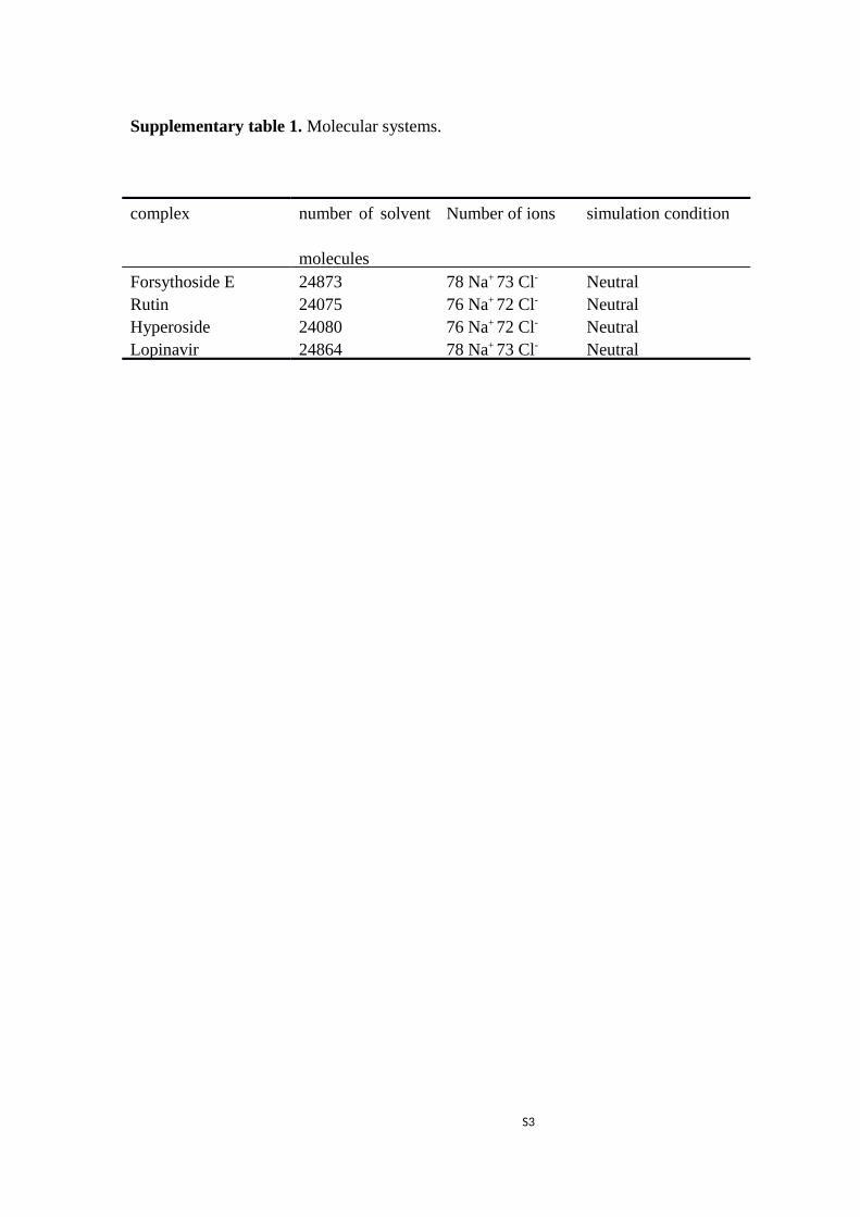
Supplementary table 1. Molecular systems.
complex number of solvent
molecules
Number of ions simulation condition
Forsythoside E 24873 78 Na+ 73 Cl- NeutralRutin 24075 76 Na+ 72 Cl- NeutralHyperoside 24080 76 Na+ 72 Cl- NeutralLopinavir 24864 78 Na+ 73 Cl- Neutral
S3
Supplementary table 2. The calculated binding free energy using MM/PBSA on each
ligand with SARS-CoV-2 main protease.
S4
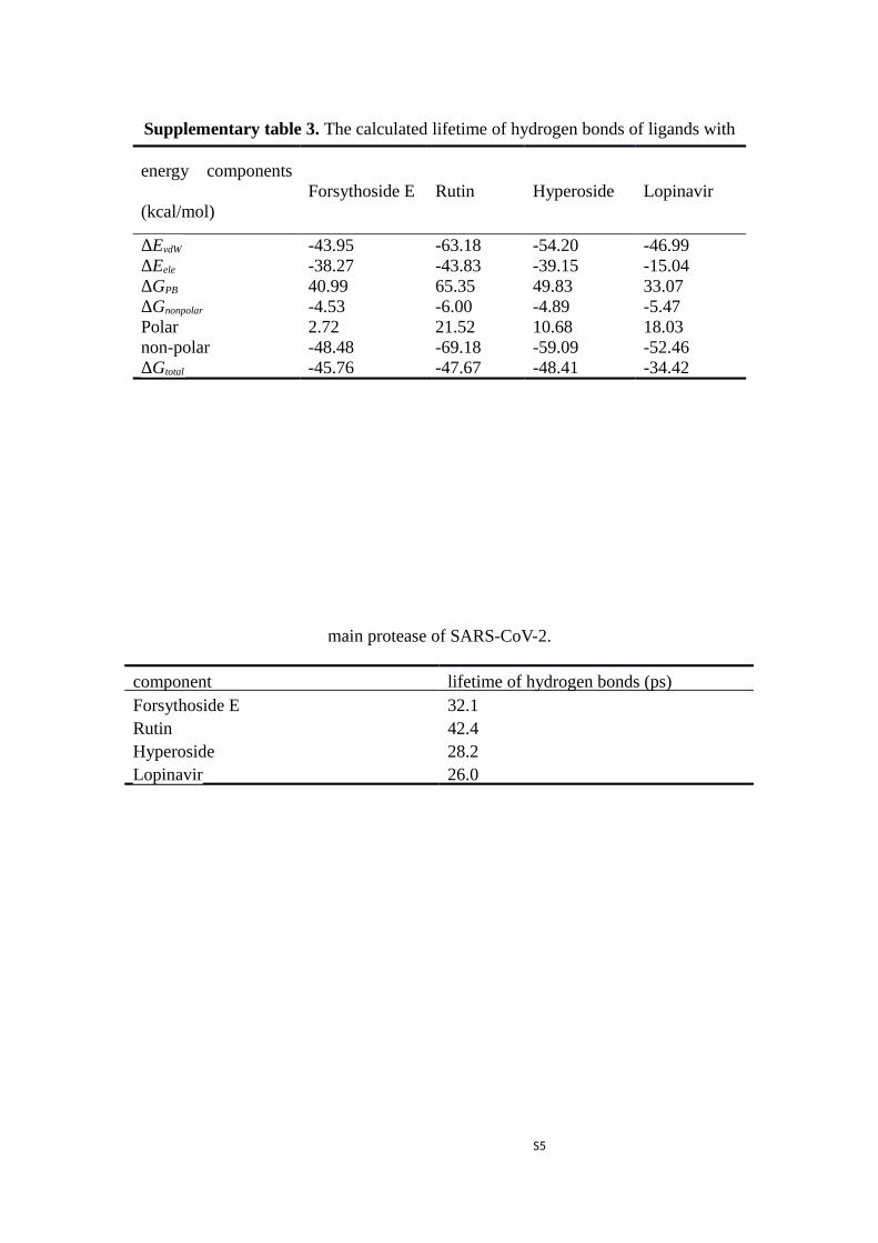
Supplementary table 3. The calculated lifetime of hydrogen bonds of ligands with
main protease of SARS-CoV-2.
component lifetime of hydrogen bonds (ps)Forsythoside E 32.1Rutin 42.4Hyperoside 28.2Lopinavir 26.0
S5
energy components
(kcal/mol)Forsythoside E Rutin Hyperoside Lopinavir
ΔEvdW -43.95 -63.18 -54.20 -46.99 ΔEele -38.27 -43.83 -39.15 -15.04 ΔGPB 40.99 65.35 49.83 33.07 ΔGnonpolar -4.53 -6.00 -4.89 -5.47 Polar 2.72 21.52 10.68 18.03non-polar -48.48 -69.18 -59.09 -52.46ΔGtotal -45.76 -47.67 -48.41 -34.42
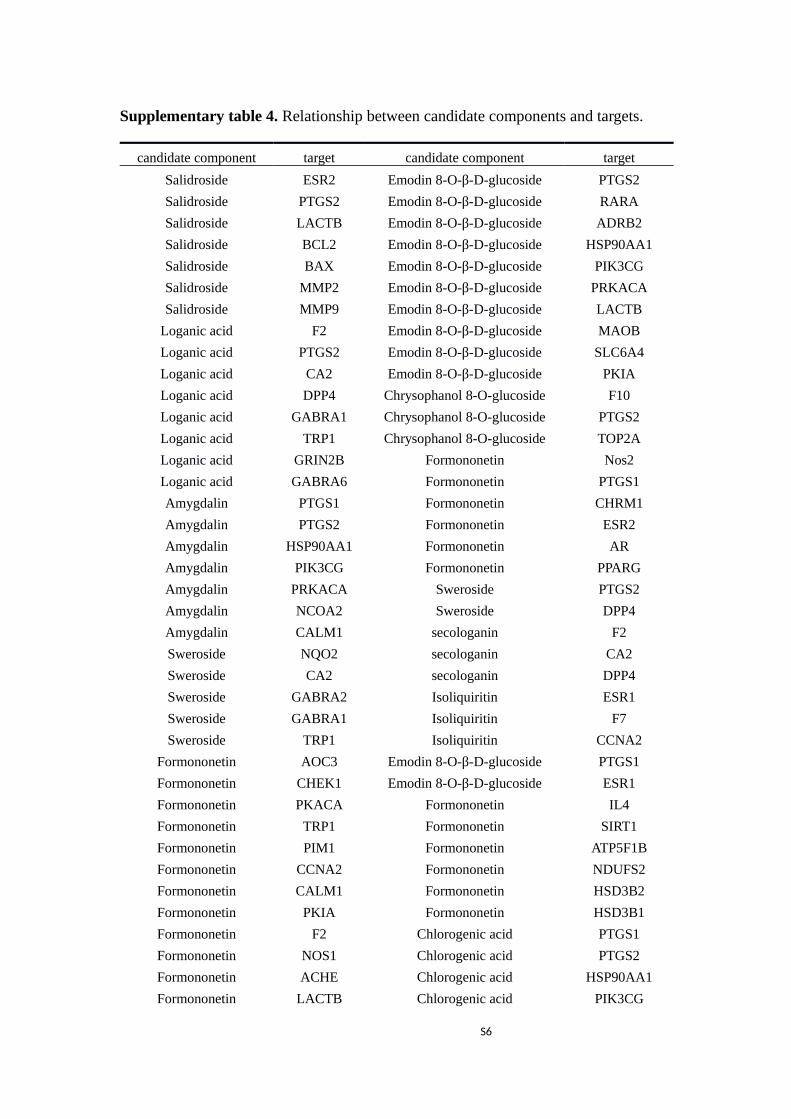
Supplementary table 4. Relationship between candidate components and targets.
candidate component target candidate component target
Salidroside ESR2 Emodin 8-O-β-D-glucoside PTGS2
Salidroside PTGS2 Emodin 8-O-β-D-glucoside RARA
Salidroside LACTB Emodin 8-O-β-D-glucoside ADRB2
Salidroside BCL2 Emodin 8-O-β-D-glucoside HSP90AA1
Salidroside BAX Emodin 8-O-β-D-glucoside PIK3CG
Salidroside MMP2 Emodin 8-O-β-D-glucoside PRKACA
Salidroside MMP9 Emodin 8-O-β-D-glucoside LACTB
Loganic acid F2 Emodin 8-O-β-D-glucoside MAOB
Loganic acid PTGS2 Emodin 8-O-β-D-glucoside SLC6A4
Loganic acid CA2 Emodin 8-O-β-D-glucoside PKIA
Loganic acid DPP4 Chrysophanol 8-O-glucoside F10
Loganic acid GABRA1 Chrysophanol 8-O-glucoside PTGS2
Loganic acid TRP1 Chrysophanol 8-O-glucoside TOP2A
Loganic acid GRIN2B Formononetin Nos2
Loganic acid GABRA6 Formononetin PTGS1
Amygdalin PTGS1 Formononetin CHRM1
Amygdalin PTGS2 Formononetin ESR2
Amygdalin HSP90AA1 Formononetin AR
Amygdalin PIK3CG Formononetin PPARG
Amygdalin PRKACA Sweroside PTGS2
Amygdalin NCOA2 Sweroside DPP4
Amygdalin CALM1 secologanin F2
Sweroside NQO2 secologanin CA2
Sweroside CA2 secologanin DPP4
Sweroside GABRA2 Isoliquiritin ESR1
Sweroside GABRA1 Isoliquiritin F7
Sweroside TRP1 Isoliquiritin CCNA2
Formononetin AOC3 Emodin 8-O-β-D-glucoside PTGS1
Formononetin CHEK1 Emodin 8-O-β-D-glucoside ESR1
Formononetin PKACA Formononetin IL4
Formononetin TRP1 Formononetin SIRT1
Formononetin PIM1 Formononetin ATP5F1B
Formononetin CCNA2 Formononetin NDUFS2
Formononetin CALM1 Formononetin HSD3B2
Formononetin PKIA Formononetin HSD3B1
Formononetin F2 Chlorogenic acid PTGS1
Formononetin NOS1 Chlorogenic acid PTGS2
Formononetin ACHE Chlorogenic acid HSP90AA1
Formononetin LACTB Chlorogenic acid PIK3CG
S6
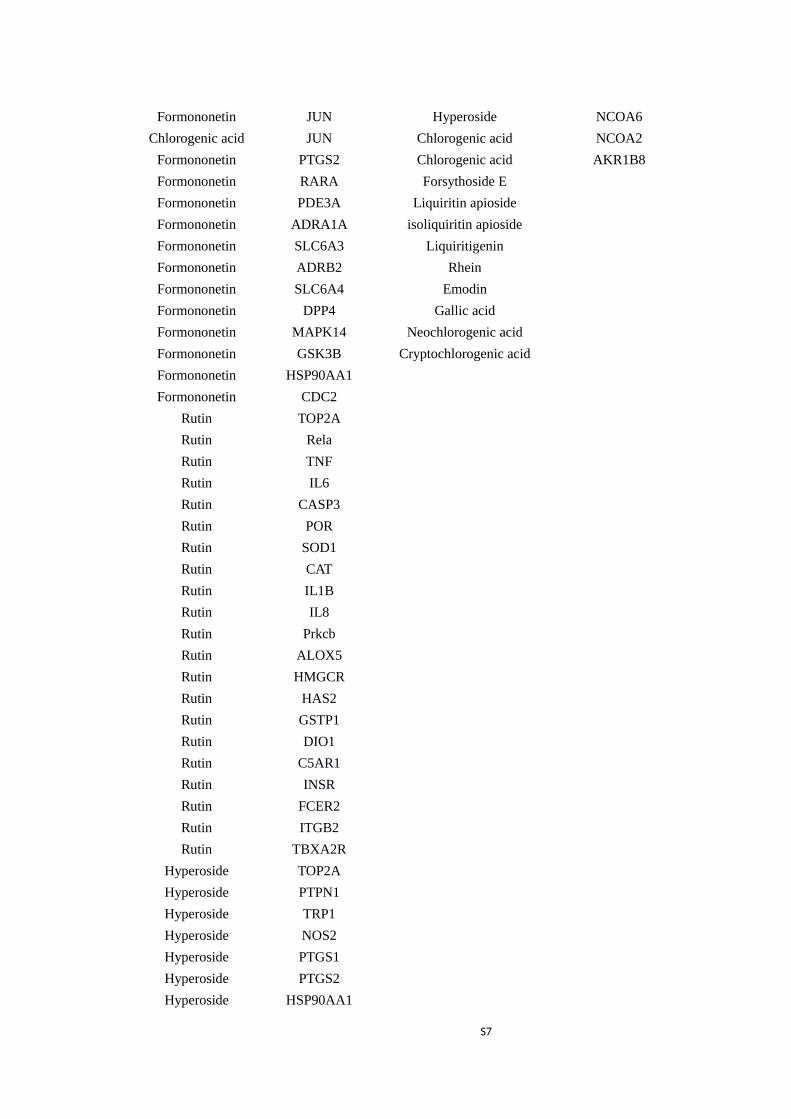
Formononetin JUN Hyperoside NCOA6
Chlorogenic acid JUN Chlorogenic acid NCOA2
Formononetin PTGS2 Chlorogenic acid AKR1B8
Formononetin RARA Forsythoside E
Formononetin PDE3A Liquiritin apioside
Formononetin ADRA1A isoliquiritin apioside
Formononetin SLC6A3 Liquiritigenin
Formononetin ADRB2 Rhein
Formononetin SLC6A4 Emodin
Formononetin DPP4 Gallic acid
Formononetin MAPK14 Neochlorogenic acid
Formononetin GSK3B Cryptochlorogenic acid
Formononetin HSP90AA1
Formononetin CDC2
Rutin TOP2A
Rutin Rela
Rutin TNF
Rutin IL6
Rutin CASP3
Rutin POR
Rutin SOD1
Rutin CAT
Rutin IL1B
Rutin IL8
Rutin Prkcb
Rutin ALOX5
Rutin HMGCR
Rutin HAS2
Rutin GSTP1
Rutin DIO1
Rutin C5AR1
Rutin INSR
Rutin FCER2
Rutin ITGB2
Rutin TBXA2R
Hyperoside TOP2A
Hyperoside PTPN1
Hyperoside TRP1
Hyperoside NOS2
Hyperoside PTGS1
Hyperoside PTGS2
Hyperoside HSP90AA1
S7

Hyperoside PIK3CG
S8

Supplementary table 5. Relationship between targets and signaling pathways.
target pathway
CASP3 MAPK signaling pathway
IL1B MAPK signaling pathway
JUN MAPK signaling pathway
MAPK14 MAPK signaling pathway
TNF MAPK signaling pathway
GSK3B B Cell Receptor Signaling pathway
JUN B Cell Receptor Signaling pathway
PIK3CG B Cell Receptor Signaling pathway
IL4 Fc epsilon RI signaling pathway
MAPK14 Fc epsilon RI signaling pathway
PIK3CG Fc epsilon RI signaling pathway
TNF Fc epsilon RI signaling pathway
CASP3 Natural killer cell mediated cytotoxicity
ITGB2 Natural killer cell mediated cytotoxicity
PIK3CG Natural killer cell mediated cytotoxicity
TNF Natural killer cell mediated cytotoxicity
MAPK14 VEGF signaling pathway
PIK3CG VEGF signaling pathway
PIGS2 VEGF signaling pathway
GSK3B ErbB signaling pathway
JUN ErbB signaling pathway
PIK3CG ErbB signaling pathway
IL1B Toll-like receptor signaling pathway
IL6 Toll-like receptor signaling pathway
JUN Toll-like receptor signaling pathway
MAPK14 Toll-like receptor signaling pathway
PIK3CG Toll-like receptor signaling pathway
F2 Complement and coagulation cascades
C5AR1 Complement and coagulation cascades
GSK3B T cell receptor signaling pathway
IL4 T cell receptor signaling pathway
JUN T cell receptor signaling pathway
MAPK14 T cell receptor signaling pathway
PIK3CG T cell receptor signaling pathway
TNF T cell receptor signaling pathway
IL4 JAK-STAT signaling pathway
IL6 JAK-STAT signaling pathway
PIK3CG JAK-STAT signaling pathway
S9
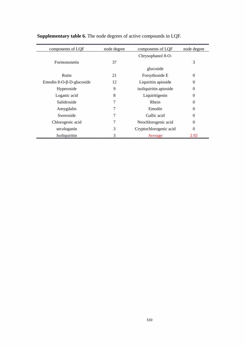
Supplementary table 6. The node degrees of active compounds in LQF.
components of LQF node degree components of LQF node degree
Formononetin 37
Chrysophanol 8-O-
glucoside
3
Rutin 21 Forsythoside E 0
Emodin 8-O-β-D-glucoside 12 Liquiritin apioside 0
Hyperoside 9 isoliquiritin apioside 0
Loganic acid 8 Liquiritigenin 0
Salidroside 7 Rhein 0
Amygdalin 7 Emodin 0
Sweroside 7 Gallic acid 0
Chlorogenic acid 7 Neochlorogenic acid 0
secologanin 3 Cryptochlorogenic acid 0
Isoliquiritin 3 Average 2.92
S10
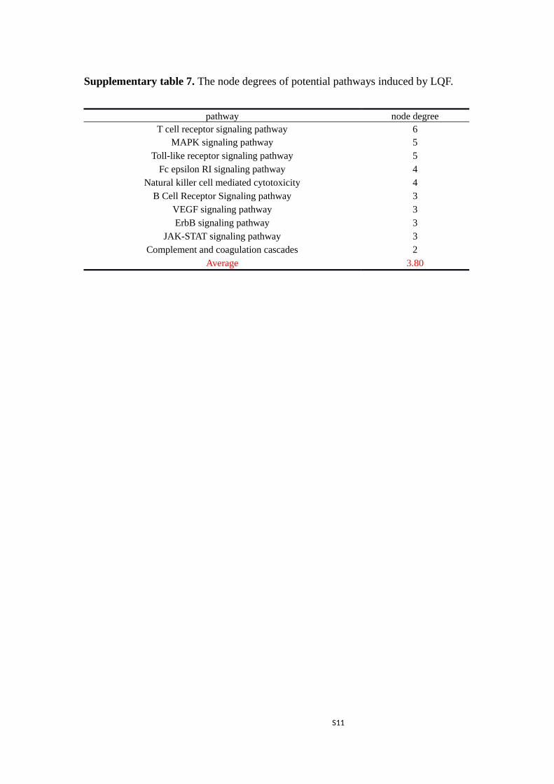
Supplementary table 7. The node degrees of potential pathways induced by LQF.
S11
pathway node degreeT cell receptor signaling pathway 6
MAPK signaling pathway 5Toll-like receptor signaling pathway 5
Fc epsilon RI signaling pathway 4Natural killer cell mediated cytotoxicity 4
B Cell Receptor Signaling pathway 3VEGF signaling pathway 3ErbB signaling pathway 3
JAK-STAT signaling pathway 3Complement and coagulation cascades 2
Average 3.80

Figure S1. RMSD plot during MD simulations. a) RMSD between Cα atoms ofprotein and ligands; b) RMSD of Cα atoms in complexes.
S12
Reference
1. Darden, T.; Perera, L.; Li, L.; Pedersen, L., New tricks for modelers from the
crystallography toolkit: the particle mesh Ewald algorithm and its use in nucleic
acid simulations. Structure 1999, 7 (3), R55-R60.
2. Wang, H.; Dommert, F.; Holm, C., Optimizing working parameters of the
smooth particle mesh Ewald algorithm in terms of accuracy and efficiency. The
Journal of chemical physics 2010, 133 (3), 034117.
3. Berendsen, H. J.; van der Spoel, D.; van Drunen, R., GROMACS: a message-
passing parallel molecular dynamics implementation. Computer physics
communications 1995, 91 (1-3), 43-56.
4. Lindahl, E.; Hess, B.; Van Der Spoel, D., GROMACS 3.0: a package for
molecular simulation and trajectory analysis. Molecular modeling annual 2001,
7 (8), 306-317.
S13

download fileview on ChemRxivms_lhqw_0321-g_SI.docx (270.77 KiB)